Effect of Ventricular Shock Strength on Cardiac Hemodynamics
8
791 Effect of Ventricular Shock Strength on Cardiac Hemodynamics TAKASHI TOKANO. M.D., DAVID BACH, M.D., JASON CHANG, JAMES DAVIS, M.D., JOSEPH J. SOUZA, M.D., ADAM ZIVIN, M.D., BRADLEY P. KNIGHT, M.D., RAJIVA GOYAL, M.D., K. CHING MAN, D,0., FRED MORADY, M.D., and S. ADAM STRICKBERGER, M.D. From the Division of Cardiology, Department of Internal Medicine, University of Michigan Medical Center, Ann Arbor, Michigan Ventricular Defibrillation and Cardiac Function. Introduction: The effect of implantable detibrillator shocks on cardiac hemodynamics is poorly understood. The purpose of this study was to test the hypothesis that ventricular defibrillator shocks adversely effect cardiac hemodynamics. Methods and Results: The cardiac index was determined by calculating the mitral valve in- flow with transesophogeal Doppler during nonthoracotomy defibrillator implantation in 17 pa- tients. Tbe cardiac index was determined before, and immediately, 1 minute, 2 minutes, and 4 minutes after sbocks were delivered during defibrillation energy requirement testing with 27- to 34-, 15-, I0-, 5-, 3-, or 1-J shocks. Tbe cardiac Index was also measured at tbe same time points after 27- to 34-, and 1-J sbocks delivered during tbe baseline rhytbm. Tbe cardiac index decreased from 2.30 ± 0.40 L/min per m- before a 27- to 34-J sbock during defibrillation en- ergy requirement testing to 2.14 ± 0.45 L/min per m- immediately afterwards (P = 0.001). Tbis effect persisted for > 4 minutes. An adverse bemodynamic effect of similar magnitude occurred after 15 J (P = 0.003) and lO-J sbocks (P = 0.01), but dissipated after 4 minutes and witbin 2 minutes, respectively. Tbere was a significant correlation between sbock strength and the per- cent cbange in cardiac index (r = 0.3, P = 0.03). The cardiac index decreased 14% after a 27- to 34-J sbock during tbe baseline rhytbm (P < 0.0001). Tbis eflect persisted for < 4 minutes. A 1- J sbock during tbe baseline rhytbm did not effect the cardiac index. Conclusion: Defibrillator sbocks > 9 J delivered during tbe baseline rbytbm or during defi- brillation energy requirement testing result in a 10% to 15% reduction in cardiac index, wbereas smaller energy sbocks do not affect cardiac bemodynamics. Tbe duration and extent of tbe adverse effect are proportional to tbe sbock strengtb. Sbock strengtb, and not ventricu- lar fibrillation, appears to be most responsible for tbis effect. Tberefore, the detrimental hemo- dynamic effects of high-energy sbocks may be avoided wben low-energy defibrillation is used. (J Cardiovasc Electrophysiol, Vol. 9, pp. 791-797. August 1998) defibrillation threshold, implantable cardioverter deflbrillator, cardiac index, cardiac output, stunning Introduction Ventricular implantable defibrillators frequently are programmed to deliver the maximum shock energy, irrespective of the defibrillation energy re- quirement. However, the effect of ventricular im- Address for correspondence: S. Adam Strickberger. M.D., Univer- sily of Michigan Medical Center, \500 Kasl Medical Center Dr., Ann Arbor, M! 48109-0022. Fax: 734-936-7026, Manuscript received 23 April 1998; Accepted for publication 28 May 1998- plantable defibrillator shock strength on cardiac hemodynamics has not been well defined.' "^ There- fore, the purpose of this study was to test the hy- pothesis that nonthoracotoiny ventricular defibril- lator shocks adversely affect cardiac hemodynamics. Methods Patient Population The study population cotisisted of 17 patients (12 men and 5 women; mean age 55 ± 16 years)
Transcript of Effect of Ventricular Shock Strength on Cardiac Hemodynamics
791
Effect of Ventricular Shock Strengthon Cardiac Hemodynamics
TAKASHI TOKANO. M.D., DAVID BACH, M.D., JASON CHANG,JAMES DAVIS, M.D., JOSEPH J. SOUZA, M.D., ADAM ZIVIN, M.D.,
BRADLEY P. KNIGHT, M.D., RAJIVA GOYAL, M.D., K. CHING MAN, D,0.,FRED MORADY, M.D., and S. ADAM STRICKBERGER, M.D.
From the Division of Cardiology, Department of Internal Medicine,University of Michigan Medical Center, Ann Arbor, Michigan
Ventricular Defibrillation and Cardiac Function. Introduction: The effect of implantabledetibrillator shocks on cardiac hemodynamics is poorly understood. The purpose of this study wasto test the hypothesis that ventricular defibrillator shocks adversely effect cardiac hemodynamics.
Methods and Results: The cardiac index was determined by calculating the mitral valve in-flow with transesophogeal Doppler during nonthoracotomy defibrillator implantation in 17 pa-tients. Tbe cardiac index was determined before, and immediately, 1 minute, 2 minutes, and 4minutes after sbocks were delivered during defibrillation energy requirement testing with 27-to 34-, 15-, I0-, 5-, 3-, or 1-J shocks. Tbe cardiac Index was also measured at tbe same timepoints after 27- to 34-, and 1-J sbocks delivered during tbe baseline rhytbm. Tbe cardiac indexdecreased from 2.30 ± 0.40 L/min per m- before a 27- to 34-J sbock during defibrillation en-ergy requirement testing to 2.14 ± 0.45 L/min per m- immediately afterwards (P = 0.001). Tbiseffect persisted for > 4 minutes. An adverse bemodynamic effect of similar magnitude occurredafter 15 J (P = 0.003) and lO-J sbocks (P = 0.01), but dissipated after 4 minutes and witbin 2minutes, respectively. Tbere was a significant correlation between sbock strength and the per-cent cbange in cardiac index (r = 0.3, P = 0.03). The cardiac index decreased 14% after a 27- to34-J sbock during tbe baseline rhytbm (P < 0.0001). Tbis eflect persisted for < 4 minutes. A 1-J sbock during tbe baseline rhytbm did not effect the cardiac index.
Conclusion: Defibrillator sbocks > 9 J delivered during tbe baseline rbytbm or during defi-brillation energy requirement testing result in a 10% to 15% reduction in cardiac index,wbereas smaller energy sbocks do not affect cardiac bemodynamics. Tbe duration and extentof tbe adverse effect are proportional to tbe sbock strengtb. Sbock strengtb, and not ventricu-lar fibrillation, appears to be most responsible for tbis effect. Tberefore, the detrimental hemo-dynamic effects of high-energy sbocks may be avoided wben low-energy defibrillation is used.(J Cardiovasc Electrophysiol, Vol. 9, pp. 791-797. August 1998)
defibrillation threshold, implantable cardioverter deflbrillator, cardiac index, cardiac output, stunning
Introduction
Ventricular implantable defibrillators frequentlyare programmed to deliver the maximum shockenergy, irrespective of the defibrillation energy re-quirement. However, the effect of ventricular im-
Address for correspondence: S. Adam Strickberger. M.D., Univer-sily of Michigan Medical Center, \500 Kasl Medical Center Dr.,Ann Arbor, M! 48109-0022. Fax: 734-936-7026,
Manuscript received 23 April 1998; Accepted for publication 28May 1998-
plantable defibrillator shock strength on cardiachemodynamics has not been well defined.' "̂ There-fore, the purpose of this study was to test the hy-pothesis that nonthoracotoiny ventricular defibril-lator shocks adversely affect cardiac hemodynamics.
Methods
Patient Population
The study population cotisisted of 17 patients(12 men and 5 women; mean age 55 ± 16 years)
792 Journal of Cardiovascular Electrophyslology Vol. 9, No. 8, August 1998
undergoing implantation of a transvenous im-plantable cardioverter defibrillator. Eigbt patientshad coronary artery disease. 8 patients had a non-ischemic cardiomyopathy, and I patient had nostructural heart disease. The mean left ventricularejection fraction was 0.32 ±0.12 (range 0.15 to0.55). and 4 patients had an ejection fraction > 0.40.Tbe mean New York Heart Association Conges-tive Heart Failure Class was 1.9 ± 0.7 {range ClassI to III). The baseline rhythm was sinus in 14 pa-tients and atrial fibrillation in 3. TransesophagealDoppler ecbocardiography was used to determinemitral valve inflow (see following). Cardiac out-put equals cardiac inflow only in tbe absence ofsignificant mitral regurgitation. Therefore., patientswere evaluated preoperatively for mitral regurgi-tation with either cardiac catheterization or withtwo-dimensional surface echocardiography. Patientswith more than minimal echocardiographically ev-ident mitral regurgitation were not eligible for par-ticipation in the study protocol.
Defibrillator System and Implantation
All patients came to the operating room in apostabsorptive state. Therapy with antiarrhythmicmedications was discontinued at least five half-livesprior to device implantation, except in eight patientsin whom amiodarone therapy had been ineftective.Angiotensin-converting enzyme inhibitor therapy,calcium channel blocker therapy, beta-blocker ther-apy, and digoxin were continued in II, 1, 4, and10 patients, respectively. General anesthesia wasinduced., the patients were intubated, and a constantlevel of anesthesia was maintained throughout theimplant procedure and study protocol using sup-plemental doses of inhalational agents, fentanyl andits derivatives, or a steroid anesthetic.
A transvenous dual coil defibrillation lead wasimplanted in 16 patients (models 75, 115, and 125,Cardiac Pacemakers, Inc. [CPIl, St. Paul, MN,USA). A transvenous defibrillation lead with a sin-gle shocking electrode (model 6936, MedtronicInc., Minneapolis, MN, USA) was used in one pa-tient. The defibrillation lead was positioned intbe right ventricular apex via the subclavian veinunder tluoroscopic guidance.
Sixteen of tbe patients in this study received adefibrillator with a truncated, fixed-tilt bipbasicwavefomi with a first phase tilt of 60% and a sec-ond phase tilt of 50% (models 1720, 1725, 1740,1742, 1743, 1762, and 1763, CPI), and one patientreceived a defibrillator with a first phase tilt andsecond phase tilt of 65% (model 7219C, Medtronic,
Inc.). The defibrillator shell functioned as a defi-briiiation electrode in six patients.
Study Protocol
Written informed consent was obtained under aprotocol approved by the Human Research Com-mittee at the University of Michigan. All shockswere delivered directly from the implantable de-fibrillator. Shocks were delivered during thebaseline rbythm and during defibrillation energyrequirement testing. Shocks were delivered duringthe baseline rhytbm to assess the isolated effect ofa shock, in the absence of ventricular fibrillation,on cardiac function. In this way, tbe effects of ashock can be separated from the effects of ven-tricular fibrillation. A 1 -J shock and tben a 27- to34-J shock (tbe maximum energy delivered by tbeimplantable defibrillator) were delivered synchro-nized to the QRS complex during the baselinerhythm.
A step-down protocol was then utilized to de-termine the defibrillation energy requirement. Thedefibrillation energy requirement was defined asthe lowest energy successful at converting ven-tricular fibrillation to the baseline rhythm. Shockenergies of 15, 10. 5, 3. and 1.0 J were delivereduntil ventricular fibrillation failed to convert to thebaseline rhythm. If tbe 15-J shock was ineffective,the first shock lor the subsequent induction was20 J. A 27- to 34-J defibrillator shock was alsoused to convert ventricular fibrillation to the base-line rhythm. Ventricular fibrillation was inducedwith ventricular pacing with a 15-V pulse deliv-ered every 30 msec with a duration of 1.1 msecfor I to 3 seconds. The mean duration of ventric-ular fibrillation was 12.7 ± 3.9 seconds for maxi-mum energy shocks, 9.5 ± 0.5 seconds for 20-Jshocks, 8.3 ± 1.8 seconds for 15-J shocks, 7.4 ±1.9 seconds for 10-J shocks, and 6.7 ± 1.8 secondsfor shocks < 10 J (P < O.OOOI)- At least 5 min-utes was allowed to elapse between shocks deliv-ered during the baseline riiythm iind between shocksthat resulted in successful defibrillation.
Cardiac Output Measurement
The cardiac output was estimated using a mea-sure of mitral valve inflow.'"̂ This technique cor-relates well with the caidiac output detennined withconventional tbermodilution techniques'' and offersthe advantage of providing an assessment of beat-by-beat changes in cardiac output. Conventionalthermodilution techniques cannot be used to assess
Tokano. etal. Ventricular Deflbritlatiun and Cardiac Function 793
rapid changes because several measurements overapproximately 2 to 3 minutes are required.
After induction of general anesthesia but beforedefibrillator implantation, a transesophageal echo-cardiography probe was inserted. The probe waspositioned to optimize visualization of mitral in-flow in a basal four-chamber view.̂ During theprotocol, the same standard view of the mitral valvewas maintained to ensure consistency of thecolor flow Doppler interrogation. At baseline, thediameter of the mitral annulus was determined inone plane. Pulsed-wave Doppler was used to de-fine left ventricular inflow at the level of the mi-tral valve annulus.*^̂ Scale and sweep speed wereoptimized for subsequent quantitation of the ve-locity time integral. Based on the assumption thatleft ventricular inflow equals left ventricular out-flow (cardiac output) in the absence of mitral re-gurgitation, transmitral inflow was calculated asan estimate of cardiac output using the mitral an-nular cross-sectional area, flow at the level of themitral annulus, and heart rate."̂ Pulsed-wave Dopplerat the level of the mitral annulus was performedat baseline and was repeated immediately aftereach shock. All images were recorded on standardVHS videotape for subsequent off-line analysis.
Images were digitized from videotape using com-mercially available hardware and software (TomTecImaging Systems, Inc., Boulder, CO, USA). Thevelocity time integral of left ventricular inflow wasaveraged for at least five consecutive cycles in allpatients; 10 cycles were used for patients with atrialfibrillation. Mitral inflow velocities associated withpremature ventricular complexes, post prematureventricular complexes, and ventricular pacedbeats were excluded from analysis. Shocks weredelivered within 30 seconds of recording thebaseline mitral inflow. The mean time to comple-tion of the first mitral valve inflow determinationafter each shock was 19.7 ± 10.8 seconds.
All measurements for mitral inflow velocity timeintegral were detennined independently by threeinvestigators. Two of the three investigators wereblinded to the shock energy and the rhythm. Themean variability of these measurements was 10.7%± 6.6%. The values determined by all investiga-tors were averaged, and the means were used foranalysis.
variables were compared by ANOVA, and then byindividual paired /-tests when the ANOVA resultwas stati.Stic ally significant. The relationship be-tween two continuous variables was assessed witha linear regression analysis. P < 0.05 was con-sidered statistically significant.
Results
Defibrillation Energy Requirements
The mean defibrillation energy requirement was8.6 ± 4.8 J in the 17 patients. The defibrillationenergy requirement was 20 J in 1 patient, 15 J in2, 10 J in 6, 5 J in 6. and 3 J in 2. In one patientwith a 5-J defibrillation energy requirement, arecording malfunction precluded the determinationof the Doppler measurement. Because of the smallnumber of successful defibrillations with energies< 10 J, these data were analyzed as a single group.
Effect of Shocks During Defibrillation EnergyRequirement Testing
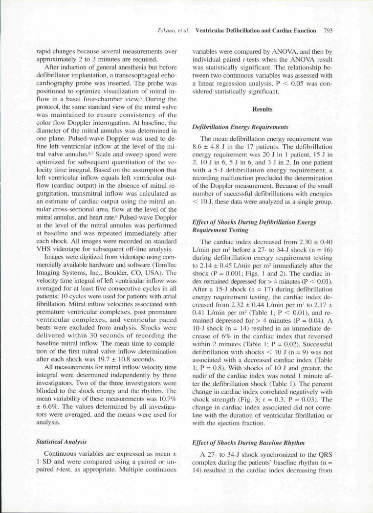
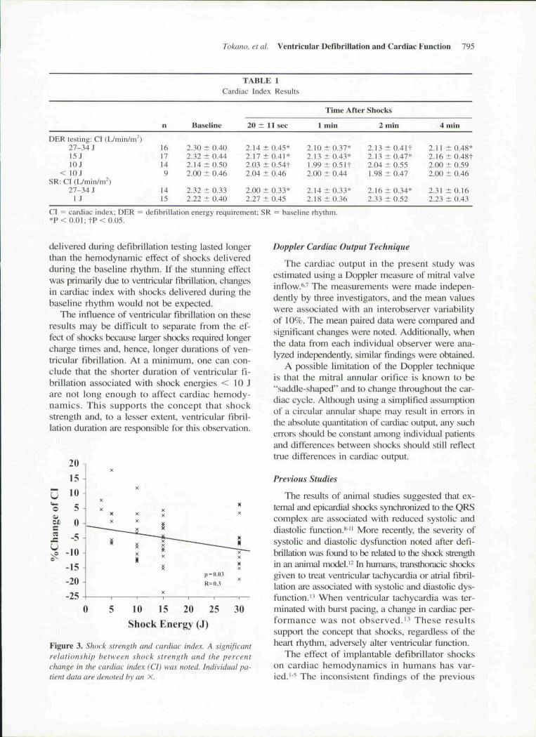
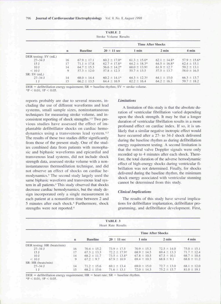
The cardiac index decreased from 2.30 ± 0.40L/min per m̂ before a 27- to 34-J shock (n = 16)during deflbrillation energy requirement testingto 2.14 ± 0.45 LVmin per m- immediately after theshock (P = 0.001; Figs. 1 and 2). The cardiac in-dex remained depressed for > 4 minutes (P < 0.01).After a 15-J shock (n = 17) during defibrillationenergy requirement testing, the cardiac index de-creased from 2.32 ± 0.44 L/min per m- to 2.17 ±0.41 L/min per m^ (Table 1; P < O.OI). and re-mained depressed for > 4 minutes (P = 0.04). A10-J shock (n = 14) resulted in an immediate de-crease of 6% in the cardiac index that reversedwithin 2 minutes (Table 1; P = 0.02). Successfuldeflbrillation with shocks < 10 J (n = 9) was notassociated with a decreased cardiac index (Table1; P = 0.8). With shocks of 10 J and greater, thenadir of the cardiac index was noted I minute af-ter the deflbrillation shock (Table 1). The percentchange in cardiac index correlated negatively withshock strength (Fig. 3; r = 0.3, P = 0.03). Thechange in cardiac index associated did not corre-late with the duration of ventricular fibrillation orwith the ejection fraction.
Statistical Analysis
Continuous variables are expressed as mean ±1 SD and were compared using a paired or un-paired /-test, as appropriate. Multiple continuous
Effect of Shocks During Baseline Rhythm
A 27- to 34-J shock synchronized to the QRScomplex during the patients' baseline rhythm (n =14) resulted in the cardiac index decreasing from
794 Journal of Cardiovascular Electrophysiology Vol. 9, No. 8, August 1998
3.0
2.5
2.0
1,5
Shock
v-i
1.0Baseline 0.3 min I min 2 min 4 min
Time After Conversion
Figure 1. Cardiac Index and 27- to 34-J shocks. The meaneffects of a 27- to 34-J shock delivered during defihrillationenergy requirement testing are shown. Cardiac inde.\ (CI)is represented on the y-axis. and time is repre.sented on theX-axis. *P <0.01; +P < 0.05.
2.32 ± 0.33 L/min per m̂ to 2.00 ± 0.33 L/minper m- immediately after the shock (Table 1; P <0.0001). This effect persisted for < 4 minutes. A1-J shock delivered during the baseline rhythm (n= 15) did not affect the cardiac index (Table 1).The relative magnitude of change in the stroke vol-ume was not different after a 27- to 34-J shock de-livered to treat ventricular fibrillation or whendelivered during the baseline rhythm (11.7% ±6.4% vs 11.6% ± 5.8%, P = 1.0).
The effect of deflbrillation on stroke volumewas nearly identical to the changes noted in car-
diac index (Table 2). Heart rate was largely unaf-fected by the defibrillation shocks (Table 3).
Discussion
Major Findings
Biphasic implantahle defibrillator shocks of 10J or more delivered during the baseline rhythmor when used to terminate ventricular fibrillationresult in a 10% to 15% reduction of cardiac index,which occurs secondary to a reduction in strokevolume. With 27- to 34-J shocks delivered dur-ing defibrillation energy requirement testing, thecardiac index returns to normal after 4 minutes,whereas when 27- to 34-J shocks are deliveredduring the baseline rhythm, the adverse hemody-namic effect dissipates within 4 minutes. The du-ration and extent of the adverse hemodynamiceffect are proportional to the shock strength.
Mechanism of Shock Effect
The detrimental hemodynamic effect of a ven-tricular defibrillator shock appears to be due to theshock itself, and not to ventricular fibrillation. Be-cause a similar degree of ventriculai" stunning wasnoted after shocks delivered during the baselinerhythm as with shocks that tenninated ventricularfibrillation, one must conclude that the shocks causethe stunning. This statement is supported by thefact that 27- to 34-J shocks delivered during ven-tricular fibrillation or during the baseline rhythmresulted in similar reduction in the cardiac index,although the adverse hemodynamic effect of shocks
Baseline 35 sec mtn mm 4 min
11)0 1
VTI(cm) 14.1 12.0 1.7 12.1
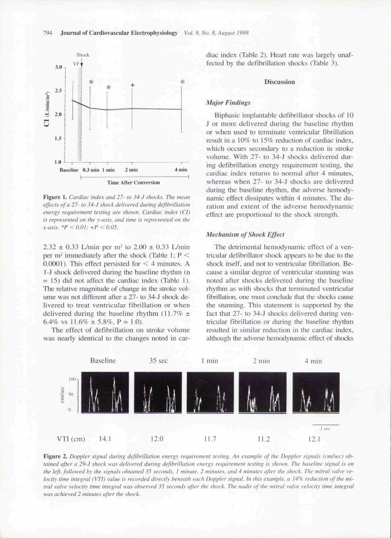
Figure 2. Doppler signat during defibriiiation energy requirement testing. An example of the Doppler signals (cm/sec) ob-tained after a 29-J shock was detivered during defihrittation energy requirement testing is stwwn. The basetine signal is onthe left, followed by the signals obtained 35 seconds. 1 minute. 2 minutes, and 4 minutes after the shock. The mitral valve ve-locity time integral fVTI) value is recorded directly beneath each Doppter signat. In this exampte. a 14% reduction of the mi-trat valve velocity time integral was observed 35 .seconds after the shock. Ttie nadir of the mitral valve velocity time integratwas achieved 2 minutes after the shock.
Tokano, et al. Ventricular Defibrillation and Cardiac Function 795
DER testing: CI (L/min/m-)27-34 i15 JlOJ
< lOJSR: CI {Umin/rrr)
27-34 J1 J
n
1617149
1415
TABLECardiac Index
Baseline
2.30 ± 0.402.32 ± 0.442.14 ± 0.502.00 ± 0.46
2.32 ± 0.332.22 ± 0.40
1
Results
20 ± 11 sec
2.14 ±2.17 ±2.03 ±2.04 ±
2.00 ±2.27 ±
0.45*0.41*0.54t0.46
0.33*0.45
Time After Shocks
1 min
2.10 ± 0.37*2.13 ±0.43*1.99 ± 0.51t2.00 i 0.44
2.14 ±0.33*2.18 ± 0.36
2
2.132.132.041.98
2.162.33
min
± 0.41t± 0.47*± 0.55± 0.47
± 0.34*± 0.52
4 min
2.11 ± 0.48*2.16 ± 0.48t2.(K) ± 0.592.00 ± 0.46
2.31 ± 0.162.23 ± 0.43
CI = cardiac index; DER = defibrillation energy requirement; SR = baseiine rhythm.*P < 0.01: t P < 0.05.
delivered during defibriliation testing lasted longerthan the hemodynamic effect of shocks deliveredduring the baseline rhythm. If the stunning effectwas primarily due to ventricular fibrillation, changesin cardiac index with shocks delivered during thebaseline rhythm would not be expected.
The influence of ventricular fibrillation on theseresults may be difficult to separate from the ef-fect of shocks because larger shocks required longercharge times and. hence, longer durations of ven-tricular fibrillation. At a minimum, one can con-clude that the shorter duration of ventricular fi-brillation associated with shock energies < 10 Jare not lotig enough to affect cardiac hemody-namics. This supports the concept that shockstrength and, to a lesser extent, ventricular fibril-lation duration are responsible for this observation.
Jo
0£B
u
20 n
15
10
5
0
-5
-10
-15
-20ye
X
X
X XX X ^
" " g————x^
»
It "ii
X
0 5 10 15 20
Shock Energy
~-—-—.
p=n.o.i
R=O..l
25
(J)
It
X
N
X "X
IfX
X
30
Figure 3. Shock sirenglh and cardiac index. A significantrelationship hetween shock strength and the percentchange in the cardiac index (CI) was noted. Individual pa-tient data are denoted by an X.
Doppler Cardiac Output Technique
The cardiac output in the present study wasestimated using a Doppler measure of mitral valveinflow.̂ ^ The measurements were made indepen-dently by three investigators, and the mean valueswere associated with an interobserver variabilityof 10%. The mean paired data were compared andsignificant changes were noted. Additionally, whenthe data from each individual observer were ana-lyzed independently, similar findings were obtained.
A possible limitation of the Doppler techniqueis that the mitral annular orifice is known to be"saddle-shaped" and to change throughout the car-diac cycle. Although using a simplified assumptionof a circular annular shape may result in errors inthe absolute quantitation of cardiac output, any sucherrors should be constant among individual patientsand differences between shocks should still reflecttrue differences in cardiac output.
Previous Studies
The results of animal studies suggested that ex-temal and epicardial shocks synchronized to the QRScomplex are associated with reduced systolic anddiastolic function.** " More recently, the severity ofsystolic and diastolic dysfunction noted after defi-brillation was found to be related to the shock strengthin an animal model.'- In humans, transthoracic shocksgiven to treat ventricular tachycardia or atrial fibril-lation are associated with systolic and diastolic dys-function.'̂ When ventricular tachycardia was ter-minated with burst pacing, a change in cardiac per-formance was not observed." These resultssupport the concept that shocks, regardless of theheart rhythm, adversely alter ventricular function.
The effect of implantabie defibrillator shockson cardiac hemodynamics in humans has var-ied.''' The inconsistent findings of the previous
796 Journal of Cardiovascular Electrophysiology Vol. 9, No. 8, August 1998
DER testing: SV (mL)27-34 J15 JlOJ
< lOJSR: SV (mL)
27-34 J1 J
n
1617149
1415
Baseline
67.9 ±17.171.1 ± 17.864.7 ± 15.357.3 ± 12.0
68.0 ± 14.466.2 ± 13.5
TABLE 2Stroke Volume Results
20 ± 11 sec
60.2 ± 17.0*62,7 ± 17.8*56.6 ± 14.2*57.8 ± 12.3
60.2 ± 14.1*64.4 ± 16.9
Time After Shocks
1 min
61.3 ± 15.0*64.2 ± 18.3*60.0 ± 13.9t55.7 ± 13.5
64.3 ± 12.3t62.2 ± 16.4
2 min
62.1 ± 14.8*64.5 ± 16.8*61.9 ± 12.757.5 ± 13.7
64.1 ± 15.064.2 ± 16.3
4 mill
57.9 ± 15.6*62.4 ±15.159.2 ± 13.158.0 ± 16.5
66.3 ± 13.759.7 ± 18.2
DER = defibrillation energy requirement: SR = baseline rhythm: SV = stroke volume.*P< 0.01; t p < 0.05.
reports probably are due to several reasons., in-cluding the use of different waveforms and leadsystems, small sample sizes, noninstantaneoustechniques for measuring stroke volume, and in-consistent reporting of shock strengths.'"^ Two pre-vious studies have assessed the effect of im-plantable defibrillator shocks on cardiac hemo-dynamics using a transvenous lead system.^"*The results of these two studies differ significantlyfrom those of the present study. One of the stud-ies combined data from patients with monopha-sic and biphasic waveforms and epicardial andtransvenous lead systems, did not include shockstrength data, assessed stroke volume with a non-instantaneous thennodilution technique, and didnot observe an effect of shocks on cardiac he-modynamics."^ Tbe second study largely used thesame biphasic waveform and transvenous lead sys-tem in all patients.̂ This study observed tbat shocksdecrease cardiac hemodynamics, but the study de-sign incorporated only a single measurement ineach patient at a nonuniform time between 2 and5 minutes after each sbock."* Furthermore, shockstrengths were not reported.''
Limitations
A limitation of this study is tbat tbe absolute du-ration of ventricular fibrillation varied dependingupon tbe shock strength. It may be that a longerduration of ventricular fibrillation results in a moreprofound effect on cardiac index. If so. it is un-likely that a similar negative inotropic effect wouldhave occurred after a 27- to 34-J shock deliveredduring the baseline rhytbm as during defibrillationenergy requirement testing. A second limitation isthat the mitral valve Doppler signals were onlyrecorded up to 4 minutes after each shock. There-fore, the total duration of tbe adverse hemodynamiceffect of high-energy shocks during ventricular fi-brillation was not detennined. Finally, for shocksdelivered during the baseline rhytbm, the minimumshock energy associated with ventricular stunningcannot be determined from this study.
Clinical Implications
Tbe results of this study have several implica-tions for deflbrillator implantation, defibrillator pro-gramming, and defibrillator development. First,
DER testing: HR (beats/min)27-34 J15 JlOJ
< lOJSR: HR (beats/min)
27-34 J1 J
n
1617149
1415
TABLE 3Heart Rate Results
Baseline
70.4 ± 15.267.3 ± 13.368.2 ±11.767.2 ± 9.7
71.2 ± 15.668.2 ± 15.6
20 ± 11 sec
73.9 ± 17.572.2 ± 17.9t73.5 ± 13.8*67.9 ± 10.9
69.1 ± 13.471.6 ± 13-1
1
70.968.967.869.4
69.072.0
Time After Shocks
min
± 15.3± 14.5± 10.5± 10-3
± \5.Q± 14.3
2 min
72.5 ± 14.069.4 ± 13.367.5 ± 10.168.8 ± 9.1
72.7 ± 13.075.2 ± 15.7
4
75.071.768.768.8
68.181.0
min
± 15.1± 12.4± 10.4±11.2
± 12.0± 19.1
DER = defibrillation energy requirement; HR - heart rate; SR = baseline rhythm.*P< 0.01; t P < 0.05.
Tokano. et al. Ventricular Deflbrillation and Cardiac Function 797
these results suggest that defihriliators should notbe routinely programmed to the maximum avail-able energy. Lower-energy shocks that consistentlyresult in successful defibrillation are likely to beassociated with improved postshock hemodynam-ics. In the present study, this was demonstrated tobe true with shocks delivered during the baselinerhythm and during ventricular fibrillation. The sameis probably also true when defibrillator shocks areused to treat ventricular tachycardia and supraven-tricular tachycardias. Given that ventricular andsupraventricular tachycardias are sometimes treatedin the ventricular fibrillation zone of the implantabledefibrillator, patients probably also receive high-output shocks for these arrhythmias. Therefore, ir-respective of the rhythm being treated by the de-fibrillator, if an adequate defibriiiation safety mar-gin can be achieved with a lower energy shock,then lower-energy defibrillation may be preferred.Second, although the mechanism of presumed de-fibrillation-induced congestive heart failure is notknown, these data suggest the use of lower-energyshocks may prevent this phenomenon.''*'̂ One mayargue that a 10% reduction in cardiac index maynot be important for normal patients. However, pa-tients with implanted defibrillators typically havea low ejection fraction, an advanced New YorkHeart Association heart failure classification, anda reduced cardiac reserve. These frequent comor-bidities suggest that the 10% reduction in cardiacindex after defibrillation may be clinically impor-tant, and that the current industry direction towardlower maximum energy defibrillators is appropri-ate. Additionally, lower-energy defibrillation re-sults in a shorter arrhythmia duration, which maylimit the ischemic effects of ventricular fibrillation.Finally, these data have implications for the wait-ing time between inductions of ventricular fibril-lation used to determine the defibriiiation energyrequirement. During defibrillation energy require-ment testing using a step-down protocol, a mini-mum of 4 minutes should be allowed to elapse be-tween inductions of ventricular fibrillation.
References
1. Antunes ML, Spotnitz HM, LivellJ FD, et al: Effect ofelectrophysiological testing on ejection fraction duringcardioverter/defibrillator implantation. Ann ThoracSurg I988;45:3I5-318.
2. Hachenberg T, Hammel D, Mollhoff T, et al: Car-diopulmonary effects on internal cardioverter/defibril-iator Implantation. Acta Anaeslhesiol Scand 1991;35:626-630.
3. Stoddard MF. Redd RR. Buckingham TA. et al: Ef-fects of electrophysiologic testing of the automatic im-plantable cardioverter-defibrillator on left ventricularsystolic function and diastolic filling. Am Heart JI99!;122:714-719.
4. Steinbeck G. Dorwarth U. Mattke S. et al: Hemody-namic deterioration during ICD implant: Predictors ofhigh-risk patients. Am HeartJ 1994:127:1064-1067.
5. Meyer J, Mollhoff T, Selfert T. et al: Cardiac output isnot affected during iniraoperative testing of the auto-matic implantable cardioverter defibrillator. J Cardio-vasc Electrophysiol 1996:7:211-216.
6. Lewis JF. Kuo LC. Nelson. JG. et al: Pulsed Dopplerechocardiographic determination of stroke volume andcardiac output: Clinical validation of two new methodsusing the apical window. Circulation 1984;70:425-43L
7. Dennig K, Rudolph W: Mitral valve apparatus. In Roe-landt JRTC, Pandian NG, eds: Multiplane Trans-esophageal Fctiocardiography. Churchill Livingstone,New York, 1996. pp. 69-85.
8. Osswald S. Trouton TG. O'Nunain SS. et a!: Relationbetween shock-related myocardial injury and defibrii-iation efficacy of monophasic and biphasic shocks in acanine model. Circulation 1994:90:2501-2509.
9. Laniado S. Buckley NM. Frank CW. et a!: Effects ofDC electric couniershock on ventricular function.cation balance and endogenous norcpinephrine in thedog heart. Cardiology 1974:59:253-267.
10. Kerber RE, Martins JB. Gascho JA, et al: Effect of di-rect-current countershocks on regional myocardial con-tractility and perfusion. Experimental studies. Circula-tion 1981:63:323-332.
11. Ditchey RV. LeWinter MM: Effects of direct-currentelectrical shocks on systolic and diastolic left ventricu-lar function in dogs. Am Heart J 1983:105:727-731.
12. Xie J. Weil MH. Sun S. et al: High-energy defibrilla-tion increases the severity of postresuscitation myocar-dial dysfunction. Circulation 1997:96:683-688.
13. Stoddard MF. Labovitz AJ. Stevens LL. et al: Effectsof electrophysiologic studies resulting in electricalcountershock or burst pacing on left ventricular sys-tolic and diastolic function. Am Heart J 1988:116:364-370.
14. Frame R. Brodman R. Gross J, et al: Initial experiencewith transvenous implantable cardioverter defibrillatorlead systems: Operative morbidity and mortality,PACE 1993:16:149-152.
15. Mansourati J, Larlet JM, Salaun G. et al: Safety of highenergy internal cardioversion for atrial fibrillation.PACE 1997:20:1919-1923.