Crosstalk between mitochondrial (dys)function and mitochondrial abundance
Upload
independentCategory
view
1download
0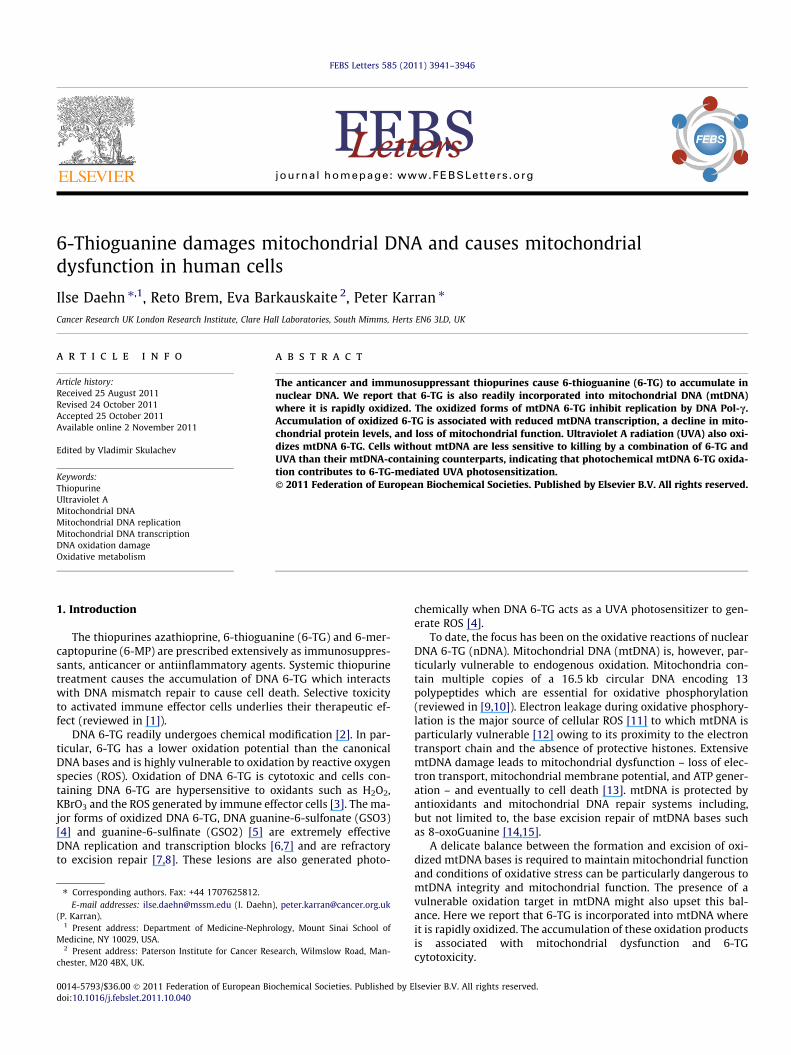
FEBS Letters 585 (2011) 3941–3946
journal homepage: www.FEBSLetters .org
6-Thioguanine damages mitochondrial DNA and causes mitochondrialdysfunction in human cells
Ilse Daehn ⇑,1, Reto Brem, Eva Barkauskaite 2, Peter Karran ⇑Cancer Research UK London Research Institute, Clare Hall Laboratories, South Mimms, Herts EN6 3LD, UK
a r t i c l e i n f o
Article history:Received 25 August 2011Revised 24 October 2011Accepted 25 October 2011Available online 2 November 2011
Edited by Vladimir Skulachev
Keywords:ThiopurineUltraviolet AMitochondrial DNAMitochondrial DNA replicationMitochondrial DNA transcriptionDNA oxidation damageOxidative metabolism
0014-5793/$36.00 � 2011 Federation of European Biodoi:10.1016/j.febslet.2011.10.040
⇑ Corresponding authors. Fax: +44 1707625812.E-mail addresses: [email protected] (I. Daehn
(P. Karran).1 Present address: Department of Medicine-Nephro
Medicine, NY 10029, USA.2 Present address: Paterson Institute for Cancer Res
chester, M20 4BX, UK.
a b s t r a c t
The anticancer and immunosuppressant thiopurines cause 6-thioguanine (6-TG) to accumulate innuclear DNA. We report that 6-TG is also readily incorporated into mitochondrial DNA (mtDNA)where it is rapidly oxidized. The oxidized forms of mtDNA 6-TG inhibit replication by DNA Pol-c.Accumulation of oxidized 6-TG is associated with reduced mtDNA transcription, a decline in mito-chondrial protein levels, and loss of mitochondrial function. Ultraviolet A radiation (UVA) also oxi-dizes mtDNA 6-TG. Cells without mtDNA are less sensitive to killing by a combination of 6-TG andUVA than their mtDNA-containing counterparts, indicating that photochemical mtDNA 6-TG oxida-tion contributes to 6-TG-mediated UVA photosensitization.� 2011 Federation of European Biochemical Societies. Published by Elsevier B.V. All rights reserved.
1. Introduction
The thiopurines azathioprine, 6-thioguanine (6-TG) and 6-mer-captopurine (6-MP) are prescribed extensively as immunosuppres-sants, anticancer or antiinflammatory agents. Systemic thiopurinetreatment causes the accumulation of DNA 6-TG which interactswith DNA mismatch repair to cause cell death. Selective toxicityto activated immune effector cells underlies their therapeutic ef-fect (reviewed in [1]).
DNA 6-TG readily undergoes chemical modification [2]. In par-ticular, 6-TG has a lower oxidation potential than the canonicalDNA bases and is highly vulnerable to oxidation by reactive oxygenspecies (ROS). Oxidation of DNA 6-TG is cytotoxic and cells con-taining DNA 6-TG are hypersensitive to oxidants such as H2O2,KBrO3 and the ROS generated by immune effector cells [3]. The ma-jor forms of oxidized DNA 6-TG, DNA guanine-6-sulfonate (GSO3)[4] and guanine-6-sulfinate (GSO2) [5] are extremely effectiveDNA replication and transcription blocks [6,7] and are refractoryto excision repair [7,8]. These lesions are also generated photo-
chemical Societies. Published by E
logy, Mount Sinai School of
earch, Wilmslow Road, Man-
chemically when DNA 6-TG acts as a UVA photosensitizer to gen-erate ROS [4].
To date, the focus has been on the oxidative reactions of nuclearDNA 6-TG (nDNA). Mitochondrial DNA (mtDNA) is, however, par-ticularly vulnerable to endogenous oxidation. Mitochondria con-tain multiple copies of a 16.5 kb circular DNA encoding 13polypeptides which are essential for oxidative phosphorylation(reviewed in [9,10]). Electron leakage during oxidative phosphory-lation is the major source of cellular ROS [11] to which mtDNA isparticularly vulnerable [12] owing to its proximity to the electrontransport chain and the absence of protective histones. ExtensivemtDNA damage leads to mitochondrial dysfunction – loss of elec-tron transport, mitochondrial membrane potential, and ATP gener-ation – and eventually to cell death [13]. mtDNA is protected byantioxidants and mitochondrial DNA repair systems including,but not limited to, the base excision repair of mtDNA bases suchas 8-oxoGuanine [14,15].
A delicate balance between the formation and excision of oxi-dized mtDNA bases is required to maintain mitochondrial functionand conditions of oxidative stress can be particularly dangerous tomtDNA integrity and mitochondrial function. The presence of avulnerable oxidation target in mtDNA might also upset this bal-ance. Here we report that 6-TG is incorporated into mtDNA whereit is rapidly oxidized. The accumulation of these oxidation productsis associated with mitochondrial dysfunction and 6-TGcytotoxicity.
lsevier B.V. All rights reserved.
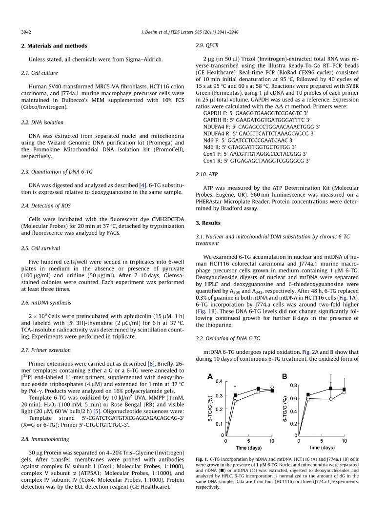
Fig. 1. 6-TG incorporation by nDNA and mtDNA. HCT116 (A) and J774a.1 (B) cellswere grown in the presence of 1 lM 6-TG. Nuclei and mitochondria were separatedand nDNA (j) or mtDNA (s) was extracted, digested to deoxynucleosides andanalyzed by HPLC. 6-TG incorporation is normalized to the amount of dG in thesame DNA sample. Data are from four (HCT116) or three (J774a-1) experiments,respectively.
3942 I. Daehn et al. / FEBS Letters 585 (2011) 3941–3946
2. Materials and methods
Unless stated, all chemicals were from Sigma–Aldrich.
2.1. Cell culture
Human SV40-transformed MRC5-VA fibroblasts, HCT116 coloncarcinoma, and J774a.1 murine macrophage precursor cells weremaintained in Dulbecco’s MEM supplemented with 10% FCS(Gibco/Invitrogen).
2.2. DNA isolation
DNA was extracted from separated nuclei and mitochondriausing the Wizard Genomic DNA purification kit (Promega) andthe Promokine Mitochondrial DNA Isolation kit (PromoCell),respectively.
2.3. Quantitation of DNA 6-TG
DNA was digested and analyzed as described [4]. 6-TG substitu-tion is expressed relative to deoxyguanosine in the same sample.
2.4. Detection of ROS
Cells were incubated with the fluorescent dye CMH2DCFDA(Molecular Probes) for 20 min at 37 �C, detached by trypsinizationand fluorescence was analyzed by FACS.
2.5. Cell survival
Five hundred cells/well were seeded in triplicates into 6-wellplates in medium in the absence or presence of pyruvate(100 lg/ml) and uridine (50 lg/ml). After 7–10 days, Giemsa-stained colonies were counted. Each experiment was performedat least three times.
2.6. mtDNA synthesis
2 � 106 Cells were preincubated with aphidicolin (15 lM, 1 h)and labeled with [50 3H]-thymidine (2 lCi/ml) for 6 h at 37 �C.TCA-insoluble radioactivity was determined by scintillation count-ing. Experiments were performed in triplicate.
2.7. Primer extension
Primer extensions were carried out as described [6]. Briefly, 26-mer templates containing either a G or a 6-TG were annealed to[32P] end-labeled 11-mer primers, supplemented with deoxyribo-nucleoside triphosphates (4 lM) and extended for 1 min at 37 �Cby Pol-c. Products were analyzed on 16% polyacrylamide gels.
Template 6-TG was oxidized by 10 kJ/m2 UVA, MMPP (1 mM,20 min), H2O2 (100 mM, 5 min) or Rose Bengal (RB) and visiblelight (20 lM, 60 W bulb/2 h) [5]. Oligonucleotide sequences were:
Template strand 50-CGATCTGATGTXCGAGCAGACAGCAG-30
(X@G or 6-TG); Primer 50-CTGCTGTCTGC-30.
2.8. Immunoblotting
30 lg Protein was separated on 4–20% Tris–Glycine (Invitrogen)gels. After transfer, membranes were probed with antibodiesagainst complex IV subunit I (Cox1; Molecular Probes, 1:1000),complex V subunit a (ATP5A1; Molecular Probes, 1:1000), andcomplex IV subunit IV (Cox4; Molecular Probes, 1:1000). Proteindetection was by the ECL detection reagent (GE Healthcare).
2.9. QPCR
2 lg (in 50 ll) Trizol (Invitrogen)-extracted total RNA was re-verse-transcribed using the Illustra Ready-To-Go RT–PCR beads(GE Healthcare). Real-time PCR (BioRad CFX96 cycler) consistedof 10 min initial denaturation at 95 �C, followed by 40 cycles of15 s at 95 �C and 60 s at 58 �C. Reactions were prepared with SYBRGreen (Fermentas), using 1 ll cDNA and 10 pmoles of each primerin 25 ll total volume. GAPDH was used as a reference. Expressionratios were calculated with the DD ct method. Primers were:
GAPDH F: 50 GAAGGTGAAGGTCGGAGTC 30
GAPDH R: 50 GAAGATGGTGATGGGATTTC 30
NDUFA4 F: 50 CAGAGCCCTGGAACAAACTGGG 30
NDUFA4 R: 50 GACCTTCATTCTAAAGCAGCG 30
Nd6 F: 50 GGATCCTCCCGAATCAAC 30
Nd6 R: 50 GTAGGATTGGTGCTGTGG 30
Cox1 F: 50 AACGTTGTAGGCCCCTACGGG 30
Cox1 R: 50 GTGAGAGCTAAGGTCGGGGCG 30
2.10. ATP
ATP was measured by the ATP Determination Kit (MolecularProbes, Eugene, OR). 560 nm luminescence was measured on aPHERAstar Microplate Reader. Protein concentrations were deter-mined by Bradford assay.
3. Results
3.1. Nuclear and mitochondrial DNA substitution by chronic 6-TGtreatment
We examined 6-TG accumulation in nuclear and mtDNA of hu-man HCT116 colorectal carcinoma and J774a.1 murine macro-phage precursor cells grown in medium containing 1 lM 6-TG.Deoxynucleoside digests of nuclear and mtDNA were separatedby HPLC and deoxyguanosine and 6-thiodeoxyguanosine werequantified by A260 and A342, respectively. After 48 h, 6-TG replaced0.3% of guanine in both nDNA and mtDNA in HCT116 cells (Fig. 1A).6-TG incorporation by J774.a cells was around two-fold higher(Fig. 1B). These DNA 6-TG levels did not change significantly fol-lowing continued growth for further 8 days in the presence ofthe thiopurine.
3.2. Oxidation of DNA 6-TG
mtDNA 6-TG undergoes rapid oxidation. Fig. 2A and B show thatduring 10 days of continuous 6-TG treatment, the oxidized form of
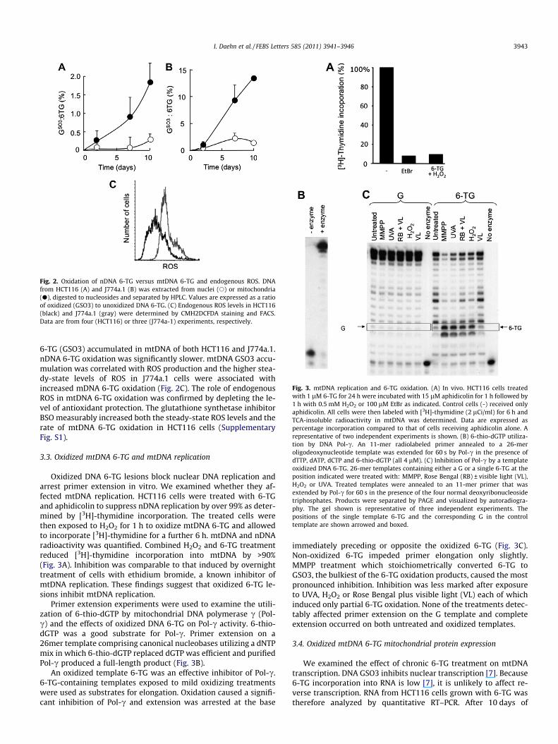
Fig. 2. Oxidation of nDNA 6-TG versus mtDNA 6-TG and endogenous ROS. DNAfrom HCT116 (A) and J774a.1 (B) was extracted from nuclei (s) or mitochondria(d), digested to nucleosides and separated by HPLC. Values are expressed as a ratioof oxidized (GSO3) to unoxidized DNA 6-TG. (C) Endogenous ROS levels in HCT116(black) and J774a.1 (gray) were determined by CMH2DCFDA staining and FACS.Data are from four (HCT116) or three (J774a-1) experiments, respectively.
Fig. 3. mtDNA replication and 6-TG oxidation. (A) In vivo. HCT116 cells treatedwith 1 lM 6-TG for 24 h were incubated with 15 lM aphidicolin for 1 h followed by1 h with 0.5 mM H2O2 or 100 lM EtBr as indicated. Control cells (-) received onlyaphidicolin. All cells were then labeled with [3H]-thymidine (2 lCi/ml) for 6 h andTCA-insoluble radioactivity in mtDNA was determined. Data are expressed aspercentage incorporation compared to that of cells receiving aphidicolin alone. Arepresentative of two independent experiments is shown. (B) 6-thio-dGTP utiliza-tion by DNA Pol-c. An 11-mer radiolabeled primer annealed to a 26-meroligodeoxynucleotide template was extended for 60 s by Pol-c in the presence ofdTTP, dATP, dCTP and 6-thio-dGTP (all 4 lM). (C) Inhibition of Pol-c by a templateoxidized DNA 6-TG. 26-mer templates containing either a G or a single 6-TG at theposition indicated were treated with: MMPP, Rose Bengal (RB) ± visible light (VL),H2O2 or UVA. Treated templates were annealed to an 11-mer primer that wasextended by Pol-c for 60 s in the presence of the four normal deoxyribonucleosidetriphosphates. Products were separated by PAGE and visualized by autoradiogra-phy. The gel shown is representative of three independent experiments. Thepositions of the single template 6-TG and the corresponding G in the controltemplate are shown arrowed and boxed.
I. Daehn et al. / FEBS Letters 585 (2011) 3941–3946 3943
6-TG (GSO3) accumulated in mtDNA of both HCT116 and J774a.1.nDNA 6-TG oxidation was significantly slower. mtDNA GSO3 accu-mulation was correlated with ROS production and the higher stea-dy-state levels of ROS in J774a.1 cells were associated withincreased mDNA 6-TG oxidation (Fig. 2C). The role of endogenousROS in mtDNA 6-TG oxidation was confirmed by depleting the le-vel of antioxidant protection. The glutathione synthetase inhibitorBSO measurably increased both the steady-state ROS levels and therate of mtDNA 6-TG oxidation in HCT116 cells (SupplementaryFig. S1).
3.3. Oxidized mtDNA 6-TG and mtDNA replication
Oxidized DNA 6-TG lesions block nuclear DNA replication andarrest primer extension in vitro. We examined whether they af-fected mtDNA replication. HCT116 cells were treated with 6-TGand aphidicolin to suppress nDNA replication by over 99% as deter-mined by [3H]-thymidine incorporation. The treated cells werethen exposed to H2O2 for 1 h to oxidize mtDNA 6-TG and allowedto incorporate [3H]-thymidine for a further 6 h. mtDNA and nDNAradioactivity was quantified. Combined H2O2 and 6-TG treatmentreduced [3H]-thymidine incorporation into mtDNA by >90%(Fig. 3A). Inhibition was comparable to that induced by overnighttreatment of cells with ethidium bromide, a known inhibitor ofmtDNA replication. These findings suggest that oxidized 6-TG le-sions inhibit mtDNA replication.
Primer extension experiments were used to examine the utili-zation of 6-thio-dGTP by mitochondrial DNA polymerase c (Pol-c) and the effects of oxidized DNA 6-TG on Pol-c activity. 6-thio-dGTP was a good substrate for Pol-c. Primer extension on a26mer template comprising canonical nucleobases utilizing a dNTPmix in which 6-thio-dGTP replaced dGTP was efficient and purifiedPol-c produced a full-length product (Fig. 3B).
An oxidized template 6-TG was an effective inhibitor of Pol-c.6-TG-containing templates exposed to mild oxidizing treatmentswere used as substrates for elongation. Oxidation caused a signifi-cant inhibition of Pol-c and extension was arrested at the base
immediately preceding or opposite the oxidized 6-TG (Fig. 3C).Non-oxidized 6-TG impeded primer elongation only slightly.MMPP treatment which stoichiometrically converted 6-TG toGSO3, the bulkiest of the 6-TG oxidation products, caused the mostpronounced inhibition. Inhibition was less marked after exposureto UVA, H2O2 or Rose Bengal plus visible light (VL) each of whichinduced only partial 6-TG oxidation. None of the treatments detec-tably affected primer extension on the G template and completeextension occurred on both untreated and oxidized templates.
3.4. Oxidized mtDNA 6-TG mitochondrial protein expression
We examined the effect of chronic 6-TG treatment on mtDNAtranscription. DNA GSO3 inhibits nuclear transcription [7]. Because6-TG incorporation into RNA is low [7], it is unlikely to affect re-verse transcription. RNA from HCT116 cells grown with 6-TG wastherefore analyzed by quantitative RT–PCR. After 10 days of
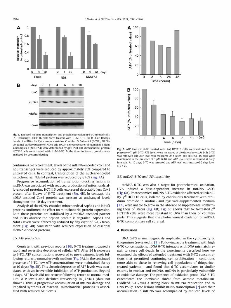
Fig. 4. Reduced mt gene transcription and protein expression in 6-TG-treated cells.(A) Transcripts. HCT116 cells were treated with 1 lM 6-TG for 0, 4 or 10 days.Levels of mRNAs for Cytochrome c oxidase Complex IV Subunit I (COX1), NADH-ubiquinol oxidoreductase 6 (ND6), and NADH dehydrogenase (ubiquinone) 1 alphasubcomplex 4 (NDUFA4) were determined by qRT–PCR. (B) Mitochondrial protein.HCT116 cells were treated with 1 lM 6-TG. At the times indicated, proteins wereanalyzed by Western blotting.
Fig. 5. ATP levels in 6-TG treated cells. (A) HCT116 cells were cultured in thepresence of 1 lM 6-TG. ATP levels were measured at the times shown. At 24 h, 6-TGwas removed and ATP level was measured 24 h later (48). (B) HCT116 cells weremaintained in the presence of 1 lM 6-TG and ATP levels were measured at dailyintervals. At 10 days, 6-TG was removed and ATP level was measured 2 days later(10 + 2).
3944 I. Daehn et al. / FEBS Letters 585 (2011) 3941–3946
continuous 6-TG treatment, levels of the mtDNA-encoded cox1 andnd6 transcripts were reduced by approximately 70% compared tountreated cells. In contrast, transcription of the nuclear-encodedmitochondrial Ndufa4 protein was reduced by 640% (Fig. 4A).
Progressive accumulation of transcription-blocking lesions inmtDNA was associated with reduced production of mitochondrial-ly-encoded proteins. HCT116 cells expressed detectably less Cox1protein after 8 days of 6-TG treatment (Fig. 4B). In contrast, thenDNA-encoded Cox4 protein was present at unchanged levelsthroughout the 10-day treatment.
Analysis of the nDNA-encoded mitochondrial Atp5a1 and Nduf3proteins confirmed the effect on mitochondrial protein expression.Both these proteins are stabilized by a mtDNA-encoded partnerand in its absence the orphan protein is degraded. Atp5a1 andNduf3 levels were detectably reduced by day eight of 6-TG treat-ment (Fig. 4B) consistent with reduced expression of essentialmtDNA-encoded proteins.
3.5. ATP production
Consistent with previous reports [16], 6-TG treatment caused arapid and reversible depletion of cellular ATP. After 24 h exposureto 6-TG, ATP concentrations recovered to pre-treatment levels fol-lowing return to normal growth medium (Fig. 5A). In the continuedpresence of 6-TG, low ATP concentrations were maintained for upto 10 days (Fig. 5B). This chronic depression of ATP levels was asso-ciated with an irreversible inhibition of ATP production. Beyond4 days, ATP levels did not recover following return to normal med-ium. ATP levels also declined irreversibly in J774a.1 (data notshown). Thus, a progressive accumulation of mtDNA damage andimpaired synthesis of essential mitochondrial proteins is associ-ated with reduced ATP levels.
3.6. mtDNA 6-TG and UVA sensitivity
mtDNA 6-TG was also a target for photochemical oxidation.UVA induced a dose-dependent increase in mtDNA GSO3(Fig. 6A). Photochemical mtDNA 6-TG oxidation affected cell viabil-ity. q0 HCT116 cells, isolated by continuous treatment with ethi-dium bromide in uridine- and pyruvate-supplemented medium[17], were unable to grow in the absence of supplements, confirm-ing their q0 status (Fig. 6B). Fig. 6C shows that 6-TG-treated q0
HCT116 cells were more resistant to UVA than their q+ counter-parts. This suggests that the photochemical oxidation of mtDNA6-TG compromises cell survival.
4. Discussion
DNA 6-TG is unambiguously implicated in the cytotoxicity ofthiopurines (reviewed in [1]). Following acute treatment with high6-TG concentrations, nDNA 6-TG interacts with DNA mismatch re-pair to cause cell death. In the experiments described here weexamined the effects of extended treatment with 6-TG concentra-tions that permitted continuing cell proliferation – conditionsmore akin to those in renewing cell populations of thiopurine-treated patients – and found that 6-TG accumulates to similarextents in nuclear and mtDNA. mtDNA is particularly vulnerableto oxidative damage. The presence of oxidation-prone DNA 6-TGexacerbates the inevitable threat from aerobic metabolism.Oxidized 6-TG was a strong block to mtDNA replication and toDNA Pol-c. These lesions inhibit nDNA transcription [7] and theiraccumulation in mtDNA was accompanied by reduced levels of
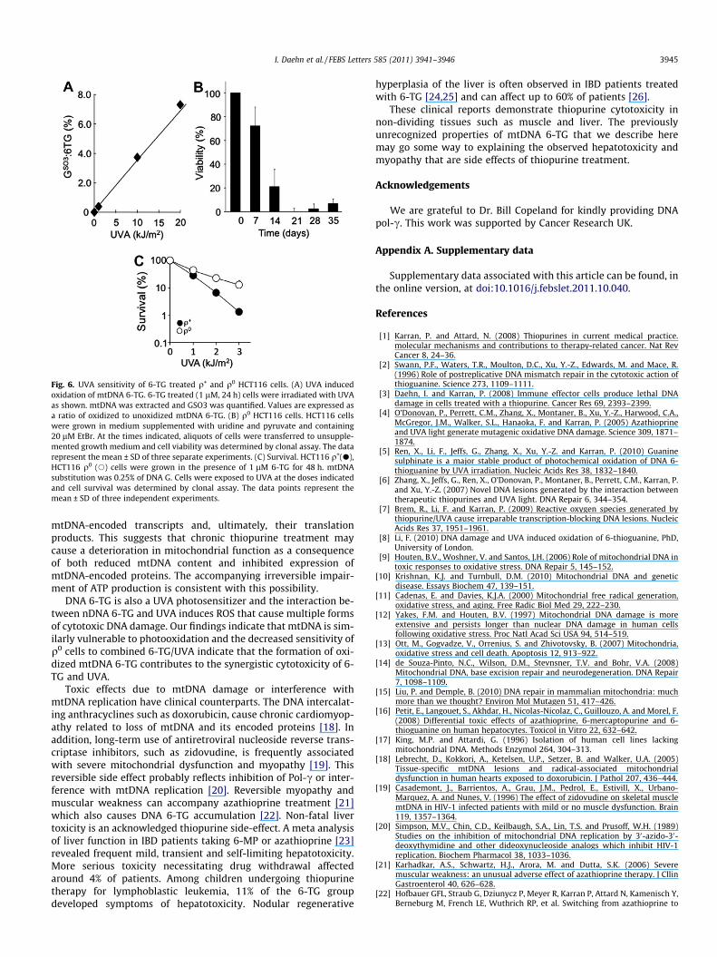
Fig. 6. UVA sensitivity of 6-TG treated q+ and q0 HCT116 cells. (A) UVA inducedoxidation of mtDNA 6-TG. 6-TG treated (1 lM, 24 h) cells were irradiated with UVAas shown. mtDNA was extracted and GSO3 was quantified. Values are expressed asa ratio of oxidized to unoxidized mtDNA 6-TG. (B) q0 HCT116 cells. HCT116 cellswere grown in medium supplemented with uridine and pyruvate and containing20 lM EtBr. At the times indicated, aliquots of cells were transferred to unsupple-mented growth medium and cell viability was determined by clonal assay. The datarepresent the mean ± SD of three separate experiments. (C) Survival. HCT116 q+(d),HCT116 q0 (s) cells were grown in the presence of 1 lM 6-TG for 48 h. mtDNAsubstitution was 0.25% of DNA G. Cells were exposed to UVA at the doses indicatedand cell survival was determined by clonal assay. The data points represent themean ± SD of three independent experiments.
I. Daehn et al. / FEBS Letters 585 (2011) 3941–3946 3945
mtDNA-encoded transcripts and, ultimately, their translationproducts. This suggests that chronic thiopurine treatment maycause a deterioration in mitochondrial function as a consequenceof both reduced mtDNA content and inhibited expression ofmtDNA-encoded proteins. The accompanying irreversible impair-ment of ATP production is consistent with this possibility.
DNA 6-TG is also a UVA photosensitizer and the interaction be-tween nDNA 6-TG and UVA induces ROS that cause multiple formsof cytotoxic DNA damage. Our findings indicate that mtDNA is sim-ilarly vulnerable to photooxidation and the decreased sensitivity ofq0 cells to combined 6-TG/UVA indicate that the formation of oxi-dized mtDNA 6-TG contributes to the synergistic cytotoxicity of 6-TG and UVA.
Toxic effects due to mtDNA damage or interference withmtDNA replication have clinical counterparts. The DNA intercalat-ing anthracyclines such as doxorubicin, cause chronic cardiomyop-athy related to loss of mtDNA and its encoded proteins [18]. Inaddition, long-term use of antiretroviral nucleoside reverse trans-criptase inhibitors, such as zidovudine, is frequently associatedwith severe mitochondrial dysfunction and myopathy [19]. Thisreversible side effect probably reflects inhibition of Pol-c or inter-ference with mtDNA replication [20]. Reversible myopathy andmuscular weakness can accompany azathioprine treatment [21]which also causes DNA 6-TG accumulation [22]. Non-fatal livertoxicity is an acknowledged thiopurine side-effect. A meta analysisof liver function in IBD patients taking 6-MP or azathioprine [23]revealed frequent mild, transient and self-limiting hepatotoxicity.More serious toxicity necessitating drug withdrawal affectedaround 4% of patients. Among children undergoing thiopurinetherapy for lymphoblastic leukemia, 11% of the 6-TG groupdeveloped symptoms of hepatotoxicity. Nodular regenerative
hyperplasia of the liver is often observed in IBD patients treatedwith 6-TG [24,25] and can affect up to 60% of patients [26].
These clinical reports demonstrate thiopurine cytotoxicity innon-dividing tissues such as muscle and liver. The previouslyunrecognized properties of mtDNA 6-TG that we describe heremay go some way to explaining the observed hepatotoxicity andmyopathy that are side effects of thiopurine treatment.
Acknowledgements
We are grateful to Dr. Bill Copeland for kindly providing DNApol-c. This work was supported by Cancer Research UK.
Appendix A. Supplementary data
Supplementary data associated with this article can be found, inthe online version, at doi:10.1016/j.febslet.2011.10.040.
References
[1] Karran, P. and Attard, N. (2008) Thiopurines in current medical practice.molecular mechanisms and contributions to therapy-related cancer. Nat RevCancer 8, 24–36.
[2] Swann, P.F., Waters, T.R., Moulton, D.C., Xu, Y.-Z., Edwards, M. and Mace, R.(1996) Role of postreplicative DNA mismatch repair in the cytotoxic action ofthioguanine. Science 273, 1109–1111.
[3] Daehn, I. and Karran, P. (2008) Immune effector cells produce lethal DNAdamage in cells treated with a thiopurine. Cancer Res 69, 2393–2399.
[4] O’Donovan, P., Perrett, C.M., Zhang, X., Montaner, B., Xu, Y.-Z., Harwood, C.A.,McGregor, J.M., Walker, S.L., Hanaoka, F. and Karran, P. (2005) Azathioprineand UVA light generate mutagenic oxidative DNA damage. Science 309, 1871–1874.
[5] Ren, X., Li, F., Jeffs, G., Zhang, X., Xu, Y.-Z. and Karran, P. (2010) Guaninesulphinate is a major stable product of photochemical oxidation of DNA 6-thioguanine by UVA irradiation. Nucleic Acids Res 38, 1832–1840.
[6] Zhang, X., Jeffs, G., Ren, X., O’Donovan, P., Montaner, B., Perrett, C.M., Karran, P.and Xu, Y.-Z. (2007) Novel DNA lesions generated by the interaction betweentherapeutic thiopurines and UVA light. DNA Repair 6, 344–354.
[7] Brem, R., Li, F. and Karran, P. (2009) Reactive oxygen species generated bythiopurine/UVA cause irreparable transcription-blocking DNA lesions. NucleicAcids Res 37, 1951–1961.
[8] Li, F. (2010) DNA damage and UVA induced oxidation of 6-thioguanine, PhD,University of London.
[9] Houten, B.V., Woshner, V. and Santos, J.H. (2006) Role of mitochondrial DNA intoxic responses to oxidative stress. DNA Repair 5, 145–152.
[10] Krishnan, K.J. and Turnbull, D.M. (2010) Mitochondrial DNA and geneticdisease. Essays Biochem 47, 139–151.
[11] Cadenas, E. and Davies, K.J.A. (2000) Mitochondrial free radical generation,oxidative stress, and aging. Free Radic Biol Med 29, 222–230.
[12] Yakes, F.M. and Houten, B.V. (1997) Mitochondrial DNA damage is moreextensive and persists longer than nuclear DNA damage in human cellsfollowing oxidative stress. Proc Natl Acad Sci USA 94, 514–519.
[13] Ott, M., Gogvadze, V., Orrenius, S. and Zhivotovsky, B. (2007) Mitochondria,oxidative stress and cell death. Apoptosis 12, 913–922.
[14] de Souza-Pinto, N.C., Wilson, D.M., Stevnsner, T.V. and Bohr, V.A. (2008)Mitochondrial DNA, base excision repair and neurodegeneration. DNA Repair7, 1098–1109.
[15] Liu, P. and Demple, B. (2010) DNA repair in mammalian mitochondria: muchmore than we thought? Environ Mol Mutagen 51, 417–426.
[16] Petit, E., Langouet, S., Akhdar, H., Nicolas-Nicolaz, C., Guillouzo, A. and Morel, F.(2008) Differential toxic effects of azathioprine, 6-mercaptopurine and 6-thioguanine on human hepatocytes. Toxicol in Vitro 22, 632–642.
[17] King, M.P. and Attardi, G. (1996) Isolation of human cell lines lackingmitochondrial DNA. Methods Enzymol 264, 304–313.
[18] Lebrecht, D., Kokkori, A., Ketelsen, U.P., Setzer, B. and Walker, U.A. (2005)Tissue-specific mtDNA lesions and radical-associated mitochondrialdysfunction in human hearts exposed to doxorubicin. J Pathol 207, 436–444.
[19] Casademont, J., Barrientos, A., Grau, J.M., Pedrol, E., Estivill, X., Urbano-Marquez, A. and Nunes, V. (1996) The effect of zidovudine on skeletal musclemtDNA in HIV-1 infected patients with mild or no muscle dysfunction. Brain119, 1357–1364.
[20] Simpson, M.V., Chin, C.D., Keilbaugh, S.A., Lin, T.S. and Prusoff, W.H. (1989)Studies on the inhibition of mitochondrial DNA replication by 30-azido-30-deoxythymidine and other dideoxynucleoside analogs which inhibit HIV-1replication. Biochem Pharmacol 38, 1033–1036.
[21] Karhadkar, A.S., Schwartz, H.J., Arora, M. and Dutta, S.K. (2006) Severemuscular weakness: an unusual adverse effect of azathioprine therapy. J CllinGastroenterol 40, 626–628.
[22] Hofbauer GFL, Straub G, Dziunycz P, Meyer R, Karran P, Attard N, Kamenisch Y,Berneburg M, French LE, Wuthrich RP, et al. Switching from azathioprine to

3946 I. Daehn et al. / FEBS Letters 585 (2011) 3941–3946
mycophenolate mofetil improves skin photosensitivity in kidney transplantrecipients. Am J Transplant (E pub ahead of print, September 2011).
[23] Vora, A., Mitchell, C.D., Lennard, L., Eden, T.O., Kinsey, S.E., Lilleyman, J. andRichards, S.M. The Medical Research Council; National Cancer ResearchNetwork Childhood Leukaemia Working Party (2006) Toxicity and efficacyof 6-thioguanine versus 6-mercaptopurine in childhood lymphoblasticleukaemia: a randomised trial. Lancet 368, 1339–1348.
[24] Geller, S.A., Dubinsky, M.C., Poordad, F.F., Vasiliauskas, E.A., Cohen, A.H., Abreu,M.T., Tran, T., Martin, P., Vierling, J.M. and Targan, S.R. (2004) Early hepaticnodular hyperplasia and submicroscopic fibrosis associated with 6-
thioguanine therapy in inflammatory bowel disease. Am J Surg Pathol 28,1204–1211.
[25] Ferlitsch, A., Teml, A., Reinisch, W., Ulbrich, G., Wrba, F., Homoncik, M., Gangl,A., Peck-Radoslavljevic, M. and Vogelsang, H. (2007) 6-thioguanine associatednodular regenerative hyperplasia in patients with inflammatory bowel diseasemay induce portal hypertension. Am J Gastroenterol 102, 2495–2503.
[26] Dubinsky, M.C., Vasiliauskas, E.A., Singh, H., Abreu, M.T., Papadakis, K.A., Tran,T., Martin, P., Vierling, J.M., Geller, S.E., Targan, S.R., et al. (2003) 6-Thioguaninecan cause serious liver injury in inflammatory bowel disease patients.Gastroenterology 125, 298–303.
Copyright © 2022 FDOKUMEN