The effects on Trypanosoma cruzi of novel synthetic naphthoquinones are mediated by mitochondrial...
11
A comparative assessment of mitochondrial function in epimastigotes and bloodstream trypomastigotes of Trypanosoma cruzi Renata L. S. Gonçalves & Rubem F. S. Menna Barreto & Carla R. Polycarpo & Fernanda R. Gadelha & Solange L. Castro & Marcus F. Oliveira Received: 9 August 2011 /Accepted: 28 September 2011 /Published online: 12 November 2011 # Springer Science+Business Media, LLC 2011 Abstract Trypanosoma cruzi is a hemoflagellate protozoan that causes Chagas’ disease. The life cycle of T. cruzi is complex and involves different evolutive forms that have to encounter different environmental conditions provided by the host. Herein, we performed a functional assessment of mitochondrial metabolism in the following two distinct evolutive forms of T. cruzi: the insect stage epimastigote and the freshly isolated bloodstream trypomastigote. We observed that in comparison to epimastigotes, bloodstream trypomastigotes facilitate the entry of electrons into the electron transport chain by increasing complex II-III activity. Interestingly, cytochrome c oxidase (CCO) activity and the expression of CCO subunit IV were reduced in bloodstream forms, creating an “electron bottleneck” that favored an increase in electron leakage and H 2 O 2 forma- tion. We propose that the oxidative preconditioning provided by this mechanism confers protection to blood- stream trypomastigotes against the host immune system. In this scenario, mitochondrial remodeling during the T. cruzi life cycle may represent a key metabolic adaptation for parasite survival in different hosts. Keywords Energy metabolism . Reactive oxygen species . Hormesis . Differentiation Introduction Chagas’ disease, once thought to be an endemic illness in Latin America, affects approximately 12–14 million people (Dias 2007) and has spread to other regions, such as North America, Europe and Japan (Tanowitz et al. 2011). The life cycle of the etiological agent Trypanosoma cruzi is complex, involving four different evolutive forms that have to develop inside hematophagous triatomine insect vectors and mammalian hosts. Inside the vertebrate host, T. cruzi transforms into a bloodstream non-dividing form referred to as a trypomastigote, which subsequently transforms into an intracellular dividing form, the amastigote. Inside the insect host, T. cruzi develops into either proliferative or non- This work is dedicated to the memory of the honorable Brazilian scientist, teacher and human being Dr. Henrique Leonel Lenzi (1943–2011). R. L. S. Gonçalves : M. F. Oliveira (*) Laboratório de Bioquímica de Resposta ao Estresse, Programa de Biologia Molecular e Biotecnologia, Instituto de Bioquímica Médica, Universidade Federal do Rio de Janeiro, Rio de Janeiro, RJ, Brazil e-mail: [email protected] R. L. S. Gonçalves : M. F. Oliveira Laboratório de Inflamação e Metabolismo, Instituto Nacional de Ciência e Tecnologia de Biologia Estrutural e Bioimagem (INBEB), Universidade Federal do Rio de Janeiro, Rio de Janeiro, RJ, Brazil R. F. S. M. Barreto : S. L. Castro Laboratório de Biologia Celular, Instituto Oswaldo Cruz, FIOCRUZ, Rio de Janeiro, RJ, Brazil C. R. Polycarpo Laboratório de Biologia Molecular, Programa de Biologia Molecular e Biotecnologia, Instituto de Bioquímica Médica, Universidade Federal do Rio de Janeiro, Rio de Janeiro, RJ, Brazil F. R. Gadelha Departamento de Bioquímica, Instituto de Biologia, Universidade Estadual de Campinas, Campinas, SP, Brazil J Bioenerg Biomembr (2011) 43:651–661 DOI 10.1007/s10863-011-9398-8
-
Upload
independent -
Category
Documents
-
view
4 -
download
0
Transcript of The effects on Trypanosoma cruzi of novel synthetic naphthoquinones are mediated by mitochondrial...
A comparative assessment of mitochondrial functionin epimastigotes and bloodstream trypomastigotesof Trypanosoma cruzi
Renata L. S. Gonçalves & Rubem F. S. Menna Barreto &
Carla R. Polycarpo & Fernanda R. Gadelha &
Solange L. Castro & Marcus F. Oliveira
Received: 9 August 2011 /Accepted: 28 September 2011 /Published online: 12 November 2011# Springer Science+Business Media, LLC 2011
Abstract Trypanosoma cruzi is a hemoflagellate protozoanthat causes Chagas’ disease. The life cycle of T. cruzi iscomplex and involves different evolutive forms that have toencounter different environmental conditions provided bythe host. Herein, we performed a functional assessment ofmitochondrial metabolism in the following two distinct
evolutive forms of T. cruzi: the insect stage epimastigoteand the freshly isolated bloodstream trypomastigote. Weobserved that in comparison to epimastigotes, bloodstreamtrypomastigotes facilitate the entry of electrons into theelectron transport chain by increasing complex II-IIIactivity. Interestingly, cytochrome c oxidase (CCO) activityand the expression of CCO subunit IV were reduced inbloodstream forms, creating an “electron bottleneck” thatfavored an increase in electron leakage and H2O2 forma-tion. We propose that the oxidative preconditioningprovided by this mechanism confers protection to blood-stream trypomastigotes against the host immune system. Inthis scenario, mitochondrial remodeling during the T. cruzilife cycle may represent a key metabolic adaptation forparasite survival in different hosts.
Keywords Energy metabolism . Reactive oxygen species .
Hormesis . Differentiation
Introduction
Chagas’ disease, once thought to be an endemic illness inLatin America, affects approximately 12–14 million people(Dias 2007) and has spread to other regions, such as NorthAmerica, Europe and Japan (Tanowitz et al. 2011). The lifecycle of the etiological agent Trypanosoma cruzi iscomplex, involving four different evolutive forms that haveto develop inside hematophagous triatomine insect vectorsand mammalian hosts. Inside the vertebrate host, T. cruzitransforms into a bloodstream non-dividing form referred toas a trypomastigote, which subsequently transforms into anintracellular dividing form, the amastigote. Inside the insecthost, T. cruzi develops into either proliferative or non-
This work is dedicated to the memory of the honorable Brazilian scientist,teacher and human being Dr. Henrique Leonel Lenzi (1943–2011).
R. L. S. Gonçalves :M. F. Oliveira (*)Laboratório de Bioquímica de Resposta ao Estresse,Programa de Biologia Molecular e Biotecnologia,Instituto de Bioquímica Médica,Universidade Federal do Rio de Janeiro,Rio de Janeiro, RJ, Brazile-mail: [email protected]
R. L. S. Gonçalves :M. F. OliveiraLaboratório de Inflamação e Metabolismo,Instituto Nacional de Ciência e Tecnologia deBiologia Estrutural e Bioimagem (INBEB),Universidade Federal do Rio de Janeiro,Rio de Janeiro, RJ, Brazil
R. F. S. M. Barreto : S. L. CastroLaboratório de Biologia Celular, Instituto Oswaldo Cruz,FIOCRUZ,Rio de Janeiro, RJ, Brazil
C. R. PolycarpoLaboratório de Biologia Molecular,Programa de Biologia Molecular e Biotecnologia,Instituto de Bioquímica Médica,Universidade Federal do Rio de Janeiro,Rio de Janeiro, RJ, Brazil
F. R. GadelhaDepartamento de Bioquímica, Instituto de Biologia,Universidade Estadual de Campinas,Campinas, SP, Brazil
J Bioenerg Biomembr (2011) 43:651–661DOI 10.1007/s10863-011-9398-8
proliferative infective forms known as epimastigotes andmetacyclic trypomastigotes, respectively. Thus, the adapta-tion of different parasite evolutive forms to changes inenvironmental and physico-chemical conditions is animportant survival mechanism.
The members of the Trypanosomatidae family exhibitseveral distinct features, such as the presence of glycosomes,which are peroxisome-like organelles that compartmentalizethe first reactions of glycolysis (Michels et al. 2006). Theseparasites possess a single mitochondrion that remainsmorphologically similar throughout their life cycle (deMeirelles and De Souza 1982). Regarding energy metabo-lism, the life stages of T. cruzi and Leishmania exhibit acomparatively more complex metabolic capacity than othertrypanosomatids because they are able to metabolize glucoseand amino acids (Rogerson and Gutteridge 1980). AlthoughT. cruzi metabolism changes considerably over its life cycle,the different stages have functional tricarboxylic acid cycles(Adroher et al. 1988; Rogerson and Gutteridge 1980) andoxidative phosphorylation machinery (Tielens and VanHellemond 2009). T. cruzi experiences differences in glucoseavailability being the vertebrate blood rich in glucose(Lehane 2005), whereas the insect digestive tract has limitedamounts of free glucose (Billingsley 1988). The vertebrateform trypomastigotes exhibits the highest glucose transportactivity (Silber et al. 2009), and its transition to amastigotesis accompanied by a shift from a carbohydrate- to a lipid-based energy metabolism (Atwood et al. 2005).
Mitochondria are organelles that are implicated not onlyin aerobic ATP synthesis via oxidative phosphorylation butare also involved in the cellular redox balance, representingone of the major sources of reactive oxygen species (ROS)in the cell. During mitochondrial respiration, a smallportion of oxygen is partially reduced to superoxide (O2
•−)radicals, which are then dismutated to hydrogen peroxide(H2O2) by superoxide dismutase (SOD) (Boveris andChance 1973; Brookes et al. 2002) or spontaneousreduction. The redox state of the mitochondrial electrontransport chain (ETC) is crucial for ROS generation(Nicholls and Ferguson 2002). Under a higher mitochon-drial membrane potential (ΔΨm), the half-life of thereduced ETC components increases, favoring the leakageof electrons (Kowaltowski et al. 2009; Korshunov et al.1997). Controlled levels of ROS are important for signalingand adaptation to a number of different insults (Pan et al.2011). Mitochondrial, microsomal and cytosolic enzymescontribute to H2O2 generation at fairly high rates in T. cruziepimastigotes (Boveris and Stoppani 1977; Carranza et al.2009). Interestingly, parasites with increased antioxidantdefenses are more resistant to redox insults and are morevirulent, suggesting a relationship between redox balanceand infectivity (Piacenza et al. 2009). Finally, the transfor-mation of epimastigotes to infective metacyclic trypomasti-
gotes entails an increase in some of the parasite antioxidantdefenses, which can be seen as important mechanisms tocircumvent the redox challenge mediated by the hostimmune system (Atwood et al. 2005).
Recent findings demonstrate that complex I of therespiratory chain has limited functions in T. cruzi metabo-lism (Carranza et al. 2009; Silva et al. 2011). Additionally,succinate has been shown to be the main substrate thatsupports oxygen consumption in epimastigotes (Denicola-Seoane et al. 1992; Vercesi et al. 1991). The levels ofcytochromes b and a are significantly lower in T. cruziepimastigotes in comparison with those in mammalian cells(Cazzulo 1994). Cytochrome a is part of complex IV, whichis the rate limiting step in mitochondrial respiration (Poytonand McEwen 1996; Villani et al. 1998; Villani and Attardi2000). The inhibition of complex IV activity increases thereduction state of the upstream ETC components, impairingthe respiratory rates and oxidative phosphorylation,thereby favoring ROS formation (Ferguson et al. 2005;Zuckerbraun et al. 2007). Another peculiarity of thetrypanosomatid respiratory chain is the presence of acyanide-insensitive salicylhydroxamic acid (SHAM)-sensitiveterminal oxidase (alternative oxidase), which is important forregenerating glycosomal NADH (Chaudhuri et al. 2006).
Using an enhanced method to culture trypomastigotes,Docampo et al. demonstrated select features of the T. cruzimitochondrion (Docampo et al. 1993; Docampo 1993).Aside from these previous reports, the efforts to character-ize mitochondrial physiology in different T. cruzi formshave been hampered by the difficulties in obtaining freshlyisolated bloodstream trypomastigotes. Therefore, we aimedto compare the mitochondrial function between freshlyisolated bloodstream T. cruzi trypomastigotes with epimas-tigotes. Because T. cruzi experiences strikingly distinctenvironmental challenges in hosts, we hypothesized thatmitochondrion functional plasticity would be central inenabling the parasite to adapt to such variations. Wedemonstrated that the mitochondrion of T. cruzi bloodstreamtrypomastigotes exhibits lower oxygen consumption ratesand increased H2O2 production in comparison to those of theinsect stage epimastigotes. The increased electron leakageobserved in bloodstream trypomastigotes could be a conse-quence of the increased activity of complex II–III and thereduced activity of complex IV. We propose that this is aredox-mediated preconditioning that would confer someprotection to bloodstream trypomastigotes against an oxida-tive challenge induced by host immune system activation.
Material and methods
Parasites All experiments were performed with the T. cruziY strain. Epimastigotes were maintained axenically at 28 °C
652 J Bioenerg Biomembr (2011) 43:651–661
in a liver infusion and tryptose (LIT) medium supplementedwith 10% fetal bovine serum (FBS) (Cultilab, Campinas,Brazil). The medium was changed weekly, and epimasti-gotes were harvested during the exponential growth phase(5-day old cultures). Bloodstream trypomastigotes wereisolated from the blood of albino Swiss mice 7 days afterintraperitoneal injection with 5×10⁵ parasites. Citratedblood was collected by heart puncture. Red and whiteblood cells were removed by differential centrifugation(500 x g for 30 min at 4 °C), and the supernatant wascollected to obtain the purified parasites. To improve theyield of parasites, the pellet was resuspended in Dulbecco’smodified Eagle’s medium (DMES, Sigma-Aldrich, St.Louis, MO, USA) supplemented with 10% FBS and re-centrifuged at 500 x g for 15 min. This step was repeatedtwo times, the supernatants were centrifuged (1,500 x g for15 min) and bloodstream trypomastigote pellets wereresuspended in 3–5 mL of DMES+10% FBS. Finally, theparasites were washed with phosphate buffered saline(PBS) and kept on ice until use. The yield of this procedurewas approximately 2.5×10⁷ parasites/mouse. The proteinconcentration in both parasite forms was determined by theLowry method (Lowry et al. 1951) using bovine serumalbumin as the standard.
Susceptibility of T. cruzi to different compounds Blood-stream trypomastigotes and epimastigotes were respectivelyresuspended in DMES and LIT media supplemented with10% FBS. An aliquot of 100 μL (106 parasites) was addedto the same volume of H2O2, antimycin A (AA) oriodoacetamide (IAA) previously prepared at twice thedesired final concentration in 96-well microplates andincubated at 37 °C (trypomastigotes) or 28 °C (epimasti-gotes) for 2 h. Cell counts were performed on a Neubauerchamber, and the activity of the compounds upon parasitesurvival was expressed as LD50, corresponding to theconcentration that leads to the lysis of 50% parasites.
Mitochondrial membrane potential Parasite mitochondrialmembrane potential (ΔΨm) was evaluated fluorimetricallyby the following two different approaches: i) TMRE probeon intact cells; ii) safranine O in digitonin-permeabilizedcells. For the TMRE analysis, epimastigotes and blood-stream trypomastigotes were incubated with 50 nM of theprobe for 20 min. TMRE-positive parasites were analyzedusing a FACSCalibur flow cytometer (Becton Dickinson,Franklin Lakes, NJ, USA) equipped with Cell Questsoftware (Joseph Trotter, Scripps Research Institute, LaJolla, CA, USA). The specificity of TMRE staining wasevaluated by inducing mitochondrial uncoupling after theaddition of 1 μM FCCP. A total of 10,000 events wereacquired in the region previously established to correspondto parasites. Safranin O fluorescence was measured with a
spectrofluorometer (excitation and emission at 495 and586 nm wavelengths, respectively) (Varian, Cary EclipseModel, Oberkochen, Germany). A sample of 5×10⁷ para-sites was added to 2 mL of respiration buffer containing125 mM sucrose, 65 mM KCl, 2 mM KH2PO4, 0.5 mMMgCl2, 10 mM HEPES-KOH (pH 7.2), 1 mg/mL fatty acidfree bovine serum albumin (FAF-BSA), and 1 mM EGTA(Vercesi et al. 1991 with minor modifications). Succinate(5 mM) was added to energize the mitochondria. Thedigitonin concentration was titrated based on the loss offluorescence of safranin O. Optimal digitonin concentra-tions for each parasite form, 15 μM for epimastigotes and7.5 μM for the trypomastigotes, were set based on themaintenance of the ΔΨm for at least 30 min. The ΔΨm
collapsed after adding 1.25 μg/mL AA.
ETC complex activities Mitochondrial ETC complex activ-ities were measured in triplicate at room temperature in atotal reaction volume of 1 mL using a spectrophotometer(UV 2550 Shimadzu Co., Shimadzu, Japan) as previouslydescribed (Ferguson et al. 2005). The AA-sensitivesuccinate:cytochrome c oxidoreductase activity (complexII–III) was measured by the increase in the absorbance at550 nm due to the reduction of ferricytochrome c(ε=19 mM−1 cm−1) (Chance and Williams 1955). Thereaction mixture consisted of 100 mM potassium phos-phate buffer (pH 7.4), 50 μM horse heart cytochrome c,5 mM succinate and 1 mM KCN. KCN-sensitive cyto-chrome c oxidase (complex IV) activity was measuredbased on the decrease in absorbance due to the oxidationof ferrocytochrome c at 550 nm (ε=19 mM−1 cm−1). Thereaction mixture consisted of 100 mM potassium phos-phate buffer (pH 7.4) and 50 μM sodium dithionite-reduced cytochrome c. Decreases in absorbance weremonitored after the addition of frozen-thawed parasitehomogenates (70 μg of protein).
F1Fo ATP synthase functional content To assess thefunctional content of F1Fo ATP synthase, basal respiratoryrates of epimastigotes and bloodstream trypomastigoteskept in respiration buffer were titrated with oligomycin(0.1 μg/mL) until they reached a state-4 like respiratoryrates, meaning that any further addition of oligomycin wasunable to decrease the oxygen consumption rates. The basalrespiratory rates were plotted against the doses of oligomy-cin, and the functional content was calculated as describedin Japiassu et al. (2011).
Oxygen consumption rates O2 consumption rates of epi-mastigotes or trypomastigotes (5×10⁷ parasites/chamber)were evaluated by high-resolution respirometry (Oxygraph-2 K; OROBOROS Instruments, Innsbruck, Austria) undercontinuous stirring. The temperature was maintained at 28 °C
J Bioenerg Biomembr (2011) 43:651–661 653
for experiments with epimastigotes and at 37 °C fortrypomastigotes. Both reactions were performed in 2 mL ofrespiration buffer. Oxygen concentration and flux wererecorded using DatLab software (Oxygraph-2 K; ORO-BOROS Instruments, Innsbruck, Austria). Digitonin (15 μMfor epimastigotes and 7.5 μM for trypomastigotes) was addedto permeabilize the parasites. Subsequently, 5 mM succinateand 200 μM ADP were added to stimulate state 3 mitochon-drial oxygen consumption. State 4-like respiration wasinduced with the addition of 2.5 μg/mL oligomycin asdescribed by Vercesi et al. (1991). Uncoupled respirationwas stimulated after the addition of up to 3 μM FCCP,resulting in increased oxygen consumption. Mitochondrialrespiration was inhibited by the addition of 1.25 μg/mL ofAA to reach residual oxygen consumption (ROX).
Mitochondrial H2O2 release H2O2 release was measuredusing the Amplex Red probe (Molecular Probes, Carlsbad,CA, USA) and horseradish peroxidase (HRP; Sigma-Aldrich, St. Louis, MO, USA) (Votyakova and Reynolds2004). Epimastigotes or trypomastigotes (5 x 10⁷/chamber)were incubated in the respiration buffer described abovecontaining 2.5 μM Amplex Red reagent and 3 U/mL HRP.Parasites were permeabilized with the same digitoninconcentrations set for the respirometry assays. Mitochon-drial metabolic states were measured following the sameprotocol employed for the oxygen consumption measure-ments. Amplex Red fluorescence was monitored at excita-tion and emission wavelengths at 530 nm (slit 5 nm) and590 nm (slit 5 nm), respectively, in a Varian spectrofluo-rometer (Cary Eclipse Model, Oberkochen, Germany). Acalibration curve was obtained using H2O2 as a standard(Menna-Barreto et al. 2009).
Mitochondrial free radical leak Both oxygen consumptionrates and H2O2 release were measured in the same media,as well as at the same temperature and substrate concen-tration. The free radical leak was considered as thepercentage of electrons out of sequence that reducedoxygen to superoxide (which is further dismutated toH2O2) instead of reaching complex IV to reduce oxygento water. The rates of H2O2 release were divided by twicethe rate of oxygen consumed in the same mitochondrialmetabolic state; percentages of these values were obtainedby multiplication by 100 (Herrero and Barja 1997).
Real time PCR Parasite total RNA was extracted usingTRIzol reagent according to the manufacturer’s instruc-tions. Up to 1 μg of RNA was treated with DNAse I(Fermentas, Thermo Fisher Scientific Inc., Canada) to avoidgenomic DNA contamination. The complementary DNA(cDNA) was synthesized using the High-capacity ReverseTranscriptase Polymerase Chain Reaction (RT-PCR) kit
(Applied Biosystems, USA). Quantitative PCR was per-formed in a StepOnePlus Real time PCR machine (AppliedBiosystems, USA) using the Power SYBR-Green PCRMaster Mix (Applied Biosystems, USA). The 2−ΔΔCt
method was used to analyze the relative changes in geneexpression levels (Livak and Schmittgen 2001). Alphatubulin (accession number: Tc00.1047053411235.9) wasused as an endogenous control. The PCR programconsisted of a heating at 95 °C for 10 min followed by 40cycles at 95 °C for 15 s, 60 °C for 1 min and a meltingcurve. The oligonucleotides were designed using primer3software (Rozen and Skaletsky 2000) after the identifica-tion of T. cruzi orthologs for succinate dehydrogenase(accession number: Tc00.1047053505843.24), cytochromeb (accession number: Tc00.1047053509395.100) and cyto-chrome c oxidase, and subunit IV (accession number:Tc00.1047053510889.50). The following primer sequenceswere used: L- GGCTTTGGAAACAACCCATA and R-TCAATCAACCAGCGATACGA for succinate dehydroge-nase; L- GGTCACAGTGAACAGGCAAA and R-CCCCAACGAAAGAAATACCA for cytochrome b andcytochrome c oxidase; and L-CTACGTGAAAAGACGCGTTG and R- GCATACTCCCGCTCAACATT for subunit IV.
Statistical analyses Comparisons between groups wereperformed by one-way ANOVA followed by Tukey’s testfor pairwise comparison. When appropriate, unpairedStudent’s t-tests were employed. Statistical analysis wasperformed using GraphPad Prism version 4.00 for Win-dows (GraphPad Software, San Diego, USA). Significancelevels were indicated by P≤0.05. All experiments wereperformed with at least three independent sample groups.
Results
Epimastigotes and bloodstream trypomastigotes exhibitdifferent susceptibilities to energy and redox metabolismmodulators
Our first goal was to determine the susceptibility of T. cruziepimastigotes and bloodstream trypomastigotes to iodoace-tamide (IAA) and antimycin a (AA), which are classicalinhibitors of glyceraldehyde-3-phosphate dehydrogenase(glycolysis) and mitochondrial ETC complex III, respec-tively. Table 1 shows that bloodstream trypomastigotes aremore sensitive to the inhibition of glycolysis than epimas-tigotes due to their significantly lower LD50 value for IAA(171.2±38.7 μM vs. 1818.4±258.4 μM, p<0.004). How-ever, bloodstream trypomastigotes were more resistant toETC inhibition than epimastigotes, exhibiting a higherLD50 value for AA (193.4±13.3 μM vs. 40.6±5.9 μM, p<0.02). Interestingly, the bloodstream forms were also more
654 J Bioenerg Biomembr (2011) 43:651–661
resistant to oxidative stress, as revealed by higher LD50
values for H2O2 (1430.5±140.3 μM vs. 694.2±33.1 μM,p<0.02). These results indicate that T. cruzi bloodstreamtrypomastigotes rely more on glycolysis than oxidativephosphorylation to meet their energy demand, and aremore resistant to redox challenges than epimastigotes.
Bloodstream trypomastigotes exhibit lower mitochondrialmembrane potential than epimastigotes
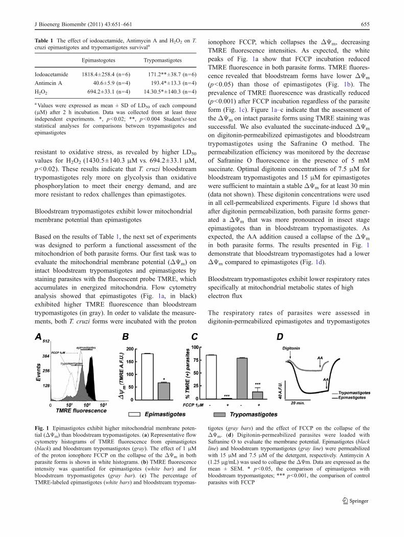
Based on the results of Table 1, the next set of experimentswas designed to perform a functional assessment of themitochondrion of both parasite forms. Our first task was toevaluate the mitochondrial membrane potential (ΔΨm) onintact bloodstream trypomastigotes and epimastigotes bystaining parasites with the fluorescent probe TMRE, whichaccumulates in energized mitochondria. Flow cytometryanalysis showed that epimastigotes (Fig. 1a, in black)exhibited higher TMRE fluorescence than bloodstreamtrypomastigotes (in gray). In order to validate the measure-ments, both T. cruzi forms were incubated with the proton
ionophore FCCP, which collapses the ΔΨm, decreasingTMRE fluorescence intensities. As expected, the whitepeaks of Fig. 1a show that FCCP incubation reducedTMRE fluorescence in both parasite forms. TMRE fluores-cence revealed that bloodstream forms have lower ΔΨm
(p<0.05) than those of epimastigotes (Fig. 1b). Theprevalence of TMRE fluorescence was drastically reduced(p<0.001) after FCCP incubation regardless of the parasiteform (Fig. 1c). Figure 1a–c indicate that the assessment ofthe ΔΨm on intact parasite forms using TMRE staining wassuccessful. We also evaluated the succinate-induced ΔΨm
on digitonin-permeabilized epimastigotes and bloodstreamtrypomastigotes using the Safranine O method. Thepermeabilization efficiency was monitored by the decreaseof Safranine O fluorescence in the presence of 5 mMsuccinate. Optimal digitonin concentrations of 7.5 μM forbloodstream trypomastigotes and 15 μM for epimastigoteswere sufficient to maintain a stable ΔΨm for at least 30 min(data not shown). These digitonin concentrations were usedin all cell-permeabilized experiments. Figure 1d shows thatafter digitonin permeabilization, both parasite forms gener-ated a ΔΨm that was more pronounced in insect stageepimastigotes than in bloodstream trypomastigotes. Asexpected, the AA addition caused a collapse of the ΔΨm
in both parasite forms. The results presented in Fig. 1demonstrate that bloodstream trypomastigotes had a lowerΔΨm compared to epimastigotes (Fig. 1d).
Bloodstream trypomastigotes exhibit lower respiratory ratesspecifically at mitochondrial metabolic states of highelectron flux
The respiratory rates of parasites were assessed indigitonin-permeabilized epimastigotes and trypomastigotes
Table 1 The effect of iodoacetamide, Antimycin A and H2O2 on T.cruzi epimastigotes and trypomastigotes survivala
Epimastogotes Trypomastigotes
Iodoacetamide 1818.4±258.4 (n=6) 171.2**±38.7 (n=6)
Antimcin A 40.6±5.9 (n=4) 193.4*±13.3 (n=4)
H2O2 694.2±33.1 (n=4) 14.30.5*±140.3 (n=4)
a Values were expressed as mean ± SD of LD50 of each compound(μM) after 2 h incubation. Data was collected from at least threeindependent experiments. *, p<0.02; **, p<0.004 Student’s t-teststatistical analyses for comparisons between trypamastigotes andepimastigotes
Fig. 1 Epimastigotes exhibit higher mitochondrial membrane poten-tial (ΔΨm) than bloodstream trypomastigotes. (a) Representative flowcytometry histograms of TMRE fluorescence from epimastigotes(black) and bloodstream trypomastigotes (gray). The effect of 1 μMof the proton ionophore FCCP on the collapse of the ΔΨm in bothparasite forms is shown in white histograms. (b) TMRE fluorescenceintensity was quantified for epimastigotes (white bar) and forbloodstream trypomastigotes (gray bar). (c) The percentage ofTMRE-labeled epimastigotes (white bars) and bloodstream trypomas-
tigotes (gray bars) and the effect of FCCP on the collapse of theΔΨm. (d) Digitonin-permeabilized parasites were loaded withSafranine O to evaluate the membrane potential. Epimastigotes (blackline) and bloodstream trypomastigotes (gray line) were permeabilizedwith 15 μM and 7.5 μM of the detergent, respectively. Antimycin A(1.25 μg/mL) was used to collapse the ΔΨm. Data are expressed as themean ± SEM. * p<0.05, the comparison of epimastigotes withbloodstream trypomastigotes; *** p<0.001, the comparison of controlparasites with FCCP
J Bioenerg Biomembr (2011) 43:651–661 655
using high-resolution respirometry with the same respirato-ry media as previously described (Vercesi et al. 1991;Carranza et al. 2009) in the presence of 5 mM succinate.Table 2 shows that ADP-induced state 3 respiration wassignificantly lower in bloodstream trypomastigotes than inepimastigotes (2.24±0.20 nmol O2.min−1 mg−1 ptn vs. 3.5±0.64 nmol O2.min−1 mg−1 ptn, respectively, p<0.05).Induction of state 4 respiration by oligomycin stronglyinhibited oxygen consumption rates down to similar levelsin both parasite forms (bloodstream trypomastigotes:1.14±0.18 nmol O2.min−1 mg−1 ptn vs. epimastigotes:1.29±0.16 nmol O2.min−1 mg−1 ptn). The respiratory ratesmeasured for epimastigotes in Table 2 agree with valuesobtained with strains CL Brener, Esmeraldo, and 115epimastigote (Carranza et al. 2009). Uncoupled respirationin both parasite forms was achieved by titrating state 4with the proton ionophore FCCP. As observed in state3, uncoupled respiratory rates of trypomastigotes (1.9±0.14 nmol O2.min−1 mg−1 ptn) were significantly lower(p<0.05) than that in epimastigotes (3.51±0.44 nmolO2.min−1 mg−1 ptn). Uncoupled respiratory rates werealso indistinguishable from those in state 3. Residualoxygen consumption (ROX) was measured upon theaddition of AA (1.25 μg/mL), which caused a reductionin oxygen consumption of 99.9% and 88% in epimasti-gotes and bloodstream trypomastigotes, respectively.Finally, the respiratory control ratio (RCR) in blood-stream trypomastigotes was determined to be 1.88±0.32,whereas epimastigotes exhibited a significantly highervalue of 2.83±0.32 (p<0.05). The reduced RCR value inbloodstream trypomastigotes was essentially due toreduced uncoupled respiratory rates (Table 2).
Increased complex II–III and reduced complex IV activitiescreate an “electron bottleneck” in bloodstreamtrypomastigotes mitochondria
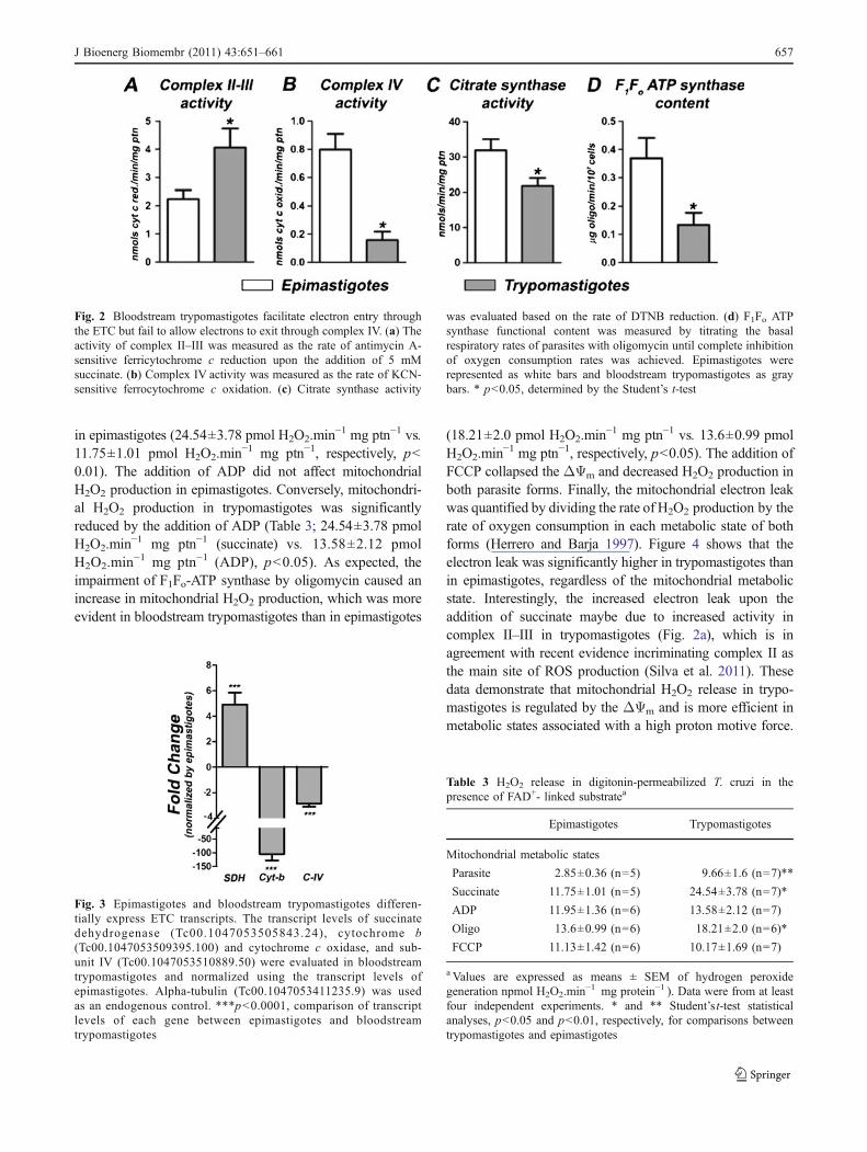
The lower oxygen consumption rates exhibited by trypo-mastigotes at the metabolic states associated with a higherelectron flow (state 3 and uncoupled state) suggest that theobserved functional changes are derived from the inhibitionof the transport/oxidation machinery (substrate transport,tricarboxylic acid cycle, and the electron transport chain)and not the phosphorylation machinery (F1Fo-ATP syn-thase, adenine nucleotide translocator, and phosphatecarrier). Therefore, we assessed the activity of the mito-chondrial complex II–III and complex IV of both parasiteforms (Figs. 2a and b). Surprisingly, complex II–IIIactivities were significantly higher in bloodstream trypo-mastigotes than in epimastigotes (Fig. 2a, p<0.05). How-ever, cytochrome c oxidase activity was significantly higherin epimastigotes than in bloodstream trypomastigotes(Fig. 2b, p<0.05).
Citrate synthase activity is commonly employed todetermine the content of functional mitochondria ofdifferent cells. Despite the kinetoplastids possessing onlya single mitochondrion, we observed that citrate synthaseactivity significantly decreased in bloodstream trypomasti-gotes as compared to epimastigotes (Fig. 2c, p<0.05),suggesting that the mitochondrial functional content of bothforms is different. A similar trend was also observed whenmeasuring the functional content of the F1Fo-ATP synthase,which was significantly lower in bloodstream trypomasti-gotes (Fig. 2d, p<0.05).
We quantified the expression levels of genes related tothe ETC by quantitative PCR (Fig. 3). Consistent withenzymatic activity, the expression of succinate dehydroge-nase (SDH) was significantly higher in bloodstreamtrypomastigotes (p<0.001). Also, trypomastigote mRNAlevels of cytochrome b (a component of complex III) andsubunit IV of cytochrome c oxidase (a component ofcomplex IV) were significantly lower (Fig. 3).
Trypomastigotes at mitochondrial metabolic stateswith a high proton motive force producemore mitochondrial H2O2
We evaluated mitochondrial H2O2 release in digitonin-permeabilized parasites at different metabolic states (Table 3).In the absence of succinate, the rates of H2O2 release intrypomastigotes were significantly higher than those inepimastigotes (9.66±1.60 pmol H2O2.min−1 mg ptn−1 vs.2.85±0.36 pmol H2O2.min−1 mg ptn−1, respectively, p<0.01). A similar pattern was also observed when succinatewas added to the permeabilized parasites, with rates of H2O2
formation in trypomastigotes being significantly higher than
Table 2 Oxygen consumption rates in digitonin-permeabilized T.cruzi in the presence of a FAD+-linked substratea
Epimastigotes Trypomastigotes
Mitochondrial metabolic states
Succinate 1.89±0.38 (n=10) 1.73±0.33 (n=5)
ADP 3.5±0.64 (n=10) 2.24±0.29 (n=7)*
Oligo 1.29±0.16 (n=10) 1.14±0.18 (n=7)
FCCP 3.51±0.44 (n=10) 1.9±0.14 (n=7)*
ROX 0.15±0.09 (n=10) 0.23±0.06 (n=7)
RCRmax 2.839±0.32 (n=10) 1.887±0.32 (n=7)
a Values are expressed as means ± SEM of oxygen consumption(nmol O2.min−1 mg−1 protein). Data were from at least fourindependent experiments. The residual oxygen consumption (ROX)was evaluated after 1.25 μg/mL AA addition. The respiratorycontrol ratio (RCR) was calculated by dividing the uncoupledrespiration by state 4-like respiratory rates.*, Student’s t-test statis-tical analyses (p<0.05) for comparisons between trypomastigotesand epimastigotes
656 J Bioenerg Biomembr (2011) 43:651–661
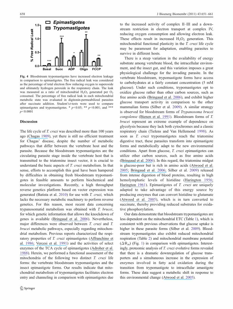
in epimastigotes (24.54±3.78 pmol H2O2.min−1 mg ptn−1 vs.11.75±1.01 pmol H2O2.min−1 mg ptn−1, respectively, p<0.01). The addition of ADP did not affect mitochondrialH2O2 production in epimastigotes. Conversely, mitochondri-al H2O2 production in trypomastigotes was significantlyreduced by the addition of ADP (Table 3; 24.54±3.78 pmolH2O2.min−1 mg ptn−1 (succinate) vs. 13.58±2.12 pmolH2O2.min−1 mg ptn−1 (ADP), p<0.05). As expected, theimpairment of F1Fo-ATP synthase by oligomycin caused anincrease in mitochondrial H2O2 production, which was moreevident in bloodstream trypomastigotes than in epimastigotes
(18.21±2.0 pmol H2O2.min−1 mg ptn−1 vs. 13.6±0.99 pmolH2O2.min−1 mg ptn−1, respectively, p<0.05). The addition ofFCCP collapsed the ΔΨm and decreased H2O2 production inboth parasite forms. Finally, the mitochondrial electron leakwas quantified by dividing the rate of H2O2 production by therate of oxygen consumption in each metabolic state of bothforms (Herrero and Barja 1997). Figure 4 shows that theelectron leak was significantly higher in trypomastigotes thanin epimastigotes, regardless of the mitochondrial metabolicstate. Interestingly, the increased electron leak upon theaddition of succinate maybe due to increased activity incomplex II–III in trypomastigotes (Fig. 2a), which is inagreement with recent evidence incriminating complex II asthe main site of ROS production (Silva et al. 2011). Thesedata demonstrate that mitochondrial H2O2 release in trypo-mastigotes is regulated by the ΔΨm and is more efficient inmetabolic states associated with a high proton motive force.
Fig. 3 Epimastigotes and bloodstream trypomastigotes differen-tially express ETC transcripts. The transcript levels of succinatedehydrogenase (Tc00.1047053505843.24), cytochrome b(Tc00.1047053509395.100) and cytochrome c oxidase, and sub-unit IV (Tc00.1047053510889.50) were evaluated in bloodstreamtrypomastigotes and normalized using the transcript levels ofepimastigotes. Alpha-tubulin (Tc00.1047053411235.9) was usedas an endogenous control. ***p<0.0001, comparison of transcriptlevels of each gene between epimastigotes and bloodstreamtrypomastigotes
Fig. 2 Bloodstream trypomastigotes facilitate electron entry throughthe ETC but fail to allow electrons to exit through complex IV. (a) Theactivity of complex II–III was measured as the rate of antimycin A-sensitive ferricytochrome c reduction upon the addition of 5 mMsuccinate. (b) Complex IV activity was measured as the rate of KCN-sensitive ferrocytochrome c oxidation. (c) Citrate synthase activity
was evaluated based on the rate of DTNB reduction. (d) F1Fo ATPsynthase functional content was measured by titrating the basalrespiratory rates of parasites with oligomycin until complete inhibitionof oxygen consumption rates was achieved. Epimastigotes wererepresented as white bars and bloodstream trypomastigotes as graybars. * p<0.05, determined by the Student’s t-test
Table 3 H2O2 release in digitonin-permeabilized T. cruzi in thepresence of FAD+- linked substratea
Epimastigotes Trypomastigotes
Mitochondrial metabolic states
Parasite 2.85±0.36 (n=5) 9.66±1.6 (n=7)**
Succinate 11.75±1.01 (n=5) 24.54±3.78 (n=7)*
ADP 11.95±1.36 (n=6) 13.58±2.12 (n=7)
Oligo 13.6±0.99 (n=6) 18.21±2.0 (n=6)*
FCCP 11.13±1.42 (n=6) 10.17±1.69 (n=7)
a Values are expressed as means ± SEM of hydrogen peroxidegeneration npmol H2O2.min−1 mg protein−1 ). Data were from at leastfour independent experiments. * and ** Student’s t-test statisticalanalyses, p<0.05 and p<0.01, respectively, for comparisons betweentrypomastigotes and epimastigotes
J Bioenerg Biomembr (2011) 43:651–661 657
Discussion
The life cycle of T. cruzi was described more than 100 yearsago (Chagas 1909), yet there is still no efficient treatmentfor Chagas’ disease, despite the number of metabolicpathways that differ between the vertebrate host and theparasite. Because the bloodstream trypomastigotes are thecirculating parasite stage inside the vertebrate host that istransmitted to the triatomine insect vector, it is crucial tounderstand the basic aspects of T. cruzi metabolism. In thatsense, efforts to accomplish this goal have been hamperedby difficulties in obtaining fresh bloodstream trypomasti-gotes in feasible amounts to perform biochemical andmolecular investigations. Recently, a high throughputreverse genetics platform based on vector expression wasgenerated (Batista et al. 2010) for use with T. cruzi, whichlacks the necessary metabolic machinery to perform reversegenetics. For this reason, most recent data concerningtrypanosomatid metabolism was obtained with T. brucei,for which genetic information that allows the knockdown ofgenes is available (Bringaud et al. 2006). Nevertheless,major differences were observed between T. cruzi and T.brucei metabolic pathways, especially regarding mitochon-drial metabolism. Previous reports characterized the respi-ratory properties of T. cruzi epimastigotes (Affranchino etal. 1986; Vercesi et al. 1991) and the activities of selectenzymes of the TCA cycle of epimastigotes (Adroher et al.1988). Herein, we performed a functional assessment of themitochondria of the following two distinct T. cruzi lifeforms: the vertebrate bloodstream trypomastigotes and theinsect epimastigote forms. Our results indicate that mito-chondrial metabolism of trypomastigotes facilitates electronentry and channeling in comparison with epimastigotes due
to the increased activity of complex II–III and a down-stream restriction in electron transport at complex IV,reducing oxygen consumption and allowing electron leak.These effects result in increased H2O2 generation. Thismitochondrial functional plasticity in the T. cruzi life cyclemay be paramount for adaptation, enabling parasites tosurvive in different hosts.
There is a steep variation in the availability of energysubstrate among vertebrate blood, the intracellular environ-ment, and the insect gut, and this variation imposes a greatphysiological challenge for the invading parasite. In thevertebrate bloodstream, trypomastigote forms have accessto carbohydrates at a fairly constant concentration (5 mMglucose). Under such conditions, trypomastigotes opt tooxidize glucose rather than other carbon sources, such asfree amino acids (Bringaud et al. 2006), and exhibit higherglucose transport activity in comparison to the othermammalian forms (Silber et al. 2009). A similar strategyis observed for bloodstream forms of Trypanosoma bruceicongolense (Bienen et al. 1991). Bloodstream forms of T.brucei represent an extreme example of dependence onglycolysis because they lack both cytochromes and a classicrespiratory chain (Tielens and Van Hellemond 1998). Assoon as T. cruzi trypomastigotes reach the triatominedigestive tract, these parasites transform into epimastigoteforms and metabolically adapt to the new environmentalconditions. Apart from glucose, T. cruzi epimastigotes canutilize other carbon sources, such as free amino acids(Bringaud et al. 2006). In this regard, the triatomine midgutis glucose-poor but is rich in amino acids (Atwood et al.2005; Bringaud et al. 2006; Silber et al. 2009) releasedfrom intense digestion of blood proteins, resulting in highhemolymphatic levels of histidine (Harington 1956;Harington 1961). Epimastigotes of T. cruzi are uniquelyadapted to take advantage of this energy source byproducing enzymes that can convert histidine to glutamate(Atwood et al. 2005), which is in turn converted tosuccinate, thereby providing reduced substrates for oxida-tive phosphorylation.
Our data demonstrate that bloodstream trypomastigotes areless dependent on the mitochondrial ETC (Table 1), which isconsistent with previous observations that glucose uptake ishigher in these parasite forms (Silber et al. 2009). Blood-stream trypomastigotes also exhibit reduced mitochondrialrespiration (Table 2) and mitochondrial membrane potential(ΔΨm) (Fig. 1) in comparison with epimastigotes. Interest-ingly, proteomic analysis of T. cruzi evolutive forms revealedthat there is a dramatic downregulation of glucose trans-porters and a simultaneous increase in the expression ofenzymes involved in fatty acid oxidation during thetransition from trypomastigote to intracellular amastigoteforms. These data suggest a metabolic shift in response tothis environmental change (Atwood et al. 2005).
Fig. 4 Bloodstream trypomastigotes have increased electron leakagein comparison to epimastigotes. The free radical leak was consideredas the percentage of total electron flow reducing oxygen to superoxideand ultimately hydrogen peroxide in the respiratory chain. The leakwas measured as a ratio of mitochondrial H2O2 generated per O2
consumed. The percentage of free radical leak in each mitochondrialmetabolic state was evaluated in digitonin-permeabilized parasitesafter succinate addition. Student’s t-tests were used to compareepimastigotes and trypomastigotes. * p<0.05; ** p<0.001; and ***p<0.0001
658 J Bioenerg Biomembr (2011) 43:651–661

Extracellular glucose levels regulate energy metabolismin different cells and organisms (Coustou et al. 2003;Lamour et al. 2005). Coustou and co-workers demonstratedthat intracellular ATP levels of procyclic T. brucei grown ina glucose-rich medium are not altered by oligomycin,suggesting that oxidative phosphorylation is not fundamen-tal for the survival of these parasite forms. In contrast, thedownregulation of pyruvate kinase reduced ATP levels andincreased the parasite doubling time (Coustou et al. 2003).Interestingly, procyclic forms of T. brucei kept in a mediummimicking the insect digestive tract containing limitedglucose availability showed an increase in proline uptakeand oligomycin sensitivity, suggesting that under limitedavailability of glucose, oxidative phosphorylation becomesthe main source of ATP instead of glycolysis (Lamour et al.2005). Similarly, T. cruzi bloodstream trypomastigotes wereless sensitive to AA and were highly sensitive to IAAincubation (Table 1), indicating that these forms are lessdependent on oxidative phosphorylation and rely more onglycolysis for ATP synthesis. These results were furthersupported by the reduced mitochondrial O2 consumption andΔΨm of bloodstream trypomastigotes (Table 2 and Fig. 1).
In comparison with epimastigotes, metacyclic trypo-mastigotes of T. cruzi are better equipped with scavengerantioxidant defenses (Atwood et al. 2005; Piacenza et al.2008), which can explain why they are more resistant tooxidative challenges despite their higher rates of mitochon-drial H2O2 generation (Tables 1 and 3 and Fig. 5).Proteomic analyses have demonstrated an upregulation ofseveral enzymes involved in antioxidant defenses inmetacyclic trypomastigotes in comparison with epimasti-gotes, such as trypanothione synthetase, ascorbate peroxi-dase, mitochondrial and cytosolic tryparedoxin peroxidase,iron superoxide dismutase and tryparedoxin (Atwood et al.2005; Piacenza et al. 2008). Differentiation from epimasti-gotes to metacyclic trypomastigotes was also correlatedwith increased resistance to peroxynitrite challenge(Piacenza et al. 2008). It is important to emphasize thatthe susceptibility of T. cruzi to H2O2 varies not only inany given individual parasite in the same strain, but alsoamong different strains of the same parasite form(Boveris and Stoppani 1977; Mielniczki-Pereira et al.2007). In addition, mitochondrial H2O2 generation variesamong different strains of T. cruzi epimastigotes (Carranza
Fig. 5 Mitochondrial functionalremodeling along T. cruzi lifecycle. The main source ofelectrons for T. cruzi mitochon-dria was set to complex II by theaddition of succinate. In thisschematic representation,complex I was not representedas it has limited function in T.cruzi metabolism (Carranza etal. 2009). Bloodstreamtrypomastigotes exhibitedincreased complex II-IIIactivities in comparison toepimastigotes (II, UQH2, III).Conversely, complex IV (IV)activity in bloodstream formswas significantly reduced whencompared to insect forms,creating an electron bottleneckthat facilitates electron leakageand ROS formation. As aresult, the ΔΨm is reduced inbloodstream forms (representedas low amounts of H+) as wellas oxygen consumption. Furtherdetails are described in the text
J Bioenerg Biomembr (2011) 43:651–661 659
et al. 2009). Our findings are consistent with the conceptthat bloodstream trypomastigotes are preadapted tooxidative challenges triggered by the host immuneresponses, such as the respiratory burst of phagocyticcells (Atwood et al. 2005). Nevertheless, additionalresearch is required to determine which factors areinvolved in mitochondrial functional remodeling over theT. cruzi life cycle.
The mitochondrial functional changes observed in thetwo T. cruzi forms investigated in this study are schemat-ically summarized in Fig. 5. In general, the mitochondria ofboth epimastigotes and bloodstream trypomastigotes oper-ate in the classical way; oxidation of succinate supports notonly the transference of electrons to oxygen, but alsoincreases the membrane potential and supports H2O2
formation. However, striking differences were observed inthe magnitude of all these parameters between the twoparasite forms. Based on our data, we hypothesize that thereduced oxygen consumption and the increased electronleakage and H2O2 formation observed in bloodstreamtrypomastigotes could be a result of functional ETCremodeling. In bloodstream trypomastigotes, increasedSDH expression and complex II–III activity facilitate theentry of electrons into the ETC (Figs. 2a and 3) whilereducing cytochrome b and cytochrome c oxidase expres-sion (Fig. 3) and activity (Fig. 2b). These alterations couldrestrict electron transport to the final site of ETC, resultingin an impairment of oxygen reduction to H2O (Table 2),thereby creating an “electron bottleneck” effect. In thissense, higher electron leakage and H2O2 formation rates inbloodstream trypomastigotes could be a direct consequenceof the restricted electron transport along the ETC (Fig. 5).Recent evidence indicates that mitochondrial-derived su-peroxide is an important molecule that drives precondition-ing against stress conditions in yeast. In the same context,our data, in addition to previously reported data, show thattrypomastigotes are more resistant to H2O2 and peroxyni-trite incubation (Alvarez et al. 2011; Tanaka et al. 1983). Aprevious study also shows that bloodstream trypomastigotesare more equipped with antioxidant defenses (Irigoin et al.2008). Additionally, more virulent parasite strains weremore resistant to pro-oxidant injury and exhibitedincreased levels of antioxidant enzymes (Piacenza etal. 2009) compared to those in attenuated strains.Conceivably, increased mitochondrial ROS generationwould precondition bloodstream trypomastigotes againsta severe oxidative insult, in a hormetic-type response,protecting these parasite forms from the host immuneresponse. Our results indicate that there is significantfunctional plasticity in the T. cruzi mitochondrion duringdifferent phases of its life cycle, which may be importantfor adaptation, enabling parasite survival in distinct hostenvironments.
Acknowledgments This work was supported by grants fromDECIT/SCTIE/MS, FIOCRUZ, FAPERJ and CNPq (through theInstituto Nacional de Ciência e Tecnologia em Biologia Estrutural eBioimagem). MFO is a researcher scholar from CNPq and a recipientof the Jovens Cientistas do Nosso Estado grant from FAPERJ (2010).RLSG was funded by CAPES/FAPERJ. We are thankful for thetechnical support of Marcos Meuser and Michelle Fernandes. We arealso grateful Dr. Jose Henrique M.C. Oliveira and Prof. Anibal E.Vercesi (Unicamp, Brazil) for the helpful discussions. Lastly, we thankDr. Eduardo Fox for his assistance in manuscript revision.
References
Adroher FJ, Osuna A, Lupianez JA (1988) Differential energeticmetabolism during Trypanosoma cruzi differentiation. I. Citratesynthase, NADP-isocitrate dehydrogenase, and succinate dehy-drogenase. Arch Biochem Biophys 267(1):252–261
Affranchino JL, Schwarcz de Tarlovsky MN, Stoppani AO (1986)Terminal oxidases in the trypanosomatid Trypanosoma cruzi.Comp Biochem Physiol B 85(2):381–388
Alvarez MN, Peluffo G, Piacenza L, Radi R (2011) Intraphagosomalperoxynitrite as a macrophage-derived cytotoxin against inter-nalized Trypanosoma cruzi: consequences for oxidative killingand role of microbial peroxiredoxins in infectivity. J Biol Chem286(8):6627–6640
Atwood JA 3rd, Weatherly DB, Minning TA, Bundy B, Cavola C,Opperdoes FR et al (2005) The Trypanosoma cruzi proteome.Science 309(5733):473–476
Batista M, Marchini FK, Celedon PA, Fragoso SP, Probst CM, Preti Het al (2010) A high-throughput cloning system for reversegenetics in Trypanosoma cruzi. BMC Microbiol 10:259
Bienen EJ, Webster P, Fish WR (1991) Trypanosoma (Nannomonas)congolense: changes in respiratory metabolism during the lifecycle. Exp Parasitol 73(4):403–412
Billingsley PF (1988) Morphometric analysis of Rhodnius prolixusStal (Hemiptera:Reduviidae) midgut cells during blood digestion.Tissue & Cell 20(2):291–301
Boveris A, Chance B (1973) The mitochondrial generation ofhydrogen peroxide. General properties and effect of hyperbaricoxygen. Biochemical Journal 134(3):707–716
Boveris A, Stoppani AO (1977) Hydrogen peroxide generation inTrypanosoma cruzi. Experientia 33(10):1306–1308
Bringaud F, Riviere L, Coustou V (2006) Energy metabolism oftrypanosomatids: adaptation to available carbon sources. MolBiochem Parasitol 149(1):1–9
Brookes PS, Levonen AL, Shiva S, Sarti P, Darley-Usmar VM (2002)Mitochondria: regulators of signal transduction by reactive oxygenand nitrogen species. Free Radic Biol Med 33(6):755–764
Carranza JC, Kowaltowski AJ, Mendonca MA, de Oliveira TC,Gadelha FR, Zingales B (2009) Mitochondrial bioenergetics andredox state are unaltered in Trypanosoma cruzi isolates withcompromised mitochondrial complex I subunit genes. J BioenergBiomembr 41(3):299–308
Cazzulo JJ (1994) Intermediate metabolism in Trypanosoma cruzi. JBioenerg Biomembr 26(2):157–165
Chagas, C. (1909). Nova tripanozomiaze humana: estudos sobre amorfolojia e o ciclo evolutivo do Schizotrypanum cruzi n. gen., n.sp., ajente etiolojico de nova entidade morbida do homem. Mem.Inst. Oswaldo Cruz, 1(2).
Chance B, Williams GR (1955) Respiratory enzymes in oxidativephosphorylation. II. Difference spectra. J Biol Chem 217(1):395–407
Chaudhuri M, Ott RD, Hill GC (2006) Trypanosome alternative oxidase:from molecule to function. Trends Parasitol 22(10):484–491
660 J Bioenerg Biomembr (2011) 43:651–661
Coustou V, Besteiro S, Biran M, Diolez P, Bouchaud V, Voisin P et al(2003) ATP generation in the Trypanosoma brucei procyclicform: cytosolic substrate level is essential, but not oxidativephosphorylation. J Biol Chem 278(49):49625–49635
de Meirelles MN, De Souza W (1982) Trypanosoma cruzi: ultrastruc-tural cytochemistry of mitochondrial enzymes. Exp Parasitol 53(3):341–354
Denicola-Seoane A, Rubbo H, Prodanov E, Turrens JF (1992)Succinate-dependent metabolism in Trypanosoma cruzi epimas-tigotes. Mol Biochem Parasitol 54(1):43–50
Dias JC (2007) Globalization, inequity and Chagas disease. CadSaude Publica 23(Suppl 1):S13–22
Docampo R (1993) Calcium homeostasis in Trypanosoma cruzi. BiolRes 26(1–2):189–196
Docampo R, Moreno SN, Vercesi AE (1993) Effect of thapsigargin oncalcium homeostasis in Trypanosoma cruzi trypomastigotes andepimastigotes. Mol Biochem Parasitol 59(2):305–313
Ferguson M, Mockett RJ, Shen Y, Orr WC, Sohal RS (2005) Age-associated decline in mitochondrial respiration and electrontransport in Drosophila melanogaster. Biochem J 390(Pt2):501–511
Harington JS (1956) Histamine and histidine in excreta of the blood-sucking bug Rhodnius prolixus. Nature 178(4527):268
Harington JS (1961) Studies of the amino acids of Rhodnius prolixusI. Analysis of the haemolymph. Parasitology 51:309–318
Herrero A, Barja G (1997) ADP-regulation of mitochondrial freeradical production is different with complex I-or complex II-linked substrates: implications for the exercise paradox and brainhypermetabolism. J Bioenerg Biomembr 29(3):241–249
Irigoin F, Cibils L, Comini MA, Wilkinson SR, Flohe L, Radi R(2008) Insights into the redox biology of Trypanosoma cruzi:Trypanothione metabolism and oxidant detoxification. FreeRadic Biol Med 45(6):733–742
Japiassu AM, Santiago AP, d’Avila Jda C, Garcia-Souza LF, Galina A,Castro Faria-Neto HC et al (2011) Bioenergetic failure of humanperipheral blood monocytes in patients with septic shock ismediated by reduced F1Fo adenosine-5′-triphosphate synthaseactivity. Crit Care Med 39(5):1056–1063
Korshunov SS, Skulachev VP, Starkov AA (1997) High protonicpotential actuates a mechanism of production of reactive oxygenspecies in mitochondria. FEBS Lett 416(1):15–18
Kowaltowski AJ, de Souza-Pinto NC, Castilho RF, Vercesi AE (2009)Mitochondria and reactive oxygen species. Free Radic Biol Med47(4):333–343
Lamour N, Riviere L, Coustou V, Coombs GH, Barrett MP, BringaudF (2005) Proline metabolism in procyclic Trypanosoma brucei isdown-regulated in the presence of glucose. J Biol Chem 280(12):11902–11910
Lehane MJ (2005) The Biology of Blood-sucking in Insects, 2nd edn.Cambridge University, press, UK
Livak KJ, Schmittgen TD (2001) Analysis of relative gene expressiondata using real-time quantitative PCR and the 2(−Delta Delta C(T)) Method. Methods 25(4):402–408
Lowry OH, Rosebrough NJ, Farr AL, Randall RJ (1951) Proteinmeasurement with the Folin phenol reagent. J Biol Chem 193(1):265–275
Menna-Barreto RF, Goncalves RL, Costa EM, Silva RS, Pinto AV,Oliveira MF et al (2009) The effects on Trypanosoma cruzi ofnovel synthetic naphthoquinones are mediated by mitochondrialdysfunction. Free Radic Biol Med 47(5):644–653
Michels PA, Bringaud F, Herman M, Hannaert V (2006) Metabolicfunctions of glycosomes in trypanosomatids. Biochim BiophysActa 1763(12):1463–1477
Mielniczki-Pereira AA, Chiavegatto CM, Lopez JA, Colli W, AlvesMJ, Gadelha FR (2007) Trypanosoma cruzi strains, Tulahuen 2and Y, besides the difference in resistance to oxidative stress,display differential glucose-6-phosphate and 6-phosphogluconatedehydrogenases activities. Acta Trop 101(1):54–60
Nicholls DG, Ferguson SJ (2002) Bioenergetics 3, 3rd edn. London,Academic Press, pp 82–83
Pan Y, Schroeder EA, Ocampo A, Barrientos A, Shadel GS(2011) Regulation of Yeast Chronological Life Span byTORC1 via Adaptive Mitochondrial ROS Signaling. CellMetab 13(6):668–678
Piacenza L, Peluffo G, Alvarez MN, Kelly JM, Wilkinson SR, Radi R(2008) Peroxiredoxins play a major role in protecting Trypano-soma cruzi against macrophage-and endogenously-derived per-oxynitrite. Biochem J 410(2):359–368
Piacenza L, Zago MP, Peluffo G, Alvarez MN, Basombrio MA, RadiR (2009) Enzymes of the antioxidant network as noveldeterminers of Trypanosoma cruzi virulence. Int J Parasitol 39(13):1455–1464
Poyton RO, McEwen JE (1996) Crosstalk between nuclear andmitochondrial genomes. Annu Rev Biochem 65:563–607
Rogerson GW, Gutteridge WE (1980) Catabolic metabolism inTrypanosoma cruzi. Int J Parasitol 10(1):131–135
Rozen S, Skaletsky H (2000) Primer3 on the WWW for generalusers and for biologist programmers. Methods Mol Biol132:365–386
Silber AM, Tonelli RR, Lopes CG, Cunha-e-Silva N, Torrecilhas AC,Schumacher RI et al (2009) Glucose uptake in the mammalianstages of Trypanosoma cruzi. Mol Biochem Parasitol 168(1):102–108
Silva TM, Peloso EF, Vitor SC, Ribeiro LH, & Gadelha FR (2011). O(2) consumption rates along the growth curve: new insights intoTrypanosoma cruzi mitochondrial respiratory chain. J BioenergBiomembr. [ahead of print]
Tanaka Y, Tanowitz H, Bloom BR (1983) Growth of Trypanosomacruzi in a cloned macrophage cell line and in a variant defectivein oxygen metabolism. Infect Immun 41(3):1322–1331
Tanowitz HB, Weiss LM, Montgomery SP (2011) Chagas disease hasnow gone global. PLoS Negl Trop Dis 5(4):e1136
Tielens AG, Van Hellemond JJ (1998) Differences in energymetabolism between trypanosomatidae. Parasitol Today 14(7):265–272
Tielens AG, van Hellemond JJ (2009) Surprising variety in energymetabolism within Trypanosomatidae. Trends Parasitol 25(10):482–490
Vercesi AE, Bernardes CF, Hoffmann ME, Gadelha FR, Docampo R(1991) Digitonin permeabilization does not affect mitochondrialfunction and allows the determination of the mitochondrialmembrane potential of Trypanosoma cruzi in situ. J Biol Chem266(22):14431–14434
Villani G, Attardi G (2000) In vivo control of respiration bycytochrome c oxidase in human cells. Free Radic Biol Med 29(3–4):202–210
Villani G, Greco M, Papa S, Attardi G (1998) Low reserve ofcytochrome c oxidase capacity in vivo in the respiratory chain ofa variety of human cell types. J Biol Chem 273(48):31829–31836
Votyakova TV, Reynolds IJ (2004) Detection of hydrogen peroxidewith Amplex Red: interference by NADH and reduced glutathioneauto-oxidation. Arch Biochem Biophys 431(1):138–144
Zuckerbraun BS, Chin BY, Bilban M, d’Avila JC, Rao J, Billiar TR etal (2007) Carbon monoxide signals via inhibition of cytochromec oxidase and generation of mitochondrial reactive oxygenspecies. FASEB J 21(4):1099–1106
J Bioenerg Biomembr (2011) 43:651–661 661