Biological Role of Aldo–Keto Reductases in Retinoic Acid Biosynthesis and Signaling
13
REVIEW ARTICLE published: 17 April 2012 doi: 10.3389/fphar.2012.00058 Biological role of aldo–keto reductases in retinoic acid biosynthesis and signaling F. Xavier Ruiz † , Sergio Porté, Xavier Parés and Jaume Farrés* Department of Biochemistry and Molecular Biology, Universitat Autònoma de Barcelona, Barcelona, Spain Edited by: Yi Jin, University of Pennsylvania, USA Reviewed by: Ana Cristina Carvalho Rego, University of Coimbra, Portugal Natalia Kedishvili, University of Alabama at Birmingham, USA *Correspondence: Jaume Farrés, Department of Biochemistry and Molecular Biology, Universitat Autònoma de Barcelona, E-08193 Bellaterra, Barcelona, Spain. e-mail: [email protected] † Present address: F. Xavier Ruiz, Structural Biology and Genomics Department, IGBMC, CNRS, INSERM, University of Strasbourg, 67404 Illkirch, France. Several aldo–keto reductase (AKR) enzymes from subfamilies 1B and 1C show retinalde- hyde reductase activity, having low K m and k cat values. Only AKR1B10 and 1B12, with all-trans-retinaldehyde, and AKR1C3, with 9-cis-retinaldehyde, display high catalytic effi- ciency. Major structural determinants for retinaldehyde isomer specificity are located in the external loops (A and C for AKR1B10, and B for AKR1C3), as assessed by site-directed mutagenesis and molecular dynamics. Cellular models have shown that AKR1B and 1C enzymes are well suited to work in vivo as retinaldehyde reductases and to regulate retinoic acid (RA) biosynthesis at hormone pre-receptor level. An additional physiological role for the retinaldehyde reductase activity of these enzymes, consistent with their tissue local- ization, is their participation in β-carotene absorption. Retinaldehyde metabolism may be subjected to subcellular compartmentalization, based on enzyme localization. While reti- naldehyde oxidation to RA takes place in the cytosol, reduction to retinol could take place in the cytosol by AKRs or in the membranes of endoplasmic reticulum by microsomal reti- naldehyde reductases. Upregulation of some AKR1 enzymes in different cancer types may be linked to their induction by oxidative stress and to their participation in different signaling pathways related to cell proliferation. AKR1B10 and AKR1C3, through their retinaldehyde reductase activity, trigger a decrease in the RA biosynthesis flow, resulting in RA depriva- tion and consequently lower differentiation, with an increased cancer risk in target tissues. Rational design of selective AKR inhibitors could lead to development of novel drugs for cancer treatment as well as reduction of chemotherapeutic drug resistance. Keywords: aldo–keto reductase, retinaldehyde, retinoic acid, retinol, cancer INTRODUCTION Members of the aldo–keto reductase (AKR) superfamily are NADP(H)-dependent cytosolic enzymes which fold into a typical (α/β) 8 -barrel. AKRs catalyze the reduction of a wide variety of car- bonyl compounds (Jez et al., 1997; Barski et al., 2008). With regard to the biological function of human AKRs, they may be involved in the detoxification of electrophilic compounds, such as 4- hydroxy-trans -2-nonenal generated under oxidative stress condi- tions. AKRs are also able to reduce a variety of lipophilic substrates, such as ketosteroids, ketoprostaglandins, and retinoids, which are hormone precursors. Thus, another function may be related to pre-receptor hormone regulation of the amount of ligand avail- able for some nuclear receptors and, therefore, to participation in transcriptional gene control. Lastly, they are considered as phase I drug-metabolizing enzymes and can either detoxify or activate Abbreviations: ADH, alcohol dehydrogenase; AKR, aldo–keto reductase; ALDH, aldehyde dehydrogenase; BCO1, β-carotene 15,15 -monooxygenase 1; CRABP, cel- lular retinoic acid binding protein; CRBP-I, -II, cellular retinol binding protein type I, II; CYP26, cytochrome P450 family 26; LRAT, lecithin:retinol acyl transferase; MD, molecular dynamics; MDR, medium-chain dehydrogenase/reductase; PPARγ, peroxisome proliferator-activated receptor γ; RA, retinoic acid; RALDH, retinalde- hyde dehydrogenase; RAR,retinoic acid receptor; RBP,retinol binding protein; REH, retinyl ester hydrolase; ROS, reactive oxygen species; RXR, retinoid X receptor; SDR, short-chain dehydrogenase/reductase; STRA6, stimulated by retinoic acid gene 6; TTR, transthyretin. xenobiotic compounds (i.e., polycyclic aromatic hydrocarbons; Penning and Drury, 2007). With respect to the human AKRs, members of the AKR1B subfamily are of special interest. AKR1B1, the classical aldose reductase, is related to secondary diabetic complications, while AKR1B10 is induced in cancer cells. Although these two AKRs share 71% sequence identity, they show very different kinetic properties with some relevant substrates like retinaldehyde or glu- cose. Thus, AKR1B10 is a retinaldehyde reductase with a much higher k cat value than that of AKR1B1, while glucose is reduced by AKR1B1 but not by AKR1B10. Recently, AKR1B10 has emerged as a tumor marker since it is overexpressed in different types of cancers, featuring hepatocellular carcinoma and lung cancer cor- related with tobacco smoking (Ruiz et al., 2009; Wang et al., 2009; Díez-Dacal et al., 2011). AKR1C1–AKR1C4, also known as human hydroxysteroid dehydrogenases, share 86% sequence identity, but the individual enzymes show different substrate specificity, inhibitor selectiv- ity, and tissue expression pattern. AKR1C3 is one of the most interesting enzymes, with increasing evidence strongly supporting its involvement in cancer development (Jin and Penning, 2007; Penning and Byrns, 2009; Ruiz et al., 2011b). The term “retinoid” refers to compounds derived from vita- min A (retinol), where their basic structure is divided into three domains: a cyclohexene ring or β-ionone, an aliphatic chain with www.frontiersin.org April 2012 |Volume 3 | Article 58 | 1
-
Upload
independent -
Category
Documents
-
view
0 -
download
0
Transcript of Biological Role of Aldo–Keto Reductases in Retinoic Acid Biosynthesis and Signaling

REVIEW ARTICLEpublished: 17 April 2012
doi: 10.3389/fphar.2012.00058
Biological role of aldo–keto reductases in retinoic acidbiosynthesis and signaling
F. Xavier Ruiz†, Sergio Porté, Xavier Parés and Jaume Farrés*
Department of Biochemistry and Molecular Biology, Universitat Autònoma de Barcelona, Barcelona, Spain
Edited by:
Yi Jin, University of Pennsylvania, USA
Reviewed by:
Ana Cristina Carvalho Rego,University of Coimbra, PortugalNatalia Kedishvili, University ofAlabama at Birmingham, USA
*Correspondence:
Jaume Farrés, Department ofBiochemistry and Molecular Biology,Universitat Autònoma de Barcelona,E-08193 Bellaterra, Barcelona, Spain.e-mail: [email protected]†Present address:
F. Xavier Ruiz, Structural Biology andGenomics Department, IGBMC,CNRS, INSERM, University ofStrasbourg, 67404 Illkirch, France.
Several aldo–keto reductase (AKR) enzymes from subfamilies 1B and 1C show retinalde-hyde reductase activity, having low K m and kcat values. Only AKR1B10 and 1B12, withall-trans-retinaldehyde, and AKR1C3, with 9-cis-retinaldehyde, display high catalytic effi-ciency. Major structural determinants for retinaldehyde isomer specificity are located inthe external loops (A and C for AKR1B10, and B for AKR1C3), as assessed by site-directedmutagenesis and molecular dynamics. Cellular models have shown that AKR1B and 1Cenzymes are well suited to work in vivo as retinaldehyde reductases and to regulate retinoicacid (RA) biosynthesis at hormone pre-receptor level. An additional physiological role forthe retinaldehyde reductase activity of these enzymes, consistent with their tissue local-ization, is their participation in β-carotene absorption. Retinaldehyde metabolism may besubjected to subcellular compartmentalization, based on enzyme localization. While reti-naldehyde oxidation to RA takes place in the cytosol, reduction to retinol could take placein the cytosol by AKRs or in the membranes of endoplasmic reticulum by microsomal reti-naldehyde reductases. Upregulation of some AKR1 enzymes in different cancer types maybe linked to their induction by oxidative stress and to their participation in different signalingpathways related to cell proliferation. AKR1B10 and AKR1C3, through their retinaldehydereductase activity, trigger a decrease in the RA biosynthesis flow, resulting in RA depriva-tion and consequently lower differentiation, with an increased cancer risk in target tissues.Rational design of selective AKR inhibitors could lead to development of novel drugs forcancer treatment as well as reduction of chemotherapeutic drug resistance.
Keywords: aldo–keto reductase, retinaldehyde, retinoic acid, retinol, cancer
INTRODUCTIONMembers of the aldo–keto reductase (AKR) superfamily areNADP(H)-dependent cytosolic enzymes which fold into a typical(α/β)8-barrel. AKRs catalyze the reduction of a wide variety of car-bonyl compounds (Jez et al., 1997; Barski et al., 2008). With regardto the biological function of human AKRs, they may be involvedin the detoxification of electrophilic compounds, such as 4-hydroxy-trans-2-nonenal generated under oxidative stress condi-tions. AKRs are also able to reduce a variety of lipophilic substrates,such as ketosteroids, ketoprostaglandins, and retinoids, which arehormone precursors. Thus, another function may be related topre-receptor hormone regulation of the amount of ligand avail-able for some nuclear receptors and, therefore, to participation intranscriptional gene control. Lastly, they are considered as phase Idrug-metabolizing enzymes and can either detoxify or activate
Abbreviations: ADH, alcohol dehydrogenase; AKR, aldo–keto reductase; ALDH,aldehyde dehydrogenase; BCO1, β-carotene 15,15′-monooxygenase 1; CRABP, cel-lular retinoic acid binding protein; CRBP-I, -II, cellular retinol binding protein typeI, II; CYP26, cytochrome P450 family 26; LRAT, lecithin:retinol acyl transferase;MD, molecular dynamics; MDR, medium-chain dehydrogenase/reductase; PPARγ,peroxisome proliferator-activated receptor γ; RA, retinoic acid; RALDH, retinalde-hyde dehydrogenase; RAR, retinoic acid receptor; RBP, retinol binding protein; REH,retinyl ester hydrolase; ROS, reactive oxygen species; RXR, retinoid X receptor; SDR,short-chain dehydrogenase/reductase; STRA6, stimulated by retinoic acid gene 6;TTR, transthyretin.
xenobiotic compounds (i.e., polycyclic aromatic hydrocarbons;Penning and Drury, 2007).
With respect to the human AKRs, members of the AKR1Bsubfamily are of special interest. AKR1B1, the classical aldosereductase, is related to secondary diabetic complications, whileAKR1B10 is induced in cancer cells. Although these two AKRsshare 71% sequence identity, they show very different kineticproperties with some relevant substrates like retinaldehyde or glu-cose. Thus, AKR1B10 is a retinaldehyde reductase with a muchhigher kcat value than that of AKR1B1, while glucose is reduced byAKR1B1 but not by AKR1B10. Recently, AKR1B10 has emergedas a tumor marker since it is overexpressed in different types ofcancers, featuring hepatocellular carcinoma and lung cancer cor-related with tobacco smoking (Ruiz et al., 2009; Wang et al., 2009;Díez-Dacal et al., 2011).
AKR1C1–AKR1C4, also known as human hydroxysteroiddehydrogenases, share 86% sequence identity, but the individualenzymes show different substrate specificity, inhibitor selectiv-ity, and tissue expression pattern. AKR1C3 is one of the mostinteresting enzymes, with increasing evidence strongly supportingits involvement in cancer development (Jin and Penning, 2007;Penning and Byrns, 2009; Ruiz et al., 2011b).
The term “retinoid” refers to compounds derived from vita-min A (retinol), where their basic structure is divided into threedomains: a cyclohexene ring or β-ionone, an aliphatic chain with
www.frontiersin.org April 2012 | Volume 3 | Article 58 | 1
Ruiz et al. Aldo–keto reductases in retinoid metabolism
four conjugated double bonds, and a polar group which may showdifferent oxidation states. The different configuration of thesedouble bonds leads to different stereoisomers, being the most fre-quent all-trans, 9-cis, 11-cis, and 13-cis (Blomhoff and Blomhoff,2006). Retinol and its derivatives retinaldehyde and retinoic acid(RA) are essential for the growth and maintenance of many bodytissues, such as skin, bone, and vasculature, as well as for the visualcycle (11-cis-retinaldehyde) and immune function. They also playa role in reproduction, embryonic growth, and development. Inthis sense, RA is a key molecule in the development of differentvertebrate organs and tissues by promoting cell differentiation andapoptosis. RA has also a role in several pathological conditions,
such as skin diseases, premature birth, and rheumatoid arthritis.Various human cancers have altered retinoid metabolism and lowRA levels, which favor tumor progression (Theodosiou et al., 2010;Tang and Gudas, 2011).
Vitamin A incorporation in the body occurs through dietaryintake of animal products, which are a source of retinyl esters,and from the consumption of fruits and vegetables, which area source of carotenoids, mainly β-carotene (Figure 1A). In thefirst case, retinol is absorbed by the enterocyte cell after retinylesters have been hydrolyzed in the small intestine. In the secondcase, β-carotene is absorbed directly by passive diffusion, and thenin the enterocyte is split into two retinaldehyde molecules due
FIGURE 1 | Schematic diagram of retinoid absorption, transport, and
metabolism. (A) Upon dietary intake and intestinal absorption, retinoids,and carotenoids are converted to retinol, which is bound to CRBP-II.Retinol is further processed to retinyl esters, incorporated intochylomicrons, and transported to the liver. In hepatocytes, retinyl estersare hydrolyzed to retinol which forms a complex with CRBP-I. Retinol canthen be transferred to stellate cells for storage as retinyl esters.Alternatively, retinol may bind to RBP, and the retinol:RBP complex isbound to TTR and transferred to circulation for transport to target tissues.Binding to TTR reduces renal clearance. Adapted with permission from(Theodosiou et al., 2010). Copyright©2010, Birkhäuser Verlag, Basel. (B) Inthe target tissues, STRA6, a cell-surface receptor for RBP, facilitates retinolentry. Carotenoids may be also a major source of cellular retinoids. Levels
of CRBP-I, LRAT, REH, and oxidoreductases (ADH, SDR, AKR, and RALDH)influence the retinoid flow either toward the storage pathway or thesynthesis of RA, which is bound to CRABP. RA enters the nucleus, whereit binds to nuclear receptors. RA is degraded and eliminated by CYP26enzymes. Adapted with permission from (Parés et al., 2008).Copyright©2008, Birkhäuser Verlag, Basel. ADH, alcohol dehydrogenase;AKR, aldo–keto reductase; BCO1, β-carotene 15,15′-monooxygenase 1;CRABP, cellular retinoic acid binding protein; CRBP-I, -II, cellular retinolbinding protein type I, -II; CYP26, cytochrome P450 family 26; LRAT,lecithin:retinol acetyltransferase; RA, retinoic acid; RALDH, retinaldehydedehydrogenase; RBP, retinol binding protein; REH, retinyl ester hydrolase;SDR, short-chain dehydrogenase/reductase; STRA6, stimulated by retinoicacid gene 6; TTR, transthyretin.
Frontiers in Pharmacology | Experimental Pharmacology and Drug Discovery April 2012 | Volume 3 | Article 58 | 2

Ruiz et al. Aldo–keto reductases in retinoid metabolism
to the activity of β-carotene 15,15′-monooxygenase 1 (BCO1).The liver is another tissue which contributes significantly to β-carotene cleavage. Later, retinaldehyde is reduced to retinol. In thecell, retinol has two metabolic alternatives: storage or oxidativemetabolism. One form of storage is through binding to cellu-lar retinol binding protein type I and type II (CRBP-I and II,respectively). CRBP-I shows wide tissue expression, while CRBP-II is expressed in the small intestine. In the enterocyte, retinol isattached to CRBP-II, and through the action of lecithin:retinol acyltransferase (LRAT) is esterified with fatty acids to yield long-chainesters of retinol. These are packaged into chylomicrons and aretransported to the liver parenchymal cells where they are capturedby specific receptors and transferred to stellate cells for storage,which constitutes 50–80% of body retinol in the form of retinylesters. When required in the peripheral tissues, retinyl esters arehydrolyzed to retinol by retinol ester hydrolase (REH). The retinolgenerated is secreted and transported in the blood as a complexbetween retinol and the plasmatic retinol binding protein (RBP),which in turn is complexed with transthyretin (TTR) to reduce itsglomerular filtration. Over 90% of retinol entering the cell is recy-cled to the plasma, and only a small part is esterified for storage,activated to RA or catabolized (Theodosiou et al., 2010; Harrison,2012; Shirakami et al., 2012).
Biosynthesis of RA, the most potent biologically active metabo-lite of vitamin A, requires two oxidative steps (Figure 1B). Mem-bers of three oxidoreductase superfamilies have been implicatedin the reversible oxidation of retinol to retinaldehyde, which is thepathway rate-limiting step (Napoli, 1999; Belyaeva et al., 2008).The participation of cytosolic alcohol dehydrogenases (ADH)from the medium-chain dehydrogenase/reductase (MDR) super-family and of microsomal short-chain dehydrogenases/reductases(SDR) has been deeply studied. More recently, the AKR super-family has been added as a novel group of cytosolic enzymes thatcould contribute to retinoid redox conversions. Based on the cofac-tor specificity, ADH (NAD-dependent) may work in the oxidativedirection, AKRs (NADPH-dependent) in the reductive direction,while SDR show examples of both specificities.
The control of retinaldehyde levels is essential in the regula-tion of RA synthesis and, therefore, of its signaling role. Onceretinaldehyde is synthesized, it has two alternative metabolic fates,its irreversible oxidation to RA by the action of aldehyde dehy-drogenases (ALDH or RALDH) or its reduction back to retinolby retinaldehyde reductases. Synthesized RA binds to cellular RAbinding protein (CRABP) and is transported to the cell nucleuswhere it binds to retinoid receptors. These can be divided into twosubgroups: RA receptors (RAR), binding the isomers all-trans and9-cis-RA, and retinoid X receptors (RXR), binding 9-cis-RA withhigh affinity, but not all-trans-RA. RAR/RXR heterodimers arebound to RA response elements (RARE) in DNA. Ligand bindinginduces a conformational change in the RAR/RXR heterodimerswhich promotes gene transcription. While RAR only forms het-erodimers with RXR receptor (RAR–RXR), RXR is capable also toform heterodimers with thyroid hormone (RXR–TR), vitamin D(RXR–DR), and peroxisomal proliferator-activated (RXR–PPAR)receptors. It appears that the RXR activity is subordinated to thepresence of ligand bound to RAR in the heterodimer (Pogenberget al., 2005). There is a balance between synthesis and catabolism
to control RA levels. RA catabolism to more oxidized metabo-lites such as 4-hydroxy-RA or 4-oxo-RA occurs primarily throughenzymes from the CYP26 family (Parés et al., 2008; Theodosiouet al., 2010; Tang and Gudas, 2011; Kumar et al., 2012).
The first AKR enzyme reported to display activity with retinoidswas chicken AKR or AKR1B12 (Crosas et al., 2001). Since then,a significant amount of data has been reported on AKRs andretinoids. In this review, this relevant topic will be discussed, asAKRs are being established as enzymes that participate in the pre-cise fine tuning required for the first and regulatory step of RAbiosynthesis. Regulation of this pathway by AKRs and its relation-ship with retinoid body uptake and cell proliferation will also beapproached because of its pathological relevance.
CHARACTERIZATION OF AKRs AS RETINALDEHYDEREDUCTASESACTIVITY ASSAY METHODOLOGIES AND KINETIC RESULTS FOR AKRENZYMES WITH RETINOIDSIn vitro kinetic studies on AKR enzymes with retinoids are fun-damental to investigate isomer specificity, inhibitor selectivity,and structure–function relationships. Retinoids are highly unsta-ble hydrophobic compounds displaying very low solubility inwater-based solvents and being susceptible to photodegradation,double-bond isomerization, and oxidation reactions. Thus, theyneed to be handled under dim red light, and properly solubilizedand stabilized. In order to overcome these difficulties, two differ-ent methodologies have been used to perform kinetic studies withretinoids: (1) the ADH enzymatic assay (or Tween 80 assay), and(2) the SDR enzymatic assay (or HPLC assay), both reviewed inParés et al. (2008).
The ADH enzymatic assay (or Tween 80 assay)This assay is characterized by the use of an aqueous buffer con-taining a low amount of the non-ionic detergent Tween 80 (poly-oxyethylene (20) sorbitan monooleate) and the spectrophotomet-ric measurement of the reaction at 25˚C, following retinaldehydeabsorbance at 400 nm, where retinol does not absorb. Table 1lists the kcat values of the AKR1 enzymes obtained by using thismethod. K m values are not included because Tween 80 behavesas an apparently competitive inhibitor and thus, at the concentra-tion used in the assay, there is a 10- to 100-fold increase of theretinoid K m values (Martras et al., 2004; Ruiz et al., 2009). Someof the values in Table 1 may not be fully comparable since kineticstudies were performed in different laboratories using differentprotein sources and methodologies, under various experimentalconditions. Thus, AKR1B12 results were obtained by using tissue-purified instead of recombinant protein. Constants for AKR1B13,1B14, 1B17, and 1C15 were measured with 0.01% (v/v) Tween80 instead of the widely used 0.02% concentration. Other labora-tories used a high concentration of organic solvent (e.g., 8%, v/v,dimethylsulfoxide). Another added trouble with this methodologyis its low reproducibility due to the high absorbance of retinalde-hyde at 400 nm combined with the low activity rate shown byAKR1 enzymes, resulting in high and variable background whichmust be subtracted during activity assays. While this methodol-ogy may still serve to perform routine determination of enzymeactivity, it is not useful for comparison of kinetic parameters with
www.frontiersin.org April 2012 | Volume 3 | Article 58 | 3
Ruiz et al. Aldo–keto reductases in retinoid metabolism
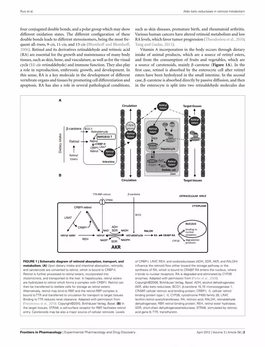
Table 1 | Catalytic constants (k cat, min−1) of AKR1 enzymes with
retinaldehyde isomers, obtained spectrophotometrically in the
presence ofTween 80.
Enzyme All-trans-retinaldehyde 9-cis-Retinaldehyde
1A2 N.A.a N.A.a
1B1 0.37b 2.84
1B10 17.8b 39.0c 5.20
1B3 11.6d N.D.
1B7 9.73d N.D.
1B8 15.3d N.D.
1B9 0.11 N.A.
1B12 17.0e 8.62e
1B13 N.A.f N.D.
1B14 N.A.g N.D.
1B17 0.58h N.D.
1C7* L.A.i N.D.
1C15 1.2j 2.8j
Activity was determined in 0.1 M sodium phosphate, pH 7.5, 0.02% Tween
80, 0.2 mM NADPH, at 25˚C. ND, not determined; NA, no activity was
detected or it was less than 0.5 nmol min−1 mg−1. LA, low activity was detected,
4 nmol min-1 mg-1. *Kinetics was performed in 0.1 M potassium phosphate buffer,
pH 6.5, at 37˚C, and products were analyzed by reverse phase HPLC equilibrated
with 80% (v/v) acetonitrile in water. aCrosas et al. (2003), bGallego et al. (2007b),cQuinn et al. (2008), dJoshi et al. (2010), eCrosas et al. (2001), fEndo et al. (2009),gEndo et al. (2010a), hEndo et al. (2010b), iEndo et al. (2001), jEndo et al. (2007).
other enzymes, such as SDR, where activity has been typicallydetermined by using detergent-free buffer (Gough et al., 1998;Parés et al., 2008).
The SDR enzymatic assay (or HPLC assay)This assay uses an aqueous buffer containing retinoid/bovineserum albumin at 1:1 molar ratio, reaction at 37˚C, followed byretinoid extraction with hexane and analysis of the reaction prod-ucts by HPLC. Determination of the reaction rate is based onthe percentage of substrate conversion. Compared to the Tween80 assay, the HPLC assay is far more reproducible, it does notalter the K m values, requires a lower amount of substrate and isalso suitable for cell culture experiments. Indeed, the compara-tive activity analysis side-by-side of ADH, SDR, and AKRs withretinoids revealed that all enzymes exhibited low and similar K m
values, 1 μM or lower, while they differed in their kcat values.Therefore, when possible, it is highly recommended to choose thismethod instead of the Tween 80 assay.
Tables 2 and 3 show kinetic constants of AKR1 enzymes usingthe HPLC assay. As it can be observed, K m values keep close to1 μM or lower, which is in the physiological range of retinol con-centration (Hollander and Muralidhara, 1977; Quick and Ong,1990; Harrison, 2012). In contrast, kcat values are clearly distinctbetween different enzymes (Gallego et al., 2006; Ruiz et al., 2011b).None of the rodent enzymes characterized displayed high kcat val-ues, leaving human AKR1B10 and chicken AKR1B12 as the solepotent all-trans-retinaldehyde reductases of the superfamily (Gal-lego et al., 2006, 2007b; Ruiz et al., 2011a). On the other hand,human AKR1C enzymes have been shown to use preferentially
Table 2 | Kinetic constants of AKR1 enzymes with retinaldehyde
isomers, obtained by the HPLC assay in the presence of BSA.
Enzyme All-trans-retinaldehyde 9-cis-Retinaldehyde
K m k cat k cat/K m K m k cat k cat/K m
1B1a 1.1 0.35 320 0.4 0.7 1500
1B3b 1.0 0.52 540 N.D.
1B7b 0.5 0.02 42 N.D.
1B8b 2.1 0.05 22 N.D.
1B9b 2.0 0.27 140 N.D.
1B10a 0.6 27 45000 0.7 0.9 1300
1B12 0.6 2.5 4100 N.D.
1C1c L.A. 0.48 0.18 370
1C2c N.A. N.A.
1C3c 1.4 0.60 430 0.40 13 32,500
1C4c 0.31 0.24 790 0.80 0.40 500
Activities were determined in 90 mM KH2PO4, 40 mM KCl, pH 7.4, 0.5 mM
NADPH, 37˚C. NA, no activity; ND, not determined; LA, low activity was detected,
0.56 nmol min−1 mg−1. Units: Km (μM), kcat (min−1), kcat/Km (mM−1 min−1). aGallego
et al. (2007b), bRuiz et al. (2011a), c Ruiz et al. (2011b).
Table 3 | Kinetic constants of AKR1 enzymes with retinol isomers,
obtained by the HPLC assay in the presence of BSA.
Enzyme All-trans-retinol 9-cis-Retinol
K m k cat k cat/K m K m k cat k cat/K m
1B10a 0.4 4.3 12300 N.A.
1B12 0.5 1.4 2900 N.A.
1C3b N.A. 0.30 0.26 850
Activities were determined in 90 mM KH2PO4, 40 mM KCl, pH 7.4, 2.3 mM
NADP+, 37˚C. NA, no activity. Units: Km (μM), kcat (min−1), kcat/Km (mM−1 min−1).aGallego et al. (2007b), bRuiz et al. (2011b).
9-cis-retinaldehyde, especially AKR1C3, which is unique in hav-ing a high kcat value and a catalytic efficiency in the same order asthat of AKR1B10 with all-trans-retinaldehyde (Ruiz et al., 2011b).
Some AKR1B enzymes are also active toward ring-oxidized retinoids, like all-trans-4-hydroxy, 4-oxo, and 3,4-didehydroretinaldehyde (Ruiz et al., 2009). Reminiscent of what isobserved with ADH, kcat values increase between 4- and 20-foldin AKRs with ring-oxidized retinoids in comparison with under-ivatized retinoids (Martras et al., 2004; Ruiz et al., 2009). Thesederivatives are involved in degradation pathways, but in some casesthey are bioactive compounds which can bind to nuclear recep-tors. These results suggest that AKR1B enzymes may be potentiallyinvolved in these pathways in vivo.
EFFECT OF CRBP-I AND MICROSOMAL MEMBRANES ON RETINOIDOXIDOREDUCTASE ACTIVITYCellular retinol binding protein type I is a cytosolic protein witha widespread tissue distribution. Since CRBP-I binds retinol withvery high affinity (K d for retinol ≈0.1 nM), retinol availability for
Frontiers in Pharmacology | Experimental Pharmacology and Drug Discovery April 2012 | Volume 3 | Article 58 | 4

Ruiz et al. Aldo–keto reductases in retinoid metabolism
enzymes in a cellular environment is an important issue. There-fore, the activity of AKR1B10 with retinol, along with that of someother oxidoreductases from the MDR and SDR superfamilies, waschecked in the presence of CRBP-I (Gallego et al., 2006) by usingthe HPLC assay. The presence of CRBP-I seriously hampered enzy-matic activity with retinol. All the enzymes analyzed could usefree retinol as a substrate but not retinol bound to CRBP-I. Thesmall activity found with holo-CRBP-I was attributed to the disso-ciated free retinol which was in equilibrium with CRBP-I. Thispoint was confirmed for microsomal RDH by other laborato-ries (Belyaeva et al., 2005, 2008; Farjo et al., 2011) and suggeststhat oxidoreductases may have limited access to retinol bound toCRBP-I.
In addition, evidence indicates that a fraction of cellular retinolis associated with membranes. In fact, retinol needs to be trans-ferred to membranes of the endoplasmic reticulum in order tobe esterified by LRAT, and most SDR retinol dehydrogenases aremicrosomal. Thus, the effect of added microsomal membranes onthe retinol dehydrogenase activity of AKR1B10 and representativeenzymes of two other oxidoreductase superfamilies was investi-gated. Enzyme activity was inhibited in a dose-dependent mannerby microsomal membranes (Gallego et al., 2007a) and, in somecases, by adding phospholipid-based liposomes (Farjo et al., 2011).In conclusion, cytosolically located enzymes may not have accessto retinol bound to CRBP-I nor to that absorbed into membranes,while microsomal enzymes could use the latter.
Retinaldehyde reductase activity of AKR1B1 and 1B10, side-by-side with that of cytosolic ADH and microsomal SDR enzymes,was also analyzed in the presence of CRBP-I (Gallego et al., 2006).Although the enzymes examined only could use free retinalde-hyde, they could still function in the presence of CRBP-I becauseof a higher dissociation constant (K d for retinaldehyde ≈50 nM),which results in a significant fraction of free retinaldehyde foundin solution (Parés et al., 2008).
STRUCTURAL DETERMINANTS OF RETINALDEHYDE REDUCTASEACTIVITY IN AKRsAKR1B1 is likely one of the best characterized proteins at struc-tural level, with a large number of high quality structures solvedand some at unprecedented ultrahigh resolution (0.66 Å; Howardet al., 2004). Many crystallographic structures of AKR1 ternarycomplexes have been solved using enzyme inhibitors as ligands,mostly because of their pharmacological relevance (Lovering et al.,2004; El-Kabbani and Podjarny, 2007; Barski et al., 2008). In con-trast, only one crystal structure of an AKR1B enzyme complexwith a substrate or analog has been obtained (AKR1B1–NADP-glucose-6-phosphate, PDB 2ACQ) and only few for some AKR1Cenzymes. In this latter case, the studies were based on the use ofsubstrates showing low kcat values, which helped stabilizing theternary complex (i.e., AKR1C2–NADP-testosterone, PDB 1J96;AKR1C3–NADP-prostaglandin D2, PDB 1RY0). As for retinoidmolecules bound to AKR enzymes, structural data are lackingand only have been inferred from docking and molecular dynam-ics (MD) simulations (Gallego et al., 2007b; Ruiz et al., 2009,2011a,b). From these crystallographic and modeling studies, themain interacting residues in the cofactor- and substrate-bindingsites could be identified. In the active site, the vast majority
of AKRs have a catalytic tetrad featuring Asp44, Tyr49, Lys78,and His111 (AKR1B10 numbering). The most variable area inthe substrate-binding site between different enzymes is found inloops A (residues 112–136), B (residues 212–226), and C (residues297–307). Substrate specificity and inhibitor selectivity is mostlydetermined by the interaction with residues located in these threehighly variable loops.
Members of subfamily AKR1A (aldehyde reductase) did notshow activity with retinoids. A larger loop C, with an insertionof nine amino acid residues, not present in AKR1B and AKR1Cenzymes, could be the determinant for the absence of activity ofpig aldehyde reductase (AKR1A2) with retinoids, as it restricts theaccess of bulky substrates and inhibitors to the cavity (Barski et al.,1996; Crosas et al., 2003).
Regarding the subfamily 1B, as mentioned above, severalAKR1B1 inhibitor complexes have been obtained. In contrast, forAKR1B10, only one three-dimensional structure, the ternary com-plex AKR1B10–NADP+-tolrestat has been solved (PDB 1ZUA;Gallego et al., 2007b). The comparison between AKR1B1 andAKR1B10 structures allowed explaining the difference of 100times in catalytic efficiency with all-trans-retinaldehyde. MD sim-ulations showed that all-trans-retinaldehyde is a bulky mole-cule which, in contrast to smaller molecules like tolrestat, inter-acted through its cyclohexene ring with the outer region of thesubstrate-binding pocket, especially with loops A and C. The mostremarkable amino acid substitutions in residues participating inretinoid binding between AKR1B1 and AKR1B10 were observedat positions 125 (Leu to Lys) and 304 (Cys to Ser).
Site-directed mutagenesis exchanging the AKR1B10 residuesfor those of AKR1B1 (K125L and S304C) was performed and theresulting single and double mutant enzymes were kinetically char-acterized (Table 4). With all-trans-retinaldehyde, the kcat value ofboth single mutants decreased more than 10 fold compared to thewild-type enzyme, while the double mutant showed a similar valueto that of the low-retinoid activity AKR1B1. The kinetics with dl-glyceraldehyde was not affected by the substitutions likely becausethese residues are located far from where small substrates bind.Thus the kcat value for dl-glyceraldehyde was similar for all theenzymes listed in Table 4, suggesting a common rate-limiting step,likely cofactor dissociation. In contrast, large differences in the kcat
value with all-trans-retinaldehyde suggest that the rate-limiting
Table 4 | Catalytic constants (k cat, min−1) of AKR1B10, AKR1B10
K125L, and AKR1B10 S304C mutants, and AKR1B1.
Enzyme DL-Glyceraldehyde All-trans-retinaldehyde
AKR1B10 35 27
K125L 35.6 2.0
S304C 29 2.0
K125L/S304C 28 0.12
AKR1B1 31 0.35
Activities were determined in 0.1 M sodium phosphate, pH 7.5, 0.2 mM NADPH,
25˚C, with DL-glyceraldehyde, and in 90 mM KH2PO4, 40 mM KCl, pH 7.4, 0.5 mM
NADPH, 37˚C, with all-trans-retinaldehyde, using the HPLC assay. Data from Ruiz
et al. (2011a).
www.frontiersin.org April 2012 | Volume 3 | Article 58 | 5

Ruiz et al. Aldo–keto reductases in retinoid metabolism
step for this substrate differs between AKR1B1 and AKRB10. Itis conceivable that either the chemical step or product release isslower when retinaldehyde is the substrate in AKR1B enzymes,the exception being AKR1B10, which has a similar kcat valuefor all-trans-retinaldehyde and dl-glyceraldehyde. MD simulationand kinetics with ring-oxidized retinoids supported this notion,although transient kinetic experiments would be required to con-firm it unequivocally (Gallego et al., 2007b; Ruiz et al., 2009,2011a).
Molecular dynamics simulations showed that binding of all-trans-retinaldehyde to AKR1B10 required Lys125 to swivel towardthe solvent, something not required in the other models tested(Figure 2; Gallego et al., 2007b). One interesting finding is that inorder to achieve a higher enzyme activity with retinoids, AKR1Benzymes require a hydrophilic group to be present either inthe substrate cyclohexene ring or in enzyme residue 125, facil-itating proper orientation of substrate for catalysis or productrelease. When exchanging Ser304 by Cys in AKR1B10, some
FIGURE 2 | Models of all-trans-retinaldehyde docked into the AKR1B10
and AKR1B1 structures. (A) Tolrestat-binding pocket in theAKR1B10–NADP+-tolrestat crystal. (B) All-trans-retinaldehyde bindingpocket of AKR1B10 predicted by our model. (C) Tolrestat-binding pocket inthe AKR1B1–NADP+-tolrestat crystal (PDB entry 2FZD). (D)
All-trans-retinaldehyde binding pocket of AKR1B1 predicted by docking andMD. The molecular surface is colored according to the local electrostaticpotential as calculated with the program PYMOL (www.pymol.org).Residues around the substrate define a highly hydrophobic and welladjusted pocket, protecting the retinaldehyde molecule from the polarsolvent. Reproduced with permission from Gallego et al. (2007b).Copyright©2007, National Academy of Sciences, USA.
hydrogen bonds and hydrophobic interactions between loops Aand C of AKR1B1 are predicted to be broken. Somehow, struc-tural variations in the K125L/S304C double mutant may haveinduced protein loop rearrangements and product–protein inter-action changes (Ruiz et al., 2011a), which may explain the largedecrease in its kcat value, similar to that of the low-retinoid activ-ity AKR1B1 (Table 4). Thus, Lys125 and Ser304, the latter beingunique to 1B10 and chicken 1B12 (also a highly active retinalde-hyde reductase), are major structural determinants for all-trans-retinaldehyde specificity of 1B10, as assessed by site-directed muta-genesis and molecular dynamics. In summary, retinoid specificityin the 1B subfamily seems to be governed by hydrophilic contactsin the outer part of the substrate-binding site and by loop–loopinteractions.
As described above, human 1C subfamily members, espe-cially AKR1C3, are predominantly 9-cis-retinaldehyde reductases.AKR1C enzymes have some amino acid insertions at their N-terminal region and also a shorter loop B in the part interactingwith the cofactor. This smaller loop makes the chemical step andproduct release to be more rate-limiting than cofactor dissocia-tion in comparison to 1A and 1B enzymes (Jin and Penning, 2006;Barski et al., 2008). Binding of retinaldehyde isomers to AKR1C1,a low activity enzyme, and AKR1C3, a high activity enzyme, wascompared and the computer models obtained were very similarin their overall structure, but with some subtle changes. Trp227,located in loop B and conserved in the two proteins, adopteddifferent conformations in the complexes, resulting in differentloop arrangements. Structural analysis suggested that the vari-ation in the conformation could be due to residue 226, Arg inAKR1C3, and Pro in AKR1C1, resulting in a more rigid andtighter loop B. The importance of this residue was partially sup-ported by a moderate drop in the catalytic efficiency of AKR1C3R226P and R226Q mutants. Additional structural features mightcontribute to the kcat difference between the two enzymes. Prob-ably, the wider and more hydrophilic site of AKR1C3 couldfacilitate binding of 9-cis-retinaldehyde. The lower activity ofAKR1C3 with all-trans-retinaldehyde could also be explained bya distinct loop B conformation in the complex with this iso-mer, whose different geometry resulted in a positioning similarto that of the AKR1C1:9-cis-retinaldehyde complex (Ruiz et al.,2011b).
BIOLOGICAL ROLE OF AKRs AS RETINALDEHYDEREDUCTASESRETINALDEHYDE REDUCTASE ACTIVITY OF AKRs IN CELLULAR MODELSSince AKR enzymes had been characterized in vitro as retinalde-hyde reductases, their activity was also tested in different cellularmodels, namely, primary cell cultures as well as tumor cell lines.In order to identify endogenous or transfected AKRs as the ori-gin of retinaldehyde reductase activity, two different experimentalapproaches were used, i.e., enzyme overexpression and/or the useof enzyme inhibitors.
Primary cultures of human aortic smooth muscle cells, whenstimulated to proliferate, overexpressed AKR1B1 and converted35% of added retinaldehyde to retinol. This conversion decreasedby 40% when cells were incubated in the presence of tolrestat, anAKR1B1 inhibitor. Therefore, AKR1B1, which typically shows low
Frontiers in Pharmacology | Experimental Pharmacology and Drug Discovery April 2012 | Volume 3 | Article 58 | 6

Ruiz et al. Aldo–keto reductases in retinoid metabolism
in vitro enzyme activity, acted as a retinaldehyde reductase in acellular environment, which points out to a significant role in vivo(Gallego et al., 2006).
Monkey kidney COS-1 cells, when transiently expressingAKR1B10, doubled their capacity for all-trans-retinaldehydereduction (Gallego et al., 2007b). Overexpression of AKR1B10in human airway epithelial 16HBE cells treated with retinalde-hyde also increased retinol production (Wang et al., 2010). Lastly,human lung adenocarcinoma A549 cells showed a retinaldehydereducing activity of 9.6 ± 0.8 nmol min−1 mg−1, which was inhib-ited by 50% upon treatment with ponalrestat (an AKR1B1 andAKR1B10 inhibitor; Quinn et al., 2008).
Breast adenocarcinoma MCF-7 cells were used as a model tostudy retinaldehyde reductase activity of AKR1C enzymes. Thesecells exhibit very low retinol oxidation activity, down-regulatedretinol esterification and low retinaldehyde oxidation. Retinalde-hyde reduction was found to be very high and approximately30% of this activity was due to AKR1C enzymes, presumably toAKR1C3, as shown by flufenamic acid inhibition (an AKR1C1and AKR1C3 inhibitor). The reductive metabolism of 9-cis-retinaldehyde was less significant, although AKR1C3 and AKR1C1were responsible for approximately 90% of this activity (Ruiz et al.,2011b).
As for the oxidizing activity, transfection in COS-1 cells withAKR1B10 did not increase conversion of externally added retinolto retinaldehyde (Gallego et al., 2007b) and AKR1C3 inhibitionhad no effect on the retinol conversion in MCF-7 cells (Ruiz et al.,2011b). Thus, both AKR1B and AKR1C enzymes could not func-tion as retinol dehydrogenases in cell culture. These results wereexpected since, in metabolically active cells, NADP is mostly foundin its reduced state and therefore reductive metabolism by NADP-dependent enzymes is favored over oxidative reactions (Barskiet al., 2008). A similar finding had been previously described inCOS-1 cells, where transient transfection of AKR1C enzymes indi-cated that they function as ketosteroid reductases (Penning et al.,2004).
Overall, these studies using different cellular models confirmthat the retinaldehyde reductase activity of human AKRs may havean in vivo role in the RA biosynthetic pathway.
EFFECT OF AKR ACTIVITY ON RA SIGNALING THROUGH PRE-RECEPTORREGULATIONHaving demonstrated that AKRs are able to decrease in vitro andcellular retinaldehyde levels, we explored whether their retinalde-hyde reductase activity might also deplete RA levels thus affectingRA signaling. For this purpose, HeLa cells were transiently cotrans-fected with an AKR expression plasmid and a RARE reporterplasmid, and treated with either all-trans or 9-cis-retinol. Over-expression of each of AKR1B1, 1B10, 1C3, and 1C4 decreasedboth all-trans- and 9-cis-RA-dependent trans-activation, meaninga lower amount of RA being produced due to their retinaldehydereductase activity (Ruiz et al., 2009, 2011b). This result implies thatAKR1B and 1C enzymes are part of a fine tuning regulating systemof RA biosynthesis, carried out by proteins from different super-families, such as AKR, MDR, and SDR, and described earlier in thisreview. Overall this indicates that AKR enzymes may contributeto pre-receptor down-regulation of RAR and RXR transcriptional
activity, which could lead to cell proliferation and increased cancerrisk in target tissues.
It is well known that these enzymes and other members ofthe human AKR1A, 1B, and 1D subfamilies are able to use assubstrates lipophilic molecules other than retinoids, includingsteroids, prostaglandins, and polycyclic aromatic hydrocarbons.Interestingly, these compounds or their derivatives act as ligands ofa wide variety of nuclear hormone receptors (Penning and Drury,2007), which suggests a biological function in the regulation at pre-receptor level. Consequently, the retinaldehyde reductase activityof AKR1B and 1C enzymes can now be added to this complexregulatory interplay.
ROLE OF AKR1C3 IN THE CONTROL OF 9-cis-RA LEVELSThe 9-cis isomer of RA binds to both RAR and RXR with highaffinity in vitro, and has diverse pharmacological actions which aredistinct from those of all-trans-RA. Intensive analytical research inquantifying RA isomers had not detected 9-cis-RA in serum and ina variety of tissues, until the recent identification of this isomer inpancreas, where it plays a role in regulation of glucose-stimulatedinsulin secretion (Kane et al., 2010; Kane, 2012). About 80% ofpancreatic 9-cis-RA is concentrated in β-cells, which represent lessthan 5% of the total pancreatic cell population. This indicates avery discrete cellular localization of this isomer. It is possible that9-cis-RA may only be present in a local cell population in othertissues, and thus dilution of the isomer in the homogenate ofthe whole tissue may preclude its detection. A source of 9-cis-RAcould be 9-cis-retinol, which has been detected in pancreas andother tissues (Kane, 2012), while enzymes oxidizing 9-cis-retinoland 9-cis-retinaldehyde are widespread (Table 5). In addition, 9-cis carotenoids found in the diet can produce 9-cis-retinaldehydeby the action of BCO1 (Kane, 2012). Levels of 9-cis-retinaldehydewould be controlled by the action of oxidoreductases. In generalretinaldehyde oxidoreductases are more specific for the all-transthan for the 9-cis isomer (Table 5), except for several AKR enzymes,especially AKR1C3. The robust AKR1C3 activity with the 9-cisform is comparable or higher than that of the members of otherenzyme superfamilies, supporting a role in the control of 9-cis-RAsignaling. Specificity for the 9-cis over the all-trans isomer has alsobeen observed in other enzymes, such as RDH5 (Mertz et al., 1997)and ALDH8A1 (Lin and Napoli, 2000).
ROLE OF CYTOSOLIC AKRs IN SUBCELLULAR RETINALDEHYDEMETABOLISMAs discussed above, in vitro and cellular studies indicate that AKRscould be involved in the reduction of retinaldehyde to retinol. Fur-thermore, this activity could modulate RA synthesis, confirmingthat the control of retinaldehyde levels is essential in the regulationof RA function.
Available evidence supports cellular compartmentalization ofretinoid metabolism. The enzymes involved in RA synthesis arelocalized in different subcellular compartments. In addition, thelow solubility of retinol and retinaldehyde in water also influencestheir distribution in the cell. In the cytoplasm, retinol is tightlybound to CRBP-I (Napoli, 1999). Retinol is also found in freeform incorporated into endoplasmic reticulum membranes,whichis supported by the observation that CRBP-I can transfer retinol
www.frontiersin.org April 2012 | Volume 3 | Article 58 | 7

Ruiz et al. Aldo–keto reductases in retinoid metabolism
Table 5 | Properties of human retinaldehyde oxidoreductases with reported kinetic constants.
Enzyme Substrate Reaction
(cofactor)
Subcellular
localization
Tissue distribution Reference
All-trans-Ral 9-cis-Ral
k cat/K m V max/K m k cat/K m V max/K m
AKR1B1 320 8.3 1500 45
Reduction
(NADPH)Cytosolic
Widespread Gallego et al. (2007b),
Grimshaw and Mathur (1989)
AKR1B10 45000 1200 1300 33 Small intestine, adrenal
gland, colon � liver,
thymus
Gallego et al. (2007b),
Hyndman and Flynn (1998),
Cao et al. (1998)
AKR1C1 L.A. 370 10 Lung, liver � testis,
mammary gland
Ruiz et al. (2011b), Penning
et al. (2000)
AKR1C3 430 11 32,500 850 Mammary gland,
prostate � liver, lung
Ruiz et al. (2011b), Penning
et al. (2000)
AKR1C4 790 20 500 13 Liver Ruiz et al. (2011b), Penning
et al. (2000)
RDH11 N.D. 4200 N.D. 8.4*
Reduction
(NADPH)Microsomal
Widespread Belyaeva et al. (2003),
Kedishvili et al. (2002)
RDH12 900000 25000 100000 2800 Retina �> kidney >
pancreas � other
Belyaeva et al. (2005)
RDH14 N.D. 340* N.D. Widespread Belyaeva and Kedishvili (2002)
ALDH1A1 N.D. 3700, 4.2** N.D. 5.6**
Oxidation
(NAD+)Cytosolic
Widespread Yoshida et al. (1992), Xi and
Yang (2008), King and Holmes
(1998), Gagnon et al. (2002)
ALDH1A2 N.D. 6.5†
N.D. 1.2†
Testis, ovary > pancreas,
placenta � lung,
intestine, liver
Xi and Yang (2008), Gagnon
et al. (2002)
ALDH1A3 1170000 5000 N.D. Widespread Xi and Yang (2008), Graham
et al. (2006)
ALDH8A1 L.A.# N.D. 0.23# Kidney and liver Lin and Napoli (2000)
All activities were measured using purified enzymes unless indicated otherwise. kcat/Km, min−1 mM−1; Vmax/Km, nmol min−1 mg−1 μM−1; kcat values of AKRs were cal-
culated using Mr = 38500; Values with purified SDR enzymes were taken from Parés et al. (2008); ND, not determined; LA, low activity; Ral, retinaldehyde; *Kinetic
constants using microsomal fractions of transiently transfected cells; **Kinetics were performed with the presence of Tween 80 in the activity buffer; † Kinetic
constants of mouse enzyme; #Kinetic constants using whole cell lysates of transiently transfected cells.
to phospholipid membranes (Herr et al., 1999). LRAT and REHare both membrane-bound enzymes and LRAT-enriched micro-somal fraction uses efficiently retinol bound to membranes or toCRBP-I (Ghyselinck et al., 1999; Gallego et al., 2006). As we havepreviously seen, the human enzymes involved in the redox trans-formations of retinol and retinaldehyde are not active with theCRBP-I-retinol complex, but only with free retinol (Gallego et al.,2006; Farjo et al., 2011). In fact, CRBP-I is not needed for retinoloxidation since mice lacking CRBP-I did not exhibit decreased RAsynthesis but instead they had greatly reduced their stores of liverretinyl esters (Ghyselinck et al., 1999). Interestingly, double trans-genic mice lacking CRBP-I and ADH recovered normal levels ofretinyl esters, meaning that CRBP-I might protect free retinol frombeing oxidized to retinaldehyde and converted to RA. Interestingly,mouse loss-of-function models indicate that the highly activecytosolic ADH show a minor role in RA synthesis during embry-onic development, while the less active microsomal RDH10 (an
SDR member,also inactive with CRBP-I-bound retinol) is essential(Farjo et al., 2011; Kumar et al., 2012). This suggests that RDH10,but not ADH, has access to free retinol absorbed into the mem-brane, and that the physiological oxidation of retinol occurs pre-dominantly in a membrane-bound cellular compartment (Farjoet al., 2011).
As opposed to retinol, retinaldehyde metabolism has distinctlocalization features. As we have discussed previously, CRBP-Ibinds less tightly retinaldehyde (Gallego et al., 2006; Kane et al.,2011), which can be more easily released to its free form thanretinol in the cytoplasm. A physiological implication of this fact isthat the presence of CRBP-I appears to favor retinaldehyde metab-olism over that of retinol oxidation in the cytosol. Oxidation toRA is a cytosolic step since the four ALDH involved (1A1, 1A2,1A3, and 8A1) are soluble proteins. Interestingly, although addi-tional ALDH are localized in other subcellular compartments (e.g.,endoplasmic reticulum, mitochondria, or peroxisomes), they are
Frontiers in Pharmacology | Experimental Pharmacology and Drug Discovery April 2012 | Volume 3 | Article 58 | 8

Ruiz et al. Aldo–keto reductases in retinoid metabolism
not active with retinaldehyde. In contrast, retinaldehyde reductioncould be catalyzed either by microsomal SDR or by the cytosolicAKRs. The contribution of each enzyme, and the direction (oxi-dation or reduction) of the reaction, will depend on its amount ina specific tissue in a precise moment during fetal development oradult life, and on its kinetic properties. Table 5 lists the kineticand localization properties of major enzymes of retinaldehyderedox transformations. Although comparison of kinetic constantsshould be done with caution because of different methodologiesused, it is apparent that SDR and ALDH are the most efficientenzymes. Using knockout animals, it has been well demonstratedthat the three most active ALDH involved are essential for nor-mal embryonic development of mice (Farjo et al., 2011; Kumaret al., 2012). It is also clear that AKR1B10 and AKR1C3 exhibitremarkable efficiencies, comparable to those of enzymes from theother superfamilies. Thus, in tissues with increased expression,either normal tissues or in the pathological conditions discussedbelow, participation of AKR1B10 and AKR1C3 in the control ofretinaldehyde levels could be relevant, which would directly affectthe RA signaling pathway.
In summary, collected data situates the first step of RA biosyn-thesis (retinol to retinaldehyde) mainly in the membrane ofendoplasmic reticulum, while the second step (retinaldehyde oxi-dation) is cytosolic. Finally, retinaldehyde reduction, which reg-ulates the flow of RA synthesis, can happen in either subcellularcompartment, with the likely contribution of cytosolic AKRs.
PUTATIVE FUNCTION OF AKRs IN THE METABOLISM OF DIETARYPROVITAMIN A CAROTENOIDSDietary carotenoids are a major source of vitamin A metabolites.In humans, about one half of absorbed β-carotene is cleaved inthe intestinal mucosa by BCO1 to yield two molecules of reti-naldehyde. This is then reduced to retinol by poorly characterizedreductases, esterified, and the resulting retinyl ester incorporatedinto chylomicrons. Intact carotenoids can be directly incorporatedinto chylomicrons and delivered to the liver, and in associationwith lipoproteins can be also taken up by extrahepatic tissues (Har-rison, 2012; Lobo et al., 2012). BCO1 is abundant in intestine andliver, but it is also expressed in a variety of tissues in the embryoand adult, suggesting that local specific conversion of β-carotenemay contribute to retinoid metabolism in peripheral tissues (Loboet al., 2012). AKR1B10 and other AKRs are also present in intestineand in several tissues (Table 5), and could have a role in the reduc-tion of retinaldehyde produced by the BCO1 action. Furthermore,the cytosolic localization of the BCO1 catalyzed reaction may favorthe participation of either AKRs or ALDH in the metabolic fate ofretinaldehyde.
ROLE OF RETINALDEHYDE REDUCTASE ACTIVITY OF AKRs INPROLIFERATION AND TUMORIGENESISUpregulation of some AKR1 enzymes in different types of cancerhas been widely reported (reviewed in Ruiz et al., 2009, 2011b).AKR1B10 was initially identified as a protein expressed in hepa-tocellular carcinoma and its mRNA was also found elevated ina number of cancer cell lines, especially in non-small cell lungcancer and adenocarcinoma (Zeindl-Eberhart et al., 2004; Fuku-moto et al., 2005). AKR1B1 has been shown to be involved in colon
carcinomas (Tammali et al., 2006). AKR1C3 was overexpressed in awide variety of tumors, including breast and prostate cancer, eitherin an hormone-dependent or independent manner (Penning andByrns, 2009) and leukemia (Desmond et al., 2003).
Generally, the induction of AKR1 enzymes could be explainedas a part of the cellular defense response against oxidative stress(Jin and Penning, 2007). Tobacco components and other xenobi-otics generate reactive oxygen species which induce AKR1 enzymesin their role as phase I drug-metabolizing enzymes (Figure 3).Indeed, AKR1B1, AKR1B10, and AKR1C1–AKR1C3 belong to thebattery of genes regulated by antioxidant response elements viathe Nrf2–Keap-1 complex in humans (Jin and Penning, 2007;Penning and Lerman, 2008; MacLeod et al., 2009; Ebert et al.,2011). AKR1 enzymes, in some cases, contribute to chemother-apeutic drug resistance, which could be rationalized by a directaction through enzymatic activity in the case of carbonyl-bearingcompounds, i.e., daunorubicin (Novotna et al., 2008; Verma et al.,2008; Zhong et al., 2011), or indirectly, by reducing oxidative stressgenerated by these agents (Deng et al., 2004). Consequently, AKR1enzyme inhibitors could be useful since they would be able toenhance the therapeutic efficacy of anticancer drugs by prevent-ing chemoresistance (Jin and Penning, 2007; Martin and Maser,2009; Wang et al., 2009; Shen et al., 2011).
A putative mechanism by which the activity of AKR1 enzymescould promote tumor growth is the conversion of retinaldehydeto retinol which would provoke RA deprivation and blockage ofits differentiating effect, promoting cell proliferation, and foster-ing tumorigenesis (Figure 3). According to the model for retinoidpre-receptor metabolism and regulation in target tissues, CRBP-Iacts favoring retinol storage into membranes and because of itshigh affinity for retinol and low one for retinaldehyde restrictsthe availability of the first and allows retinaldehyde reductases todisplay their activity (Parés et al., 2008). Overexpression of AKR1enzymes found in some cancers would favor retinaldehyde reduc-tion (Penning, 2005). In fact, lung adenocarcinoma cell line A549possesses a robust retinaldehyde reductase activity, due mostlyto AKR1B10 (and to AKR1B1 to a lesser extent; Quinn et al.,2008).
Regarding AKR1B10, due to its high catalytic efficiency withall-trans-retinaldehyde, the importance of the RA-related effecton tumorigenesis is a strong possibility. RA is a crucial factor inairway epithelial differentiation and its local deficiency could pro-mote carcinogenesis of the airway epithelium. Upregulation ofAKR1B10 may be an early event in carcinogenesis, as its expres-sion is elevated in squamous metaplasia and precancerous lesionsof non-small cell lung carcinoma. This is why it has been suggestedas an early detection marker and treatment target for non-smallcell lung carcinoma (Fukumoto et al., 2005). Also, AKR1B10 is asmoking-responsive gene, and its upregulation is not limited tosquamous metaplasia and cancer tissues but occurs in the airwayepithelium in response to smoking (even healthy smokers showinduction of AKR1B10 expression). AKR1B10 high activity wouldalso explain the failure of β-carotene supplementation to reducelung cancer risk, because AKR1B10 would prevent RA generation(Penning, 2005; Quinn et al., 2008).
AKR1C3 induction is related to hormone-dependent breast andprostate cancer and now with a putative relevant role in cancer
www.frontiersin.org April 2012 | Volume 3 | Article 58 | 9

Ruiz et al. Aldo–keto reductases in retinoid metabolism
FIGURE 3 | Relationship of AKR1 enzymes with carcinogenesis at
different levels. Firstly, reactive oxygen species (ROS), such as thesegenerated by tobacco smoke, trigger the expression of some AKR1enzymes (steps 1, 2, 3). Subsequently, activity of AKR1 enzymes withretinaldehyde provokes retinoic acid (RA) deprivation blocking itsdifferentiating effect (steps 4′ and 5′), and favoring cell proliferation.
Besides, AKR1 enzymes participate in the detoxification of lipidperoxidation aldehydes and can also metabolize various antitumoralagents bearing a carbonyl group (step 4). Through these activities, theenzymes promote cell survival and chemotherapeutic drug resistance(step 5). Overall, induction of AKR1 enzymes foster tumorigenesis (steps6 and 6′).
related to RA signaling. Recently, cell proliferation was studied inhuman promyelocytic leukemia HL-60 cells, which endogenouslyexpress AKR1C3 (Ruiz et al., 2011b). It was demonstrated that thepro-proliferative action of AKR1C3 was mediated in part by theRA signaling pathway. Specifically, the addition of lithocholic acid(an AKR1C3 inhibitor) enhanced the antiproliferative effect of 9-cis-retinol. The inhibition of AKR1C3 could cause an increasedflow in the synthesis of 9-cis-RA and, consequently, a lower pro-liferation rate. The fact that HL-60 cells possess the enzymaticmachinery to produce RA, as well as RAR and RXR, makes plausi-ble that the observed effect was due to the action of 9-cis-RA. Theuse of an RAR antagonist restored the proliferation levels obtainedwith 9-cis-retinol alone, confirming that the observed effects weremediated via the RA signaling pathway.
Desmond et al. (2003), by using the same HL-60 model, hadpreviously shown that AKR1C3 inhibition caused cell differentia-tion and increased synthesis of 15-deoxy-Δ12,14-prostaglandin J2
(15Δ-PGJ2), a PPARγ ligand. Taken together these results indi-cate that induced AKR1C3 activity may deplete the agonists ofboth PPARγ and RAR, resulting in cell proliferation and tumorprogression. Therefore, an intriguing possibility arises: AKR1C3may regulate antiproliferative signaling mediated by heterodimericcomplexes between PPARγ and RXR and thus enzyme inhibitorscould increase the 9-cis-retinol/RA antiproliferative effect, witha potential use in leukemia treatment (Figure 4; Ruiz et al.,2011b).
FIGURE 4 | Putative effect of AKR1C3 inhibition in HL-60 cells. A modelfor AKR1C3 role in leukemia is shown: AKR1C3 inhibition leads to anincrease of the 9-cis-RA synthesis flow, favoring ligand binding to RAR andRXR. In addition, AKR1C3 inhibition causes an increased synthesis of15-deoxy-Δ12,14-prostaglandin J2 (15Δ-PGJ2), which is the ligand for PPARγ.Activated RXR and PPARγ form heterodimeric complexes and bind to generegulatory elements. Thus, AKR1C3 inhibition promotes differentiationthrough these two signaling pathways, inhibiting tumor progression.
Frontiers in Pharmacology | Experimental Pharmacology and Drug Discovery April 2012 | Volume 3 | Article 58 | 10

Ruiz et al. Aldo–keto reductases in retinoid metabolism
In conclusion, accumulated experimental evidence with AKRsand retinoids adds further support to the previous notion thatenzymatic activity of AKRs plays a role in the hormonal regulationof cell proliferation at pre-receptor level. In addition, theAKR-mediated modulation of RXR and their ability to form het-erodimeric complexes with other nuclear receptors raises the pos-sibility of cross-talk between different signaling pathways. Unbal-anced AKR expression may lead to dedifferentiation and increasedcancer risk in target tissues, and provides a rationale for the design
of AKR inhibitor-based drugs. The structural determinants, herereported, of the unique retinaldehyde specificity of AKR1B10 and1C3, the most relevant AKRs in cancer generation, may facilitatethe finding of selective inhibitors with therapeutic interest.
ACKNOWLEDGMENTSThis work was supported by grants from the Spanish DirecciónGeneral de Investigación (BFU2008-02945 and BFU2011-24176)and Generalitat de Catalunya (2009 SGR 795).
REFERENCESBarski, O. A., Gabbay, K. H., and
Bohren, K. M. (1996). The C-terminal loop of aldehyde reduc-tase determines the substrate andinhibitor specificity. Biochemistry35, 14276–14280.
Barski, O. A., Tipparaju, S. M., andBhatnagar, A. (2008). The aldo-ketoreductase superfamily and its role indrug metabolism and detoxification.Drug Metab. Rev. 40, 553–624.
Belyaeva, O. V., Johnson, M. P., andKedishvili, N. Y. (2008). Kineticanalysis of human enzyme RDH10defines the characteristics of a phys-iologically relevant retinol dehy-drogenase. J. Biol. Chem. 283,20299–20308.
Belyaeva, O. V., and Kedishvili, N. Y.(2002). Human pancreas protein 2(PAN2) has a retinal reductase activ-ity and is ubiquitously expressedin human tissues. FEBS Lett. 531,489–493.
Belyaeva, O. V., Korkina, O. V., Stet-senko, A. V., Kim, T., Nelson, P.S., and Kedishvili, N. Y. (2005).Biochemical properties of purifiedhuman retinol dehydrogenase 12(RDH12): catalytic efficiency towardretinoids and C9 aldehydes andeffects of cellular retinol-bindingprotein type I (CRBPI) and cel-lular retinaldehyde-binding protein(CRALBP) on the oxidation andreduction of retinoids. Biochemistry44, 7035–7047.
Belyaeva, O. V., Stetsenko, A. V., Nel-son, P., and Kedishvili, N. Y. (2003).Properties of short-chain dehydro-genase/reductase RalR1: characteri-zation of purified enzyme, its ori-entation in the microsomal mem-brane, and distribution in humantissues and cell lines. Biochemistry42, 14838–14845.
Blomhoff, R., and Blomhoff, H. K.(2006). Overview of retinoid metab-olism and function. J. Neurobiol. 66,606–630.
Cao, D., Fan, S. T., and Chung, S. S.(1998). Identification and charac-terization of a novel human aldosereductase-like gene. J. Biol. Chem.273, 11429–11435.
Crosas, B., Cederlund, E., Torres,D., Jörnvall, H., Farrés, J., andParés, X. (2001). A vertebrate aldo-keto reductase active with retinoidsand ethanol. J. Biol. Chem. 276,19132–19140.
Crosas, B., Hyndman, D. J., Gallego, O.,Martras, S., Parés, X., Flynn, T. G.,and Farrés, J. (2003). Human aldosereductase and human small intes-tine aldose reductase are efficientretinal reductases: consequences forretinoid metabolism. Biochem. J.373, 973–979.
Deng, H. B., Adikari, M., Parekh, H. K.,and Simpkins, H. (2004). Ubiqui-tous induction of resistance to plat-inum drugs in human ovarian, cer-vical, germ-cell and lung carcinomatumor cells overexpressing isoforms1 and 2 of dihydrodiol dehydroge-nase. Cancer Chemother. Pharmacol.54, 301–307.
Desmond, J. C., Mountford, J. C.,Drayson, M. T., Walker, E. A., Hewi-son, M., Ride, J. P., Luong, Q. T., Hay-den, R. E., Vanin, E. F., and Bunce,C. M. (2003). The aldo-keto reduc-tase AKR1C3 is a novel suppres-sor of cell differentiation that pro-vides a plausible target for the non-cyclooxygenase-dependent antineo-plastic actions of nonsteroidal anti-inflammatory drugs. Cancer Res. 63,505–512.
Díez-Dacal, B., Gayarre, J., Gharbi,S., Timms, J. F., Coderch, C.,Gago, F., and Pérez-Sala, D. (2011).Identification of aldo-keto reduc-tase AKR1B10 as a selective targetfor modification and inhibition byprostaglandin A(1): implications forantitumoral activity. Cancer Res. 71,4161–4171.
Ebert, B., Kisiela, M., Wsol, V.,and Maser, E. (2011). Proteasomeinhibitors MG-132 and bortezomibinduce AKR1C1, AKR1C3, AKR1B1,and AKR1B10 in human colon can-cer cell lines SW-480 and HT-29.Chem. Biol. Interact. 191, 239–249.
El-Kabbani, O., and Podjarny, A.(2007). Selectivity determinants ofthe aldose and aldehyde reductaseinhibitor-binding sites. Cell. Mol.Life Sci. 64, 1970–1978.
Endo, K., Fukui, M., Mishima, M., andWatanabe, K. (2001). Metabolism ofvitamin A affected by prostaglandinF synthase in contractile interstitialcells of bovine lung. Biochem. Bio-phys. Res. Commun. 287, 956–961.
Endo, S., Matsunaga, T., Fujita, A.,Tajima, K., El-Kabbani, O., and Hara,A. (2010a). Rat aldose reductase-like protein (AKR1B14) efficientlyreduces the lipid peroxidation prod-uct 4-oxo-2-nonenal. Biol. Pharm.Bull. 33, 1886–1890.
Endo, S., Matsunaga, T., Kuragano, T.,Ohno, S., Kitade, Y., Tajima, K., El-Kabbani, O., and Hara, A. (2010b).Properties and tissue distributionof a novel aldo-keto reductaseencoding in a rat gene (Akr1b10).Arch. Biochem. Biophys. 503,230–237.
Endo, S., Matsunaga, T., Horie, K.,Tajima, K., Bunai, Y., Carbone,V., El-Kabbani, O., and Hara, A.(2007). Enzymatic characteristics ofan aldo-keto reductase family pro-tein (AKR1C15) and its localizationin rat tissues. Arch. Biochem. Biophys.465, 136–147.
Endo, S., Matsunaga, T., Mamiya, H.,Hara, A., Kitade, Y., Tajima, K., andEl-Kabbani, O. (2009). Characteri-zation of a rat NADPH-dependentaldo-keto reductase (AKR1B13)induced by oxidative stress. Chem.Biol. Interact. 178, 151–157.
Farjo, K. M., Moiseyev, G., Nikolaeva,O., Sandell, L. L., Trainor, P. A.,and Ma, J. X. (2011). RDH10 isthe primary enzyme responsible forthe first step of embryonic Vita-min A metabolism and retinoic acidsynthesis. Dev. Biol. 357, 347–355.
Fukumoto, S.,Yamauchi, N., Moriguchi,H., Hippo, Y., Watanabe, A., Shiba-hara, J., Taniguchi, H., Ishikawa, S.,Ito, H., Yamamoto, S., Iwanari, H.,Hironaka, M., Ishikawa, Y., Niki, T.,Sohara, Y., Kodama, T., Nishimura,M., Fukayama, M., Dosaka-Akita,H., and Aburatani, H. (2005). Over-expression of the aldo-keto reduc-tase family protein AKR1B10 ishighly correlated with smokers’non-small cell lung carcinomas. Clin.Cancer Res. 11, 1776–1785.
Gagnon, I., Duester, G., and Bhat,P. V. (2002). Kinetic analysis ofmouse retinal dehydrogenase type-2(RALDH2) for retinal substrates.Biochim. Biophys. Acta 1596,156–162.
Gallego, O., Belyaeva, O. V., Porté,S., Ruiz, F. X., Stetsenko, A. V.,Shabrova, E. V., Kostereva, N. V., Far-rés, J., Parés, X., and Kedishvili, N.Y. (2006). Comparative functionalanalysis of human medium-chaindehydrogenases, short-chain dehy-drogenases/reductases and aldo-keto reductases with retinoids.Biochem. J. 399, 101–109.
Gallego, O., Belyaeva, O. V., Porté, S.,Ruiz, F. X., Stetsenko,A.V., Shabrova,E. V., Kostereva, N. V., Martras, S.,Parés, X., Kedishvili, N. Y., and Far-rés, J. (2007a). “Kinetic analysis ofSDRs, ADHs and AKRs toward freeand CRBPI-bound retinoids: effectof tween-80 and microsomal mem-branes,” in Enzymology and Mol-ecular Biology of Carbonyl Metab-olism 13, eds H. Weiner, R. Lin-dahl, E. Maser, and B. Plapp (WestLafayette, IN: Purdue UniversityPress), 144–151.
Gallego, O., Ruiz, F. X., Ardèvol, A.,Domínguez, M., Álvarez, R., DeLera, A. R., Rovira, C., Farrés, J.,Fita, I., and Parés, X. (2007b).Structural basis for the high all-trans-retinaldehyde reductase activ-ity of the tumor marker AKR1B10.Proc. Natl. Acad. Sci. U.S.A. 104,20764–20769.
Ghyselinck, N. B., Bavik, C., Sapin,V., Mark, M., Bonnier, D., Hinde-lang, C., Dierich, A., Nilsson, C. B.,Hakansson, H., Sauvant, P., Azais-Braesco, V., Frasson, M., Picaud, S.,and Chambon, P. (1999). Cellularretinol-binding protein I is essentialfor vitamin A homeostasis. EMBO J.18, 4903–4914.
Gough, W. H.,Vanooteghem, S., Sint, T.,and Kedishvili, N. Y. (1998). cDNAcloning and characterization of anew human microsomal NAD+-dependent dehydrogenase that oxi-dizes all-trans-retinol and 3alpha-hydroxysteroids. J. Biol. Chem. 273,19778–19785.
www.frontiersin.org April 2012 | Volume 3 | Article 58 | 11

Ruiz et al. Aldo–keto reductases in retinoid metabolism
Graham, C. E., Brocklehurst, K.,Pickersgill, R. W., and Warren,M. J. (2006). Characterization ofretinaldehyde dehydrogenase 3.Biochem. J. 394, 67–75.
Grimshaw, C. E., and Mathur, E.J. (1989). Immunoquantitation ofaldose reductase in human tissues.Anal. Biochem. 176, 66–71.
Harrison, E. H. (2012). Mechanismsinvolved in the intestinal absorptionof dietary vitamin A and provita-min A carotenoids. Biochim. Bio-phys. Acta 1821, 70–77.
Herr, F. M., Li, E., Weinberg, R. B.,Cook, V. R., and Storch, J. (1999).Differential mechanisms of retinoidtransfer from cellular retinol bindingproteins types I and II to phospho-lipid membranes. J. Biol. Chem. 274,9556–9563.
Hollander, D., and Muralidhara, K.S. (1977). Vitamin A1 intestinalabsorption in vivo: influence ofluminal factors on transport. Am. J.Physiol. 232, E471–E477.
Howard, E. I., Sanishvili, R., Cachau, R.E., Mitschler, A., Chevrier, B., Barth,P., Lamour, V., Van Zandt, M., Sib-ley, E., Bon, C., Moras, D., Schnei-der, T. R., Joachimiak, A., and Pod-jarny, A. (2004). Ultrahigh resolu-tion drug design I: details of inter-actions in human aldose reductase-inhibitor complex at 0.66 A. Proteins55, 792–804.
Hyndman, D. J., and Flynn, T. G.(1998). Sequence and expressionlevels in human tissues of a newmember of the aldo-keto reductasefamily. Biochim. Biophys. Acta 1399,198–202.
Jez, J. M., Flynn, T. G., and Penning,T. M. (1997). A new nomenclaturefor the aldo-keto reductase super-family. Biochem. Pharmacol. 54,639–647.
Jin, Y., and Penning, T. M. (2006).Multiple steps determine theoverall rate of the reductionof 5alpha-dihydrotestosteronecatalyzed by human type 3 3alpha-hydroxysteroid dehydrogenase:implications for the eliminationof androgens. Biochemistry 45,13054–13063.
Jin, Y., and Penning, T. M. (2007).Aldo-keto reductases and bioactiva-tion/detoxication. Annu. Rev. Phar-macol. Toxicol. 47, 263–292.
Joshi, A., Rajput, S., Wang, C., Ma, J.,and Cao, D. (2010). Murine aldo-keto reductase family 1 subfamilyB: identification of AKR1B8 as anortholog of human AKR1B10. Biol.Chem. 391, 1371–1378.
Kane, M. A. (2012). Analysis, occur-rence, and function of 9-cis-retinoic
acid. Biochim. Biophys. Acta 1821,10–20.
Kane, M. A., Bright, F. V., and Napoli,J. L. (2011). Binding affinitiesof CRBPI and CRBPII for 9-cis-retinoids. Biochim. Biophys. Acta1810, 514–518.
Kane, M. A., Folias, A. E., Pingitore, A.,Perri, M., Obrochta, K. M., Krois,C. R., Cione, E., Ryu, J. Y., andNapoli, J. L. (2010). Identificationof 9-cis-retinoic acid as a pancreas-specific autacoid that attenuatesglucose-stimulated insulin secre-tion. Proc. Natl. Acad. Sci. U.S.A. 107,21884–21889.
Kedishvili, N. Y., Chumakova, O.V., Chetyrkin, S. V., Belyaeva,O. V., Lapshina, E. A., Lin, D.W., Matsumura, M., and Nelson,P. S. (2002). Evidence that thehuman gene for prostate short-chaindehydrogenase/reductase (PSDR1)encodes a novel retinal reduc-tase (RalR1). J. Biol. Chem. 277,28909–28915.
King, G., and Holmes, R. (1998).Human ocular aldehyde dehydro-genase isozymes: distribution andproperties as major soluble proteinsin cornea and lens. J. Exp. Zool. 282,12–17.
Kumar, S., Sandell, L. L., Trainor, P. A.,Koentgen, F., and Duester, G. (2012).Alcohol and aldehyde dehydroge-nases: retinoid metabolic effects inmouse knockout models. Biochim.Biophys. Acta 1821, 198–205.
Lin, M., and Napoli, J. L. (2000).cDNA cloning and expression ofa human aldehyde dehydrogenase(ALDH) active with 9-cis-retinaland identification of a rat ortholog,ALDH12. J. Biol. Chem. 275,40106–40112.
Lobo, G. P., Amengual, J., Palczewski, G.,Babino, D., and Von Lintig, J. (2012).Mammalian carotenoid-oxygenases:key players for carotenoid functionand homeostasis. Biochim. Biophys.Acta 1821, 78–87.
Lovering, A. L., Ride, J. P., Bunce, C.M., Desmond, J. C., Cummings,S. M., and White, S. A. (2004).Crystal structures of prostaglandinD(2) 11-ketoreductase (AKR1C3) incomplex with the nonsteroidal anti-inflammatory drugs flufenamic acidand indomethacin. Cancer Res. 64,1802–1810.
MacLeod, A. K., Mcmahon, M., Plum-mer, S. M., Higgins, L. G., Pen-ning, T. M., Igarashi, K., and Hayes,J. D. (2009). Characterization ofthe cancer chemopreventive NRF2-dependent gene battery in humankeratinocytes: demonstration thatthe KEAP1-NRF2 pathway, and not
the BACH1-NRF2 pathway, controlscytoprotection against electrophilesas well as redox-cycling compounds.Carcinogenesis 30, 1571–1580.
Martin, H. J., and Maser, E. (2009).Role of human aldo-keto-reductaseAKR1B10 in the protection againsttoxic aldehydes. Chem. Biol. Interact.178, 145–150.
Martras, S., Álvarez, R., Gallego, O.,Domínguez, M., De Lera, A. R., Far-rés, J., and Parés, X. (2004). Kinet-ics of human alcohol dehydrogenasewith ring-oxidized retinoids: effectof Tween 80. Arch. Biochem. Biophys.430, 210–217.
Mertz, J. R., Shang, E., Piantedosi,R., Wei, S., Wolgemuth, D. J.,and Blaner, W. S. (1997). Identi-fication and characterization of astereospecific human enzyme thatcatalyzes 9-cis-retinol oxidation. Apossible role in 9-cis-retinoic acidformation. J. Biol. Chem. 272,11744–11749.
Napoli, J. L. (1999). Interactions ofretinoid binding proteins andenzymes in retinoid metabolism.Biochim. Biophys. Acta 1440,139–162.
Novotna, R., Wsol, V., Xiong, G., andMaser, E. (2008). Inactivation of theanticancer drugs doxorubicin andoracin by aldo-keto reductase (AKR)1C3. Toxicol. Lett. 181, 1–6.
Parés, X., Farrés, J., Kedishvili, N.,and Duester, G. (2008). Medium-and short-chain dehydroge-nase/reductase gene and proteinfamilies: medium-chain and short-chain dehydrogenases/reductases inretinoid metabolism. Cell. Mol. LifeSci. 65, 3936–3949.
Penning, T. M. (2005). AKR1B10: a newdiagnostic marker of non-small celllung carcinoma in smokers. Clin.Cancer Res. 11, 1687–1690.
Penning, T. M., Burczynski, M. E.,Jez, J. M., Hung, C. F., Lin, H.K., Ma, H., Moore, M., Palackal,N., and Ratnam, K. (2000). Human3alpha-hydroxysteroid dehydroge-nase isoforms (AKR1C1-AKR1C4)of the aldo-keto reductase superfam-ily: functional plasticity and tissuedistribution reveals roles in the inac-tivation and formation of male andfemale sex hormones. Biochem. J.351, 67–77.
Penning, T. M., and Byrns, M. C. (2009).Steroid hormone transforming aldo-keto reductases and cancer. Ann. N.Y. Acad. Sci. 1155, 33–42.
Penning, T. M., and Drury, J. E. (2007).Human aldo-keto reductases: func-tion, gene regulation, and singlenucleotide polymorphisms. Arch.Biochem. Biophys. 464, 241–250.
Penning, T. M., Jin, Y., Steckelbroeck,S., Lanisnik Rizner, T., and Lewis,M. (2004). Structure-function ofhuman 3 alpha-hydroxysteroiddehydrogenases: genes and proteins.Mol. Cell. Endocrinol. 215, 63–72.
Penning, T. M., and Lerman, C. (2008).Genomics of smoking exposure andcessation: lessons for cancer preven-tion and treatment. Cancer Prev. Res.(Phila.) 1, 80–83.
Pogenberg, V., Guichou, J. F., Vivat-Hannah, V., Kammerer, S., Perez,E., Germain, P., De Lera, A.R., Gronemeyer, H., Royer, C.A., and Bourguet, W. (2005).Characterization of the interac-tion between retinoic acid recep-tor/retinoid X receptor (RAR/RXR)heterodimers and transcriptionalcoactivators through structural andfluorescence anisotropy studies. J.Biol. Chem. 280, 1625–1633.
Quick, T. C., and Ong, D. E. (1990). Vit-amin A metabolism in the humanintestinal Caco-2 cell line. Biochem-istry 29, 11116–11123.
Quinn, A. M., Harvey, R. G., andPenning, T. M. (2008). Oxida-tion of PAH trans-dihydrodiolsby human aldo-keto reductaseAKR1B10. Chem. Res. Toxicol. 21,2207–2215.
Ruiz, F. X., Gallego, O., Ardèvol,A., Moro, A., Domínguez, M.,Álvarez, S., Álvarez, R., De Lera,A. R., Rovira, C., Fita, I., Parés,X., and Farrés, J. (2009). Aldo-keto reductases from the AKR1Bsubfamily: retinoid specificity andcontrol of cellular retinoic acidlevels. Chem. Biol. Interact. 178,171–177.
Ruiz, F. X., Moro, A., Gallego, O.,Ardèvol, A., Rovira, C., Petrash, J.M., Parés, X., and Farrés, J. (2011a).Human and rodent aldo-keto reduc-tases from the AKR1B subfam-ily and their specificity with reti-naldehyde. Chem. Biol. Interact. 191,199–205.
Ruiz, F. X., Porté, S., Gallego, O.,Moro, A., Ardèvol, A., Del Río, A.,Rovira, C., Farrés, J., and Parés, X.(2011b). Retinaldehyde is a substratefor human aldo-keto reductases ofthe 1C subfamily. Biochem. J. 440,335–344.
Shen, Y., Zhong, L., Johnson, S., andCao, D. (2011). Human aldo-ketoreductases 1B1 and 1B10: a compar-ative study on their enzyme activitytoward electrophilic carbonyl com-pounds. Chem. Biol. Interact. 191,192–198.
Shirakami, Y., Lee, S. A., Clugston, R.D., and Blaner, W. S. (2012). Hepaticmetabolism of retinoids and disease
Frontiers in Pharmacology | Experimental Pharmacology and Drug Discovery April 2012 | Volume 3 | Article 58 | 12

Ruiz et al. Aldo–keto reductases in retinoid metabolism
associations. Biochim. Biophys. Acta1821, 124–136.
Tammali, R., Ramana, K. V., Sing-hal, S. S., Awasthi, S., and Srivas-tava, S. K. (2006). Aldose reduc-tase regulates growth factor-inducedcyclooxygenase-2 expression andprostaglandin E2 production inhuman colon cancer cells. CancerRes. 66, 9705–9713.
Tang, X. H., and Gudas, L. J. (2011).Retinoids, retinoic acid receptors,and cancer. Annu. Rev. Pathol. 6,345–364.
Theodosiou, M., Laudet, V., and Schu-bert, M. (2010). From carrot toclinic: an overview of the retinoicacid signaling pathway. Cell. Mol.Life Sci. 67, 1423–1445.
Verma, M., Martin, H. J., Haq,W., O’Connor, T. R., Maser, E.,and Balendiran, G. K. (2008).Inhibiting wild-type and C299Smutant AKR1B10; a homologue
of aldose reductase upregulated incancers. Eur. J. Pharmacol. 584,213–221.
Wang, C., Yan, R., Luo, D., Watabe,K., Liao, D. F., and Cao, D. (2009).Aldo-keto reductase family 1 mem-ber B10 promotes cell survival byregulating lipid synthesis and elim-inating carbonyls. J. Biol. Chem. 284,26742–26748.
Wang, R., Wang, G., Ricard, M. J., Fer-ris, B., Strulovici-Barel, Y., Salit, J.,Hackett, N. R., Gudas, L. J., andCrystal, R. G. (2010). Smoking-induced upregulation of AKR1B10expression in the airway epitheliumof healthy individuals. Chest 138,1402–1410.
Xi, J., and Yang, Z. (2008). Expression ofRALDHs (ALDH1As) and CYP26sin human tissues and during theneural differentiation of P19 embry-onal carcinoma stem cell. Gene Expr.Patterns 8, 438–442.
Yoshida, A., Hsu, L. C., and Dave, V.(1992). Retinal oxidation activityand biological role of humancytosolic aldehyde dehydrogenase.Enzyme 46, 239–244.
Zeindl-Eberhart, E., Haraida, S., Lieb-mann, S., Jungblut, P. R., Lamer, S.,Mayer, D., Jager, G., Chung, S., andRabes, H. M. (2004). Detection andidentification of tumor-associatedprotein variants in human hepato-cellular carcinomas. Hepatology 39,540–549.
Zhong, L., Shen, H., Huang, C., Jing,H., and Cao, D. (2011). AKR1B10induces cell resistance to daunoru-bicin and idarubicin by reducingC13 ketonic group. Toxicol. Appl.Pharmacol. 255, 40–47.
Conflict of Interest Statement: Theauthors declare that the research wasconducted in the absence of any com-mercial or financial relationships that
could be construed as a potential con-flict of interest.
Received: 23 December 2011; paper pend-ing published: 31 January 2012; accepted:19 March 2012; published online: 17 April2012.Citation: Ruiz FX, Porté S, Parés X andFarrés J (2012) Biological role of aldo–keto reductases in retinoic acid biosynthe-sis and signaling. Front. Pharmacol. 3:58.doi: 10.3389/fphar.2012.00058This article was submitted to Frontiersin Experimental Pharmacology and DrugDiscovery, a specialty of Frontiers inPharmacology.Copyright © 2012 Ruiz, Porté, Parésand Farrés. This is an open-access articledistributed under the terms of the Cre-ative Commons Attribution Non Com-mercial License, which permits non-commercial use, distribution, and repro-duction in other forums, provided theoriginal authors and source are credited.
www.frontiersin.org April 2012 | Volume 3 | Article 58 | 13


![Introduction: Remembering Aldo Moro [co-authored with Giancarlo Lombardi]](https://static.fdokumen.com/doc/165x107/6315661f511772fe451054ce/introduction-remembering-aldo-moro-co-authored-with-giancarlo-lombardi.jpg)