Spx mediates oxidative stress regulation of the methionine sulfoxide reductases operon in Bacillus...
13
BioMed Central Page 1 of 13 (page number not for citation purposes) BMC Microbiology Open Access Research article Spx mediates oxidative stress regulation of the methionine sulfoxide reductases operon in Bacillus subtilis CongHui You 1,2,4 , Agnieszka Sekowska 1 , Olivera Francetic 3 , Isabelle Martin- Verstraete 1 , YiPing Wang 2 and Antoine Danchin* 1 Address: 1 Institut Pasteur, Unité de Génétique des Génomes Bactériens, 28 rue du Docteur Roux, 75724 Paris Cedex 15, France, 2 National Laboratory of Protein Engineering and Plant Genetic Engineering, The College of Life Sciences, Peking University, 100871, Beijing, PR China, 3 Institut Pasteur, Unité de Génétique Moléculaire, 25 rue du Docteur Roux, 75724 Paris Cedex 15, France and 4 Department of Physics and Center for Theoretical Biological Physics, University of California at San Diego, La Jolla, CA 92093-0374, USA Email: CongHui You - [email protected]; Agnieszka Sekowska - [email protected]; Olivera Francetic - [email protected]; Isabelle Martin-Verstraete - [email protected]; YiPing Wang - [email protected]; Antoine Danchin* - [email protected] * Corresponding author Abstract Background: All aerobically grown living cells are exposed to oxidative damage by reactive oxygen species (ROS). A major damage by ROS to proteins is caused by covalent modifications of methionine residues giving methionine sulfoxide (Met-SO). Methionine sulfoxide reductases are enzymes able to regenerate methionine and restore protein function after oxidative damage. Results: We characterized the methionine sulfoxide reductase genes msrA and msrB in Bacillus subtilis, forming an operon transcribed from a single sigma A-dependent promoter. The msrAB operon was specifically induced by oxidative stress caused by paraquat (PQ) but not by H 2 O 2 . Spx, a global oxidative stress regulator in B. subtilis, is primarily responsible for this PQ-specific induction of msrAB expression. In support of this finding, an spx deletion mutant is extremely sensitive to PQ, and increased expression of msrA was identified in a clpX mutant in which Spx accumulated. However, the Spx effect was also visible under conditions where the protein did not accumulate (PQ treatment), suggesting a specific molecular effect at the level of the Spx protein. Indeed, the CXXC motif of Spx was found essential for its function in the PQ-specific induction of msrAB expression. PQ caused a modification of Spx requiring at least one of the cysteines of the CXXC motif of Spx. The PQ modified form of Spx showed a dynamic change in vivo. Conclusion: The Spx mediated PQ-specific regulation pathway of the msrAB operon in B. subtilis is reported. Our results suggest that PQ induced the expression of msrAB partially through an oxidation on Spx via modification of its CXXC motif. Background Among the 20 protein-building amino acids, methionine has a special status. It is the first amino acid in all nascent peptides, and often retained in the mature polypeptides. Methionine residues in proteins are also involved in cata- lytic centers [1-5] and binding of metals, copper in partic- ular [6,7]. However, its major role in the cell is sometimes attributed to the reactivity of its sulfur atom. Methionine is highly sensitive to reactive oxygen species (ROS) that modify it covalently, yielding methionine sulfoxide in Published: 28 July 2008 BMC Microbiology 2008, 8:128 doi:10.1186/1471-2180-8-128 Received: 28 April 2008 Accepted: 28 July 2008 This article is available from: http://www.biomedcentral.com/1471-2180/8/128 © 2008 You et al; licensee BioMed Central Ltd. This is an Open Access article distributed under the terms of the Creative Commons Attribution License (http://creativecommons.org/licenses/by/2.0 ), which permits unrestricted use, distribution, and reproduction in any medium, provided the original work is properly cited.
-
Upload
sorbonne-fr -
Category
Documents
-
view
1 -
download
0
Transcript of Spx mediates oxidative stress regulation of the methionine sulfoxide reductases operon in Bacillus...

BioMed CentralBMC Microbiology
ss
Open AcceResearch articleSpx mediates oxidative stress regulation of the methionine sulfoxide reductases operon in Bacillus subtilisCongHui You1,2,4, Agnieszka Sekowska1, Olivera Francetic3, Isabelle Martin-Verstraete1, YiPing Wang2 and Antoine Danchin*1Address: 1Institut Pasteur, Unité de Génétique des Génomes Bactériens, 28 rue du Docteur Roux, 75724 Paris Cedex 15, France, 2National Laboratory of Protein Engineering and Plant Genetic Engineering, The College of Life Sciences, Peking University, 100871, Beijing, PR China, 3Institut Pasteur, Unité de Génétique Moléculaire, 25 rue du Docteur Roux, 75724 Paris Cedex 15, France and 4Department of Physics and Center for Theoretical Biological Physics, University of California at San Diego, La Jolla, CA 92093-0374, USA
Email: CongHui You - [email protected]; Agnieszka Sekowska - [email protected]; Olivera Francetic - [email protected]; Isabelle Martin-Verstraete - [email protected]; YiPing Wang - [email protected]; Antoine Danchin* - [email protected]
* Corresponding author
AbstractBackground: All aerobically grown living cells are exposed to oxidative damage by reactiveoxygen species (ROS). A major damage by ROS to proteins is caused by covalent modifications ofmethionine residues giving methionine sulfoxide (Met-SO). Methionine sulfoxide reductases areenzymes able to regenerate methionine and restore protein function after oxidative damage.
Results: We characterized the methionine sulfoxide reductase genes msrA and msrB in Bacillussubtilis, forming an operon transcribed from a single sigma A-dependent promoter. The msrABoperon was specifically induced by oxidative stress caused by paraquat (PQ) but not by H2O2. Spx,a global oxidative stress regulator in B. subtilis, is primarily responsible for this PQ-specific inductionof msrAB expression. In support of this finding, an spx deletion mutant is extremely sensitive to PQ,and increased expression of msrA was identified in a clpX mutant in which Spx accumulated.However, the Spx effect was also visible under conditions where the protein did not accumulate(PQ treatment), suggesting a specific molecular effect at the level of the Spx protein. Indeed, theCXXC motif of Spx was found essential for its function in the PQ-specific induction of msrABexpression. PQ caused a modification of Spx requiring at least one of the cysteines of the CXXCmotif of Spx. The PQ modified form of Spx showed a dynamic change in vivo.
Conclusion: The Spx mediated PQ-specific regulation pathway of the msrAB operon in B. subtilisis reported. Our results suggest that PQ induced the expression of msrAB partially through anoxidation on Spx via modification of its CXXC motif.
BackgroundAmong the 20 protein-building amino acids, methioninehas a special status. It is the first amino acid in all nascentpeptides, and often retained in the mature polypeptides.Methionine residues in proteins are also involved in cata-
lytic centers [1-5] and binding of metals, copper in partic-ular [6,7]. However, its major role in the cell is sometimesattributed to the reactivity of its sulfur atom. Methionineis highly sensitive to reactive oxygen species (ROS) thatmodify it covalently, yielding methionine sulfoxide in
Published: 28 July 2008
BMC Microbiology 2008, 8:128 doi:10.1186/1471-2180-8-128
Received: 28 April 2008Accepted: 28 July 2008
This article is available from: http://www.biomedcentral.com/1471-2180/8/128
© 2008 You et al; licensee BioMed Central Ltd. This is an Open Access article distributed under the terms of the Creative Commons Attribution License (http://creativecommons.org/licenses/by/2.0), which permits unrestricted use, distribution, and reproduction in any medium, provided the original work is properly cited.
Page 1 of 13(page number not for citation purposes)

BMC Microbiology 2008, 8:128 http://www.biomedcentral.com/1471-2180/8/128
two enantiomeric forms: R- and S-. Remarkably, this reac-tion is reversible and two non-homologous methioninesulfoxide reductases MsrA and MsrB can restore intactmethionines from the S- and R- forms, respectively(reviewed in [8]). Despite a long interest in this modifica-tion and repair, we still lack much understanding aboutthe control of the cognate regulation pathway. A series ofthorough analyses have been performed in Enterobacteria(Escherichia coli [9] and Erwinia chrysanthemi [10]), but ourknowledge is still scarce in Gram-positive organisms,despite the obvious importance of ROS in pathogenicity[11].
In the genome of B. subtilis, the two methionine sulfoxidereductase genes, msrA and msrB (yppQ), are adjacent in thechromosome and probably form an operon [12]. Thefunction of these enzymes and the regulation of their syn-thesis have not been studied. The only experimentalreport related to methionine sulfoxide reductase in B. sub-tilis is the demonstration of MsrA activity in wild-typespores [13]. Here, we report an oxidative stress inducedregulation pathway of msrA and msrB expression in B. sub-tilis and identify genes that are involved in the regulationof methionine oxidation and repair.
ResultsPromoter localization of the msrAB operonTo identify the promoter of the putative msrA-msrB (for-merly yppQ) operon, we mapped the transcriptional startsites of both msrA and msrB genes using the 5' RACEmethod. The same transcriptional start site was detectedin each case. The msrA and msrB genes, which belonged toa common transcriptional unit, form therefore an operonin B. subtilis. The transcriptional start site of the msrA-msrBoperon is located 30 nt upstream of the ATG translationstart codon of msrA and regions similar to consensus -35(TTTTCA) and -10 (TATAAT) boxes separated by 16 nt arefound upstream of this start point (Fig. 1). In addition, there-sequencing of the msrAB region confirmed that msrAand msrB are individual genes in B. subtilis. They are sepa-rated by a TAA stop codon (data not shown).
The effect of oxidative stress on expression of the msrAB operonThe physiological role of MsrA and MsrB in vivo is protec-tion against oxidative damage [14]. However, in most ofthe bacteria investigated so far, the expression of msrA hasnot been shown to be induced by oxidative stress [12]. Toour knowledge, only two reports show an unambiguousregulatory effect of oxidative stress: the expression of msrAfrom Xanthomonas campestris pv. phaseoli [15] and Helico-bacter pylori [16] is strongly induced by exposure to oxi-dants. To study the regulation of the msrAB operon in B.subtilis in response to oxidative stress, we used lacZ as asensitive reporter of gene expression.
H2O2 had no effect on the expression of either a msrA::lacZfusion or a msrB::lacZ fusion even at a concentration of100 mM, which resulted in growth inhibition, while a 2-fold increase of a peroxide inducible katA::lacZ fusionexpression was detected here in the presence of 200 μMH2O2 as previously reported [17]. In contrast, the additionof 100 μM PQ to the culture medium led to a 3.5-foldinduction of the expression of the msrA::lacZ fusion ascompared to the level observed in the absence of the oxi-dizing agent (Fig. 2A). Another superoxide generatingmolecule, diamide, had no effect (data not shown),prompting us to explore in some more depth the PQeffect, which was further confirmed by real time RT-PCRassays, with a 8-fold increase of the quantity of the msrAmRNA after PQ treatment (Fig. 2B). This induction factoris higher than that obtained with lacZ fusions, possiblybecause of the β-galactosidase sensitivity to PQ treatment.Thus, under the conditions tested in this study, the expres-sion of the msrAB operon in B. subtilis was induced by PQbut not by H2O2, indicating the existence of a specific PQ-responsive regulation in B. subtilis.
The global thiol stress response regulator Spx participates in PQ-specific induction of the msrAB operonTo identify regulators that may be responsible for the PQ-induced enhancement of msrAB expression, we studiedthe possible involvement of some previously reported oxi-dative stress response regulators in B. subtilis.
Spx is a global thiol stress response regulator in B. subtilis,and higher msrA expression has been observed in a tran-scriptome analysis when the Spx protein was maintainedat a high concentration in vivo [18]. We tested the expres-sion of the msrA gene in a spx mutant by real time RT-PCRassays. In the absence of oxidative stress, the msrA expres-sion levels in strain BSY5000 (spx::aphA-3) and in thewild-type strain were similar (data not shown). This result
Identification of the transcriptional start point of the msrAB operonFigure 1Identification of the transcriptional start point of the msrAB operon. The gene organization of msrA and msrB in B. subtilis chromosome is shown. The arrowheads indicate the direction of transcription. 'P' means promoter. The tran-scriptional start point (+1 site) is indicated in bold case. Pre-dicted -10 and -35 regions are shown in bold and boxed. The translational start site is in bold case and underlined.
Page 2 of 13(page number not for citation purposes)

BMC Microbiology 2008, 8:128 http://www.biomedcentral.com/1471-2180/8/128
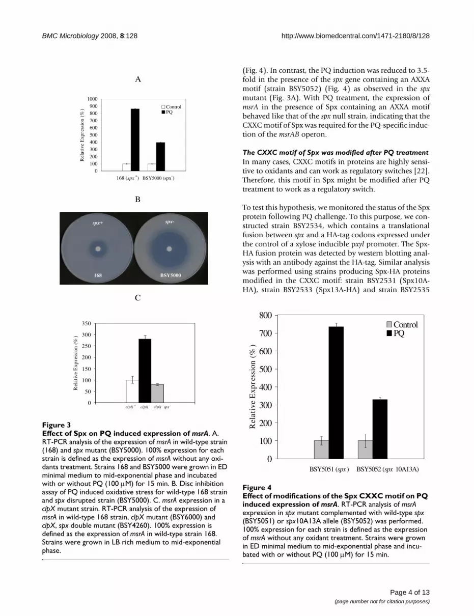
was not surprising because Spx was barely detectable inthe wild-type strain under these conditions [19]. How-ever, in the spx mutant, the PQ-specific induction of msrAexpression was reduced to approximately 50% of thatobserved in the spx wild-type strain (Fig. 3A) as shown byreal time RT-PCR assays. Therefore, Spx was involved in apart of the PQ-specific induction of msrA expression.Interestingly, compared to the wild-type strain, the spxmutant is extremely sensitive to PQ (Fig. 3B), suggestingthat Spx participates directly or indirectly in PQ responsein B. subtilis. As reported previously, while present at a lowlevel in the wild type, the Spx protein accumulates to ahigh concentration in a B. subtilis clpX mutant [20]. Tosubstantiate the involvement of Spx in the regulation ofthe msrAB operon, we measured the expression of msrA ina clpX nullbackground or in a clpX and spx double mutantusing real time RT-PCR (Fig. 3C). The quantity of the msrAtranscript increased 3-fold in the clpX mutant as comparedto that in the wild-type strain. However, the expression ofthe msrA transcript in the clpX and spx double mutant wassimilar to that in the wild-type strain. This indicates thatSpx is required for the increased expression of msrAobserved in the clpX mutant, supporting our finding thatthe Spx protein takes part in PQ induced msrA expression.
The increased expression of msrA observed in the clpXmutant suggested that the PQ challenge could lead to anincrease of the Spx protein concentration in vivo. Indeed,our real time RT-PCR experiment showed a 6-foldincreased expression of the spx gene after 100 μM PQ treat-ment (see Additional file 1). However, no significantchange of the Spx amount in the cells was identified afterPQ challenge when compared to the dramatic accumula-tion of Spx in a clpX mutant by western blotting analysis(data not shown). This suggests that some other mecha-nism than simply the quantity of the Spx protein isinvolved in the increase in msrA expression.
The CXXC motif of Spx is essential for the activation of msrAB operon expression following PQ challengeIt has been proposed that the transcriptional activationeffect of Spx on trxA and trxB expression could be due tothe formation of an intra-molecular disulfide bond gener-ated by oxidation of the two cysteine residues in the CXXCmotif of the protein [21]. This prompted us to test theinvolvement of the Spx CXXC motif in the PQ-inducedactivation of the msrAB operon.
To this purpose, the msrA expression was tested by realtime RT-PCR assays in a spx mutant that contained eitherthe wild-type spx gene or the spx gene with a modifiedCXXC motif expressed under the control of a xylose induc-ible pxyl promoter (strains BSY5051 and BSY5052). AfterPQ treatment, the msrA expression was induced 7.5-foldin the presence of the wild-type spx gene (strain BSY5051)
Effect of oxidative stresses on the expression of msrAFigure 2Effect of oxidative stresses on the expression of msrA. A. β-galactosidase activity of the msrA::lacZ transcriptional fusion. BSY4546 (msrA::lacZ) was grown in ED minimal medium to mid-exponential phase and treated with or with-out PQ (100 μM) or H2O2 (100 μM) for 1.5 hours. B. Real time RT-PCR analysis of msrA expression in wild-type strain (168) subjected to oxidative stresses. 100% expression is defined as the expression of msrA without any oxidant treat-ment. Strain 168 was grown in ED minimal medium to mid-exponential phase and incubated with or without PQ (100 μM) or H2O2 (100 μM) for 15 min.
A
0
5
10
15
20
25
Spec
ific
Act
ivit
y (U
/mg)
Control H2O2PQ
B
0
100
200
300
400
500
600
700
800
900
1000
Rel
ativ
e E
xpre
ssio
n (%
) ControlH2O2PQ
Page 3 of 13(page number not for citation purposes)
BMC Microbiology 2008, 8:128 http://www.biomedcentral.com/1471-2180/8/128
(Fig. 4). In contrast, the PQ induction was reduced to 3.5-fold in the presence of the spx gene containing an AXXAmotif (strain BSY5052) (Fig. 4) as observed in the spxmutant (Fig. 3A). With PQ treatment, the expression ofmsrA in the presence of Spx containing an AXXA motifbehaved like that of the spx null strain, indicating that theCXXC motif of Spx was required for the PQ-specific induc-tion of the msrAB operon.
The CXXC motif of Spx was modified after PQ treatmentIn many cases, CXXC motifs in proteins are highly sensi-tive to oxidants and can work as regulatory switches [22].Therefore, this motif in Spx might be modified after PQtreatment to work as a regulatory switch.
To test this hypothesis, we monitored the status of the Spxprotein following PQ challenge. To this purpose, we con-structed strain BSY2534, which contains a translationalfusion between spx and a HA-tag codons expressed underthe control of a xylose inducible pxyl promoter. The Spx-HA fusion protein was detected by western blotting anal-ysis with an antibody against the HA-tag. Similar analysiswas performed using strains producing Spx-HA proteinsmodified in the CXXC motif: strain BSY2531 (Spx10A-HA), strain BSY2533 (Spx13A-HA) and strain BSY2535
Effect of modifications of the Spx CXXC motif on PQ induced expression of msrAFigure 4Effect of modifications of the Spx CXXC motif on PQ induced expression of msrA. RT-PCR analysis of msrA expression in spx mutant complemented with wild-type spx (BSY5051) or spx10A13A allele (BSY5052) was performed. 100% expression for each strain is defined as the expression of msrA without any oxidant treatment. Strains were grown in ED minimal medium to mid-exponential phase and incu-bated with or without PQ (100 μM) for 15 min.
0
100
200
300
400
500
600
700
800
BSY5051 (spx ) BSY5052 (spx 10A13A)
Rel
ativ
e E
xpre
ssio
n (%
)
ControlPQ
Effect of Spx on PQ induced expression of msrAFigure 3Effect of Spx on PQ induced expression of msrA. A. RT-PCR analysis of the expression of msrA in wild-type strain (168) and spx mutant (BSY5000). 100% expression for each strain is defined as the expression of msrA without any oxi-dants treatment. Strains 168 and BSY5000 were grown in ED minimal medium to mid-exponential phase and incubated with or without PQ (100 μM) for 15 min. B. Disc inhibition assay of PQ induced oxidative stress for wild-type 168 strain and spx disrupted strain (BSY5000). C. msrA expression in a clpX mutant strain. RT-PCR analysis of the expression of msrA in wild-type 168 strain, clpX mutant (BSY6000) and clpX, spx double mutant (BSY4260). 100% expression is defined as the expression of msrA in wild-type strain 168. Strains were grown in LB rich medium to mid-exponential phase.
A
0
100
200
300
400
500
600
700
800
900
1000
168 (spx +) BSY5000 (spx-)
Rel
ativ
e E
xpre
ssio
n (%
) ControlPQ
B
C
0
50
100
150
200
250
300
350
clpX + clpX - clpX - spx -
Rel
ativ
e E
xpre
ssio
n (%
)
Page 4 of 13(page number not for citation purposes)

BMC Microbiology 2008, 8:128 http://www.biomedcentral.com/1471-2180/8/128
(Spx10A13A-HA). To show that the HA tag did not inter-fere with the function of Spx, we verified that the presenceof the HA-tag did not modify the Spx-dependent regula-tion of msrA expression during PQ stress (see Additionalfile 2).
In the western blot (Fig. 5A), two strong signal bandsobtained in the 20 KDa region in each lane were non-spe-cific bands (lane 1–10), and three forms of Spx-HA pro-
teins (lanes 1 and 2) were detected compared to thenegative control without a Spx-HA tagged protein (lanes 9and 10). The major band corresponded to the predictedmolecular mass of Spx-HA (around 17 KDa) while twoadditional forms named Spx-M1 and Spx-M2 appeared inthe higher molecular mass region (lane 1). Interestingly,after PQ treatment (lane 2), the amount of Spx-M1 signif-icantly increased but the amount of the native Spx and toa less extend of the Spx-M2 decreased as compared to the
Analysis and characterization of PQ modification effect on Spx and PQ modified form of SpxFigure 5Analysis and characterization of PQ modification effect on Spx and PQ modified form of Spx. A. PQ-induced modifications of the CXXC motif of Spx. Strains producing wild-type and CXXC motif mutated Spx-HA fusion proteins were treated with or without 100 μM PQ for 15 min. Crude cell extracts from each strain were analyzed by western blot using an antibody against HA. lane 1–2 (wt): strain BSY2534 producing Spx-HA protein; lane 3–4 (10A): strain BSY2531 producing Spx10A-HA protein; lane 5–6 (13A): strain BSY2533 producing Spx13A-HA protein; lane 7–8 (10A13A): strain BSY2535 pro-ducing Spx10A13A-HA protein; lane 9–10: vector control (strain BSY2530 that did not express any HA fusion protein). lane 2, 4, 6, 8, 10: with PQ treatment; lane 1, 3, 5, 7, 9: without PQ treatment. The modified forms of Spx were marked as Spx-M1, Spx-M2 and native forms of Spx were marked as Spx. B. Time course changes of PQ-induced modifications of Spx in vivo. Strain BSY2534 producing Spx-HA was incubated with 100 μM PQ for different time (0–6 hr). 40 μg protein from cell crude extracts obtained at each time point was analyzed by western blot using anti-HA antibody. lane 1: 0 min; lane 2: 15 min; lane 3: 1 hr; lane 4: 2 hr; lane 5:4 hr; lane 6: 6 hr.
A
B
Page 5 of 13(page number not for citation purposes)

BMC Microbiology 2008, 8:128 http://www.biomedcentral.com/1471-2180/8/128
untreated sample (lane 1). Similar patterns were observedwith Spx10A (lanes 3 and 4) and Spx13A (lanes 5 and 6)proteins. In both cases, PQ treatment led to an increase ofthe amount of Spx10A-M1 and Spx13A-M1 forms. In con-trast, no Spx-M1 or Spx-M2 forms were detected for theSpx10A13A protein with and without PQ treatment(lanes 7 and 8). These results strongly suggested that PQtreatment led to a post-translational modification of Spxinvolving the cysteine rich motif (CXXC) and that at leastone cysteine residue of the CXXC motif was required forthe PQ mediated modification of Spx. We then studiedthe time course of changes in Spx in vivo after PQ treat-ment. As shown in Fig. 5B, a 15 minute treatment led tothe highest amount of Spx-M1 (lane 2) in vivo. Theamount of Spx-M1 further decreased until it totally disap-peared at six hours after treatment while the Spx-M2 accu-mulated between four and six hours (lane 6). In parallel,the amount of the native form of Spx decreased to reachthe lowest level one hour after treatment (lane 3), thenstarted to increase (lane 4–6). At six hours, it reached alevel similar to that detected before PQ treatment (Lane1). Therefore, both the modified form of Spx and thenative Spx showed dynamic changes in vivo after a PQtreatment.
In many cases, oxidative reagents modify the CXXC motifsof proteins through the formation of disulfide bonds [23].This modification is reversible in vitro after incubationwith reducing reagents, such as DTT [24]. Therefore, wetested whether PQ induced modification of the CXXCmotif of Spx could be reversed by DTT. The total cellularextracts of strain BSY2534 obtained after a PQ treatmentwere incubated with or without 50 mM DTT for differenttime. Interestingly, the Spx-M1 form of Spx did not disap-pear after DTT treatment under the conditions tested (datanot shown) indicating that this modification is unlikely tobe mediated by disulfide bonds formation.
DiscussionFactors involved in the regulation of msrA and msrB genesin bacteria are still largely unknown [12]. In the presentstudy, we demonstrated that the expression of the msrABoperon in B. subtilis was significantly induced under PQ-mediated oxidative stress conditions (Fig. 2). PQ is an oxi-dant that is commonly recognized to generate superoxidein vivo. It may also generate other ROS, including singletoxygen. However, in our experiments a msrAB mutant wasnot more sensitive to PQ, H2O2, or diamide treatmentthan the wild-type strain (data not shown), suggestingthat methionine sulfoxide reductases in B. subtilis are notthe key enzymesagainst oxidative stresses. Remarkably, inour hands, msrAB expression was not regulated by H2O2oxidative stress (Fig. 2). The finding that the response toPQ and H2O2 induced oxidative stresses differ is in linewith some previous observations in different bacterial
species. For example, the oxidant-induced expression ofmsrA in X. campestris varies according to the nature of theoxidants used [15]. Furthermore, in B. subtilis many geneswere reported to selectively respond either to PQ or toH2O2 [25,26]. Similar to the situation of the msrABoperon, the expression of cysK and other genes involved incysteine metabolism is only induced by PQ but not byH2O2 [25,26]. PerR, the specific peroxide responsive regu-lator in B. subtilis, is not involved in the regulation of themsrAB operon, in agreement with previous results indicat-ing that msrAB does not belong to the PerR regulon [27].In addition, a peroxide induced sigma B-dependent regu-lation of msrAB seems unlikely since no sigma B promoteris found upstream of the msrAB operon ([28,29], thiswork).
In the present study, we showed that Spx, a global tran-scriptional regulator of the disulfide specific oxidativestress response in B. subtilis plays a central role in the PQ-specific induction of msrAB expression. Surprisingly,diamide, a reagent that causes disulfide stress in organ-isms, did not induce the expression of msrAB in all theconditions tested, while we found a complex involvementof Spx, originally identified as a disulfide stress regulator(data not shown). This finding seems to be in contradic-tion with the fact of the participation of Spx in the PQinduced expression of msrAB. Several reasons mightaccount for that observation: (i) disulfide bond formationmight not involve in this PQ induced expression of msrAB(see below); (ii) Spx could respond to other oxidativestress beside disulfide stress; (iii) the expression of spx isregulated in a particularly complex way, with effectorsthat might compensate each other action on msrABexpression [30]. As a case in point, the spx mutant showedmore sensitivity to PQ treatment compared to the wild-type stain (Fig. 3B).
In line with (iii), the Spx mediated regulatory effect onmsrAB is only partial since PQ has a residual inductioneffect on msrA in a spx mutant (Fig. 3A). Therefore, besidesSpx, there were some unidentified PQ responding factorsregulating msrAB expression, or PQ could affect msrABmRNA stability in vivo. More studies are required tounravel the processes underlying these complex effects.
Further experiments indicated that the PQ effect on Spxoccurs at the post-translational level and that the CXXCmotif of Spx may be a target of PQ oxidation because thismotif was essential for the PQ-induced enhancement ofthe msrA expression mediated by Spx (Fig. 4). This hintedthe possibility that PQ activates Spx through an oxidativemodification of its CXXC motif.
Interestingly, PQ challenge brought a significantlyincreased amount of a new form of Spx, Spx-M1. This
Page 6 of 13(page number not for citation purposes)

BMC Microbiology 2008, 8:128 http://www.biomedcentral.com/1471-2180/8/128
form, which has an apparent mass 400 Da higher thanthat of the native Spx, probably corresponds to a post-translational modification of Spx (Fig. 5A). The Spx-M1form is still observed with Spx10A or Spx13A but disap-pears in a strain producing the Spx10A13A protein. Thisindicates the requirement for at least one of the twocysteines of the CXXC motif to observe the PQ-inducedmodification of Spx. So far, we failed to purify sufficientamounts of Spx-M1 in B. subtilis to identify the nature ofthis modification. Since the amount of Spx-M2 onlyshowed a moderate change in all the conditions testedhere, extensive studies on Spx-M2 will be carried out in afurther work.
In Xanthomonas campestris, a cysteine motif of the OhrRregulator that senses hydroperoxides is modified ascysteine-sulfenic acid which rapidly reacts with anothercysteine of the protein [31]. The OhrR regulator of B. sub-tilis has recently been shown to be modified in the sameway after oxidative stress with the formation of a disulfidebond (S-thiolation) between the Cys15 of OhrR and anew but still uncharacterized low molecular mass motif of398 Da [27]. This chemical modification has also beendetected in Bacillus anthracis [32]. In the present report thesize increase of Spx after PQ treatment could correspondto the same sulfenic acid modification followed by reac-tion with a thiol present in the cytoplasm of B. subtilis,playing the role of glutathione in other bacteria. However,since the PQ modified form of Spx is resistant to DTTunder the conditions tested (data not shown), a modifica-tion by S-thiolation seems unlikely. We cannot excludethat a derivative of PQ might have reacted with an acti-vated thiol of Spx. Finally, we could not completely ruleout the possibility that conformational changes broughtby PQ could be responsible for the different forms of Spxdetected. In any event, the direct covalent modification orconformational modification of Spx seems to be removedin vivo, and the amount of native Spx also showed adynamic change after PQ treatment (Fig. 5B). In moredetails, with short PQ treatment time, the native Spxshowed no much changes (lane 2), but longer PQ treat-ment seemed to trigger the degradation of the native Spxin vivo (lane 3), which fits observations reported previ-ously on the effects of oxidative stress on proteins in vivo,which is causing protein degradation [33,34]. It appearsthat subsequent to a degradative process Spx was regener-ated in vivo after the cells have adapted to the PQ treat-ment (lane 4–6). These facts suggested that Spx proteinexperienced complex processes of modification, degrada-tion and regeneration in vivo following PQ treatment.
ConclusionIn summary, this study reports a Spx mediated PQ-specificregulation pathway of the msrAB operon in B. subtilis. PQinduces the expression of msrAB partially through its pos-
sible oxidation of the Spx CXXC motif. More studies areexpected to discover the molecular mechanism of PQ-induced modification of Spx as well as other factorsinvolved in the PQ-specific induction of the msrABoperon.
MethodsBacterial strains, plasmids, and culture conditionsE. coli and B. subtilis strains and plasmids used in this workare listed in Table 1. E. coli cells were grown in LBmedium. B. subtilis cells were grown in LB medium, sporu-lation (SP) medium (0.8% nutrient broth, 1 mM MgSO4,13.4 mM KCl, 0.5 mM CaCl2, 10 μM MnCl2, 13.4 μM fer-ric ammonium citrate), or ED minimal medium (6 mMK2HPO4, 4.4 mM KH2PO4, 27 mM glucose (or fructose),0.3 mM Na3-citrate, 15 mM L-glutamine, 0.244 mM L-tryptophan, 33.5 μM ferric ammonium citrate, 1 mMMgSO4, 4 mM MgCl2, 0.25 mM CaCl2, 10 μM MnCl2).Antibiotics were added with the following concentrationwhen required: ampicillin (Amp), 100 μg ml-1 for E. coli;spectinomycin (Spc), 60 μg ml-1 for both E. coli and B. sub-tilis; erythromycin (Ery), 1 μg ml-1, chloramphenicol(Cam), 5 μg ml-1, and kanamycin (Kan), 5 μg ml-1 for B.subtilis. When xylose was added (at 0.5% (w/v) final con-centration), fructose was used as carbon source. Solidmedia were prepared by adding agar to 1.5% (w/v) to therespective liquid media. Amylase activity was detectedafter growth of B. subtilis strains on Tryptose Blood AgarBase (TBAB, Difco) supplemented with 10 g/liter hydro-lyzed starch (Sigma). Starch degradation was detected bysublimating iodine onto the plates. All experiments wereperformed in accordance with the European regulationrequirements concerning the contained use of GeneticallyModified Organisms of Group-I (French agreementN°2735).
DNA techniquesDNA purification, restriction enzyme digestion, ligationand transformation of E. coli were performed according tostandard protocols [35]. For cloning purpose, YieldAceDNA polymerase (Stratagene) was used. All the primersused for PCR in this study are listed in Table 2. DNAsequence of plasmid constructed in this work was deter-mined using the dideoxy-chain termination method andThermo Sequenase Kit (Amersham Pharmacia Biotech).B. subtilis cells were transformed with either plasmid DNAor chromosomal DNA following the two-step protocol asdescribed previously [36].
Plasmids and strains constructionsTo construct msrA transcriptional fusions with the lacZgene, DNA fragments of the msrA gene (nucleotides from+ 16 to +534 relative to the translation start point) wereamplified by PCR from genomic DNA of B. subtilis 168using primers CHY45 and CHY46, then inserted into the
Page 7 of 13(page number not for citation purposes)

BMC Microbiology 2008, 8:128 http://www.biomedcentral.com/1471-2180/8/128
EcoRI and BamHI sites of plasmid pDIA5307 (in situ lacZtranscriptional fusion vector) [37], producing plasmidpCHY4546. The plasmid pCHY4546 was introduced intothe chromosome of B. subtilis 168 by a single crossoverevent, giving strain BSY4546. Strain BSY4546 was furtherverified by PCR. In strain BSY4546, the lacZ gene wasinserted after the translation stop codon of msrA, thereforea complete copy of msrA is present.
The spx and clpX genes were individually disrupted by akanamycin resistance cassette using three overlappingfragments with primers running PCR. DNA fragments Iand II, which correspond to the upstream (nucleotides -
519 to +29 relative to spx translation start point) anddownstream (nucleotides -25 to +565 relative to spx trans-lation stop point) regions of spx, were amplified by PCRfrom genomic DNA of B. subtilis 168 using primersCHY101 and CHY102, or CHY103 and CHY104, respec-tively. DNA fragments I and II, which correspond to theupstream region (nucleotides -580 to +85 relative to clpXtranslation start point) and the downstream region(nucleotides -81 to +543 relative to clpX translation stoppoint) of clpX, were amplified by PCR from genomic DNAof B. subtilis 168 using primers CHY128 and CHY129, orCHY130 and CHY131, respectively. DNA fragment III(kanamycin cassette gene (aphA-3) (1486 bp)) for both of
Table 1: Strains and plasmids used in this study
Strains Genotype Source or Reference
Escherichia coli
DH5α F-, Δ80dlacZΔM15, Δ(lacZYA-argF)U169, deoR, recA1, endA1 Laboratory collectionhsdR17(rk-, mk+), phoA, supE44, λ -, thi-1, gyrA96, relA1
Bacillus subtilis
168 trpC2 Laboratory collectionBFS2842 trpC2 spx::lacZ erm Functional analysis projecta
BSY2530 trpC2 amyE:: pX vector cat This workBSY2531 trpC2 amyE:: (pxyl-spx10A-HA) cat This workBSY2533 trpC2 amyE:: (pxyl-spx13A-HA) cat This workBSY2534 trpC2 amyE:: (pxyl-spx-HA) cat This workBSY2535 trpC2 amyE:: (pxyl-spx10A13A-HA) cat This workBSY4260 trpC2 clpX::aphA-3 spx::lacZ erm This workBSY4546 trpC2 msrA::lacZ cat This workBSY5000 trpC2 spx::aphA-3 This workBSY5051 trpC2 spx::aphA-3 thrC::(pxyl-spx) spc This workBSY5052 trpC2 spx::aphA-3 thrC::(pxyl-spx10A13A) spc This workBSY5531 trpC2 spx::aphA-3 amyE:: (pxyl-spx10A-HA) cat This workBSY5533 trpC2 spx::aphA-3 amyE:: (pxyl-spx13A-HA) cat This workBSY5534 trpC2 spx::aphA-3 amyE:: (pxyl-spx-HA) cat This workBSY5535 trpC2 spx::aphA-3 amyE:: (pxyl-spx10A13A-HA) cat This workBSY6000 trpC2 clpX::aphA-3 This work
Plasmids Description Source or Reference
pDIA5307 lacZ transcriptional fusion vector, AmpR CamR [37]pDG784 aphA-3 gene delivery vector, KanR [38]pXT gene expression vector AmpRSpcR [40]pX gene expression vector AmpRCamR [41]pCHY109 pXT spx AmpRSpcR This workpCHY110 pXT spx10A13A AmpRSpcR This workpCHY111 pX spx10A-HA AmpRCamR This workpCHY113 pX spx13A-HA AmpRCamR This workpCHY115 pX spx10A13A-HA AmpRCamR This workpCHY253 pX spx-HA AmpRCamR This workpCHY4546 pDIA5307 msrA::lacZ AmpR CamR This work
aThis strain has been constructed during the frame of the project for the functional characterization of the genome of B. subtilis in Japan [46].aphA-3 is a kanamycin resistance gene; cat is a chloramphenical acetyl transferase gene; spc is a spectinomycin resistance gene, erm is a erythromycin resistance gene.
Page 8 of 13(page number not for citation purposes)

BMC Microbiology 2008, 8:128 http://www.biomedcentral.com/1471-2180/8/128
them was amplified by PCR from plasmid pDG784 [38]using primers CHY13 and CHY14. In each case, the threepurified PCR fragments were mixed in equal amounts.Primers CHY101 and CHY104 were used to carry out thethree fragments PCR for spx, while primers CHY128 andCHY131 were used for clpX. The resulting PCR fragments(2624 bp for spx and 2775 bp for clpX) were purified andused individually to transform B. subtilis 168. Kanamycinresistant (KanR) transformants selected on SP plates con-taining kanamycin were further verified by PCR, resultingin the spx and clpX knockout strains BSY5000 andBSY6000. The chromosomal DNA of strains BSY5000 andBSY6000 was prepared and used to transform strainsBSY4546 respectively, resulting in strains BSY4550 andBSY4560.
To obtain a spx and clpX double mutant strain, the chro-mosomal DNA of the clpX knockout strain BSY6000 wastransformed into a spx null mutant strain BFS2842 (con-structed in the functional analysis project of the B. subtilisgenome [39]). Consequently, kanamycin and erythromy-cin resistant colonies were selected, resulting in the spxand clpX double mutant strain BSY4260.
To obtain a spx gene containing two mutations modifyingthe CXXC motif of Spx by AXXA (C10A, C13A), theupstream part of the spx coding sequence (nucleotides -519 to +60 relative to the translational start site) and thedownstream part of spx (nucleotides +30 to +961 relativeto the translational start site) were amplified by PCR fromgenomic DNA of B. subtilis 168 using primers CHY101and CHY114, or CHY115 and CHY104, respectively,introducing the point mutations C10A and C13A. Thesetwo purified PCR fragments were mixed in equalamounts, and primers CHY109 and CHY110 were used tocarry out overlap PCR. The resulting PCR fragment con-tained the complete coding sequences of spx (nucleotides-36 to +467 relative to the translational start site) contain-ing mutations C10A and C13A. The complete codingsequence of spx (nucleotides -36 to +467 relative to thetranslational start site) was also amplified by PCR fromgenomic DNA of B. subtilis 168 using primers CHY109and CHY110. Plasmid pXT [40] was then used to clonethe wild-type and the modified spx10A13A genes underthe control of the xylose-inducible promoter. The PCRfragments were inserted between the EcoRI and BamHIsites of plasmid pXT, resulting in plasmid pCHY109 and
Table 2: Primers used in this study
Primer Name Sequence (5'-3')
CHY13 GATAAACCCAGCGAACCATTTGAGGTGCHY14 GATACAAATTCCTCGTAGGCGCTCGGGACCHY45 CCGGAATTCGAAATCGCCACATTTGCAGGCHY46 CGCGGATCCTTATTTAGCCCCCCAATGCTCGCHY101 AAGCCCATATTGCTCGAGGTGGCHY102 TATCACCTCAAATGGTTCGCTGGGTTTATCCAGCTTGGTGATGTGTATAGTGCHY103 GGTCCCGAGCGCCTACGAGGAATTTGTATCAGAAGCACAGCGTTTGGCAAACCHY104 GGAACATTTATTGCCGTTGCCCHY109 AAGGAGGAAGCAGGTATGGTTACACTATACACATCCHY110 GACACGCACGAGGTTTAGTTTGCCAAACGCTGTGCHY111 CTCGCCTTTCTGCATGAAGTCGCGCTTGGTGATGTGTATAGTGCHY112 CACCAAGCGCGACTTCATGCAGAAAGGCGAGCHY113 GAGCCACGCTCTCGCCTTTCTCGCTGAAGTACAGCTTGGTGATGCHY114 ACTTCAGCGAGAAAGGCGAGAGCGTGGCTCCHY115 GAGCCACGCTCTCGCCTTTCTCGCTGAAGTCGCGCTTGGTGATGTGTATAGTGCHY128 GTCTGCAAATGCAAGGCATGCHY129 TATCACCTCAAATGGTTCGCTGGGTTTATCCAGCTACAAGCTTACGAACCTGCHY130 GGTCCCGAGCGCCTACGAGGAATTTGTATCGCAACTGTGACACACGGAGAGCHY131 AGTTCCACAAAGACAGCCTGCHY134 CGGCCATACTGAAAACCCTACHY135 ATCTGTCGGATCGATTTGCTCHY137 GCAGTAACGAAGTCCGTTTGCHY140 CCGCATGGTTCAAACATAAACHY141 CGTCAGACTTTCGTCCATTGCHY173 CCACTTCTTCTTCAATCGGCCHY253 GGACTAGTAGAGGAGTGAAGATGAATGGCHY254 CGCGGATCCTTAAGCGTAATCCGGAACATCGTATGGGTAGTTTGCCAAACGCTGTGCTTCYGSP3 TTTCCGCCAGACGTTGCTTGYGSP4 GCATCTGTCGGATCGATTTG
Page 9 of 13(page number not for citation purposes)
BMC Microbiology 2008, 8:128 http://www.biomedcentral.com/1471-2180/8/128
PCH110. Plasmids pCHY109 and pCHY110 weredigested by ScaI and used to transform strains BSY 5000.The pxyl-spx gene or the pxyl-spx(10A,13A) gene wasinserted by a double crossover event at the thrC locus ofthese strains, giving rise to strains BSY5051 and BSY5052.
To construct a C-terminal HA (Hemagglutinin, YPYDVP-DYA)-tag fusion with the Spx protein, a DNA fragmentcorresponding to the spx gene (nucleotides from -16 to+393 relative to the translation start point) was amplifiedby PCR from genomic DNA of B. subtilis 168 using prim-ers CHY253 and CHY254, leading to a translationalfusion between spx and HA tag codons at the N-terminal.Plasmid pX [41] was used to clone the spx gene fused withHA codons under the control of a xylose-inducible pro-moter. This PCR fragment was then inserted into the SpeIand BamHI sites of plasmid pX, producing plasmidpCHY253. This plasmid was digested by ScaI and inte-grated into the amyE locus on the chromosome of B. sub-tilis 168 by a double crossover event, giving strainBSY2534. The loss of amylase activity was detected asreported before [42]. Strain BSY2534 was further verifiedby PCR. Chromosomal DNA of the BSY2534 strain wastransformed into the spx mutant strain BSY5000, givingrise to strain BSY5534.
To obtain a spx gene containing three mutations modify-ing the CXXC motif of Spx-HA fusion protein by AXXC(C10A), CXXA (C13A) and AXXA (C10AC13A), theupstream part of the spx coding sequence(nucleotides -519 to +60 relative to the translational start site) wereamplified by PCR from genomic DNA of B. subtilis 168using primers CHY101 and CHY111, CHY101 andCHY113, and CHY101 and CHY114, individually, andthe individually corresponding downstream part of spx(nucleotides +30 to +393 relative to the translational startsite) were amplified by PCR from genomic DNA of B. sub-tilis 168 using primers CHY112 and CHY254, CHY114and CHY254, and CHY115 and CHY254, introducing thepoint mutations C10A, C13A, and C10AC13A. Each ofabove two corresponding purified PCR fragments wasmixed in equal amount, and primers CHY253 andCHY254 were used to carry out overlap PCR. The resultingPCR fragments contained the complete coding sequencesof spx with the HA tag gene containing mutations at C10A,C13A and C10AC13A, individually. These PCR fragmentswere inserted between the SpeI and BamHI sites of plas-mid pX, resulting in plasmids pCHY111, pCH113 andpCHY115. These plasmids were digested by ScaI and usedto transform strains 168 and BSY 5000, respectively. Thespx with HA tag gene, spx10A with HA tag gene, spx13Awith HA tag gene and the spx10A13A with HA tag genewas inserted by a double crossover event at the amyE locusof these strains, giving rise to strains BSY2531, BSY2533,BSY2535 and BSY5531, BSY5533, BSY5535. To make a
control, the plasmid pX was digested by ScaI and insertedinto the amyE locus of strain 168 by a double crossoverevent, producing strain BSY2530.
Oxidative stress induction, RNA extraction and β-galactosidase assaysOvernight B. subtilis cultures in LB were diluted to an OD600 nm of 0.1 in ED minimal medium and cultured at37°C. When OD 600 nm reached 0.6, cell cultures weresplit into three aliquots with one incubated with H2O2(100 μM), one incubated with PQ (100 μM), and onekept as a control. In some cases, cell cultures were splitinto two aliquots, treated with or without PQ (100 μM),respectively. After 15 min treatment, 10 ml of cells werecentrifuged at 7000 rpm at 4°C for 1 min and RNA wasextracted as reported previously [43].
β-galactosidase assay was performed as described byMiller [44]. One Unit of β-galactosidase was defined asthe amount of enzyme that produced 1 nmol of o-nitro-phenol per min at 28°C. Specific activity was expressed inUnits per mg protein. Protein concentration was deter-mined by the Bradford's method [45]. The experimentswere performed in triplicates.
Real time PCR assays and 5' RACE analysis3 μg of the DNase I-treated total RNA extracted from dif-ferent strains treated with or without oxidants was reverse-transcribed to generate cDNA using AMV Reverse Tran-scriptase (Roche) and random primer pDN(6) (Roche)according to the manufacturer's instructions. Real-timePCR (RT-PCR) analyzing the transcription of msrA and16S rRNA using each cDNA as a template was performedusing SYBR Green PCR Master Mix and SDS 2.1 softwareon a PRISM 7900 HT platform (Applied Biosystems). Rel-ative quantification of msrA transcription in various con-ditions and various strains was analyzed by thecomparative Ct (threshold cycle) method according to themanufacturer's instructions. The internal control usedhere was 16S rRNA. Primers used for msrA and 16S rRNAare CHY134 and CHY135, and CHY140 and CHY141,respectively.
The start site of the msrA or msrB transcript was identifiedby 5' RACE System for Rapid Amplification of cDNA Endskit (Invitrogen). The first strand cDNA was generated withprimers YGSP3 (complementary to nucleotides +345 to+364 relative to msrA translation start point) and CHY137(complementary to nucleotides +325 to +345 relative tomsrB translation start point) using 2 μg total RNA from B.subtilis strain 168 as template. After dC-tail treatment ofthe two cDNA obtained, the msrA cDNA was furtheramplified by PCR using the Abridged Anchor Primer fromthe kit and the nested primer YGSP4 (nucleotides +239 to+257 relative to msrA translation start point) while the
Page 10 of 13(page number not for citation purposes)

BMC Microbiology 2008, 8:128 http://www.biomedcentral.com/1471-2180/8/128
msrB cDNA was amplified using the Abridged AnchorPrimer from the kit and the nested primer CHY173(nucleotides +206 to +226 relative to msrB translationstart point). The two resulting PCR fragments were loadedon 1% agarose gel, and a single band was detected in eachcase. The two PCR fragments were sequenced to deter-mine the transcriptional start site.
Western blotting analysisTo study the in vivo effect of PQ-induction on the Spx pro-tein in B. subtilis, strains BSY2534, BSY2531, BSY2533,BSY2535, BSY2530, which contain Spx-HA, Spx (10A)-HA, Spx (13A)-HA, Spx (10A13A)-HA fusion proteins andthe negative vector control, individually, were grown inED minimal medium to an OD 600 nm of 0.6. Then, eachstrain culture was divided into two fractions. One wasincubated with 100 μM PQ for 15 minutes, and the sec-ond was kept as a control. 50 ml of cells from each culturewas harvested by centrifugation, resuspended in 1 ml of10 mM TrisHCl, pH 6.8 with 40 μl of Complete ProteaseInhibitor (Roche), and disrupted by sonication. The celldebris was removed by centrifugation at 16,000 g for 40min. Protein concentration was determined by the Brad-ford's method [45]. 40 μg of protein crude extracts fromeach culture was loaded onto a 17% SDS-PAGE. Proteinswere subsequently transferred to nitrocellulose by iBlot™Dry Blotting System (Invitrogen) and incubated withAnti-HA-Peroxidase antibody (Roche). Blots were devel-oped with Amersham ECL Plus Western Blotting Detec-tion System (GE healthcare) and signals were detected bya phosphorimager.
To study the effect of the addition of the dithiothreitol(DTT) on the PQ-induced modifications of Spx, the abovecrude extracts of strain BSY2534 obtained after 15 min PQ(100 μM) treatment was incubated with or without 50mM DTT at 37°C for 0 hr, 2 hr or 4 hr. Then 40 μg proteinobtained at each time point was loaded onto a 17% SDS-PAGE. A western blotting experiment was performed andSpx-HA was detected with Anti-HA-Peroxidase antibodyas described above.
To monitor the dynamic changes of the different Spxforms in vivo, strain BSY2534 was grown in ED minimalmedium to an OD 600 nm of 0.6, and was treated by 100μM PQ. At different time point (from 0 to 6 hours), 50 mlcell culture was collected and disrupted. 40 μg proteinfrom each cell crude extracts was analyzed by western blotusing the anti-HA-Peroxidase antibody as describedabove.
Disc inhibition assayB. subtilis strains wild-type 168 and spx disrupted strainBSY5000 were grown in ED minimal medium. 2 ml of cellculture from each strain at mid-logarithmic phase was
poured evenly onto ED minimal medium plate. After 5min, the liquid cell culture on the plate surface wasremoved carefully. When the plate was dried, a filter disc(diameter: 0.7 cm) was placed in the middle of the plateand impregnated with 8 μl of 10 mM PQ. The diametersof the inhibition zone were measured after the plates wereincubated at 37°C for 16 hours.
Authors' contributionsAD and CHY designed the whole experiments. AS, OF,IM–V and YPW helped to design some of the experiments.CHY carried out all the experiments. CHY and AD draftthis manuscript and all the others contributed in the writ-ing. All authors approved the final manuscript.
Additional material
AcknowledgementsWe thank Undine Mechold, Evelyne Turlin, Evelyne Krin, Marie-Françoise Hullo, Yanjiong Chen for help in some experiments and Yuko Makita for bioinformatics analysis, and Terence Hwa, Dalai Yan, the members of CHY's PhD thesis review committee and former anonymous reviewers for their careful reading and suggestions to this manuscript. CHY was sup-ported by a consulting grant to AD managed by EGIDE (France) and the BioSapiens European Union's 6th Framework Program: BioSapiens-Net-work of Excellence, Grant LSHG CT-2003-503265, as well as NSFC (No. 30470940), the Program of Introducing Talents of Discipline to Universi-ties, No. B06001.
Additional file 1Figure S1 – Effect of PQ on the expression of spx. RT-PCR analysis of spx expression in wild-type strain (168) subjected to a PQ induced oxida-tive stress. 100% expression is defined as the expression of spx without PQ treatment. Strain 168 was grown in ED minimal medium to a mid-expo-nential phase and incubated with or without PQ (100 μM) for 15 min. Primers CHY255 (5'tgcaagatttgtaccgcttg3') and CHY256 (5'tgcaagatttgtaccgcttg3') specific for spx gene were used.Click here for file[http://www.biomedcentral.com/content/supplementary/1471-2180-8-128-S1.doc]
Additional file 2Figure S2 – Wild-type and CXXC motif mutated Spx-HA fusion pro-teins effect on PQ induced expression of msrA. Real time RT-PCR anal-ysis of msrA expression in a spx mutant (BSY5000) and in spx mutant complemented with either spx-HA (BSY5534), or spx10A-HA (BSY5531), or spx13A-HA (BSY5533), or spx10A13A-HA (BSY5535) was performed. Each strain was grown in ED minimal medium to a mid-exponential phase and treated with or without 100 μM PQ for 15 min. 100% expression for each strain is defined as the expression of msrA with-out any oxidants treatment.Click here for file[http://www.biomedcentral.com/content/supplementary/1471-2180-8-128-S2.doc]
Page 11 of 13(page number not for citation purposes)

BMC Microbiology 2008, 8:128 http://www.biomedcentral.com/1471-2180/8/128
References1. Ezraty B, Grimaud R, El Hassouni M, Moinier D, Barras F: Methio-
nine sulfoxide reductases protect Ffh from oxidative dam-ages in Escherichia coli. Embo J 2004, 23(8):1868-1877.
2. Caldwell P, Luk DC, Weissbach H, Brot N: Oxidation of themethionine residues of Escherichia coli ribosomal proteinL12 decreases the protein's biological activity. Proc Natl AcadSci USA 1978, 75(11):5349-5352.
3. Abulimiti A, Qiu X, Chen J, Liu Y, Chang Z: Reversible methioninesulfoxidation of Mycobacterium tuberculosis small heat shockprotein Hsp16.3 and its possible role in scavenging oxidants.Biochem Biophys Res Commun 2003, 305(1):87-93.
4. Khor HK, Fisher MT, Schoneich C: Potential role of methioninesulfoxide in the inactivation of the chaperone GroEL byhypochlorous acid (HOCl) and peroxynitrite (ONOO-). J BiolChem 2004, 279(19):19486-19493.
5. Tang YC, Chang HC, Roeben A, Wischnewski D, Wischnewski N,Kerner MJ, Hartl FU, Hayer-Hartl M: Structural features of theGroEL-GroES nano-cage required for rapid folding of encap-sulated protein. Cell 2006, 125(5):903-914.
6. Jiang J, Nadas IA, Kim MA, Franz KJ: A Mets motif peptide foundin copper transport proteins selectively binds Cu(I) withmethionine-only coordination. Inorg Chem 2005,44(26):9787-9794.
7. Poger D, Fuchs JF, Nedev H, Ferrand M, Crouzy S: Moleculardynamics study of the metallochaperone Hah1 in its apo andCu(I)-loaded states: role of the conserved residue M10. FEBSLett 2005, 579(24):5287-5292.
8. Cabreiro F, Picot CR, Friguet B, Petropoulos I: Methionine sulfox-ide reductases: relevance to aging and protection againstoxidative stress. Ann N Y Acad Sci 2006, 1067:37-44.
9. Moskovitz J, Rahman MA, Strassman J, Yancey SO, Kushner SR, BrotN, Weissbach H: Escherichia coli peptide methionine sulfoxidereductase gene: regulation of expression and role in protect-ing against oxidative damage. J Bacteriol 1995, 177(3):502-507.
10. Hassouni ME, Chambost JP, Expert D, Van Gijsegem F, Barras F: Theminimal gene set member msrA, encoding peptide methio-nine sulfoxide reductase, is a virulence determinant of theplant pathogen Erwinia chrysanthemi. Proc Natl Acad Sci USA1999, 96(3):887-892.
11. Ruan H, Tang XD, Chen ML, Joiner ML, Sun G, Brot N, Weissbach H,Heinemann SH, Iverson L, Wu CF, Hoshi T: High-quality lifeextension by the enzyme peptide methionine sulfoxidereductase. Proc Natl Acad Sci USA 2002, 99(5):2748-2753.
12. Ezraty B, Aussel L, Barras F: Methionine sulfoxide reductases inprokaryotes. Biochim Biophys Acta 2005, 1703(2):221-229.
13. Hayes CS, Illades-Aguiar B, Casillas-Martinez L, Setlow P: In vitroand in vivo oxidation of methionine residues in small, acid-soluble spore proteins from Bacillus species. J Bacteriol 1998,180(10):2694-2700.
14. Weissbach H, Resnick L, Brot N: Methionine sulfoxide reduct-ases: history and cellular role in protecting against oxidativedamage. Biochim Biophys Acta 2005, 1703(2):203-212.
15. Vattanaviboon P, Seeanukun C, Whangsuk W, Utamapongchai S,Mongkolsuk S: Important role for methionine sulfoxidereductase in the oxidative stress response of Xanthomonascampestris pv. phaseoli. J Bacteriol 2005, 187(16):5831-5836.
16. Alamuri P, Maier RJ: Methionine sulfoxide reductase in Helico-bacter pylori: interaction with methionine-rich proteins andstress-induced expression. J Bacteriol 2006, 188(16):5839-5850.
17. Herbig AF, Helmann JD: Roles of metal ions and hydrogen per-oxide in modulating the interaction of the Bacillus subtilisPerR peroxide regulon repressor with operator DNA. MolMicrobiol 2001, 41(4):849-859.
18. Nakano S, Kuster-Schock E, Grossman AD, Zuber P: Spx-depend-ent global transcriptional control is induced by thiol-specificoxidative stress in Bacillus subtilis. Proc Natl Acad Sci USA 2003,100(23):13603-13608.
19. Nakano MM, Hajarizadeh F, Zhu Y, Zuber P: Loss-of-functionmutations in yjbD result in ClpX- and ClpP-independentcompetence development of Bacillus subtilis. Mol Microbiol2001, 42(2):383-394.
20. Nakano S, Zheng G, Nakano MM, Zuber P: Multiple pathways ofSpx (YjbD) proteolysis in Bacillus subtilis. J Bacteriol 2002,184(13):3664-3670.
21. Nakano S, Erwin KN, Ralle M, Zuber P: Redox-sensitive transcrip-tional control by a thiol/disulphide switch in the global regu-lator, Spx. Mol Microbiol 2005, 55(2):498-510.
22. Paget MS, Buttner MJ: Thiol-based regulatory switches. Annu RevGenet 2003, 37:91-121.
23. Linke K, Jakob U: Not every disulfide lasts forever: disulfidebond formation as a redox switch. Antioxid Redox Signal 2003,5(4):425-434.
24. Storz G, Tartaglia LA, Ames BN: Transcriptional regulator ofoxidative stress-inducible genes: direct activation by oxida-tion. Science 1990, 248(4952):189-194.
25. Antelmann H, Bernhardt J, Schmid R, Mach H, Volker U, Hecker M:First steps from a two-dimensional protein index towards aresponse-regulation map for Bacillus subtilis. Electrophoresis1997, 18(8):1451-1463.
26. Mostertz J, Scharf C, Hecker M, Homuth G: Transcriptome andproteome analysis of Bacillus subtilis gene expression inresponse to superoxide and peroxide stress. Microbiology 2004,150(Pt 2):497-512.
27. Lee JW, Soonsanga S, Helmann JD: A complex thiolate switchregulates the Bacillus subtilis organic peroxide sensor OhrR.Proc Natl Acad Sci USA 2007, 104(21):8743-8748.
28. Hecker M, Volker U: General stress response of Bacillus subtilisand other bacteria. Adv Microb Physiol 2001, 44:35-91.
29. Hecker M, Volker U: Non-specific, general and multiple stressresistance of growth-restricted Bacillus subtilis cells by theexpression of the sigmaB regulon. Mol Microbiol 1998,29(5):1129-1136.
30. Leelakriangsak M, Kobayashi K, Zuber P: Dual negative control ofspx transcription initiation from the P3 promoter by repres-sors PerR and YodB in Bacillus subtilis. J Bacteriol 2007,189(5):1736-1744.
31. Panmanee W, Vattanaviboon P, Poole LB, Mongkolsuk S: Novelorganic hydroperoxide-sensing and responding mechanismsfor OhrR, a major bacterial sensor and regulator of organichydroperoxide stress. J Bacteriol 2006, 188(4):1389-1395.
32. Nicely NI, Parsonage D, Paige C, Newton GL, Fahey RC, Leonardi R,Jackowski S, Mallett TC, Claiborne A: Structure of the type IIIpantothenate kinase from Bacillus anthracis at 2.0 A resolu-tion: implications for coenzyme A-dependent redox biology.Biochemistry 2007, 46(11):3234-3245.
33. Bader N, Grune T: Protein oxidation and proteolysis. Biol Chem2006, 387(10–11):1351-1355.
34. Grune T, Klotz LO, Gieche J, Rudeck M, Sies H: Protein oxidationand proteolysis by the nonradical oxidants singlet oxygen orperoxynitrite. Free Radic Biol Med 2001, 30(11):1243-1253.
35. Sambrook JFE, Maniatis T: Molecular Cloning: a LaboratoryManual. Cold Spring Harbor, N.Y.: Cold Spring Harbor LaboratoryPress; 1989.
36. Kunst F, Rapoport G: Salt stress is an environmental signalaffecting degradative enzyme synthesis in Bacillus subtilis. JBacteriol 1995, 177(9):2403-2407.
37. Calogero S, Gardan R, Glaser P, Schweizer J, Rapoport G, Debar-bouille M: RocR, a novel regulatory protein controllingarginine utilization in Bacillus subtilis, belongs to the NtrC/NifA family of transcriptional activators. J Bacteriol 1994,176(5):1234-1241.
38. Guerout-Fleury AM, Shazand K, Frandsen N, Stragier P: Antibiotic-resistance cassettes for Bacillus subtilis. Gene 1995, 167(1–2):335-336.
39. Kobayashi K, Ehrlich SD, Albertini A, Amati G, Andersen KK, ArnaudM, Asai K, Ashikaga S, Aymerich S, Bessieres P, Boland F, Brignell SC,Bron S, Bunai K, Chapuis J, Christiansen LC, Danchin A, DebarbouilleM, Dervyn E, Deuerling E, Devine K, Devine SK, Dreesen O, Err-ington J, Fillinger S, Foster SJ, Fujita Y, Galizzi A, Gardan R, EschevinsC, et al.: Essential Bacillus subtilis genes. Proc Natl Acad Sci USA2003, 100(8):4678-4683.
40. Nair S, Derre I, Msadek T, Gaillot O, Berche P: CtsR controls classIII heat shock gene expression in the human pathogen Liste-ria monocytogenes. Mol Microbiol 2000, 35(4):800-811.
41. Mogk A, Hayward R, Schumann W: Integrative vectors for con-structing single-copy transcriptional fusions between Bacillussubtilis promoters and various reporter genes encoding heat-stable enzymes. Gene 1996, 182(1–2):33-36.
42. You C, Lu H, Sekowska A, Fang G, Wang Y, Gilles AM, Danchin A:The two authentic methionine aminopeptidase genes are
Page 12 of 13(page number not for citation purposes)
http://www.ncbi.nlm.nih.gov/entrez/query.fcgi?cmd=Retrieve&db=PubMed&dopt=Abstract&list_uids=7836279
http://www.ncbi.nlm.nih.gov/entrez/query.fcgi?cmd=Retrieve&db=PubMed&dopt=Abstract&list_uids=7836279
http://www.ncbi.nlm.nih.gov/entrez/query.fcgi?cmd=Retrieve&db=PubMed&dopt=Abstract&list_uids=7836279
http://www.ncbi.nlm.nih.gov/entrez/query.fcgi?cmd=Retrieve&db=PubMed&dopt=Abstract&list_uids=9927663
http://www.ncbi.nlm.nih.gov/entrez/query.fcgi?cmd=Retrieve&db=PubMed&dopt=Abstract&list_uids=9573155
http://www.ncbi.nlm.nih.gov/entrez/query.fcgi?cmd=Retrieve&db=PubMed&dopt=Abstract&list_uids=2183352
http://www.ncbi.nlm.nih.gov/entrez/query.fcgi?cmd=Retrieve&db=PubMed&dopt=Abstract&list_uids=2183352
http://www.ncbi.nlm.nih.gov/entrez/query.fcgi?cmd=Retrieve&db=PubMed&dopt=Abstract&list_uids=2183352
http://www.ncbi.nlm.nih.gov/entrez/query.fcgi?cmd=Retrieve&db=PubMed&dopt=Abstract&list_uids=9298659
http://www.ncbi.nlm.nih.gov/entrez/query.fcgi?cmd=Retrieve&db=PubMed&dopt=Abstract&list_uids=9767581
http://www.ncbi.nlm.nih.gov/entrez/query.fcgi?cmd=Retrieve&db=PubMed&dopt=Abstract&list_uids=9767581
http://www.ncbi.nlm.nih.gov/entrez/query.fcgi?cmd=Retrieve&db=PubMed&dopt=Abstract&list_uids=7730271
http://www.ncbi.nlm.nih.gov/entrez/query.fcgi?cmd=Retrieve&db=PubMed&dopt=Abstract&list_uids=8113162
http://www.ncbi.nlm.nih.gov/entrez/query.fcgi?cmd=Retrieve&db=PubMed&dopt=Abstract&list_uids=8113162
http://www.ncbi.nlm.nih.gov/entrez/query.fcgi?cmd=Retrieve&db=PubMed&dopt=Abstract&list_uids=8566804

BMC Microbiology 2008, 8:128 http://www.biomedcentral.com/1471-2180/8/128
Publish with BioMed Central and every scientist can read your work free of charge
"BioMed Central will be the most significant development for disseminating the results of biomedical research in our lifetime."
Sir Paul Nurse, Cancer Research UK
Your research papers will be:
available free of charge to the entire biomedical community
peer reviewed and published immediately upon acceptance
cited in PubMed and archived on PubMed Central
yours — you keep the copyright
Submit your manuscript here:http://www.biomedcentral.com/info/publishing_adv.asp
BioMedcentral
differentially expressed in Bacillus subtilis. BMC Microbiol 2005,5:57.
43. Sekowska A, Robin S, Daudin JJ, Henaut A, Danchin A: Extractingbiological information from DNA arrays: an unexpected linkbetween arginine and methionine metabolism in Bacillus sub-tilis. Genome Biol 2001, 2(6):RESEARCH0019.
44. Miller CG, Kukral AM, Miller JL, Movva NR: pepM is an essentialgene in Salmonella typhimurium. J Bacteriol 1989,171(9):5215-5217.
45. Bradford MM: A rapid and sensitive method for the quantita-tion of microgram quantities of protein utilizing the princi-ple of protein-dye binding. Anal Biochem 1976, 72:248-254.
46. Bacillus subtilis Genome Database [http://bacillus.genome.jp]
Page 13 of 13(page number not for citation purposes)
![fff SPX[h_X^]TTa R^\ - Daily Pioneer](https://static.fdokumen.com/doc/165x107/6334ea98b9085e0bf5093f10/fff-spxhxtta-r-daily-pioneer.jpg)