Phenotype Enhancement Screen of a Regulatory spx Mutant Unveils a Role for the ytpQ Gene in the...
11
Phenotype Enhancement Screen of a Regulatory spx Mutant Unveils a Role for the ytpQ Gene in the Control of Iron Homeostasis Peter Zuber 1 *, Shefali Chauhan 1 , Praseeda Pilaka 1 , Michiko M. Nakano 1 , Sairam Gurumoorthy 1 , Ann A. Lin 1 , Skye M. Barendt 1 , Bui Khanh Chi 2 , Haike Antelmann 2 , Ulrike Ma ¨ der 3 1 Division of Environmental and Biomolecular Systems, Institute of Environmental Health, Oregon Health and Science University, Beaverton, Oregon, United States of America, 2 Institute for Microbiology, Ernst-Moritz-Arndt-University of Greifswald, Greifswald, Germany, 3 Interfaculty Institute for Genetics and Functional Genomics, Ernst-Moritz-Arndt-University of Greifswald, Greifswald, Germany Abstract Spx is a global regulator of genes that are induced by disulfide stress in Bacillus subtilis. The regulon that it governs is comprised of over 120 genes based on microarray analysis, although it is not known how many of these are under direct Spx control. Most of the Spx-regulated genes (SRGs) are of unknown function, but many encode products that are conserved in low %GC Gram-positive bacteria. Using a gene-disruption library of B. subtilis genomic mutations, the SRGs were screened for phenotypes related to Spx-controlled activities, such as poor growth in minimal medium and sensitivity to methyglyoxal, but nearly all of the SRG mutations showed little if any phenotype. To uncover SRG function, the mutations were rescreened in an spx mutant background to determine which mutant SRG allele would enhance the spx mutant phenotype. One of the SRGs, ytpQ was the site of a mutation that, when combined with an spx null mutation, elevated the severity of the Spx mutant phenotype, as shown by reduced growth in a minimal medium and by hypersensitivity to methyglyoxal. The ytpQ mutant showed elevated oxidative protein damage when exposed to methylglyoxal, and reduced growth rate in liquid culture. Proteomic and transcriptomic data indicated that the ytpQ mutation caused the derepression of the Fur and PerR regulons of B. subtilis. Our study suggests that the ytpQ gene, encoding a conserved DUF1444 protein, functions directly or indirectly in iron homeostasis. The ytpQ mutant phenotype mimics that of a fur mutation, suggesting a condition of low cellular iron. In vitro transcription analysis indicated that Spx stimulates transcription from the ytpPQR operon within which the ytpQ gene resides. The work uncovers a link between Spx and control of iron homeostasis. Citation: Zuber P, Chauhan S, Pilaka P, Nakano MM, Gurumoorthy S, et al. (2011) Phenotype Enhancement Screen of a Regulatory spx Mutant Unveils a Role for the ytpQ Gene in the Control of Iron Homeostasis. PLoS ONE 6(9): e25066. doi:10.1371/journal.pone.0025066 Editor: Christophe Herman, Baylor College of Medicine, United States of America Received April 15, 2011; Accepted August 25, 2011; Published September 20, 2011 Copyright: ß 2011 Zuber et al. This is an open-access article distributed under the terms of the Creative Commons Attribution License, which permits unrestricted use, distribution, and reproduction in any medium, provided the original author and source are credited. Funding: Deutsche Forschungsgemeinschaft AN 746/2-1 to HA. GM045898 from the National Institutes of Health USA to PZ. The funders had no role in study design, data collection and analysis, decision to publish, or preparation of the manuscript. Competing Interests: The authors have declared that no competing interests exist. * E-mail: [email protected] Introduction Transcriptome profiling can place genes into regulons or stimulons by providing evidence for coordinate control, governed by a transcriptional regulator and responsive to a specific metabolic or environmental stimulus [1]. In Gram-positive bacteria, some regulons, such as those controlled by alternative RNA polymerase sigma subunits [2] and global regulators [3] are large and complex. For example, the general stress SigmaB regulon, transcription of which requires the alternative RNA polymerase form bearing the s B subunit, is estimated to include over 200 genes in L. monocytogenes [4] and over 120 genes in B. subtilis [5,6,7,8]. Many of the genes within complex regulons are of unknown function and the sites of mutations having no detectable phenotype. Hence our view of the roles global regulators play in bacterial physiology remains incomplete. We can imagine that the genes within these complex regulons reside in functionally redundant or genetically buffered subgroups required for allevi- ating stress by detoxifying or removing harmful agents, or repairing the damage such agents inflict upon macromolecules and supramolecular structure. The Spx protein is a global regulator of the Gram-positive bacterium’s stress response [9]. It is highly conserved in low G+C Gram-positive bacteria [10], and in B. subtilis it interacts with RNA polymerase to exert positive and negative transcriptional control over a genome-wide scale [11,12,13]. The products of genes having known function that are induced by Spx include thioredoxin, thioredoxin reductase, and products that function in cysteine biosynthesis as well as synthesis of the low molecular weight redox buffer, bacillithiol [12,14,15]. Spx activates the transcription of its regulon in response to disulfide stress and in cells treated with various toxic agents including paraquat, nitric oxide, cell wall-acting agents, toxic electrophiles and hypochloric acid [12,16,17,18,19,20,21]. Spx is under tight regulation that involves positive and negative transcriptional control [22,23,24] and proteolytic control by a substrate-binding factor, YjbH, together with the ATP-dependent protease, ClpXP [25,26,27,28]. Additionally, its activity is controlled by a disulfide redox switch involving a CXXC motif at the N-terminal end of Spx that affects the protein’s productive interaction with RNA polymerase [11]. Spx governs a large regulon with about 120 of its members designated as ‘‘y’’ genes of unknown function (Table S1). Because PLoS ONE | www.plosone.org 1 September 2011 | Volume 6 | Issue 9 | e25066
Transcript of Phenotype Enhancement Screen of a Regulatory spx Mutant Unveils a Role for the ytpQ Gene in the...
Phenotype Enhancement Screen of a Regulatory spxMutant Unveils a Role for the ytpQ Gene in the Controlof Iron HomeostasisPeter Zuber1*, Shefali Chauhan1, Praseeda Pilaka1, Michiko M. Nakano1, Sairam Gurumoorthy1, Ann A.
Lin1, Skye M. Barendt1, Bui Khanh Chi2, Haike Antelmann2, Ulrike Mader3
1 Division of Environmental and Biomolecular Systems, Institute of Environmental Health, Oregon Health and Science University, Beaverton, Oregon, United States of
America, 2 Institute for Microbiology, Ernst-Moritz-Arndt-University of Greifswald, Greifswald, Germany, 3 Interfaculty Institute for Genetics and Functional Genomics,
Ernst-Moritz-Arndt-University of Greifswald, Greifswald, Germany
Abstract
Spx is a global regulator of genes that are induced by disulfide stress in Bacillus subtilis. The regulon that it governs iscomprised of over 120 genes based on microarray analysis, although it is not known how many of these are under directSpx control. Most of the Spx-regulated genes (SRGs) are of unknown function, but many encode products that areconserved in low %GC Gram-positive bacteria. Using a gene-disruption library of B. subtilis genomic mutations, the SRGswere screened for phenotypes related to Spx-controlled activities, such as poor growth in minimal medium and sensitivityto methyglyoxal, but nearly all of the SRG mutations showed little if any phenotype. To uncover SRG function, the mutationswere rescreened in an spx mutant background to determine which mutant SRG allele would enhance the spx mutantphenotype. One of the SRGs, ytpQ was the site of a mutation that, when combined with an spx null mutation, elevated theseverity of the Spx mutant phenotype, as shown by reduced growth in a minimal medium and by hypersensitivity tomethyglyoxal. The ytpQ mutant showed elevated oxidative protein damage when exposed to methylglyoxal, and reducedgrowth rate in liquid culture. Proteomic and transcriptomic data indicated that the ytpQ mutation caused the derepressionof the Fur and PerR regulons of B. subtilis. Our study suggests that the ytpQ gene, encoding a conserved DUF1444 protein,functions directly or indirectly in iron homeostasis. The ytpQ mutant phenotype mimics that of a fur mutation, suggesting acondition of low cellular iron. In vitro transcription analysis indicated that Spx stimulates transcription from the ytpPQRoperon within which the ytpQ gene resides. The work uncovers a link between Spx and control of iron homeostasis.
Citation: Zuber P, Chauhan S, Pilaka P, Nakano MM, Gurumoorthy S, et al. (2011) Phenotype Enhancement Screen of a Regulatory spx Mutant Unveils a Role forthe ytpQ Gene in the Control of Iron Homeostasis. PLoS ONE 6(9): e25066. doi:10.1371/journal.pone.0025066
Editor: Christophe Herman, Baylor College of Medicine, United States of America
Received April 15, 2011; Accepted August 25, 2011; Published September 20, 2011
Copyright: � 2011 Zuber et al. This is an open-access article distributed under the terms of the Creative Commons Attribution License, which permitsunrestricted use, distribution, and reproduction in any medium, provided the original author and source are credited.
Funding: Deutsche Forschungsgemeinschaft AN 746/2-1 to HA. GM045898 from the National Institutes of Health USA to PZ. The funders had no role in studydesign, data collection and analysis, decision to publish, or preparation of the manuscript.
Competing Interests: The authors have declared that no competing interests exist.
* E-mail: [email protected]
Introduction
Transcriptome profiling can place genes into regulons or
stimulons by providing evidence for coordinate control, governed
by a transcriptional regulator and responsive to a specific
metabolic or environmental stimulus [1]. In Gram-positive
bacteria, some regulons, such as those controlled by alternative
RNA polymerase sigma subunits [2] and global regulators [3] are
large and complex. For example, the general stress SigmaB
regulon, transcription of which requires the alternative RNA
polymerase form bearing the sB subunit, is estimated to include
over 200 genes in L. monocytogenes [4] and over 120 genes in B.
subtilis [5,6,7,8]. Many of the genes within complex regulons are of
unknown function and the sites of mutations having no detectable
phenotype. Hence our view of the roles global regulators play in
bacterial physiology remains incomplete. We can imagine that the
genes within these complex regulons reside in functionally
redundant or genetically buffered subgroups required for allevi-
ating stress by detoxifying or removing harmful agents, or
repairing the damage such agents inflict upon macromolecules
and supramolecular structure.
The Spx protein is a global regulator of the Gram-positive
bacterium’s stress response [9]. It is highly conserved in low G+C
Gram-positive bacteria [10], and in B. subtilis it interacts with RNA
polymerase to exert positive and negative transcriptional control
over a genome-wide scale [11,12,13]. The products of genes
having known function that are induced by Spx include
thioredoxin, thioredoxin reductase, and products that function in
cysteine biosynthesis as well as synthesis of the low molecular
weight redox buffer, bacillithiol [12,14,15]. Spx activates the
transcription of its regulon in response to disulfide stress and in
cells treated with various toxic agents including paraquat, nitric
oxide, cell wall-acting agents, toxic electrophiles and hypochloric
acid [12,16,17,18,19,20,21]. Spx is under tight regulation that
involves positive and negative transcriptional control [22,23,24]
and proteolytic control by a substrate-binding factor, YjbH,
together with the ATP-dependent protease, ClpXP [25,26,27,28].
Additionally, its activity is controlled by a disulfide redox switch
involving a CXXC motif at the N-terminal end of Spx that affects
the protein’s productive interaction with RNA polymerase [11].
Spx governs a large regulon with about 120 of its members
designated as ‘‘y’’ genes of unknown function (Table S1). Because
PLoS ONE | www.plosone.org 1 September 2011 | Volume 6 | Issue 9 | e25066

the Spx regulon is induced under a variety of stress conditions,
uncovering the function of the Spx-regulated genes (SRGs) would
further define the role of Spx in the cell’s response to encounters
with harmful agents.
In recent years, methods of genetic analysis have been
developed to exploit the vast collections of genomic data generated
from whole genome sequencing projects. Large gene knock-out
libraries have been created and utilized to uncover functional
genetic modules consisting of genes that influence essential cellular
processes. One way this has been accomplished is by the
systematic and automated screening of strains with paired
mutations (double mutants) to search for synthetic phenotypes
indicative of genetic interaction [29,30,31,32,33,34,35]. The
rationale for uncovering modules of interacting genes has its
origins in concepts of functional redundancy and genetic buffering
[36]. Elegant studies using classic genetic systems, and screens for
unlinked non-complementation and synthetic lethality, uncovered
genes that reside within functional modules that affect, for
example, morphogenesis and the dynamics of cytoskeletal
components [37,38,39,40,41]. More recent studies of genome-
wide synthetic genetic arrays uncovered new factors involved in
iron metabolism and in the activity of the transcription complex
[29]. Thus genetic screens for synthetic interaction and phenotype
enhancement can shed light on the functions of genes for which no
known function has been assigned. Hence, we undertook a
phenotype enhancement screen of spx mutants bearing knock-outs
of each of the SRGs of unknown function. Strains with the spx
mutation paired with each srg mutation that have defects in growth
or elevated sensitivity to methylglyoxal, to which spx mutants are
sensitive, were detected. One such srg mutation, in the ytpQ gene,
was studied using proteomic and transcriptomic analyses, which
uncovered a role for ytpQ in iron metabolism/homeostasis.
Results and Discussion
Results of previously published microarray hybridization data
[12], uncovered about 125 genes of the ‘‘y’’ designation that were
more than 3-fold upregulated by high Spx concentrations (Table
S1). The majority of the genes are of unknown function, although
many encode products that are highly conserved in other, mostly
Gram-positive, bacterial species. While we do not know at this
point if all of the genes are under the direct transcriptional control
of Spx, we decided to designate the genes as SRGs, or Spx-
regulated genes.
The spx regulon is induced by a number of toxic agents and
stress conditions, [10,12,16,17,20,22,42]. To understand further
the role of Spx in the cell’s response to these diverse stress
conditions, we sought to gain information on the function of the
individual SRGs. A B. subtilis ORF knock-out library, obtained
from K. Kobayashi and N. Ogasawara (NAIST, Japan), contains
gene disruptions in over 2,000 ORFs that are assigned the ‘‘y’’
genomic designation [43]. Each disruption was created by an
integrated DNA fragment, derived from plasmid pMUTIN [43],
that was inserted within the target gene’s coding sequence. The
fragment contains a promoter-less lacZ gene at its 59end, followed
by the E. coli lacI gene, the product of which controls an IPTG-
inducible Pspac promoter [44] residing at the 39 end of the fragment
and oriented in the 39 direction. The Pspac promoter in this position
can drive expression of genes located downstream of the insertion
[43], thus alleviating potential insertion-dependent polar effects.
The use of pMUTIN-mediated gene disruption was used in
previous genome-wide mutational screens and in gene interaction
studies [45,46]. Gene disruptions in the SRGs listed in Table S1
were tested by screening for growth on nutrient broth sporulation
medium (DSM) and minimal glucose (TSS) medium plates. Some
slight defect in growth on TSS agar plates was detected for some of
the SRG mutants, but the majority showed no obvious defect in
growth based on colony size. We next tested the SRG mutant
strains for defects in growth on DSM agar plates containing
methylglyoxal (MG), a toxic alpha-oxoaldehyde to which spx
mutants are sensitive (data not shown). Again, we observed only
minor growth defects compared with the wild-type parent on
DSM agar medium containing concentrations of methylglyoxal to
which spx mutants are sensitive.
Phenotype enhancement screen of srg mutations in thespx mutant background
Possible reasons for the absence of SRG mutant phenotype
related to stress resistance include genetic interactions built into
the spx regulon that contribute to functional redundancy and
genetic buffering. We reasoned that uncovering phenotype would
require overcoming regulon genetic interactions that mask defects
conferred by the SRG mutations. Hence, we undertook a screen of
the SRG mutations within the spx deletion mutant background.
The reasoning is illustrated in Figure 1, where the spx mutation is
shown to cause a reduction in overall SRG expression, thereby
reducing the contributions of genetic interactions among SRGs.
Introduction of the srg mutation into the spx mutant background
potentially confers hypersensitivity to the agents to which the spx
mutant is sensitive, but the srg spx mutant would now be predicted
to exhibit further reduction in growth on minimal medium, and
hypersensitivity to lower toxic agent concentrations that have little
to no effect on the parent spx mutant.
Each plate was inoculated with 10 ml of a dilution series of a log
phase culture. The size of the isolated colonies was then measured
using the Pixicillus application (Material and Methods). An
example of a plate and data collected is shown in Figure S1,
where four strains, the wild type JH642, the spx mutant, the SRG
mutant, yitV, and the spx yitV double mutant plated on DSM and
DSM plus MG is shown. The average isolated colony size as
determined by Pixicillus indicated that the double mutant has a
slight growth defect on the DSM-MG plate compared with the spx
mutant parent. Most of the double mutants screened showed a
result similar to that uncovered in the yitV spx mutant.
Disruption of ytpQ, an Spx-regulated gene of unknownfunction, enhances spx mutant phenotype whencombined with an spx null mutation
Other double mutants showed more dramatic phenotype
enhancement on TSS agar plates than was observed in the case
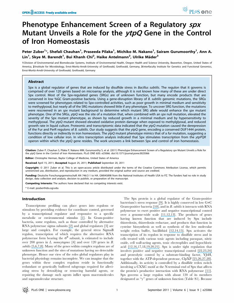
of the yitV mutant. The linked genes ytoQ and ytpQ (Figure 2A)
were the sites of two gene disruptions that showed growth defects
in the spx mutant background. The ytpQ spx mutant showed a
severe growth defect on TSS minimal medium (Figure 2B and
2C). The phenotypes of the ytoQ spx and ytpQ spx mutants were
examined in growth curves of cultures in liquid TSS medium
(Figure 2D and E). Both ytpQ and ytoQ mutants and the spx null
mutant showed defects in growth as evident in the slope of the log
phase portion of the growth curves or final OD600 (doubling times:
JH642; 42.8 min. spx null mutant; 93.8 min. ytoQ; 60.3 min. ytpQ;
73.3 min). The double mutants showed a further reduction in
growth rate (ytoQ spx; 103.8 min, ytpQ spx; 164.9 min) and had a
lower growth yield. While the introduction of the ytpQ mutation
into the spx null mutant resulted in a slower growth rate than the
spx null, the doubling time of the ytoQ spx strain was not
significantly longer than that of the spx null. Hence, we did not
further analyze the ytoQ mutant.
Function of Spx Regulon
PLoS ONE | www.plosone.org 2 September 2011 | Volume 6 | Issue 9 | e25066

The ytpQ and ytoQ genes are linked and divergently oriented in
the B. subtilis chromosome (Figure 2A). The ytpQ gene product is a
member of the DUF1444 family of bacterial proteins with no
known function. The ytpQ gene is part of a tricistronic operon that
also contains ytpP and ytpR. Disruption of ytpR showed no
noticeable phenotype on DSM or TSS medium with or without
MG, and confers no phenotype enhancement in the spx mutant
background (data not shown). The disruption of the ytpP gene,
encoding a thioredoxin-like protein [47], conferred phenotype
enhancement in the spx background (Figure 2B), but this was
reversed by addition of IPTG, showing that the defect was due to a
polar effect of the ytpP insertion (data not shown), most likely
causing reduction in ytpQ gene expression.
Complementation was conducted using an IPTG-inducible
version of the ytpQ gene ectopically expressed from the amyE locus
of the B. subtilis genome. For these experiments, a deletion ytpQ
mutation was constructed in which part of the ytpQ coding
sequence was replaced by a spectinomycin-resistance cassette. An
spx ytpQ double mutant (ORB7816) was then constructed by
introducing the ytpQ::spc mutation into a spx::tet (tetracycline
resistance) mutant. As was observed with the ytpQ::pMUTIN spx
double mutant, the ORB7816 strain enhanced the growth-
defective phenotype compared with either the ytpQ or spx mutants
in the presence or absence of MG (Figure S2). Thus, a strain
bearing the new ytpQ::spc allele and the ectopic inducible ytpQ
construct was grown in TSS minimal medium in the presence and
absence of 0.5 mM IPTG. The results (Figure S3) showed that the
reduced growth rate of the ytpQ mutant (doubling time of
79.4 min) was reversed when the ectopically expressed ytpQ gene
was induced. The induced complemented strain (ORB8011) had a
growth rate similar to the wild type parent (JH642; doubling time
of 47.3 min. OR8011; doubling time of 46.5 min).
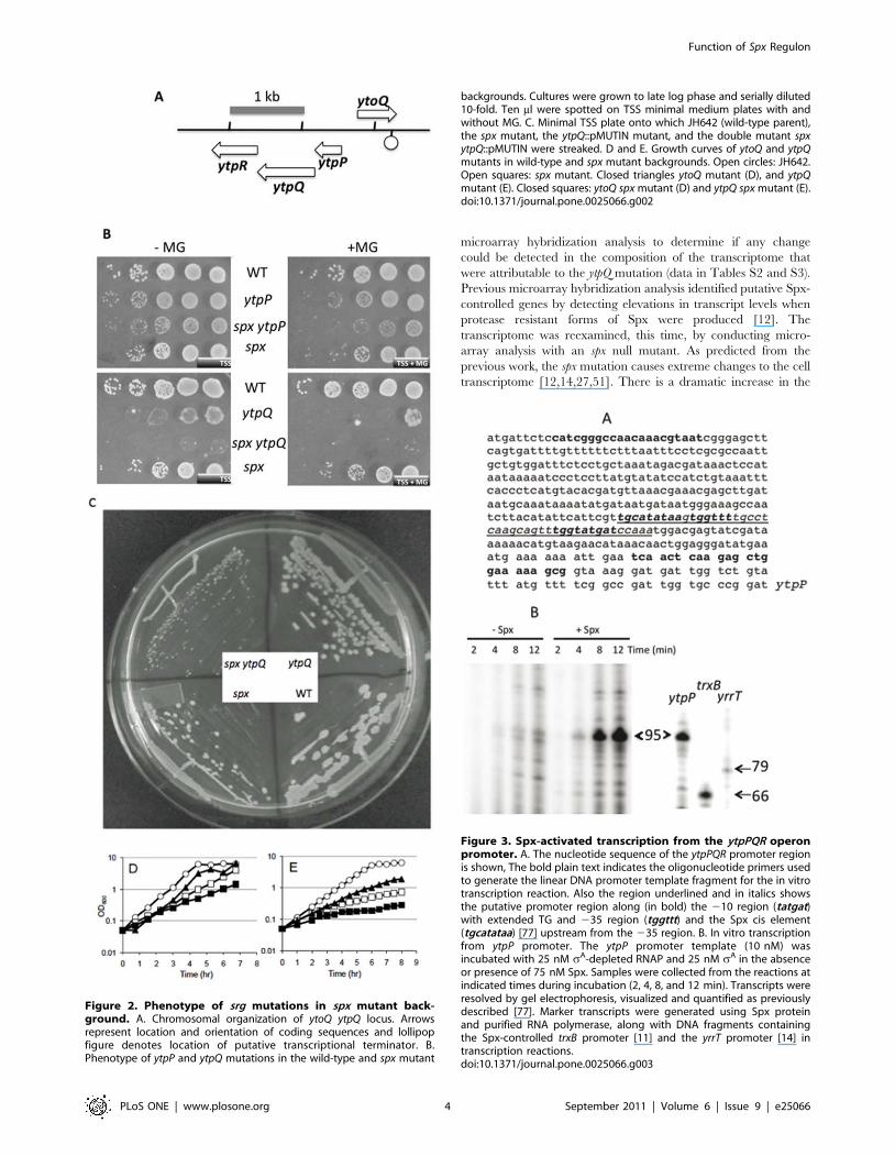
The ytpPQR operon is under direct Spx controlTo validate the previous microarray results, the ytoQ and ytpQ
pMUTIN insertions, both generating transcriptional lacZ fusions,
were used to measure ytoQ- and ytpQ-directed b-galactosidase
activity in cells expressing an IPTG-inducible, protease resistant
form of Spx (SpxDD) that were grown in liquid DSM medium
(Figure S4). In keeping with the microarray results, the fusions
showed elevated expression when the spxDD allele was induced.
Furthermore, microarray analysis (described below) indicated
reduced ytpQ transcript levels in the spx mutant (Table S2).
Validation was also accomplished by transcription analysis
performed in vitro, which showed that addition of Spx in a
reaction with the ytpPQR promoter region DNA and RNA
polymerase resulted in the synthesis of a transcript of the predicted
size (Figure 3). Synthesis of the transcript is stimulated by the
addition of Spx and initiates near a sA-recognized promoter
sequence (Figure 3A and B). The reaction utilized His-tagged
RNA polymerase from an rpoDL366A mutant from which RNA
polymerase lacking sA was obtained. Purified sA was required for
each reaction in order to observe a transcript from the ytpP
promoter template (data not shown). This result is consistent with
the presence of a sA-utilized promoter upstream of the ytpP coding
sequence.
The ytpQ mutant has increased levels of protein damageafter MG treatment
We further examined the phenotype of the ytpQ mutant in order
to gain more information about its possible role in the B. subtilis
stress response. Previous microarray hybridization studies indicat-
ed that ytpQ was derepressed in a perR [peroxide regulator [48]]
mutant background [49]. That ytpQ is derepressed in a perR
mutant and activated by Spx suggests that its product might
function in the oxidative/electrophile stress response. We
determined if the ytpQ mutant shows elevated levels of protein
carbonylation damage by performing an oxyblot [50] on cell
extracts from cultures of JH642, spx mutant, and ytpQ::spc mutant
cells that were untreated or treated with MG. The untreated wild-
type cells showed some evidence of oxidative protein damage
(Figure 4), which was increased upon MG treatment. The
untreated spx mutant cells showed more damage than the wild
type parent, with some increase in damage following MG
treatment. The untreated ytpQ mutant cells resembled the
untreated wild-type parent in the level of oxidatively damaged
protein, but the mutant underwent a dramatic increase in the level
of protein damage upon MG treatment that was much higher than
the treated wild-type parent or spx mutant. The result suggests that
the ytpQ product plays a role in preventing protein damage
resulting from an encounter with a toxic electrophile (MG).
Transcriptomic and proteomic analyses indicate a role ofytpQ in iron homeostasis
To gain more insight into the function of ytpQ, the transcrip-
tome of the ytpQ mutant was analyzed, and compared with that of
the wild type and the spx mutant. Similarities in the transcriptomic
changes conferred by the spx and ytpQ mutations would provide
clues to the role played by YtpQ within the Spx regulon. The wild-
type parent, ytpQ and spx mutants were grown in a glucose
minimal medium, with and without 2.8 mM MG, to mid-log
phase. Cells were harvested and RNA was extracted for
Figure 1. Rationale for phenotype enhancement screen.Horizontal line denotes genotype and vertical arrows represent levelof expression of each gene. SRG is Spx-regulated gene. The X on thehorizontal line is a null mutation, eliminating expression. The SRGmutation yields little observable phenotype with respect to electrophilestress. The spx mutation reduces overall SRG expression, thus reducingeffect of genetic buffering and functional redundancy. The srg spxdouble mutant is now found to be hypersensitive to electrophile(methylgyoxal), or shows other defects such as reduced growth rate.doi:10.1371/journal.pone.0025066.g001
Function of Spx Regulon
PLoS ONE | www.plosone.org 3 September 2011 | Volume 6 | Issue 9 | e25066
microarray hybridization analysis to determine if any change
could be detected in the composition of the transcriptome that
were attributable to the ytpQ mutation (data in Tables S2 and S3).
Previous microarray hybridization analysis identified putative Spx-
controlled genes by detecting elevations in transcript levels when
protease resistant forms of Spx were produced [12]. The
transcriptome was reexamined, this time, by conducting micro-
array analysis with an spx null mutant. As predicted from the
previous work, the spx mutation causes extreme changes to the cell
transcriptome [12,14,27,51]. There is a dramatic increase in the
Figure 2. Phenotype of srg mutations in spx mutant back-ground. A. Chromosomal organization of ytoQ ytpQ locus. Arrowsrepresent location and orientation of coding sequences and lollipopfigure denotes location of putative transcriptional terminator. B.Phenotype of ytpP and ytpQ mutations in the wild-type and spx mutant
Figure 3. Spx-activated transcription from the ytpPQR operonpromoter. A. The nucleotide sequence of the ytpPQR promoter regionis shown, The bold plain text indicates the oligonucleotide primers usedto generate the linear DNA promoter template fragment for the in vitrotranscription reaction. Also the region underlined and in italics showsthe putative promoter region along (in bold) the 210 region (tatgat)with extended TG and 235 region (tggttt) and the Spx cis element(tgcatataa) [77] upstream from the 235 region. B. In vitro transcriptionfrom ytpP promoter. The ytpP promoter template (10 nM) wasincubated with 25 nM sA-depleted RNAP and 25 nM sA in the absenceor presence of 75 nM Spx. Samples were collected from the reactions atindicated times during incubation (2, 4, 8, and 12 min). Transcripts wereresolved by gel electrophoresis, visualized and quantified as previouslydescribed [77]. Marker transcripts were generated using Spx proteinand purified RNA polymerase, along with DNA fragments containingthe Spx-controlled trxB promoter [11] and the yrrT promoter [14] intranscription reactions.doi:10.1371/journal.pone.0025066.g003
backgrounds. Cultures were grown to late log phase and serially diluted10-fold. Ten ml were spotted on TSS minimal medium plates with andwithout MG. C. Minimal TSS plate onto which JH642 (wild-type parent),the spx mutant, the ytpQ::pMUTIN mutant, and the double mutant spxytpQ::pMUTIN were streaked. D and E. Growth curves of ytoQ and ytpQmutants in wild-type and spx mutant backgrounds. Open circles: JH642.Open squares: spx mutant. Closed triangles ytoQ mutant (D), and ytpQmutant (E). Closed squares: ytoQ spx mutant (D) and ytpQ spx mutant (E).doi:10.1371/journal.pone.0025066.g002
Function of Spx Regulon
PLoS ONE | www.plosone.org 4 September 2011 | Volume 6 | Issue 9 | e25066

level of transcripts from CymR-controlled genes, whose products
function in organosulfur metabolism and the synthesis of cysteine
(Figure 5, Table S2) [14,52,53]. In fact, the impaired growth of the
spx mutant on minimal medium can be reversed by addition of
cysteine, indicating that the spx mutant is a mild Cys auxotroph.
The reduced expression of the trxA gene might account for spx
growth phenotype on minimal medium, since thioredoxin is
required for sulfate utilization [54]. There is an increase in the
level of transcript from genes controlled by PerR and Fur, both Fe-
binding proteins that regulate, respectively, the peroxide response
and genes that are activated under iron starvation (Figure 5, Table
S2). ComK-dependent transcription was also derepressed con-
firming previously reported results [55,56]. Synthesis of transcripts
encoded by early sporulation genes controlled by sH, sE, and sF
was also up-regulated in the spx mutant background. ClpX was
known to be required for sH-dependent transcription (Figure 6,
Table S2) [57,58], which was partially relieved by a mutation in
the spx gene [55].
The most striking result from the transcriptome analysis of the
ytpQ mutant is the dramatic derepression of the Fur (iron uptake
regulator) and PerR (peroxide response regulator) regulons
(Figure 5, Table S2 and S3). The ytpQ mutation seems to mimic
the reported phenotype of the B. subtilis fur mutant [59]. The
expression of genes (dhbABCDEF) encoding the enzyme complex
that catalyzes dihydroxybenzoyl-glycine (DHB-Gly, or itoic acid)
synthesis was dramatically increased. The B. subtilis strain, JH642,
bears a mutation in the sfp gene [60] encoding phosphopantethei-
Figure 4. Assay of protein damage in wild-type, spx, and ytpQstrains in the presence and absence of methylglyoxal (MG).Cells were incubated in 100 ml 2xYT cultures to an OD600 of 0.6.Cultures were split and MG to 2 mM was added to one of the twocultures. Crude cell extracts were prepared for SDS-PAGE and oxyblotanalysis was performed as described in Materials and Methods. Thesame amounts of protein were applied to the SDS-PAGE gel. WTdenotes blot of JH642 culture cell extracts. MG – methylglyoxaltreatment.doi:10.1371/journal.pone.0025066.g004
Figure 5. Hierarchical clustering analysis of gene expression profiles up- and downregulated in spx and ytpQ mutants. Geneexpression data were clustered based on the induction or repression ratios in the spx and ytpQ mutants leading to different nodes specific toregulons. Nodes enriched for genes that belong to the thiol-specific stress regulons (CymR, Spx, PerR, Fur, HxlR, CatR) are shown.doi:10.1371/journal.pone.0025066.g005
Function of Spx Regulon
PLoS ONE | www.plosone.org 5 September 2011 | Volume 6 | Issue 9 | e25066

nyl transferase [61] required for non-ribosomal peptide synthetase
activity. Hence, JH642 cells are unable to synthesize the
siderophore, bacillibactin (DHB-Gly-Thr) [62] despite the fact
that genes required for its biosynthesis (the dhb operon) show
elevated expression. The genes specifying hydroxamate side-
rophore utilization (yxeB, fhuB, fhuD) were also upregulated in the
ytpQ mutant. The expression of the Fur-regulated ykuNOP operon
was elevated higher than 10-fold in the ytpQ mutant. The operon
encodes two flavodoxins that serve as reductases for nitric oxide
synthase catalysis and for two-electron transfer to cytochrome
P450 BioI. This latter function can be fulfilled by the product of
the fer gene, which is a 4Fe-4S ferridoxin. Under conditions of low
iron, the ykuN, and ykuP products provide an iron-free substitute.
Another characteristic of the ytpQ mutant that mimics the fur null
phenoytpe is the derepression of the genes belonging to the cryptic
prophage, PBSX (Figure S5) [59]. It is not known why these genes
show elevated expression in the fur mutant.
Notably, several of the genes derepressed in the ytpQ mutant
were further up-regulated upon MG treatment (Figure 5, Tables
S2, S3). These include genes controlled by PerR and Fur, as well
as the spx gene itself. Elevated expression of genes encoding the
transporter for elemental iron (yfmLMN) was observed in the ytpQ
mutant cells treated with MG.
Another class of genes showing elevated expression in the spx and
ytpQ mutant is the sD regulon, which includes genes that function in
motility and chemotaxis [63,64]. The level of the hag gene
transcript, encoding flagellin, was increased 80-fold in the ytpQ
mutant (Figure 6). It is not clear why the expression of the sD
regulon is elevated in the ytpQ mutant. Reduced growth rate of the
ytpQ mutant could be related to the elevated expression of genes that
Figure 6. Hierarchical clustering analysis of gene expression profiles up- and downregulated in spx and ytpQ mutants. Geneexpression data were clustered based on the induction or repression ratios in the spx and ytpQ mutants leading to different nodes specific toregulons. Nodes including regulons involved in motility, competence and sporulation are shown (Com, SigmaD, SigmaH, CodY, SinR, Spo0A, SigmaL,AhrC, Rok, SigmaE, F, G, K regulons). Red indicates induction and green repression in the spx or ytpQ mutants under control conditions, and MG stress.doi:10.1371/journal.pone.0025066.g006
Function of Spx Regulon
PLoS ONE | www.plosone.org 6 September 2011 | Volume 6 | Issue 9 | e25066

function in motility and chemotaxis. Possibly linked to this
phenotype is the elevated expression of ywaC in the ytpQ mutant
(3.5-fold). The ywaC gene product is a GTP pyrophosphokinase that
can catalyze ppGpp formation at the expense of GTP [65,66].
Induction of the stringent response has been observed to heighten
expression of the motility/chemotaxis regulons through reduced
activity of the CodY repressor [67]. Another report has linked iron-
dependent control and CodY with the stringent response [68].
2D-gel based cytoplasmic proteome analysis provides validation
of the transcriptomic results (Figure 7). Consistent with the
microarray data of the spx mutant, the most strongly up-regulated
proteins in the proteome are controlled by the CymR repressor.
These include YtmI, YtmO, YtnJ, YrhB, CysK, SsuA, SsuD.
Furthermore, the PerR-controlled proteins KatA, AhpC, AhpF
were strongly elevated in the spx mutant proteome and less
strongly in the proteome of the ytpQ mutant. The proteins of the
Fur-regulated genes dhbA, dhbB, and dhbE were also observed at
increased amounts in the ytpQ mutant. Finally, the Hag protein
amount was also elevated in mutant cells. The elevated protein
levels in the ytpQ mutant are likely the result of the enhanced
transcription uncovered in the transcriptome analysis.
The phenotype enhancement screen is one way to gain
information about SRG function. In the present study, Spx
phenotype enhancement was tested by examining growth on
minimal medium and sensitivity to methylglyoxal. However, the
Spx regulon is induced under a variety of harsh conditions, and an
spx null mutant is sensitive to other toxic agents including
electrophiles, paraquat, selenite, hypochlorite and certain antibi-
otics (unpublished data). Resistance to each of these agents might
require the contributions of one or more specific SRGs. With this
in mind, further phenotype enhancement screens can be
conducted in the presence of each toxic agent with SRG mutations
in the spx mutant background to initiate an effort to uncover
function of other Spx regulon members.
The spx phenotype enhancement screen of SRGs uncovered
the ytpQ gene as being an important member of the spx regulon.
The double spx ytpQ mutant exhibited severely impaired growth
in minimal medium. Phenotype analysis of the ytpQ mutant
showed that it had an elevated level of oxidative protein damage
after methylglyoxal treatment compared to that of the wild-type
parent. The increased protein damage upon MG treatment could
be a consequence of dysfunctional iron metabolism, which was
evident from the transcriptome results showing heightened
expression of Fur regulon genes in ytpQ mutant cells. While
phenotype of the ytpQ mutant resembled that of a fur mutant,
indicating a condition of low cellular iron, this also creates a
condition of elevated free, chelatable iron that could mediate the
observed oxidative damage [69,70](Figure 4). These results
implicate ytpQ gene as a link between Spx-dependent control
and regulation of iron homeostasis. The study herein provides
evidence that the influence of Spx in oxidative stress management
extends to participation in the control of iron metabolism. Hence,
the severity of the spx phenotype is enhanced by the loss of ytpQ
function, and the accompanying dysfunction in iron homeostatic
mechanisms.
Attempts at gaining more information about YtpQ function will
involve a search for interacting proteins, with hopes of finding
binding partners with known function. Screening other SRG
mutations in the ytpQ background to find synthetic effects or
phenotype enhancement will uncover other spx regulon members
that reside in the same functional domain occupied by ytpQ.
Suppressor mutations that relieve the growth impairment of the
ytpQ spx mutant will also identify genes that are influenced by
YtpQ function.
Methods
Bacterial strains and growth mediaThe B. subtilis knock-out library [45] was constructed using a
previously described method [43], and was obtained from the
laboratory of N. Ogasawara (NAIST, Nara, Japan). Mutants from
the collection were re-checked by PCR using primers that hybridize
to the genomic region where the disruptions reside and primers
specific to pMUTIN. This was done to ensure that the pMUTIN
Figure 7. Dual-channel images of the protein amounts in B. subtilis wild type (green image) compared to the ytpQ (left) and spxmutants (right) (red images) under control conditions. Cytoplasmic proteins were separated by 2D PAGE as described in Materials andMethods. Image analysis was performed using the Decodon Delta 2D software. Proteins with increased levels in the mutants in at least twoindependent experiments are indicated by white labels.doi:10.1371/journal.pone.0025066.g007
Function of Spx Regulon
PLoS ONE | www.plosone.org 7 September 2011 | Volume 6 | Issue 9 | e25066

insertion was in the assigned gene. DNA from the SRG members
(Table S1) of the mutant collection was used to transform JH642 and
ORB6781 (spx::spc) competent cells to create two sets of isogenic SRG
mutants. All strains used in this study were derived from JH642 (trpC2
pheA) and are Trp2 and Phe2 auxotrophs.
B. subtilis cells were grown on TSS minimal medium [71], 2xYT
(yeast extract/tryptone [72]), or DSM nutrient broth sporulation
medium [73]. E. coli plasmid-bearing strains were propagated in
2xYT. Antibiotic concentrations used in growth media were
ampicillin (50 mg ml21), chloramphenicol (5 mg ml21), spectino-
mycin (75 mg ml21), and erythromycin/lincomycin (1 mg ml21
and 25 mg ml21, respectively).
Construction of ytpQ deletion mutantThe ytpQ gene was amplified from JH642 chromosomal DNA
using a forward primer oMN10-505 (cgCCGCAAAACAAAA-
GAAGAA; only upper case letters correspond to chromosomal
sequence) and a reverse primer oMN10-506 (cgCCA-
CACCTTCTTTATTATA) by PCR. The amplified DNA was
cloned using Topo-cloning kit (Invitrogen) to generate pMMN806.
The ytpQ gene isolated from EcoRI-digested pMMN806 was
cloned into pUC8 digested with EcoRI to generate pMMN807.
The spectinomycin-resistant cassette was isolated from pDG1727
digested with BamHI and StuI and the resistance gene was used to
replace the BglII/EcoRV-digested fragment of ytpQ in pMMN807.
The resultant plasmid pMMN808 led to a substitution of a 54-bp
DNA in ytpQ with the spectinomycin-resistance gene. The plasmid
pMMN808 was used to transform JH642 and the transformant
(ORB7815) was selected for the spectinomycin resistance. The
disruption of ytpQ in ORB7815 was confirmed by PCR using
oMN10-505 and oMN10-506. ORB7816 (spx::tet ytpQ::spc) was
constructed by transforming ORB6876 (spx::tet) with chromosomal
DNA prepared from ORB7815.
Preparation of chromosomal DNA and transformationSmall-scale chromosomal DNA preparation was conducted by
growing a culture of 3 ml 2xYT. The cells were harvested at
14,000 rpm in a Sorvall super T21 centrifuge with SL-50T rotor at
4uC for 10 min. The cells were resuspended in EDTA buffer
(25 mM NaCL, 50 mM EDTA). Lysozyme was added and the
mixture was then incubated for 15 min. Later sarkosyl was added
and phenol/chloroform extraction was performed. The chromo-
somal DNA was then stored at 4uC. The chromosomal DNA from
the mutants was used to transform JH642 competent cells and spx
mutant (ORB6781) competent cells. The transformation mixture
was incubated for 30 min at 37uC and then 1 ml of 2xYT was
added in each test tube. The transformation mixture was incubated
for 1 h and plated on DSM Erm-Ln (1 mg/ml erythromycin and
25 mg/ml lincomycin) and DSM Spc (75 mg/ml spectinomycin)
plates, respectively. The colonies were then streaked for single cell
clones. After genotype testing, strains were stored at 280uC.
Mutant ScreeningThe SRG mutations in the wild-type and spx mutant backgrounds
were tested on TSS, TSS+IPTG (0.5 mM), TSS+MG (2 mM),
TSS+MG+IPTG plates and the phenotype of each strain was
recorded. The mutants in both backgrounds along with JH642 and
spx::spc were grown in TSS media with appropriate antibiotics. The
cells were diluted to OD600 = 0.1 and then serially diluted to 1026 in
TSS medium. Ten ml from each dilution were transferred to TSS
plates and TSS and MG (2 mM) plates.
To assess the extent of growth impairment on minimal medium
or caused by treatment with methylglyoxal, we employed a Matlab
graphical user interface to measure colony size on agar medium
containing the toxic agent. The application, Pixicillus, utilizes
triangulation of coordinates corresponding to the dimensions of
the petri plate as a standard to calculate colony size (the design and
use of the program is available upon request).
Construction of ytpQ complementation strainsThe ytpQ ORF as well as 37 bases upstream harboring the ytpQ
Shine-Dalgarno sequence was amplified by PCR from JH642
purified genomic DNA using primers oSB1 (TAGGGAAGCTTCA-
GACTCTCTCGCTAAAGCGTAAAGGA, HindIII cut site un-
derlined) and oSB2 (TAGGCTCTAGACTAATCCTTTTTCG-
GACGGCTTTTCGC, XbaI cut site underlined). The HindIII -
XbaI digested linear amplicon was cloned into pDR67 [74], which
contains an IPTG-inducible spac promoter [44], a chloramphenicol
resistance cassette, and flanking amyE homologous regions for
chromosome integration. pDR67::Pspac-(SDytpQ)-ytpQ (pSB1) was
sequenced and introduced by transformation into several competent
isogenic strains of B. subtilis: i) spx::tet (ORB6876), ii) ytpQ::spc
(ORB7815), iii) spx::tet ytpQ::spc (ORB7816), and iv) parental JH642.
Strains were tested for the disruption of the amyE locus by plating on
LB plus 0.5% starch.
Growth curvesThe preculture of JH642, spx::spc and mutants constructed in
the JH642 or spx::spc genetic backgrounds were grown in TSS
media with appropriate antibiotics until mid-log phase. The cells
were then diluted to OD600 = .03 and incubation was continued
with shaking at 37uC. The OD600 was taken at time intervals of
30 min or 1 h.
OxyBlot AssayThe wild-type strain, ytpQ mutant, and spx::spc mutant were
grown in 100 ml of 2xYT at 37uc. At OD600 = 0.5, the culture was
divided equally in baffled flasks for each sample. Methylgloxal
(2.8 mM) was added into one of the two flasks. The cells were grown
for 6 h and harvested at 7000 rpm, 4uC for 20 min in a Sorvall
Super T21 centrifuge using a SL-50T rotor. The harvested cells
were resuspended in 50 mM NaCl, 25 mM EDTA pH 7.0 and
lysed using a French press. The crude cell lysate was centrifuged as
before. The supernatant was collected and the protein concentra-
tion was determined by the Bradford Assay [75]. Fifteen mg of the
crude sample protein was derivatized with 2, 4 dinitrophenyl
hydrazine. A control reaction with extract of untreated cells was also
assembled. The samples were then applied to a 12% SDS
polyacrylamide gel and the protein was electroblotted onto a
nitrocellulose membrane. The membrane was treated with primary
antibody provided in the Oxyblot kit (Millipore) specific for 2,4
dinitrophenol hydrazone. After the treatment with antibodies the
membrane was treated with chemi-luminescent reagent (Millipore)
and labeled protein was visualized on a photographic film (Fuji).
In vitro transcriptionRNA polymerase and Spx protein was purified as previously
described [28,76]. For the study reported herein, RNA polymerase
was purified from an rpoDL366A mutant (A gift from C. P. Moran,
Jr., Emory University), which lacks SigA protein after a three-
column purification procedure [76]. The enzyme does not
transcribe a consensus sA promoter DNA fragment (from the
rpsD gene) unless purified sA [58] is added to the reaction. In vitro
transcription reactions were performed according to previously
published work [11,76,77], with further details in Figure legend 3.
Function of Spx Regulon
PLoS ONE | www.plosone.org 8 September 2011 | Volume 6 | Issue 9 | e25066
Proteome and mass spectrometry analysisB. subtilis wild type (JH642), ytpQ and spx mutant cells were
grown in Belitsky minimal medium [78] to an OD500 = 0.4 and
harvested before (control) and 20 min after exposure to 2.8 mM
methylglyoxal. Preparation of cytoplasmic protein extracts and
separation by two-dimensional gel electrophoresis (2D-PAGE)
were performed as described [79]. The protein content was
determined using the Bradford assay [75]. For two-dimensional gel
electrophoresis (2D-PAGE), 200 mg of the protein extracts were
separated using the non-linear immobilized pH gradients (IPG) in
the pH range 4–7 for cytoplasmic proteins (Amersham Bioscienc-
es) and a Multiphor II apparatus (Amersham Pharmacia Biotech)
as described previously [80]. The resulting 2D gels were fixed in
40% (v/v) ethanol, 10% (v/v) acidic acid and stained with
Colloidal Coomassie Brilliant Blue (Amersham Biosciences). The
image analysis was performed from the Coomassie-stained 2D gels
using the DECODON Delta 2D software (http://www.decodon.
com).
For protein identification from 2D gels, spot-cutting, tryptic
digestion of the proteins, and spotting of the resulting peptides
onto the MALDI-targets (Voyager DE-STR, PerSeptive Biosys-
tems) were performed using the Ettan Spot Handling Workstation
(Amersham-Biosciences, Uppsala, Sweden) as described previously
[81]. The MALDI-TOF-TOF measurement of spotted peptide
solutions was carried out on a Proteome-Analyzer 4800 (Applied
Biosystems, Foster City, CA, USA) as described previously [81].
Transcriptome analysisFor microarray analysis, B. subtilis wild type, ytpQ and spx
mutant cells were grown in Belitsky minimal medium to OD500 of
0.4 and harvested before and after exposure to 2.8 mM
methylglyoxal. Total RNA was isolated by the acid phenol
method as described [82]. For transcriptome analysis, 35 mg RNA
were DNase-treated using the RNase-Free DNase Set (Qiagen)
and purified using the RNA Clean-Up and Concentration Micro
Kit (Norgen). The quality of the RNA preparations was assessed
by means of the Agilent 2100 Bioanalyzer according to the
manufacturer’s instructions.
Synthesis and purification of fluorescently labeled cDNA were
carried out according to Charbonnier et al. [83] with minor
modifications. In detail, 10 mg of total RNA were mixed with
random primers (Promega) and spike-ins (Two-Color RNA Spike-
In Kit, Agilent Technologies). The RNA/primer mixture was
incubated at 70uC for 10 min followed by 5 min incubation on
ice. Then, the following reagents were added: 10 ml of 56 First
Strand Buffer (Invitrogen), 5 ml of 0.1 M DTT (Invitrogen), 0.5 ml
of a dNTP mix (10 mM dATP, dGTP, and dTTP, 2.5 mM
dCTP), 1.25 ml of Cy3-dCTP or Cy5-dCTP (GE Healthcare) and
2 ml of SuperScript II reverse transcriptase (Invitrogen). The
reaction mixture was incubated at 42uC for 60 min and then
heated to 70uC for 10 min. After 5 min on ice, the RNA was
degraded by incubation with 2 units of RNaseH (Invitrogen) at
room temperature for 30 min. Labeled cDNA was then purified
using the CyScribe GFX Purification Kit (GE Healthcare). Five
hundred ng of Cy5-labeled cDNA and 500 ng of Cy3-labeled
cDNA were hybridized together to the microarray following
Agilent’s hybridization, washing and scanning protocol (Two-
Color Microarray-based Gene Expression Analysis, version 5.5).
Data were extracted and processed using the Feature Extraction
software (version 10.5, Agilent Technologies). For each gene on a
microarray, the error-weighted average of the log ratio values of
the individual probes was calculated using the Rosetta Resolver
software (version 7.2.1, Rosetta Biosoftware). Genes showing
induction or repression ratios of at least three-fold in three
independent experiments were considered as significantly induced.
The averages ratios and standard deviations for all induced or
repressed genes in the ytpQ and spx mutants compared to the wild
type were calculated from three independent transcriptome
experiments each and listed in Table S3. All microarray datasets
and accompanying descriptions are MIAME compliant and the
datasets are available in the GEO database under accession
numbers GSE28872.
Hierarchical clustering analysisClustering of gene expression profiles up- and downregulated in
spx and ytpQ mutants compared to the wild type under control
conditions and in the ytpQ mutant under methylglyoxal stress were
performed using Cluster 3.0 [84]. The transcriptome datasets
included log2-fold expression changes in the spx and ytpQ mutant
strains versus wild type. After hierarchical clustering, the output
was visualized using TreeView [85]. For clustering, genes were
used that are induced or repressed in the ytpQ and/or spx mutants
(e.g. CymR, Spx, PerR, Fur, HxlR, CatR, Com, SigmaD,
SigmaH, CodY, SinR, Spo0A, SigmaL, AhrC, Rok, SigmaE, F,
G, K regulons and SPb-related genes).
Supporting Information
Figure S1 Colonies of serially diluted, spotted cultureson TSS plates with and without 3 mM methylglyoxal.Strains are JH642 (wild type parent), spx mutant, yitV, and yitV spx
mutant. Values are average colony size as determined by Pixicillus.
(TIF)
Figure S2 TSS minimal medium agar containing 0, 0.5,1, and 2 mM MG. From top to bottom rows: JH642 (Wild type
parent), spx::tet mutant, ytpQ deletion mutant, spx::tet ytpQ deletion
double mutant. Cells were grown in TSS medium to mid-log
phase, then serially diluted 10-fold. Ten ml of each dilution was
then spotted onto the agar surface. Plates were incubated at 37uC.
(TIF)
Figure S3 Complementation of the DytpQ::spc mutationby an IPTG-inducible allele of ytpQ expressed from theamyE locus (Strain ORB8011). Growth curves are shown in
which cultures of JH642 cells and ORB8011 cells are grown in
TSS medium containing Trp and Phe auxotrophic requirements.
(TIF)
Figure S4 Assay of ytpQ- and ytoQ-directed b-galacto-sidase activity in wild type cells and cells bearing theIPTG inducible spxDD allele. A. Open circles: ytpQ::pMUTIN
Physpank-spxDD without IPTG. Closed circles: with IPTG. B. Open
triangles: ytoQ::pMUTIN Physpank-spxDD without IPTG. Closed
triangles: with IPTG. C. The expression of the ytpQ- and ytoQ-lacZ
fusions was measured in ytpQ::pMUTIN and ytoQ::pMUTIN cells in
the absence of the IPTG-inducible spxDD construct. Open circles:
ytpQ::pMUTIN without IPTG. Closed circles: ytpQ::pMUTIN with
IPTG. Open triangles: ytoQ::pMUTIN without IPTG. Closed
triangles: ytoQ::pMUTIN with IPTG.
(TIF)
Figure S5 Hierarchical clustering analysis of geneexpression profiles up- and downregulated in spx andytpQ mutants. Gene expression data were clustered based on
the induction or repression ratios in the spx and ytpQ mutants.
Nodes enriched for phage-related genes of B. subtilis are shown.
(TIF)
Function of Spx Regulon
PLoS ONE | www.plosone.org 9 September 2011 | Volume 6 | Issue 9 | e25066

Table S1 Expression levels of spx-controlled genes incells bearing an IPTG-inducible allele of spx (spxDD)encoding a protease resistant form of Spx protein. The
values are log2 of transcript level ratio between cells grown in
presence and absence of IPTG [12].
(TIF)
Table S2 Induction and repression of genes in the spxand ytpQ mutants under control conditions and inresponse to 2.8 mM methylglyoxal stress as revealedby transcriptome analyses. The averages ratios and standard
deviations as well as log2-fold changes for all induced and
repressed genes are calculated from three transcriptome replicate
experiments at control conditions and after 10 min of exposure to
2.8 mM methylglyoxal. All genes with induction ratios of at least
three-fold were listed and classified according to previously
described regulons according to http://dbtbs.hgc.jp and Sub-
tiWiki database (http://subtiwiki.uni-goettingen.de/wiki/index.
php/Main_Page).
(XLS)
Table S3 Induction and repression of genes undercontrol conditions and in response to methylglyoxal inthe spx and ytpQ mutants as revealed by transcriptome
analyses. The transcriptome datasets included log2-fold expres-
sion changes of average values of three transcriptome replicates
according to Table S2. All induced and repressed genes under
control and methylglyoxal stress were listed and classified into
regulons according to http://dbtbs.hgc.jp and SubtiWiki database
(http://subtiwiki.uni-goettingen.de/wiki/index.php/Main_Page).
These data were used for the hierarchical clustering analysis
presented in Figures 5 and 6.
(XLS)
Acknowledgments
We thank the Decodon company for support with the Decodon Delta 2D
software, Dana Clausen and Marc Schaffer for excellent technical
assistance, Dirk Albrecht for mass spectrometry analysis and Claudia
Schurmann for support in array data analysis. We also thank Amit Kumar
(Avnera Corp.) for design of Pixilus software.
Author Contributions
Conceived and designed the experiments: PZ HA. Performed the
experiments: SC PP MMN SG BKC HA UM AAL SMB. Analyzed the
data: PZ HA UM. Contributed reagents/materials/analysis tools: PZ
MMN HA UM. Wrote the paper: PZ HA.
References
1. Conway T, Schoolnik GK (2003) Microarray expression profiling: capturing
a genome-wide portrait of the transcriptome. Mol Microbiol 47: 879–889.
2. Gruber TM, Gross CA (2003) Multiple sigma subunits and the partitioning of
bacterial transcription space. Annu Rev Microbiol 57: 441–466.
3. Helmann JD, Wu MF, Kobel PA, Gamo FJ, Wilson M, et al. (2001) Global
transcriptional response of Bacillus subtilis to heat shock. J Bacteriol 183:
7318–7328.
4. Hain T, Hossain H, Chatterjee SS, Machata S, Volk U, et al. (2008) Temporal
transcriptomic analysis of the Listeria monocytogenes EGD-e sigmaB regulon. BMC
Microbiol 8: 20.
5. Petersohn A, Antelmann H, Gerth U, Hecker M (1999) Identification and
transcriptional analysis of new members of the sigmaB regulon in Bacillus subtilis.
Microbiology 145(Pt 4): 869–880.
6. Petersohn A, Bernhardt J, Gerth U, Hoper D, Koburger T, et al. (1999)
Identification of sigma(B)-dependent genes in Bacillus subtilis using a promoter
consensus-directed search and oligonucleotide hybridization. J Bacteriol 181:
5718–5724.
7. Price CW, Fawcett P, Ceremonie H, Su N, Murphy CK, et al. (2001) Genome-
wide analysis of the general stress response in Bacillus subtilis. Mol Microbiol 41:
757–774.
8. Volker U, Maul B, Hecker M (1999) Expression of the sigmaB-dependent
general stress regulon confers multiple stress resistance in Bacillus subtilis.
J Bacteriol 181: 3942–3948.
9. Zuber P (2009) Management of oxidative stress in Bacillus. Annu Rev Microbiol
63: 575–597.
10. Zuber P (2004) Spx-RNA polymerase interaction and global transcriptional
control during oxidative stress. J Bacteriol 186: 1911–1918.
11. Nakano S, Erwin KN, Ralle M, Zuber P (2005) Redox-sensitive transcriptional
control by a thiol/disulphide switch in the global regulator, Spx. Mol Microbiol
55: 498–510.
12. Nakano S, Kuster-Schock E, Grossman AD, Zuber P (2003) Spx-dependent
global transcriptional control is induced by thiol-specific oxidative stress in
Bacillus subtilis. Proc Natl Acad Sci USA 100: 13603–13608.
13. Newberry KJ, Nakano S, Zuber P, Brennan RG (2005) Crystal structure of the
Bacillus subtilis anti-alpha, global transcriptional regulator, Spx, in complex with
the alpha C-terminal domain of RNA polymerase. Proc Natl Acad Sci USA 102:
15839–15844.
14. Choi SY, Reyes D, Leelakriangsak M, Zuber P (2006) The global regulator Spx
functions in the control of organosulfur metabolism in Bacillus subtilis. J Bacteriol
188: 5741–5751.
15. Gaballa A, Newton GL, Antelmann H, Parsonage D, Upton H, et al. (2010)
Biosynthesis and functions of bacillithiol, a major low-molecular-weight thiol in
Bacilli. Proc Natl Acad Sci U S A 107: 6482–6486.
16. Antelmann H, Hecker M, Zuber P (2008) Proteomic signatures uncover thiol-
specific electrophile resistance mechanisms in Bacillus subtilis. Expert Rev
Proteomics 5: 77–90.
17. Chi BK, Gronau K, Maeder U, Hessling B, Becher D, et al. (2011) S-
bacillithiolation protects against hypochlorite stress in Bacillus subtilis as revealed
by transcriptomics and redox proteomics. Mol Cell Proteomics.
18. Eiamphungporn W, Helmann JD (2008) The Bacillus subtilis sigma(M) regulon
and its contribution to cell envelope stress responses. Mol Microbiol 67:830–848.
19. Morimoto T, Rukmana A, Takahashi H, Giyanto, Ogasawara N (2009)
Assessment of transcriptional responses of Bacillus subtilis cells to the antibiotic
enduracidin, which interferes with cell wall synthesis, using a high-density tilingchip. Genes Genet Syst 84: 253–267.
20. Nguyen TT, Eiamphungporn W, Mader U, Liebeke M, Lalk M, et al. (2009)
Genome-wide responses to carbonyl electrophiles in Bacillus subtilis: control of thethiol-dependent formaldehyde dehydrogenase AdhA and cysteine proteinase
YraA by the MerR-family regulator YraB (AdhR). Mol Microbiol 71: 876–
894.
21. You C, Sekowska A, Francetic O, Martin-Verstraete I, Wang Y, et al. (2008)Spx mediates oxidative stress regulation of the methionine sulfoxide reductases
operon in Bacillus subtilis. BMC Microbiol 8: 128.
22. Cao M, Moore CM, Helmann JD (2005) Bacillus subtilis paraquat resistance is
directed by sigmaM, an extracytoplasmic function sigma factor, and is conferredby YqjL and BcrC. J Bacteriol 187: 2948–2956.
23. Leelakriangsak M, Kobayashi K, Zuber P (2007) Dual negative control of spx
transcription initiation from the P3 promoter by repressors PerR and YodB inBacillus subtilis. J Bacteriol 189: 1736–1744.
24. Leelakriangsak M, Zuber P (2007) Transcription from the P3 promoter of theBacillus subtilis spx gene is induced in response to disulfide stress. J Bacteriol 189:
1727–1735.
25. Garg SK, Kommineni S, Henslee L, Zhang Y, Zuber P (2009) The YjbH
protein of Bacillus subtilis enhances ClpXP-catalyzed proteolysis of Spx.J Bacteriol 191: 1268–1277.
26. Larsson JT, Rogstam A, von Wachenfeldt C (2007) YjbH is a novel negative
effector of the disulphide stress regulator, Spx, in Bacillus subtilis. Mol Microbiol66: 669–684.
27. Nakano S, Nakano MM, Zhang Y, Leelakriangsak M, Zuber P (2003) Aregulatory protein that interferes with activator-stimulated transcription in
bacteria. Proc Natl Acad Sci USA 100: 4233–4238.
28. Nakano S, Zheng G, Nakano MM, Zuber P (2002) Multiple pathways of Spx(YjbD) proteolysis in Bacillus subtilis. J Bacteriol 184: 3664–3670.
29. Butland G, Babu M, Diaz-Mejia JJ, Bohdana F, Phanse S, et al. (2008) eSGA:E. coli synthetic genetic array analysis. Nat Methods 5: 789–795.
30. Decourty L, Saveanu C, Zemam K, Hantraye F, Frachon E, et al. (2008)
Linking functionally related genes by sensitive and quantitative characterizationof genetic interaction profiles. Proc Natl Acad Sci U S A 105: 5821–5826.
31. Gray JV, Krause SA (2009) Synthetic genetic interactions allele dependence,uses, and conservation. Adv Genet 66: 61–84.
32. Ooi SL, Pan X, Peyser BD, Ye P, Meluh PB, et al. (2006) Global synthetic-
lethality analysis and yeast functional profiling. Trends Genet 22: 56–63.
33. Tong AH, Evangelista M, Parsons AB, Xu H, Bader GD, et al. (2001)Systematic genetic analysis with ordered arrays of yeast deletion mutants.
Science 294: 2364–2368.
34. Tong AH, Lesage G, Bader GD, Ding H, Xu H, et al. (2004) Global mapping of
the yeast genetic interaction network. Science 303: 808–813.
Function of Spx Regulon
PLoS ONE | www.plosone.org 10 September 2011 | Volume 6 | Issue 9 | e25066

35. Typas A, Nichols RJ, Siegele DA, Shales M, Collins SR, et al. (2008) High-
throughput, quantitative analyses of genetic interactions in E. coli. Nat Methods5: 781–787.
36. Hartman JLt, Garvik B, Hartwell L (2001) Principles for the buffering of genetic
variation. Science 291: 1001–1004.37. Dutcher SK, Lux FG, 3rd (1989) Genetic interactions of mutations affecting
flagella and basal bodies in Chlamydomonas. Cell Motil Cytoskeleton 14:104–117.
38. Huffaker TC, Hoyt MA, Botstein D (1987) Genetic analysis of the yeast
cytoskeleton. Annu Rev Genet 21: 259–284.39. James SW, Silflow CD, Thompson MD, Ranum LP, Lefebvre PA (1989)
Extragenic suppression and synthetic lethality among Chlamydomonas reinhardtii
mutants resistant to anti-microtubule drugs. Genetics 122: 567–577.
40. Lux FG, 3rd, Dutcher SK (1991) Genetic interactions at the FLA10 locus:suppressors and synthetic phenotypes that affect the cell cycle and flagellar
function in Chlamydomonas reinhardtii. Genetics 128: 549–561.
41. Stearns T, Botstein D (1988) Unlinked noncomplementation: isolation of newconditional-lethal mutations in each of the tubulin genes of Saccharomyces cerevisiae.
Genetics 119: 249–260.42. Hochgrafe F, Wolf C, Fuchs S, Liebeke M, Lalk M, et al. (2008) Nitric oxide
stress induces different responses but mediates comparable protein thiol
protection in Bacillus subtilis and Staphylococcus aureus. J Bacteriol 190: 4997–5008.43. Vagner V, Dervyn E, Ehrlich SD (1998) A vector for systematic gene
inactivation in Bacillus subtilis. Microbiol 144: 3097–3104.44. Yansura DG, Henner DJ (1985) Use of the E. coli lac repressor and operator to
control gene expression in Bacillus subtilis. Proc Natl Acad Sci USA 81: 439–443.45. Kobayashi K, Ehrlich SD, Albertini A, Amati G, Andersen KK, et al. (2003)
Essential Bacillus subtilis genes. Proc Natl Acad Sci U S A 100: 4678–4683.
46. Thomaides HB, Davison EJ, Burston L, Johnson H, Brown DR, et al. (2007)Essential bacterial functions encoded by gene pairs. J Bacteriol 189: 591–602.
47. Kouwen TR, Dubois JY, Freudl R, Quax WJ, van Dijl JM (2008) Modulation ofthiol-disulfide oxidoreductases for increased production of disulfide-bond-
containing proteins in Bacillus subtilis. Appl Environ Microbiol 74: 7536–7545.
48. Lee JW, Helmann JD (2006) The PerR transcription factor senses H2O2 bymetal-catalysed histidine oxidation. Nature 440: 363–367.
49. Helmann JD, Wu MF, Gaballa A, Kobel PA, Morshedi MM, et al. (2003) Theglobal transcriptional response of Bacillus subtilis to peroxide stress is coordinated
by three transcription factors. J Bacteriol 185: 243–253.50. Kurien BT, Scofield RH (2009) A brief review of other notable protein detection
methods on blots. Methods Mol Biol 536: 557–571.
51. Nakano MM, Zhu Y, Liu J, Reyes DY, Yoshikawa H, et al. (2000) Mutationsconferring amino acid residue substitutions in the carboxy-terminal domain of
RNA polymerase a can suppress clpX and clpP with respect to developmentallyregulated transcription in Bacillus subtilis. Mol Microbiol 37: 869–884.
52. Even S, Burguiere P, Auger S, Soutourina O, Danchin A, et al. (2006) Global
Control of Cysteine Metabolism by CymR in Bacillus subtilis. J Bacteriol 188:2184–2197.
53. Tanous C, Soutourina O, Raynal B, Hullo MF, Mervelet P, et al. (2008) TheCymR Regulator in Complex with the Enzyme CysK Controls Cysteine
Metabolism in Bacillus subtilis. J Biol Chem 283: 35551–35560.54. Mostertz J, Hochgrafe F, Jurgen B, Schweder T, Hecker M (2008) The role of
thioredoxin TrxA in Bacillus subtilis: a proteomics and transcriptomics approach.
Proteomics 8: 2676–2690.55. Nakano MM, Hajarizadeh F, Zhu Y, Zuber P (2001) Loss-of-function mutations
in yjbD result in ClpX- and ClpP-independent competence development ofBacillus subtilis. Mol Microbiol 42: 383–394.
56. Nakano MM, Nakano S, Zuber P (2002) Spx (YjbD), a negative effector of
competence in Bacillus subtilis, enhances ClpC-MecA-ComK interaction. MolMicrobiol 44: 1341–1349.
57. Liu J, Cosby WM, Zuber P (1999) Role of Lon and ClpX in the post-translational regulation of a sigma subunit of RNA polymerase required for
cellular differentiation of Bacillus subtilis. Mol Microbiol 33: 415–428.
58. Liu J, Zuber P (2000) The ClpX protein of Bacillus subtilis indirectly influencesRNA polymerase holoenzyme composition and directly stimulates sigmaH-
dependent transcription. Mol Microbiol 37: 885–897.59. Ollinger J, Song KB, Antelmann H, Hecker M, Helmann JD (2006) Role of the
Fur regulon in iron transport in Bacillus subtilis. J Bacteriol 188: 3664–3673.60. Nakano MM, Corbell N, Besson J, Zuber P (1992) Isolation and characterization
of sfp: a gene required for the production of the lipopeptide biosurfactant,
surfactin in Bacillus subtilis. Mol Gen Genet 232: 313–321.61. Lambalot RH, Gehring AM, Flugel RS, Zuber P, LaCelle M, et al. (1996) A new
enzyme superfamily - the phosphopantetheinyl transferases. Chem Biol 3:923–936.
62. May JJ, Wendrich TM, Marahiel MA (2001) The dhb operon of Bacillus subtilis
encodes the biosynthetic template for the catecholic siderophore 2,3-dihydrox-
ybenzoate-glycine-threonine trimeric ester bacillibactin. J Biol Chem 276:
7209–7217.
63. Fredrick KL, Helmann JD (1994) Dual chemotaxis signaling pathways in Bacillus
subtilis: a sigma D-dependent gene encodes a novel protein with both CheW and
CheY homologous domains. J Bacteriol 176: 2727–2735.
64. Marquez LM, Helmann JD, Ferrari E, Parker HM, Ordal GW, et al. (1990)
Studies of sigma D-dependent functions in Bacillus subtilis. J Bacteriol 172:
3435–3443.
65. Nanamiya H, Kasai K, Nozawa A, Yun CS, Narisawa T, et al. (2008)
Identification and functional analysis of novel (p)ppGpp synthetase genes in
Bacillus subtilis. Mol Microbiol 67: 291–304.
66. Natori Y, Tagami K, Murakami K, Yoshida S, Tanigawa O, et al. (2009)
Transcription activity of individual rrn operons in Bacillus subtilis mutants
deficient in (p)ppGpp synthetase genes, relA, yjbM, and ywaC. J Bacteriol 191:
4555–4561.
67. Bergara F, Ibarra C, Iwamasa J, Patarroyo JC, Aguilera R, et al. (2003) CodY is
a nutritional repressor of flagellar gene expression in Bacillus subtilis. J Bacteriol
185: 3118–3126.
68. Miethke M, Westers H, Blom EJ, Kuipers OP, Marahiel MA (2006) Iron
starvation triggers the stringent response and induces amino acid biosynthesis for
bacillibactin production in Bacillus subtilis. J Bacteriol 188: 8655–8657.
69. Faulkner MJ, Helmann JD (2011) Peroxide stress elicits adaptive changes in
bacterial metal ion homeostasis. Antioxid Redox Signal 15: 175–189.
70. Imlay JA (2003) Pathways of oxidative damage. Annu Rev Microbiol 57:
395–418.
71. Fouet A, Jin SF, Raffel G, Sonenshein AL (1990) Multiple regulatory sites in the
Bacillus subtilis citB promoter region. J Bacteriol 172: 5408–5415.
72. Nakano MM, Marahiel MA, Zuber P (1988) Identification of a genetic locus
required for biosynthesis of the lipopeptide antibiotic surfactin in Bacillus subtilis.
J Bacteriol 170: 5662–5668.
73. Schaeffer P, Millet J, J.-P. A (1965) Catabolic repression of bacterial sporulation.
Proc Natl Acad Sci 54: 704–711.
74. Ireton K, Rudner DZ, Siranosian KJ, Grossman AD (1993) Integration of
multiple developmental signals in Bacillus subtilis through the Spo0A transcription
factor. Genes Dev 7: 283–294.
75. Bradford MM (1976) A rapid and sensitive method for the quantitation of
microgram quantities of protein utilizing the principle of protein-dye binding.
Anal Biochem 72: 248–254.
76. Reyes DY, Zuber P (2008) Activation of transcription initiation by Spx:
formation of transcription complex and identification of a Cis-acting element
required for transcriptional activation. Mol Microbiol 69: 765–779.
77. Nakano MM, Lin A, Zuber CS, Newberry KJ, Brennan RG, et al. (2010)
Promoter Recognition by a Complex of Spx and the C-Terminal Domain of the
RNA Polymerase Œ6 Subunit. PLoS ONE 5: e8664.
78. Stulke J, Hanschke R, Hecker M (1993) Temporal activation of beta-glucanase
synthesis in Bacillus subtilis is mediated by the GTP pool. J Gen Microbiol 139:
2041–2045.
79. Tam LT, Antelmann H, Eymann C, Albrecht D, Bernhardt J, et al. (2006)
Proteome signatures for stress and starvation in Bacillus subtilis as revealed by a 2-
D gel image color coding approach. Proteomics 6: 4565–4585.
80. Antelmann H, Tjalsma H, Voigt B, Ohlmeier S, Bron S, et al. (2001) A
proteomic view on genome-based signal peptide predictions. Genome Res 11:
1484–1502.
81. Eymann C, Dreisbach A, Albrecht D, Bernhardt J, Becher D, et al. (2004) A
comprehensive proteome map of growing Bacillus subtilis cells. Proteomics 4:
2849–2876.
82. Majumdar D, Avissar YJ, Wyche JH (1991) Simultaneous and rapid isolation of
bacterial and eukaryotic DNA and RNA: a new approach for isolating DNA.
Biotechniques 11: 94–101.
83. Charbonnier Y, Gettler B, Francois P, Bento M, Renzoni A, et al. (2005) A
generic approach for the design of whole-genome oligoarrays, validated for
genomotyping, deletion mapping and gene expression analysis on Staphylococcus
aureus. BMC Genomics 6: 95.
84. de Hoon MJ, Imoto S, Nolan J, Miyano S (2004) Open source clustering
software. Bioinform 20: 1453–1454.
85. Eisen MB, Spellman PT, Brown PO, Botstein D (1998) Cluster analysis and
display of genome-wide expression patterns. Proc Natl Acad Sci U S A 95:
14863–14868.
Function of Spx Regulon
PLoS ONE | www.plosone.org 11 September 2011 | Volume 6 | Issue 9 | e25066


![fff SPX[h_X^]TTa R^\ - Daily Pioneer](https://static.fdokumen.com/doc/165x107/6334ea98b9085e0bf5093f10/fff-spxhxtta-r-daily-pioneer.jpg)