A Global Value Chain Approach to Food, Healthy Diets, and ...
Upload
khangminh22Category
view
1download
0
RESEARCH ARTICLE Open Access
Very low protein diets supplemented withketo-analogues in ESRD predialysis patientsand its effect on vascular stiffness and AVFMaturationCristiana David1,2, Ileana Peride1,2* , Andrei Niculae1,2, Alexandra Maria Constantin3
and Ionel Alexandru Checherita1,2
Abstract
Background: Native arteriovenous fistula (AVF) is the most appropriate type of vascular access for chronic dialysis.Its patency rates depend on vascular wall characteristics. Ketoacid analogues of essential amino acids (KA/EAA) areprescribed in end-stage renal disease (ESRD) pre-dialysis patients to lower toxic metabolic products generation andimprove nutritional status. We hypothesized that very-low protein diet (VLPD) supplemented with KA/EAA mayinfluence arterial wall stiffness and affect AVF maturation rates and duration in pre-dialysis ESRD patients.
Methods: In a prospective, cohort, 3 years study we enrolled 67 consecutive non-diabetic early referral ESRDpatients that underwent AVF creation in our hospital. Patients were divided in two groups based on their regimen12 months prior to surgery: a VLPD supplemented with KA/EAA study group versus a low protein diet non-KA/EAA-supplemented control group. For each patient we performed serum analysis for the parameters of bone mineraldisease, inflammation and nutritional status, one pulse wave velocity (PWV) measurement and one Dopplerultrasound (US) determination prior the surgery, followed by consequent Doppler US assessments at 4, 6, 8 and12 weeks after it. Rates and duration of mature AVF achievement were noted. We used logistic regression toanalyze the association between AVF maturation and KA/EAA administration, by comparing rates and durationsbetween groups, unadjusted and adjusted for systolic blood pressure, C-reactive protein, PWV, phosphorus values.All parameters in the logistic model were transformed in binary variables. A p-value < α = 0.05 was consideredsignificant; data were processed using SPSS 16 software and Excel.
Results: In the study group (n = 28, aged 57 ± 12.35, 13 females) we registered better serum phosphate (p = 0.022)and C-reactive protein control (p = 0.021), lower PWV (p = 0.007) and a higher percent of AVF creation success (33.3 % versus 17.8 %, p < 0.05). AVF maturation duration was lower in study group (5.91 versus 7.15 weeks, p < 0.001).
Conclusions: VLPD supplemented with KA/EAA appear to improve the native AVF primary outcome, decreasingthe initial vascular stiffness, possible by preserving vascular wall quality in CKD patients through a better serumphosphate levels control and the limitation of inflammatory response.
Keywords: End-stage renal disease, Arterial stiffness, Arteriovenous fistula maturation, Ketoacid analogues ofessential amino acids
* Correspondence: [email protected] Department No. 3, “Carol Davila” University of Medicine andPharmacy Bucharest, 37th Dionisie Lupu Street, 020021 Sector 2, Bucharest,Romania2Department of Nephrology and Dialysis, “St. John” Emergency ClinicalHospital Bucharest, Bucharest, RomaniaFull list of author information is available at the end of the article
© 2016 The Author(s). Open Access This article is distributed under the terms of the Creative Commons Attribution 4.0International License (http://creativecommons.org/licenses/by/4.0/), which permits unrestricted use, distribution, andreproduction in any medium, provided you give appropriate credit to the original author(s) and the source, provide a link tothe Creative Commons license, and indicate if changes were made. The Creative Commons Public Domain Dedication waiver(http://creativecommons.org/publicdomain/zero/1.0/) applies to the data made available in this article, unless otherwise stated.
David et al. BMC Nephrology (2016) 17:131 DOI 10.1186/s12882-016-0347-y
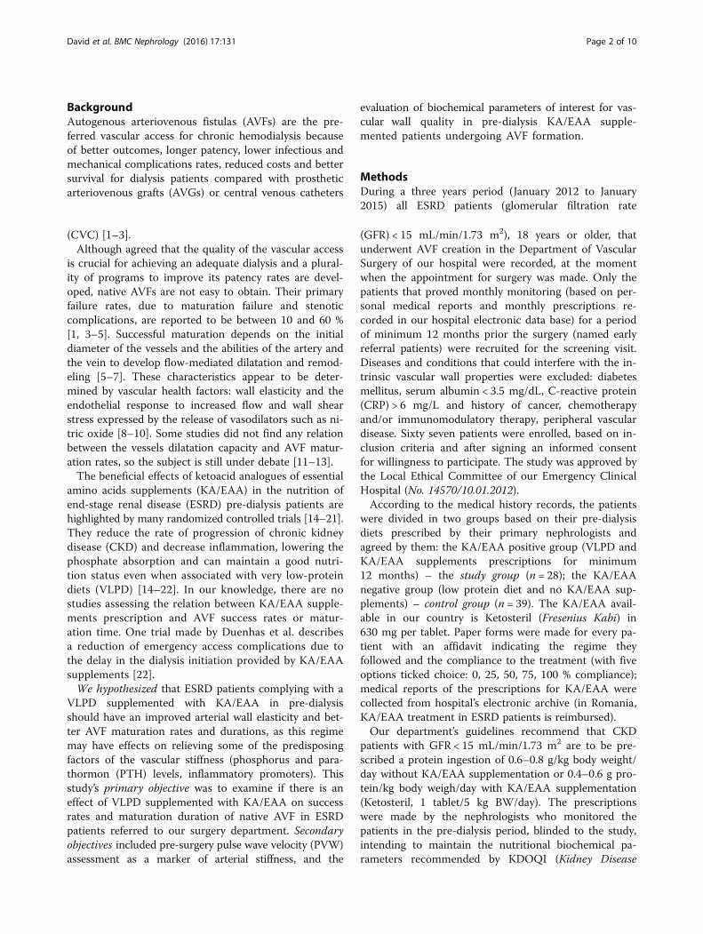
BackgroundAutogenous arteriovenous fistulas (AVFs) are the pre-ferred vascular access for chronic hemodialysis becauseof better outcomes, longer patency, lower infectious andmechanical complications rates, reduced costs and bettersurvival for dialysis patients compared with prostheticarteriovenous grafts (AVGs) or central venous catheters
(CVC) [1–3].Although agreed that the quality of the vascular access
is crucial for achieving an adequate dialysis and a plural-ity of programs to improve its patency rates are devel-oped, native AVFs are not easy to obtain. Their primaryfailure rates, due to maturation failure and stenoticcomplications, are reported to be between 10 and 60 %[1, 3–5]. Successful maturation depends on the initialdiameter of the vessels and the abilities of the artery andthe vein to develop flow-mediated dilatation and remod-eling [5–7]. These characteristics appear to be deter-mined by vascular health factors: wall elasticity and theendothelial response to increased flow and wall shearstress expressed by the release of vasodilators such as ni-tric oxide [8–10]. Some studies did not find any relationbetween the vessels dilatation capacity and AVF matur-ation rates, so the subject is still under debate [11–13].The beneficial effects of ketoacid analogues of essential
amino acids supplements (KA/EAA) in the nutrition ofend-stage renal disease (ESRD) pre-dialysis patients arehighlighted by many randomized controlled trials [14–21].They reduce the rate of progression of chronic kidneydisease (CKD) and decrease inflammation, lowering thephosphate absorption and can maintain a good nutri-tion status even when associated with very low-proteindiets (VLPD) [14–22]. In our knowledge, there are nostudies assessing the relation between KA/EAA supple-ments prescription and AVF success rates or matur-ation time. One trial made by Duenhas et al. describesa reduction of emergency access complications due tothe delay in the dialysis initiation provided by KA/EAAsupplements [22].We hypothesized that ESRD patients complying with a
VLPD supplemented with KA/EAA in pre-dialysisshould have an improved arterial wall elasticity and bet-ter AVF maturation rates and durations, as this regimemay have effects on relieving some of the predisposingfactors of the vascular stiffness (phosphorus and para-thormon (PTH) levels, inflammatory promoters). Thisstudy’s primary objective was to examine if there is aneffect of VLPD supplemented with KA/EAA on successrates and maturation duration of native AVF in ESRDpatients referred to our surgery department. Secondaryobjectives included pre-surgery pulse wave velocity (PVW)assessment as a marker of arterial stiffness, and the
evaluation of biochemical parameters of interest for vas-cular wall quality in pre-dialysis KA/EAA supple-mented patients undergoing AVF formation.
MethodsDuring a three years period (January 2012 to January2015) all ESRD patients (glomerular filtration rate
(GFR) < 15 mL/min/1.73 m2), 18 years or older, thatunderwent AVF creation in the Department of VascularSurgery of our hospital were recorded, at the momentwhen the appointment for surgery was made. Only thepatients that proved monthly monitoring (based on per-sonal medical reports and monthly prescriptions re-corded in our hospital electronic data base) for a periodof minimum 12 months prior the surgery (named earlyreferral patients) were recruited for the screening visit.Diseases and conditions that could interfere with the in-trinsic vascular wall properties were excluded: diabetesmellitus, serum albumin < 3.5 mg/dL, C-reactive protein(CRP) > 6 mg/L and history of cancer, chemotherapyand/or immunomodulatory therapy, peripheral vasculardisease. Sixty seven patients were enrolled, based on in-clusion criteria and after signing an informed consentfor willingness to participate. The study was approved bythe Local Ethical Committee of our Emergency ClinicalHospital (No. 14570/10.01.2012).According to the medical history records, the patients
were divided in two groups based on their pre-dialysisdiets prescribed by their primary nephrologists andagreed by them: the KA/EAA positive group (VLPD andKA/EAA supplements prescriptions for minimum12 months) – the study group (n = 28); the KA/EAAnegative group (low protein diet and no KA/EAA sup-plements) – control group (n = 39). The KA/EAA avail-able in our country is Ketosteril (Fresenius Kabi) in630 mg per tablet. Paper forms were made for every pa-tient with an affidavit indicating the regime theyfollowed and the compliance to the treatment (with fiveoptions ticked choice: 0, 25, 50, 75, 100 % compliance);medical reports of the prescriptions for KA/EAA werecollected from hospital’s electronic archive (in Romania,KA/EAA treatment in ESRD patients is reimbursed).Our department’s guidelines recommend that CKD
patients with GFR < 15 mL/min/1.73 m2 are to be pre-scribed a protein ingestion of 0.6–0.8 g/kg body weight/day without KA/EAA supplementation or 0.4–0.6 g pro-tein/kg body weigh/day with KA/EAA supplementation(Ketosteril, 1 tablet/5 kg BW/day). The prescriptionswere made by the nephrologists who monitored thepatients in the pre-dialysis period, blinded to the study,intending to maintain the nutritional biochemical pa-rameters recommended by KDOQI (Kidney Disease
David et al. BMC Nephrology (2016) 17:131 Page 2 of 10

Outcomes Quality Initiative) for ESRD patients [14]. Ac-cording to the same KDOQI guidelines, patients fromboth groups received phosphate binders, calcium andvitamin D supplements when necessary.The medical history was recorded and a set of biochem-
ical tests was taken at the first visit (one week priorto the surgery), along with the PWV measurementand a Doppler ultrasound (Doppler US) evaluation.At one week after the first visit AVF were created, bythe same surgeon. Pre-surgery US Doppler evaluationsfor AVF placement are a routine in our center forcases with unsatisfactory data on clinical examination.The intervention was considered suitable at an arterydiameter > 2 mm, a vein diameter > 2.5 mm, and itrespected the standard surgical procedure. In patientswith poor quality distal vessels we approach proximalsites from the beginning. 1 % lidocaine is used forlocal anesthesia and we do not prescribe perioperativeantibiotics. Our surgeon mostly uses the artery-sideto vein-end technique, mobilizing the vein to adaptthe artery. We do not ligate collateral veins in thecreation session. When needed, the two-steps superfi-cialization is performed: first the creation, than thetransposition of the AVF in another separate session.The management policy in our center for all the new
created AVF is the clinical examinations at 1, 4 and, ifneeded, 6 weeks after the surgery. Doppler US is pre-scribed only in clinical inconclusive cases. As we are aninitiation center for renal replacement therapy and weprovide patients for several centers in and aroundBucharest, further examinations usually are at the pre-scription of the center’s nephrologists.For the patients enrolled in the study, visits 2, 3, 4 and
5 consisted in clinical and Doppler US evaluations at 4,6, 8 and 12 weeks after the surgery. AVF maturation wasdefined with Doppler US measurements of diameter >0.6 cm and access flow > 600 mL/min [3]; failure cre-ation of the native AVF was considered the failed surgeryand failed maturation was defined when the above men-tioned criteria for success maturation were not fulfilled.When a patient fulfilled these criteria simultaneously, itwas recorded as a mature AVF at that interval of time(4, 6, 8 or 12 weeks).Biochemical determinations included albumin, calcium,
phosphorus, creatinine, C-reactive protein and cholesterollevels, determinations made at hospital laboratory using aMindray analyzer; for iPTH (intact parathormon) one pri-vate lab performing ECLIA (ElectrochemiluminescenceImmunoassay) determination was used.PWV measurements were done with the validated
oscillometric device Mobil-O-graph PWA device (Indus-trielle Entwicklung Medizintechnik, Germany) withincorporated IEM-Hypertension Management Software,handled by the same research technician who was
blinded to the clinical data. The device measures centralblood pressure (BP) values and displays them along withthe PWV values. All the guidelines for this examinationwere respected [23]. Briefly, patients were placed in aquiet room, in the seated position, and the cuff of thedevice was placed on the left arm – brachial artery; dataincluding age, weight and height, along with the smoker/nonsmoker status were introduced. The device makestwo recordings for BP values and then it measures thevelocity of arterial pulse wave based on the oscillationsdetected on the upper-arm cuff during systole anddisplaying it together with the normal range for eachpatient using the integrated PWV algorithm.Doppler ultrasound was performed with the same de-
vice (Aloha Cardiology P.C.) by one cardiologist, withthe patient in the upright seated position; two determi-nations were made in a session for a single data record-ing. To avoid turbulence determined accuracy variations,the flow was measured in the distal part of the access,with the transducer (a 7.0 MHz probe) in the longitu-dinal position; the diameter was measured with thetransducer perpendicular to the vessel [3]. Althoughmany studies recommend AVFs’ flow measurement inthe feeding artery, we performed the diameter and flowmeasurements in the vein, distal to the anastomosis(>2 cm), since the venous walls’ characteristics are sig-nificant for a good access and the post-procedural com-plications develop mostly at this level (stenosis, sclerosisor other narrowing causes) [24, 25].The primary outcome was to evaluate the differences
of the AVF success creation rates and maturation dura-tions between the study and the control group. Thesecondary outcome was to compare the arterial stiffness– evaluated using PWV determination – and biochem-ical profiles between the two groups.
Statistical analysisBaseline pre-surgery biochemical characteristics werecompared between the two groups using Fisher testfor categorical parameters (to validate data). Normaldistributed values were analyzed using the meanvalues and standard deviation. Parameters expressedby percentages were also compared using Chi-squaredtest. We used logistic regression to analyze the associ-ation between AVF maturation and KA/EAA adminis-tration, by comparing the average AVF maturationduration between the two groups, unadjusted andthen adjusted for systolic BP, C-reactive protein,PWV, and phosphorus values. All the considered pa-rameters in the logistic model were transformed inbinary variables. A p value < α = 0.05 was consideredstatistically significant, and data processing was per-formed using SPSS 16 software and Excel.
David et al. BMC Nephrology (2016) 17:131 Page 3 of 10

ResultsSixty seven subjects were monitored until the end of thisprospective observational study: 28 patients who re-ceived KA/EAA supplements for 12 months before thestudy enrollment (study group) and 39 patients withoutKA/EAA supplements in the last 12 months prior tostudy enrollment (control group). Three patients werelost to follow-up during the 3 months period and wereexcluded from the results. The diagram of patient’s en-rollment scheme is shown in Fig. 1.
Demographical data and other characteristicsDemographical data in the study and control groups areshown in Table 1, observing a mean age of 57 ± 12.35 yearsin the study group and 54 ± 11.7 in the control group, re-spectively (p < 0.001; SD = 2.471); additionally, 46.4 % inthe study group were female, 48.7 % in the control group,respectively (p < 0.001; SD = 2.298). In this table we alsoshowed other characteristics which can influence the ar-terial wall stiffness evolution and the PWV values – thenumber of smokers and the mean BP values (recordedduring the PWV determination) in each group of patients.There was no significant difference regarding the percent
of smokers in studied groups; BP values showed a signifi-cant difference between groups – p = 0.0141; SD =12.122.
AVFs outcomesThe primary outcome parameters – surgery results andcharacteristics of AVF maturation – are presented andcompared between the two groups in Table 2.A success rate of 83.33 % was registered in the cohort,
with an average maturation time of 6.57 +/− 2.64 weeks.Twenty two lower arm AVFs were performed in the studygroup – 6 radiocephalic (wrist) and 16 brachiocephalicfistulas (middle-arm). In the control group, 30 lower armAVFs were performed – 7 radiocephalic (wrist) and 23brachiocephalic fistulas (middle-arm). In 15 cases we wereforced to perform upper arm fistulas – 6 in the studygroup (21.4 %) and 9 in the control group (23.07 %). In 6cases we managed to save or improve the newly createdAVF (thrombosis, stenosis or collateral veins) by surgicalprocedures during the study (two cases in the study groupand four in the control group).Eighteen patients failed to achieve a suitable vascular
access for hemodialysis in the 3 months interval: 5 fromthe study group and 13 from the control group (p <
Fig. 1 Enrollment chart and study design scheme
David et al. BMC Nephrology (2016) 17:131 Page 4 of 10
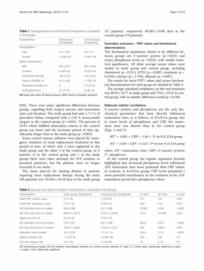
0.05). There were many significant differences betweengroups, regarding both surgery success and maturationrates and duration. The study group had only a 7.1 % ofprocedure failure compared with a 12.8 % unsuccessfulsurgery in the control group (p = 0.021). The percent ofAVF’s which fulfilled maturation criteria in the controlgroup was lower and the necessary period of time sig-nificantly longer than in the study group (p < 0.001).Seven central venous catheters were placed for emer-
gency initiation of renal replacement treatment in thisperiod of time, of which only 2 were registered in thestudy group and the other 5 in the control group. In 4patients (3 in the control group and 1 in the studygroup) there were other attempts for AVF creation, inproximal positions, but the patients were no longerrecorded in our study.The mean interval for starting dialysis in patients
requiring renal replacement therapy during the study(30 patients) was: 50.454 ± 24.18 days in the study group
(11 patients), respectively 49.263 ± 23.00 days in thecontrol group (19 patients).
Secondary outcomes – PWV values and biochemicaldeterminationsThe biochemical parameters found to be different be-tween groups are C-reactive protein (p = 0.021) andserum phosphorus levels (p = 0.022), with similar statis-tical significance. All other average serum values weresimilar in study group and control group, includingcholesterol (p = 0.311), iPTH (p = 0.269), creatinine (p =0.2324), calcium (p = 1.703), albumin (p = 0.887).The results for mean PWV values and serum biochem-
ical determinations for each group are detailed in Table 3.The average calculated compliance on diet and treatment
was 80.76 ± 23.77 in study group and 79.41 ± 23.41 in con-trol group, with no statistic differences noted (p = 0.158).
Elaborate statistic correlationsC-reactive protein and phosphorus are the only bio-chemical parameters that have directly influencedmaturation time, as it follows: in KA/EAA group, dueto lower levels of phosphorus and CRP, the matur-ation time was shorter than in the control group(Figs. 2 and 3):
MT ¼ 3:891� CRP þ 1:44� P–in KA=EAA group
MT ¼ 1:624� CRP þ 6:123� P–in non−KA=EAA group
where MT =maturation time; CRP = C-reactive protein;P = phosphorus.In the control group, the logistic regression formula
highlighted that increased phosphorus levels influencedAVF maturation time more profound than CRP values.In contrast, in KA/EAA group, CRP levels presented amore powerful contribution on the evolution of the AVFmaturation period than phosphorus values.
Table 1 Demographical and biochemical characteristics comparedin the groups
Characteristics Study group(28 patients)
Control group(39 patients)
Demographics
Age 57 ± 12.35 54 ± 11.7
Female 13 (46.4 %) 19 (48.7 %)
Other characteristics
BMI 28.3 ± 6.13 28.1 ± 6.08
Smokers 8 (28.5 %) 10 (25.6 %)
Systolic BP (mmHg) 148 ± 7.79 154 ± 6.58
Vitamin D/VDRA (n) 6 (21.4 %) 11 (28.2 %)
Phosphorus binders (n) 0 5 (12.8 %)
Antihypertensive 21 (75 %) 32 (82 %)
BMI body mass index, BP blood pressure, VDRA vitamin D receptor activation
Table 2 Vascular and dialysis initiation characteristics compared in the groups
Characteristics Study group (28 patients) Control group (39 patients) χ2 value SD-value p-value
Failed AVF creation cases 2 (7.1 %) 5 (12.8 %) 3.11 2.01 0.021
Failed AVF maturation cases 3 (10.7 %) 8 (20.5 %) 2.82 3.19 0.017
AVF diameter (cm) at 4 weeks 0.60 ± 0.09 0.51 ± 0.08 73.396 0.226 <0.001
AVF flow (mL/min) at 4 weeks 698.07 ± 136.13 579.12 ± 105.84 317.2 337.034 0.017
Upper arm AVF (n) 6 (21.4 %) 9 (23.1 %)
AVF diameter (cm) at 3 months 0.70 ± 0.07 0.67 ± 0.08 84.35 0.191 <0.001
AVF flow (mL/min) at 3 months 756.92 ± 136.62 722.65 ± 122.25 364 338.25 0.007
Maturation time (weeks) 5.91 ± 0.92 7.15 ± 1.19 34.45 2.775 <0.001
Dialysis initiation (%) 11 (39.3 %) 19 (48.7 %) 6.31 5.61 0.5
CVC-HD initiation (%) 2 (7.1 %) 5 (12.8 %) 4.5 2.121 0.5
AVF arteriovenous fistula, CVC-HD initiation hemodialysis initiation on central venous catheter, χ2 value > χ2 critical value, statistically significant, p-value< α-value = 0.05, statistically significant
David et al. BMC Nephrology (2016) 17:131 Page 5 of 10

When applying linear regression, regarding the influ-ence of AVF diameter and flow on maturation time, at4 weeks and 3 months, respectively, it could be demon-strated that, in the study group, the AVF diameter had amore significant impact on the maturation period thanAVF flow, especially at 4 weeks. In contrast, in thecontrol group, both AVF diameter and flow equallyinfluenced the maturation time (Table 4).Furthermore, when comparing the influence of BP on
PWV values (χ2 = 21.44, p < 0.001 in the study group,and 22.78, p < 0.001 in the control group), we noticedthat BP had a higher influence on PWV levels in thecontrol group than in the study group (0.143 versus0.137 – correlation coefficient).
DiscussionsThis study focuses on the impact of KA/EAA supple-mented VLPD on the vascular wall quality and on thechances for a successful vascular access creation. At themoment of fistula surgery we analyzed the differences ofseveral biochemical parameters considered at interestbetween a VLPD KA/EAA supplemented group ofpatients (n = 28) and a conventional low protein diet,non-KA/EAA, control group (n = 39). The repartition ofthe patients in the two groups was made based on theirdocumented history (the registered prescriptions for
keto-analogues, a reimbursed treatment for ESRD patientsin our country). After the vascular access creation, we ana-lyzed AVF characteristics and maturation process in all thesurgery-successful patients (60 cases): 26 patients in thestudy group and 34 patients in the control group.There is no consensus on the criteria to define fistula
maturation. We chose the KDOQI definition, accordingto which an adequate AVF has a flow > 600 mL/min anda diameter of the vein of > 0.6 cm [3, 26]. One third toover a half of fistula creation surgery procedures world-wide resides in failures – failure of creation, delayedmaturation or early fistula failure [8, 13, 26, 27]. In ourstudy, the percent of failure was 26.86 % in whole group,a reasonable percent considering that the study cohortwas made of monthly monitored patients for at least oneyear before renal replacement therapy preparations. TheAVF maturation rate was 83.33 % (50 patients) and themean AVF maturation duration in the whole group was6.57 ± 2.64 weeks, with pronounced differences betweencontrol group and the KA/EAA group (p < 0.001).We also evaluated the situation of the vascular access at
the 4 weeks measurement, when an AVF flow > 500 mL/min and a vein diameter after the anastomoses of > 0.4 cmcan predict the future AVF adequacy [24, 28, 29]. At thatpoint we noted that a difference already exists betweenthe two groups, and it deepens at 6 weeks and at
Table 3 Significant statistical differences between the studied groups (KA/EAA group and control group) regarding the analyzedparameters
Characteristics Study group (28 patients) Control group (39 patients) χ2 value SD-value p-value
Creatinine (mg/dL) 6.9 ± 0.16 7.0 ± 0.15 8.27 0.416 0.2324
Calcium (mg/dL) 9.1 ± 0.15 8.8 ± 0.11 3.867302 0.389 1.703
Phosphorus (mg/dL) 4.0 ± 0.19 5.2 ± 0.33 253.2 0.608 0.022
iPTH (pg/mL) 118 ± 25.56 129 ± 27.95 0.625 56.061 0.269
C-reactive protein (mg/L) 1.2 ± 0.38 2.3 ± 0.48 222.8 0.988 0.021
Albumin (g/dL) 4.2 ± 0.11 3.9 ± 0.10 13.93 0.278 0.887
Cholesterol (mg/dL) 206 ± 44.63 214 ± 46.17 0.48 44.35 0.311
PWV [90 % CI], (m/s) 9.39 ± 0.83 10.61 ± 1.07 513.5 2.741 0.007
iPTH intact parathormon, PWV pulse wave velocity, χ2 value > χ2 critical value, statistically significant, p-value < α-value = 0.05, statistically significant
Fig. 2 The influence of phosphorus values on AVF maturation time trend in both groups
David et al. BMC Nephrology (2016) 17:131 Page 6 of 10

12 weeks measurements. This is in contrast with thefindings of Robbin et al. who determined no signifi-cant improvements in the development of AVF in thesecond and third months [24]. A gradual continuous in-crease in AVF blood flow until 12 weeks is sustained byother studies [8, 29, 30]. At the end of the study, a higherpercent of failures in AVF creation was recorded in thenon-KA/EAA control group (33.3 % versus 17.8 %). Wepropose some explanations for the differences registeredbetween the studied groups.For fistulas to mature, the vessels have to dilate: their
diameters must increase by 40–60 % and their flows 10–20 times [5, 8, 12, 13, 30, 31]. In ESRD patients thisprocess is hampered by an increased wall stiffness, dueto medial thickening and calcifications plus intimal in-flammation and hyperplasia [12, 13, 32–36].The elasticity of the vessel wall is defined by its char-
acteristics: the quality of its constituents, de degree ofthe stiffness and vascular calcifications [8, 37]. Arterialstiffness is recognized as an important factor of vascularhealth [38]. Its levels were widely analyzed in relation tothe cardiovascular risk in CKD patients [39]. There aremany indicators proposed to predict the dilatation cap-acity and, through it, the AVF success: PVW, peripheralarterial tonometry (PAT), flow mediated dilation (FMD).
None of them proved their supremacy [40]. PWV is themost important indicative for global vascular stiffnessbut it was rarely explored in ESRD patients as a predictorof AVF success.We determined PWV values in both groups of patients
and we found them lower than other data from the litera-ture, probably due to the enrollment policy – early referralpatients monitored for minimum 12 months [41, 42].Nevertheless, values were significantly different be-tween the two groups (p = 0.007). It appears that VLPDand KA/EAA administration had a protective effectagainst vascular stiffness.Analyzing the biochemical parameters list, the variable
that is significantly different between groups and can beassociated with arterial stiffness by attending vascularcalcifications is the serum phosphorus level. Phosphorusplays a central role as a promoter in vascular inflamma-tory processes, affecting vascular wall elasticity andstimulating vascular calcifications [43–45]. VLPD andKA/EAA supplements ameliorate calcium-phosphate de-regulations in ESRD by lowering serum phosphoruslevels through several mechanisms. Initially, the smallerthe protein intake, the lower the amount of phosphatesprovided for absorption. Secondly, the calcium salts ofketo analogues in the KA/EAA composition acts like
Fig. 3 The influence of CRP values on AVF maturation time trend in both groups
Table 4 The influence of AVF diameter and flow at 4 weeks and 3 months on maturation time in both groups
Parameters Study group (28 patients) Control group (39 patients)
Coefficient p-value Coefficient p-value
AVF flow at 4 weeks 0.582 0.415 0.876 0.013
AVF diameter at 4 weeks −1.099 0.012 −1.330 0.001
AVF flow at 3 months 0.462 0.558 −0.063 0.822
AVF diameter at 3 months −0.755 0.095 −0.371 0.232
AVF arteriovenous fistula
David et al. BMC Nephrology (2016) 17:131 Page 7 of 10
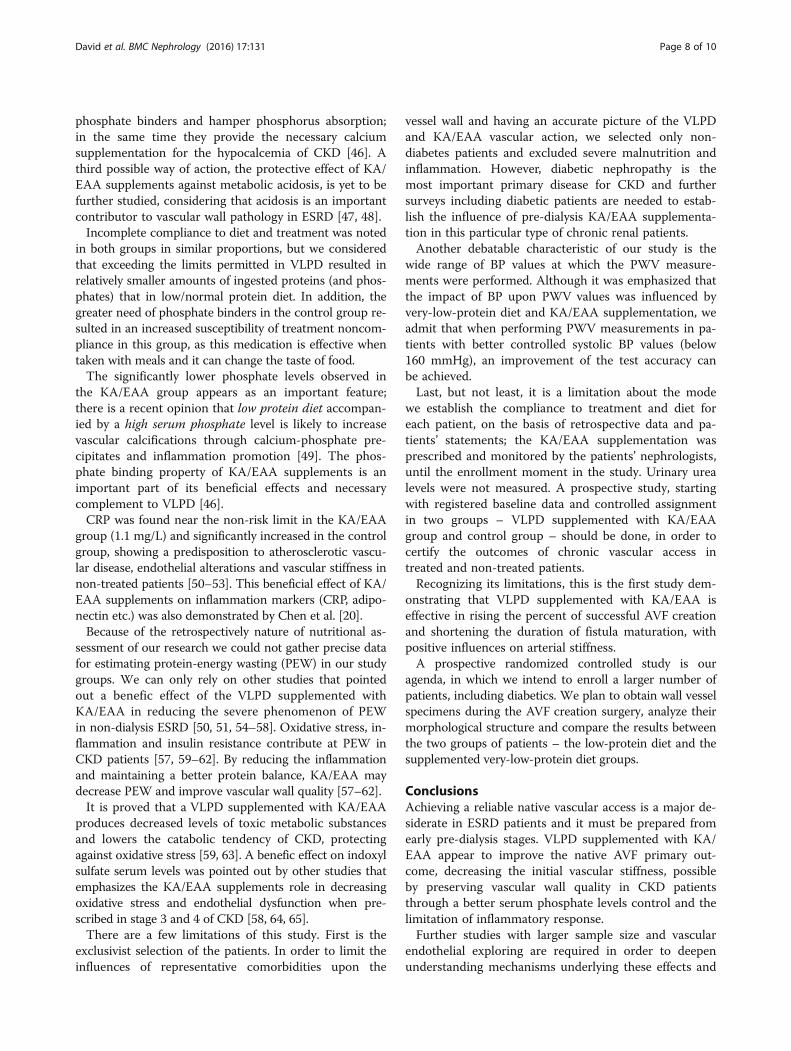
phosphate binders and hamper phosphorus absorption;in the same time they provide the necessary calciumsupplementation for the hypocalcemia of CKD [46]. Athird possible way of action, the protective effect of KA/EAA supplements against metabolic acidosis, is yet to befurther studied, considering that acidosis is an importantcontributor to vascular wall pathology in ESRD [47, 48].Incomplete compliance to diet and treatment was noted
in both groups in similar proportions, but we consideredthat exceeding the limits permitted in VLPD resulted inrelatively smaller amounts of ingested proteins (and phos-phates) that in low/normal protein diet. In addition, thegreater need of phosphate binders in the control group re-sulted in an increased susceptibility of treatment noncom-pliance in this group, as this medication is effective whentaken with meals and it can change the taste of food.The significantly lower phosphate levels observed in
the KA/EAA group appears as an important feature;there is a recent opinion that low protein diet accompan-ied by a high serum phosphate level is likely to increasevascular calcifications through calcium-phosphate pre-cipitates and inflammation promotion [49]. The phos-phate binding property of KA/EAA supplements is animportant part of its beneficial effects and necessarycomplement to VLPD [46].CRP was found near the non-risk limit in the KA/EAA
group (1.1 mg/L) and significantly increased in the controlgroup, showing a predisposition to atherosclerotic vascu-lar disease, endothelial alterations and vascular stiffness innon-treated patients [50–53]. This beneficial effect of KA/EAA supplements on inflammation markers (CRP, adipo-nectin etc.) was also demonstrated by Chen et al. [20].Because of the retrospectively nature of nutritional as-
sessment of our research we could not gather precise datafor estimating protein-energy wasting (PEW) in our studygroups. We can only rely on other studies that pointedout a benefic effect of the VLPD supplemented withKA/EAA in reducing the severe phenomenon of PEWin non-dialysis ESRD [50, 51, 54–58]. Oxidative stress, in-flammation and insulin resistance contribute at PEW inCKD patients [57, 59–62]. By reducing the inflammationand maintaining a better protein balance, KA/EAA maydecrease PEW and improve vascular wall quality [57–62].It is proved that a VLPD supplemented with KA/EAA
produces decreased levels of toxic metabolic substancesand lowers the catabolic tendency of CKD, protectingagainst oxidative stress [59, 63]. A benefic effect on indoxylsulfate serum levels was pointed out by other studies thatemphasizes the KA/EAA supplements role in decreasingoxidative stress and endothelial dysfunction when pre-scribed in stage 3 and 4 of CKD [58, 64, 65].There are a few limitations of this study. First is the
exclusivist selection of the patients. In order to limit theinfluences of representative comorbidities upon the
vessel wall and having an accurate picture of the VLPDand KA/EAA vascular action, we selected only non-diabetes patients and excluded severe malnutrition andinflammation. However, diabetic nephropathy is themost important primary disease for CKD and furthersurveys including diabetic patients are needed to estab-lish the influence of pre-dialysis KA/EAA supplementa-tion in this particular type of chronic renal patients.Another debatable characteristic of our study is the
wide range of BP values at which the PWV measure-ments were performed. Although it was emphasized thatthe impact of BP upon PWV values was influenced byvery-low-protein diet and KA/EAA supplementation, weadmit that when performing PWV measurements in pa-tients with better controlled systolic BP values (below160 mmHg), an improvement of the test accuracy canbe achieved.Last, but not least, it is a limitation about the mode
we establish the compliance to treatment and diet foreach patient, on the basis of retrospective data and pa-tients’ statements; the KA/EAA supplementation wasprescribed and monitored by the patients’ nephrologists,until the enrollment moment in the study. Urinary urealevels were not measured. A prospective study, startingwith registered baseline data and controlled assignmentin two groups – VLPD supplemented with KA/EAAgroup and control group – should be done, in order tocertify the outcomes of chronic vascular access intreated and non-treated patients.Recognizing its limitations, this is the first study dem-
onstrating that VLPD supplemented with KA/EAA iseffective in rising the percent of successful AVF creationand shortening the duration of fistula maturation, withpositive influences on arterial stiffness.A prospective randomized controlled study is our
agenda, in which we intend to enroll a larger number ofpatients, including diabetics. We plan to obtain wall vesselspecimens during the AVF creation surgery, analyze theirmorphological structure and compare the results betweenthe two groups of patients – the low-protein diet and thesupplemented very-low-protein diet groups.
ConclusionsAchieving a reliable native vascular access is a major de-siderate in ESRD patients and it must be prepared fromearly pre-dialysis stages. VLPD supplemented with KA/EAA appear to improve the native AVF primary out-come, decreasing the initial vascular stiffness, possibleby preserving vascular wall quality in CKD patientsthrough a better serum phosphate levels control and thelimitation of inflammatory response.Further studies with larger sample size and vascular
endothelial exploring are required in order to deepenunderstanding mechanisms underlying these effects and
David et al. BMC Nephrology (2016) 17:131 Page 8 of 10

certify the role of VLPD supplemented with KA/EAA inrelieving vascular stiffness and ameliorating AVFs matur-ation rates and durations.
AbbreviationsAVFs: Arteriovenous fistulas; AVGs: Arteriovenous grafts; BMI: Body massindex; BP: Blood pressure; CKD: Chronic kidney disease; CRP: C-reactiveprotein; CVC: Central venous catheters; CVC-HD initiation: Hemodialysisinitiation on central venous catheter; Doppler US: Doppler ultrasound;ECLIA: Electrochemiluminescence immunoassay; ESRD: End-stage renaldisease; FMD: Flow mediated dilation; GFR: Glomerular filtration rate;iPTH: Intact parathormon; KA/EAA: Ketoacid analogues of essential aminoacids supplements; KDOQI: Kidney Disease Outcomes Quality Initiative;PAT: Peripheral arterial tonometry; PEW: Protein-energy wasting;PTH: Parathormon; PVW: Pulse wave velocity; VDRA: Vitamin D receptoractivation; VLPD: Very low-protein diets
AcknowledgementsWe would like to thank all participants for their cooperation. We are gratefulto cardiologist Găvănescu Mădălina for performing all US Doppler examinations,and to vascular surgeon Elfarra Mazen for his contribution.
FundingNo funding was received for writing this study.
Availability of data and materialsAll data supporting this manuscript are presented in the article or availableupon request from the corresponding author, Peride I.
Authors’ contributionsCD: conception and design; interpretation of analyzed data; provision ofstudy patients; manuscript writing. PI: conception and design; interpretationof analyzed data; provision of study patients; manuscript writing. NA: conceptionand design; interpretation of analyzed data; provision of study patients;manuscript writing. CAM: statistical analysis of collected data and resultstabulation. CIA: conception and design; interpretation of analyzed data;provision of study patients; manuscript writing. All authors read and approvedthe final manuscript and agreed on its submission.
Competing interestsPotential competing interests declared by Cristiana DAVID and AndreiNICULAE, who received congress attendance fees to Romanian NationalCongress of Nephrology from Fresenius Kabi in November 2015.
Consent for publicationAs only summary average data are presented, consent of publication forpatient is not applicable.
Ethics approval and consent to participateThe study protocol was approved by the Ethics Committee of “St. John”Emergency Clinical Hospital Bucharest. The study design was described toresponders and they signed a consent to participate.
Author details1Clinical Department No. 3, “Carol Davila” University of Medicine andPharmacy Bucharest, 37th Dionisie Lupu Street, 020021 Sector 2, Bucharest,Romania. 2Department of Nephrology and Dialysis, “St. John” EmergencyClinical Hospital Bucharest, Bucharest, Romania. 3Bucharest University ofEconomic Studies, Bucharest, Romania.
Received: 10 May 2016 Accepted: 8 September 2016
References1. Kim SM, Han Y, Kwon H, Hong HS, Choi JY, Park H, Kwon TW, Cho YP. Impact
of a preoperative evaluation on the outcomes of an arteriovenous fistula.Ann Surg Treat Res. 2016;90(4):224–30.
2. Lafrance JP, Rahme E, Lelorier J, Iqbal S. Vascular access-related infections:definitions, incidence rates, and risk factors. Am J Kidney Dis. 2008;52(5):982–93.
3. Foundation NK. KDOQI clinical practice guidelines for vascular access: update2006. Am J Kidney Dis. 2006;48 Suppl 1:S176–322.
4. Schinstock CA, Albright RC, Williams AW, Dillon JJ, Bergstralh EJ, Jenson BM,McCarthy JT, Nath KA. Outcomes of arteriovenous fistula creation after theFistula First Initiative. Clin J Am Soc Nephrol. 2011;6(8):1996–2002.
5. van der Linden J, Lameris TW, van den Meiracker AH, de Smet AA, BlankestijnPJ, van den Dorpel MA. Forearm venous distensibility predicts successfularteriovenous fistula. Am J Kidney Dis. 2006;47(6):1013–9.
6. Allon M, Litovsky S, Young CJ, Deierhoi MH, Goodman J, Hanaway M, LockhartME, Robbin ML. Medial fibrosis, vascular calcification, intimal hyperplasia, andarteriovenous fistula maturation. Am J Kidney Dis. 2011;58(3):437–43.
7. KhavaninZadeh M, Gholipour F, Naderpour Z, Porfakharan M. Relationshipbetween Vessel Diameter and Time to Maturation of Arteriovenous Fistulafor Hemodialysis Access. Int J Nephrol. 2012;2012:942950.
8. Dixon BS. Why don’t fistula mature? Kidney Int. 2006;70(8):1413–22.9. MacRae JM, Ahmed S, Hemmelgarn B, Sun Y, Martin BJ, Roifman I, Anderson
T. Role of vascular function in predicting arteriovenous fistula outcomes: anobservational pilot study. Can J Kidney Health Dis. 2015;2:19.
10. Verbeke FH, Pannier B, Guérin AP, Boutouyrie P, Laurent S, London GM.Flow-mediated vasodilation in end-stage renal disease. Clin J Am SocNephrol.2011;6(8):2009–15.
11. Mitchell GF, Parise H, Vita JA, Larson MG, Warner E, Keaney Jr JF, Keyes MJ, LevyD, Vasan RS, Benjamin EJ. Local shear stress and brachial artery flow-mediateddilation: the Framingham Heart Study. Hypertension. 2004;44(2):134–9.
12. Allon M, Ornt DB, Schwab SJ, Rasmussen C, Delmez JA, Greene T, Kusek JW,Martin AA, Minda S. Factors associated with the prevalence of arteriovenousfistulas in hemodialysis patients in the HEMO study. Hemodialysis (HEMO)Study Group. Kidney Int. 2000;58(5):2178–85.
13. Kheda MF, Brenner LE, Patel MJ, Wynn JJ, White JJ, Prisant LM, Jones SA, PaulsonWD. Influence of arterial elasticity and vessel dilatation on arteriovenous fistulamaturation: a prospective cohort study. Nephrol Dial Transpl. 2010;25(2):525–31.
14. Clinical practice guidelines for nutrition in chronic renal failure. K/DOQI,National Kidney Foundation. Am J Kidney Dis. 2000;35(6 Suppl 2):S1-S140.
15. Bellizzi V, Chiodini P, Cupisti A, Viola BF, Pezzotta M, De Nicola L, Minutolo R,Barsotti G, Piccoli GB, Di Iorio B. Very low-protein diet plus ketoacids inchronic kidney disease and risk of death during end-stage renal disease: ahistorical cohort controlled study. Nephrol Dial Transplant. 2015;30(1):71–7.
16. Khan IA, Nasiruddin M, Haque SF, Khan RA. A randomized clinical trial to evaluatethe efficacy and safety of α-keto amino acids supplementation in stage 3 and 4of chronic kidney disease. Asian J Pharm Clin Res. 2014;7(3):21–4.
17. Walser M. Does prolonged protein restriction preceding dialysis lead toprotein malnutrition at the onset of dialysis? Kidney Int. 1993;44(5):1139–44.
18. Feiten SF, Draibe SA, Watanabe R, Duenhas MR, Baxmann AC, Nerbass FB,Cuppari L. Short-term effects of a very-low-protein diet supplemented withketoacids in nondialyzed chronic kidney disease patients. Eur J Clin Nutr.2005;59(1):129–36.
19. Brunori G, Viola BF, Parrinello G, De Biase V, Como G, Franco V, Garibotto G,Zubani R, Cancarini GC. Efficacy and safety of a very-low-protein diet whenpostponing dialysis in the elderly: a prospective randomized multicentercontrolled study. Am J Kidney Dis. 2007;49(5):569–80.
20. Chen N, Jin Y, Ren H, Xu J, Shen P, Huang X. Anti-inflammatory effects oflow protein diet supplemented with keto-amino acid in the treatment oftype 2 diabetic nephropathy. Kidney Res Clin Pract. 2012;31(2):A24.
21. Shah AP, Kalantar-Zadeh K, Kopple JD. Is there a role for ketoacid supplementsin the management of CKD? Am J Kidney Dis. 2015;65(5):659–73.
22. Duenhas M, Gonçalves E, Dias M, Leme G, Laranja S. Reduction of morbidityrelated to emergency access to dialysis with very low protein dietsupplemented with ketoacids (VLPD + KA). Clin Nephrol. 2013;79(5):387–93.
23. Van Bortel LM, Duprez D, Starmans-Kool MJ, Safar ME, Giannattasio C, CockroftJ, Kaiser DR, Thuillez C. Clinical applications of arterial stiffness. Task Force III:recommendations for user procedures. Am J Hypertens. 2002;15(5):445–52.
24. Robbin ML, Chamberlain NE, Lockhart ME, Gallichio MH, Young CJ, DeierhoiMH, Allon M. Hemodialysis arteriovenous fistula maturity: US evaluation.Radiology. 2002;225(1):59–64.
25. Wiese P, Nonnast-Daniel B. Colour Doppler ultrasound in dialysis access.Nephrol Dial Transplant. 2004;19(8):1956–63.
26. Zangan MS, Falk A. Optimizing arteriovenous fistula maturation. SeminIntervent Radiol. 2009;26(2):144–50.
27. Patel ST, Hughes J, Mills JRSR. Failure of arteriovenous fistula maturation: anunintended consequence of exceeding dialysis outcome quality Initiativeguidelines for hemodialysis access. J Vasc Surg. 2003;38(3):439–45.
28. Beathard GA. Maturation and evaluation of the newly created hemodialysisarteriovenous fistula. UpToDate. 2014; http://www.uptodate.com/contents/
David et al. BMC Nephrology (2016) 17:131 Page 9 of 10

maturation-and-evaluation-of-the-newly-created-hemodialysis-arteriovenous-fistula. Accessed 19 Oct 2015.
29. Corpataux JM, Haesler E, Silacci P, Ris HB, Hayoz D. Low-pressure environmentand remodelling of the forearm vein in Brescia-Cimino haemodialysis access.Nephrol Dial Transplant. 2002;17(6):1057–62.
30. Lomonte C, Casucci F, Antonelli M, Giammaria B, Losurdo N, Marchio G,Basile C. Is there a place for duplex screening of the brachial artery in thematuration of arteriovenous fistulas? Semin Dial. 2005;18(3):243–6.
31. Sidawy AN, Gray R, Besarab A, Henry M, Ascher E, Silva Jr M, Miller A, ScherL, Trerotola S, Gregory RT, Rutherford RB, Kent KC. Recommended standardsfor reports dealing with arteriovenous hemodialysis accesses. J Vasc Surg.2002;35(3):603–10.
32. Back MR, Maynard M, Winkler A, Bandyk DF. Expected flow parameterswithin hemodialysis access and selection for remedial intervention ofnonmaturing conduits. Vasc Endovascular Surg. 2008;42(2):150–8.
33. London GM, Marchais SJ, Guerin AP, Metivier F, Adda H. Arterial structure andfunction in end-stage renal disease. Nephrol Dial Transplant. 2002;17(10):1713–24.
34. Nemcsik J, Kiss I, Tislér A. Arterial stiffness, vascular calcification and bonemetabolism in chronic kidney disease. World J Nephrol. 2012;1(1):25–34.
35. Palit S, Kendrick J. Vascular calcification in chronic kidney disease: role ofdisordered mineral metabolism. Curr Pharm Des. 2014;20(37):5829–33.
36. Hunt JL, Fairman R, Mitchell ME, Carpenter JP, Golden M, Khalapyan T,Wolfe M, Neschis D, Milner R, Scoll B, Cusack A, Mohler 3rd ER. Boneformation in carotid plaques: a clinicopathological study. Stroke. 2002;33(5):1214–9.
37. Bonetti PO, Lerman LO, Lerman A. Endothelial dysfunction: a marker ofatherosclerotic risk. Arterioscler Thromb Vasc Biol. 2003;23(2):168–75.
38. Taal MW. Arterial stiffness in chronic kidney disease: an update. Curr OpinNephrol Hypertens. 2014;23(2):169–73.
39. Karras A, Haymann JP, Bozec E, Metzger M, Jacquot C, Maruani G, HouillierP, Froissart M, Stengel B, Guardiola P, Laurent S, Boutouyrie P, Briet M,Nephro Test Study Group. Large artery stiffening and remodeling areindependently associated with all-cause mortality and cardiovascular eventsin chronic kidney disease. Hypertension. 2012;60(6):1451–7.
40. McGrogan DG, Maxwell AP, Khawaja AZ, Inston NG. Current tools for predictionof arteriovenous fistula outcomes. Clin Kidney J. 2015;8(3):282–9.
41. Townsend RR, Wimmer NJ, Chirinos JA, Parsa A, Weir M, Perumal K, Lash JP,Chen J, Steigerwalt SP, Flack J, Go AS, Rafey M, Rahman M, Sheridan A,Gadegbeku CA, Robinson NA, Joffe M. Aortic PWV in chronic kidney disease:a CRIC ancillary study. Am J Hypertens. 2010;23(3):282–9.
42. Townsend RR. Arterial stiffness and chronic kidney disease: lessons from theChronic Renal Insufficiency Cohort study. Curr Opin Nephrol Hypertens.2015;24(1):47–53.
43. Zoungas S, Cameron JD, Kerr PG, Wolfe R, Muske C, McNeil JJ, McGrath BP.Association of carotid intima-medial thickness and indices of arterial stiffness withcardiovascular disease outcomes in CKD. Am J Kidney Dis. 2007;50(4):622–30.
44. Yamada S, Tokumoto M, Tatsumoto N, Taniguchi M, Noguchi H, Nakano T,Masutani K, Ooboshi H, Tsuruya K, Kitazono T. Phosphate overload directlyinduces systemic inflammation and malnutrition as well as vascularcalcification in uremia. Am J Physiol Renal Physiol. 2014;306(12):F1418–28.
45. Yamada S, Tatsumoto N, Tokumoto M, Noguchi H, Ooboshi H, Kitazono T,Tsuruya K. Phosphate binders prevent phosphate-induced cellular senescenceof vascular smooth muscle cells and vascular calcification in a modified,adenine-based uremic rat model. Calcif Tissue Int. 2015;96(4):347–58.
46. Schaefer K, Erley CM, von Herrath D, Stein G. Calcium salts of ketoacids as a newtreatment strategy for uremic hyperphosphatemia. Kidney Int. 1989;27:S136–9.
47. Bushinsky DA, Ori Y. Effects of metabolic and respiratory acidosis on bone.Curr Opin Nephrol Hypertens. 1993;2(4):588–96.
48. Verove C, Maisonneuve N, El AA, Boldron A, Azar R. Effect of the correctionof metabolic acidosis on nutritional status in elderly patients with chronicrenal failure. J Ren Nutr. 2002;12(4):224–8.
49. Yamada S, Tokumoto M, Tsuruya K, Tatsumoto N, Noguchi H, Kitazono T,Ooboshi H. Fetuin-A decrease induced by a low-protein diet enhancesvascular calcification in uremic rats with hyperphosphatemia. Am J RenalPhyisiol. 2015;309(8):F744–54.
50. CP, Kopple JD, Kalantar-Zadeh K. Inflammation in renal insufficiency. UpToDate.2015; http://www.uptodate.com/contents/inflammation-in-renal-insufficiency.com. Accessed 21 Oct 2015.
51. Singh AK. Protein restriction and progression of chronic kidney disease.UpToDate. 2014; http://www.uptodate.com/contents/protein-restriction-and-progression-of-chronic-kidney-disease.com. Accessed 21 Oct 2015.
52. Lacson Jr E, Levin NW. C-reactive protein and end-stage renal disease.Semin Dial. 2004;17(6):438–48.
53. van der Sande FM, Kooman JP, Leunissen KM. The predictive value of C-reactive protein in end-stage renal disease: is it clinically significant? BloodPurif. 2006;24(4):335–41.
54. Rattazzi M, Puato M, Faggin E, Bertipaglia B, Grego F, Pauletto P. Newmarkers of accelerated atherosclerosis in end-stage renal disease. J Nephrol.2003;16(1):11–20.
55. Wang DT, Lu L, Shi Y, Geng ZB, Yin Y, Wang M, Wei LB. Supplementation ofketoacids contributes to the up-regulation of the Wnt7a/Akt/p70S6K pathwayand the down-regulation of apoptotic and ubiquitin-proteasome systems inthe muscle of 5/6 nephrectomised rats. Br J Nutr. 2014;111(9):1536–48.
56. Wang XH, Mitch WE. Mechanisms of muscle wasting in chronic kidneydisease. Nat Rev Nephrol. 2014;10(9):504–16.
57. Kovesdy CP, Kopple JD, Kalantar-Zadeh K. Management of protein-energywasting in non-dialysis-dependent chronic kidney disease: reconciling lowprotein intake with nutritional therapy. Am J Clin Nutr. 2013;97(6):1163–77.
58. Piccoli GB, Vigotti FN, Leone F, Capizzi I, Daidola G, Cabiddu G, Avagnina P.Low-protein diets in CKD: how can we achieve them? A narrative,pragmatic review. Clin Kidney J. 2015;8(1):61–70.
59. Guarnieri G, Barazzoni R. Fighting protein-energy wasting in chronic kidneydisease: a challenge of complexity. J Ren Nutr. 2011;21(1):2–6.
60. Baldassarre D, De Jong A, Amato M, Werba JP, Castelnuovo S, Frigerio B, Veglia F,Tremoli E, Sirtori CR. Carotid intima-media thickness and markers of inflammation,endothelial damage and hemostasis. Ann Med. 2008;40(1):21–44.
61. Perunicić-Peković G, Rasić-Milutinović Z, Pljesa S. Predictors of mortality indialysis patients–association between malnutrition, inflammation andatherosclerosis (MIA syndrome). Med Pregl. 2004;57(3–4):149–52.
62. Ikizler TA, Cano NJ, Franch H, Fouque D, Himmelfarb J, Kalantar-Zadeh K,Kuhlmann MK, Stenvinkel P, TerWee P, Teta D, Wang AY, Wanner C,International Society of Renal Nutrition and Metabolism. Prevention andtreatment of protein energy wasting in chronic kidney disease patients: aconsensus statement by the International Society of Renal Nutrition andMetabolism. Kidney Int. 2013;84(6):1096–107.
63. Gao X, Wu J, Dong Z, Hua C, Hu H, Mei C. A low-protein diet supplementedwith ketoacids plays a more protective role against oxidative stress of ratkidney tissue with 5/6 nephrectomy than a low-protein diet alone. Br JNutr. 2010;103(4):608–16.
64. Niwa T, Ise M. Indoxyl sulfate, a circulating uremic toxin, stimulates theprogression of glomerular sclerosis. J Lab Clin Med. 1994;124(1):96–104.
65. Marzocco S, Dal Piaz F, Di Micco L, Torraca S, Sirico ML, Tartaglia D, AutoreG, Di Iorio B. Very low protein diet reduces indoxyl sulfate levels in chronickidney disease. Blood Purif. 2013;35(1–3):196–201.
• We accept pre-submission inquiries
• Our selector tool helps you to find the most relevant journal
• We provide round the clock customer support
• Convenient online submission
• Thorough peer review
• Inclusion in PubMed and all major indexing services
• Maximum visibility for your research
Submit your manuscript atwww.biomedcentral.com/submit
Submit your next manuscript to BioMed Central and we will help you at every step:
David et al. BMC Nephrology (2016) 17:131 Page 10 of 10
Copyright © 2022 FDOKUMEN