Clinical Impact of Left Ventricular Hypertrophy and Implications for Regression
15
Clinical Impact of Left Ventricular Hypertrophy and Implications for Regression Surya M. Artham, Carl J. Lavie ⁎ , Richard V. Milani, Dharmendrakumar A. Patel, Anil Verma, Hector O. Ventura Ochsner Medical Center, New Orleans, LA Abstract Left ventricular hypertrophy (LVH) is an independent risk factor and predictor of cardiovascular (CV) events and all-cause mortality. Patients with LVH are at increased risk for stroke, congestive heart failure, coronary heart disease, and sudden cardiac death. Left ventricular hypertrophy represents both a manifestation of the effects of hypertension and other CV risk factors over time as well as an intrinsic condition causing pathologic changes in the CV structure and function. We review the risk factors for LVH and its consequences, concentric remodeling, and its prognostic significance, clinical benefits and supporting evidence for LVH regression, and its implications for management. We conclude our review summarizing the various pharmacological and nonpharmacological therapeutic options approved for the treatment of hypertension and LVH regression and the supporting clinical trial data for these therapeutic strategies. (Prog Cardiovasc Dis 2009;52:153-167) © 2009 Elsevier Inc. All rights reserved. Left ventricular (LV) hypertrophy (LVH) is both a target organ response to chronic arterial hypertension (HTN) and other cardiovascular (CV) disorders and is an independent risk factor for heart failure (HF), coronary heart disease (CHD), stroke, arrhythmias, sudden cardiac death (SCD) and for CV morbidity and mortality. 1-10 In the short term, increases in LV mass (LVM) may be beneficial by allowing the myocardium to compensate for increased LV wall stress and potential hemodynamic compromise; in the long term, however, LVH can deteriorate to maladaptive hypertrophy and to increased risk of HF. 11 Substantial evidence exists to show that the fundamental response to an isolated increase in afterload consists of LVH of the concentric type, without chamber dilatation, whereas obesity and other conditions that increase preload, including signif- icant valvular regurgitation such as aortic insufficiency and mitral regurgitation, arteriovenous fistula, and volume overload due to chronic kidney disease, lead to chamber dilatation with a minimal increase in LV wall thickness (Fig 1). 3-5 Numerous epidemiological studies have demonstrated that LVH is associated with an extremely high prevalence (63% over 5 years in Veterans Cooperative study) 12 of morbid CV events, and in most instances, LVH is a more powerful risk factor than other conventional CV risk factors for major morbidity and mortality. 13-16 The risk of death or nonfatal complications is increased 2 to 4-fold in the presence of LVH, independent of age, sex, and other risk factors. 13,14 Numerous experimental and observa- tional studies demonstrated that blunting of the hyper- trophic response is associated with preserved CV function and improved survival. 17 In this review, we discuss the various types of LVH, risk factors for LVH and its consequences, and the pharmacological and nonpharmacological interventions for HTN and LVH regression. We also review the current literature available on concentric remodeling (CR), its predictive value for CV events as well as implications for Progress in Cardiovascular Diseases 52 (2009) 153 – 167 www.progcardvascdis.com Statement of Conflict of Interest: see page 164. ⁎ Address reprint requests to Carl J. Lavie, MD, FACC, FACP, FCCP, Ochsner Medical Center, 1514 Jefferson Highway, New Orleans, LA 70121-2483. E-mail address: [email protected] (C.J. Lavie). 0033-0620/$ – see front matter © 2009 Elsevier Inc. All rights reserved. doi:10.1016/j.pcad.2009.05.002 153
-
Upload
independent -
Category
Documents
-
view
0 -
download
0
Transcript of Clinical Impact of Left Ventricular Hypertrophy and Implications for Regression

Progress in Cardiovascular Diseases 52 (2009) 153–167www.progcardvascdis.com
Clinical Impact of Left Ventricular Hypertrophy andImplications for Regression
Surya M. Artham, Carl J. Lavie⁎, Richard V. Milani, Dharmendrakumar A. Patel,Anil Verma, Hector O. Ventura
Ochsner Medical Center, New Orleans, LA
Abstract Left ventricular hypertrophy (LVH) is an independent risk factor and predictor of
Statement of Conf⁎ Address reprint
FCCP, Ochsner MedicLA 70121-2483.
E-mail address: cl
0033-0620/$ – see frodoi:10.1016/j.pcad.20
cardiovascular (CV) events and all-cause mortality. Patients with LVH are at increased riskfor stroke, congestive heart failure, coronary heart disease, and sudden cardiac death. Leftventricular hypertrophy represents both a manifestation of the effects of hypertension and otherCV risk factors over time as well as an intrinsic condition causing pathologic changes in the CVstructure and function. We review the risk factors for LVH and its consequences, concentricremodeling, and its prognostic significance, clinical benefits and supporting evidence for LVHregression, and its implications for management. We conclude our review summarizing thevarious pharmacological and nonpharmacological therapeutic options approved for thetreatment of hypertension and LVH regression and the supporting clinical trial data for thesetherapeutic strategies. (Prog Cardiovasc Dis 2009;52:153-167)
© 2009 Elsevier Inc. All rights reserved.Left ventricular (LV) hypertrophy (LVH) is both atarget organ response to chronic arterial hypertension(HTN) and other cardiovascular (CV) disorders and isan independent risk factor for heart failure (HF),coronary heart disease (CHD), stroke, arrhythmias,sudden cardiac death (SCD) and for CV morbidity andmortality.1-10 In the short term, increases in LV mass(LVM) may be beneficial by allowing the myocardiumto compensate for increased LV wall stress and potentialhemodynamic compromise; in the long term, however,LVH can deteriorate to maladaptive hypertrophy and toincreased risk of HF.11 Substantial evidence exists toshow that the fundamental response to an isolatedincrease in afterload consists of LVH of the concentrictype, without chamber dilatation, whereas obesity andother conditions that increase preload, including signif-
lict of Interest: see page 164.requests to Carl J. Lavie, MD, FACC, FACP,al Center, 1514 Jefferson Highway, New Orleans,
[email protected] (C.J. Lavie).
nt matter © 2009 Elsevier Inc. All rights reserved.09.05.002
icant valvular regurgitation such as aortic insufficiencyand mitral regurgitation, arteriovenous fistula, andvolume overload due to chronic kidney disease, leadto chamber dilatation with a minimal increase in LVwall thickness (Fig 1).3-5 Numerous epidemiologicalstudies have demonstrated that LVH is associated withan extremely high prevalence (63% over 5 years inVeterans Cooperative study)12 of morbid CV events,and in most instances, LVH is a more powerful riskfactor than other conventional CV risk factors for majormorbidity and mortality.13-16 The risk of death ornonfatal complications is increased 2 to 4-fold in thepresence of LVH, independent of age, sex, and otherrisk factors.13,14 Numerous experimental and observa-tional studies demonstrated that blunting of the hyper-trophic response is associated with preserved CVfunction and improved survival.17
In this review, we discuss the various types of LVH,risk factors for LVH and its consequences, and thepharmacological and nonpharmacological interventionsfor HTN and LVH regression. We also review the currentliterature available on concentric remodeling (CR), itspredictive value for CV events as well as implications for
153

Fig 1. Cardiac structural adaptation to obesity (mid wall thickening with chamber dilatation), hypertension (significant wall thickening without chamberdilatation), and combined obesity-hypertension. CHF, chronic heart failure. Adapted from Lancet 1982;1:1165-1168.4
154 S.M. Artham et al. / Progress in Cardiovascular Diseases 52 (2009) 153–167
preventing the conversion of CR to frank LVH, and theimpact of reversing LVH on major CV outcomes.
Table 1Risk factors for LVH
Major risk factors• Age• Weight• Arterial pressure (especially ambulatory, at work site, or duringexercise)
Other risk factors• Renin-angiotensin-aldosterone system• Race (higher in African Americans)• Increased sodium intake• Diabetes• Obesity• Hypercholesterolemia• Aortic stenosis and regurgitant valvular heart disease• Job strain• Catecholamines• Various growth factors
Risk Factors
Epidemiologic studies have determined that age is avery important risk factor for the development of LVH.18
Because both elevated blood pressure (BP) and increasedbody weight, which by themselves are independent riskfactors for the development of LVH, increase with age inmost populations, it is not surprising that there is a veryhigh prevalence of LVH in the elderly.1,6,7,18 In fact,most studies suggest that age is an independent risk factorfor LVH.3-5
Left ventricular hypertrophy is also related to numberof other conditions including obesity,19 diabetes,20
hypercholesterolemia,21,22 prior myocardial infarction(MI),23,24 aortic stenosis,25 and regurgitant valvular heartdisease.25 Studies have also indicated that LVH is moreprevalent in African Americans,26 in individuals with ahigh sodium intake (independent of arterial pressure), andin subjects with high “job strain.” Various neurohumoralinfluences, particularly the renin-angiotensin and adren-ergic systems (RAAS),8-10,27 insulin, and other growthfactors28 have been implicated in the pathogenesis of LVH(Table 1). In addition, the Hypertension Genetic Epide-
miology Network (HyperGEN) study22 demonstrated thatthe combination of metabolic risk factors (obesity,diabetes, and hypercholesterolemia) is associated withhigher LVM and higher prevalence of LVH.
Consequences of LVH
For decades, it has been known that LVH is not benignbut is accompanied by numerous adverse events including

Fig 2. Consequences of hypertension and hypertensive LVH. Reprinted with permission from Drugs 1991;42:945-961.1
155S.M. Artham et al. / Progress in Cardiovascular Diseases 52 (2009) 153–167
myocardial ischemia and MI, ventricular dysrhythmias,ventricular dysfunction, and increased CV and all-causemorbidity and mortality (Fig 2).2,3,9,14,15,29-31 More than 3decades ago, data from the Framingham cohort demon-strated that definite electrocardiographic (ECG) evidence ofLVH was associated with an 8-fold increase in CVmortality and a 6-fold increase in CHD mortality.24
Moreover, the relationship between LVH and increasedCV risk in HTN is continuous, with increased risk at levelsbelow conventional diagnostic thresholds for LVH and astepwise increase in risk from the lowest to the highest
Fig 3. Progressive increase in cardiovascular morbidity (A) and all-cause mortalitindex. Reprinted with permission from Hypertension 2000;35:580-586.32
quintiles of LVM (Fig 3).32 Although LVHmay initially becompensatory by reducing LV wall stress, clearly as LVHprogresses, it is not benign but is associated with numerousCV abnormalities and morbid CV events (Table 2).1
Coronary Flow Reserve
The ability of the coronary arteries to increase bloodflow under stress is referred to as coronary flow reserve.Several epidemiological studies33,34 have demonstratedthat patients with LVH, particularly those with repolari-
y (B) rates from first to fifth quintile of distribution of left ventricular mass
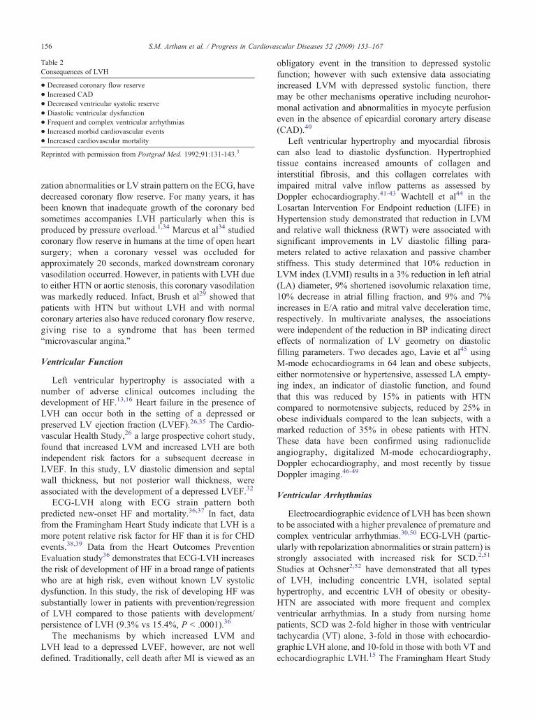
Table 2Consequences of LVH
• Decreased coronary flow reserve• Increased CAD• Decreased ventricular systolic reserve• Diastolic ventricular dysfunction• Frequent and complex ventricular arrhythmias• Increased morbid cardiovascular events• Increased cardiovascular mortality
Reprinted with permission from Postgrad Med. 1992;91:131-143.3
156 S.M. Artham et al. / Progress in Cardiovascular Diseases 52 (2009) 153–167
zation abnormalities or LV strain pattern on the ECG, havedecreased coronary flow reserve. For many years, it hasbeen known that inadequate growth of the coronary bedsometimes accompanies LVH particularly when this isproduced by pressure overload.1,34 Marcus et al34 studiedcoronary flow reserve in humans at the time of open heartsurgery; when a coronary vessel was occluded forapproximately 20 seconds, marked downstream coronaryvasodilation occurred. However, in patients with LVH dueto either HTN or aortic stenosis, this coronary vasodilationwas markedly reduced. Infact, Brush et al29 showed thatpatients with HTN but without LVH and with normalcoronary arteries also have reduced coronary flow reserve,giving rise to a syndrome that has been termed“microvascular angina.”
Ventricular Function
Left ventricular hypertrophy is associated with anumber of adverse clinical outcomes including thedevelopment of HF.13,16 Heart failure in the presence ofLVH can occur both in the setting of a depressed orpreserved LV ejection fraction (LVEF).26,35 The Cardio-vascular Health Study,26 a large prospective cohort study,found that increased LVM and increased LVH are bothindependent risk factors for a subsequent decrease inLVEF. In this study, LV diastolic dimension and septalwall thickness, but not posterior wall thickness, wereassociated with the development of a depressed LVEF.32
ECG-LVH along with ECG strain pattern bothpredicted new-onset HF and mortality.36,37 In fact, datafrom the Framingham Heart Study indicate that LVH is amore potent relative risk factor for HF than it is for CHDevents.38,39 Data from the Heart Outcomes PreventionEvaluation study36 demonstrates that ECG-LVH increasesthe risk of development of HF in a broad range of patientswho are at high risk, even without known LV systolicdysfunction. In this study, the risk of developing HF wassubstantially lower in patients with prevention/regressionof LVH compared to those patients with development/persistence of LVH (9.3% vs 15.4%, P b .0001).36
The mechanisms by which increased LVM andLVH lead to a depressed LVEF, however, are not welldefined. Traditionally, cell death after MI is viewed as an
obligatory event in the transition to depressed systolicfunction; however with such extensive data associatingincreased LVM with depressed systolic function, theremay be other mechanisms operative including neurohor-monal activation and abnormalities in myocyte perfusioneven in the absence of epicardial coronary artery disease(CAD).40
Left ventricular hypertrophy and myocardial fibrosiscan also lead to diastolic dysfunction. Hypertrophiedtissue contains increased amounts of collagen andinterstitial fibrosis, and this collagen correlates withimpaired mitral valve inflow patterns as assessed byDoppler echocardiography.41-43 Wachtell et al44 in theLosartan Intervention For Endpoint reduction (LIFE) inHypertension study demonstrated that reduction in LVMand relative wall thickness (RWT) were associated withsignificant improvements in LV diastolic filling para-meters related to active relaxation and passive chamberstiffness. This study determined that 10% reduction inLVM index (LVMI) results in a 3% reduction in left atrial(LA) diameter, 9% shortened isovolumic relaxation time,10% decrease in atrial filling fraction, and 9% and 7%increases in E/A ratio and mitral valve deceleration time,respectively. In multivariate analyses, the associationswere independent of the reduction in BP indicating directeffects of normalization of LV geometry on diastolicfilling parameters. Two decades ago, Lavie et al45 usingM-mode echocardiograms in 64 lean and obese subjects,either normotensive or hypertensive, assessed LA empty-ing index, an indicator of diastolic function, and foundthat this was reduced by 15% in patients with HTNcompared to normotensive subjects, reduced by 25% inobese individuals compared to the lean subjects, with amarked reduction of 35% in obese patients with HTN.These data have been confirmed using radionuclideangiography, digitalized M-mode echocardiography,Doppler echocardiography, and most recently by tissueDoppler imaging.46-49
Ventricular Arrhythmias
Electrocardiographic evidence of LVH has been shownto be associated with a higher prevalence of premature andcomplex ventricular arrhythmias.30,50 ECG-LVH (partic-ularly with repolarization abnormalities or strain pattern) isstrongly associated with increased risk for SCD.2,51
Studies at Ochsner2,52 have demonstrated that all typesof LVH, including concentric LVH, isolated septalhypertrophy, and eccentric LVH of obesity or obesity-HTN are associated with more frequent and complexventricular arrhythmias. In a study from nursing homepatients, SCD was 2-fold higher in those with ventriculartachycardia (VT) alone, 3-fold in those with echocardio-graphic LVH alone, and 10-fold in those with both VT andechocardiographic LVH.15 The Framingham Heart Study
157S.M. Artham et al. / Progress in Cardiovascular Diseases 52 (2009) 153–167
in a community-based cohort reported that increasedechocardiographic LVH was associated with an increasedrisk of SCD.53 A recent study from Wachtell et al54 alsoshowed that regression of LVH is associated with markedreduction in risk of SCD. Another interesting observationfrom this analysis is that a reduction in both Cornellvoltage duration product and Sokolow-Lyon voltage ECGcriterion was associated with an even lower rate of SCDcompared with reduction in either criterion alone.
Left ventricular hypertrophy is also strongly associatedwith the development of atrial fibrillation (AF). In a 16-year follow-up of 2482 subjects with untreated essentialHTN in sinus rhythm at baseline, Verdecchia et al55 foundthat age and LVM (both P b .001) were the soleindependent predictors of AF. For every 1 SD increasein LVM, the risk of AF increased 1.20 times (95% CI,1.07-1.34). Similarly, in the LIFE trial, in 8851 hyperten-sive patients free of AF at baseline examination, ECG-LVH was a strong independent predictor of new-onsetAF.56 In a substudy by Okin et al57 from the LIFE trial,regression of ECG-LVH was associated with a decreasedincidence of new-onset AF. Patients with ECG-LVH have40 times more premature ventricular contractions thannormotensive subjects or hypertensive patients withoutLVH, including ventricular couplets, multifocal ventricu-lar ectopy, or runs of VT.1,2
Increased Morbid CV Events
Early data from the Framingham Heart Studydemonstrated that ECG evidence of LVH, particularlywith strain pattern, was associated with all types of CHD,including acute coronary syndromes, angina, MI, andSCD.24,38 Over the last 2 decades, Casale et al,16 Korenet al,14 and 2 Framingham studies13,58 (one in the elderlyand the other in younger subjects) have all clearlydemonstrated a marked increase in the incidence of CVevents and an increase in all-cause and CV morbidity andmortality in those subjects with echocardiographicevidence of LVH.
A landmark report from the Framingham Heart Studynearly 40 years ago reported that mortality rate associatedwith ECG-LVH is remarkably similar to the high risknoted in patients following a Q-wave MI.24 Additionaldata from the Framingham Study,13 as well as data fromCornell University14 and the Cardiovascular HealthStudy,59 among others, have demonstrated that a stronggradient exists between increased echocardiographic LVMand increased CV risk. In an elderly cohort of 1141followed up for 4 years prospectively in the Framinghamcohort,12 echocardiographically determined LVM wasperhaps the strongest independent predictor of CHDevents. Levy et al13 of the Framingham Heart Studyexamined the impact of LVM on prognosis in 3220younger subjects followed up prospectively for 4 years. In
this cohort, LVM was strongly and independentlyassociated with the incidence of CV disease, mortalityfrom CV disease, as well as all-cause mortality. Cooperet al31 demonstrated that LVH in patients with CHD isassociated with increased mortality that is independent ofLV function and angiographic extent of CAD. In a studyof 172 prospectively identified patients with essentialHTN in whom echocardiography was performed atbaseline and after a mean of 5 years, almost 30% ofpatients with normal LVM at baseline and LVH at follow-up had a morbid CV event; among those with normalLVM at both baseline and follow-up, less than 10% had asimilar morbid CV event.60
Left ventricular hypertrophy also appears to bepredictive of renal end points. In the Reduction ofEndpoints in Non–insulin-dependent diabetes mellituswith the Angiotensin II Antagonist Losartan trial in 1513patients, Boner et al61 demonstrated that LVH at baselinesignificantly predicted end-stage renal disease or death(hazard ratio [HR] = 1.44; P = .011) and CV events (HR =1.68; P = .001).
Despite having extensive evidence linking ECG andechocardiographic LVM to increased CV risk, LVH isoften not thought of as a standard or conventional CV riskfactor. The most likely explanation for this is that LVH isconsidered to be a subclinical disease lying in the center ofthe continuum from standard risk factors to clinicallymanifest CV disease, as well as the fact that there was arelative paucity of data until recently on the impact ofreversing LVH on major CV outcomes.
Concentric Remodeling
HTN induces four distinct patterns of LV geometrybased on the relationships between cavity size and wallthickness, including eccentric and concentric LVH as wellas CR (Fig 4).62 Concentric remodeling, a subtle abnormalLV geometric pattern associated with increased CV risk, isdefined as an increase in RWT with normal LVMI (RWT≥0.45 and LVMI b125 g/m2, although many studiesutilize slightly different cut-points for RWT and LVH).
Alterations in LV geometry in the absence of LVHhave not always been appreciated as an important CV riskfactor. The type of LVH and pattern of LV geometry maybe an important prognostic factor, with a stepwise increasein risk as hypertensive patients progress from CR toeccentric and concentric LVH (Fig 5).14 Koren et al14
originally reported a CV event rate of 4.2 per 100 patient-years in those with CR compared to 1.8 per 100 patientyears when normal LV geometry is present. concentricremodeling represents a more subtle LV geometric patterndistinct from LVH and has been shown to independentlypredict adverse CV outcomes in populations with HTN,

Fig 4. Hemodynamic and geometric profiles in hypertensive patients with the 4 patterns of left ventricular geometry. The short axis/long axis ratio (b/a) wasdescribed by 2-dimensional echocardiography. CI, cardiac index (l/min/m2); SBP, systolic blood pressure; TPR, total peripheral resistance (dynes s cm−5).Reprinted with permission from J Am Coll Cardiol. 1992;19:1550-1558.62
158 S.M. Artham et al. / Progress in Cardiovascular Diseases 52 (2009) 153–167
and recently, Milani et al8 and Lavie et al9 assessed theprevalence (Fig 6) and validated the prognostic signifi-cance of various LV geometric patterns, including CR, inpatients with normal LVEF, including elderly and alsoconfirmed that CR is associated with increased all-causemortality (Fig 7).63
Milani et al,8 from a large clinical echocardiographicdatabase of 35 602 patients with normal LVEF, identified
Fig 5. Relation of left ventricular geometry to mortality in patients withuncomplicated hypertension. CH, concentric hypertrophy; EH, eccentrichypertrophy; NL, normal. Relative wall thickness is the ratio of theposterior wall thickness to one-half the left ventricular internal dimensionat end-diastole. Reprinted with permission from Ann Intern Med.1991;114:345-352.14
abnormal LV geometry in 46% (n = 16 320) of patients,with CR present in 35% and LVH (both concentric andeccentric) in 11% and also described the effect of changein cardiac structure on subsequent survival. In this study,there was a strong relationship between abnormal LVgeometry and mortality, and patients with CR and LVHexhibited considerably higher relative risk for all-causemortality compared to subjects with normal LV geometry
Fig 6. Frequency of LV geometric patterns in patients with normalsystolic function. Reprinted with permission from Am J Cardiol.2006;97:959-963.8

Fig 7. Actuarial cumulative hazard plot for survival time based on cardiac structure. A, Normal structure, CR, and frank LVH. B, Normal structure, CR,eccentric hypertrophy (EH), and concentric hypertrophy (CH). Reprinted with permission from Am J Cardiol. 2006;97:959-963.8
159S.M. Artham et al. / Progress in Cardiovascular Diseases 52 (2009) 153–167
(relative risk [RR] 1.99, P b .0001; RR 2.13, P b .0001,respectively). Another noteworthy finding from this studyis that subjects with CR who reverted to a normal LVgeometric pattern had improved survival (RR 0.64, P =.03) compared with those who progressed to LVH (RR1.54, P = .05; Fig 8). In the study by Lavie et al,9 inpatients N70 years of age (mean age of 78 ± 5 years), CR(43%) was the most common LV geometric pattern withsignificantly increased mortality compared with similarlyaged patients with either normal structure or eccentric
Fig 8. Patients with CR who progress to LVH versus subjects who convert to norand 95% CIs of all-cause mortality. Reprinted with permission from Am J Car
LVH, with similar increased mortality to those withconcentric LVH.
There is very little experimental and clinical data toexplain why CR predisposed these patients to an increasein all-cause mortality, but potential mechanisms mightinclude elevated arterial load, reduced coronary flowreserve, elevated neuroendocrine components, and abnor-mal growth factors.8,9,27,28 Furthermore, in some studies,RWT, and not LVM, was independently linked todecreased coronary flow reserve. The study by Lavie et
mal (NL) cardiac structure. A, Kaplan-Meier survival over 3 years. B, RRsdiol. 2006;97:959-963.8

160 S.M. Artham et al. / Progress in Cardiovascular Diseases 52 (2009) 153–167
al9 in the older cohort reemphasizes this observation byfurther demonstrating that, in multivariate analysis, RWTand not LVM was an independent predictor of all-causemortality. Preliminary data from our laboratory demon-strate that LA volume index is also a potent predictor ofmortality and adds to the mortality-risk prediction in all 4major categories of LV geometry.64,65 Moreover, our datahave determined that both RWT and LVMI predictmortality, but RWT seems to do so more robustly. Inaddition, we have extended our findings to women, whereLV geometry, including CR and LVH, are potentpredictors of all-cause mortality.66,67
Determination of LVH
The prevalence of LVH in patients with HTN varies byseverity of HTN, age and the methods of detection. Ratesof 20% to nearly 50% have been reported in populationswith mild-to-moderate HTN.68 Electrocardiogram andechocardiography are the 2 most widely used diagnostictools for the detection of LVH: ECG is highly specific butnot nearly as sensitive when compared with anatomicallyvalidated echocardiographic methods.69
Quantitative echocardiography, which accurately esti-mates LVM, not only is a more precise noninvasive toolfor determining the prevalence of LVH but also permitsthe evaluation of serial changes in LVM and LVfunction.13,69,70 Left ventricular mass index is routinelycalculated using the Devereux and Reichek formula,although the exact cut-off for RWT and LVMI havevaried somewhat in various studies.68 In general, normalLV structure has normal RWT and LVMI, whereaselevated RWT and normal LVMI represent CR, elevatedLVMI but normal RWT represent eccentric LVH, andelevation in both RWT and LVMI represent concentricLVH. Relative wall thickness below 0.45 and LVMIbelow 125 g/m2 is suggestive of normal LV geometry,increase in RWT 0.45 or greater and normal LVMI below125 g/m2 denote CR, normal RWT below 0.45 andincrease in LVMI 125g/m2 or greater indicate eccentricLVH, whereas increase in both RWT 0.45 or greater andLVMI above 125 g/m2 indicates concentric LVH.65 In theFramingham Heart Study, LVH was detected in 2.1%based on ECG criteria and in 16% based on echocardio-graphic criteria.69
Recent clinical data using newer imaging techniques,such as 3-dimensional echocardiography and cardiacmagnetic resonance imaging (CMR), appear to be verypromising.71 Cardiac magnetic resonance imaging pro-vides the most accurate and highly reproducible evaluationof cardiac size and structure. The large epidemiologicstudy, Multi-Ethnic Study of Atherosclerosis, evaluatedthe relationship between LVM and geometry and incident
CV events in 5098 participants (free of clinically apparentCV disease) who underwent baseline and follow-up CMRimaging. Over 4 years of median follow-up, CR wasstrongly related to stroke and CHD (stroke, HR: 4.2 per g/mL, P = .005; CHD, HR: 2.1 per g/mL, P = .02;), whereasLVM showed the strongest association with incident HFevents (HR: 1.4 per 10% increment, P b .0001).71
Impact of Obesity on LVH Determination
As we discussed above, obesity is one of the mostpowerful predictors of LVH. In fact, we have recentlydemonstrated that abnormal LV geometry, including CRas well as both eccentric and concentric LVH, occur morecommonly in obese than leaner patients.63,65,72
Moreover, although we have identified an “obesityparadox” (obese have better prognosis than their leanercounterparts) among our echocardiographic cohorts withpreserved LV systolic function, similar to the finding byour group and others studying cohorts with establishedheart disease, in every category of body mass index, wedemonstrated progressive worsening in mortality withprogression from normal structure to CR and to LVH.63
We have also extended these findings to cohorts of elderlyolder than 70 years72 as well as to a large cohort ofwomen.66 Despite an obesity paradox (obese have moreLV geometric abnormalities but lower mortality), at anygiven category of body weight, LV geometry was apowerful predictor of prognosis.
Our data specifically used LVM corrected for bodysurface area (BSA) or LVMI criteria to determine LVH,as recommended by the American Society of Echocar-diography and used in many major trials. Obviously,because increasing body weight increases BSA,indexing LVM to BSA may lead to underestimation ofLVH in obesity. Therefore, to avoid this pitfall, manyresearchers have recommended correcting LVM forsome height or some allometric correction (eg, height2.7,using a cut-off of 50 g/m2.7) that seems to correlatebetter with lean body mass and reduces variability incohorts of “normal” subjects.73
Nevertheless, our data using BSA and standardmethods with LVMI criteria for LVH clearly demonstratethe higher prevalence of LV geometric abnormalitiesassociated with obesity as well as the fact that LVgeometry was a potent predictor of all-cause mortality inboth obese and nonobese cohorts.
Central Aortic Pressure
Findings of the Conduit Artery Function Evaluationstudy74 showed that central aortic pressure might be abetter predictor of CV outcomes compared to peripheral

161S.M. Artham et al. / Progress in Cardiovascular Diseases 52 (2009) 153–167
brachial BP. Central aortic BP is measured noninva-sively by using applantation tonometer to record radialartery pulse waves, which are then transformed toderive a central arterial wave form and central aorticmean systolic and diastolic pressures. In this particularstudy, the calcium-channel blocker (CCB) amlodipinewas proven to be a better central aortic pressurelowering agent compared to the β-blocker atenolol,suggesting that different classes of BP-lowering agentshave different effects on brachial versus aortic systolicand pulse pressures. Subsequent data from the StrongHeart Study75 have also confirmed the above findingsthat central aortic pressures might be better predictor ofCV outcomes.
In the Conduit Artery Function Evaluation study,74
although the brachial pressure lowering effect was almostidentical with both CCB and β-blocker–based treatmentstrategies, hard CV end points were lower with CCBstrategy, indicating that β-blocker–based therapy might beless effective at regressing end-organ damage.'
Regression of LVH
It is well known from both animal and human studiesthat antihypertensive therapy could cause regression ofLVH.76,77 In addition, regression of LVH is associatedwith lower CV risk, independent of BP lowering. As aresult, prevention or reversal of hypertensive LVH iswidely accepted as a desirable treatment goal.
It is evident from numerous studies that prevention and/or regression of ECG-LVH is associated with a reducedrisk of CV morbidity and mortality.78-80 Fouad et al81 firstdemonstrated that treatment of HTN could be accompa-nied by a reduction of echocardiographic LVM. ReversingLVH has been shown to improve systolic and diastolicfunction, improve coronary flow reserve, with reductionsin CV risk.13,60,80,82,83
An observational study in 524 participants in theFramingham Heart Study with ECG- LVH84 found thatreduction in Cornell voltage was associated with lowerCV risk, whereas increase in voltage identified indivi-duals at increased risk of CV disease. Recent evidencefrom the Heart Outcomes Prevention Evaluation trial80
provides the strongest evidence supporting the hypothesisthat regression of ECG-LVH improves prognosis,demonstrating that the combined end point of eitherregression or prevention of progression to LVH usingramipril-based therapy was associated with reduced riskof death, MI, stroke, and HF.80
In a substudy of the LIFE trial, 941 prospectivelyidentified patients 55 to 80 years of age with essentialHTN and ECG-LVH had LVM measured by echocar-diography at enrollment in the trial and, thereafter, were
followed up annually for a mean of 4.8 years for CVevents.85,86 The composite endpoint of CV death, fatalor nonfatal MI, and fatal or nonfatal stroke occurred in104 patients (11%). The multivariable Cox regressionmodel demonstrated a strong association between lowerLVMI and reduced rate of the composite endpoint (HR,0.78 per 1 SD [25.3 g/m2] decrease in LVMI; 95% CI,0.65-0.94; P = .009) over and above that predicted byreduction in BP. There were parallel associationsbetween lower LVMI and lower CV mortality (HR,0.62; P = .0001), stroke (HR, 0.76; P = .02), MI (HR,0.85; P = .33), and all-cause mortality (HR, 0.72; P =.002), independent of systolic BP and assigned treat-ment. When patients were dichotomized into those withversus those without LVH by predefined criteria onserial echocardiograms, substantially lower rates of thecomposite end point, CV mortality and all-cause deathwere demonstrated in patients without echocardiograph-ic LVH.86 Of note, losartan was more effective thanatenolol in the LIFE trial in the reduction of clinical CVend points.87,88
Similar findings were reported by Okin et al87 usingECG-LVH criteria. The greater the decrease in LVM asassessed by ECG, the greater the reduction in major CVevents. In both studies, the reduction in LVM waspredictive of lower event rates independent of the degreeof BP reduction, as well as other potential confounders.
Okin et al89 for the first time demonstrated a novelpotential benefit of LVH reversal. In a large cohort of 7998nondiabetic patients with HTN and LVH from the LIFEstudy who were followed up for over 4.6 years, persistentabsence of or regression of ECG-LVH during antihyper-tensive treatment was associated with a lower rate of new-onset diabetes. Research from the same group also showedthat regression of ECG-LVH is associated with a 36%lower incidence of HF and fewer HF hospitalizations.90
Therefore, both nonpharmacological91 and pharma-cological92-95 strategies towards prevention and reductionof LVH are probably warranted. Studies from our center andothers have shown that even after LVH is reduced and theantihypertensive therapy is withdrawn, resulting in return ofBP to pretreatment levels, both systolic and diastolic LVperformance remain enhanced.17,96
Themechanisms bywhich changes in LVH translate intobeneficial clinical outcomes are not clear. One major theoryis that angiotensin II promotes intracellular reactions thatmay lead to both LVH and progression of atheroscleroticlesions through proliferation of vascular smooth musclecells.41,97 Further support that elevated RAAS activitycorrelates with LVH comes from a recent study by Pitt etal98 who compared LVH regression during treatment withangiotensin-converting enzyme (ACE) inhibitor enalapril,the selective aldosterone blocker eplerenone, or theircombination in patients with HTN. A 9-month, double-blind, randomized study was performed in 202 patients with

162 S.M. Artham et al. / Progress in Cardiovascular Diseases 52 (2009) 153–167
LVH and HTN who received eplerenone 200 mg daily,enalapril 40 mg daily, or eplerenone 200 mg and enalapril10 mg daily. Change in LVM as assessed by magneticresonance imaging was the primary end point. Eplerenonesignificantly reduced LVM from baseline (−14.5 ± 3.36 g;n = 50) similar to that noted with enalapril (−19.7 ± 3.20 g;n = 54; P = .258), but eplerenone/enalapril (−27.2 ± 3.39 g;n = 49) was more effective than eplerenone alone (P = .007)for reducing LVM.
The beneficial effect of these 2 agents is mediated viaangiotensin and aldosterone blockade, resulting in anantihypertrophic effect on the myocardium and anantiproliferative effect on smooth muscles (reversal ofmyocardial and vascular remodeling).99
Furthermore, HTN is associated with endothelialdysfunction, and the ACE inhibitors are known to improveendothelial function. It is also suggested that ACEinhibitors might improve peripheral insulin resistance; infact, in one particular study, this property was associatedwith LVH regression.100
In addition, longitudinal studies in patients withHTN83,101 and in the general population24 have reportedthat individuals with LVH regression during 2 to 5 years offollow-up had lower CV event rates than those who havean increase in LVM. The Treatment of Mild HypertensionStudy (TOMHS)102 and an echocardiographic substudyfrom the LIFE103 trial are the only 2 large-scale (n = 902 vsn = 754), long-term (4 vs 2 years) studies to examine thetime course of LV geometric response to BP control. In asubstudy from the LIFE trial,103 the proportion ofparticipants with eccentric LVH, concentric LVH, andCR substantially decreased over 2 years of study treatment(44% to 30%; 24% to 2%; 10% to 4%), and the number ofparticipants with normal LV geometry substantiallyincreased from 22% to 64% (P b .01). These LV geometricchanges occurred during the second year of systematicantihypertensive therapy, during a time period when BPdecreased only slightly (and statistically insignificantly).In contrast, the TOMHS102 found little further change inLVM after the first year of therapy, and this can beexplained by the much lower mean BP (mean, 140/91 vs173/95 mm Hg) and LVH prevalence (15% vs 75%) atbaseline in TOMHS participants compared to LIFE studyparticipants. In addition, this group had largely eradicatedHTN (mean BP, 130/88 mm Hg) and normalized LVMI(mean, 89 g/m2) after 1 year of treatment, thus leavinglittle opportunity for further LVH regression.
Analysis of data from the Anglo-Scandinavian CardiacOutcomes Trial ECG-LVH substudy104 demonstrated thatantihypertensive therapy with an amlodipine-based regi-men reduced ECG LVH more effectively than atenolol-based therapy. In this substudy, the severity of ECG LVHwas assessed at baseline, at 2 years and at the last visit usingthe Cornell and Sokolow-Lyon Voltage criteria. At 5.5years of follow-up, amlodipine-based treatment strategywas associated with a greater regression in LVH compared
to atenolol-based strategy as measured by Cornell criteria(means, −133.1 vs −73.4 mm/millisecond, adjusted P b.0001) and by Sokolow-Lyon (means, −3.4 vs −2.3 mm,adjusted P b .0001). These differences in regression ofLVH were significant after 2 years of treatment.104
It is hypothesized that dual blockade of RAAS at 2different sites will incur incremental benefits both in BPreduction and LVH regression. Aliskiren, a novel directrenin inhibitor that blocks the RAAS, in the Aliskiren inLeft Ventricular Hypertrophy trial,105 suggested that it isas effective as losartan in regressing LVH. This studyrandomized 465 overweight patients with HTN and withevidence of LVH to receive aliskiren 300 mg/d (n = 154),losartan 100 mg/d (n = 152), or combination of these 2 (n =154) for 9 months. The primary end point, reduction inLVM, as measured by CMR imaging was reducedsignificantly from baseline in all treatment groups (−4.9,−4.8, and −5.8 g/m2 reductions in the aliskiren, losartan,and combination arms, respectively; P b .0001 for alltreatment groups). This study also shows that the reductionin LVM with the combination therapy is not significantlydifferent from that achieved with losartan alone.
Pharmacologic Interventions
The above data suggest that antihypertensive treatmentshould be directed both towards BP reduction and LVHregression. With the exception of minoxidil and hydral-azine, other classes of hypertensive drugs have beenshown to produce some reduction in LVH.106 Because ofthe heterogeneity of the trials and inconsistent data, clearrecommendations in favor of one group of medications arenot yet available. However, overall evidence is in favor ofACE inhibitors and angiotensin receptor blockers (ARBs)and CCBs in terms of LVM reduction.89,91-93 The effect ofβ-blockers on the regression of LVH is not consistent, andthe diuretics provide a moderate level of LVH reduction.
Dahlof et al93 in 1992 and Schmieder et al94 in 1996, intheir meta-analyses of the trials evaluating the effects ofdifferent antihypertensive agents on LVM reduction,demonstrated the superiority of ACE inhibitors over β-blockers. Later in 2003, Klingbeil et al92 extended theirmeta-analysis by including 80 more studies and recon-firmed their previous findings by showing that ARBs,CCBs, and ACE inhibitors reduced LVM by approxi-mately 10% to 13%, whereas the reduction with β-blockers and diuretics was from 6% to 8%.
The greater ECG-LVH and echocardiographic LVHregression with the ARB losartan compared to the β-blocker atenolol in the LIFE study seems to represent aclass effect, which is supported by the preliminary datafrom a trial with patients randomized to the ARBtelmisartan or the β-blocker carvedilol. In this study,reductions in LVMI were greater with telmisartan than

163S.M. Artham et al. / Progress in Cardiovascular Diseases 52 (2009) 153–167
carvedilol (15.7% vs. 9.1% and 15.5% vs. 9.6%,respectively; both P = .001), presumably due to amechanism beyond that of BP lowering.48 In addition toLIFE, the Prospective Randomized Enalapril StudyEvaluating Regression of Ventricular Enlargement95 andthe LVH: Indapamide Sustained Release VersusEnalapril107 are other large, well-designed trials withadequate power which evaluated the various effects of BPtherapy on LVH progression/regression. ProspectiveRandomized Enalapril Study Evaluating Regression ofVentricular Enlargement (n = 303) in an ethnically diversepopulation with HTN and increased LVM has shown thatboth enalapril and nifedipine gastrointestinal therapeuticsystem have moderately beneficial and statisticallyindistinguishable effects on regression of LVH.95 TheLVH: Indapamide Sustained Release Versus Enalaprilstudy was a 1-year, prospective, randomized, double-blindstudy with 505 hypertensive men and women with LVHrandomized to treatment with indapamide SR (slowrelease) or enalapril. After 48 weeks, patients treatedwith indapamide SR had a significant reduction in LVMIfrom baseline values (mean reduction 8.4 g/m2; P b .001).By contrast, enalapril did not significantly reduce LVMIfrom baseline values (mean reduction 1.9 g/m2).107
Most antihypertensive trials using β-blockers over thedecades have been conducted using atenolol as the studydrug, and we are not certain that these data can necessarilybe extrapolated to other beta-blocking agents. One reasonwhy β-blockers do not perform as well as otherantihypertensive therapies is due to the fact that they donot effectively lower central systolic BP as much as otherantihypertensive agents and regression of LVH is moreclosely correlated with central BP than with brachial BP.To further clarify the efficacy of other drugs in this group,a randomized, double-blind, multicenter study comparingthe effects of carvedilol modified-release formulation(Coreg CR) and atenolol in combination with andcompared to an ACE inhibitor lisinopril on LVMregression in subjects with HTN and LVH (CLEVER)108
is underway evaluating the effects of combination ofcarvedilol CR and lisinopril vs atenolol plus lisinopril vslisinopril alone on LVMI regression. This study is beingconducted with the premise that carvedilol with itsperipheral α-blocking effects, might have LVM regressioneffects due to its after load reduction. This has been testedpreviously in two small open-label studies with 14 and 22subjects, each suggesting favorable results with respect toLVMI regression.108
Importantly it is worth noting that most patients withHTN have multiple other CV risk factors in addition toelevated BP, and most of these patients do not adequatelyrespond to monotherapy but require treatment withmultiple antihypertensives. In this regard, 2 recent trialsprovide data regarding the superiority of the low-dosecombination therapy in terms of both BP reduction andLVH regression. The pREterax in regression of Arterial
Stiffness in a contrOlled double-bliNd study109 demon-strated that the low-dose combination therapy ACEinhibitor perindopril plus diuretic indapamide produced asignificantly greater change in LVM after 1 year than theβ-blocker atenolol, despite inducing a similar change inmean BP. In addition, perindopril/indapamide reducedcentral (carotid) and peripheral (brachial) systolic BP andpulse pressure to a significantly greater extent thanatenolol. The PICXEL (Preterax In a double-blindControlled study versus Enalapril in LVH)110 studydemonstrated that the fixed dose combination of ACEinhibitor perindopril and diuretic indapamide reducedLVH more effectively than monotherapy with high dosesof ACE inhibitor enalapril and provided better BP control.This reduction cannot be entirely explained by the betterefficacy of the low-dose combination on BP reduction.
Even lifestyle and nonpharmacological treatmentstrategies lead to LVH reduction.91 The nonpharmacolo-gic strategy for LVH reduction includes efforts aimed atsodium restriction, weight reduction, and increasingaerobic or dynamic exercise. Studies highlighting weightreduction have demonstrated significant reductions inLVH, even greater than that obtained with pharmacologicagents. In obese hypertensive patients, MacMahon et al111
demonstrated that modest weight loss (8-kg loss) resultedin a mall, but statistically greater reduction in LV wallthickness compared with conventional pharmacologicagents. Data from our laboratory and elsewhere suggestedthat dietary sodium intake is directly related to LVHincidence, independent of its effect on BP. Therefore,sodium restriction should be encouraged for bothtreatment of HTN and LVH reduction.112
Bariatric surgery is the other well-established weightloss therapeutic strategy with proven benefits on metabolicprofile and improvements in LV structure and function. Ina recent prospective longitudinal study in 43 consecutiveobese patients, the impact of bariatric surgery on LVdiastolic function was assessed. Nine months postsurgerynormalization of diastolic dysfunction variables, includingmitral inflow velocities and deceleration time on tissueDoppler imaging, was noted.113 Although bariatricsurgery has resulted in improvements in CV risk scoresas well as morbidity and mortality related with diabetes,CV diseases, as well as certain cancers, the initial BPlowering effects of this surgery have not been maintainedover several year follow-up periods.114,115
Conclusions
These results in hypertensive subjects show that areduction in LVM is associated with a reduction in majorCV morbidity and mortality exceeding that achieved by BPcontrol alone, strongly suggesting that emphasis is needed

164 S.M. Artham et al. / Progress in Cardiovascular Diseases 52 (2009) 153–167
not only on BP lowering but also on reducing LVH.Evidence suggests that RAAS inhibition with either anACE inhibitor or an ARB should be primary choice, asthese agents appear to lower LVM to a greater extentcompared to other antihypertensive agents. In view ofrecent evidence from the Anglo-Scandinavian CardiacOutcomes Trial, addition of CCBs to RAAS inhibition maybe particularly effective in achieving LVH regression.84
Statement of Conflict of Interest
All authors declare that there are no conflicts of interest.
References
1. Lavie CJ, Ventura HO, Messerli FH: Regression of increased leftventricular mass by antihypertensives. Drugs 42:945-961, 1991
2. Messerli FH, Ventura HO, Elizardi DJ, et al: Hypertension andsudden death: increased ventricular ectopic activity in leftventricular hypertrophy. Am J Med 77:18-22, 1984
3. Lavie CJ, Ventura HO, Messerli FH: Left ventricular hypertrophy.Its relationship to obesity and hypertension. Postgrad Med 91:131-143, 1992
4. Messerli FH: Cardiovascular effects of obesity and hypertension.Lancet 1:1165-1168, 1982
5. Lavie CJ, Messerli FH: Cardiovascular adaptation to obesity andhypertension. Chest 90:275-279, 1986
6. Lavie CJ, Milani RV, Messerli FH: Prevention and reduction of leftventricular hypertrophy in the elderly. Clin Geriatr Med 12:57-68,1996
7. Lavie CJ, Ventura HO,Messerli FH: Left ventricular hypertrophy inthe elderly. Cardiol Elderly 2:362-369, 1994
8. Milani RV, Lavie CJ, Mehra MR, et al: Left ventricular geometryand survival in patients with normal left ventricular ejectionfraction. Am J Cardiol 97:959-963, 2006
9. Lavie CJ, Milani RV, Ventura HO, et al: Left ventricular geometryand mortality in patients N70 years of age with normal ejectionfraction. Am J Cardiol 98:1396-1399, 2006
10. Lavie CJ, Milani RV, Shah SB, et al: Impact of left ventriculargeometry on prognosis-A review of Ochsner studies. Ochsner J 8:11-17, 2008
11. Katz AM: Cardiomyopathy of overload: a major determinant ofprognosis in congestive heart failure. N Engl J Med 322:100-110,1990
12. Veterans Administration Cooperative Study on AntihypertensiveAgents: Double blind control study of antihypertensive agents. III.Chlorothiazide alone and in combination with other agents;preliminary results. Arch Intern Med 110:230-236, 1962
13. Levy D, Garrison RJ, Savage DD, et al: Prognostic implications ofechocardiographically determined left ventricular mass in theFramingham Heart Study. N Engl J Med 322:1561-1566, 1990
14. Koren MJ, Devereux RB, Casale PN, et al: Relation of leftventricular mass and geometry to morbidity and mortality inuncomplicated essential hypertension. Ann Intern Med 114:345-352, 1991
15. Aronow WS, Epstein S, Kuenigsberg M, et al: Usefulness ofechocardiographic left ventricular hypertrophy, ventricular tachy-cardia, and complex ventricular arrhythmias in predicting ventric-ular fibrillation or sudden death among elderly patients. Am JCardiol 62:1124-1125, 1988
16. Casale PN, Devereux RB, Milner M, et al: Value of echocardio-graphic measurement of left ventricular mass in predictingcardiovascular morbid events in hypertensive men. Ann InternMed 105:173-178, 1986
17. Schmieder RE, Messerli FH, Sturgill D, et al: Cardiac performanceafter reduction of myocardial hypertrophy. Am J Med 87:22-27,1989
18. Lavie CJ, Milani RV, Mehra MR, et al: Hypertension and leftventricular hypertrophy in the elderly. In: Tresch DD, Aronow WS,editors. Cardiovascular disease in the elderly patient. 2nd ed. NewYork: Marcel Dekker, Inc; 1998. p. 109-127, 1998
19. Lavie CJ, Ventura HO, Messerli FH: Cardiopathy of obesity. IntMed Spec 9:57-71, 1988
20. Lee M, Gardin JM, Lynch JC, et al: Diabetes mellitus andechocardiographic left ventricular function in free-living elderlymen and women: the Cardiovascular Health Study. AmHeart J 133:36-43, 1997
21. de Simone G, Celentano A, Devereux R: Metabolic risk factors andleft ventricular hypertrophy. Nutr Metab Cardiovac Dis 8(Suppl 5):40-45, 1998
22. de Simone G, Palmieri V, Bella JN, et al: Association of leftventricular hypertrophy with metabolic risk factors: the HyperGENstudy. J Hypertens 20:323-331, 2002
23. Jilaihawi H, Greaves S, Rouleau JL, et al: Left ventricularhypertrophy and the risk of subsequent left ventricular remodelingfollowing myocardial infarction. Am J Cardiol 91:723-726, 2003
24. Kannel WB, Gordon T, Castelli WP, et al: Electrocardiographic leftventricular hypertrophy and risk of coronary heart disease: theFramingham study. Ann Intern Med 72:813-822, 1970
25. Carabello BA: The relationship of left ventricular geometry andhypertrophy to left ventricular function in valvular heart disease.J Heart Valve Dis(Suppl 2):S132-S138, 1995
26. Drazner MH, Dries DL, Peshock RM, et al: Left ventricularhypertrophy is more prevalent in blacks than whites in the generalpopulation: the Dallas Heart Study. Hypertension 4:124-129, 2005
27. Olsen MH, Wachtell K, Hermann KL, et al: Is cardiovascularremodeling in patients with essential hypertension related to morethanhigh blood pressure?ALIFE substudy. Losartan Intervention ForEndpoint-Reduction in Hypertension. AmHeart J 144:530-537, 2002
28. Malatino LS, Cataliotti A, Benedetto FA, et al: Hepatocyte growthfactor and left ventricular geometry in end-stage renal disease.Hypertension 41:88-92, 2003
29. Brush SE, Cannon RO, Schenke WH, et al: Angina due to coronarymicrovascular disease in hypertensive patients without leftventricular hypertrophy. N Engl J Med 319:1302-1307, 1988
30. Levy D, Anderson KM, Savage DD, et al: Risk of ventriculararrhythmias in left ventricular hypertrophy: the Framingham HeartStudy. Am J Cardiol 60:560-565, 1987
31. Cooper RS, Simmons BE, Castaner A, et al: Left ventricularhypertrophy is associated with worse survival independent ofventricular function and number of coronary arteries severelynarrowed. Am J Cardiol 65:441-445, 1990
32. Schillaci G, Verdecchia P, Porcellati C, et al: Continuous relationbetween left ventricular mass and cardiovascular risk in essentialhypertension. Hypertension 35:580-586, 2000
33. Lauer MS, Anderson KM, Kannel WB, et al: The impact of obesityon left ventricular mass and geometry: the Framingham HeartStudy. JAMA 266:231-236, 1991
34. Marcus ML, Koyanayi S, Harrison DG, et al: Abnormalities in thecoronary circulation that occur as a consequence of cardiachypertrophy. Am J Med 75:62-66, 1983
35. Devereux RB, Roman MJ, Liu JE, et al: Congestive heart failuredespite normal left ventricular systolic function in a population-basedsample: the Strong Heart Study. Am J Cardiol 86:1090-1096, 2000
36. Mathew J, Lonn E, Johnstone D, et al: Left ventricular hypertrophyby electrocardiogram predicts death and development of heart
165S.M. Artham et al. / Progress in Cardiovascular Diseases 52 (2009) 153–167
failure in high-risk patients without systolic dysfunction. J Am CollCardiol 35(Suppl A):212A Abstract, 2000
37. Okin PM, Devereux RB, Nieminen MS, et al: Electrocardiographicstrain pattern and prediction of new-onset congestive heart failure inhypertensive patients: the Losartan Intervention for EndpointReduction inHypertension (LIFE) study. Circulation 113:67-73, 2006
38. Kannel WB: Prevalence and natural history of electrocardiographicleft ventricular hypertrophy. Am J Med 75:4-11, 1983
39. Kannel WB: Hypertension in the elderly: epidemiologic appraisalfrom the Framingham study. Cardiol Elderly 1:359-363, 1993
40. Marcus ML, Harrison DG, Chilian WM, et al: Alterations in thecoronary circulation in hypertrophied ventricles. Circulation 75:119-125, 1987
41. Krauser DG, Devereux RB: Ventricular hypertrophy and hyperten-sion: prognostic elements and implications for management. Herz31:305-316, 2006
42. Diez J, Querejeta R, Lopez B, et al: Losartan-dependent regressionof myocardial fibrosis is associated with reduction of left ventricularchamber stiffness in hypertensive patients. Circulation 105:2512-2517, 2002
43. Villari B, Campbell SE, Hess OM, et al: Influence of collagennetwork on left ventricular systolic and diastolic function in aorticvalve disease. J Am Coll Cardiol 22:1477-1484, 1993
44. Wachtell K, Bella JN, Rokkedal J, et al: Change in diastolic leftventricular filling after one year of antihypertensive treatment: theLosartan Intervention For Endpoint Reduction in Hypertension(LIFE) study. Circulation 105:1071-1076, 2002
45. Lavie CJ, Amodeo C, Ventura HO, et al: Left atrial abnormalitiesindicating diastolic ventricular dysfunction in cardiopathy ofobesity. Chest 92:1042-1046, 1987
46. Wang M, Gabriel WK, Wang A, et al: Tissue Doppler imaging pro-vides incremental prognostic value in patients with systemic hyper-tension and left ventricular hypertrophy. J Hypertens 23:183-191,2005
47. Cuocolo A, Sax FL, Brush JE, et al: Left ventricular hypertrophyand impaired diastolic filling in essential hypertension. Diastolicmechanisms for systolic dysfunction during exercise. Circulation81:978-986, 1990
48. Galzerano D, Tammaro P, del Viscovo L, et al: Three-dimensionalechocardiographic and magnetic resonance assessment of the effectof telmisartan compared with carvedilol on left ventricular mass amulticenter, randomized, longitudinal study. Am J Hypertens 18:1563-1569, 2005
49. Granier P, Esquerre JP, Tredez P, et al: Correlations between M-mode markers of left ventricular hypertrophy and radionuclideangiographic indices of left ventricular diastolic function in mild tomoderate hypertension. J Hypertens 7:S100-S101, 1989
50. McLenachan JM, Henderson E, Morris KI, et al: Ventriculararrhythmias in patients with hypertensive left ventricular hypertro-phy. N Engl J Med 317:787-792, 1987
51. Kannel WB, Schatzkin A: Sudden death: lessons from subsets inpopulation studies. J Am Coll Cardiol 5:141B-149B, 1985
52. Lavie CJ, Ventura HO, Messerli FH, et al: Hypertension, obesity,left ventricular hypertrophy, complex ventricular ectopic activity,and increased risk for sudden death: Review of Ochsner Studies andthe literature. J Cardiopulm Rehabil 13:264-270, 1993
53. Haider AW, Larson MG, Benjamin EJ, et al: Increased leftventricular mass and hypertrophy are associated with increased riskfor sudden death. J Am Coll Cardiol 32:1454-1459, 1998
54. Wachtell K, Okin PM, Olsen MH, et al: Regression ofelectrocardiographic left ventricular hypertrophy during antihyper-tensive therapy and reduction in sudden cardiac death. Circulation116:700-705, 2007
55. Verdecchia P, Reboldi G, Gattobigio R, et al: Atrial fibrillation inhypertension: predictors and outcome. Hypertension 41:218-223,2003
56. Wachtell K, LehtoM,Gerdts E, et al: Angiotensin II receptor blockadereduces new-onset atrial fibrillation and subsequent stroke comparedto atenolol: the Losartan Intervention For Endpoint reduction inhypertension (LIFE) study. J Am Coll Cardiol 45:712-719, 2005
57. Okin PM, Wachtell K, Devereux RB, et al: Regression ofelectrocardiographic left ventricular hypertrophy and decreasedincidence of new-onset atrial fibrillation in patients with hyperten-sion. JAMA 296:1242-1248, 2006
58. Levy D, Garrison RJ, Savage DD, et al: Left ventricular mass andincidence of coronary heart disease in an elderly cohort. Ann InternMed 110:101-107, 1989
59. Drazner MH, Rame JE, Marino EK, et al: Increased left ventricularmass is a risk factor for the development of a depressed leftventricular ejection fraction within five years: the CardiovascularHealth Study. J Am Coll Cardiol 43:2207-2215, 2004
60. Koren MJ, Ulin RJ, Koren AT, et al: Left ventricular mass changeduring treatment and outcome in patients with essential hyperten-sion. Am J Hypertens 15:1021-1028, 2002
61. Boner G, Cooper ME, McCarroll K, et al: Adverse effects of leftventricular hypertrophy in the Reduction of Endpoints in NIDDMwith the Angiotensin II Antagonist Losartan (RENAAL) study.Diabetologia 48:1980-1987, 2005
62. Ganau A, Devereux RB, Roman MJ, et al: Patterns of leftventricular hypertrophy and geometric remodeling in essentialhypertension. J Am Coll Cardiol 19:1550-1558, 1992
63. Lavie CJ, Milani RV, Ventura HO, et al: Disparate effects of leftventricular geometry and obesity on mortality in patients withpreserved left ventricular ejection fraction. Am J Cardiol 100:1460-1464, 2007
64. Patel DA, Lavie CJ, Milani RV, et al: Left atrial volume index andleft ventricular geometry independently predict mortality in 16,904elderly patients with preserved ejection fraction, abstracted. J AmColl Cardiol 53:10(Suppl A):A273, 2009
65. Patel DA, Lavie CJ, Milani RV, et al: Impact of left atrial volumeindex and left ventricular geometry on mortality in obese versusnon-obese elderly patients with preserved ejection fraction,abstracted. J Am Coll Cardiol 53:10(Suppl A):A238, 2009
66. Patel D, Lavie CJ, Milani RV, et al: Disparate effects of leftventricular geometry and obesity on mortality in women withpreserved systolic function—“the obesity paradox” in women.Circulation 118:S1153, 2008
67. Patel D, Lavie CJ, Milani RV, et al: Left ventricular geometrypredicts mortality in 26,216 women with preserved systolicfunction. Circulation 118:S602, 2008
68. Devereux RB, Reichek N: Echocardiographic determination of leftventricular mass in man: anatomic validation of the method.Circulation 55:613-618, 1977
69. Levy D, Labib SB, Anderson KM, et al: Determinants of sensitivityand specificity of electrocardiographic criteria for left ventricularhypertrophy. Circulation 81:815-820, 1990
70. Levy D, Anderson KM, Savage DD, et al: Echocardiographicallydetected left ventricular hypertrophy: prevalence and risk factors.Ann Intern Med 108:7-13, 1988
71. Bluemke DA, Kronmal RA, Lima JA, et al: The relationship of leftventricular mass and geometry to incident cardiovascular events:the MESA (Multi-Ethnic Study of Atherosclerosis) study. J AmColl Cardiol 52:2148-2155, 2008
72. Lavie CJ, Milani RV, Patel D, et al: Disparate effects of obesityand left ventricular geometry on mortality in 8,088 elderlypatients with preserved systolic function. Postgrad Med 121.In press, 2009
73. De Simone SR, Daniels RB, Devereux RA, et al: Left ventricularmass in normotensive children and adults: assessment of allometricrelations and impact of overweight. J Am Coll Cardiol 20:1251-1260, 1992

166 S.M. Artham et al. / Progress in Cardiovascular Diseases 52 (2009) 153–167
74. Williams B, Lacy PS, Thom SM, et al, and CAFE Investigators,Anglo-Scandinavian Cardiac Outcomes Trial Investigators, CAFESteering Committee and Writing Committee: Differential impact ofblood pressure-lowering drugs on central aortic pressure andclinical outcomes. Circulation 113:1213-1225, 2006
75. Roman MJ, Devereux RB, Kizer JR, et al: Central pressure morestrongly relates to vascular disease and outcome than doesbrachial pressure: the Strong Heart study. Hypertension 50:197-203, 2007
76. Nagano M, Higaki J, Mikami H, et al: Converting enzymeinhibitors regressed cardiac hypertrophy and reduced tissueangiotensin II in spontaneously hypertensive rats. J Hypertens 9:595-599, 1991
77. Helmcke JG, Schneckloth R, Corcoran AC, et al: Electrocardio-graphic changes of left ventricular hypertrophy: effects ofantihypertensive treatment. Am Heart J 53:549-557, 1957
78. Hypertension Detection and Follow-up Program CooperativeGroup: Five-year findings of the Hypertension Detection andFollow-up Program: prevention and reversal of left ventricularhypertrophy with antihypertensive drug therapy. Hypertension 7:105-112, 1985
79. Prineas RJ, Rautaharju PM, Grandits G, et al: Independent risk forcardiovascular disease predicted by modified continuous scoreelectrocardiographic criteria for 6-year incidence and regression ofleft ventricular hypertrophy among clinically disease free men:16-year follow-up for the Multiple Risk-Factor InterventionTrial. J Electrocardiol 34:91-101, 2001
80. Mathew J, Sleight P, Lonn E, et al: Reduction of cardiovascular riskby regression of electrocardiographic markers of left ventricularhypertrophy by the angiotensin-converting enzyme inhibitorramipril. Circulation 104:1615-1621, 2001
81. Fouad FM, Nakashima Y, Tarazi RC, et al: Reversal of leftventricular hypertrophy in hypertensive patients treated withmethyldopa. Lack of association with blood pressure control.Am J Cardiol 49:795-801, 1982
82. Wachtell K, Palmieri V, Olsen MH, et al: Change in systolic leftventricular performance after 3 years of antihypertensive treatment:the Losartan Intervention For Endpoint reduction in hypertension(LIFE) study. Circulation 106:227-232, 2002
83. Verdecchia P, Schillaci G, Borgioni C, et al: Prognostic significanceof serial changes in left ventricular mass in essential hypertension.Circulation 97:48-54, 1998
84. Levy D, Salomon M, D'Agostino RB, et al: Prognostic implicationsof baseline electrocardiographic features and their serial changes insubjects with left ventricular hypertrophy. Circulation 90:1786-1793, 1994
85. Devereux RB, Wachtell K, Gerdts E, et al: Prognostic significanceof left ventricular mass change during treatment of hypertension.JAMA 292:2350-2356, 2004
86. Devereux RB, Dahlöf B, Gerdts E, et al: Regression of hypertensiveleft ventricular hypertrophy by losartan compared with atenolol: theLosartan Intervention for Endpoint Reduction in Hypertension(LIFE) trial. Circulation 110:1456-1462, 2004
87. Okin PM, Devereux RB, Jern S, et al, for the LIFE StudyInvestigators: Regression of electrocardiographic left ventricularhypertrophy during antihypertensive treatment and the prediction ofmajor cardiovascular events. JAMA 292:2343-2349, 2004
88. Dahlöf B, Devereux RB, Kjeldsen SE, et al: Cardiovascularmorbidity and mortality in the Losartan Intervention For Endpointreduction in hypertension study (LIFE): a randomised trial againstatenolol. Lancet 359:995-1003, 2002
89. Okin PM, Devereux RB, Harris KE, et al, for the LIFE Studyinvestigators: In-treatment resolution or absence of electrocardio-graphic left ventricular hypertrophy is associated with decreasedincidence of new-onset diabetes mellitus in hypertensive patients:
the Losartan Intervention for Endpoint reduction in hypertension(LIFE) Study. Hypertension 50:984-990, 2007
90. Okin PM, Devereux RB, Harris KE, et al: Regression ofelectrocardiographic left ventricular hypertrophy is associatedwith less hospitalization for heart failure in hypertensive patients.Ann Intern Med 147:311-319, 2007
91. MacMahon SW, Wilcken DEL, McDonald GJ: The effect of weightreduction on left ventricular mass: a randomized controlled trial inyoung, overweight hypertensive patients. N Engl J Med 314:334-339,1986
92. Klingbeil AU, Schneider M, Martus P, et al: A meta-analysis of theeffects of treatment on left ventricular mass in essential hyperten-sion. Am J Med 115:41-46, 2003
93. Dahlöf B, Pennert K, Hansson L: Reversal of left ventricularhypertrophy in hypertensive patients: a meta-analysis of 109treatment studies. Am J Hypertens 5:95-110, 1992
94. Schmieder RE, Martus P, Klingbeil A: Reversal of left ventricularhypertrophy in essential hypertension: a meta-analysis of random-ized double-blind studies. JAMA 275:1507-1513, 1996
95. Devereux RB, Palmieri V, Sharpe N, et al: Effects of once-dailyangiotensin-converting enzyme inhibition and calcium channelblockade-based antihypertensive treatment regimens on left ventric-ular hypertrophy and diastolic filling in hypertension: the ProspectiveRandomized Enalapril Study Evaluating Regression of VentricularEnlargement (PRESERVE) trial. Circulation 104:1248-1254, 2001
96. Trimarco B, De Luca N, Ricciardelli B, et al: Cardiac function insystemic hypertension before and after reversal of left ventricularhypertrophy. Am J Cardiol 62:745-750, 1988
97. Sadoshima J, Izumo S: Molecular characterization of angiotensin II:induced hypertrophy of cardiac myocytes and hyperplasia ofcardiac fibroblasts: critical role of the AT1 receptor subtype. CircRes 73:413-423, 1993
98. Pitt B, Reichek N, Willenbrock R, et al: Effects of eplerenone,enalapril, and eplerenone/enalapril in patients with essentialhypertension and left ventricular hypertrophy: the 4E-left ventric-ular hypertrophy study. Circulation 108:1831-1838, 2003
99. Beevers G, Lip GYH, O'Brien E: ABC of Hypertension. 4th ed.London: BMJ Books; 2001, 2001
100. Kuperstein R, Sasson Z: Effects of antihypertensive therapy onglucose and insulin metabolism and on left ventricular mass: arandomized, double-blind, controlled study of 21 obese hyperten-sives. Circulation 102:1802-1806, 2000
101. Muiesan ML, Salvetti M, Rizzoni D, et al: Association of change inleft ventricular mass with prognosis during long-term antihyper-tensive treatment. J Hypertens 13:1091-1105, 1995
102. Neaton JD, Grimm Jr RH, Prineas RJ, et al, for the treatment of mildhypertension research group: Treatment of Mild HypertensionStudy: final results. JAMA 270:713-724, 1993
103. Devereux RB, Palmieri V, Liu JE, et al: Progressive hypertrophyregression with sustained pressure reduction in hypertension: theLosartan Intervention For Endpoint Reduction study. J Hypertens20:1445-1450, 2002
104. Berglund T, Dahlof B, Sever P, et al: Differential regression ofelectrocardiographic left ventricular hypertrophy by amlodipine versusatenolol in the ASCOT trial. J Hypertens 26(Suppl 1):S254, 2008
105. Solomon SD, Appelbaum E, Manning WJ, et al: Effect of the directrennin inhibitor aliskiren, the angiotensin receptor blocker losartan,or both on left ventricular mass in patients with hypertension andleft ventricular hypertrophy. Circulation 119:530-537, 2009
106. Leibson PR: Left ventricular hypertrophy. Curr Treat OptionsCardiovasc Med 1:219-230, 1999
107. Gosse P, Sheridan DJ, Zannad F, et al: Regression of left ventricularhypertrophy in hypertensive patients treated with indapamide SR1.5 mg versus enalapril 20 mg: the LIVE study. J Hypertens 18:1465-1475, 2000

167S.M. Artham et al. / Progress in Cardiovascular Diseases 52 (2009) 153–167
108. Bakris GL, Tarka EA, Waterhouse B, et al: Cardiovascular riskfactors in hypertension: rationale and design of studies toinvestigate the effects of controlled-release carvedilol on regressionof left ventricular hypertrophy and lipid profile. Am J Cardiol 98:46-52, 2006
109. De Luca N, et al, on behalf of the REASON Project investigators:Selective reduction of cardiac mass and central blood pressure onlow-dose combination perindopril/indapamide in hypertensivesubjects. J Hypertens 22:1623-1630, 2004
110. Dahlof B, et al: The PICXEL study: benefit of perindopril/indapamide on LVH reduction. J Hypertens 22(Suppl 2):S410,2004
111. MacMahon S, Collins G, Rautaharju P, et al: Electrocardiographicleft ventricular hypertrophy and effects of antihypertensive drug
therapy in hypertensive participants in the multiple risk factorintervention trial. Am J Cardiol 63:202-210, 1989
112. Schmeider RE, Messerli FH, Garavaglia GE, et al: Dietary saltintake: a determinant of cardiac involvement in essential hyperten-sion. Circulation 78:951-956, 1988
113. Leichman JG, Wilson EB, Scarborough T, et al: Dramatic reversalof derangements in muscle metabolism and left ventricular functionafter bariatric surgery. Am J Med 121:966-973, 2008
114. Sjöström L, Narbro K, Sjöström CD, et al: Effects of bariatricsurgery on mortality in Swedish obese subjects. N Engl J Med 357:741-752, 2007
115. Sjöström CD, Peltonen M, Wedel H, et al: Differentiated long-termeffects of intentional weight loss on diabetes and hypertension.Hypertension 36:20-25, 2000