Thyroid dysfunction in mental disorders
52
THYROID DYSFUNCTION IN MENTAL DISORDERS Subho Chakrabarti Dept. of Psychiatry, PGIMER, Chandigarh INTRODUCTION The relationship between brain and the thyroid functions was established a long time ago. In the early 1800s Parry and Graves related “various nervous affectations” and symptoms of hysteria, restlessness, hyperactivity, and impaired concentration to the diagnosis of hyperthyroidism or thyrotoxicosis. These descriptions were followed by reports of psychiatric symptoms associated with hypothyroidism by Gull in the latter part of the century, subsequently confirmed by the Committee of the Clinical Society of London. In 1949 Asher coined the term ‘myxedema madness’ to describe the association between hypothyroidism and mental disturbances and also proposed the use of thyroid hormones to correct these disturbances. [1] Seminal accounts such as these paved the way for further research on the interaction between psychiatric symptoms and the endocrine system. The field of ‘endocrinological psychiatry,’ a term first used by Laignel-Lavastine in 1908, was thus born. The concept of ‘endocrinological psychiatry’ encouraged several attempts to influence brain function with drugs; unfortunately, early hormone treatments of psychiatric disorders were not very successful. Interest therefore shifted from the mental symptoms associated with endocrine disorders to the other end of the continuum, i.e. endocrinological disturbances that emerge as a part of psychiatric disorders. This triggered a number of studies explicitly describing the nature of neuroendocrinological changes under baseline conditions and after specific probes. [2] The association between congenital hypothyroidism and profound mental retardation had also been recognised for over a century; therefore, the extraordinary influence of thyroid hormones on developing nervous systems continued to 1
Transcript of Thyroid dysfunction in mental disorders
THYROID DYSFUNCTION IN MENTAL DISORDERS
Subho ChakrabartiDept. of Psychiatry, PGIMER, Chandigarh
INTRODUCTION
The relationship between brain and the thyroid functions wasestablished a long time ago. In the early 1800s Parry and Graves related “various nervousaffectations” and symptoms of hysteria, restlessness, hyperactivity, and impaired concentration to the diagnosis of hyperthyroidism or thyrotoxicosis. These descriptions were followed by reports of psychiatric symptoms associated with hypothyroidism by Gull in the latter part of the century, subsequently confirmed by the Committee of the Clinical Society of London. In 1949 Asher coined the term ‘myxedema madness’ to describe the association between hypothyroidism and mental disturbances and also proposed theuse of thyroid hormones to correct these disturbances. [1]
Seminal accounts such as these paved the way for further research on the interaction between psychiatric symptoms andthe endocrine system. The field of ‘endocrinological psychiatry,’ a term first used by Laignel-Lavastine in 1908,was thus born. The concept of ‘endocrinological psychiatry’encouraged several attempts to influence brain function withdrugs; unfortunately, early hormone treatments of psychiatric disorders were not very successful. Interest therefore shifted from the mental symptoms associated with endocrine disorders to the other end of the continuum, i.e. endocrinological disturbances that emerge as a part of psychiatric disorders. This triggered a number of studies explicitly describing the nature of neuroendocrinological changes under baseline conditions and after specific probes.[2] The association between congenital hypothyroidism and profound mental retardation had also been recognised for over a century; therefore, the extraordinary influence of thyroid hormones on developing nervous systems continued to
1

be widely studied. In contrast, early functional studies suggested that the mature mammalian brain may not be a target site for thyroid hormone. However, the rapid progressin biotechnology allowed the examination of complex interactions between thyroid hormones and the developed central nervous system (CNS). This eventually provided the much needed biological framework for investigating the role of thyroid hormones in various psychiatric disorders. [3]
Abbreviations used
5HT 5 hydroxy tryptamine (serotonin)BPAD Bipolar affective disordersCNS Central nervous systemCSF Cerebrospinal fluidDSM Diagnostic and Statistical Manual for
mental disorders FT4/FT4 I Free thyroxine/free thyroxine index FT3/FT3 I Free triiodothyronine/ free
triiodothyronine indexHPT Hypothalamo-pituitary-thyroidHPA Hypothalamo-pituitary adrenalOATP1C1 Organic anion- transporting polypeptidemcg/d micrograms per dayMCT 8 Monocarboxylate transporterMAOI Monoamine oxidase inhibitorsRCAD Rapid cycling affective disordersRCTs Randomised controlled trials
2

RDC Research Diagnostic CriteriarT3 Reverse triiodothyronineSSRIs Selective serotonin reuptake inhibitorsTCAs Tricyclic antidepressantsT3 TriodothyronineT4 ThyroxineTRH Thyrotropin-releasing hormoneTSH Thyroid-stimulating hormoneTTR TransthyretinTRs Thyroid hormone receptors
THYROID AND THE HYPOTHALAMO-PITUITARY-THYROID (HPT) AXIS
The thyroid is small gland located in the neck, anterior to the trachea, weighing anything between 10- 45 grams in normally developed humans. It contains follicular (or epithelial) cells which are involved in producing thyroid hormones. Individual secretory units of the gland or follicles are circular cyst-like structures filled with a jelly-like substance called colloid. Colloid is a major constituent of the thyroid gland. It contains the protein thyroglobulin, which plays key roles in both synthesizing and storing thyroid hormones. Thyroid hormones are bioaminesmade by combining iodine with the amino acid tyrosine. The thyroid gland regulates cellular activity by releasing two different thyroid hormones thyroxine (T4) and triiodothyronine (T3). [4]
The HPT system has a hierarchical structure similar to that of the hypothalamo-pituitary adrenal (HPA) axis, with the thyrotropin-releasing hormone (TRH) as the hypothalamic master hormone that is released from nerve endings in the median eminence, from where it enters the anterior pituitarythrough the portal system. There, it induces synthesis and release of thyrotropin or the thyroid-stimulating hormone (TSH) from pituitary thyrotrophs, which enters the circulation and acts on the thyroid causing release of T3
3

and T4 (Figure 1). T4 is the main product of thyroid secretion and local deiodination in peripheral tissues produces T3, the biologically active thyroid hormone. In serum, more than 99% of the thyroid hormones are bound to specific proteins mainly to T4-binding globulin, and to a lesser extent to transthyretin (TTR) and albumin. Thyroid hormones can be rapidly released from these proteins; this process facilitates their entry into cells; only free hormones are active. All T4 comes from the thyroid, but under usual circumstances, only about 20% of T3 is derived from the gland. The remaining 80% comes from removal of iodine from the T4 molecule by enzymes called deiodinases. These deiodinating enzymes, including type-I, type-II and type-III deiodinases, are present in peripheral tissues. Type-I deiodinase is located primarily in the liver and kidney. Type-II deiodinase, which converts T4 to T3, is located mostly in brain glial cells and in muscles. Its activity is primarily responsible for brain T3 concentrations. Type-III deiodinase converts T4 to reverse triiodothyronine (rT3). The amount of T3 derived from the plasma or from local conversion of T4 varies among tissues. In the cerebral cortex, about 80% of T3 is derived from local tissue conversion. [2, 3, 5-9]
Most T4 is transported into the brain by a carrier-mediated process involving hormone transporters primarily TTR, although there may be additional transporting systems. Once in the brain, T4 is converted to T3 through the action of Type-II deiodinase. In the adult brain, the process of deiodination is different from that in peripheral tissues, and is associated with a region-specific expression of type-II and type-III isoenzymes. Type –II deiodinases are primarily expressed in glial cells (tanycytes and astrocytes) of various regions of the brain, principally thecortical areas and the anterior pituitary. Their activity also varies widely across brain areas, with the highest levels found in cortical areas and lesser activity in the midbrain, pons, hypothalamus and brainstem. Moreover, in
4

contrast to peripheral tissues where T4 concentrations usually far exceed those of T3, in the brain T4 and T3 concentrations are in an equimolar range. Levels of T3 within the brain are tightly controlled within narrow limitseven under adverse conditions, and because of this capacity of the brain to auto-regulate T3 conversion from T4, peripheral thyroid indices do not always reflect central thyroid activity. [5, 6, 9]
Stress, circadian rhythms, neurotransmitters
negative feedback HYPOTHALAMUS
TRH
5
FIGURE 1
HPT AXIS: Components and feedback regulation

PITUITARY negative feedback
TSH
THYROID
T3, T4
The actions of thyroid hormones at the cellular level are initiated by the intracellular binding of T3 to nuclear thyroid hormone receptors (TRs), which belong to a large super-family of steroid hormone receptors such as vitamin D receptors and retinoic acid receptors (Figure 2). These
6
nuclear thyroid hormone receptors (TRa and TRb in diverse isoforms) are widely distributed in the adult brain, with higher densities in phylogenetically younger brain regions (e.g. amygdala and hippocampus) and lower densities in the brain stem and cerebellum. Since both the deiodinases and the T3 target receptors are located intracellularly, the action and metabolism of thyroid hormones are intracellular events that require entry across the cell membrane via plasma membrane carriers. Recently, two major representatives of such carriers have been characterised in the central nervous system of both rodents and humans; the monocarboxylate transporter (MCT 8) and the organic anion-transporting polypeptide (OATP1C1). After the coupling of T3to nuclear receptors, the transcriptionally active complex binds to thyroid-hormone responsive elements and thus altersgene expression and, accordingly, synthesis of messenger ribonucleic acid and proteins. Genes that are controlled by thyroid hormones are known to encode proteins of myelin, neurotrophins and their receptors, transcription factors, splicing regulators and proteins involved in intracellular signalling pathways. Apart from these genomic actions of T3,an effect on neurotransmission directly at the synapse is also plausible. However, to date, the mechanisms of thyroid hormone effects on the brain are not fully known. Besides influencing brain metabolism, they seem to modulate gene expression and affect intracellular signalling pathways and neurotransmitter systems, including the noradrenergic, serotonergic, and gamma-aminobutyric acid systems. Downstream of receptors, thyroid hormones appear to exert important influences on the activity and synthesis of G-proteins in the adult brain. Lack of these hormones, therefore, leads to impairments in adenylate cyclase and phosphoinositide-based signalling pathways. [3, 5-7, 9]
The HPT axis is regulated by several complex feed back mechanisms at all levels. Unbound or free T3 and T4 feed back at the hypothalamus to inhibit TRH release and at the
7

anterior pituitary to inhibit TSH release. However, there isprovision for override of this negative feedback system, probably by central noradrenergic input. Furthermore, serotonin and probably gamma-aminobutyric acid, somatostatin, and corticotropin-releasing factor inhibit hypothalamic TRH and anterior pituitary TSH release.
Dopamine, acting through D2 receptors, appears to produce ageneral stimulatory effect on the median eminence, elicitingrelease of both TRH and somatostatin. However, dopamine can also directly inhibit TSH secretion. The HPT axis is also regulated by stress-responsive elements at the level of TRH and by the circadian system’s influence on TSH. At the levelof brain, additional mechanisms such as intracellular transport and metabolism regulate local concentrations of thyroid hormones (Figure 1). [3, 5-10]
Gene transcription
Nucleus
RNApolymerase
TRE THRG
TR
Cytoplasm T3
8
Deiodination (Type II deiododinase)
TTR
T4
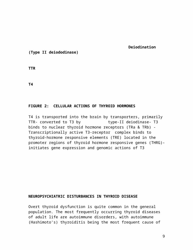
FIGURE 2: CELLULAR ACTIONS OF THYROID HORMONES
T4 is transported into the brain by transporters, primarily TTR- converted to T3 by type-II deiodinase- T3 binds to nuclear thyroid hormone receptors (TRa & TRb) - Transcriptionally active T3-receptor complex binds to thyroid-hormone responsive elements (TRE) located in the promoter regions of thyroid hormone responsive genes (THRG)-initiates gene expression and genomic actions of T3
NEUROPSYCHIATRIC DISTURBANCES IN THYROID DISEASE
Overt thyroid dysfunction is quite common in the general population. The most frequently occurring thyroid diseases of adult life are autoimmune disorders, with autoimmune (Hashimoto’s) thyroiditis being the most frequent cause of
9

hypothyroidism (inadequate hormone production), and Graves disease being the most frequent cause of hyperthyroidism (excess hormone production). The prevalence of hyperthyroidism in the general population varies from 0.2% to about 0.5% and is associated with Graves's disease in 80%of cases. The prevalence of clinical hypothyroidism is approximately 2% in women and less than 0.1% in men. Subclinical thyroid dysfunction, defined as serum TSH levelsoutside the normal reference range with normal levels of free thyroxine (FT4) and free triiodothyronine (FT3), is even more common, being found in 2%-10% of the general population. Approximately 5% to 15% of patients with subclinical hypothyroidism progress to overt hypothyroidism per year. Both overt and subclinical thyroid dysfunction are more common among women and the elderly [3, 6, 7, 9]
Both hyperthyroidism and hypothyroidism are associated with a number of changes in mood and intellectual performance. [3,
7] Neuropsychiatric symptoms commonly associated with thyroid dysfunction are listed in table-1.
Table -1: Common neuropsychiatric symptoms in thyroid dysfunction
Hyperthyroidism Hypothyroidism
Insomnia RetardationAgitation Poor appetiteIrritability Poor sleepAnxiety Fatigue Emotional lability Lethargy Poor concentration ApathyDysphoria WeaknessApathy Low moodLethargy Poor concentrationCognitive Cognitive
10

impairments impairments
Though symptoms of hyperthyroidism may overlap with those ofa manic episode, instances of hyperthyroidism-induced mania are rare, and appear to be limited to patients with a previous history or strong family history of bipolar disorder. Severe hypo-thyroidism often mimics melancholic depression and dementia. [3]
Neuropsychiatric impairments accompanying dysfunction of thethyroid gland are usually reversed rapidly following hormonal supplementation, although severe hypothyroidism, ifleft untreated, may rarely result in irreversible dementia.
[3, 7]
Even though thyroid disorders are associated with psychiatric symptoms in clinical populations, existence of asimilar association in general population is controversial and inconsistent. Certain large community-based studies havefound no association between different parameters of thyroiddysfunction and psychiatric symptoms such as depression or anxiety. [11-14] On the other hand other community studies have reported increased rates of hospitalization for mood disorders, or the existence of persistent psychological impairments in a subset of patients. [15 -17] Reviews of the subject are equally inconclusive. Some have suggested only aweak link between overt or subclinical thyroid alterations and neuropsychiatric disturbances, [18, 19] while others have found that higher levels of thyroxine in the normal range are associated with increased risk of depression, indicatingthat the effects of thyroid hormone on mood may differ in normal populations and patients with clinical thyroid dysfunction. [13]
THYROID DYSFUNCTION IN PSYCHIATRIC DISORDERS
11
It is now widely accepted that thyroid hormone continues to play a critical role in the adult brain, influencing mood and cognition, although the details remain to be elucidated.There is also general consensus that small changes in the HPT axis, even within the normal range, among patients with psychiatric disorders can have significant effects on brain functions. Such changes in thyroid hormone status may produce a cascade of biological events eventually leading tothe onset of psychiatric disorders. Additionally, in patients with primary psychiatric disorders, thyroid hormones appear to be capable of modulating the phenotypic expression of their illness, or their response to treatment.
.In psychiatric populations, regardless of specific diagnoses, thyroid dysfunction is more common than in the general population. Accordingly, disturbances of the HPT axis have been identified in a number of different psychiatric conditions. However, the bulk of the research evidence relates to the broad spectrum of mood disorders including unipolar and bipolar affective disorders. Therefore, the predominant focus of this chapter is on HPT axis dysfunction in mood disorders.
HPT AXIS DYSFUNCTION IN DEPRESSION
The vast majority of patients with depression do not have overt thyroid disease.Although most patients with depression have normal T3, T4 and TSH circulating levels, there is evidence of altered activity of the HPT axis in some of them.
Iodothyronines in depression: thyroxine (T4), triiodothyronine (T3) and reverse T3 (rT3) The most commonly observed abnormality in thyroid function among patients with depression is"euthyroid
12
hyperthyroxinemia" - a relative increase of total or free serum T4 and a relative decrease of T3 levels. Nearly 20% - 30% of the patients with depression have levels of total andfree T4 levels above the normal limit. As patients achieve remission, T4 and FT4 levels have been observed to drop. Transient elevations of thyroxine have similarly been observed in patients hospitalized for medical or psychiatricillnesses, with spontaneous normalization within 2 weeks after admission; indicating that this change could be due tonon-specific effects of stress. T4 levels also decline with treatment with a variety of pharmacological agents such as antidepressants, carbamazepine or lithium, as well as non-pharmacological treatments, such as sleep- deprivation, light therapy, electroconvulsive therapy and psychotherapy. [1, 3, 7, 20, 21] The association between clinical improvement and a drop in T4 levels has led to the speculation that depression results from a state of relative T4 excess, with the mood-enhancing effects of T3 resulting from its down-regulation of T4 levels. [7] Turnover studies using radiolabelled T4 have also shown increased daily production rates of T4, indicating that the thyroid gland is abnormallystimulated in depression. [22] Increase in peripheral T4 levels is paralleled by reports of increased concentration of free T4 in the cerebrospinal fluid (CSF) during depression, which declines with clinical recovery. This further suggests that depression is associated with a state of relative hyperthyroidism. [23] Hypercortisolism, which is commonly prevalent in depression, can lead to an activation of the hypothalamic neurons producing excess TRH,and causing over-activity of the HPT axis. Excess cortisol may also inhibit conversion of T4 to T3, leading to the observed increase in T4. [1] However, the pathophysiological significance of these changes still remains uncertain, more so because not all studies have reported similar changes among either patients with major depression, or following their treatment. [24- 27] However, this inconsistency may reflect heterogeneity in patient populations and other methodological differences across different studies, rather that the lack of an abnormality.
13

Serum T3 levels in depressed patients are often found to be normal in depression, though some studies have found reducedlevels, typically in patients with more severe depression. [1, 20] The combination of an increased T4 and an unaltered T3in depression suggests a reduced deiodination of T4 into T3,which is also seen in physical (non-thyroidal) illnesses, but the availability of substrate T4 is higher in depression. [20]
Reverse T3 (rT3) is the inactive analogue of T3. Elevated levels of serum total, as well as free rT3, have been reported among patients with unipolar depression and women with mania, but other studies have been unable to replicate this finding. [20] CSF levels of rT3 have been studied in different types of depression, and have been found to be highest in the endogenous type. [28] Elevated rT3 levels in depression suggest the possibility of a defect in the brain deiodinases. It has been proposed that depression causes an inhibition in the deiodinase type-II enzyme, probably due tothe increase in cortisol levels. This enzyme is responsible for the transformation of T4 into T3 in the brain, and, consequently, its inhibition triggers the conversion of T4 by the other enzyme, type-III brain deiodinase, into rT3.
Increase in the CSF and serum rT3 further inhibits deiodinase type-II and reinforces the altered conversion of T4. However, the ratio of CSF to serum rT3 is very high in normal subjects and those with depression. This ratio does not change even on recovery from depression. This indicates that rT3 changes found in depression are unlikely to be involved in its pathogenesis. [1, 20]
Thyroid stimulating hormone in depression
Currently, the most diagnostically sensitive tests to detectthyroid disease are the ultra-sensitive immunoradiometric
14

assays of serum TSH. They are, therefore, the primary screening tests for both subclinical and overt hypothyroidism and hyperthyroidism. [3] Although some studies have found reduced basal serum levels of TSH among patients with depression, compared to healthy controls, [29] these levels are often within the normal range. [30]
However, the presence of altered TSH secretion in depressionis revealed by the loss of the nocturnal TSH rise, which usually occurs between midnight and 3 a.m. In this regard, the mean plasma concentration of TSH during sleep, its nocturnal peak concentration, and the amplitude of its circadian rhythm, have all been reported to be lower in subjects with depression, compared with both normal controlsand remitted patients. [31, 32] Finally, the time of the nocturnal TSH peak has also been found to be advanced duringa depressive episode. The absence of a nocturnal surge in TSH is believed to be a more sensitive indicator of an HPT abnormality than a blunted TSH response to TRH. After the complete recovery from depression this nocturnal variation of TSH is re-established, but in instances in which the recovery from depression does not occur, or is incomplete, this change in TSH remains attenuated or absent. Partial sleep deprivation which has an antidepressant effect, leads to the reestablishment of the nocturnal increase of TSH withthe consequent increase in T3 and T4 serum levels. Therefore, this nocturnal reduction in TSH could lead to a global decrease in the secretion of thyroid hormones, generating a certain degree of central hypothyroidism in some patients with depression. [1, 2, 20, 25, 31, 32] One putative mechanism to explain the decreased TSH secretion would be chronic hypersecretion of hypothalamic TRH, which would decrease the number of pituitary TRH receptors. Such a hypothesis is consonant with the finding of increased CSF –TRH in some, but not all studies, of patients with depression. [2] There is also some evidence to suggest that the reduction in TSH could be secondary to the
15

hypercortisolism prevalent in depression, since it is well known that glucocorticoids inhibit TSH secretion. However, other data do not support the notion that HPT abnormalities in depression are secondary to HPA axis dysfunction. [2, 25]
In direct contrast, certain studies have reported increased levels of serum TSH among patients with depression. In one such study, patients with endogenous depression had significantly higher serum TSH levels than subjects with hypothyroidism being treated with thyroxine. [33] The authors concluded that this indicated an inappropriate secretion of TSH (in relation to the elevated thyroidal production of T4)in patients with endogenous depression, compatible with somedegree of central over-stimulation of the thyroid in the untreated depressed state. Nevertheless, these findings may have only indicated that patients with hypothyroidism were receiving high doses of hormonal supplements. Nevertheless, others have also reported higher serum TSH levels among patients with depression, particularly among outpatients. [1,
34] Increased TSH in depression has been attributed to chronic stimulation of pituitary thyrothrophs by elevated TRH or reduced somatostatin, which normally inhibits TSH, but these changes have not been consistently replicated. [1,
2, 20] Elevated TSH also appears to be more likely among certain subgroups such as atypical, treatment-resistant and bipolar depressions. [20, 34]
TSH response to TRH in depression
Prior to the development of highly specific and sensitive TSH assays, the TSH response to an intravenous dose of TSH was the most widely used test for detecting HPT dysfunction.Though many variations of the test exist, the standardized protocol consists of a drawing a baseline sample of TSH and then infusing 500 micrograms of TRH intra-venously over 1 minute, following which a second TSH sample is drawn 30 minutes later. A normal response is a rise in TSH of 5 muU/mL or more above the baseline level. The response is exaggerated in hypothyroidism and blunted in
16
hyperthyroidism. More liberal definitions (i.e. less than 7 muU/mL), as well as more conservative definitions (i.e., less than 2.5 muU /mL) of ‘blunting’ have also been used across studies. [35] Another modification of the test utilizesa parameter called TSH, which is defined as the difference between morning and evening TSH responses. This has been demonstrated to detect HPT abnormality at a higher sensitivity rate of 89% among patients with major depression, than the conventional TRH test. [2, 8]
Soon after the advent of the TRH test in endocrinology came the discovery of a blunted TSH response to TRH in some patients with depression, originally reported in 1972, [36]
and subsequently confirmed by several other groups. As data accrued it became clear that a blunted TSH response occurs in 25%–30% of patients with DSM-III- or RDC-defined major depression. [37] Whether patients with a blunted response represented a distinct subgroup, or the lower end of a single population, however, remained unclear. Rates of blunted TSH response were found to be much lower (3%-16%) inambulatory patients with major depression than those hospitalized for depression. [1]
Moreover, as additional diagnostic groups were studied, similar blunted responses were observed among diverse groupsof patients such as those with schizophrenia, panic disorder, alcohol problems, personality disorders and eatingdisorders etc. [2, 8] Given these findings, it became apparentthat the specificity of the blunted TSH response to TRH for depression was poor. This, combined with its low sensitivity, prevented the use of the TRH test as a screening tool for depression. Separate from standard diagnostic categories, psychopathological features reported to be associated with a blunted TSH response have included suicidality, agitation, and panic. [2, 8] However, these findings have not been sufficiently replicated to establish them as reliable correlates of the blunted TSH response. Some evidence also indicated that patients, whose blunted TSH response to TRH failed to normalize after a successful
17
course of antidepressant medication or ECT, were more prone to develop a relapse. [2, 7, 8] Then again, not all studies of patients being treated with antidepressants have confirmed the value of a blunted TRH test in predicting recovery from depression. [1, 20]
The simplest explanation for the blunted TSH response could be that it results from increased feedback inhibition by thyroid hormones resulting from the transient hyperthyroxinemia commonly accompanying depression. However,this would not explain the persistence of the blunted TSH response even after remission of depression. Another leadinghypothesis to explain the blunted TSH response in depressionis hypersecretion of hypothalamic TRH, which should lead to desensitization of pituitary TRH receptors and impair the process of TSH release. In support of the TRH hypersecretionhypothesis are reports of increased CSF-TRH among patients with major depression compared to neurological controls or medically healthy controls. However, this increase has neither been reported consistently, nor have any of these reports found an association between CSF-TRH and a blunted TSH response. [2, 35] Another possible explanation for the blunted TSH response in depression is the hypercortisolemia commonly observed in the condition, since cortisol is known to inhibit TSH release. Then again, the evidence of blunted TSH response being secondary to such HPA axis changes is also not always consistent. [2, 35]
While a blunted TSH response to TRH occurs in some patients with depression, an exaggerated response occurs in others. It is estimated that nearly 10% to 17% of patients with depression show an exaggerated TSH response to the TRH test,though higher rates have been reported by others. [1] This latter phenomenon supports the concept of an association of depression with subclinical hypothyroidism. Patients with subclinical hypothyroidism show neither classical signs and symptoms, nor reductions in levels of thyroid hormones. The diagnosis depends upon either an elevated basal TSH or an
18
exaggerated TSH response to TRH. [1, 2] An exaggerated response to TRH has been found among patients positive for thyroid autoantibodies. [38] The combination of an exaggeratedTRH test, elevated basal TSH, subclinical hypothyroidism andasymptomatic autoimmune thyroiditis is more likely among patients with bipolar depressions, particularly those suffering from rapid cycling affective disorders (RCAD), as well as patients with treatment-refractory depressions. [1, 2,
8, 20]
Thyrotropin releasing hormone in depression
As mentioned above, a few studies have evaluated levels of CSF-TRH in relation to the blunted TSH response to TRH, among patients with depression; the results of these do not reveal a consistent picture. Some of these reports among untreated patients have suggested that CSF –TRH is elevated among those with endogenous depression and “severe melancholic depression with psychosis. [42, 43] However, a correlation with severity of depression or blunting of TSH response to TRH has not been found. Others have either been unable to find elevated CSF-TRH levels and its associations with basal TSH levels, [35, 44] or have found a negative association between the two measures. [45] These findings are thus not consistent with proposed hypothesis of hypothalamicTRH hypersecretion and subsequent pituitary down-regulation,causing a blunted TSH response to TRH in depression. [35]
Transthyretin in depression
Transthyretin is one of the T4 serum-transporting proteins, which displays a certain specificity for transportation of T4 into the brain.[1] Lowered CSF -TTR in patients with depression, compared to healthy controls or subjects with neurological disorders, have been found in some studies. [46 -
48] It has been suggested that suggested that the low levels of this transporting protein may cause a degree of ‘brain
19

hypothyroidism’ accompanied by peripheral concentrations of thyroid hormones within the normal range. This could lead tothe treatment-resistance found among patients of one such study. [46] An association with serotonergic dysfunction and suicidal ideation was also demonstrated, [48] but could not be replicated subsequently. [49] Moreover, animal models have shown that complete absence of transthyretin has no impact on thyroid functions, thus indicating the importance of other membrane carrier systems such as MCT8 or OATP1C1 to maintain thyroid hormone homeostasis within the brain. [3]
Subclinical hypothyroidism in depression
Different grades of hypothyroidism are enumerated in table-3. Subclinical hypothyroidism is characterised by a serum TSH level elevated above the statistically defined upper limit of the reference range, in association with a serum free T4 level within the reference range. [1, 3] Subclinicalhypothyroidism, by definition, does not produce the featuresof clinical hypothyroidism, but subtle somatic symptoms, cognitive deficits, and mood changes have been observed in association with this condition. These symptoms generally improve following thyroid hormone treatment. [1, 7] Approximately 5% to 15% of patients with subclinical hypothyroidism progress to overt hypothyroidism per year. Patients with antithyroid antibodies are at a particularly high risk, with 80% progressing to overt hypothyroidism within 4 years. [7] Among patients with depression, the prevalence of subclinical hypothyroidism has been reported to be both lower and higher than the general population rate. [7] At arough estimate approximately 15% of patients with depressionhave subclinical hypothyroidism. [3, 39-41] By contrast, the prevalence of subclinical hypothyroidism in adults without known thyroid disease is approximately 2%-10%, although the general population prevalence increases with age and in women, such that subclinical hypothyroidism is present in upto 20% of women aged over 60 years. [7, 9, 19] Treatment response to antidepressants may be impaired among patients
20

with subclinical hypothyroidism. [1, 7] Indeed, much higher rates of the condition (15% -52%) have often been reported among patients with treatment-refractory depression. [1]
Treatment-resistant patients with subclinical hypothyroidismalso appear to be significantly more likely to respond to T3augmentation, than euthyroid patients with treatment-resistant depression. [7] However, recent data have raised doubts about the utility of thyroid measures such as serum TSH in predicting antidepressant response. [41]
Table-2: Grades of hypothyroidism
Grade I Clinical features of overt hypothyroidism T3, T4 low TSH elevated
Grade II No overt featuresT3, T4 – normalTSH elevated
Grade III T3, T4, TSH – normal Exaggerated TSH response toTRH
Grade IV Presence of antibodies, other thyroid functions normal
Antithyroid antibodies in depression
There are three important antibodies that are involved in thyroid autoimmunity - anticolloid antibodies which mainly react with thyroglobulin, anticytoplasmic antibodies which react with components in the microsomal fraction of the thyrocytes (antimicrosomal antibodies) and are specific for the enzyme thyroid peroxidase, and TSH-receptor antibodies. [1, 3] Autoimmune thyroid disease, marked by the presence of antithyroid antibodies, is a major cause of thyroid dysfunction. In general, people with antithyroid antibodies
21
have diminished thyroid gland function compared with people who are free of these antibodies. [7] Nine to twenty percent of patients hospitalized for depression are positive for antithyroid antibodies. [38, 40] Patients with depression seem to have a higher prevalence of autoimmune thyroiditis compared to the normal population rates of 5% to 10%. [40]
Other indices of thyroid function are generally normal in these patients, hence they are said to be suffering from ‘symptom less autoimmune thyroiditis.’ However, elevated thyroid antibodies have only been reported in some studies of patients with unipolar depression, [38, 40, 50-53] but not in others. [34, 54-56] Autoimmune thyroiditis is found in nearly 15% of depressed patients with exaggerated TSH response to the TRH stimulation test. Associations with features such as apathy,atypical symptoms, melancholia and treatment resistance havealso been noted. [1] This has led to the suggestion that subclinical thyroid hypofunction, especially in patients with melancholic or refractory depressions are possibly of an autoimmune origin. Antithyroid antibodies could produce the state of relative CNS hypothyroidism proposed to the principal pathogenetic mechanism in such patients.
Postpartum changes in mood seem to be particularly associated with the presence of anti-thyroid antibodies, andthyroid dysfunction. Though not consistently noted in all such patients, [7]] thyroid autoimmnunity may play a key rolein onset of the condition in a subgroup of women with postpartum depression. [50, 57 - 59] Among otherwise euthyroid women, rates of autoimmune conditions rise following delivery. In the 6 months following childbirth, approximately 5% to 9% of women experience thyroid dysfunction, compared with a rate of 3% to 4% in the generalpopulation, and up to 12% have been found positive for thyroid antibodies. These increased rates are believed to result from a "rebound" immune phenomenon after delivery, following the immunosuppressant effect of high cortisol levels during pregnancy. [7] In contrast, women suffering from postpartum depression have been noted to have a much higher
22
rate of antithyroid antibodies in several studies. [50, 58, 60, 61]
Evidence also suggests that thyroid autoimmunity may be affected by the HPA axis abnormalities, which mediate the balance between pro-inflammatory and anti-inflammatory cytokines. This could explain the association between thyroid antibodies and postpartum mood disorders. [52]
HPT AXIS DYSFUNCTION IN BIPOLAR AFFECTIVE DISORDER
Although most patients with bipolar disorder are euthyroid, thyroid measures in the low-normal or below-normal range seem to be relevant for the pathophysiology of bipolar affective disorders (BPAD). Overt hypothyroidism is much more common in patients suffering from BPAD treated with lithium carbonate than in unselected psychiatric inpatients.[62]
Little data exist on the prevalence of hyperthyroxinemia in patients diagnosed with mania. In a study of 31 manic patients, one third had elevated free and total T4 levels atadmission, although other mean thyroid indices were within the normal range. [63] However, this finding is not specific to mania, as transient mild elevations of free and total T4 (“euthyroid hyperthyroxinemia”) have been commonly noted in acutely admitted psychiatric patients, including those with depression. [7]
Indices of thyroid dysfunction may, however, predict a less than optimal outcome among patients with BPAD. An association of low levels of T4, within the normal range, with greater mood instability and depression severity, has been reported during prophylactic lithium treatment in patients with bipolar disorder. [64] Similarly, lower FT4 index and higher TSH, although within the normal range, weresignificantly associated with a poorer treatment response during the acute depressed phase of bipolar disorder. [65]
Furthermore, the free T4 index was found to be inversely
23
related to the length of hospital- stay in males with affective disorders. [66]
Patients with a bipolar disorder may show an exaggerated response of TSH to TRH, consistent with the high prevalence of subclinical hypothyroidism in this condition. This difference between patients with unipolar and bipolar disorder, however, remains controversial. [62]
Subclinical forms of hypothyroidism are common in BPAD. Lithium treatment predisposes not only to clinical, but alsoto subclinical hypothyroidism. This may account for the higher rates of subclinical hypothyroidism found among patients with bipolar disorder, compared to the general population. [7, 62] However, the fact that both overt and subclinical hypothyroidism has been found to be more common in those with the rapid cycling disease than in the unselected patients with bipolar disorder, suggests that hypothyroidism is unlikely to be due solely to lithium, as lithium treatment was a common feature of both groups. [62]
Studies are equivocal as to whether thyroid antibodies are elevated in patients with bipolar disorder, and as to whether lithium exposure is associated with the occurrence of the higher prevalence of thyroid antibodies in BPAD. One study found a higher prevalence of thyroid peroxidase antibodies in bipolar disorder (28%) than in the general population or in non-bipolar psychiatric disorders. [67] A positive antibody status was associated with an increased risk for lithium-induced hypothyroidism but not with currentor former lithium treatment. However, results from other studies are inconsistent with reported rates in the range 0–43%. [68 -73] These are thus not very different community prevalence rates of approximately 12–18% of thyroid peroxidase antibodies. [3] Age, gender and sensitivity of antibody assays are important confounding factors. [73] It hasbeen proposed that autoimmune thyroiditis, with thyroid peroxidase antibodies as markers may be a potential endophenotype for bipolar disorder, and is related to the
24

genetic vulnerability to develop bipolar disorder, rather than to the disease process itself. [74] Although the offspring of parents with bipolar disorder were found to have increased vulnerability to develop thyroid autoimmunitycompared to high-school aged controls, this was independent of any psychiatric disorder or symptoms. [75]
Many studies on the prevalence of antibodies in patients with mood disorders have not controlled for previous exposure to lithium, which may promote the production of antithyroid antibodies. However, one study that did control for lithium exposure found no difference in the prevalence of antibodies in patients with unipolar depression, althoughpatients with bipolar depression did have a slightly higher rate (13%) of antithyroid antibodies than in the general population. [70] Thus the prognostic significance of antithyroid antibodies in patients with bipolar disorders, including the implications for likelihood of treatment response, still remains to be clarified. [7]
HPT AXIS DYSFUNCTION RAPID CYCLING, MIXED AFFECTIVE AND SEASONAL AFFECTIVE DISORDERS
Certain subtypes of bipolar disorder may be particularly associated with HPT axis dysfunction.
Rapid-cycling affective disorder (RCAD) has been associated with HPT axis abnormalities across several studies. All manner of HPT axis dysfunctions have been reported in RCAD. These have included abnormalities of T4 and TSH, exaggeratedTSH response to TRH, higher prevalence of thyroid diseases, indices of lowered thyroid function indicating subclinical hypothyroidism, elevated rates of thyroid antibodies, as well as overt hypothyroidism. [3, 6, 7, 76-80] However, other studies have refuted such associations. Methodological factors such as retrospective designs and lack of controls hinder any consistent conclusions from this data. [77-79] Morepertinently, many of these studies have included patients
25
being treated with lithium, which has known antithyroid effects. Accordingly, cross-sectional studies of unmedicatedpatients with RCAD have usually found little evidence of HPTaxis dysfunction. [77-79] Evidence has also been presented to suggest that RCAD is associated with a latent hypofunction of the HPT axis; this dysfunction may become manifest with lithium treatment. [78] Although the outcome of research is inconsistent, clinical consensus suggests that that even biochemical evidence of hypothyroidism can induce rapid cycling. However, correction of biochemical hypothyroidism is not always effective in rectifying rapid cycling. [77]
Unrelated to lithium use, mixed states have also been associated with reduced thyroid functioning in certain studies, [7, 25, 79, 81, 82] although others have failed to documentthis association. [7, 79] It has been well-documented that in healthy people T3, T4 and TSH levels as well as TSH responseto TRH are somewhat different in winter than in summer. This has prompted the investigation of HPT axis dysfunction in patients with seasonal affective disorders. However, the few studies that have explored thyroid indices among such patients have been unable to find any evidence of HPT axis disturbance. [83, 84]
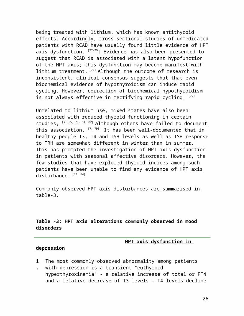
Commonly observed HPT axis disturbances are summarised in table-3.
Table -3: HPT axis alterations commonly observed in mood disorders
HPT axis dysfunction in depression
1.
The most commonly observed abnormality among patients with depression is a transient "euthyroid hyperthyroxinemia" - a relative increase of total or FT4and a relative decrease of T3 levels - T4 levels decline
26

as patients remit, or receive antidepressant treatment. 2.
Absence of a nocturnal surge in TSH, which is believed to be the most sensitive indicator of a HPT abnormality,is also present – indicative of a certain degree of central hypothyroidism in some patients with depression.
3.
Elevated TSH has been reported - appears to be more likely among certain subgroups such as atypical, treatment-resistant and bipolar depressions.
4.
A blunted TSH response to TRH occurs in 25%–30% of patients with DSM-III- or RDC-defined major depression. Specificity and sensitivity of the test is poor. Some evidence also indicates that patients whose blunted TSH response to TRH fails to normalize after a successful course of antidepressant medication or ECT are more prone to develop a relapse.
5.
Nearly 10% to 17% of patients with depression show an exaggerated TSH response to the TRH test, indicative of subclinical hypothyroidism. Often found among patients positive for thyroid antibodies. More likely among patients with bipolar depressions, particularly those suffering from rapid cycling affective disorders, as well as patients with treatment-refractory depressions.
6.
Approximately 15% of patients with depression have subclinical hypothyroidism. Treatment response to antidepressants may be impaired among patients with subclinical hypothyroidism. Rates of subclinical hypothyroidism are higher in treatment-resistant depression.
7.
About 9% -20% of patients hospitalized for depression are positive for antithyroid antibodies; these rates areconsiderably higher than the general population rates ofautoimmune thyroiditis, but only in some studies. Postpartum depression is particularly associated with thyroid autoimmunity.
HPT axis dysfunction in bipolar affective disorder
1 HPT axis dysfunction is often associated with greater
27

. mood instability and poorer treatment-outcome.2.
An exaggerated response of TSH to TRH found is consistent with the high prevalence of subclinical hypothyroidism in BPAD. Some, but not all of the subclinical pathology, results from treatment with lithium.
3.
Inconsistent elevations of antithyroid antibodies – not always attributable to effect of lithium are present.
4.
RCAD has been associated with overt and subclincal hypothyroidism in some, but not all of the studies. RCADmay be associated with latent dysfunction of the HPT axis, which is unmasked by treatment with lithium.
HPT AXIS DYSFUNCTION IN OTHER PSYCHIATRIC DISORDERS
Apart from mood disorders HPT axis disturbances have been occasionally reported among other psychiatric disorders. Theassociation of thyroid deficiency and mental retardation is well known. In the perinatal period, thyroid deficiency results in irreversible brain damage and mental retardation.This syndrome is referred to as cretinism. [3, 7] More recently, it has been demonstrated that mutations in transporter proteins such as MCT8 can also result in severe mental retardation, axial hypotonia, and speech retardation.[3, 6] The potential association between Alzheimer’s disease and thyroid dysfunction has also been investigated and foundto be ambiguous. [3, 9] Non-specific decrease in serum concentrations of T4 or FT4 I, or both, has been associated with the diagnosis of alcohol dependence, but is possibly due to the direct toxic effects of alcohol upon thyroid function. [14] HPT axis abnormalities have also been documented in psychotic conditions such as schizophrenia, aswell as eating disorders. [85] The association between thyroid dysfunction and anxiety disorders, particularly panic disorder, generalized anxiety disorder and post-
28
traumatic stress disorder has been documented more consistently. [10, 14, 86, 87]
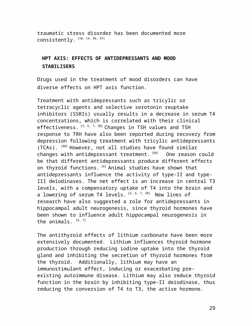
HPT AXIS: EFFECTS OF ANTIDEPRESSANTS AND MOOD STABILISERS
Drugs used in the treatment of mood disorders can have diverse effects on HPT axis function.
Treatment with antidepressants such as tricylic or tetracyclic agents and selective serotonin reuptake inhibitors (SSRIs) usually results in a decrease in serum T4concentrations, which is correlated with their clinical effectiveness. [3, 6, 7, 20] Changes in TSH values and TSH response to TRH have also been reported during recovery fromdepression following treatment with tricylic antidepressants(TCAs). [88] However, not all studies have found similar changes with antidepressant treatment. [88] One reason couldbe that different antidepressants produce different effects on thyroid functions. [6] Animal studies have shown that antidepressants influence the activity of type-II and type-III deiodinases. The net effect is an increase in central T3levels, with a compensatory uptake of T4 into the brain and a lowering of serum T4 levels. [3, 6, 7, 20] New lines of research have also suggested a role for antidepressants in hippocampal adult neurogenesis, since thyroid hormones have been shown to influence adult hippocampal neurogenesis in the animals. [6, 7]
The antithyroid effects of lithium carbonate have been more extensively documented. Lithium influences thyroid hormone production through reducing iodine uptake into the thyroid gland and inhibiting the secretion of thyroid hormones from the thyroid. Additionally, lithium may have an immunostimulant effect, inducing or exacerbating pre-existing autoimmune disease. Lithium may also reduce thyroidfunction in the brain by inhibiting type-II deiodinase, thusreducing the conversion of T4 to T3, the active hormone.
29

These effects produce persistent elevation of serum TSH concentrations, inconsistent reductions in the serum concentrations of T4 and/or T3, abnormal TSH responses to TRH and production of antithyroid antibodies. Clinically this may result in goitre or overt hypothyroidism. Rates of overt hypothyroidism among patients on long-term treatment with lithium vary widely from 2% to 40%. Subclinical hypothyroidism is estimated to occur in about 20% of the patients. The prevalence of lithium-induced goitre has generally ranged between 5% to 15%, although some reports find prevalence rates up to 50%. Antithyroid antibodies arefound in about 15% of patients on lithium, though rates are higher among those with longer exposure to lithium. Risks ofhypothyroidism are somewhat higher in middle-aged women who have a preexisting vulnerability to autoimmune thyroiditis, and those with a rapid cycling course. The average duration of lithium therapy before the diagnosis of hypothyroidism isusually 6-18 months, though hypothyroidism can develop as early as 2-3 months. HPT axis dysfunction can normalize following prolonged treatment or following cessation of therapy. [6, 7, 20, 89, 90] The development of hypothyroidism is not a contraindication to continuing lithium. Thyroid supplementation is recommended instead, though there is considerable disagreement about when this should be started.Nevertheless, overt hypothyroidism, clear evidence of subclinical pathology and the presence of rapid cycling or treatment-resistance are unequivocal indications for thyroxine supplementation. [7]
Carbamazepine also influences the HPT axis in a complex manner. Carbamazepine increases activity of type-II deiodinase and inhibits type-III deiodinase, thus resulting in increased T3 levels. Carbamazepine also increases T4 and T3 metabolism through induction of hepatic microsomal drug-metabolizing enzymes. Patients on carbamazepine usually havelow to normal serum levels of T4, T3 and TSH levels. However, they generally do not develop overt hypothyroidism.[7, 20]
30

THYROID HORMONES IN TREATMENT OF MOOD DISORDERS
In the 1930s, Norwegian physicians first used desiccated sheep thyroid gland to treat patients with cyclic mood disorders. [3, 7] This was followed by several attempts at hormone treatments of psychiatric disorders, which were not very successful. However, beginning in the late 1960s a series of open and controlled clinical trials confirmed the therapeutic value of adjunctive treatment with thyroid hormones in mood disorders. Since then many more similar trials have been conducted. The evidence suggests that as a means of augmenting and accelerating response to antidepressant treatment, thyroid hormones are the next bestalternative to lithium carbonate.
Treatment of depression
Thyroid hormones have occasionally been used alone to treat depressive disorders. In the 1970s a number of studies demonstrated a mild but transient improvement in mood in depressed patients treated with TRH, which persisted for a few days or at most 3 weeks. Later other studies, including controlled double-blind trials (RCTs), failed to demonstrateany beneficial effect of TRH. [8, 20] Promising short-term and long-term antidepressant effects of intrathecal and intravenous TRH have been documented subsequently. However, these small open trials have not been replicated. [91, 92] Sole treatment with either T3 or T4 has been compared to antidepressants in a few trials; results have been equivocaland mostly disappointing. [20]
Most of the research evidence, however, relates to the use of thyroid hormones in augmenting effects of antidepressant drugs. Older trials investigated the effects of the combination of T3 or T4 with TCAs; more recently several trials have been conducted among patients being treated withSSRIs. An initial meta-analysis of T3 augmentation in patients who showed inadequate response to tricyclic
31

antidepressants included eight studies with a total of 292 euthyroid patients with depression. [93] Patients treated withT3 augmentation were twice as likely to respond to treatmentcompared with the various control groups; improvements in depression scores were moderately large (effect size 0.62). A subsequent meta-analysis investigated the use of thyroid hormone supplementation to accelerate the treatment of depression. [94]. Six studies comprising of 125 patients were identified. All were conducted with T3 and a TCA. Five of the six studies found T3 to be significantly more effective than placebo in accelerating clinical response. The pooled, weighted effect size index was 0.58, and the average effect was highly significant. Further, the effects of T3 acceleration were greater as the percentage of women participating in the study increased. T3 has also been used to augment ECT. [95] Patients receiving T3 needed significantly fewer ECTs, and demonstrated less damage to memory functions, in comparison to the ECT and placebo in this study.
Augmentation of TCAs by T3 has been compared to augmentationwith lithium. In the initial randomized-controlled trial, 50patients with non-psychotic major depression, who were TCA non-responders were treated for 2 weeks with T3, lithium or placebo. [96] Results demonstrated that T3 and lithium weresuperior to placebo augmentation, and the augmenting efficacy of T3 and lithium was similar. More recently a muchlarger trial of T3 augmentation of SSRI treatment has been conducted as a part of STAR* D study. [97] A total of 142 adult outpatients with nonpsychotic major depressive disorder who had not achieved remission or who were intolerant to an initial prospective treatment with citalopram and a second switch or augmentation trial, were randomly assigned to augmentation with lithium or T3 for up to 14 weeks. T3 augmentation compared favourably with lithium augmentation in terms of remission rates, and was better tolerated. The lower side effect burden and ease of use of T3 augmentation thus actually suggested that it has
32
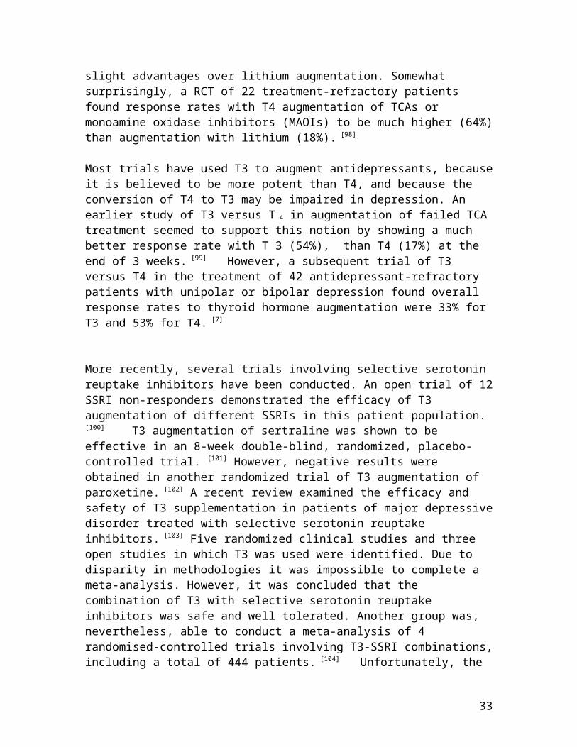
slight advantages over lithium augmentation. Somewhat surprisingly, a RCT of 22 treatment-refractory patients found response rates with T4 augmentation of TCAs or monoamine oxidase inhibitors (MAOIs) to be much higher (64%)than augmentation with lithium (18%). [98]
Most trials have used T3 to augment antidepressants, becauseit is believed to be more potent than T4, and because the conversion of T4 to T3 may be impaired in depression. An earlier study of T3 versus T 4 in augmentation of failed TCAtreatment seemed to support this notion by showing a much better response rate with T 3 (54%), than T4 (17%) at the end of 3 weeks. [99] However, a subsequent trial of T3 versus T4 in the treatment of 42 antidepressant-refractory patients with unipolar or bipolar depression found overall response rates to thyroid hormone augmentation were 33% for T3 and 53% for T4. [7]
More recently, several trials involving selective serotonin reuptake inhibitors have been conducted. An open trial of 12SSRI non-responders demonstrated the efficacy of T3 augmentation of different SSRIs in this patient population.
[100] T3 augmentation of sertraline was shown to be effective in an 8-week double-blind, randomized, placebo-controlled trial. [101] However, negative results were obtained in another randomized trial of T3 augmentation of paroxetine. [102] A recent review examined the efficacy and safety of T3 supplementation in patients of major depressivedisorder treated with selective serotonin reuptake inhibitors. [103] Five randomized clinical studies and three open studies in which T3 was used were identified. Due to disparity in methodologies it was impossible to complete a meta-analysis. However, it was concluded that the combination of T3 with selective serotonin reuptake inhibitors was safe and well tolerated. Another group was, nevertheless, able to conduct a meta-analysis of 4 randomised-controlled trials involving T3-SSRI combinations,including a total of 444 patients. [104] Unfortunately, the
33

results did not support the notion that simultaneous initiation of treatment of major depression with an SSRI andT3 was more effective than SSRI monotherapy. Finally, preliminary evidence has suggested than differences in efficacy of the T3-SSRI combination could be mediated by functional polymorphisms of type-II deiodinase. [105]
Therefore, although lithium carbonate is still the best validated and most preferred drug used for augmenting treatment of depression, thyroid hormones are perhaps the next best agents in this regard. Either T3 (up to 50 mcg/d) or T4 (up to 100-500 mcg/d) can be used as augmentation. T3 is preferred because of its faster onset/offset of action and less risk of toxicity, but is not available in India. T3augmentation can be started at 25 mcg/d and if tolerated increased to 37.5 - 50 mcg/d after 1 week. Patients should be monitored for thyrotoxicosis, which usually develops at doses of 75 mcg/day and above. Thyroid functions should be monitored closely. T 4 is usually started at 100 mcg/day increased by 25-50 mcg per week to a maximum of 500 mcg per day. After 2 weeks response is assessed, and in patients who are judged to have responded to the hormone can be continued for a further period of 2-3 months before it is gradually tapered off. Close monitoring of side effects is recommended. Potential interactions with oral anticoagulants, oestrogens, digitalis, cathecholamines and cholestyramine have also been reported. T3 and T4 are contraindicated in pregnancy and cardiovascular diseases. [7,
25]
Hypermetabolic doses of thyroxine in treatment of bipolar affective disorders
In a series of open-label studies, adjunctive treatment withhypermetabolic or supraphysiological doses of T4 was found to be effective in the maintenance treatment of patients with severe rapid cycling or resistant bipolar disorder, whodid not respond to standard measures. [3, 7, 20, 62]
34

Supraphysiological doses ofT4 may also have immediate therapeutic value in patients with antidepressant-resistant bipolar and unipolar depressions, during a phase of refractory depression. Doses of 150-500 mcg/d are employed to raise the FT4 index by 150%, which is adequate to suppress TSH and produce a response. Although treatment withsupraphysiological T4 requires close monitoring, the hyperthyroxinemia is tolerated surprisingly well. No seriouseffects, including loss of bone mineral density, were observed even in patients treated for extended periods. Given the paucity of effective treatment interventions for rapid-cycling bipolar disorder, the role of T4 merits further study in controlled trials. Till such time this remains an option of last resort. [7]
Treatment recommendations are summarised in table-4.
Table- 4: Adjunctive treatment with thyroid hormones in mooddisorders
1. Evidence from open trials, RCTs and meta-analyses has demonstrated that thyroid hormones are safe and effective agents for augmenting and accelerating antidepressant treatment.
2. The evidence favours adjunctive treatment of tricyclic antidepressants; evidence for selective serotonin reuptake inhibitors is somewhat equivocal
3. Female patients appear to benefit the most. 4. Though T3 (25-50 mcg/d) is the preferred agent, T4
(100-500 mcg/d) can also be used. 5. Response is usually evident by about 2 weeks;
35
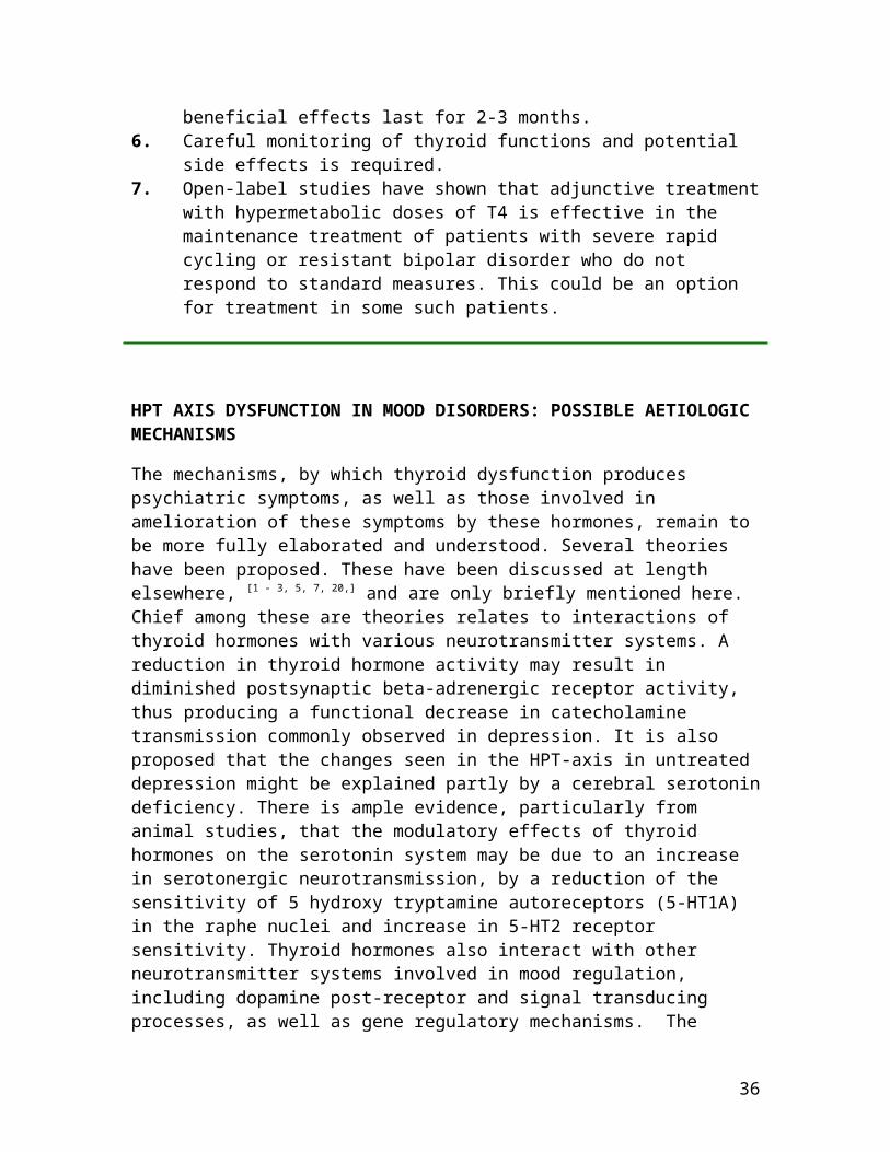
beneficial effects last for 2-3 months.6. Careful monitoring of thyroid functions and potential
side effects is required. 7. Open-label studies have shown that adjunctive treatment
with hypermetabolic doses of T4 is effective in the maintenance treatment of patients with severe rapid cycling or resistant bipolar disorder who do not respond to standard measures. This could be an option for treatment in some such patients.
HPT AXIS DYSFUNCTION IN MOOD DISORDERS: POSSIBLE AETIOLOGIC MECHANISMS
The mechanisms, by which thyroid dysfunction produces psychiatric symptoms, as well as those involved in amelioration of these symptoms by these hormones, remain to be more fully elaborated and understood. Several theories have been proposed. These have been discussed at length elsewhere, [1 - 3, 5, 7, 20,] and are only briefly mentioned here.
Chief among these are theories relates to interactions of thyroid hormones with various neurotransmitter systems. A reduction in thyroid hormone activity may result in diminished postsynaptic beta-adrenergic receptor activity, thus producing a functional decrease in catecholamine transmission commonly observed in depression. It is also proposed that the changes seen in the HPT-axis in untreated depression might be explained partly by a cerebral serotonindeficiency. There is ample evidence, particularly from animal studies, that the modulatory effects of thyroid hormones on the serotonin system may be due to an increase in serotonergic neurotransmission, by a reduction of the sensitivity of 5 hydroxy tryptamine autoreceptors (5-HT1A) in the raphe nuclei and increase in 5-HT2 receptor sensitivity. Thyroid hormones also interact with other neurotransmitter systems involved in mood regulation, including dopamine post-receptor and signal transducing processes, as well as gene regulatory mechanisms. The
36

underlying mechanism leading to the beneficial response of T3 is unknown, but may reflect brain hypothyroidism in the context of systemic euthyroidism. It is postulated that a similar situation prevails with regards to the thyroid axis and that the increased T4 in depression, as well as the blunted TSH response to exogenous TRH, reflects glucocorticoid activation of the TRH neurons leading to increased TRH secretion with resultant down regulation of the TRH receptors on the thyrotrope. Furthermore, within theCNS, the regulatory cascade through which thyroid hormones, particularly T3, exert their effects could involve deiodinase activity, nuclear binding to genetic loci, altered signal transduction and, ultimately, altered proteinsynthesis.
HPT AXIS DYSFUNCTION IN PSYCHIATRIC DISORDERS: IMPLICATIONS
Considerable evidence has accumulated over the years to suggest that overt and subclinical hypothyroidism is likely to be associated with the onset of psychiatric disorders, particularly mood disorders in a subset of patients. Even subtle thyroid abnormalities, such as the presence of thyroid antibodies in otherwise euthyroid individuals, may produce neurobehavioral symptoms and adversely affect treatment response.Routine thyroid function testing of psychiatric patients is not necessary, but certain populations should be screened carefully. These include patients with signs and symptoms suggesting overt hypothyroidism, those with rapid-cycling bipolar disorder, and with treatment-refractory depression. Women, who are within 6 months of childbirth, or are in their mid-40s, and patients on lithium, should also be monitored for thyroid function, as these are populations at relatively higher risk for thyroid abnormalities. Screening tests optimally include FT4 and TSH, and should be repeated at least yearly. More expensive tests such as antibody titres might be required for some.
37

Thyroid supplementation should be initiated for patients with elevated TSH and low FT4. Supplementation for patients with subclinical hypothyroidism is still a matter of considerable debate. Thyroid replacement in such patients can be considered in situations where serum TSH is in the high-normal range (over 10 muU/L), where antithyroid antibodies are detected, where patients have severe, refractory mood disorders such as RCAD, and where they exhibit deficits in memory or cognitive functioning.
Some patients receiving antidepressants appear likely to experience an augmentation and acceleration of treatment response on addition of thyroid hormones; patients with RCADnot responding to standard measures could also benefit from such treatment. Therefore, such patients should receive the benefit of hormonal interventions, wherever feasible.
REFERENCES
1. . Saint-Clair Bahlsa S-C, de Carvalho GA. The relation between thyroid function and depression: a review. Rev Bras Psiquiatr 2004; 26: 40-8.
2. Holsboer F. Neuroendocrinology of mood disorders. In: Bloom FE, Kupfer DJ, editors. Psychopharmacology: the
38

Fourth Generation of Progress. New York: Raven Press; 1995. pp 957-69.
3. Bauer M, Goetz T, Glenn T, Whybrow PC. The thyroid-brain interaction in thyroid disorders and mood disorders. J Neuroendocrinol 2008; 20: 1101-14.
4. Molina PE. Endocrine Physiology. 2nd ed. New York: McGraw-Hill Companies, Inc.; 2006.
5. Bauer M, Heinz A, Whybrow PC. Thyroid hormones, serotonin and mood: of synergy and significance in the adult brain. Mol Psychiatry 2002; 7: 140 – 56.
6. Bunevicius R. Thyroid disorders in mental patients. Curr Opin Psychiatry. 2009; 22: 391- 5.
7. Hendrick V, Altshuler L, Whybrow PC. Psychoneuroendocrinology of mood disorders. The hypothalamic-pituitary-thyroid axis. Psychiatr Clin North Am 1998; 21: 277-92.
8. Mason GA, Garbutt JC, Prange Jr AJ. Thyrotropin-releasing hormone. Focus on basic neurobiology. In: Bloom, FE, Kupfer DJ, editors. Psychopharmacology: The Fourth Generation of Progress. New York: Raven Press; 1995. pp 493-503.
9. Boelaert K, Franklyn JA. Thyroid hormone in health and disease. J Endocrinol 2005; 187: 1–15.
10.Prange Jr. AJ. Thyroid axis sustaining hypothesis of posttraumatic stress disorder. Psychosom Med 1999: 61: 139 – 40.
11.Patten SB, Williams JV, Esposito E, Beck CA. Self-reported thyroid disease and mental disorder prevalence
39
in the general population. Gen Hosp Psychiatry 2006; 28: 503 - 8.
12.um A, Bjøro T, Mykletun A, Dahl AA. An association between depression, anxiety and thyroid function–a clinical fact or an artefact? Acta Psychiatr Scand 2002; 106: 27–34.
13.Williams MD, Harris R, Dayan CM, Evans J, Gallacher J, Ben-Shlomo Y. Thyroid function and the natural history of depression: findings from the Caerphilly ProspectiveStudy (CaPS) and a meta-analysis. Clin Endocrinol. 2009; 70: 484- 92.
14.Hermann D, Hewer W, Lederbogen F. Testing the association between thyroid dysfunction and psychiatricdiagnostic group in an iodine-deficient area. J Psychiatry Neurosci 2004; 29: 444-9.
15.Thomsen AF, Kvist TK, Andersen PK, Kessing LV. Increased risk of developing affective disorder in patients with hypothyroidism: a register based study. Thyroid 2005; 15: 700 - 7.
16.Saravanan P, Chau WF, Roberts N, Vedhara K, Greenwood R, Dayan CM. Psychological well-being in patients on ‘adequate’ doses of l-thyroxine: results of a large, controlled community-based questionnaire study. Clin Endocrinol (Oxf) 2002; 57: 577 - 85.
17.Wekking EM, Appelhof BC, Fliers E, Schene AH, Huyser J,Tijssen JG, Wiersinga WM. Cognitive functioning and well-being in euthyroid patients on thyroxine replacement therapy for primary hypothyroidism. Eur J Endocrinol 2005; 153: 747– 53.
40
18.Samuels MH. Cognitive function in untreated hypothyroidism and hyperthyroidism. Curr Opin Endocrinol Diabetes Obes 2008; 15: 429 - 33.
19.Surks MI, Ortiz E, Daniels GH et al. Subclinical thyroid disease: scientific review and guidelines for diagnosis and management. JAMA 2004; 291: 228 - 38.
20.Kirkegaard C, Faber J. The role of thyroid hormones in depression. Eur J
Endocrinol 1998; 138:1-9.
21.Joffe RT. Is the thyroid still important in major depression? J Psychiatry Neurosci 2006; 31: 367-8.
22.Kirkegaard C, Kørner A, Faber J. Increased production of thyroxine and inappropriately elevated serum thyrotropin levels in endogenous depression. Biol Psychiatry 1990; 27: 472 - 76.
23.Kirkegaard C, Faber J. Free thyroxine and 3, 3’, 5’-triiodothyronine levels in cerebrospinal fluid in patients with endogenous depression. Acta Endocrinol 1991; 124:166-72.
24.Sullivan PF, Wilson DA, Mulder RT et al. The hypothalamic-pituitary-thyroid axis in major depression. Acta Psychiatr Scand 1997; 95: 370-78.
25.Seidman SN. Psychoneuroendocrinology of mood disorders.In: Stein DJ, Kupfer DJ, Schatzberg AF, editors. Textbook of mood disorders. Volume 1. Washington DC: American Psychiatric Publishing Inc.; 2006.pp 117-30.
26.Linnoila M, Gold P, Potter WZ, et al: Tricyclic antidepressants do not alter thyroid hormone levels in patients suffering from major affective disorder. Psychiatry Res 1981; 4: 457 – 60.
41
27.Joffe RT, Singer W: Effect of phenelzine on thyroid function in depressed patients. Biol Psychiatry 1987; 22:1033- 35.
28.Linnoila M, Cowdry R, Lamberg B-A, Makinen T, Rubinow D. CSF triiodothyronine (rT3) levels in patients with affective disorders. Biol Psychiatry 1983; 18:1489-92.
29.Frey A, Lampert A, Dietz K et al. Thyrotropin serum concentrations in healthy volunteers are associated with depression-related personality traits. Neuropsychobiology 2007; 56:123 – 26.
30.Maes M, Meltzer HY, Cosyns P, Suy E, Schotte C. An evaluation of basal
hypothalamic-pituitary-thyroid axis function in depression: Results of a large -scaled and controlled study. Psychoneuroendocrinol 1993; 18:607-20.
31.Bartalena L, Placidi GF, Martino E et al. Nocturnal serum thyrotropin (TSH) surge and the TSH releasing hormone: dissociated behavior in untreated depressives.J Clin Endocrinol Metab 1990; 71: 650-5.
32.Souetre E, Salvati E, Belugou J-L, et al. Circadian rhythms in depression and recovery: evidence for blunted amplitude as the main chronobiological abnormality. Psychiatry Res.1989; 28: 263 - 78.
33.Kirkegaard C, Korner A, Faber J. Increased production of thyroxine and inappropriately elevated serum thyrotropin levels in endogenous depression. Biol Psychiatry 1990; 27: 472-6.
34.Brouwer JP, Appelhof BC, Hoogendijk WJ et al. Thyroid and adrenal axis in major depression: a controlled study in outpatients. Eur J Endocrinol 2005; 152: 185–191.
42
35.Frye MA, Dunn RT, Gary KA et al. Lack of correlation between cerebrospinal fluid thyrotropin-releasing hormone (TRH) and TRH-stimulated thyroid-stimulating hormone in patients with depression. Biol Psychiatry 1999; 45:1049 – 52.
36.Prange AJ Jr, Wilson IC, Lara PP, Alltop LB, Breese GR.Effects of thyroropin-releasing hormone in depression. Lancet 1972;2:999-1002
37.Loosen PT, Prange Jr AJ. Serum thyrotropin (TSH) response to thyrotropin releasing hormone (TRH) in psychiatric patients: a review. Am J Psychiatry 1982; 139:405-16.
38.Gold MS, Pottash ALC, Extein I. “Symptomless” autoimmune thyroiditis in depression. Psychiatry Res 1982; 6:261-9.
39.Gold MS, Pottash AL, Extein I. Hypothyroidism and depression. Evidence from complete thyroid function evaluation. JAMA 1981; 245: 1919 - 22.
40.Nemeroff CB, Simon JS, Haggerty JJ Jr, Evans DL. Antithyroid antibodies
in depressed patients. Am J Psychiatry 1985; 142: 840–3.
41.Joffe RT, Levitt AJ. Basal thyrotropin and major depression: relation to clinical variables and treatment outcome. Can J Psychiatry 2008; 53: 833 – 8.
43

42.Kirkegaard C, Faber J, Hummer L, Rogowski P. Increased levels of TRH in cerebospinal fluid from patients with endogenous depression. Psychoneuroendocrinol 1979; 4:227-35.
43.Banki CM, Bissette G, Arato M, Nemeroff CB. Elevation of immunoreactive CSF TRH in depressed patients. Am J Psychiatry 1988; 145:1526-31.
44.Roy A, Wolkowitz OM, Bissette G, Nemeroff CB. Differences in CSF concentrations of thyrotropin-releasing hormone in depressed patients and normal subjects: negative findings. Am J Psychiatry 1994; 151:600-2.
45.Adinoff B, Nemeroff CB, Bissette G, Martin PR, LinnoilaM. Inverse relationship between CSF TRH concentrations and the TSH response to TRH in abstinent alcohol dependent patients. Am J Psychiatry 1991: 148:1586 - 8.
46.Hatterer JA, Herbert J, Hidaka C, Roose SP, Gorman JM. CSF transthyretin in patients with depression. Am J Psychiatry 1993: 150:813– 15.
47.Sullivan GM, Hatterer JA, Herbert J et al. Low levels of transthyretin in the CSF of depressed patients. Am JPsychiatry 1999; 156:710 – 15.
48.Sullivan GM, Mann JJ, Oquendo MA. et al. Low cerebrospinal fluid transthyretin levels in depression:correlations with suicidal ideation and low serotonin function. Biol Psychiatry 2006; 60:500-506.
49.Schultz K, Träskman-Bendz L, Petersén A. Transthyretin in cerebrospinal fluid from suicide attempters. J Affect Disord 2008; 109:205-208.
44
50.Pop VJ, Maartens LH, Leusink G. et al. Are autoimmune thyroid dysfunction and depression related? J Clin Endocrinol Metab 1998; 83: 3194–31
51.Fountoulakis KN, Iacovides A, Grammaticos P: Thyroid function in
clinical subtypes of major depression. BMC Psychiatry 2004, 4(6); 1-9.
52.Carta MG, Loviselli A, Hardoy MC, Massa S, Cadeddu M, Sardu C, Carpiniello
B, Dell’Osso L, Mariotti S. The link between thyroid autoimmunity (antithyroid peroxidase autoantibodies) with anxiety and mooddisorders in the community: a field of interest for public health in the future. BMC Psychiatry 2004; 4(5): 1-5.
53.Laske C, Zank M, Klein R, et al. Autoantibody reactivityin serum of patients with major depression, schizophrenia and healthy controls. Psychiatry Res 2008; 158:83-86.
54.Legros S, Mendlewicz J, Wybran J: Immunoglobulins, autoantibodies and other serum protein fractions in psychiatric disorders. Eur Arch Psychiatr Neurol Sci 1985; 235: 9-11.
55.Joffe RT: Antithyroid antibodies in major depression. Acta Psychiatr Scand 1987; 76:598-599.
56.Haggerty JJ Jr, Evans DL, Golden RN, Pedersen CA, SimonJS, Nemeroff
CB. The presence of antithyroid antibodies in patients with affective
45

and nonaffective psychiatric disorders. Biol Psychiatry 1990; 27: 51–60.
57.Harris B, Fung H, Johns S, et al. Transient postpartum thyroid dysfunction and
thyroid antibodies and depression. J Affect Disord 17:243-249, 1989
58.Harris B, Othman S, Davies JA, et al. Association between postpartum thyroid dysfunction and thyroid antibodies and depression. Br Med J 1992: 305:152 – 56.
156.
59.Pedersen CA, Stern RA, Pate J, et al: Thyroid and adrenal measures during late pregnancy and the puerperium in women who have been major depressed or who become dysphoric postpartum. J Affect Disord 1993; 29:201-11.
60.Esposito S, Prange AJ, Golden RN. The thyroid axis, andmood disorders: overview and future prospectives. Psychopharmacol Bull 1997; 33:205 - 17.
61.Pop VJ, de Rooy HA, Vader HL, Heide van der D, van Son MJ, Komproe I.
Microsomal antibodies during gestation in relation to postpartum thyroid dysfunction and depression. Acta Endocrinol Scand 1993; 129:26 –30.
62.Szabadi E. Thyroid dysfunction and affective illness. Check the hypothalamic-pituitary-thyroid axis in patients resistant to treatment. Br Med J 1991; 302:923-4.
46
63.Joyce PR. The prognostic significance of thyroid function in mania. J Psychiatr Res 1991; 25:1-6.
64.Frye MA, Denicoff KD, Bryan AL, et al. Association between lower serum free T4 and greater mood instability and depression in lithium-maintained bipolar patients. Am J Psychiatr 1999; 156:1909–14.
65.Cole DP, Thase ME, Mallinger AG, et al. Slower treatment response in bipolar depression predicted by lower pre-treatment thyroid function. Am J Psychiatry 2002; 159: 116–21.
66.Abulseoud O, Sane N, Cozzolino A, et al. Free T4 index and clinical outcome in patients with depression. J Affect Disord 2007; 100: 271 - 77.
67.Kupka RW, Nolen WA, Post RM, et al. High rate of autoimmune thyroiditis in bipolar disorder: lack of association with lithium exposure. Biol Psychiatry 2002; 51: 305–311.
68.Lazarus JH, John R, Bennie EH, Chalmers RJ, Crockett G.Lithium therapy and thyroid function: a long-term study. Psychol Med. 1981; 85-92.
69.Leroy MC, Villeneuve A, Lajeunesse C. Lithium, thyroid function and antithyroid antibodies. Prog Neuropsychopharmacol Biol Psychiatry. 1988; 12: 483-490.
70.Haggerty JJ Jr, Evans DL, Golden RN, Pedersen CA, SimonJS, Nemeroff CB. The presence of antithyroid antibodiesin patients with affective and nonaffective psychiatricdisorders. Biol Psychiatry 1990; 27: 51–60.
71.Oomen HA, Schipperijn AJ, Drexhage HA. The prevalence of affective disorder and in particular of a rapid
47

cycling of bipolar disorder in patients with abnormal thyroid function tests. Clin Endocrinol (Oxf) 1996; 45:215 - 23.
72.Haggerty JJ Jr, Silva SG, Marquardt M, et al. Prevalence of antithyroid antibodies in mood disorders.Depress Anxiety 1997; 5: 91– 6.
73.Hornig M, Amsterdam JD, Kamoun M, Goodman DB. Autoantibody disturbances in affective disorders: a function of age and gender? J Affect Disord 1999; 55: 29–37.
74.Vonk R, van der Schot AC, Kahn RS, Nolen WA, Drexhage HA. Is autoimmune thyroiditis part of the genetic vulnerability (or an endophenotype) for bipolar disorder? Biol Psychiatry 2007; 62: 135– 40.
75.Hillegers MH, Reichart CG, Wals M, et al. Signs of a higher prevalence of autoimmune thyroiditis in female offspring of bipolar parents. Eur Neuropsychopharmacol 2007; 17: 394–99.
76.Roy-byrne PP, Joffe RT, Udhe TW, Post RM. Approaches tothe evaluation and treatment of rapid-cycling affectiveillness. Br J Psychiatry 1984; 145: 543-50.
77.Ahmed M, Richard Morriss R. Assessment and management of rapid-cycling bipolar affective disorder Adv Psy Treatment 1997; 3: 367-73.
78.Gyulai L, Bauer M, Bauer MS, Garcia-Espana F, Cnaan A, Whybrow PC. Thyroid hypofunction in patients with rapid-cycling bipolar disorder after lithium challenge.Biol Psychiatry. 2003; 53:899-905.
79.Brieger P. Comorbidity in mixed states and rapid-cycingforms of bipolar disorder. In: Marneros A’ Goodwin F, editors. Bipolar disorders. Mixed states, rapid cycling
48

and atypical forms. Cambridge: Cambridge University Press; 2005. pp 263- 76.
80.Azorin JM, Kaladjian A, Adida M, et al. Factors associated with rapid cycling in bipolar I manic patients: findings from a French national study. CNS Spectr 2008; 13:780-787.
81.Zarate CA, Tohen M, Zarate SB: Thyroid function tests in first-episode bipolar disorder manic and mixed types. Biol Psychiatry 1997; 42:302 – 04.
82.Chang KD, Keck PE, Stanton SP, et al. Differences in thyroid function between bipolar manic and mixed states. Biol Psychiatry 1998: 43: 730 – 33.
83.Lingjaerde O, Reichborn-Kjennerud T, Haug E. Thyroid function in seasonal affectivre disorder. J Affect Disord 1995; 33: 39 – 45.
84.Sher L, Rosenthal NE, Wehr TA. Thyroid function tests in patients with seasonal affective disorder and control subjects Biol psychiatry 2000; 47:1S–173S.
85.Targum SD. Neuroendocrine dysfunction in schizophreniform disorder: correlation with six-month clinical outcome. Am J Psychiatry 1983; 140: 309 -13.
86.Simon NM, Blacker D, Korbly NB, et al. Hypothyroidism and hyperthyroidism in anxiety disorders revisited: newdata and literature review. J Affect Disord 2002; 69: 209 -17.
87.Friedman MJ, Wang S, Jalowiec JE, McHugo GJ, McDonagh-Coyle A. Thyroid hormone alterations among women with posttraumatic stress disorder due to childhood sexual abuse Biol Psychiatry 2005;57:1186– 92.
49
88.Kusalic M, Engelsmann F, Bradwejn J. Thyroid functioning during treatment for depression. J PsychiatrNeurosci 1993; 18: 260-63.
89.Ferrier IN, Tyrer SP, Bell AJ. Lithium therapy. Adv Psy Treatment 1995; 1: 102-10.
90.Johnston AM, Eagles JM. Lithium-associated clinical hypothyroidism. Prevalence and risk factors. Br J Psychiatry 1999; 175: 336 -39.
91.Marangell LB, Callahan AM, George MS, et al. Effects ofintrathecal protirelin in refractory depressed patients. Arch Gen Psychiatry 1997; 54:214- 22.
92.Callahan AM, Frye MA, Marangell LB, et al. Comparative antidepressant effects of intravenous and intrathecal thyrotropin-releasing hormone: confounding effects of tolerance and implications for therapeutics. Biol Psychiatry 1997; 1: 264-72.
93.Aronson R, Offman HJ, Joffe RT, Naylor CD. Triiodothyronine augmentation in the treatment of refractory depression. A meta-analysis. Arch Gen Psychiatry 1996; 53:842- 48.
94.Altshuler LL, Bauer M, Frye MA, et al. Does thyroid supplementation accelerate tricyclic antidepressant response? A review and meta-analysis of the literature.Am J Psychiatry 2001; 158:1617- 22.
95.Stern. RA, Nevels CT, Sheihorse ME, Prohaska ML, Mason GA, Prange AJ. Antidepressant and memory effects of combined thyroid hormone treatment and ECT: preliminaryfindings Biol Psychiatry 1991; 30: 623-27.
96.Joffe RT, SingerW, Levitt AJ, MacDonald C. A placebo-controlled comparison of lithium and triiodothyronine augmentation of tricyclic antidepressants in unipolar
50
refractory depression. Arch Gen Psychiatry 1993; 50: 387–93.
97.Nierenberg AA, Fava M, Trivedi MH, et al. A comparison of lithium and T3 augmentation following two failed medication treatments for depression: a STAR*D report. Am J Psychiatry 2006; 163:1519-30.
98.Spoov J, Lahdelma L. Should thyroid augmentation precede lithium augmentation—a pilot study. J Affect Disord 1998; 49:235–9.
99.Joffe RT, Singer W. A comparison of triiodothyronine and thyroxine in the potentiation of tricyclic antidepressants. Psychiat Res 1990; 32:241-51.
100. Abraham G, Milev R, Lawson S. T3 augmentation of SSRI resistant depression. J Affect Disord 1998; 49: 235–39.
101. Cooper-Kazaz R, Apter JT, Cohen R, et al. Combinedtreatment with sertraline and liothyronine in major depression: a randomized, double-blind, placebo-controlled trial. Arch Gen Psychiatry 2007; 64:679–88.
102. Appelhof BC, Brouwer JP, van Dyck R, et al. Triiodothyronine addition to paroxetine in the treatment of major depressive disorder. J Clin Endocrinol Metab 2004; 89:6271–76.
103. Cooper-Kazaz R, Lerer B. Efficacy and safety of triiodothyronine supplementation in patients with majordepressive disorder treated with specific serotonin reuptake inhibitors. Int J Neuropsychopharmacol 2008; 11:685- 99.
51

104. Papakostas GI, Cooper-Kazaz R, Appelhof BC, et al.Simultaneous initiation (coinitiation) of pharmacotherapy with triiodothyronine and a selective serotonin reuptake inhibitor for major depressive disorder: a quantitative synthesis of double-blind studies. Int Clin Psychopharmacol 2009; 24:19-25.
105. Cooper-Kazaz R, van der Deure WM, Medici M, et al.Preliminary evidence that a functional polymorphism in type 1 deiodinase is associated with enhanced potentiation of the antidepressant effect of sertralineby triiodothyronine. J Affect Disord 2009; 116: 113-16.
52