White blood disorders _dent.pdf
37
-
Upload
khangminh22 -
Category
Documents
-
view
1 -
download
0
Transcript of White blood disorders _dent.pdf

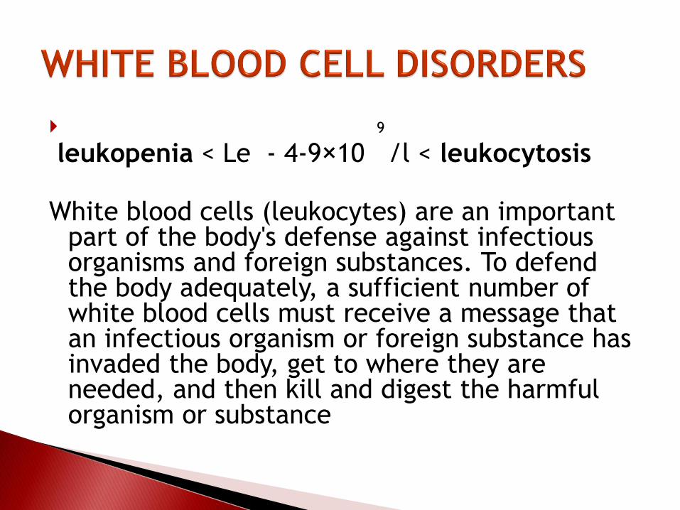
9
leukopenia < Le - 4-9×10 /l < leukocytosis
White blood cells (leukocytes) are an important part of the body's defense against infectious organisms and foreign substances. To defend the body adequately, a sufficient number of white blood cells must receive a message that an infectious organism or foreign substance has invaded the body, get to where they are needed, and then kill and digest the harmful organism or substance
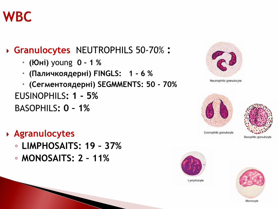
Granulocytes NEUTROPHILS 50-70% : (Юні) young 0 – 1 %
(Паличкоядерні) FINGLS: 1 - 6 %
(Сегментоядерні) SEGMMENTS: 50 - 70%
EUSINOPHILS: 1 - 5%
BASOPHILS: 0 – 1%
Agranulocytes
◦ LIMPHOSAITS: 19 – 37%
◦ MONOSAITS: 2 – 11%
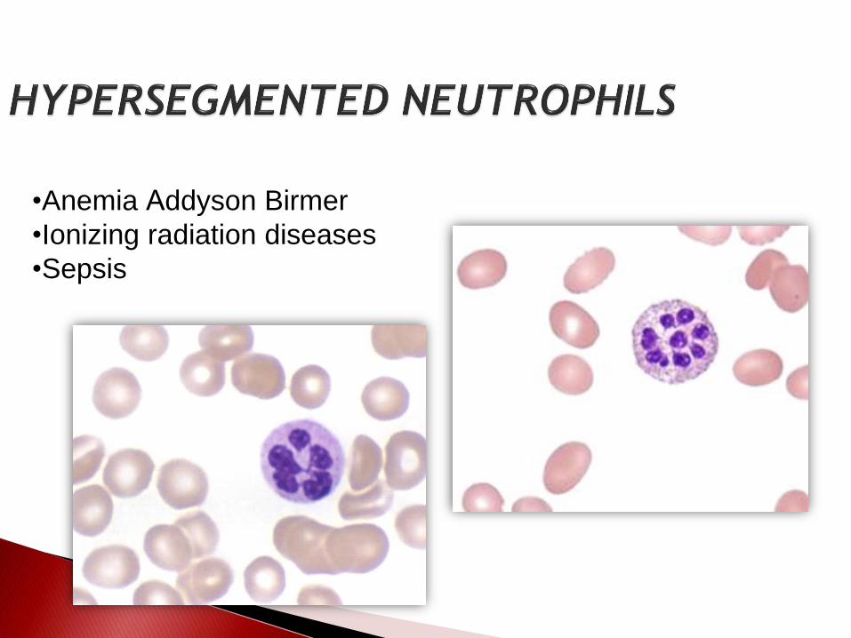
•Anemia Аddyson Birmer
•Ionizing radiation diseases
•Sepsis

Peter Maslak
ASH Image Bank 2009; 2009: 9-00018.
•Purulent-septic state
•Krupouse pneumonia
•Dysentery

Acute infections diseases

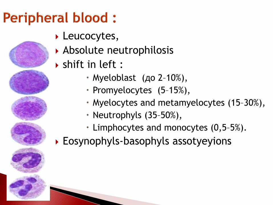
Leucocytes,
Absolute neutrophilosis
shift in left :
Myeloblast (до 2–10%),
Promyelocytes (5–15%),
Myelocytes and metamyelocytes (15–30%),
Neutrophyls (35–50%),
Limphocytes and monocytes (0,5–5%).
Eosynophyls-basophyls assotyeyions

Normally, people produce about 100 million
white blood cells a day. The number of white
blood cells in a given volume of blood is
expressed as cells per microliter of blood. The
total white blood cell count normally ranges
between 4,000 and 9,000 (11,000) cells per
microliter.

1 Inadequate production
A) increase of production of the Le
B) disorders of the maturity of the Le
C) production of pathological Le
2 Disorders of the number of Le

Leukopenia
Leukopenia, a decrease in the number of white
blood cells to fewer than 4,000 cells per
microliter of blood, makes people more
susceptible to infections.

Leukopenia results most commonly from a decrease in granulocytes, which are the most prevalent circulating white cells.
Lymphopenias are much less common; they are associated with
congenital immunodeficiency diseases
association with specific clinical states, such as advanced human immunodeficiency virus (HIV) infection
treatment with corticosteroids.
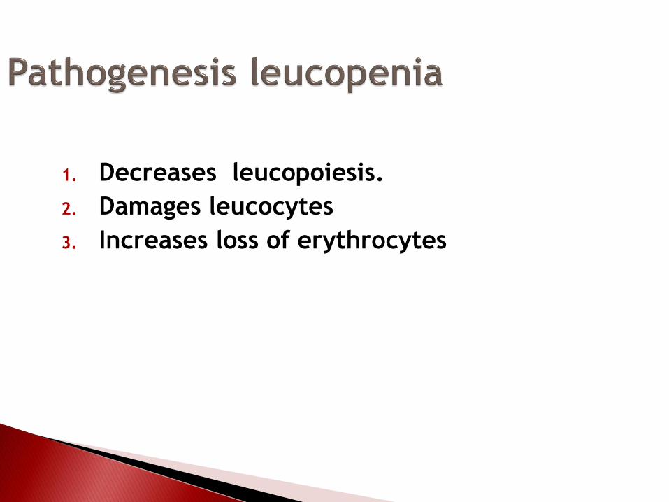
1. Decreases leucopoiesis.
2. Damages leucocytes
3. Increases loss of erythrocytes
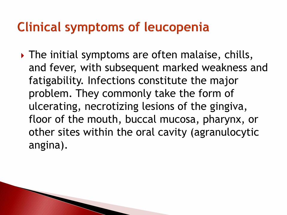
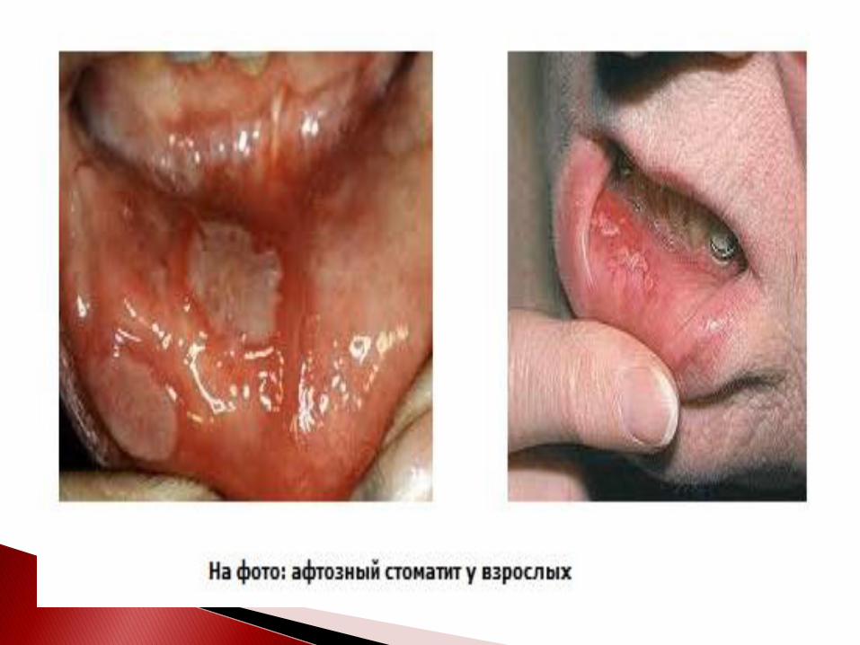
The initial symptoms are often malaise, chills,
and fever, with subsequent marked weakness and
fatigability. Infections constitute the major
problem. They commonly take the form of
ulcerating, necrotizing lesions of the gingiva,
floor of the mouth, buccal mucosa, pharynx, or
other sites within the oral cavity (agranulocytic
angina).

These lesions often show a massive growth of
microorganisms, due to the inability to mount a
leukocyte response. In addition to removal of the
offending drug and control of infections,
treatment efforts may also include the
administration of granulocyte colony-stimulating
factor, which stimulates neutrophil production by
the bone marrow.

Leukocytosis, an increase in the number of white blood cells to more than 11,000 cells per microliter of blood. May result from the normal response of the body to help fight an infection. However, an increase in the number of white blood cells can also result when the regulation of white blood cell development is disrupted and immature or abnormal cells are released into the blood.
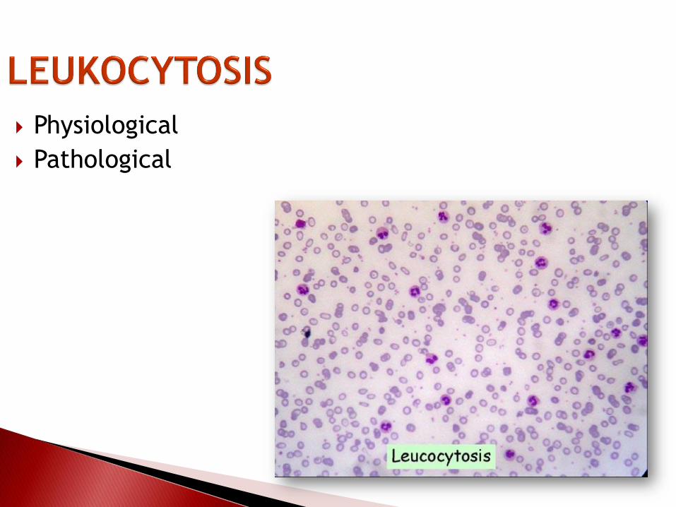
Physiological
Pathological

Food (nutritional)
Myogenic (muscle)
Infants (first 2 days)
Pregnant women (5-6th month of
pregnancy)
Pregnant women (up to 2 weeks after birth)
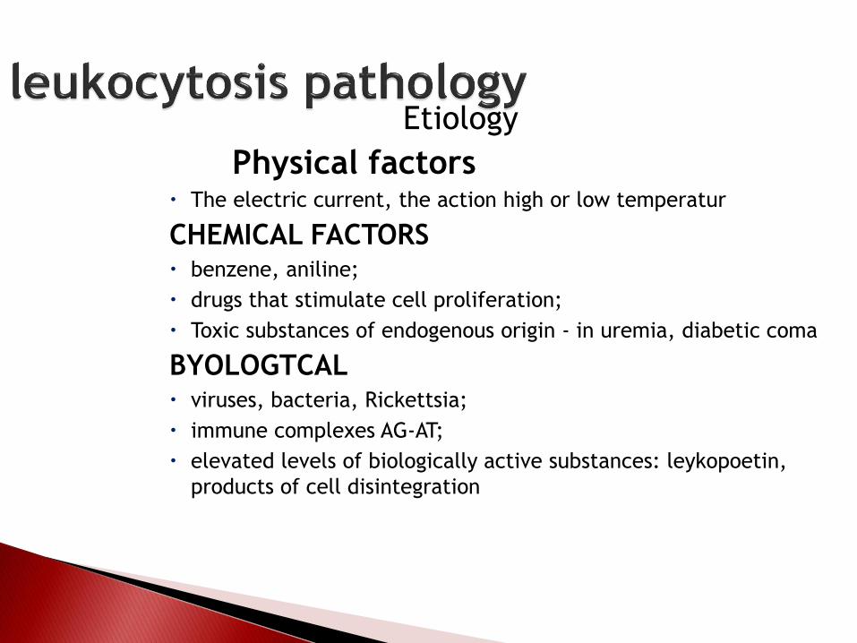
Etiology
Physical factors The electric current, the action high or low temperatur
CHEMICAL FACTORS benzene, aniline;
drugs that stimulate cell proliferation;
Toxic substances of endogenous origin - in uremia, diabetic coma
BYOLOGTCAL viruses, bacteria, Rickettsia;
immune complexes AG-AT;
elevated levels of biologically active substances: leykopoetin,
products of cell disintegration

Absolute leukocytosis
Mechanism:◦ increase the level and activity of stimulants leykopoezis
(leykopoetini)
◦ lowering of the level and activity
of inhibitors leykopoezis
(lipoprotein keylon, lactoferrin)
◦ tumor damage gemopoetic tissue (in leukemia)
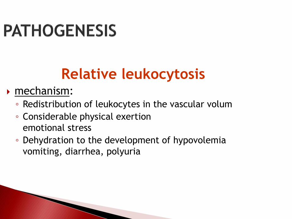
Relative leukocytosis mechanism:◦ Redistribution of leukocytes in the vascular volum
◦ Considerable physical exertion
emotional stress
◦ Dehydration to the development of hypovolemia
vomiting, diarrhea, polyuria

Reactive Leukocytosis An increase in the number of white cells is common in a variety of reactive inflammatory states caused by microbial and nonmicrobial stimuli.
Neutrophilic LeukocytosisAcute bacterial infections, especially those caused by pyogenic organisms; sterile inflammation caused by, for example, tissue necrosis (myocardial infarction, burns).
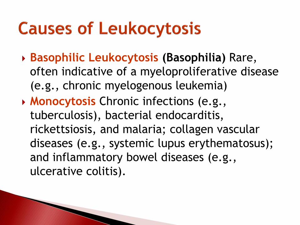
Basophilic Leukocytosis (Basophilia) Rare,
often indicative of a myeloproliferative disease
(e.g., chronic myelogenous leukemia)
Monocytosis Chronic infections (e.g.,
tuberculosis), bacterial endocarditis,
rickettsiosis, and malaria; collagen vascular
diseases (e.g., systemic lupus erythematosus);
and inflammatory bowel diseases (e.g.,
ulcerative colitis).

Аabsolute – increase number of lymphocytes
(> 40%).
Chronic limpholeucosis
Chronic bacterial diseases (туберкулез,
сифилис, лепра)
Endocrine disiases (базедова болезнь)
Disorders of white cells include
deficiencies (leukopenias) and
proliferations, which may be reactive (often
microbial disease ) neoplastic. Neoplastic
disorders, they cause approximately 9% of all
cancer deaths in adults and a staggering 40% in
children younger than 15 years.

Tumors represent the most important of the white cell disorders. They can be divided into three broad categories based on the origin of the tumor cells:
Lymphoid neoplasms, which include non-Hodgkin lymphomas (NHLs), Hodgkin lymphomas, lymphocytic leukemias, and plasma cell dyscrasias and related disorders. In many instances these tumors are composed of cells that resemble normal stages of lymphocyte differentiation, a feature that serves as one of the bases for their classification.
Myeloid neoplasms arise from stem cells thatnormally give rise to the formed elements ofthe blood: granulocytes, red cells, andplatelets. The myeloid neoplasms fall intothree fairly distinct subcategories: acutemyelogenous leukemias, in which immatureprogenitor cells accumulate in the bonemarrow; chronic myeloproliferativedisorders, in which inappropriately increasedproduction of formed blood elements leads toelevated blood cell counts; andmyelodysplastic syndromes, which arecharacteristically associated with ineffectivehematopoiesis and cytopenias.

Histiocytic neoplasms represent
proliferative lesions of histiocytes.
Of special interest is a spectrum of
proliferations comprising Langerhans
cells (the Langerhans cell
histiocytoses).

Yang pupils
Nondyferentyeited myeloblasts its general cells
(развития являются миелобласты, потерявшие
способность к диференциации).
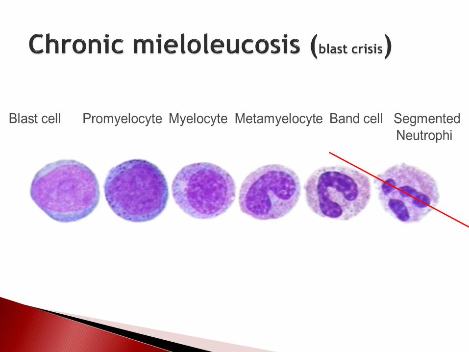
Hiatus leucemicus
Blast cell Promyelocyte Myelocyte Metamyelocyte Band cell Segmented
Neutrophil
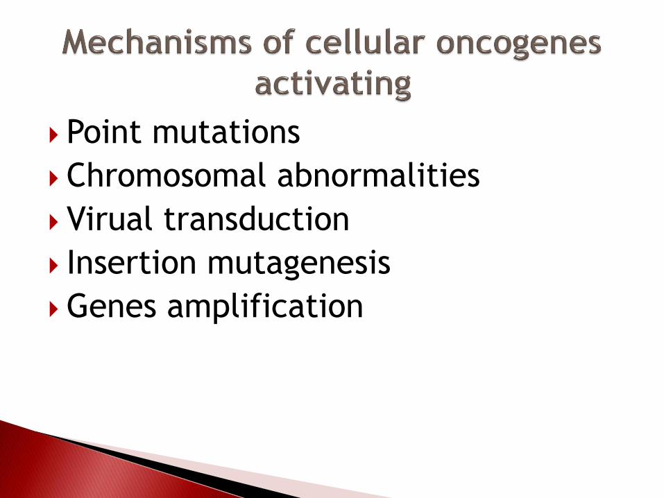
Point mutations
Chromosomal abnormalities
Virual transduction
Insertion mutagenesis
Genes amplification

Metaplastic anemia
Thrombocytopenia
Hemorrhagic syndrome
Secondary immunedeficiency
Decrease of resistance

In the Western world, infectious mononucleosis is an acute, self-limited disease of adolescents and young adults that is caused by B lymphocytotropic EBV, a member of the herpesvirus family. The infection is characterized by
(1) fever, sore throat, and generalized lymphadenitis;
(2) increase of lymphocytes in blood, many of which have an atypical morphology;
(3) antibody and T cell response to EBV. It should be noted that cytomegalovirus infection induces a similar syndrome, which can be differentiated only by serologic methods.

Etiologic Factors for Glossodynia Local Causes Candidiasis climacteric postmenopausal anxiety Migratory glossitits (geographic tongue) diabetes
depression Lichen planus Sjogren's Syndrome (xerostomia)
cancerphobia Trauma drug reactions (xerostomia) Oral cancer deficiency states Denture faults anemias (Iron, B12, Folic Acid) Impression surface lingual artery atherosclerosis Polished surface fheumatoid arthritis