Baroreflex sensitivity, heart rate, and blood pressure variability in hypertensive pregnancy...
8
Baroreflex Sensitivity, Heart Rate, and Blood Pressure Variability in Normal Pregnancy Andreas Voss, Hagen Malberg, Agnes Schumann, Niels Wessel, Thomas Walther, Holger Stepan, and Renaldo Faber Heart rate variability is a relevant predictor of cardiovascular risk in humans. However, to use heart and blood pressure (BP) variability or baroreflex sensitivity as markers for hypertensive pregnancy disorders, it is first necessary to describe these parameters in normal pregnancy. To accommodate the complexities of autonomic cardiovascular control we added parameter domains of nonlinear dynamics to conventional linear methods of time and frequency domains. The BP of 27 women with normal pregnancy and 14 nonpregnant women were monitored at a high resolution (200 Hz sampling frequency) using a Portapres for 30 min. The pregnant women were divided into groups of 32 or less or greater than 32 weeks of gestation. Pregnant and nonpregnant women were classified into subclasses of maternal age of less than 28 or 28 or more years. Except for two single parameter domains, we found no significant differences in heart rate and BP variability for pregnant women with different gestational age or different maternal age. Moreover, no significant differences in spontaneous baroreflex sensitivity could be found between pregnant women regardless of either their age or gestational age. In contrast, all measures of nonlinear dynamics of heart rate variability as well as all parameter domains of spontaneous baroreflex sensitivity showed significant changes between pregnant and nonpregnant women, whereas BP variability did not differ between those groups. This complex assessment of autonomic cardiovascular regulation has shown that the parameters tested are stable in the second half of normal pregnancy, and might have the potential to be excellent indicators of pathophysiologic conditions. Am J Hypertens 2000;13:1218 –1225 © 2000 American Journal of Hypertension, Ltd. KEY WORDS: Heart rate variability, blood pressure variability, baroreflex sensitivity, pregnancy, linear dynamics, nonlinear dynamics. B lood pressure variability (BPV) and heart rate variability (HRV) are generated by the rhythmic actions of cardiovascular hor- mones and neuronal pathways on effector organs such as the heart, kidneys, and vessels. Heart rate variability has been shown to be a relevant pre- dictor for the mortality of patients with myocardial infarction. The clinical importance of HRV has been confirmed in several studies since the late 1980s. HRV is a strong and independent predictor of mortality after an acute myocardial infarction, 1–7 but even age and gender can influence blood pressure (BP) and heart rate, and lead to variability changes. 8 –12 Simi- larly, the sinoaortic baroreflex sensitivity (BRS), which is one of the primary mechanisms that regulates BP, is correlated to the incidence of pathophysiologic condi- tions. For instance, numerous studies have reported baroreflex resetting with aging and after long-term exposure to chronic hypertension. 13,14 Received January 14, 2000. Accepted April 4, 2000. From the University of Applied Sciences (AV, HM, AS, NW), Jena, Germany; Max-Delbru ¨ ck-Center for Molecular Medicine (TW), Berlin-Buch, Germany; and Department of Obstetrics and Gynecol- ogy (HS, RF), University of Leipzig, Leipzig, Germany. Address correspondence and reprint requests to Renaldo Faber, MD, Universita ¨tsfrauenklinik, Philipp-Rosenthalstr. 55, 04103 Leipzig, Germany; e-mail: [email protected] AJH 2000;13:1218 –1225 © 2000 by the American Journal of Hypertension, Ltd. 0895-7061/00/$20.00 Published by Elsevier Science, Inc. PII S0895-7061(00)01199-7
-
Upload
independent -
Category
Documents
-
view
8 -
download
0
Transcript of Baroreflex sensitivity, heart rate, and blood pressure variability in hypertensive pregnancy...

Baroreflex Sensitivity, Heart Rate, and BloodPressure Variability in Normal PregnancyAndreas Voss, Hagen Malberg, Agnes Schumann, Niels Wessel, Thomas Walther, Holger Stepan,and Renaldo Faber
Heart rate variability is a relevant predictor ofcardiovascular risk in humans. However, to useheart and blood pressure (BP) variability orbaroreflex sensitivity as markers for hypertensivepregnancy disorders, it is first necessary to describethese parameters in normal pregnancy. Toaccommodate the complexities of autonomiccardiovascular control we added parameterdomains of nonlinear dynamics to conventionallinear methods of time and frequency domains.The BP of 27 women with normal pregnancy and14 nonpregnant women were monitored at a highresolution (200 Hz sampling frequency) using aPortapres for 30 min. The pregnant women weredivided into groups of 32 or less or greater than 32weeks of gestation. Pregnant and nonpregnantwomen were classified into subclasses of maternalage of less than 28 or 28 or more years.
Except for two single parameter domains, wefound no significant differences in heart rate andBP variability for pregnant women with different
gestational age or different maternal age.Moreover, no significant differences inspontaneous baroreflex sensitivity could be foundbetween pregnant women regardless of either theirage or gestational age. In contrast, all measures ofnonlinear dynamics of heart rate variability as wellas all parameter domains of spontaneous baroreflexsensitivity showed significant changes betweenpregnant and nonpregnant women, whereas BPvariability did not differ between those groups.This complex assessment of autonomiccardiovascular regulation has shown that theparameters tested are stable in the second half ofnormal pregnancy, and might have the potential tobe excellent indicators of pathophysiologicconditions. Am J Hypertens 2000;13:1218–1225© 2000 American Journal of Hypertension, Ltd.
KEY WORDS: Heart rate variability, blood pressurevariability, baroreflex sensitivity, pregnancy, lineardynamics, nonlinear dynamics.
Blood pressure variability (BPV) and heartrate variability (HRV) are generated by therhythmic actions of cardiovascular hor-mones and neuronal pathways on effector
organs such as the heart, kidneys, and vessels. Heart
rate variability has been shown to be a relevant pre-dictor for the mortality of patients with myocardialinfarction. The clinical importance of HRV has beenconfirmed in several studies since the late 1980s. HRVis a strong and independent predictor of mortalityafter an acute myocardial infarction,1–7 but even ageand gender can influence blood pressure (BP) andheart rate, and lead to variability changes.8–12 Simi-larly, the sinoaortic baroreflex sensitivity (BRS), whichis one of the primary mechanisms that regulates BP, iscorrelated to the incidence of pathophysiologic condi-tions. For instance, numerous studies have reportedbaroreflex resetting with aging and after long-termexposure to chronic hypertension.13,14
Received January 14, 2000. Accepted April 4, 2000.From the University of Applied Sciences (AV, HM, AS, NW),
Jena, Germany; Max-Delbruck-Center for Molecular Medicine (TW),Berlin-Buch, Germany; and Department of Obstetrics and Gynecol-ogy (HS, RF), University of Leipzig, Leipzig, Germany.
Address correspondence and reprint requests to Renaldo Faber,MD, Universitatsfrauenklinik, Philipp-Rosenthalstr. 55, 04103Leipzig, Germany; e-mail: [email protected]
AJH 2000;13:1218–1225
© 2000 by the American Journal of Hypertension, Ltd. 0895-7061/00/$20.00Published by Elsevier Science, Inc. PII S0895-7061(00)01199-7
The brain seems to be the source for most of thesignals thought to be responsible for the setting of BPand HR.15 The molecular mechanisms involved in theregulation of BPV and HRV or BRS are only poorlyunderstood, although initial analysis of gene-manipu-lated animals had shown that genes involved in car-diovascular regulation and expressed in the centralnervous system should be interesting candidates forthe molecular regulation of BPV and HRV.16–19
Although significant adaptations of the maternalcardiovascular system are known to occur during nor-mal pregnancy, our specific knowledge of the mater-nal HRV and BPV during pregnancy is poor. How-ever, it has been reported that BPV20,21 and HRV22,23
are decreased in normal pregnancy. In addition, En-eroth and Storck24 described an unchanged HRV inwomen with preeclampsia and women hospitalizeddue to different complications compared to healthypregnant women, whereas other groups described dif-ferences in cardiovascular variabilities in preeclampticpregnancies.22,25–28
This study was performed to determine whether theparameter domains of HRV, BPV, or BRS in normalpregnancy are influenced by either the gestational ageor by the age of the pregnant women. To investigatethe complex modulation of the autonomic system weintroduced new methods of nonlinear dynamics andcombined these with traditional methods and an ad-vanced technology of BRS analysis by the Dual Se-quence Method.29
METHODS
Subjects In this cross-sectional study, 27 healthypregnant women (age: 28.9 6 5.5 years; gestationalweek: 32 6 6; range: 21st to 40th week of gestation)were recruited from the Department of Obstetrics,University of Leipzig, from May to December 1998.Informed consent was obtained from all patients. Allwomen had single pregnancies, normal BP during thepregnancy, and normal pregnancy outcome. Womenwith diabetes, or cardiovascular and renal diseaseswere excluded. None of the women had a history ofhypertensive disorders in a previous pregnancy. Bet-amimetics or other drugs with cardiovascular effectswere not given to the women. As a control group, 14age-matched healthy women (age: 27.9 6 5.6 years)were investigated. None of these controls had a car-diovascular or renal disease or took drugs with car-diovascular effects.
Data Acquisition and Preprocessing The beat-to-beat BP values were recorded under standardizedresting conditions for all pregnant women and thecontrols using the Portapres device (portable, volume-clamp photoplethysmographic BP device, Model 2;BMI-TNO, Amsterdam, The Netherlands), a 30 min
recording time (to have a clinical relevant and feasibletime of monitoring), and a 200 Hz sampling fre-quency. Simultaneously, a Holter ECG (Avionics,Leesburg, Virginia) with a resolution of 8 ms wasrecorded. The women were lying comfortably on theirleft side with an approximately 30° tilt towards theobserver. To exclude an influence of the period ofthe day, all values were recorded between 8 am and12 am.
The recorded data were filtered to exclude ventric-ular premature beats, artifacts, and noise. Further-more, the time series were filtered using a movingaverage filter (2nd order) before calculating the BRS,to smooth and amplify the dominant signal compo-nents. Standard frequency domain parameters of HRVand BPV presuppose an equidistant sampled timeseries. Neither the beat-to-beat interval (BBI) time se-ries (sequence of successive beat-to-beat differences)nor the BP time series are equidistant in terms of time,but they are in terms of the actual beat number. It wastherefore necessary to create equidistant time series byinterpolation (Dt 5 500 ms), to get spectra with fre-quency scaling. The normal and edited BBI after fil-tering are denoted as NN intervals for HRV and BPV.
Analysis of HRV From the time domain the follow-ing parameters25,26 were calculated:
• mean NN 5 the mean value of the NN intervals inms (NN means intervals between successive normalheart beats);
• SDNN 5 standard deviation of the NN intervals, inms;
• RMSSD 5 the root mean square of successive NNinterval differences (HRV), in ms;
• Renyi4 5 the Renyi entropy of order 4 of the histo-gram (HRV), without unit;
In the frequency domain we used these frequencybands:
• ULF 5 the power in the frequency band 0–0.0033Hz, in s2;
• VLF 5 the power in the frequency band 0.0033–0.04Hz, in s2;
• LF 5 the power in the frequency band 0.04–0.15 Hz,in s2;
• HF 5 the power in the frequency band 0.15 Hz–0.4Hz, in s2.
The spectra were estimated using the Fast FourierTransform. To avoid the leakage effect a BlackmanHarris window function was applied. Furthermore,the measures of normalized LFn and UVLF as the sumof ULF 1 VLF 1 LF were calculated.
Symbolic dynamics, as a nonlinear approach toinvestigate complex systems, facilitates the analysisof dynamic aspects of heart rate and BP variability.
AJH–NOVEMBER 2000–VOL. 13, NO. 11 VARIABILITY IN PREGNANCY 1219

The concept of symbolic dynamics is based on acoarse-graining of the dynamics.27,28,30,31 The Renyientropy calculated from these distributions of words(FWRen025 2 a 5 0.25) is a suitable measure for thecomplexity within the time series. Higher values ofthis entropy refer to a higher complexity within thecorresponding tachograms and lower values to lowercomplexity. A high percentage of words consistingonly of the symbols ‘0’ and ‘2’ (WPSUM02) is a mea-sure for a decreased HRV. Forbidden words (FW) arethose words that seldom or never occur (a probabilityless than .001) within the distribution of words with alength of 3.
Additionally, a second mode of symbolic dynamicsfor high-variability analysis was used in this study. Inthis case we observed six successive symbols of asimplified alphabet {0,1}. Here, the symbol ‘0’ standsfor a difference between two successive beats lowerthan a special limit of 20 ms, whereas ‘1’ representsthose cases where the difference between two succes-sive beats exceeds this special limit. Words consistingonly of unique type of symbols (all ‘1’) were counted.This measure is called PHVAR20 and quantifies anincreased variability.
Analysis of BPV From the time domain the follow-ing parameters were calculated:
• bmeanNN 5 the mean value of the systolic BP ofsuccessive normal heart beats;
• bSDNN 5 standard deviation of the systolic BP timeseries.
In the frequency domain these frequency bands wereused:
• bULF 5 the power in the frequency band 0–0.0033Hz, in s2;
• bVLF 5 the power in the frequency band 0.0033–0.04 Hz, in s2.
The measures of normalized bLFn and bUVLF (sum ofbULF 1 bVLF 1 bLF) were calculated.
Estimation of Baroreflex Sensitivity The parameterdomains with the most relevance to estimation of thespontaneous baroreflex are the slopes as a measure ofthe sensitivity.32,33 The slopes were usually calculatedby linear regression BRS 5 DBBI/DBP (ms/mm Hg)with BP as systolic BP. Using the Dual Sequence Meth-od29 two kinds of BBI responses were analyzed (withdifferent synchronization modes and splitted sectors):bradycardiac fluctuations (an increase in BP causes anincrease in BBI) and tachycardic fluctuations (a de-crease in BP causes a decrease in BBI). The analysis ofthe tachycardic and bradycardic fluctuations containsthree consecutive BP and BBI values.
The following parameters were calculated:
• the mean bradycardiac and tachycardic slopeswhere an increased BP causes a reduced heart rate(all_brady) and a decreased BP causes an increasedheart rate (all_tachy);
• the mean bradycardic and tachycardic slopes in-cluding only slopes greater than 5 where an in-creased BP of 1 mm Hg causes an increase of BBI ofat least 5 ms (all_.5_brady) and a decreased BP of1 mm Hg causes a decrease in BBI of at least 5 ms(all_.5_tachy).29
Statistics The two-tailed univariate t test for equal-ity of means was used to test whether the parametermeans of the various groups differed significantlyfrom each other. The analysis includes seven differenttests:
• Test 1: Influence of the age of pregnant women onvariability parameters (, 28 versus $ 28 years;
• Test 2: Influence of the age of non-pregnant womenon variability parameters (, 28 versus $ 28 years);
• Test 3: Differences in variability between agematched healthy pregnant and healthy nonpregnantwomen , 28 years of age;
• Test 4: Differences in variability between age-matched healthy pregnant and healthy nonpregnantwomen aged 28 years or more;
• Test 5: Influence of gestational age (# 32 versus . 32weeks) on variability parameters;
• Test 6: Differences in variability between age-matched healthy nonpregnant women and healthypregnant women with a gestational age # 32 weeks;
• Test 7: Differences in variability between age-matched healthy nonpregnant women and healthypregnant women with a gestational age . 32 weeks.
Additionally, multivariate analysis of variance(MANOVA) with all parameter domains was per-formed to test whether age or pregnancy have a sig-nificant effect on the population means of the param-eter domains considered. Afterwards, one-factorialMANOVA tests were performed to prove whether thegestational age has a significant effect on the popula-tion means. Kolmogorov-Smirnov test and Shapiro-Wilk test were used to prove normal distributionwithin the groups.
RESULTS
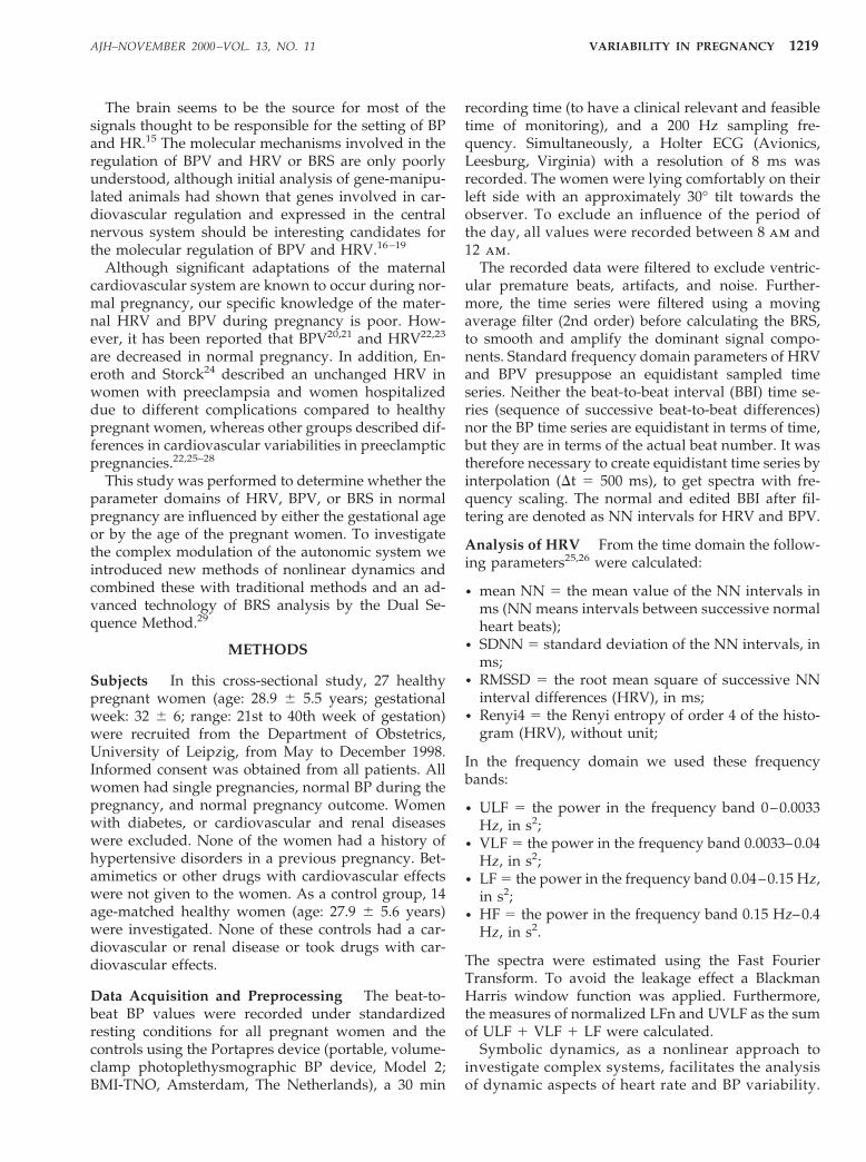
Figs. 1 and 2 show examples of the HRV and BPV of ahealthy pregnant woman in comparison to the vari-abilities of a nonpregnant healthy woman.
We found no significant differences in HRV, BPV,and BRS associated with maternal age (Test 1: meanage difference, 9 years) and parity (data not shown).Also the respiratory frequency was unchanged. A sim-ilar result was obtained within the nonpregnant con-trols (Test 2) with one exception: the LFn of HRV was
AJH–NOVEMBER 2000–VOL. 13, NO. 111220 VOSS ET AL

significantly reduced with increasing age (Table 1);however, MANOVA analysis using all variable sets ofall parameter domains showed no significant effect ofage on all parameter domains.
Advanced gestational age influenced only one fre-quency-domain parameter of HRV (Test 5). A highergestational age is correlated with an increase of thepower within the ULF frequency band. Parameter do-mains of BPV remain stable with advancing gesta-tional age, except for a decrease of the mean value ofsystolic BP from 113 to 100 mg Hg. Also the parameterdomains of BRS are unchanged after the 32nd week ofgestation (Test 5). Multivariate MANOVA excluded asignificant dependency of the gestational age on allinvestigated parameter domains, including mean sys-tolic BP (bmeanNN, P , .64).
In comparing pregnant to nonpregnant womenwith an age , 28 or $ 28 years (Tests 3 and 4), therewere considerable differences in HRV and BRS. Incontrast, for BPV no significant differences could befound between the groups. In test 3, all four time-domain parameters of HRV were decreased in preg-nant women, as were two of the four frequency-do-main parameters of HRV. All four parameter domainsof nonlinear dynamics showed significant changes,too. In Test 4 only one time-domain parameter of HRV(RMSSD), but the same two frequency-domain param-eters of HRV as in Test 3, were significantly decreased.Finally, we calculated high significance for all fourparameter domains of BRS in Tests 3 and 4 (Table 1).
The comparison of pregnant women # 32 weeks tononpregnant controls (Test 6) showed a significantdecline in all parameter domains of the HRV, exceptLFn, and all parameter domains of BRS, except anunchanged BPV. Compared to Test 6, the group withgestational age . 32 weeks was characterized by asmaller number of significantly altered parameter do-mains of HRV, compared to the nonpregnant controls(Test 7). However, in both tests all parameter domainsof BRS were significantly decreased (Table 2).
DISCUSSION
The analysis of heart rate and BP variability is a non-invasive diagnostic tool that provides important prog-nostic information concerning individual risk, for in-stance, after acute myocardial infarction.3–7 In contrastto previous investigations on HRV and BPV in preg-nant women,20–24 our study provides a more complexand multiparametric approach, including traditionalmethods of HRV and BPV; time domain measures;frequency domain measures combined with newmethods of nonlinear dynamics;26,30 and BRS analy-sis.34
Thus, we analyzed the cardiovascular parameters of27 pregnant women to determine whether the gesta-tional age or the age of the pregnant women influenceparameter domains of HRV, BPV, or BRS in normalpregnancy. With the exception of two parameter do-mains of HRV (LFn and ULF), there were no signifi-cant differences. Investigation on the influence of bi-
FIG. 1. Systolic blood pressure (SBP) and beat-to-beat intervals(RR) of a pregnant healthy woman.
FIG. 2. Systolic blood pressure (SBP) and beat-to-beat intervals(RR) of a nonpregnant healthy woman.
AJH–NOVEMBER 2000–VOL. 13, NO. 11 VARIABILITY IN PREGNANCY 1221

ologic age on HRV have shown that the alterations inthese parameters can occur as long-term effects overdecades.8,10–12 For this reason, significant changes dueto aging were not to be expected in our study, mainlybecause of the relatively short time frame observed,with a mean age difference of 9 years in the pregnantwomen and 10 years in the nonpregnant controls,respectively. On the other hand, the decrease of LFnwith maternal age might be caused by an aging effect,as LF power declines linearly across the whole agerange.30
Increased power within the ULF frequency band ofthe HRV depending on the gestational age can resultfrom several reasons. A number of investigators haveshown that systems like the renin–angiotensin systemor the thermoregulation system influence the ULF pa-rameter domain.34 However, instabilities caused by
physical or mental stress can specifically alter the ULFband. Possibly, the fetal movements could trigger ma-ternal reactions, inducing changes in this frequencyband.
In addition, the systolic BP was significantly de-creased in the group over 32 weeks of gestation, but alllinear and nonlinear parameter domains of BPV wereunchanged. These data are in contrast to those ofAyala and Hermida et al,20,21 who showed decreasedparameters of BPV as well as an increase in systolic BPfrom the 21st week until delivery, and are contrary toroutine clinical observations. The most likely explana-tion for this observation is our methodical approach,which uses continuous finger arterial pressure record-ing. Furthermore, all parameters of BRS remain stablewith advancing gestational age. In addition, the dataof 14 healthy nonpregnant women were acquired to
TABLE 1. RESULTS PRESENTED AS MEAN 6 STANDARD DEVIATION OF TEST 1 (DIFFERENCESCAUSED BY MATERNAL AGE), TEST 2 (DIFFERENCES CAUSED BY AGE OF THE NONPREGNANT
CONTROLS), TEST 3 (DIFFERENCES BETWEEN PREGNANT AND NONPREGNANT < 28 YEARS), ANDTEST 4 (DIFFERENCES BETWEEN PREGNANT AND NONPREGNANT > 28 YEARS)
P<28 NP<28 P>28 NP>28Test 1
PTest 2
PTest 3
PTest 4
PMean SD Mean SD Mean SD Mean SD
HRVmeanNN 695 89 805 103 771 139 856 60 NS NS .023 NSSDNN 42 11 70 21 45 17 67 24 NS NS .002 NSRMSSD 25 10 51 21 27 7 51 22 NS NS .004 .007Renyi4 1.8 0.2 2.3 0.3 1.8 0.4 2.2 0.4 NS NS .000 NSULF 0.07 0.06 0.09 0.05 0.15 0.24 0.24 0.22 NS NS NS NSVLF 0.23 0.14 0.56 0.58 0.19 0.13 0.34 0.19 NS NS NS NSLF 0.15 0.11 0.46 0.2 0.09 0.05 0.33 0.32 NS NS .001 0.037HF 0.07 0.06 0.27 0.21 0.07 0.04 0.29 0.22 NS NS .015 0.007LFn 0.66 0.12 0.66 0.08 0.6 0.13 0.53 0.14 NS .045 NS NSFW 24 9 9 8 25 7 12 10 NS NS .001 .008FWRen025 3.7 0.2 3.9 0.1 3.6 0.2 3.9 0.1 NS NS .003 .005phvar20 0.008 0.013 0.068 0.081 0.006 0.007 0.079 0.086 NS NS .034 .016WPSUM02 0.44 0.14 0.21 0.1 0.48 0.17 0.25 0.18 NS NS .001 .029
BPVbmeanNN 104 15 114 11 110 14 113 8 NS NS NS NSbsDNN 8 2 8 1 7 3 8 3 NS NS NS NSbULF 0.006 0.008 0.004 0.003 0.003 0.003 0.008 0.006 NS NS NS NSbVLF 0.011 0.008 0.007 0.002 0.007 0.006 0.006 0.003 NS NS NS NSbUVLF 0.017 0.015 0.011 0.003 0.01 0.008 0.014 0.009 NS NS NS NSbLFn 0.86 0.08 0.75 0.16 0.78 0.17 0.79 0.06 NS NS NS NS
BRSall_tachy 11 3 17 5 13 4 21 4 NS NS .003 .003all_.5_tachy 13 3 19 4 14 4 22 5 NS NS .002 .004all_brady 10 4 19 6 12 3 21 7 NS NS .001 .005all_.5_brady 12 3 20 6 13 3 22 7 NS NS .001 .005
P 5 pregnant women; NP 5 nonpregnant controls; P value 5 level of significance; HRV 5 heart rate variability; NS 5 not significant; for otherabbreviations, see the Methods section.
AJH–NOVEMBER 2000–VOL. 13, NO. 111222 VOSS ET AL

investigate differences between pregnant and non-pregnant women. During pregnancy significant adap-tive changes of the cardiovascular system occur. Inprevious studies a decreased HRV with a reducedhigh-frequency power in the second trimester of nor-mal pregnancy compared to nonpregnant women hasbeen described.22,35 Our data support these findings,with a reduced power in the HF (0.15–0.4 Hz) and inthe LF (0.04–0.15 Hz). In addition, we were able toshow that these alterations are also detectable in thethird trimester, which implicates a reduced vagallymediated activity in the second half of pregnancy.Furthermore, our data show significantly decreasedmeasures of nonlinear dynamics such as FW,FWRen025, phvar20, and WPSUM02. These methodsof nonlinear dynamics describe complex fluctuationsin rhythm and separate structures of nonlinear behav-ior in the heart rate time series more successfully thanclassical methods of time and frequency domainsdo.26,30 This provides additional support for the de-
scribed decrease in HRV in normal pregnant women.Moreover, the differences in the HRV between preg-nant and nonpregnant women seem to be more drasticin the second trimester than in the third trimester. Thiscorresponds to the known clinical finding that thehighest increase of heart rate and volume load occuruntil the end of the second trimester in normal preg-nancy. The physiologic reduction in HRV may resultnot only from an increased resting heart rate, but alsofrom the state of chronic volume overload, with anincreased preload in pregnancy that is also highest atthe end of the second trimester. Thus, physiologiccardiovascular changes may lead to an impaired adap-tive capacity. Why this impairment, which is consid-ered to be of negative prognostic value in patients witha cardiovascular disease,3–7 is observable in healthypregnant women remains an open question. Interest-ingly, none of the time and frequency domains of BPVdiffered between the pregnant and nonpregnantwomen, which is in contrast to previous studies.20,21
TABLE 2. RESULTS PRESENTED AS MEAN 6 STANDARD DEVIATION OF TEST 5 (DIFFERENCESCAUSED BY GESTATIONAL AGE IN WEEKS), TEST 6 (DIFFERENCES BETWEEN PREGNANT WOMEN
WITH A GESTATIONAL AGE < 32 WEEKS AND NONPREGNANT CONTROLS), AND TEST 7(DIFFERENCES BETWEEN PREGNANT WOMEN WITH A GESTATIONAL AGE > 32 WEEKS AND
NONPREGNANT CONTROLS)
< 32 weeks > 32 weeks NPTest 5
PTest 6
PTest 7
PMean SD Mean SD Mean SD
HRVmeanNN 709 91 770 124 843 70 NS .001 NSSDNN 43 10 46 18 68 18 NS .000 .007RMSSD 26 9 25 8 50 16 NS .000 .000Renyi4 1.8 0.2 1.8 0.4 2.3 0.3 NS .000 .006ULF 0.06 0.04 0.2 0.22 0.16 0.16 .036 .033 NSVLF 0.2 0.11 0.24 0.16 0.38 0.17 NS .006 NSLF 0.11 0.08 0.13 0.11 0.41 0.25 NS .001 .003HF 0.07 0.04 0.07 0.07 0.25 0.15 NS .001 .002LFn 0.64 0.14 0.64 0.1 0.61 0.13 NS NS NSFW 24 7 27 8 9.4 7.6 NS .000 .000FWRen025 3.6 0.2 3.6 0.2 3.9 0.1 NS .000 .000phvar20 0.008 0.013 0.007 0.011 0.065 0.062 NS .005 .006WPSUM02 0.45 0.12 0.49 0.21 0.23 0.12 NS .000 .002
BPVbmeanNN 113 14 100 9 111 8 .02 NS .013bsDNN 7 2 7 2 8 2 NS NS NSbULF 0.004 0.004 0.004 0.003 0.006 0.005 NS NS NSbVLF 0.008 0.007 0.008 0.006 0.006 0.003 NS NS NSbUVLF 0.012 0.01 0.012 0.009 0.012 0.006 NS NS NSbLFn 0.82 0.15 0.76 0.16 0.81 0.06 NS NS NS
BRSall_tachy 13 3 11 4 19 5 NS .001 .000all_.5_tachy 14 3 12 4 20 5 NS .001 .000all_brady 11 3 10 4 20 6 NS .000 .000all_.5_brady 12 2 12 4 21 6 NS .000 .000
Abbreviations as in Table 1.
AJH–NOVEMBER 2000–VOL. 13, NO. 11 VARIABILITY IN PREGNANCY 1223

Using first the Dual Sequence Method29 to analyzethe BRS in pregnancy, we have shown that tachycardicas well as bradycardic fluctuations are decreased inpregnant women. Moreover, the interpretation ofchanges in spontaneous BRS may explain changes invagal HR modulation due to the predominant role ofthe parasympathetic system and HR regulation,within the time constants explored by these methods.Secondly, we interpret this decreased BRS as a paralleleffect to the decreased HRV, as both quantify theeffects of autonomic cardiac modulation. In addition,we can exclude an influence of the respiratory fre-quency on all investigated parameters, because nodifferences could be detected between pregnant andnonpregnant women, as was also shown previously.36
With respect to the feasibility of the method forclinical use, recording time was confined to 30 min.However, data of slower BP and HR fluctuations can-not be derived from a 30-min recording. With ourmore complex approach combining linear measures ofHRV, BPV, and BRS with the investigation of innermotions of the time series using symbolic dynamicswe have been able to provide an improved descriptionof the autonomic cardiovascular control in pregnancy.These nonlinear parameters record the interactions ofthe regulation system with a higher complexity. Thus,in the second half of normal pregnancy the parameterdomains of HRV, BPV, and BRS show a high degree ofstability related to maternal and gestational age.Therefore, we conclude that this method could be auseful tool to detect pathophysiologic changes, partic-ularly in hypertensive pregnancy disorders.
REFERENCES
1. Wolf MM, Varigos GA, Hunt D, Sloman JG: Sinusarrhythmia in acute myocardial infarction. Med J Aust1978;2:52–53.
2. Kleiger RE, Miller JP, Bigger JT Jr, Moss AJ: Decreasedheart rate variability and its association with increasedmortality after acute myocardial infarction. Am J Car-diol 1987;59:256–262.
3. Algra A, Tijssen JGP, Roelandt JRTC, Pool J, Lubsen J:Heart rate variability from 24-hour electrocardiogra-phy and the 2-year risk for sudden death. Circulation1993;88:180–184.
4. Bigger JT Jr, Fleiss JL, Steinman RC, Rolnitzky LM,Kleiger RE, Rottman JN: Frequency domain measuresof heart period variability and mortality after myocar-dial infarction. Circulation 1992;85:164–171.
5. Singh N, Mironov D, Armstrong PW, Ross AM, LangerA: Heart rate variability assessment early after acutemyocardial infarction. Circulation 1996;93:1388–1395.
6. Tsuji H, Larson MG, Venditti FJ, Manders ES, Evans JC,Feldman CL, Levy D: Impact of reduced heart ratevariability on risk for cardiac events. Circulation 1996;94:2850–2855.
7. Zuanetti G, Neilson JMM, Latini R, Santoro E, Mag-
gioni AP, Ewing DJ: Prognostic significance of heartrate variability in post-myocardial infarction patients inthe fibrinolytic era. Circulation 1996;94:432–436.
8. Shannon DC, Carley DW, Benson H: Aging of modu-lation of heart rate. Am J Physiol 1987;253:H874–H877.
9. Huikuri HV, Pikkujamsa SM, Airaksinen KE, IkaheimoMJ, Rantala AO, Kauma H, Lilja M, Kesaniemi YA:Sex-related differences in autonomic modulation ofheart rate in middle-aged subjects. Circulation 1996;94:122–125.
10. Ryan SM, Goldberger AL, Pincus SM, Mietus J, LipsitzLA: Gender- and age-related differences in heart ratedynamics: are women more complex than men? J AmColl Cardiol 1994;24:1700–1707.
11. Stein PK, Kleiger RE, Rottman JN: Differing effects ofage on heart rate variability in men and women. Am JCardiol 1997;80:302–305.
12. Umetani K, Singer DH, McCraty R, Atkinson M: Twen-ty-four hour time domain heart rate variability andheart rate: relations to age and gender over nine de-cades. J Am Coll Cardiol 1998;31:593–601.
13. Bristow JD, Honour AJ, Pickering GW, Sleight P, SmythHS: Diminished baroreflex sensitivity in high bloodpressure. Circulation 1969;39:48–54.
14. Gribbin B, Pickering TG, Sleight P, Peto R: The effect ofage and high blood pressure on baroreflex sensibility inman. Circ Res 1971;29:424–431.
15. Korpelainen JT, Sotaniemi KA, Huikuri HV, MyllyaVV: Abnormal heart rate variability as a manifestationof autonomic dysfunction in hemispheric brain infarc-tion. Stroke 1996;27:2059–2063.
16. Mansier P, Medigue C, Charlotte N, Vermeiren C,Coraboeuf E, Deroubai E, Ratner E, Chevalier B,Clairambault J, Carre F, Dahkli T, Bertin B, Briand P,Strosberg D, Swynghedauw B: Decreased heart ratevariability in transgenic mice overexpressing atrial b1-adrenoreceptors. Am J Physiol 1996;271:1465–1472.
17. Uechi M, Asai K, Osaka M, Smith A, Sato N, WagnerTE, Ishikawa Y, Hayakawa H, Vatner DE, Shannon RP,Homcy CJ, Vatner SF: Depressed heart rate variabilityand arterial baroreflex in conscious transgenic micewith overexpression of cardiac Gsalpha. Circ Res 1998;82:416–423.
18. Wickman K, Nemec J, Gendler SJ, Clapham DE: Ab-normal heart rate regulation in GIRK4 knockout mice.Neuron 1998;20:103–114.
19. Zohn IE, Symons M, Chrzanowska-Wodnicka M,Westwick JK, Der CJ: Mas oncogene signaling andtransformation require the small GTP-binding proteinRac. Mol Cell Biol 1998;18:1225–1235.
20. Hermida RC, Ayala DE, Mojon A, Fernandez JR, SilvaI, Ucieda R, Iglesias M: High sensitivity test for theearly diagnosis of gestational hypertension and pre-eclampsia. J Perinat Med 1997;25:101–109.
21. Ayala DE, Hermida RC, Mojon A, Fernandez JR, SilvaI, Ucieda R, Iglesias M: Blood pressure variability dur-ing gestation in healthy and complicated pregnancies.Hypertension 1997;30:611–618.
22. Eneroth-Grimfors E, Westgren M, Ericson M, Ihrman-Sandahl CH, Lindblad LE: Autonomic cardiovascular
AJH–NOVEMBER 2000–VOL. 13, NO. 111224 VOSS ET AL

control in normal and pre-eclamptic pregnancy. ActaObstet Gynaecol Scand 1994;73:680–684.
23. Ekholm EMK, Erkkola RU: Autonomic cardiovascularcontrol in pregnancy. Eur J Obstet Gynecol ReprodMed 1996;64:29–36.
24. Eneroth E, Storck N: Preeclampsia and maternal heartrate variability. Gynecol Obstet Invest 1998;45:170–173.
25. Task Force on ‘Heart Rate Variability’: Heart rate vari-ability, standards of measurement, physiological interpre-tation, and clinical use. Circulation 1996;93:1043–1065.
26. Voss A, Kurths J, Kleiner HJ, Witt A, Wessel N, SaparinP, Osterziel KJ, Schurath R, Dietz R: The application ofmethods of non-linear dynamics for the improved andpredictive recognition of patients threatened by suddencardiac death. Cardiovasc Res 1996;31:419–433.
27. Hadamard J: Recurrent geodesics on a surface of neg-ative curvature. J Math Pures Appl 1898;27–73.
28. Morse M: Recurrent geodesics on a surface of negativecurvature. Trans Am Math Soc 1921;22:84–110.
29. Malberg H, Wessel N, Schirdewan A, Osterziel KJ, VossA: Duale Sequenzmethode zur Analyse der spontanenBaroreflexsensitivitat bei Patienten mit dilatativer Kar-diomyopathie. Z Kardiol 1999;88:331–337.
30. Voss A, Dietz R, Fiehring H, Kleiner HJ, Kurths J,Saparin P, Vossing HJ, Witt A: High Resolution ECG,Heart Rate Variability and Nonlinear Dynamics: Tools for
High Risk Stratification. Computers in Cardiology, IEEEComputer Society Press, Los Alamitos, 1993, pp 261–264.
31. Kurths J, Voss A, Witt A, Saparin P, Kleiner HJ, WesselN: Quantitative analysis of heart rate variability. Chaos1995;5:88–94.
32. Parati G, Rienzo MD, Bertinieri G, Pomidossi G, Casa-dei R, Groppelli A, Pedotti A, Zanchetti A, Mancia G:Evaluation of the baroreceptor-heart rate reflex by 24-hour intra-arterial blood pressure monitoring in hu-mans. Hypertension 1988;12:214–222.
33. Siche JP, Herpin D, Asmar RG, Poncelet P, ChamontinB, Comparat V, Gressin V, Boutelant S, Mallion JM:Non-invasive ambulatory blood pressure variabilityand cardiac baroreflex sensitivity. J Hypertens 1995;13:1654–1659.
34. Axelrod S, Gordon D, Madwed JB, Snidman NC,Shannon DC, Cohen RG: Hemodynamic regulation:investigation by spectral analysis. Am J Physiol 1985;249:H867–H875.
35. Ekholm EMK, Tahvanainen KUO, Metsala T: Heart rateand blood pressure variability are increased in preg-nancy-induced hypertension. Am J Obstet Gynecol1997;177:1208–1212.
36. Cruikshank DP, Hays PM: Maternal physiology inpregnancy, in Gabbe SG, Niebyl JR, Simpson JL (eds):Obstetrics: Normal and Problem Pregnancies.Churchill-Livingstone, London, 1986, pp 137–156.
AJH–NOVEMBER 2000–VOL. 13, NO. 11 VARIABILITY IN PREGNANCY 1225