Influence of Esophageal Endoscopic Submucosal Dissection ...
Upload
independentCategory
view
0download
0
Reliability in endoscopic diagnosis of portal hypertensive gastropathy
George Fred Soares de Macedo, Fabio Gonçalves Ferreira, Maurício Alves Ribeiro, Luiz Arnaldo Szutan, Mauricio Saab Assef, Lucio Giovanni Battista Rossini
George Fred Soares de Macedo, Mauricio Saab Assef, Lucio Giovanni Battista Rossini, Endoscopy Service, Santa Casa de São Paulo Medical School, São Paulo, 01221-900, BrazilFabio Gonçalves Ferreira, Maurício Alves Ribeiro, Luiz Arnal-do Szutan, Department of Surgery, Liver and Portal Hyperten-sion Unit, Santa Casa de São Paulo Medical School, São Paulo, 01221-900, BrazilAuthor contributions: de Macedo GFS and Ferreira FG de-signed the study; de Macedo GFS performed the endoscopic ex-ams, and Assef MS and Rossini LGB helped with the electronic questionnaire; de Macedo GFS, Ferreira FG, Ribeiro MA and Szutan LA collaborated in the interpretation of the results and in the elaboration and critical review of the final manuscript; Fer-reira FG and de Macedo GFS wrote the manuscript.Supported by CAPES-MEC-Brazil-Grant master’s thesisCorrespondence to: Fabio Gonçalves Ferreira, MD, PhD, Department of Surgery, Liver and Portal Hypertension Unit, San-ta Casa de São Paulo Medical School, Vila Buarque, São Paulo, 01221-900, Brazil. [email protected]: +55-11-21767270 Fax: +55-11-33378164Received: February 19, 2013 Revised: May 26, 2013Accepted: June 1, 2013Published online: July 16, 2013
AbstractAIM: To analyze reliability among endoscopists in di-agnosing portal hypertensive gastropathy (PHG) and to determine which criteria from the most utilized classifi-cations are the most suitable.
METHODS: From January to July 2009, in an academ-ic quaternary referral center at Santa Casa of São Paulo Endoscopy Service, Brazil, we performed this single-center prospective study. In this period, we included 100 patients, including 50 sequential patients who had portal hypertension of various etiologies; who were previously diagnosed based on clinical, laboratory and imaging exams; and who presented with esophageal varices. In addition, our study included 50 sequential
patients who had dyspeptic symptoms and were re-ferred for upper digestive endoscopy without portal hypertension. All subjects underwent upper digestive endoscopy, and the images of the exam were digitally recorded. Five endoscopists with more than 15 years of experience answered an electronic questionnaire, which included endoscopic criteria from the 3 most commonly used Portal Hypertensive Gastropathy classifications (McCormack, NIEC and Baveno) and the presence of elevated or flat antral erosive gastritis. All five endosco-pists were blinded to the patients’ clinical information, and all images of varices were deliberately excluded for the analysis.
RESULTS: The three most common etiologies of portal hypertension were schistosomiasis (36%), alcoholic cirrhosis (20%) and viral cirrhosis (14%). Of the 50 patients with portal hypertension, 84% were Child A, 12% were Child B, 4% were Child C, 64% exhibited previous variceal bleeding and 66% were previously en-doscopic treated. The endoscopic parameters, presence or absence of mosaic-like pattern, red point lesions and cherry-red spots were associated with high inter-observer reliability and high specificity for diagnosing Portal Hypertensive Gastropathy. Sensitivity, specificity and reliability for the diagnosis of PHG (%) were as follows: mosaic-like pattern (100; 92.21; High); fine pink speckling (56; 76.62; Unsatisfactory); superficial reddening (69.57; 66.23; Unsatisfactory); red-point lesions (47.83; 90.91; High); cherry-red spots (39.13; 96.10; High); isolated red marks (43.48; 88.31; High); and confluent red marks (21.74; 100; Unsatisfactory). Antral elevated erosive gastritis exhibited high reliabil-ity and high specificity with respect to the presence of portal hypertension (92%) and the diagnosis of portal hypertensive gastropathy (88.31%).
CONCLUSION: The most suitable endoscopic criteria for the diagnosis of PHG were mosaic-like pattern, red-point lesions and cherry-red spots with no subdivisions,
ORIGINAL ARTICLE
World J Gastrointest Endosc 2013 July 16; 5(7): 1-000ISSN 1948-5190 (online)
© 2013 Baishideng. All rights reserved.
Online Submissions: http://www.wjgnet.com/esps/[email protected]:10.4253/wjge.v5.i7.1
1 July 16, 2013|Volume 5|Issue 7|WJGE|www.wjgnet.com
which were associated with a high rate of inter-observ-er reliability.
© 2013 Baishideng. All rights reserved.
Key words: Endoscopy; Cirrhosis; Portal hypertension; Portal hypertensive gastropathy; Stomach
Core tip: This article proposes a simplified approach for the diagnosis of portal hypertensive gastropathy, considering the presence or the absence of mosaic-like pattern, red point lesions and cherry-red spots, without subdivisions, as those criteria exhibit high agreement among observers and high specificity. This simplified approach is useful for future research on the natural history of this disease and its related factors, thus help-ing to clarify some of the current controversies due to the lack of homogeneity on the diagnostic criteria of portal hypertensive gastropathy.
de Macedo GFS, Ferreira FG, Ribeiro MA, Szutan LA, As-sef MS, Rossini LGB. Reliability in endoscopic diagnosis of portal hypertensive gastropathy. World J Gastrointest Endosc 2013; 5(7): 000-000 Available from: URL: http://www.wjg-net.com/1948-5190/full/v5/i7/000.htm DOI: http://dx.doi.org/10.4253/wjge.v5.i7.000
INTRODUCTIONPortal hypertensive gastropathy (PHG) is characterized by an alteration in gastric mucosa that causes digestive hemorrhage in patients with portal hypertension (PH) of any etiology. Its incidence varies in the medical literature from 4% to 80% owing to a lack of consensus on endo-scopic criteria for diagnosis[1-6].
PHG is histologically characterized by the dilation and tortuosity of the sub mucosal vessels, the thinning of the vascular wall and the increased areas of gastric mucosa occupied by vessels[7-11]. These alterations stem from he-modynamic modifications caused by portal hypertension syndrome and are not related to inflammatory infiltra-tion[7-18].
McCormack et al[7] first described PHG in 1985 and proposed the first classification, attributing a risk of bleeding of 38% to 62% for severe forms and 3.5% to 31% for mild forms of PHG. Although simplified, this classification is problematic for grading intermediate en-doscopic findings.
In 1994, the New Italian Endoscopy Club (NIEC) proposed an alternative classification including a moder-ate aspect of PHG for grading intermediate endoscopic findings[19].
Shortly after, in 1996, the Baveno Consensus, a scor-ing system for the most relevant aspects of PHG (the Baveno Score System), was developed and attributed a higher risk of bleeding in patients with the severe form of PHG and an odds ratio of 2.56[20].
The medical literature is not in agreement regarding the best classification and endoscopic criteria for diag-nosing PHG, nor is there a consensus on its therapeutic management[21-28].
The purpose of this study was to analyze reliability among endoscopists in diagnosing portal hypertensive gastropathy (PHG) and to determine which endoscopic criteria, from the most utilized classifications (McCor-mack, NIEC and Baveno), are most suitable for diagnos-ing PHG.
MATERIALS AND METHODSIn a prospective study, a total of 100 patients were se-lected from those undergoing upper digestive endoscopy between January and July 2009 at the Endoscopy Service - Santa Casa School of Medical Sciences (Santa Casa de São Paulo Medical School), São Paulo, Brazil. This study was approved by the local Research Ethics Committee, and patients were included only after signing informed consent forms.
Fifty sequential patients with portal hypertension of various etiologies previously diagnosed based on clini-cal, laboratory and imaging exams who presented with esophageal varices were selected (Table 1). All patients with clinical or endoscopic signs of upper hemorrhage were included in this study. A control group was formed, consisting of 50 sequential patients with dyspeptic symp-toms referred for upper digestive endoscopy without portal hypertension or a previous history of hepatopathy or congestive cardiopathy, abdominal ultrasounds disclos-ing normal liver and spleen, and a portal vein caliber of less than 12 mm.
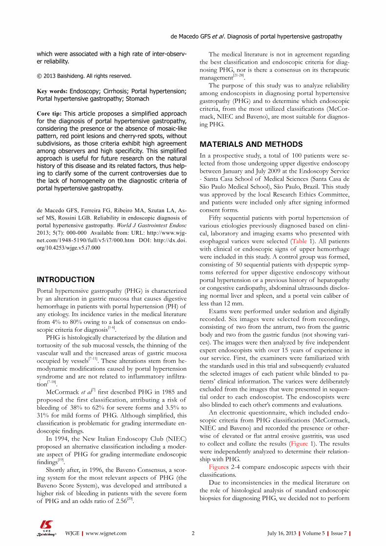
Exams were performed under sedation and digitally recorded. Six images were selected from recordings, consisting of two from the antrum, two from the gastric body and two from the gastric fundus (not showing vari-ces). The images were then analyzed by five independent expert endoscopists with over 15 years of experience in our service. First, the examiners were familiarized with the standards used in this trial and subsequently evaluated the selected images of each patient while blinded to pa-tients’ clinical information. The varices were deliberately excluded from the images that were presented in sequen-tial order to each endoscopist. The endoscopists were also blinded to each other’s comments and evaluations.
An electronic questionnaire, which included endo-scopic criteria from PHG classifications (McCormack, NIEC and Baveno) and recorded the presence or other-wise of elevated or flat antral erosive gastritis, was used to collect and collate the results (Figure 1). The results were independently analyzed to determine their relation-ship with PHG.
Figures 2-4 compare endoscopic aspects with their classifications.
Due to inconsistencies in the medical literature on the role of histological analysis of standard endoscopic biopsies for diagnosing PHG, we decided not to perform
de Macedo GFS et al . Diagnosis of portal hypertensive gastropathy
2 July 16, 2013|Volume 5|Issue 7|WJGE|www.wjgnet.com
biopsies in this study[29-31]. Due to the absence of an es-tablished gold standard for diagnosing PHG, the statisti-cal analysis was performed in two stages. The first stage verified the correlation between each endoscopic crite-rion and the presence of PH, with the group of 50 pa-tients without PH serving as a control. The second stage determined the correlation between each endoscopic cri-terion and the diagnosis of PHG. The establishment of a relationship between the endoscopic criterion and the presence of PH was a prerequisite for the potential cor-relation between the same criterion and PHG. If any cri-terion demonstrated an apparent relationship with PHG but not with PH, then it was deemed logically false.
The Statistical Package for Social Sciences version 17.0 was utilized for statistical analysis, adopting a 5% level of significance on Fisher’s Exact Test. Cronbach’s alpha was used to determine reliability among the five endoscopists, with values between 0 and 0.60 considered Unsatisfactory, values between 0.60 to 0.69 as Satisfac-tory, and values between 0.70 to 1.00 as a High degree of reliability.
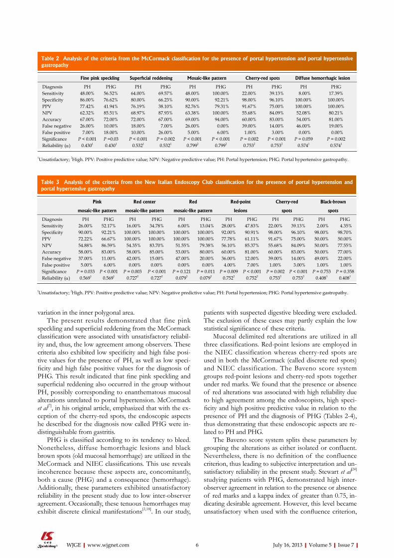
RESULTSFor criteria from the McCormack classification (Table 2), the mosaic-like pattern was associated with high reliabil-ity, specificity (90%) and positive predictive value (82.76%) for the presence of portal hypertension (PH), as well as sensitivity and negative predictive values of 100% for the diagnosis of portal hypertensive gastropathy (PHG). Fine pink speckling and superficial reddening both exhibited unsatisfactory reliability, as well as low specificity (86%) and high false positive values (7%) for the presence of
PH. In addition, these criteria exhibited low specificity (76.62%) and high false positive values (18%) for the di-agnosis of PHG.
On the NIEC classification (Table 3), pink and red mosaic-like patterns were associated with unsatisfactory reliability, but red center mosaic-like patterns exhibited high reliability, thus defining PHG as moderate.
For criteria from the Baveno classification (Table 4), only red marks demonstrated high reliability and specific-ity (92%) for the presence of PH and high reliability for the diagnosis of PHG.
Table 5 depicts the results of the statistical analysis of antral erosive gastritis and its variations, flat and elevated, in relation to the presence of portal hypertension and the diagnosis of portal hypertensive gastropathy. Antral el-evated erosive gastritis exhibited high reliability and high specificity with respect to the presence of PH (92%) and the diagnosis of PHG (88.31%).
DISCUSSIONThe analyzed classifications (McCormack, NIEC and BAVENO) comprise several common endoscopic as-pects, albeit aspects that are occasionally analyzed from different perspectives, thereby affecting the level of agreement among the observers. Others classifications have been published, including pre- and post-treatment evaluations, but these classifications have been reported without exclusive diagnostic aspects[21-22].
The presence of any mosaic-like pattern, defined as polygonal areas with whitish reticular borders, is utilized in all three classifications studied. The McCormack Clas-sification considers only its presence or absence but not variations in its inner polygonal area. In the present study, the mosaic-like pattern was associated with high reliability, specificity and positive predictive value for the presence of portal hypertension (PH), as well as sensitiv-ity and negative predictive values of 100% for the diag-nosis of portal hypertensive gastropathy (PHG), where its absence almost excluded this diagnosis. This finding corroborates the results of the study by Stewart et al[24] in which, out of the 100 patients diagnosed with PHG, 96 exhibited mosaic-like patterns.
Based on the NIEC classification, the mosaic-like pat-tern is subdivided into three and classified according to the color of the inner polygonal area as either pink, red center or red. In the present study, pink and red mosaic-like patterns were associated with unsatisfactory reli-ability, whereas a red center mosaic-like pattern had high reliability, defining PHG as moderate. Nevertheless, the red center may also be considered a red-point lesion or a cherry-red spot, characterizing the PHG as severe, there-by rendering this stratification of the pattern ambiguous and the NIEC classification inconsistent.
The Baveno Score System subdivides the mosaic-like pattern into two aspects: mild, corresponding to a pink mosaic-like pattern, and severe, which corresponds to a red mosaic-like pattern, which as mentioned above, was
Table 1 Group with portal hypertension and esophageal varices n (%)
Character n = 50
Mean age (52.7 yr)Sex Male 28 (56) Female 22 (44)Etiology Alcohol 10 (20) Schistosomiasis 18 (36) Hepatitis B 2 (4) Hepatitis C 5 (10) Alcohol and schistosomiasis 1 (2) Alcohol, schistosomiasis and Hepatitis B 1 (2) Autoimmune hepatitis 1 (2) Portal vein thrombosis 1 (2) Non-alcoholic hepatic steatosis 1 (2) Budd-Chiari Syndrome 1 (2) Biliary cirrhosis 1 (2) Idiopathic or not yet identified 8 (16)Child-pugh classification A 42 (84) B 6 (12) C 2 (4) Previous digestive bleeding 32 (64) Previous endoscopic treatment 33 (66) Using propanolol 25 (50)
3 July 16, 2013|Volume 5|Issue 7|WJGE|www.wjgnet.com
ing the presence or absence of the mosaic-like pattern, with a Kappa Index of greater than 0.75, concordance decreased when this aspect was subdivided according to
found to exhibit Unsatisfactory reliability and thus low agreement among observers. Although Stewart et al[24] also demonstrated agreement among observers analyz-
Figure 1 Electronic questionnaire.
DC
BA
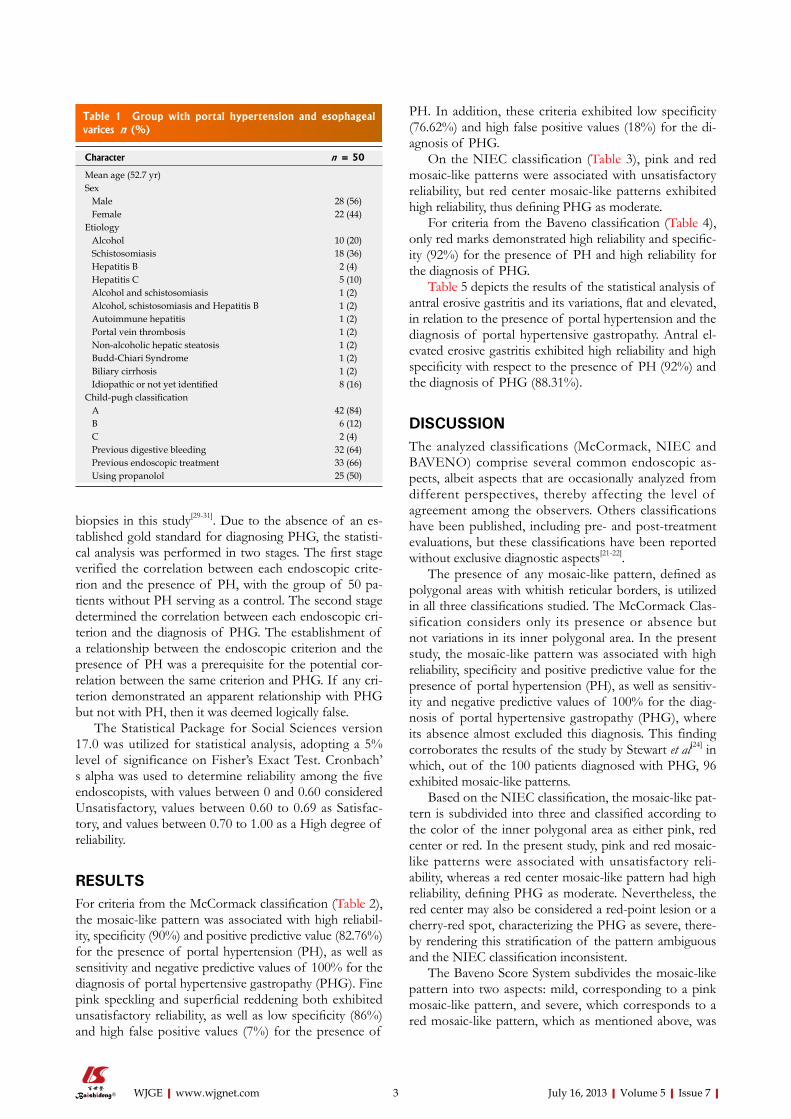
Figure 2 Endoscopic aspects of portal hypertensive gastropathy considered in the study and their corresponding classifications. A: Fine pink speckling - McCormack; B: Superficial reddening - McCormack; C: Diffuse hemorrhagic lesion - McCormack; D: Black brown spots - New Italian Endoscopy Club.
4 July 16, 2013|Volume 5|Issue 7|WJGE|www.wjgnet.com
DC
BA
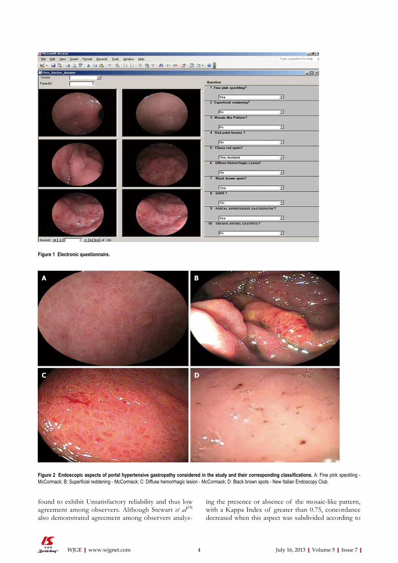
Figure 3 Endoscopic aspects of portal hypertensive gastropathy considered in the study and their corresponding classifications. A: Corresponds to “mosa-ic-like pattern” with different nomenclature in each classification as follows: mosaic-like pattern - McCormack; mild mosaic-like pattern - New Italian Endoscopy Club (NIEC); mild mosaic-like pattern - Baveno; B: Corresponds to “mosaic-like pattern” with different nomenclature in each classification as follows: red center mosaic-like pattern - McCormack; moderate mosaic-like pattern - NIEC; C: Corresponds to “mosaic-like pattern” with different nomenclature in each classification as follows: mosaic-like pattern - McCormack; severe mosaic-like pattern - NIEC; severe mosaic-like pattern - Baveno; D: GAVE - Baveno.
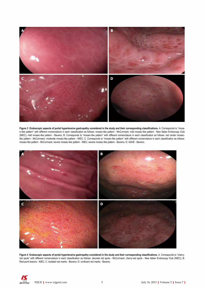
Figure 4 Endoscopic aspects of portal hypertensive gastropathy considered in the study and their corresponding classifications. A: Corresponds to “cherry-red spots” with different nomenclature in each classification as follows: discrete red spots - McCormack; cherry-red spots - New Italian Endoscopy Club (NIEC); B: Red-point lesions - NIEC; C: Isolated red marks - Baveno; D: confluent red marks - Baveno.
DC
BA
5 July 16, 2013|Volume 5|Issue 7|WJGE|www.wjgnet.com
variation in the inner polygonal area.The present results demonstrated that fine pink
speckling and superficial reddening from the McCormack classification were associated with unsatisfactory reliabil-ity and, thus, the low agreement among observers. These criteria also exhibited low specificity and high false posi-tive values for the presence of PH, as well as low speci-ficity and high false positive values for the diagnosis of PHG. This result indicated that fine pink speckling and superficial reddening also occurred in the group without PH, possibly corresponding to enanthematous mucosal alterations unrelated to portal hypertension. McCormack et al[7], in his original article, emphasized that with the ex-ception of the cherry-red spots, the endoscopic aspects he described for the diagnosis now called PHG were in-distinguishable from gastritis.
PHG is classified according to its tendency to bleed. Nonetheless, diffuse hemorrhagic lesions and black brown spots (old mucosal hemorrhage) are utilized in the McCormack and NIEC classifications. This use reveals incoherence because these aspects are, concomitantly, both a cause (PHG) and a consequence (hemorrhage). Additionally, these parameters exhibited unsatisfactory reliability in the present study due to low inter-observer agreement. Occasionally, these tenuous hemorrhages may exhibit discrete clinical manifestations[2,18]. In our study,
patients with suspected digestive bleeding were excluded. The exclusion of these cases may partly explain the low statistical significance of these criteria.
Mucosal delimited red alterations are utilized in all three classifications. Red-point lesions are employed in the NIEC classification whereas cherry-red spots are used in both the McCormack (called discrete red spots) and NIEC classification. The Baveno score system groups red-point lesions and cherry-red spots together under red marks. We found that the presence or absence of red alterations was associated with high reliability due to high agreement among the endoscopists, high speci-ficity and high positive predictive value in relation to the presence of PH and the diagnosis of PHG (Tables 2-4), thus demonstrating that these endoscopic aspects are re-lated to PH and PHG.
The Baveno score system splits these parameters by grouping the alterations as either isolated or confluent. Nevertheless, there is no definition of the confluence criterion, thus leading to subjective interpretation and un-satisfactory reliability in the present study. Stewart et al[24] studying patients with PHG, demonstrated high inter-observer agreement in relation to the presence or absence of red marks and a kappa index of greater than 0.75, in-dicating desirable agreement. However, this level became unsatisfactory when used with the confluence criterion,
Table 2 Analysis of the criteria from the McCormack classification for the presence of portal hypertension and portal hypertensive gastropathy
Fine pink speckling Superficial reddening Mosaic-like pattern Cherry-red spots Diffuse hemorrhagic lesion
Diagnosis PH PHG PH PHG PH PHG PH PHG PH PHGSensitivity 48.00% 56.52% 64.00% 69.57% 48.00% 100.00% 22.00% 39.13% 8.00% 17.39%Specificity 86.00% 76.62% 80.00% 66.23% 90.00% 92.21% 98.00% 96.10% 100.00% 100.00%PPV 77.42% 41.94% 76.19% 38.10% 82.76% 79.31% 91.67% 75.00% 100.00% 100.00%NPV 62.32% 85.51% 68.97% 87.93% 63.38% 100.00% 55.68% 84.09% 52.08% 80.21%Accuracy 67.00% 72.00% 72.00% 67.00% 69.00% 94.00% 60.00% 83.00% 54.00% 81.00%False negative 26.00% 10.00% 18.00% 7.00% 26.00% 0.00% 39.00% 14.00% 46.00% 19.00%False positive 7.00% 18.00% 10.00% 26.00% 5.00% 6.00% 1.00% 3.00% 0.00% 0.00%Significance P < 0.001 P =0.03 P < 0.001 P = 0.002 P < 0.001 P < 0.001 P = 0.002 P < 0.001 P = 0.059 P = 0.002Reliability (α) 0.4301 0.4301 0.5321 0.5321 0.7992 0.7992 0.7532 0.7532 0.5741 0.5741
1Unsatisfactory; 2High. PPV: Positive predictive value; NPV: Negative predictive value; PH: Portal hypertension; PHG: Portal hypertensive gastropathy.
Table 3 Analysis of the criteria from the New Italian Endoscopy Club classification for the presence of portal hypertension and portal hypertensive gastropathy
Pink Red center Red Red-point Cherry-red Black-brown
mosaic-like pattern mosaic-like pattern mosaic-like pattern lesions spots spots
Diagnosis PH PHG PH PHG PH PHG PH PHG PH PHG PH PHGSensitivity 26.00% 52.17% 16.00% 34.78% 6.00% 13.04% 28.00% 47.83% 22.00% 39.13% 2.00% 4.35%Specificity 90.00% 92.21% 100.00% 100.00% 100.00% 100.00% 92.00% 90.91% 98.00% 96.10% 98.00% 98.70%PPV 72.22% 66.67% 100.00% 100.00% 100.00% 100.00% 77.78% 61.11% 91.67% 75.00% 50.00% 50.00%NPV 54.88% 86.59% 54.35% 83.70% 51.55% 79.38% 56.10% 85.37% 55.68% 84.09% 50.00% 77.55%Accuracy 58.00% 83.00% 58.00% 85.00% 53.00% 80.00% 60.00% 81.00% 60.00% 83.00% 50.00% 77.00%False negative 37.00% 11.00% 42.00% 15.00% 47.00% 20.00% 36.00% 12.00% 39.00% 14.00% 49.00% 22.00%False positive 5.00% 6.00% 0.00% 0.00% 0.00% 0.00% 4.00% 7.00% 1.00% 3.00% 1.00% 1.00%Significance P = 0.033 P < 0.001 P = 0.003 P < 0.001 P = 0.121 P = 0.011 P = 0.009 P < 0.001 P = 0.002 P < 0.001 P = 0.753 P = 0.358Reliability (α) 0.5691 0.5691 0.7272 0.7272 0.0792 0.0792 0.7522 0.7522 0.7532 0.7532 0.4081 0.4081
1Unsatisfactory; 2High. PPV: Positive predictive value; NPV: Negative predictive value; PH: Portal hypertension; PHG: Portal hypertensive gastropathy.
6 July 16, 2013|Volume 5|Issue 7|WJGE|www.wjgnet.com
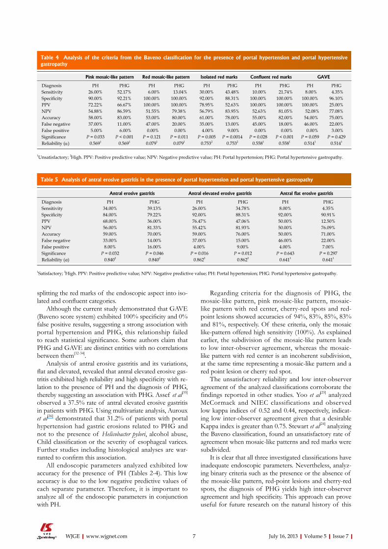
splitting the red marks of the endoscopic aspect into iso-lated and confluent categories.
Although the current study demonstrated that GAVE (Baveno score system) exhibited 100% specificity and 0% false positive results, suggesting a strong association with portal hypertension and PHG, this relationship failed to reach statistical significance. Some authors claim that PHG and GAVE are distinct entities with no correlations between them[32-34].
Analysis of antral erosive gastritis and its variations, flat and elevated, revealed that antral elevated erosive gas-tritis exhibited high reliability and high specificity with re-lation to the presence of PH and the diagnosis of PHG, thereby suggesting an association with PHG. Assef et al[35] observed a 37.5% rate of antral elevated erosive gastritis in patients with PHG. Using multivariate analysis, Auroux et al[36] demonstrated that 31.2% of patients with portal hypertension had gastric erosions related to PHG and not to the presence of Helicobacter pylori, alcohol abuse, Child classification or the severity of esophageal varices. Further studies including histological analyses are war-ranted to confirm this association.
All endoscopic parameters analyzed exhibited low accuracy for the presence of PH (Tables 2-4). This low accuracy is due to the low negative predictive values of each separate parameter. Therefore, it is important to analyze all of the endoscopic parameters in conjunction with PH.
Regarding criteria for the diagnosis of PHG, the mosaic-like pattern, pink mosaic-like pattern, mosaic-like pattern with red center, cherry-red spots and red-point lesions showed accuracies of 94%, 83%, 85%, 83% and 81%, respectively. Of these criteria, only the mosaic like-pattern offered high sensitivity (100%). As explained earlier, the subdivision of the mosaic-like pattern leads to low inter-observer agreement, whereas the mosaic-like pattern with red center is an incoherent subdivision, at the same time representing a mosaic-like pattern and a red point lesion or cherry red spot.
The unsatisfactory reliability and low inter-observer agreement of the analyzed classifications corroborate the findings reported in other studies. Yoo et al[25] analyzed McCormack and NIEC classifications and observed low kappa indices of 0.52 and 0.44, respectively, indicat-ing low inter-observer agreement given that a desirable Kappa index is greater than 0.75. Stewart et al[24] analyzing the Baveno classification, found an unsatisfactory rate of agreement when mosaic-like patterns and red marks were subdivided.
It is clear that all three investigated classifications have inadequate endoscopic parameters. Nevertheless, analyz-ing binary criteria such as the presence or the absence of the mosaic-like pattern, red-point lesions and cherry-red spots, the diagnosis of PHG yields high inter-observer agreement and high specificity. This approach can prove useful for future research on the natural history of this
Table 4 Analysis of the criteria from the Baveno classification for the presence of portal hypertension and portal hypertensive gastropathy
Pink mosaic-like pattern Red mosaic-like pattern Isolated red marks Confluent red marks GAVE
Diagnosis PH PHG PH PHG PH PHG PH PHG PH PHGSensitivity 26.00% 52.17% 6.00% 13.04% 30.00% 43.48% 10.00% 21.74% 8.00% 4.35%Specificity 90.00% 92.21% 100.00% 100.00% 92.00% 88.31% 100.00% 100.00% 100.00% 96.10%PPV 72.22% 66.67% 100.00% 100.00% 78.95% 52.63% 100.00% 100.00% 100.00% 25.00%NPV 54.88% 86.59% 51.55% 79.38% 56.79% 83.95% 52.63% 81.05% 52.08% 77.08%Accuracy 58.00% 83.00% 53.00% 80.00% 61.00% 78.00% 55.00% 82.00% 54.00% 75.00%False negative 37.00% 11.00% 47.00% 20.00% 35.00% 13.00% 45.00% 18.00% 46.00% 22.00%False positive 5.00% 6.00% 0.00% 0.00% 4.00% 9.00% 0.00% 0.00% 0.00% 3.00%Significance P = 0.033 P < 0.001 P = 0.121 P = 0.011 P = 0.005 P = 0.0014 P = 0.028 P < 0.001 P = 0.059 P = 0.429Reliability (α) 0.5691 0.5691 0.0791 0.0791 0.7532 0.7532 0.5581 0.5581 0.5141 0.5141
1Unsatisfactory; 2High. PPV: Positive predictive value; NPV: Negative predictive value; PH: Portal hypertension; PHG: Portal hypertensive gastropathy.
Table 5 Analysis of antral erosive gastritis in the presence of portal hypertension and portal hypertensive gastropathy
Antral erosive gastritis Antral elevated erosive gastritis Antral flat erosive gastritis
Diagnosis PH PHG PH PHG PH PHGSensitivity 34.00% 39.13% 26.00% 34.78% 8.00% 4.35%Specificity 84.00% 79.22% 92.00% 88.31% 92.00% 90.91%PPV 68.00% 36.00% 76.47% 47.06% 50.00% 12.50%NPV 56.00% 81.33% 55.42% 81.93% 50.00% 76.09%Accuracy 59.00% 70.00% 59.00% 76.00% 50.00% 71.00%False negative 33.00% 14.00% 37.00% 15.00% 46.00% 22.00%False positive 8.00% 16.00% 4.00% 9.00% 4.00% 7.00%Significance P = 0.032 P = 0.046 P = 0.016 P = 0.012 P = 0.643 P = 0.297Reliability (α) 0.8402 0.8402 0.8622 0.8622 0.6411 0.6411
1Satisfactory; 2High. PPV: Positive predictive value; NPV: Negative predictive value; PH: Portal hypertension; PHG: Portal hypertensive gastropathy.
7 July 16, 2013|Volume 5|Issue 7|WJGE|www.wjgnet.com
disease and related factors, thus helping to clarify some of the current controversies, including studies with his-tologic findings and comparisons with the endoscopic criteria of the classifications that we have already begun.
In conclusion, the most suitable endoscopic criteria for the diagnosis of portal hypertensive gastropathy were mosaic-like pattern, red-point lesions and cherry-red spots (without subdivisions), all of which were associated with a high rate of inter-observer reliability.
COMMENTSBackgroundPortal hypertensive gastropathy (PHG) is an alteration of gastric mucosa caus-ing occult and sometimes massive digestive hemorrhage in patients with portal hypertension of any etiology. Research frontiersPHG remains an endoscopic diagnosis, and there are many endoscopic clas-sifications. No histologic correspondence was proven, leading to an individual observer opinion in diagnosis and grading, with a low level of reliability among endoscopists.Innovations and breakthroughsThe most used classifications of PHG comprise several common endoscopic aspects, albeit aspects that are sometimes analyzed from different perspec-tives, thereby affecting the level of agreement among observers and leading to no consensus on endoscopic diagnosis and grading.ApplicationsBy separating the most suitable endoscopic criteria for the diagnosis of PHG, authors found that mosaic-like pattern, red-point lesions and cherry-red spots (without subdivisions) were associated with high inter-observer reliability and should be used to simplify and standardize the PHG diagnosis and severity.Peer reviewPHG is frequently observed on an upper gastrointestinal endoscopy in patients of portal hypertension. However, there are no objective criteria to diagnose PHG, and there is no consensus on the best classification and endoscopic cri-teria in the medical literature. The authors have attempted to analyze the data regarding reliability of various endoscopic morphological features among differ-ent endoscopists based on the criteria of McCormack, New Italian Endoscopy Club and Baveno. The authors concluded that most suitable endoscopic criteria for the diagnosis of PHG are mosaic-like pattern, red point lesions and cherry red spot.
REFERENCES1 Sarin SK, Sreenivas DV, Lahoti D, Saraya A. Factors influ-
encing development of portal hypertensive gastropathy in patients with portal hypertension. Gastroenterology 1992; 102: 994-999 [PMID: 1537536]
2 D’Amico G, Montalbano L, Traina M, Pisa R, Menozzi M, Spanò C, Pagliaro L. Natural history of congestive gastropa-thy in cirrhosis. The Liver Study Group of V. Cervello Hos-pital. Gastroenterology 1990; 99: 1558-1564 [PMID: 2227271]
3 Calès P, Oberti F, Bernard-Chabert B, Payen JL. Evaluation of Baveno recommendations for grading esophageal varices. J Hepatol 2003; 39: 657-659 [PMID: 12971983 DOI: 10.1016/S0168-8278(03)00404-5]
4 de Franchis R, Primignani M. Natural history of por-tal hypertension in patients with cirrhosis. Clin Liver Dis 2001; 5: 645-663 [PMID: 11565135 DOI: 10.1016/S1089-3261(05)70186-0]
5 Sarin SK, Agarwal SR. Gastric varices and portal hyper-tensive gastropathy. Clin Liver Dis 2001; 5: 727-67, x [PMID: 11565139 DOI: 10.1016/S1089-3261(05)70190-2]
6 Thuluvath PJ, Yoo HY. Portal Hypertensive gastropathy. Am J Gastroenterol 2002; 97: 2973-2978 [PMID: 12492178 DOI: 10.1016/S0002-9270(02)05511-9]
7 McCormack TT, Sims J, Eyre-Brook I, Kennedy H, Goepel J, Johnson AG, Triger DR. Gastric lesions in portal hyper-tension: inflammatory gastritis or congestive gastropathy? Gut 1985; 26: 1226-1232 [PMID: 3877665 DOI: 10.1136/gut.26.11.1226]
8 Albillos A, Colombato LA, Enriquez R, Ng OC, Sikuler E, Groszmann RJ. Sequence of morphological and hemody-namic changes of gastric microvessels in portal hyperten-sion. Gastroenterology 1992; 102: 2066-2070 [PMID: 1587425]
9 Panés J, Bordas JM, Piqué JM, Bosch J, García-Pagán JC, Feu F, Casadevall M, Terés J, Rodés J. Increased gastric muco-sal perfusion in cirrhotic patients with portal hypertensive gastropathy. Gastroenterology 1992; 103: 1875-1882 [PMID: 1451980]
10 Kotzampassi K, Eleftheriadis E, Aletras H. Gastric mucosal blood flow in portal hypertension patients--a laser Dop-pler flowmetry study. Hepatogastroenterology 1992; 39: 39-42 [PMID: 1533200]
11 Ohta M, Yamaguchi S, Gotoh N, Tomikawa M. Pathogenesis of portal hypertensive gastropathy: a clinical and experimen-tal review. Surgery 2002; 131: S165-S170 [PMID: 11821805 DOI: 10.1067/msy.2002.119499]
12 Dong L, Zhang ZN, Fang P, Ma SY. Portal hypertensive gas-tropathy and its interrelated factors. Hepatobiliary Pancreat Dis Int 2003; 2: 226-229 [PMID: 14599974]
13 Iwao T, Toyonaga A, Ikegami M, Sumino M, Oho K, Shi-gemori H, Sakaki M, Nakayama M, Tanikawa K, Iwao J. Portal vein hemodynamics in cirrhotic patients with portal hypertensive gastropathy: an echo-Doppler study. Hepa-togastroenterology 1994; 41: 230-234 [PMID: 7959544 DOI: 10.1016/0928-4680(94)90675-0]
14 Mezawa S, Homma H, Ohta H, Masuko E, Doi T, Miyanishi K, Takada K, Kukitsu T, Sato T, Niitsu Y. Effect of transjugu-lar intrahepatic portosystemic shunt formation on portal hypertensive gastropathy and gastric circulation. Am J Gas-troenterol 2001; 96: 1155-1159 [PMID: 11316163 DOI: 10.1016/S0002-9270(01)02257-2]
15 Bellis L, Nicodemo S, Galossi A, Guarisco R, Spilabotti L, Durola L, Dell’Unto O, Puoti C. Hepatic venous pressure gradient does not correlate with the presence and the sever-ity of portal hypertensive gastropathy in patients with liver cirrhosis. J Gastrointestin Liver Dis 2007; 16: 273-277 [PMID: 17925921]
16 Abraldes JG, Angermayr B, Bosch J. The management of portal hypertension. Clin Liver Dis 2005; 9: 685-713, vii [PMID: 16207571 DOI: 10.1016/j.cld.2005.08.001]
17 Sarin SK, Shahi HM, Jain M, Jain AK, Issar SK, Murthy NS. The natural history of portal hypertensive gastropa-thy: influence of variceal eradication. Am J Gastroen-terol 2000; 95: 2888-2893 [PMID: 11051364 DOI: 10.1016/S0002-9270(00)01993-6]
18 Primignani M, Carpinelli L, Preatoni P, Battaglia G, Carta A, Prada A, Cestari R, Angeli P, Gatta A, Rossi A, Spinzi G, De Franchis R. Natural history of portal hypertensive gastropathy in patients with liver cirrhosis. The New Italian Endoscopic Club for the study and treatment of esophageal varices (NIEC). Gastroenterology 2000; 119: 181-187 [PMID: 10889167 DOI: 10.1053/gast.2000.8555]
19 Spina GP, Arcidiacono R, Bosch J, Pagliaro L, Burroughs AK, Santambrogio R, Rossi A. Gastric endoscopic features in portal hypertension: final report of a consensus conference, Milan, Italy, September 19, 1992. J Hepatol 1994; 21: 461-467 [PMID: 7836719 DOI: 10.1016/S0168-8278(05)80329-0]
20 Sarin SK. Diagnostic issue: Portal hypertensive gastropathy and gastric varices. In: DeFranchis R, editor. Portal hyper-tension II. Proceeding of the second Baveno intenational consensus workshop on definitions, methodology and thera-peutic strategies. Oxford: Blackwell Science, 1996: 30-55
21 Hashizume M, Sugimachi K. Classification of gastric le-sions associated with portal hypertension. J Gastroenterol
8 July 16, 2013|Volume 5|Issue 7|WJGE|www.wjgnet.com
Hepatol 1995; 10: 339-343 [PMID: 7548815 DOI: 10.1111/j.1440-1746.1995.tb01105.x]
22 Tanoue K, Hashizume M, Wada H, Ohta M, Kitano S, Sugi-machi K. Effects of endoscopic injection sclerotherapy on portal hypertensive gastropathy: a prospective study. Gastro-intest Endosc 1992; 38: 582-585 [PMID: 1397916 DOI: 10.1016/S0016-5107(92)70522-7]
23 de Franchis R. Updating consensus in portal hypertension: report of the Baveno III Consensus Workshop on definitions, methodology and therapeutic strategies in portal hyper-tension. J Hepatol 2000; 33: 846-852 [PMID: 11097497 DOI: 10.1016/S0168-8278(00)80320-7]
24 Stewart CA, Sanyal AJ. Grading portal gastropathy: validation of a gastropathy scoring system. Am J Gastro-enterol 2003; 98: 1758-1765 [PMID: 12907330 DOI: 10.1016/S0002-9270(03)00446-5]
25 Yoo HY, Eustace JA, Verma S, Zhang L, Harris M, Kantsevoy S, Lee LA, Kalloo AN, Ravich WJ, Thuluvath PJ. Accuracy and reliability of the endoscopic classification of portal hy-pertensive gastropathy. Gastrointest Endosc 2002; 56: 675-680 [PMID: 12397275 DOI: 10.1016/S0016-5107(02)70116-8]
26 Chiu YC, Lu LS, Wu KL, Tam W, Hu ML, Tai WC, Chiu KW, Chuah SK. Comparison of argon plasma coagulation in man-agement of upper gastrointestinal angiodysplasia and gastric antral vascular ectasia hemorrhage. BMC Gastroenterol 2012; 12: 67 [PMID: 22681987 DOI: 10.1186/1471-230X-12-67]
27 Prachayakul V, Aswakul P, Leelakusolvong S. Massive gas-tric antral vascular ectasia successfully treated by endoscopic band ligation as the initial therapy. World J Gastrointest En-dosc 2013; 5: 135-137 [PMID: 23515642 DOI: 10.4253/wjge.v5.i3.135]
28 Fuccio L, Mussetto A, Laterza L, Eusebi LH, Bazzoli F. Di-agnosis and management of gastric antral vascular ectasia. World J Gastrointest Endosc 2013; 5: 6-13 [PMID: 23330048 DOI: 10.4253/wjge.v5.i1.6]
29 Misra SP, Dwivedi M, Misra V, Agarwal SK, Gupta R, Gupta SC, Mital VP. Endoscopic and histologic appearance of the gastric mucosa in patients with portal hypertension. Gastrointest Endosc 1990; 36: 575-579 [PMID: 2279646 DOI: 10.1016/S0016-5107(90)71167-4]
30 Piñero R, Olavarría R, Urquiola G, Yaraure M, Ramón Poleo J. [Portal hypertensive gastropathy: histologic and endo-scopic correlation]. G E N 1994; 48: 7-9 [PMID: 7926624]
31 Chaves DM, Sakai P, Mucenic M, Iriya K, Iriya Y, Ishioka S. Comparative study of portal hypertensive gastropathy in schistosomiasis and hepatic cirrhosis. Endoscopy 2002; 34: 199-202 [PMID: 11870569 DOI: 10.1055/s-2002-20291]
32 Payen JL, Calès P, Voigt JJ, Barbe S, Pilette C, Dubuisson L, Desmorat H, Vinel JP, Kervran A, Chayvialle JA. Severe por-tal hypertensive gastropathy and antral vascular ectasia are distinct entities in patients with cirrhosis. Gastroenterology 1995; 108: 138-144 [PMID: 7806035 DOI: 10.1016/0016-5085(95)90018-7]
33 Spahr L, Villeneuve JP, Dufresne MP, Tassé D, Bui B, Wil-lems B, Fenyves D, Pomier-Layrargues G. Gastric antral vascular ectasia in cirrhotic patients: absence of relation with portal hypertension. Gut 1999; 44: 739-742 [PMID: 10205216 DOI: 10.1136/gut.44.5.739]
34 Ripoll C, Garcia-Tsao G. Management of gastropathy and gastric vascular ectasia in portal hypertension. Clin Liver Dis 2010; 14: 281-295 [PMID: 20682235 DOI: 10.1016/j.cld.2010.03.013]
35 Assef MS, Valentino W, Nakamura MAC, Colaiacovo R, Rossini LG. The Study of Magnifying Endoscopy for the Diagnosis of Portal Hypertensive Gastropathy. Gastrointest Endosc 2010; 71: AB379 [DOI 10.1016/j.gie.2010.03.1046]
36 Auroux J, Lamarque D, Roudot-Thoraval F, Deforges L, Chaumette MT, Richardet JP, Delchier JC. Gastroduodenal ulcer and erosions are related to portal hypertensive gas-tropathy and recent alcohol intake in cirrhotic patients. Dig Dis Sci 2003; 48: 1118-1123 [PMID: 12822873]
P- Reviewers Abbas Z, Fouad YM, Reddy DN, Hashimoto N S- Editor Wen LL L- Editor A E- Editor Zhang DN
9 July 16, 2013|Volume 5|Issue 7|WJGE|www.wjgnet.com
Copyright © 2022 FDOKUMEN