Extraocular muscle injury during endoscopic sinus surgery
8
Extraocular muscle injury during endoscopic sinus surgery: an ophthalmologic perspective K-A Park and SY Oh Abstract Purpose The purpose of this study is to describe the clinical characteristics and treatment results of medial rectus muscle (MR) transection incurred during endoscopic sinus surgery. Methods This retrospective study included 16 patients with MR transection incurred during endoscopic sinus surgery between 1994 and 2015. The operative notes of the surgical procedure, the pattern of strabismus, the type of muscle injury, the type of corrective strabismus surgery, and the surgical outcomes were reviewed. Results Nine patients had partial resection of MR and seven patients had complete transection of MR, resulting from an injury incurred during endoscopic sinus surgery. Three of the nine patients with partial resection injury were initially diagnosed as complete resection and subsequently re- diagnosed as partial resection in a review of the images during this study. Five of the nine patients with partial MR resection underwent only simple recession/resection surgery. Patients with complete MR transection underwent muscle transposition or globe fixation surgeries and often multiple operations were required. Conclusions The results of this study showed that the treatment strategies could vary depending on the nature of muscle injury. In cases with complete transection, muscle transposition or globe fixation surgeries are often required, with multiple operations. However, partial muscle resection with only simple recession/resection surgery shows a favorable outcome in many cases. The use of proper imaging techniques, a thorough review of the images with various planes, and close follow-up are important for determining the nature of the muscle injury. Eye (2016) 30, 680–687; doi:10.1038/eye.2016.15; published online 19 February 2016 Introduction Endoscopic sinus surgery has been widely used as a mainstay of surgical treatment for disorders of the paranasal sinuses during the last 2 decades. It is well-established as a safe and effective surgical method. Despite the relative safety of the procedure and recent advances in this field, complications are still encountered. Ophthalmic injuries are rare but devastating complications. Powered devices that are widely used currently can be another source of ophthalmic injury if the suction cutting tip is misdirected into the orbit. Damage to extraocular muscles can cause severe permanent strabismus with diplopia, which has a poor prognosis even with treatment. Owing to the rare occurrence of extraocular muscle injury following endoscopic sinus surgery, there are few reports on the treatment strategies in these patients. 1–4 In the present study, we reported the results of surgical correction in patients with medial rectus (MR) injury incurred during endoscopic sinus surgery and further discussed the treatment strategies. Methods This study was approved by the Institutional Review Board of Samsung Medical Center (Seoul, Republic of Korea). All research was performed in compliance with the tenets of the Declaration of Helsinki. We retrospectively reviewed the medical records of patients with MR injury incurred during endoscopic sinus surgery, who were referred to the Samsung Medical Center between January 1994 and February 2015. Patients were identified from Department of Ophthalmology, Samsung Medical Center, Sungkyunkwan University School of Medicine, Seoul, Korea Correspondence: SY Oh, Department of Ophthalmology, Samsung Medical Center, Ilwon- dong, Kangnam-gu, Seoul 135-710, Korea Tel: +82 2 3410 3566; Fax: +82 2 3410 0074. E-mail: [email protected] Received: 9 August 2015 Accepted in revised form: 5 November 2015 Published online: 19 February 2016 CLINICAL STUDY Eye (2016) 30, 680–687 © 2016 Macmillan Publishers Limited All rights reserved 0950-222X/16 www.nature.com/eye
-
Upload
khangminh22 -
Category
Documents
-
view
0 -
download
0
Transcript of Extraocular muscle injury during endoscopic sinus surgery

Extraocular muscleinjury duringendoscopic sinussurgery: anophthalmologicperspective
K-A Park and SY Oh
Abstract
Purpose The purpose of this study is todescribe the clinical characteristics andtreatment results of medial rectus muscle(MR) transection incurred during endoscopicsinus surgery.Methods This retrospective study included16 patients with MR transection incurredduring endoscopic sinus surgery between1994 and 2015. The operative notes of thesurgical procedure, the pattern of strabismus,the type of muscle injury, the type ofcorrective strabismus surgery, and thesurgical outcomes were reviewed.Results Nine patients had partial resectionof MR and seven patients had completetransection of MR, resulting from an injuryincurred during endoscopic sinus surgery.Three of the nine patients with partialresection injury were initially diagnosed ascomplete resection and subsequently re-diagnosed as partial resection in a review ofthe images during this study. Five of the ninepatients with partial MR resection underwentonly simple recession/resection surgery.Patients with complete MR transectionunderwent muscle transposition or globefixation surgeries and often multipleoperations were required.Conclusions The results of this studyshowed that the treatment strategies couldvary depending on the nature of muscleinjury. In cases with complete transection,muscle transposition or globe fixationsurgeries are often required, with multipleoperations. However, partial muscle resectionwith only simple recession/resection surgeryshows a favorable outcome in many cases.The use of proper imaging techniques, athorough review of the images with variousplanes, and close follow-up are important fordetermining the nature of the muscle injury.
Eye (2016) 30, 680–687; doi:10.1038/eye.2016.15;published online 19 February 2016
Introduction
Endoscopic sinus surgery has been widely usedas a mainstay of surgical treatment for disordersof the paranasal sinuses during the last 2decades. It is well-established as a safe andeffective surgical method. Despite the relativesafety of the procedure and recent advances inthis field, complications are still encountered.Ophthalmic injuries are rare but devastatingcomplications. Powered devices that are widelyused currently can be another source ofophthalmic injury if the suction cutting tip ismisdirected into the orbit. Damage toextraocular muscles can cause severe permanentstrabismus with diplopia, which has a poorprognosis even with treatment. Owing to therare occurrence of extraocular muscle injuryfollowing endoscopic sinus surgery, there arefew reports on the treatment strategies in thesepatients.1–4 In the present study, we reported theresults of surgical correction in patients withmedial rectus (MR) injury incurred duringendoscopic sinus surgery and further discussedthe treatment strategies.
Methods
This study was approved by the InstitutionalReview Board of Samsung Medical Center(Seoul, Republic of Korea). All research wasperformed in compliance with the tenets of theDeclaration of Helsinki. We retrospectivelyreviewed the medical records of patients withMR injury incurred during endoscopic sinussurgery, who were referred to the SamsungMedical Center between January 1994 andFebruary 2015. Patients were identified from
Department ofOphthalmology, SamsungMedical Center,Sungkyunkwan UniversitySchool of Medicine, Seoul,Korea
Correspondence:SY Oh, Department ofOphthalmology, SamsungMedical Center, Ilwon-dong, Kangnam-gu, Seoul135-710, KoreaTel: +82 2 3410 3566;Fax: +82 2 3410 0074.E-mail: [email protected]
Received: 9 August 2015Accepted in revised form:5 November 2015Published online:19 February 2016
CLINICALSTUDY
Eye (2016) 30, 680–687© 2016 Macmillan Publishers Limited All rights reserved 0950-222X/16
www.nature.com/eye

outpatient logbooks. In this study, we included patientswith partial or complete transection of the MR incurredduring endoscopic sinus surgery, which had beenconfirmed using orbital magnetic resonance imaging(MRI) or computed tomography (CT). Some of thesepatients had participated in a study of the otolaryngologicperspective of extraocular muscle injury duringendoscopic sinus surgery in the otorhinolaryngologydepartment in the same hospital.5 We reviewed theoperative reports of all patients describing the endoscopicsinus surgery and the medical records for demographicdata, initial ocular deviation and ocular motility, surgicalmethods for strabismus, poststrabismus surgery oculardeviation and ocular motility, reoperations, andcomplications. Available preoperative and postoperativeimaging (MRI and/or CT) data were evaluated. Allpatients were classified as complete vs partial MRresection according to the orbital imaging results.All patients underwent a complete ophthalmic and
ocular motility examination. Ideally, measurements ofocular alignment were obtained using prism alternatecover testing at 6 m fixation and 33 cm fixation; however,when this was not possible, the Krimsky test at 33 cmfixation was used. Voluntary ductions were performed onall patients using a 4-point scale ranging from 0 to − 4:0=patient has full movement in the affected eye;− 2=patient is unable to move the affected eye past themidline; − 3=patient is unable to move the affected eye tothe midline; and − 4=patient has no adductingmovement in the affected eye. Forced duction and forcegeneration tests were performed on all patients. Twoexaminers (SO and KP) performed all the tests for ocularalignment and surgeries as well. The surgical strategy ineach case was individualized based on the nature of themuscle injury, the clinical presentation, and the type ofprevious orbital and muscle surgery.
Results
We identified 16 patients with MR transection incurredduring endoscopic sinus surgery. A summary of patientcharacteristics was provided in Table 1. Representativecases were shown in Figure 1. The mean age of thepatients was 47± 14 years (range 20–64 years). Allpatients had unilateral extraocular muscle damage, withmore common involvement of the right eye (10 patients,63%). Nine patients had partial resection of MR and sevenpatients had complete resection of MR from the injuryincurred during endoscopic sinus surgery (Figure 2).Three of the patients with partial resection injury werediagnosed as complete resection at their initial visit andwere re-diagnosed as partial resection during theretrospective review of the previous images. In thesecases, different planes (sagittal plane in case 9), different
imaging methods (MRI in case 11), or images taken after atime interval (case 12) showed a strand of MR connectedto the proximal and distal MR stumps, ruling outcomplete transection (Figure 3). A microdebrider,a refined endoscopic surgical tool that provides powereddissection, was used in all cases with the exception ofcase 9, in which there was no information on surgicalinstrumentation in the medical records of another hospital.A summary of findings on preoperative ophthalmic
evaluation was provided in Table 1. The mean preoperativeangle of exotropia in the primary position at distancefixation was 53± 15 prism diopters (PD) (range 15–75 PD).The mean adduction deficit was − 3.0± 1.0 (range − 1 to−4). The visual acuity in the operated eye ranged from20/20 to no light perception. Fourteen patients (88%) haddiplopia in the primary position preoperatively and allpatients had a normal head posture preoperatively. Table 2showed the previous surgeries if any, managementstrategies, and postoperative alignment of the patients. Themean time between MR injury during endoscopic sinussurgery and the first corrective surgery for the strabismuswas 9± 5 months (range 0–19 months). The mean follow-upduration was 3± 1 years (range 1.5–5 years).Four of the nine patients with partial MR transection
showed improvement of ocular movement, at 4 monthsfollow-up in 2 patients and at 2 months in 1 patient,before strabismus surgery. One patient showed completerecovery at 2 months without strabismus surgery. Five ofthe nine patients with partial MR transection underwentonly MR resection and lateral rectus muscle recession.During the MR resection surgery, care was taken not topull the MR, which could potentially convert incompletetransection to complete transection. Of the two patientswith partial MR resection, who were unable to move theaffected eye to the midline, one patient underwent theglobe tethering technique using a suture/titanium T-plateanchoring platform system6 as an initial surgery and theother patient, who previously had undergone lateralrectus muscle recession and MR resection, underwent fulltendon transposition and MR repeat resection. In twocases with complete MR transection, a proximal MRstump was found through an anterior orbitotomyapproach.7 Owing to severe fibrosis around the site ofinjury, it was impossible to completely free the musclestump from the fibrotic tissues and advance the musclestump anteriorly. Instead, the hang-back suture wasfixated tightly where the eye was positioned to themidline. In these cases, suture tightening was requiredafter the surgery due to recurrence of exodeviation overtime. Three patients with total MR transection underwentthe muscle union procedure8 with lateral rectus musclerecession. Two patients had successful postoperativeresults; however, the other patient with 70 PD exotropiaand 15 PD right hypertropia, who underwent the muscle
Extraocular muscle injury during endoscopic sinus surgeryK-A Park and SY Oh
681
Eye

Tab
le1
Patie
ntch
aracteristics,type
ofen
doscopicsinu
ssu
rgery,
type
ofinjury,a
ndsu
mmaryof
find
ings
inop
htha
lmic
evalua
tion
Case
(age
a /sex)
ESS
/injury
side
Typ
eof
MRinjury
Other
Cx.
Initial
deviation
Preop.
deviationb
Initial
add.
cPreop.a
dd.b,c
Preop.F
GTb
Preop.F
DTb
Preop.
diplopia
bLeng
thof
proxim
alstum
p
1(20/
M)
EMMA
(R)/righ
tCom
pleteresection
Non
e30
XT
50XT
−3
−3
Seve
reweakn
ess
LR:m
ildrestriction
+7mm
2(46/
F)EMMA
(R)/righ
tCom
pleteresection
Non
e50
XT
50XT
−4
−4
Mod
erateweakn
ess
LR:m
oderate
restriction
+18
mm
3(61/
M)
EMMA
(B)/righ
tCom
pleteresection
Non
e50
XT
70XT
−4
−4
Seve
reweakn
ess
MR,L
R:
mod
erate
restriction
+15
mm
4(61/
M)
EMMA
(B)/righ
tCom
pleteresection
IRinjury
80XT
15RHT
70XT15
RHT
−4
−4
Seve
reweakn
ess
Norestriction
+7mm
5(58/
M)
EMMA
(L)/left
Com
pleteresection
CRAO,o
ptic
nerve
tran
section,
complete
3rdne
rvepa
lsy
50XT,
20LHT
50XT,2
0LHT
−4
−4
Seve
reweakn
ess
Norestriction
−No
stum
pvisible
6(49/
F)EMMA
(B)/left
Com
pleteresection
Tem
porary
IRpa
lsy
50XT4
LHT
50XT
−4
−4
Seve
reweakn
ess
LR:m
oderate
restriction
+8mm
7(59/
M)
EMMA
(B)/righ
tCom
pleteresection
IRinjury
50XT
18RHT
−4
Seve
reweakn
ess
IR:m
ildrestriction
+11
mm
8(64/
F)EMMA
(L)/left
Partialresection
Non
e45
XT
45XT
−2
−2
Mod
erateweakn
ess
MR,L
R:m
ildrestriction
+−
9(49/
M)
EMMA
(R)/righ
tPa
rtialresection
Non
e30
XT
35XT
−3
−3
Mod
erateweakn
ess
LR:m
ildrestriction
+−
10(45/
M)EMMA
(B)/righ
tPa
rtialresection
CSF
leak
age
75XT
75XT
−4
−4
Seve
reweakn
ess
Norestriction
+−
11(46/
M)EMMA
(R)/righ
tPa
rtialresection
Orbital
hemorrhag
e80
XT
50XT
−4
−2
Mod
erateweakn
ess
Norestriction
+−
12(44/
M)EMMA
(R)/righ
tPa
rtialresection
Non
e60
XT
70XT
−4
−2
Seve
reweakn
ess
Norestriction
+−
13(36/
M)EMMA
(B)/righ
tPa
rtialresection
Optic
nerveinjury
35XT
40XT
−3
−3
Mod
erateweakn
ess
Norestriction
+−
14(54/
M)EMMA
(L)/left
Partialresection
Entrapm
entof
MR
35XT
30XT2L
HoT
−3
−1
Mod
erateweakn
ess
MR:m
oderate
restriction
+−
15(20/
M)EMMA
(B)/left
Partialresection
Non
e10
XT
Ortho
(Spo
nt.
recove
ry)
−1
Normal
(Spo
nt.
recove
ry)
Normal
(Spo
nt.
recove
ry)
Norestriction
−(Spo
nt.
resolution
)−
16(54/
M)EMMA
(B)/left
Partialresection
Orbital
hemorrhag
e20
XT
15XT
−2
−2
Mod
erateweakn
ess
MR,L
R,IR:
mod
erate
restriction
+−
Abb
reviations:ad
d.,ad
duc
tion
;B,b
ilateral;CRAO,c
entral
retina
lartery
occlus
ion;
CSF
,cereb
rosp
inal
fluid;C
x.,c
omplications;E
MMA,e
thmoidectomyan
dmiddle
meatalan
trostomy;
ESS
,end
oscopicsinu
ssu
rgery;
F,female;FD
T,forcedduc
tion
test;F
GT,force
gene
ration
test;IR,inferiorrectus
;L,left;LHT,lefth
ypertrop
ia;L
HoT
,lefth
ypotropia;LR,lateral
rectus
;M,m
ale;MR,m
edialrectusmus
cle;Po
st.,po
sterior;
Preo
p,preo
perative
;R,right;R
HT,right
hype
rtropia;
Spon
t.,sp
ontane
ous;XT,exo
trop
ia.aYears.bResultsof
thepreo
perative
exam
inationat
thefina
lpreop
erativevisit.
c Volun
tary
duc
tion
swerepe
rformed
using
a4-po
intscalerang
ingfrom
0to
−4:
0=pa
tien
tha
sfullmov
emen
t;−2=pa
tien
tis
unab
leto
mov
etheaffected
eyepa
stthemidlin
e;an
d−3=pa
tien
tis
unab
leto
mov
etheaffected
eyeto
themidlin
e.
Extraocular muscle injury during endoscopic sinus surgeryK-A Park and SY Oh
682
Eye

union procedure, had 20 PD exotropia and 15 PD righthypertropia postoperatively.The mean postoperative horizontal angle of ocular
deviation was 4± 6 PD (range 0–20 PD) exotropia at thefinal postoperative visit. The mean postoperative
adduction deficit was − 1.5± 1.0 (range − 0.5 to − 3). Allpatients were acceptably aligned in central gaze afterstrabismus surgery, with the exception of one patient whohad concomitant inferior rectus muscle injury. Five of the12 patients (42%) required ≥ 2 surgical procedures.
Figure 1 A 46-year-old woman with complete MR muscle transection following endoscopic sinus surgery (case 2). She underwentmultiple strabismus surgeries including muscle transposition surgery. Before (top) and 1 month following the final strabismus surgery(bottom).
Extraocular muscle injury during endoscopic sinus surgeryK-A Park and SY Oh
683
Eye
There were no intraoperative surgical complicationsreported and no cases of anterior segment ischemia. Twopatients developed a mild transient increase in intraocularpressure of the operated eye on postoperative day 1,which resolved within 1 week before discontinuation ofsteroid eye drops. There was no sign of cellular reaction inthe anterior chamber or pupillary abnormality inthese cases.
Discussion
In this study, MR injury following endoscopic sinussurgery was divided into partial vs total MR resection. Inthe partial resection group, three patients were able tomove the affected eye to the midline at the initial examand five patients were able to move the affected eye to themidline at the final preoperative exam. Four out of thenine patients with partial MR transection showedimprovement of ocular movement during the severalmonths follow-up period before the strabismus surgery.In the complete resection group, none of the patientscould move the affected eye to the midline during thepreoperative follow-up period and there was noimprovement in ocular movement during the entirepreoperative follow-up period. Small adductionmovement in some cases with complete MR resection
may be related to the relaxation of the antagonist lateralrectus muscle.In the partial resection group, simple recession–
resection surgery achieved good postoperative results inmost cases. However, in the complete resection group,muscle transposition surgery or globe fixation surgeryusing a non-absorbable suture were needed to correct thedeviation of the eyeball and resolve diplopia. In mostcomplete MR transection cases in this study, thedestruction of the extraocular muscle was severe andthere were no cases with a proximal residual stumplonger than 20 mm. The severity of destruction in thisstudy may be related to the use of the powered devices.9
We attempted to find muscle stumps in two cases withshort residual proximal muscle tissue and sutured with anon-absorbable suture; however, there was a recurrenceof exodeviation over time. Recurrence of exodeviationmay be due to loosening of the suture or remodeling ofthe tissues where the suture was anchored. Muscletransposition surgery such as the muscle union procedurewas effective in these patients with complete MRtransection. Nevertheless, the result was poorer whenaccompanied with vertical rectus muscle injury, as seen incase 4. There was no significant complication in globefixation or muscle transposition surgeries, except for atransient increase in intraocular pressure in two patients
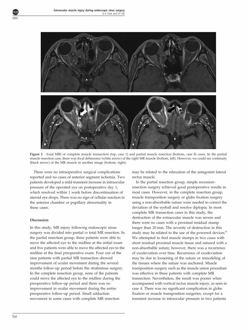
Figure 2 Axial MRI of complete muscle transection (top, case 1) and partial muscle resection (bottom, case 8) cases. In the partialmuscle resection case, there was focal dehiscence (white arrow) of the right MR muscle (bottom, left). However, we could see continuity(black arrow) of the MR muscle in another image (bottom, right).
Extraocular muscle injury during endoscopic sinus surgeryK-A Park and SY Oh
684
Eye

who underwent muscle union procedure, which might bedue to mechanical pressure. The different surgicalresponse in individual cases indicated that the response tothe same surgical technique could vary betweenindividuals. The reason for this variability may be due tosusceptibility of certain individuals, different severity,and amount and location of the injury from previousendoscopic sinus surgery. However, most patients withcomplete MR resection finally resolved diplopia and hada cosmetically fine outcome after several surgeries. Ifprimary repair of the transected muscle is impossible incomplete resection cases, surgical methods includingvarious types of transposition surgeries or globe fixationsurgeries could be selected based on the surgeon’sexperiences or availability.During the review process in this study, we
re-diagnosed three patients as partial resection, who were
diagnosed as complete resection at their initial visit. Inthese cases, the one-time axial CT scan alone was oftennot sufficient to distinguish partial muscle resection fromcomplete transection. Ela-Dalman et al10 previouslyemphasized the importance of various views of MRI scans,especially sagittal views. In our cases, we found a strand ofMR that was connected to the proximal and distal MRstumps ruling out complete transection, using a differentimage plane (sagittal plane), a different imaging method(MRI instead of CT), or with images taken after the timeinterval. A thorough review of the images is warranted inpatients with MR injury or suspected MR injury. Cautiousfollow-up is needed with repeated imaging using variousplanes of MRI scans, to evaluate possible improvement inthe adduction that could occur in partial MR resection.This study had several limitations. First, the sample size
was small due to the rare occurrence of this entity.
Figure 3 CT scans and axial MRI of case 11. The mid portion of the muscle segment seems transected on CT scans (top and middle).However, axial views of MRI shows partial damage to the MR muscle, ruling out complete transection (arrow).
Extraocular muscle injury during endoscopic sinus surgeryK-A Park and SY Oh
685
Eye

Therefore, we could not properly compare surgical resultsfrom different surgical methods. Second, there waspossible observational bias of the postoperative results.Third, it was a retrospective review, and the treatmentmethod and imaging methods were heterogeneous. Inparticular, the 20-year period over which the patientswere involved has seen much improvement in imagingtechniques. As a consequence, there were technicaldifferences between images of individual patients.In conclusion, the surgical strategy in MR transection
injury should be individualized based on the nature of themuscle injury, the clinical presentation, and the type ofprevious strabismus surgeries. In complete and severe
muscle transection injury, primary repair of the transectedmuscle is often impossible. These cases might requiremultiple globe fixation or muscle transposition surgeries.Conversely, only simple recession/resection surgery canresult in favorable outcomes in many cases of partialresection injury. If partial transection is confirmed, werecommend observation for several months beforestrabismus surgery, because some of them can showimprovement of ocular movement during initial severalmonths. In these cases, simple recession/resectionsurgery could achieve good ocular alignment even19 months after the initial injury. If partial transection issuspected, more conservative approach is needed. In
Table 2 Management strategies and postoperative results of the patients
Case Time frominjury to firststrabismussurgery (m)
Surgery Number ofstrabismussurgeries
Postop.deviationa
Postop.adductiona,b
F/Uduration (y)
Postop.diplopiain centralgazea
Postop.head
posturea
1 7 LR rec 10 mm, globe fixation surgeryusing non-absorbable suture (MRproximal stump to the sclera hang-backsuture) (orbital approach), repair of bonydefect, previous suture tightening,previous suture tightening, previoussuture tightening
5 15 XT − 3 3 − 15°
2 0 Primary repair (outside hospital), LR rec11 mm, partial tendon transposition,repair of bony defect, globe fixationsurgery using non-absorbable suture(MR proximal stump to the sclera hang-back suture) (orbital approach), SR rec5 mm, previous suture tightening
6 6 XT − 2 3 − 4°
3 8 LR rec 10 mm, muscle union procedure 1 Ortho − 1 2 − 0°4 9 LR rec 9 mm, muscle union procedure,
IO anteriorization2 20 XT 15 RHT − 2 3 + 0°
5 15 Muscle union procedure, LR rec 10 mm 1 4 XT 6 LHT − 3 1.5 − 0°6 13 Plan: muscle union procedure, LR rec7 Follow-up loss8 12 Repair of bony defect, MR res 5 mm, LR
rec 8 mm1 Ortho − 2 1.5 − 0°
9 6 LR rec 9 mm, MR res 7 mm 1 Ortho − 2 5 − 0°10 4 Globe tethering technique using a
suture/titanium T-plate anchoringplatform system, LR rec 9 mm
2 Ortho 0 4 − 0°
11 8 LR rec 9.0 mm, MR res 5.0 mm 1 15 X − 1 4 − 0°12 13 LR rec 9.5 mm, MR res 5.5 mm 1 4 XT − 0.5 2 − 0°13 7 LR rec 10 mm, MR res 10 mm (outside
hospital), full tendon transposition, MRreres 5 mm
3 Ortho − 2 5 − 0°
14 19 MR res 5.5 mm, LR rec 6 mm 1 Ortho 0 4 − 0°15 Repair of bony defect (spontaneous
recovery without strabismus surgery3 months after the injury)
0
16 6 Plan: LR rec
Abbreviations: F/U, follow-up; IO, inferior oblique muscle; LHT, left hypertropia; LR, lateral rectus; m, months; MR, medial rectus muscle; Postop.,postoperative; rec, recession surgery; res, resection surgery; reres, re-resection surgery; RHT, right hypertropia; SR, superior rectus muscle; X, exophoria;XT, exotropia; y, years. aResults of the postoperative examination at the final postoperative visit. bVoluntary ductions were performed using a 4-point scaleranging from 0 to − 4: 0=patient has full movement; − 2=patient is unable to move the affected eye past the midline; and − 3=patient is unable to movethe affected eye to the midline.
Extraocular muscle injury during endoscopic sinus surgeryK-A Park and SY Oh
686
Eye

order to determine the nature of muscle injury, a properimaging technique and a thorough review of the imagesat various planes are important. Close follow-up andrepeated imaging using MRI with various planes can behelpful in ambiguous cases.
Summary
What was known beforeK There have been changes in the type of surgical
instrumentation used during endoscopic sinus surgery.The increase in efficiency during surgery could potentiallyresult in more extensive damage when MR muscle injuryoccurs. However, owing to its rare occurrence there havebeen a few case series on this subject.
What this study addsK We were able to classify the MR muscle injury as partial vs
complete injury. There was a large difference in thetreatment strategies between the two groups. It is necessaryto carry out more extensive surgery, such as transpositionsurgery or globe fixation surgery, than simple recession/resection surgery in complete injuries. Otherwise, simplerecession/resection surgery can effectively resolve oculardeviation and diplopia in most partial resection cases. Wealso found that there had been misinterpretations of theimages before the surgery.We recommend a thoroughreview of the images in these patients with MR muscleinjury; in the cases in which the classification of MR injuryis ambiguous, cautious follow-up is required to find apossible improvement in adduction, which could occur inpartial MR resection, and repeated imaging using MRIscans with various planes will be very helpful.
Conflict of interest
The authors declare no conflict of interest.
Acknowledgements
Study design, conduction of the study, and data collectionand management (KAP and SYO); data analysis,
interpretation, and drafting the manuscript (KAP); andreview and final approval of the manuscript (SYO).
References
1 Han JK, Higgins TS. Management of orbital complications inendoscopic sinus surgery. Curr Opin Otolaryngol Head NeckSurg 2010; 18: 32–36.
2 Thacker NM, Velez FG, Demer JL, Rosenbaum AL.Strabismic complications following endoscopic sinussurgery: diagnosis and surgical management. J AAPOS 2004;8: 488–494.
3 Trotter WL, Kaw P, Meyer DR, Simon JW. Treatment ofsubtotal medial rectus myectomy complicating functionalendoscopic sinus surgery. J AAPOS 2000; 4: 250–253.
4 Yang SQ, Guo X. [Management of large exotropiawith medial rectus muscle transection followingendoscopic sinus surgery]. Zhonghua Yan Ke Za Zhi2010; 46: 974–977.
5 Sohn JH, Hong SD, Kim JH, Dhong HJ, Chung SK, Kim HYet al. Extraocular muscle injury during endoscopic sinussurgery: a series of 10 cases at a single center. Rhinology 2014;52: 238–245.
6 Tse DT, Shriver EM, Krantz KB, Tse JD, Capo H,McKeown CA. The use of titanium T-plate as platform forglobe alignment in severe paralytic and restrictivestrabismus. Am J Ophthalmol 2010; 150: 404–411 e401.
7 Underdahl JP, Demer JL, Goldberg RL, Rosenbaum AL.Orbital wall approach with preoperative orbital imaging foridentification and retrieval of lost or transected extraocularmuscles. J AAPOS 2001; 5: 230–237.
8 Park KA, Oh SY. Muscle union procedure with medial rectusrecession for unilateral abducens palsy. J Pediatr OphthalmolStrabismus 2013; 50 Online: e11–e14.
9 Thacker NM, Velez FG, Demer JL, Wang MB, RosenbaumAL. Extraocular muscle damage associated with endoscopicsinus surgery: an ophthalmology perspective. Am J Rhinol2005; 19: 400–405.
10 Ela-Dalman N, Velez FG, Rosenbaum AL. Importance ofsagittal orbital imaging in evaluating extraocular muscletrauma following endoscopic sinus surgery. Br J Ophthalmol2006; 90: 682–685.
Extraocular muscle injury during endoscopic sinus surgeryK-A Park and SY Oh
687
Eye