THREE-DIMENSIONAL ENDOSCOPIC PITUITARY SURGERY
8
ons288 | VOLUME 64 | OPERATIVE NEUROSURGERY 2 | MAY 2009 www.neurosurgery-online.com TUMOR Concepts and Innovations Abtin Tabaee, M.D. Department of Otolaryngology— Head and Neck Surgery, New York Presbyterian Hospital, Weill Medical College of Cornell University, New York, New York Vijay K. Anand, M.D. Department of Otolaryngology— Head and Neck Surgery, New York Presbyterian Hospital, Weill Medical College of Cornell University, New York, New York Justin F. Fraser, M.D. Department of Neurosurgery, New York Presbyterian Hospital, Weill Medical College of Cornell University, New York, New York Seth M. Brown, M.D., M.B.A. Department of Otolaryngology— Head and Neck Surgery, New York Presbyterian Hospital, Weill Medical College of Cornell University, New York, New York Ameet Singh, M.D. Department of Otolaryngology— Head and Neck Surgery, New York Presbyterian Hospital, Weill Medical College of Cornell University, New York, New York Theodore H. Schwartz, M.D. Departments of Neurosurgery, Otolaryngology—Head and Neck Surgery, and Neurology, New York Presbyterian Hospital, Weill Medical College of Cornell University, New York, New York Reprint requests: Theodore H. Schwartz, M.D., Departments of Neurosurgery, Otolaryngology—Head and Neck Surgery, and Neurology, New York Presbyterian Hospital, Weill Medical College of Cornell University, 525 East 68th Street, Box 99, New York, NY 10065. Email: [email protected] Received, May 27, 2008. Accepted, September 19, 2008. Copyright © 2009 by the Congress of Neurological Surgeons I nitially described by Schloffer (21) and Cushing (9) and subsequently popularized by Guiot (12) and Hardy and Wigser (14), the transsphenoidal approach to the sella now represents the preferred approach for remov- ing pituitary adenomas. Traditionally per- formed with a microscope and a sublabial inci- sion, the implementation of the endoscope and endonasal access has rendered the transsphe- noidal approach less invasive and provided improved visualization into and around the sella (4, 5, 16–18). Nevertheless, one of the pri- mary restrictions of endoscopic or endoscope- assisted surgery is the lack of binocular or stereoscopic vision. Monocular endoscopes and displays create a 2-dimensional (2-D) image that impairs depth perception, hand-eye coor- dination, and the ability to estimate size (1, 24). Operating in a 2-D environment requires sur- geons to train their hand-eye coordination to respond to visual cues received by the interac- tion of the operative instruments with the envi- ronment to accurately understand the relative depth of structures in the 2-D projection. Surgeons will often move the endoscope in and out or side to side to gain a motion parallax depth cue. This lack of stereoscopic vision has contributed to the steep learning curve in the field of neuroendoscopy. The next obvious step in the evolution of minimal access endoscopic surgery is the development of high-definition stereoendoscopes that produce a 3-dimensional (3-D) image. Although such stereoendoscopes exist (1, 2, 24), their use in neurosurgery has been limited because of the larger diameter and poor resolution of earlier generations. Hence, only 1 report exists of using stereoendoscopes ABBREVIATIONS: 3-D, 3-dimensional; 2-D, 2-dimensional THREE- DIMENSIONAL ENDOSCOPIC PITUITARY SURGERY OBJECTIVE: We describe a novel 3-dimensional (3-D) stereoendoscope and discuss our early experience using it to provide improved depth perception during transsphe- noidal pituitary surgery. METHODS: Thirteen patients underwent endonasal endoscopic transsphenoidal sur- gery. A 6.5-, 4.9-, or 4.0-mm, 0- and 30-degree rigid 3-D stereoendoscope (Visionsense, Ltd., Petach Tikva, Israel) was used in all cases. The endoscope is based on “compound eye” technology, incorporating a microarray of lenses. Patients were followed prospec- tively and compared with a matched group of patients who underwent endoscopic sur- gery with a 2-dimensional (2-D) endoscope. Surgeon comfort and/or complaints regard- ing the endoscope were recorded. RESULTS: The 3-D endoscope was used as the sole method of visualization to remove 10 pituitary adenomas, 1 cystic xanthogranuloma, 1 metastasis, and 1 cavernous sinus hemangioma. Improved depth perception without eye strain or headache was noted by the surgeons. There were no intraoperative complications. All patients without cav- ernous sinus extension (7of 9 patients) had gross tumor removal. There were no sig- nificant differences in operative time, length of stay, or extent of resection compared with cases in which a 2-D endoscope was used. Subjective depth perception was improved compared with standard 2-D scopes. CONCLUSION: In this first reported series of purely 3-D endoscopic transsphenoidal pituitary surgery, we demonstrate subjectively improved depth perception and excellent outcomes with no increase in operative time. Three-dimensional endoscopes may become the standard tool for minimal access neurosurgery. KEY WORDS: Endoscopic, Minimal access, Minimally invasive, Pituitary, 3-dimensional, Transsphenoidal Neurosurgery 64[ONS Suppl 2]:ons288–ons295, 2009 DOI: 10.1227/01.NEU.0000338069.51023.3C
-
Upload
independent -
Category
Documents
-
view
1 -
download
0
Transcript of THREE-DIMENSIONAL ENDOSCOPIC PITUITARY SURGERY

ons288 | VOLUME 64 | OPERATIVE NEUROSURGERY 2 | MAY 2009 www.neurosurgery-online.com
TUMORConcepts and InnovationsAbtin Tabaee, M.D.
Department of Otolaryngology—Head and Neck Surgery,New York Presbyterian Hospital,Weill Medical College ofCornell University,New York, New York
Vijay K. Anand, M.D.Department of Otolaryngology—Head and Neck Surgery,New York Presbyterian Hospital,Weill Medical College ofCornell University,New York, New York
Justin F. Fraser, M.D.Department of Neurosurgery,New York Presbyterian Hospital,Weill Medical College ofCornell University,New York, New York
Seth M. Brown, M.D., M.B.A.Department of Otolaryngology—Head and Neck Surgery,New York Presbyterian Hospital,Weill Medical College ofCornell University,New York, New York
Ameet Singh, M.D.Department of Otolaryngology—Head and Neck Surgery,New York Presbyterian Hospital,Weill Medical College ofCornell University,New York, New York
Theodore H. Schwartz, M.D.Departments of Neurosurgery,Otolaryngology—Head and Neck Surgery,and Neurology,New York Presbyterian Hospital,Weill Medical College ofCornell University,New York, New York
Reprint requests:Theodore H. Schwartz, M.D.,Departments of Neurosurgery,Otolaryngology—Head and Neck Surgery,and Neurology,New York Presbyterian Hospital,Weill Medical College ofCornell University,525 East 68th Street, Box 99,New York, NY 10065.Email: [email protected]
Received, May 27, 2008.
Accepted, September 19, 2008.
Copyright © 2009 by theCongress of Neurological Surgeons
Initially described by Schloffer (21) andCushing (9) and subsequently popularizedby Guiot (12) and Hardy and Wigser (14),
the transsphenoidal approach to the sella nowrepresents the preferred approach for remov-ing pituitary adenomas. Traditionally per-formed with a microscope and a sublabial inci-sion, the implementation of the endoscope andendonasal access has rendered the transsphe-noidal approach less invasive and providedimproved visualization into and around thesella (4, 5, 16–18). Nevertheless, one of the pri-mary restrictions of endoscopic or endoscope- assisted surgery is the lack of binocular orstereoscopic vision. Monocular endoscopes anddisplays create a 2-dimensional (2-D) imagethat impairs depth perception, hand- eye coor-
dination, and the ability to estimate size (1, 24).Operating in a 2-D environment requires sur-geons to train their hand- eye coordination torespond to visual cues received by the interac-tion of the operative instruments with the envi-ronment to accurately understand the relativedepth of structures in the 2-D projection.Surgeons will often move the endoscope in andout or side to side to gain a motion parallaxdepth cue. This lack of stereoscopic vision hascontributed to the steep learning curve in thefield of neuroendoscopy. The next obvious stepin the evolution of minimal access endoscopicsurgery is the development of high- definitionstereoendoscopes that produce a 3-dimensional(3-D) image. Although such stereoendoscopesexist (1, 2, 24), their use in neurosurgery hasbeen limited because of the larger diameter andpoor resolution of earlier generations. Hence,only 1 report exists of using stereoendoscopes
ABBREVIATIONS: 3-D, 3- dimensional; 2-D, 2- dimensional
THREE- DIMENSIONAL ENDOSCOPIC PITUITARY SURGERY
OBJECTIVE: We describe a novel 3-dimensional (3-D) stereoendoscope and discussour early experience using it to provide improved depth perception during transsphe-noidal pituitary surgery.METHODS: Thirteen patients underwent endonasal endoscopic transsphenoidal sur-gery. A 6.5-, 4.9-, or 4.0-mm, 0- and 30-degree rigid 3-D stereoendoscope (Visionsense,Ltd., Petach Tikva, Israel) was used in all cases. The endoscope is based on “compoundeye” technology, incorporating a microarray of lenses. Patients were followed prospec-tively and compared with a matched group of patients who underwent endoscopic sur-gery with a 2-dimensional (2-D) endoscope. Surgeon comfort and/or complaints regard-ing the endoscope were recorded.RESULTS: The 3-D endoscope was used as the sole method of visualization to remove10 pituitary adenomas, 1 cystic xanthogranuloma, 1 metastasis, and 1 cavernous sinushemangioma. Improved depth perception without eye strain or headache was notedby the surgeons. There were no intraoperative complications. All patients without cav-ernous sinus extension (7of 9 patients) had gross tumor removal. There were no sig-nificant differences in operative time, length of stay, or extent of resection comparedwith cases in which a 2-D endoscope was used. Subjective depth perception wasimproved compared with standard 2-D scopes.CONCLUSION: In this first reported series of purely 3-D endoscopic transsphenoidalpituitary surgery, we demonstrate subjectively improved depth perception and excellentoutcomes with no increase in operative time. Three- dimensional endoscopes maybecome the standard tool for minimal access neurosurgery.
KEY WORDS: Endoscopic, Minimal access, Minimally invasive, Pituitary, 3-dimensional, Transsphenoidal
Neurosurgery 64[ONS Suppl 2]:ons288–ons295, 2009 DOI: 10.1227/01.NEU.0000338069.51023.3C

NEUROSURGERY VOLUME 64 | OPERATIVE NEUROSURGERY 2 | MAY 2009 | ons289
THREE- DIMENSIONAL ENDOSCOPIC PITUITARY SURGERY
in neurosurgery, and only as an adjunct to the microscope (7).Herein, we describe the first purely stereoendoscopic 3-D neu-rosurgery in a series of patients with pituitary adenomas, andwe introduce a new high- definition 3-D stereoendoscope with adiameter suitable for neurosurgical application.
PATIENTS AND METHODS
A prospective case series of endoscopic pituitary operations incor-porating a 3-D stereoendoscope was performed after institutionalreview board approval. All procedures were performed by the seniorauthors (VKA, THS). Three- dimensional endoscopic visualization wasused for the entire operation, including the transnasal, transsphe-noidal approach, the tumor resection, and the sellar reconstruction.Patients were followed prospectively for incidence of perioperativecomplications including hemorrhage, cerebrospinal fluid leak, dete-rioration in visual acuity or fields, and new- onset diplopia. Extent ofresection was determined based on volumetric comparison of preop-erative to approximately 2-month postoperative contrast- enhancedT1-weighted magnetic resonance imaging scans performed by anindependent neuroradiologist. The surgeons completed a question-naire at the end of each operation listing the subjective benefits andlimitations of the 3-D stereoendoscope and any physical discomfortassociated with its use. Operative time and extent of resection wereretrospectively compared with a cohort of control patients whounderwent endoscopic surgery using 2-D endoscopes (including 0-,30-, and 45-degree endoscopes) during the same time period. Patientswere controlled for age, sex, location of tumor, pathology, and cav-ernous sinus extension. Statistical comparisons were performed usingStudent’s t test for parametric data and Mann- Whitney tests for non-parametric data; significance was defined as P � 0.05.
TechnologyFor 3-D visualization, a 6.5-, 4.9-, or 4.0-mm, 0- and 30-degree rigid
endoscope (Visionsense, Ltd., Petach Tikva, Israel) was used duringthe study. At the start of this project, the manufacturer had onlydeveloped a 6.0-mm, 0-degree scope, but after receiving feedbackfrom the authors, the company eventually developed smaller scopeswith angled tips. The 3-D visualization is based on a lenticular array,similar to the compound eye of an insect (Fig. 1). The spatial relationsare conveyed by the single video chip mounted in the endoscope. Theendoscope and camera unit and separate light- emitting diode illumi-nation unit connect directly to the tower and monitor (Fig. 1). Theinterpupillary distance is 0.8 mm. The images were displayed usinga Planar (Planar Systems, Inc., Beaverton, OR) 20-inch stereomirror (dual- flatscreen) system, which uses a double- coated polarized mirrorto overlay right and left images. Polarizing light- weight glasses areworn for 3-D visualization. The variance in the image by the lensarray allows for a 3-D rendering of the displayed object. The systemalso calculates volumetric information that can be used to createhybrid images with other data sets, including radiographic studies.Additionally, measurements can be taken from the 3-D images.
For 2-D visualization, the Storz endoscope system was used (KarlStorz GmbH & Co., Tuttlingen, Germany). The 2-D endoscopes wereHopkins II telescopes measuring 30 cm in length and 4 mm in diame-ter; both 0-degree and angled (30- and 45-degree) endoscopes wereused. A Radiance 23-inch high- resolution medical display (NationalDisplay Systems, Morgan Hill, CA) with a native resolution of 1920 �1200 was used.
RESULTS
Thirteen patients underwent 3-D endoscopic surgery of thesella. The final diagnoses in this cohort were: pituitary ade-noma in 10 patients, metastasis in 1 patient, cystic xanthogran-uloma in 1 patient, and cavernous sinus hemangioma in 1patient. All patients underwent a purely endoscopic transnasal,transsphenoidal approach to the sella. Additional transplanumor cavernous sinus dissection was performed in 3 of thesepatients for suprasellar extension. Endoscopic examination ofthe tumor cavity revealed gross tumor removal in 10 patients(81.8%). Intentional subtotal tumor resection was achieved in 3patients (18.2%) who presented with pituitary macroadenomawith extension into the cavernous sinus; in each case, tumorthat was adherent to the cavernous sinus was left behind. Therewere no intraoperative complications in this study. One patientexperienced transient postoperative diabetes insipidus requir-ing desmopressin.
Table 1 provides a comparison of the 3-D endoscopic cases tothe matching retrospective cohort of 2-D endoscopic cases.Patients were matched based on diagnosis, use of extendedapproaches, and cavernous sinus invasion. Hence, in eachgroup, 3 patients had extended approaches beyond the standardtranssphenoidal approach. There were no significant differencesin operative time, rate of gross total resection, or length of stay.
The senior authors reported subjectively improved depthperception throughout the procedure when using 3-D visual-ization. There were no occurrences of physical discomfort,headache, nausea, or ocular fatigue reported by the operatingsurgeons during the reported procedures. Noted advantages
FIGURE 1. Schematic representation of the imaging technology of the 3-dimensional (3-D) endoscope. The imaging objective is represented by asingle lens (L) with 2 pupil openings at the front focal plane (P). Thisarrangement generates a telecentric objective, in which all the light rayspassing through the center of each pupil emerge as a parallel beam behindthe lens. The charge- coupled device chip is covered by a lenticular array(LA)—an array of 0-power cylindrical micro lenses with their axes per-pendicular to the plane. Rays that pass through a point at the left aperture(l) are emitted as a parallel beam (dashed lines in the drawing) behind theimaging lens. These rays are focused by the lenticular array on the pixelson the right side under the lenslets. Similarly, rays that pass through theright aperture (r) (dashed- dotted lines) are focused by the lenslets on theleft pixels. A point O on the object is imaged twice, generating images onboth pixels O1 and O2. The pixels O1 and O2 are left and right views ofpoint O, and the distance between a pixel of the left view to the that of theright view (disparity) is a function of the distance of the object (O) fromthe camera.

ons290 | VOLUME 64 | OPERATIVE NEUROSURGERY 2 | MAY 2009 www.neurosurgery-online.com
TABAEE ET AL.
of the stereoendoscope included subjectively increased depthperception and “a more natural feeling” during surgery.Limitations included difficulties in cleaning the endoscope inthe absence of a sheath, lack of angled scopes, and increaseddiameter of the 3-D compared with the 2-D scope (6.5 or 4.9versus 4 mm). However, these limitations were overcome withthe development of an irrigation sheath and smaller- diameterscopes as well as angled scopes. Figures 2 and 3 illustrate com-parable images acquired with 2-D and 3-D endoscopes.
DISCUSSION
The evolution in surgical approaches to the sella has largelyparalleled advances in visualization and technology. The sub-labial, transsphenoidal approach initially described by Schloffer(21) and popularized by Cushing (9) used headlight illumina-tion through a speculum. Cushing himself largely abandonedthis technique in favor of a transcranial approach in the latterstages of his career, likely secondary to the improved visualiza-tion and direct access associated with open surgery. The inte-
gration of operative microscopy and radiofluoroscopy in thesecond half of the 20th century ushered in the era of moderntranssphenoidal pituitary surgery (12, 14). The use of rigidendoscopes represents the latest technological solution to thevisualization issues of transsphenoidal surgery, including thelong, narrow operative corridor that limits the field of view andthe extension of the pathology behind and around critical neu-rovascular structures. The placement of the visualization source(endoscope) immediately proximal to the surgical field repre-sents a clear contrast to microscopic surgery whereby the visu-alization source is outside of the patient and funneled througha speculum. Hence, endoscopic visualization using a combina-tion of 0-degree and angled endoscopes provides panoramicvisualization of the entire surgical field including the extensionof the pathology behind other structures. Additionally, theendoscope provides a mobile eye, allowing the surgeon to focusthe light and visualization on diverse regions of the operativefield. Several researchers have suggested the possibility ofimproved rates of hospital length of stay, gross tumor resec-tion, and postoperative complications of endoscopic comparedwith microscopic surgery (4, 5, 10, 17, 18).
The major criticism and limitation of endoscopic surgeryrelates to the lack of depth perception of the 2-D endoscopes.Depth perception is thought to be critical to precise motor move-ment. Two distinct aspects of the control of fine surgical move-ments have been described. The first involves initiation of a grossmovement in the general desired direction. This is followed bymultiple correctional movements that are modified based on acombination of visual cues (8). The number of required move-ments and accuracy of each movement are affected by the clar-ity of the visual feedback and experience of the surgeon. In endo-scopic surgery, the lack of tactile cues and 2-D visualizationrepresent barriers to efficient and accurate movements. Theacquisition of endoscopic skills inherently involves the ability totranslate a 2-D image into a mental 3-D representation of a givenarea. This occurs partially through monocular cues includingrelative structure, size, texture gradients, linear perspectivesalong anatomic trajectories, and motion parallax. Trained sur-geons additionally learn to infer spatial relations from hapticcues and surgical movements. Despite these compensatory fac-tors, 2-D visualization does not match the depth perceptiongained by binocular cues including vergence, stereopsis, andvertical disparities (11). Studies of human kinematics havedemonstrated the negative consequences of monocular visionincluding longer movement times and a tendency to underesti-mate distances between objects (22). In a performance analysis of252 laparoscopic bile duct injuries, for example, Way et al. (26)identified inaccurate visual perception as the cause in 97% ofcases of error. Therefore, it is not difficult to appreciate that sev-eral prior studies have shown a benefit in speed, efficiency, andlearning when the 3-D is compared with the 2-D scope (1, 2, 24).
Nevertheless, surgeons with significant experience in 2-Dendoscopic surgery overcome these limitations by using visualand tactile cues. Hence, the 3-D stereoendoscope makes a moresignificant difference for novice users (2, 3, 20, 23–25). Similarly,several independent laboratories and clinical studies using expe-
a 3-D, 3-dimensional; 2-D, 2-dimensional; SD, standard deviation; NS, not significant;GH, growth hormone.
TABLE 1. Summary of 3-dimensional and matched 2-dimen-sional casesa
Characteristics3-D 2-D P
(n � 13) (n � 13) value
Age, y (mean � SD) 55.5 � 11.3 57.3 � 13.1 NS
Sex, female, no. (%) 4 (30.8) 8 (61.5) NS
Lesion size, cm 2.2 � 0.6 2.6 � 0.8 NS(largest diameter)
Complexity, extended, 3 (23.1) 3 (23.1) NSno. (%)
Length of stay, median (d) 4 3 NS
Diagnoses
Total pituitary adenoma 10 11
GH-producing pituitary 2 2adenoma
Pituitary apoplexy 1 1 NS
Carcinoma 1 1
Cystic lesions, Rathke’scleft (2-D) and xantho-granuloma (3-D) 1 1
Cavernous hemangioma 1
Total tumors involving 2 2cavernous sinus
Operative time, 142.6 � 44.3 143.5 � 28.9 NSmean � SD (min)
Gross total resection, no. (%) 10 (76.9) 9 (69.2) NS
Unintended subtotal 0 1resection, no.
NEUROSURGERY VOLUME 64 | OPERATIVE NEUROSURGERY 2 | MAY 2009 | ons291
THREE- DIMENSIONAL ENDOSCOPIC PITUITARY SURGERY
complication rates, operativetime, and visualization be -tween 2-D and 3-D requirelarge sample sizes, which havethus far been lacking in thisarena of research. For thesereasons, it can be very difficultto find any objective signifi-cant end points when compar-ing experienced users’ out-comes between 2-D and 3-Dscopes. The purpose of thisreport was to show, first,that these endoscopes can beused successfully in neuro-surgical procedures with out- comes comparable to thoseachieved with standard 2-Dendoscopes. In addition, theauthors, experienced in 2-Dendoscopy, did not find the 3-D images disorienting orlacking in resolution. Thisreport also provides examplesof 2-D versus 3-D images ofthe same operative field sothat readers can see the differ-ences and render their ownjudgment. Note that the 2-Dimages were not acquired withthe newer high- definitioncameras that provide higher- resolution images. Hence, anycomparisons between the 2technologies must be predi-cated on the current platform,which is evolving for both 2-Dand 3-D scopes.
Technology of the ScopePrevious 3-D endoscopes
have found little use in neuro-surgery because of a variety oftechnical limitations, namely,their large diameter, the lack
of angled endoscopes, the decreased resolution compared with2-D endoscopes, and the need to wear 3-D glasses, which per-mit 3-D sensation only at certain viewing angles and result ineye strain. The Visionsense 3-D scope is based on novel tech-nology that addresses many of these obstacles. The technologi-cal designs of other systems currently available for 3-D visuali-zation are different in that they are based on dual opticalchannel technology. Dual channel technology incorporatesinformation from 2 distinct perspectives to render a single 3-Dview, similar to human vision. This can be achieved with 2 sep-arate endoscopes and cameras, or separate video chips incor-
rienced laparoscopic surgeons reported improved subjectivedepth perception but failed to identify any difference in taskperformance using 2-D versus 3-D visualization (6, 13, 19).Therefore, it is likely that the greatest impact for 3-D stereo -endoscope use would be in decreasing the learning curve fornew users. However, interpreting the results of these studies is limited by several factors. Surgeons experienced in using 2-Dscopes might also experience some learning curve in the 3-Doperative environment. Addition ally, although objective meas-ures show early improvement in learning curves with the use ofa 3-D scope, determining significant differences in such factors as
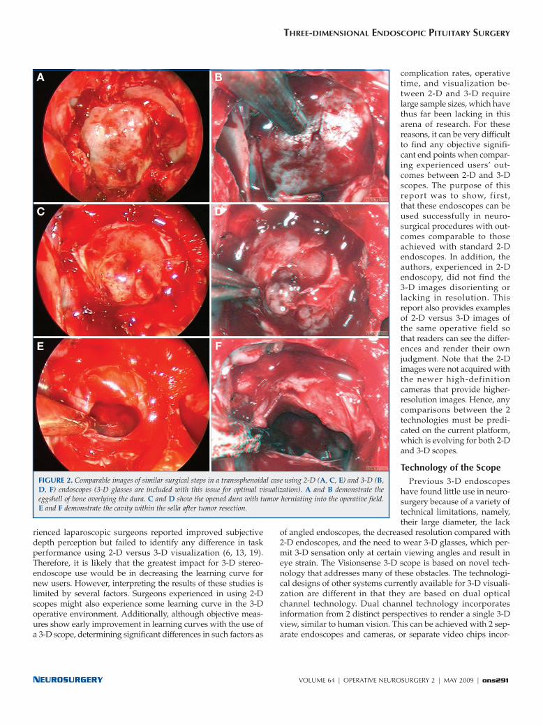
FIGURE 2. Comparable images of similar surgical steps in a transsphenoidal case using 2-D (A, C, E) and 3-D (B,D, F) endoscopes (3-D glasses are included with this issue for optimal visualization). A and B demonstrate theeggshell of bone overlying the dura. C and D show the opened dura with tumor herniating into the operative field.E and F demonstrate the cavity within the sella after tumor resection.
A B
C D
E F
ons292 | VOLUME 64 | OPERATIVE NEUROSURGERY 2 | MAY 2009 www.neurosurgery-online.com
TABAEE ET AL.
porated into a single tip. Subtle unmatched characteristics fromthe 2 visualization sources including vertical shift, focus, colorbalance, and magnification can result in ocular fatigue,headache, and nausea (15). Shutter mechanism technology,using a single camera, renders a 3-D view from the 2 pupilswith a slight temporal shift, causing discomfort from variationbetween the right and left images, caused by continuous move-ment of the camera (enhanced by the optical magnification ofthe scope). “Insect eye” technology, or a “plenoptic” camera, asdescribed herein, uses a single objective lens that splits lightinto 2 paths using 2 pupils, focusing them onto an array of
pixels, differentiated by amicrolens on top of everypixel pair. These data areprocessed through a singlevideo source to reconstruct a3-D image. The avoidance of2 separate image sources ne -gates the side effects related toocular strain. In a 2-D endo -scopic environment, the em -phasis is on increasing resolu-tion, the goal of which is toprovide more well- definedcontrast between adjacentobjects or targets to enhancethe ability of the surgeon touse visual cues to judge rela-tive depth and the 3-D natureof a nonplanar environment.In addressing the visual acu ityof a 3-D endoscopic image,using 2-D image resolution asa basis of comparison is notthe most relevant benchmark.Simply comparing the numer-ical pixel resolution of theimage on an x- y axis un der-states the ability of a 3-dimen-sionally rendered and visu- a l ized image to provide real- time, immediate depthinformation on x, y, and zaxes. For this reason, we haveendeavored to directly com-pare 2-D and 3-D systems intheir ability to help endo-scopic surgeons perform theoperative tasks. Ultimately,even if the systems are objec-tively at equipoise in affectingsuch variables as operativetime, gross total resection, andcomplication rates, the sur-geons’ subjective preferencesmight be just as important.
The limiting factor in scope/camera quality is the optics; thesmaller the scope’s outer diameter, the larger the depth of field,and the poorer the image quality. The resulting optical blurspot limits the size of the minimum pixel. The blur spot of thisplenoptic camera is smaller; thus, it is able to provide betterimage quality (27). For this reason, the resolution of the digital3-D plenoptic endoscope can be superior to a 2-D endoscopebecause it is less constrained by the blur spot. With regard todiameter, the scopes described in this article have beendecreased from 6.5 to 4.9 mm and are now 4.0 mm, which isadequately small for endoscopic endonasal surgery. The inter-
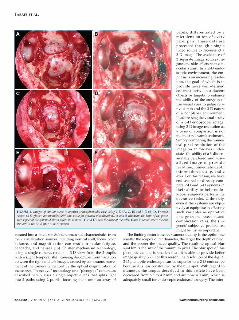
FIGURE 3. Images of similar steps in another transsphenoidal case using 2-D (A, C, E) and 3-D (B, D, F) endo-scopes (3-D glasses are included with this issue for optimal visualization). A and B illustrate the bone of the poste-rior aspect of the sphenoid sinus before its removal. C and D show the dura of the sella. E and F demonstrate the cav-ity within the sella after tumor removal.
A B
C D
E F

NEUROSURGERY VOLUME 64 | OPERATIVE NEUROSURGERY 2 | MAY 2009 | ons293
THREE- DIMENSIONAL ENDOSCOPIC PITUITARY SURGERY
pupillary distance of the system remains 0.8 mm. In addition,angled scopes are now available. Semirigid (deflecting tip)scopes are also in development. Finally, the glasses will soon bereplaced by autostereoscopic monitors that display the imagesin 3-D space without the need for glasses.
This study was also limited by the small sample size of thecohorts. Although we found no statistical difference betweenoperative times and extent of resection between groups, thesmall sample size limits the power of the study, exposing theanalysis to beta error. However, we aimed to describe an initialexperience with a rapidly evolving technology. Our goal was todemonstrate the feasibility of 3-D endoscopic technology forpractical use, rather than to statistically examine superiority of1 visualization technique. We used the 2-D group as a “goldstandard” control and succeeded in demonstrating that similarresults could be achieved with a novel visualization technology.More rigorous controlled studies will be necessary to assessthe objective value of 3-D visualization in neuroendoscopic cra-nial base surgery. This analysis was further limited by the factthat we could not compare 2-D and 3-D visualization objec-tively within each patient. Variables such as operative time andextent of resection require that 1 visualization method be usedthroughout a procedure to accurately value its effect. As such, within- patient comparisons of 2-D and 3-D endoscopy are lim-ited to subjective interval comparisons of photo- documentedportions of each case. For future research, such a limitation willrequire rigorous control of study groups to compare these visu-alization methods.
CONCLUSION
Our early experience with the use of a novel 3-D visualiza-tion system for endoscopic endonasal transsphenoidal surgerysupports the feasibility and safety of this technology. A sub-jective improvement in depth perception was appreciated byboth senior surgeons involved, and there was no increase incomplications or operative time. As this technology becomesavailable to practicing transsphenoidal surgeons, it has thepotential to correct the limitations of traditional 2-D endo-scopic technology.
DisclosureThe authors have no personal financial or institutional interest in any of the
drugs, materials, or devices described in this article.
REFERENCES1. Badani KK, Bhandari A, Tewari A, Menon M: Comparison of two-
dimensional and three- dimensional suturing: Is there a difference in a roboticsurgery setting? J Endourol 19:1212–1215, 2005.
2. Blavier A, Gaudissart Q, Cadière GB, Nyssen AS: Comparison of learningcurves and skill transfer between classical and robotic laparoscopy accordingto the viewing conditions: Implications for training. Am J Surg 194:115–121,2007.
3. Byrn JC, Schluender S, Divino CM, Conrad J, Gurland B, Shlasko E, Szold A: Three- dimensional imaging improves surgical performance for both noviceand experienced operators using the da Vinci Robot System. Am J Surg193:519–522, 2007.
4. Cappabianca P, Cavallo LM, Colao A, de Divitiis E: Surgical complicationsassociated with the endoscopic endonasal transsphenoidal approach for pitu-itary adenomas. J Neurosurg 97:293–298, 2002.
5. Cappabianca P, Cavallo LM, de Divitiis E: Endoscopic endonasal transsphe-noidal surgery. Neurosurgery 55:933–941, 2004.
6. Chan AC, Chung SC, Yim AP, Lau JY, Ng EK, Li AK: Comparison of two- dimensional vs three- dimensional camera systems in laparoscopic surgery.Surg Endosc 11:438–440, 1997.
7. Chen JC, Levy ML, Corber Z, Assifi MM: Concurrent three dimensional neu-roendoscopy: Initial descriptions of application to clinical practice. NeurosurgFocus 6:e12, 1999.
8. Chua R, Elliott D: Visual regulation of manual aiming. Hum Mov Sci 12:365–401, 1993.
9. Cushing H: III. Partial hypophysectomy for acromegaly: With remarks on thefunction of the hypophysis. Ann Surg 50:1002–1017, 1909.
10. Frank G, Pasquini E, Farneti G, Mazzatenta D, Sciarretta V, Grasso V, FaustiniFustini M: The endoscopic versus the traditional approach to pituitary sur-gery. Neuroendocrinology 83:240–248, 2006.
11. Goodale MA, Meenan JP, Bülthoff HH, Nicolle DA, Murphy KJ, Racicot CI:Separate neural pathways for the visual analysis of object shape in perceptionand prehension. Curr Biol 4:604–610, 1994.
12. Guiot G: Transsphenoidal approach in the surgical treatment of pituitaryadenomas: General principles and indications in non- functioning adenomas,in Kohler PO, Ross GT (eds): Diagnosis and Treatment of Pituitary Tumors. NewYork, American Elsevier, 1973, pp 159–178.
13. Hanna GB, Shimi SM, Cuschieri A: Randomised study of influence of two- dimensional versus three- dimensional imaging on performance of laparo-scopic cholecystectomy. Lancet 351:248–251, 1998.
14. Hardy J, Wigser SM: Trans- sphenoidal surgery of pituitary fossa tumors withtelevised radiofluoroscopic control. J Neurosurg 23:612–619, 1965.
15. Hofmeister J, Frank TG, Cuschieri A, Wade NJ: Perceptual aspects of two- dimensional and stereoscopic display techniques in endoscopic surgery:Review and current problems. Semin Laparosc Surg 8:12–24, 2001.
16. Jankowski R, Auque J, Simon C, Marchal JC, Hepner H, Wayoff M:Endoscopic pituitary tumor surgery. Laryngoscope 102:198–202, 1992.
17. Jho HD: Endoscopic transsphenoidal surgery. J Neurooncol 54:187–195, 2001.18. Laufer I, Anand VK, Schwartz TH: Endoscopic, endonasal extended
transsphenoidal, transplanum transtuberculum approach for resection ofsuprasellar lesions. J Neurosurg 106:400–406, 2007.
19. McDougall EM, Soble JJ, Wolf JS Jr, Nakada SY, Elashry OM, Clayman RV:Comparison of three- dimensional and two- dimensional laparoscopic videosystems. J Endourol 10:371–374, 1996.
20. Perez- Cruet MJ, Foley KT, Isaacs RE, Rice- Wyllie L, Wellington R, Smith MM,Fessler RG: Microendoscopic lumbar discectomy: Technical note. Neurosurgery51 [Suppl]:S129–S136, 2002.
21. Schloffer H: Erfolgreiche Operation eines Hypophysentumors auf nasalemWege [Successful operation of a hypophyseal tumor through the nasal pas-sage]. Wien Klin Wochnschr 20:621–624, 1907.
22. Servos P, Goodale MA, Jakobson LS: The role of binocular vision in prehen-sion: A kinematic analysis. Vision Res 32:1513–1521, 1992.
23. Sidhu RS, Tompa D, Jang R, Grober ED, Johnston KW, Reznick RK, HamstraSJ: Interpretation of three- dimensional structure from two- dimensionalendovascular images: Implications for educators in vascular surgery. J VascSurg 39:1305–1311, 2004.
24. Taffinder N, Smith SG, Huber J, Russell RC, Darzi A: The effect of a second- generation 3D endoscope on the laparoscopic precision of novices and expe-rienced surgeons. Surg Endosc 13:1087–1092, 1999.
25. Votanopoulos K, Brunicardi FC, Thornby J, Bellows CF: Impact of three- dimensional vision in laparoscopic training. World J Surg 32:110–118, 2008.
26. Way LW, Stewart L, Gantert W, Liu K, Lee CM, Whang K, Hunter JG: Causes andprevention of laparoscopic bile duct injuries: Analysis of 252 cases from a humanfactors and cognitive psychology perspective. Ann Surg 237:460–469, 2003.
27. Yaron A, Shechterman M, Horesh N: Blur spot limitations in distal endo-scope sensors. Presented at Stereoscopic Displays and Virtual Reality SystemsXIII, San Jose, California, January 16, 2006.
AcknowledgmentsWe thank Visionsense for technical support of this project.

ons294 | VOLUME 64 | OPERATIVE NEUROSURGERY 2 | MAY 2009 www.neurosurgery-online.com
TABAEE ET AL.
COMMENTS
A theoretical limit of the currently available scopes is the lack of 3-dimensional (3-D) images. This problem is overcome by the
reconstruction of the anatomy in the surgeon’s mind through the use ofmultiple landmarks, but this requires additional effort, a steeper learn-ing curve, and a sort of telekinetic slowing of the surgical action. Thenew 3-D endoscope studied by the authors, which until now has beenlimited to use in thoracoscopy and laparoscopy, represents a very inter-esting innovation in endoscopic transsphenoidal surgery. All those whoalready have experience with standard 2-dimensional (2-D) technologyknow that most of the improvements have been reached step by step(i.e., from mono- to 3-charge-coupled devices [CCDs] to full high-definition [HD] cameras, etc.) and have then resulted in real and majoradvantages for surgical procedures. This is why we are interested insuch new proposals and eager to test them in our own hands, too.
Paolo CappabiancaNaples, Italy
Tabaee et al. present their initial clinical experience in 13 patientswho had endonasal endoscopic tumor removal with a 6.5-, 4.9-, or
4-mm 3-D endoscope. Overall, a subjective improvement in depth per-ception was noted in operations performed with the 3-D endoscope,compared to procedures performed in a matched cohort of patientswith standard 2-D endoscopes.
The development of effective 3-D endoscopes would certainly be awelcome addition to the present endoscopic armamentarium. Animportant consideration not addressed in this assessment is how 3-Dendoscopes will compare with 2-D endoscopes using the increasinglyavailable HD cameras and video monitors. In future studies, it wouldbe useful to directly compare 3-D versus HD 2-D images in the samepatients. Given the current state of HD technology, whether 3-D endo-scopic imaging can have a tangible clinical impact beyond that pro-vided with 2-D HD imaging remains to be seen.
Daniel F. KellySanta Monica, California
This is an important article promoting technological advances in neu-roendoscopy. The authors show a practicable way to achieve a “nat-
ural” 3-D image comparable to today’s standard in microsurgery with4-mm Hopkins scopes. One of the limitations not only of endoscopy butalso of video imaging–based microsurgery is the missing 3-D informa-tion; the restriction to the use of relative size and anatomic informationalone during surgery with the 2-D-screen image requires considerableexperience, and resulting tissue lesions or even getting stuck in the tis-sue with the 2-D endoscope are some of the main risks that may causebleeding and functional damage in endoscopic neurosurgery. Thus, theauthors are right that the missing 3-D information is an obstacle in thetransition from the microscope to the endoscope. This is true even forthe experienced microsurgeon who relies on 3-D information fromtoday’s microscopes, which is far better than that obtained with thenaked eye—one of the main advantages of the surgical binocular micro-scope, aside from magnification and illumination.
The authors use a promising principle for endoscopic 3-D imaging.The conventional “telescope-type” stereoendoscopy with 2 separateendoscope channels does not allow diameters below 8 to 10 mm, asrequired in cranial neurosurgical endoscopy. Here, the authors used“compound eye technology” with a single lens and 2 pupils projectingon a CCD chip with a lenticular array, which is nicely illustrated inFigure 1 in the article. This allows an effective, less complex stereoimaging with 1 camera, even with 4-mm Hopkins II optics as the
“gold standard” in endoscopic transnasal pituitary and cranial baseapproaches; this technology is similar to the “Greenough principle,”dating back to 1896, which allows single front lens stereoscopy (2), asused in many stereomicroscopes.
One of the disadvantages is certainly the relatively small (virtual)interpupillary distance (IPD) of less than 1 mm as the main factor forthe stereoscopic effect; here, the stereoscopic performance of a micro-scope with typical stereoscopic bases of between 20 and 25 mm andalmost unlimited stereoscopic depth in telescope-principle microscopesat �3 to �13 magnification, as well as of dual-lens stereoendoscopeswith more than 3 mm (virtual) interpupillary distance (IPD), cannot beachieved (1). However, the IPD of almost 1 mm gives an adequate 2-Dimpression in a target distance of 1 to 2 cm, based on the typicallyideal IPD to target ratio of 1:10 to 1:30 for stereoscopic imaging.
According to the authors, the precision of the IPD of the endoscopecamera is controlled during manufacturing. Thus, the system will notprovide true depth information, but it gives usable 3-D information inthe high-magnification phase at short target distance in transnasal pitu-itary surgery. At longer distances, e.g., in the initial phase of thetransnasal approach and preparation of the sphenoid sinus, the stereo-scopic information will probably be marginal (no figures are providedwith an endoscopic longer-distance overview).
That the use of stereoendoscopy will achieve better results intransnasal endoscopic approaches or in general in small-diameterendoscopic neurosurgery is not demonstrated (and not stated) by theauthors. The “historical” comparison with 13 patients (similar age, sim-ilar pathology) who underwent operations with classic 4-mm glass rodHopkins scopes using 2-D imaging is certainly not significant regard-ing the reported higher gross resection rates and 0 versus 1 unintendedsubtotal resections. The comparison, however, shows, at least, that theuse of stereoendoscopy, even in this early phase, did not prolong theoperation time and gives no worse result. So I do agree that 3-Dendoscopy offers a chance for reducing the “learning curve” in endo-scopic neurosurgery; a more familiar 3-D image will facilitate the tran-sition from microsurgery to endoscopy for the microsurgically experi-enced neurosurgeon. Also the quality may improve, as has been shownfor stereolaparoscopic surgery over the course of 2 decades, e.g., witha 25% increase in speed and accuracy as measured in 3-D laparoscopicsuturing and knot tying (1).
However, 3-D endoscopy has to compete with another recent devel-opment, “full-HD endoscopy” with a resolution of 1920 � 1080 pixels.The limited resolution of conventional videoendoscopy, which onlyachieves standard television quality, is certainly an adverse factorwhich, in my opinion, might have greater importance than stereoscopicimaging. Therefore, future evaluations of stereoendoscopy should com-pare 3-D endoscopy with high-resolution imaging. Certainly, the idealsolution would be full-HD endoscopy with 3-D imaging. However,this might not be possible with today’s endoscope technology: effectiveHD endoscope resolution requires at least 4-mm or larger Hopkinsrods without split imaging; 2-mm Hopkins optics (probably compara-ble to 4-mm stereoscopy) do not profit much from HD cameras.
Also the reduced illumination and distorted colors might diminish theusefulness of 3-D small-diameter endoscopy. The figures presented bythe authors seem to be of less color fidelity than standard 2-D imaging—another factor that should be compared in future studies. In my opinion,stereo-video imaging might be more useful in microsurgery. There do notexist limitations in stereoscopic base or illumination in microscope optics;here, full-HD stereoscopic imaging could allow “video screen surgery”instead of classic binocular inspection. Advantages are more lightweight,easier-to handle microscopes; less difficult patient positioning, with thesurgeon sitting comfortably in any approach; and the possibility of inte-

NEUROSURGERY VOLUME 64 | OPERATIVE NEUROSURGERY 2 | MAY 2009 | ons295
THREE- DIMENSIONAL ENDOSCOPIC PITUITARY SURGERY
grating multimodal imaging, including navigation, endoscopy, etc., in1 optical device, as in modern electronic flight instrument system dis-plays, which allow safe precision flying and landings with artificial imag-ing at zero outside “conventional” visibility.
Michael R. GaabHannover, Germany
1. Durrani AF, Preminger GM: Three-dimensional video imaging for endoscopicsurgery. Comput Biol Med 25:237–247, 1995.
2. Grebenyuk KA, Petrov VV: The condition for eliminating spatial distortions inthe Greenough stereoscopic microscope. J Opt Technol 75:500–503, 2008.
In this article, Tabaee et al. present a novel technology that may havepotential benefits in the field of transsphenoidal surgery. Recently, the
purely endoscopic transsphenoidal approach to sellar and parasellarlesions has been a significant advance in this field of neurosurgery.Increasingly, many centers around the world are adopting and report-ing their experience with this technique.
Although there are many advantages to the endoscopic technique,among the criticisms has been the lack of stereoscopic vision. In ourhands, this has not been a major disadvantage, as the movement of theendoscope and instruments can provide many important cues that allowfor a virtual 3-D perception; however, any technology that would over-come this limitation would be welcomed. As noted by the authors, theuse of a 3-D endoscope may reduce the learning curve for surgeons. Thesize of the endoscope required (4.9 or 6.5 mm), which is significantlylarger than commonly used endoscopes, may represent a disadvantage,as a difference of a few millimeters may, in certain circumstances, add asignificant obstacle during this minimally invasive surgery.
In conclusion, we believe that the work of Tabaee et al. represents apotentially important step toward the development of a 3-D endo-scope. Nevertheless, one has to be aware that any further limitations inthe movements inside the nose, especially in expanded procedures,may preclude the use of this technology in certain types of surgery.
Fred GentiliGiorgio CarrabbaToronto, Canada
The authors describe a novel 3-D stereoendoscope and discuss theirearly experience during transsphenoidal pituitary surgery in 13
patients using 6.5-, 4.9-, or 4.0-mm 0-degree and 30-degree rigid 3-Dstereoendoscopes. They noted improved depth perception without eyestrain or headache. There were no intraoperative complications. Therewere no significant differences in operative time, length of stay, orextent of resection compared with cases in which a 2-D endoscope wasused. Depth perception, as perceived by the surgeon, was improved incomparison to depth perception with standard 2-D scopes.
The authors note no compromise after use of the endoscope.Stereoscopic depth perception, stereopsis, is a very sensitive phenom-enon. The binocular vision system can resolve angles as small asapproximately 10 arc seconds. This angle corresponds to the arc sub-tended by an IPD of 65 mm and a target at 1300 m. Furthermore, thehuman visual system is quite adept at achieving stereo fusion despiteserious artifacts. There are 2 undesirable consequences resulting fromviewing images with such artifacts. First, the viewer’s extraocular mus-cles may become fatigued, owing to the effort needed to keep the 2images fused, resulting in diplopia either during or after use. This willresolve as the viewer readapts to the real world, but this adaptationmay take several minutes. During adaptation, it is not uncommon forthe viewer to experience the nausea of simulator sickness. These
adverse effects must be avoided, by design, in any device that isintended to be useful for surgical visualization.
It is encouraging that the pursuit of 3-D endoscopy is still active. Ialways believed that it would be a tool that could only complement ourcurrent practice. In 1999, we described 4 cases in which stereoendoscopywas used as either a primary means of visualization or as an adjunct tothe operating microscope in conventional open neurosurgical proce-dures. We believed that stereoscopic vision was a significant advance inendoscope technology and was going to play a larger role in the popu-larization of minimally invasive techniques in neurosurgery. However,progress has been slow. As of 2000, we had completed 25 cases with con-current frameless stereotaxy, 12 with concurrent 3-D modeling and inte-gration, and 44 with concurrent endoscopy. To date, our experience with3-D endoscopy includes more than 300 cases in a number of approaches.Our initial error was 2-fold. We believed that the opportunity to use a 3-D endoscope would be welcomed by those practicing neurosurgery. Itturned out that this was not the case. Secondly, we sought to devise a 3-D endoscope that would eventually replace the operating microscope,and, based on this premise, we put a great deal of effort into the softwarecomponents of the unit. This was also a mistake.
Historically, we have used a Vista Medical Technologies endoscopewith an external diameter of 4.7 mm and a single glass rod optical ele-ment. A prismatic optical path separator is mounted behind the rodlens, and dual CCD devices of 640 � 480 matrixes were used to capturestereoscopic images. The effective IPD is 1.0 mm, with a working dis-tance of 15 mm. The most salient problems with our system were thelack of ports and the visualization of 640 � 480. Despite this, the endo-scope has performed without problems for more than 10 years.
Despite the theoretical advantages of stereoscopic display technol-ogy, data regarding task performance with stereoscopic versus mono-scopic displays is unclear. A number of investigators have undertakenperformance studies with various standardized tasks. In general, itappears that stereoscopic vision does not seem to improve speed orerror rate in experienced surgeons. In students or inexperienced sur-geons, however, there does appear to be a reproducible if small advan-tage to stereoscopic vision. Notable, however, is that when tasks areperformed using direct vision through the objective of the endoscope,significant improvement in performance is seen. The data, therefore,seem to suggest that a number of other factors in video processing, dis-play, and endoscope optics conspire to degrade the ability to perceiveimportant cues that might facilitate task performance. These factorshave not been adequately identified to date.
I would disagree, in a sense, with the comments that simply com-paring the numerical pixel resolution of the image on an x-y axis under-states the ability of a 3-D rendered and visualized image to providereal-time, immediate depth information on x, y, and z axes. There is nodoubt that the addition of depth contributes significantly to the abilityof an individual to work in an environment, whether virtual or surgi-cal. On the other hand, the contribution of resolution to the under-standing or perception of depth is also well documented and of sig-nificant importance.
Problems with size (I still believe that 4 mm is too large a diameter),image resolution, and the lack of working channels continue todecrease the utility of 3-D endoscopes. Despite this, we will continue touse these scopes and find them to be of significant benefit in numeroussurgical scenarios. It will be interesting to see whether the robotic endo-scopes currently in development (e.g., EndActive) will eventually sup-plant our current technologies.
C. Ben NewmanMichael L. LevySan Diego, California