BOVINE SURGERY AND LAMENESS
290
BOVINE SURGERY AND LAMENESS Second Edition A. David Weaver BSc, Dr med vet, PhD, FRCVS, Dr hc (Warsaw) Professor emeritus, College of Veterinary Medicine, University of Missouri, USA, and Bearsden, Glasgow, Scotland Guy St. Jean DMV, MS, Dipl ACVS Professor of Surgery, Former Head, Department of Veterinary Clinical Sciences, School of Veterinary Medicine, Ross University, St. Kitts, West Indies Adrian Steiner Dr med vet, FVH, MS, Dr habil, Dipl ECVS, Dipl ECBHM Professor and Head, Clinic for Ruminants, Vetsuisse-Faculty of Berne, Switzerland
-
Upload
khangminh22 -
Category
Documents
-
view
0 -
download
0
Transcript of BOVINE SURGERY AND LAMENESS

BOVINE SURGERY AND LAMENESS
Second Edition
A. David WeaverBSc, Dr med vet, PhD, FRCVS, Dr hc (Warsaw)Professor emeritus, College of Veterinary Medicine,University of Missouri, USA, and Bearsden, Glasgow, Scotland
Guy St. JeanDMV, MS, Dipl ACVSProfessor of Surgery, Former Head, Department of VeterinaryClinical Sciences, School of Veterinary Medicine,Ross University, St. Kitts, West Indies
Adrian SteinerDr med vet, FVH, MS, Dr habil, Dipl ECVS, Dipl ECBHMProfessor and Head, Clinic for Ruminants, Vetsuisse-Faculty of Berne,Switzerland
BSAA01 3/5/05 4:25 PM Page i

© 2005 David Weaver, Adrian Steiner and Guy St Jean
Editorial Offices:Blackwell Publishing Ltd, 9600 Garsington Road, Oxford OX4 2DQ, UK
Tel: +44 (0)1865 776868Blackwell Publishing Professional, 2121 State Avenue, Ames, Iowa 50014-8300, USA
Tel: +1 515 292 0140Blackwell Publishing Asia, 550 Swanston Street, Carlton, Victoria 3053, Australia
Tel: +61 (0)3 8359 1011
The right of the Authors to be identified as the Authors of this Work has been asserted inaccordance with the Copyright, Designs and Patents Act 1988.
All rights reserved. No part of this publication may be reproduced, stored in a retrieval system, or transmitted, in any form or by any means, electronic, mechanical,photocopying, recording or otherwise, except as permitted by the UK Copyright, Designs and Patents Act 1988, without the prior permission of the publisher.
First published as Bovine Surgery and Lameness in 1986 by Blackwell ScientificPublications
Library of Congress Cataloging-in-Publication Data
Weaver, A. David (Anthony David)Bovine surgery and lameness / A. David Weaver, Guy St. Jean, Adrian Steiner. – 2nd ed.
p. cm.Includes bibliographical references and index.ISBN-10: 1-4051-2382-6 (pbk. : alk. paper)ISBN-13: 978-1-4051-2382-2 (pbk. : alk. paper)1. Cattle–Surgery. 2. Lameness in cattle. I. St. Jean, Guy. II. Steiner, Adrian,
1959– III. Title.
SF961.W43 2005636.089′7–dc22
2004028333
ISBN 10: 1-4051-2382-6ISBN 13: 978-14051-2382-2
A catalogue record for this title is available from the British Library
Set in 9.5/11.5pt Photina MTby Graphicraft Limited, Hong KongPrinted and bound in Indiaby Replika Press Pvt Ltd, Kundli
The publisher’s policy is to use permanent paper from mills that operate a sustainableforestry policy, and which has been manufactured from pulp processed using acid-free andelementary chlorine-free practices. Furthermore, the publisher ensures that the text paperand cover board used have met acceptable environmental accreditation standards.
For further information on Blackwell Publishing, visit our website:www.blackwellpublishing.com
BSAA01 3/5/05 4:25 PM Page ii

Contents
Preface to First Edition vPreface to Second Edition viiAcknowledgements ix
1 General Considerations and Anaesthesia 1
2 Head and Neck Surgery 54
3 Abdominal Surgery 75
4 Female Urinogenital Surgery 140
5 Teat Surgery 158
6 Male Urinogenital Surgery 168
7 Lameness 198
Appendices1 Further Reading 2592 Abbreviations 2603 Useful Addresses 262
Index 269
BSAA01 3/5/05 4:25 PM Page iii


Preface to First Edition
This book aims to give the nuts and bolts of practical bovine surgery andlameness.
The text is directed to veterinary students in the clinical years of theirundergraduate courses, and to recent and older veterinarians, who experi-ence a limited amount of cattle surgical material. In the interests of com-pression and simplicity, a single procedure is described in some detail, whilethe alternatives, known to be equally good in the hands of others, are brieflylisted. No apology is made for such dogmatism.
The text includes frequent reference to specific types and sizes of instru-ments and suture materials. In view of the current move to the metric system,albeit late in the Anglo-American world, all figures are metric, but a com-parative scale is included.
All surgery presents a series of challenges, and this accounts for the popularity of the discipline in both the veterinary and medical professions.The challenges in bovine surgery differ from those met by the companion animal surgeon. The first is the economic question: is surgery economicallyjustified? The balance of a judgement to perform a caesarean section on aheifer with dystocia rather than to salvage the animal may be a fine one. Thisis rarely so in the companion animal field. Humane considerations often complicate an otherwise simple problem.
Other challenges include the anatomical knowledge pertinent to the proce-dure, the method of restraint and analgesia or anaesthesia, the demands ofmanual dexterity, and the physical stress imposed on patient and surgeonalike, when surgery must be performed in a sub-optimal environment, whichmay be the corner of a field or a dark spot in a dusty cowshed.
The inevitable lack of experience in bovine surgery of a young veterinarygraduate may persist as a result of a certain apathy among colleagues in the practice group. This apathy is reflected in the difficulties of maintainingequipment necessary for emergency surgery, and results in impatience at the overall time required to complete a bovine operation. The axiom ‘time is money’ applies as much to the bovine surgeon as to anyone and, sinceproper instrumentation, asepsis, effective anaesthesia and basic techniquesare the keystones to effective craftsmanship, these points are discussed in the first section.
BSAA01 3/5/05 4:25 PM Page v
vi Preface to First Edition
Succeeding chapters logically pass from the head and neck to the abdomen,thence to the female urinogenital system and teat surgery, the male urino-genital apparatus, and finally to lameness. No attempt can be made to treat asubject in depth. The further reading list is similarly selected purely as a basicliterature guide.
The reader will often have to consider ethical implications of proposed pro-phylactic or curative surgery. Animal welfare considerations are becomingincreasingly important in many countries. Is castration justified? Is it justifiedin older cattle, either on humane, economic or management grounds? Therelevant veterinary literature is sparse and the conclusions are conflicting.Responsibility rests with the veterinarian.
Why does a farm experience an ‘epidemic’ of left abomasal displacementcases? The veterinarian may search for further advice on prophylaxis. Suchan investigation will often involve other disciplines. A multi-disciplinaryapproach is particularly needed in herd lameness problems, and the checklists (see pp. 236–238) should stimulate objective recording of herd data and take the emphasis away from repetitive back-breaking treatment of theindividual cow.
Serving as a prophylactic, diagnostic and therapeutic instrument, bovinesurgery therefore fits into the concept of a herd health programme.
A. David WeaverColumbia, September 1985
BSAA01 3/5/05 4:25 PM Page vi

Preface to Second Edition
After an interval of nearly 20 years it seemed appropriate to consider anupdate. The first edition, translated into 6 languages, appeared to fulfil a need, and a minisurvey of UK veterinarians graduated 2–40 years agoshowed unanimous support for such a revision, all emphasising the need for acompact text (‘not too many words on a page’, ‘ more simple illustrations’) ofthe standard surgical techniques used in cattle on the farm.
The publishers welcomed the suggestion that two outstanding bovine surgeons, from Switzerland and North America, be invited to add their personal expertise and to provide a wider perspective. As a result this editionhas been considerably expanded and has many more line drawings. The styleand format have remained strictly instructional, sometimes even dogmatic.Only rarely are alternative techniques discussed, a notable exception beingabomasal surgery.
We consider this book should help students in their clinical training, especially when experiencing farm animal practice for the first time, as wellas new graduates, inevitably inexperienced, and those veterinarians in mixed practice who may only occasionally be called out for bovine surgicalinterventions.
More emphasis is placed on animal welfare, for example in suggestions for peri-operative analgesia and anti-inflammatory medication. The authorsare all too well aware of the problematic national (EU, other European andNorth American) regulations on permissible drug usage, and also appreciatethat each veterinarian has his or her own preferences.
Effective local, regional or occasional general anaesthesia is demandedtoday for all painful interventions in cattle. The general public, and not only the involved dairy or beef farmer, also demands that more attention be paid to food safety, and to the replacement of costly therapeutic drugs(often unavailable in developing countries) by prophylactic antimicrobials,and preferably zero drug usage. Such demands necessitate an enhanced need for good antisepsis and sterility.
Surgery may only be carried out when the condition of the patient permitsit. The correct decision may be difficult, for example in right-sided abomasaldisease, severe dystocia and digital surgery. Economical considerations andfarm facilities must also be evaluated in each potential case.
BSAA01 3/5/05 4:25 PM Page vii

viii Preface to Second Edition
The ever increasing risk over the last 20 years of litigation involving theveterinarian has resulted in more emphasis on the avoidance of potential disasters such as accidental injury to the animal or attendants, or unexpectedsequelae to standard surgical procedures. The farm manager or attendantshould always be informed beforehand of possible problems, and, we believe,could well occasionally be shown relevant sections of our text on the farm. Noveterinarian should undertake any intervention when lacking the necessaryconfidence or knowledge. We believe bovine surgeons must understand their important role in the worldwide cattle industry today.
The authors trust that this pocketbook will again provide a handy vademecum and a valuable and practical tool in daily practice.
A. David Weaver, Guy St. Jean, Adrian SteinerFebruary 2005
BSAA01 3/5/05 4:25 PM Page viii

Acknowledgements
Permission again to reproduce illustrations from the first edition was gra-ciously given by several authors and publishers (Dyce, Pavaux, Cox, Smart),while new illustrations came from several sources as below.
Figs. 1.7, 2.5, 3.2, 3.22 Dr. K.M. Dyce, Edinburgh and W.B. Saunders‘Essentials of Bovine Anatomy’, 1971 by Dyce and Wensing
Figs. 2.4, 2.9, 3.1, 3.4, 3.5 Professor Claude Pavaux, Toulouse, and Maloines.a. editeur from‘Colour Atlas of Bovine Anatomy: Splanchnology’ 1982
Fig. 3.15, Dr. John Cox, Liverpool, and Liverpool University Press ‘Surgery ofthe Reproductive Tract in Large Animals’ 1981
Fig. 3.24, Dr. M.E. Smart, Saskatoon and Veterinary Learning Systems,Yardley, PA, USA from‘Compendium of Continuing Education for the PracticingVeterinarian’ 7, S327, 1985
Fig. 3.17, Dr. H. Kümper, Giessen and Blackwell Science from ‘Innere Medizinund Chirurgie des Rindes’ 4e 2002 edited by G. Dirksen, H-D. Gründer and M. Stöber fig. 6.125)
Fig. 4.6, Dr. R.S. Youngquist, Columbia, Missouri and W.B. Saunders from‘Current Therapy in Large Animal Theriogenology’ 1997 (fig. 57.2)
Fig. 7.12, Dr. M. Steenhaut, Gent, and Blackwell Science from ‘Innere Medizinund Chirurgie des Rindes’ 4e 2002 edited by G. Dirkson, H-D. Gründer and M. Stöber (fig. 9.159)
Jan Huckin, Newbury, produced many new illustrations from rough sketchesby the first author, as did also Don Connor in Missouri. Many others comefrom the hand of Eva Steiner. John Sprout, Castle Douglas, not only com-mented usefully on the entire manuscript but also supplied some excellentsketches (Figs. 1.14, 1.15 and 2.2)
BSAA01 3/5/05 4:25 PM Page ix

x Acknowledgements
Several practising veterinarians have given useful advice including KeithCutler (Immobilon), Simon Bouisset of Toulouse (analgesics and anti-inflammatories), Bob Miller of Columbia, Missouri, Rupert Hibberd, DavidNoakes, David Ramsay, and Jan Downer. David Pritchard (DEFRA) andVincent Molony (Edinburgh) advised on certain aspects of animal welfarelegislation pertinent to the UK situation. The RCVS Library helped with some literature sources, and David Taylor checked on the ever-changingmicrobiological nomenclature, while Lesley Johnson (Veterinary MedicinesDirectorate) commented on several contentious tables in chapter 1. Appendix4 was critically assessed for accuracy by Maureen Aitken, Newbury.
The entire manuscript was typed by Christina McLachlan of Milngavie,who deserves thanks for both her accuracy and patience.
Finally thanks are given to all at Blackwell Publishing for their cooperationand advice, including Susanna Baxter, Samantha Jackson, Sophia Joyce,Emma Lonie, Sally Rawlings and Antonia Seymour, also to their copy-editorLiz Ferretti.
Guy St. Jean thanks his mentors Bruce Hull, Michael Rings and GlenHoffsis, not only for their earlier advice and encouragement during his residency, but also for their continuing friendship. He also thanks Kim Careyfor secretarial help and his wife Kathleen Yvorchuk-St. Jean for continualsupport. Adrian Steiner would like to dedicate the book to Christian.
A. David Weaver, Guy St. Jean and Adrian Steiner,April 2005
The authors have made every effort to ensure that drugs, their dosageregimes and withdrawal periods are accurate at the time of publication.Nevertheless, readers should check the product information provided by themanufacturer of each drug before its use or prescription.
Drug authorisation by regulatory authorities varies from country to coun-try, and drug withdrawal times, dependent on maximum residue limits(MRL), which are derived from residue depletion studies, can also vary from aparent (proprietary) product to a generic equivalent. Withdrawal times varywithin member states of the EU, North America and elsewhere in the world(shown in Table 1.15).
The reader should exercise individual judgement in coming to a clinicaldecision on drug usage, bearing in mind professional skill and experi-ence, and should at all times remain within the regulatory framework of thecountry.
BSAA01 3/5/05 4:25 PM Page x

CHAPTER 1
General considerations and
anaesthesia
1.1 Instrumentation 11.2 Asepsis 51.3 Sutures and suturing 61.4 Pre-operative assessment 101.5 Restraint 111.6 Premedication and sedation 131.7 Local analgesic drugs 161.8 Regional analgesia 18
1.9 General anaesthesia 361.10 Shock 401.11 Fluid therapy 421.12 Anti-microbial chemotherapy 441.13 Wound treatment 471.14 Cryotherapy 491.15 Coccygeal venepuncture 511.16 Hereditary defects 52
1.1 Instrumentation
Instruments should be maintained in good condition and, for common proce-dures, in sterile surgical packs (caesarean section, laparotomy, teat surgery,orthopedic surgery).
Sterilisation
Instruments should preferably be sterilised by one of the first two methodslisted below:
• autoclaving by steam, 750 mm/Hg at 120°C for 15 minutes or at 131°Cfor three minutes for non-packed instruments, or for a shorter time in high vacuum or high pressure autoclaves; 30 minutes for packs at 120°C,or 11 minutes at 134°C.
• gas sterilisation by ethylene oxide followed by air drying for several days to avoid diffusion of residual gases from the materials into animal tissues – some acrylic plastic materials, polystyrene and certain lensedinstruments may be damaged during this process. Note that ethyleneoxide is cancerigenic.
• cold (chemical) sterilisation in commercially available solutions, how-ever prolonged immersion is necessary. Health and safety problems existwith products such as glutaraldehyde (Rapidex® Arnolds Vet).
• simple boiling of instruments is a poor, slow and tiresome means of sterilisation particularly liable to cause damage. The minimal period ofboiling is 30 minutes, longer at altitudes over 300 m. Addition of alkali
BSAC01 3/5/05 4:28 PM Page 1

2 Chapter 1
to the steriliser increases bactericidal efficiency and boiling time may be safely reduced to 15 minutes. Corrosion is avoided by the addition of0.5–1% washing soda (Na2CO3), while accumulation of lime in serrationsor joints is removed by leaving instruments in 5% acetic acid overnight,then brushing off.
Basic instruments for caesarean section or laparotomy
The following is a suggested list of equipment to cover most eventualities.
• towel clamps (Backhaus) × 4, 8.8 cm
• haemostatic forceps (Spencer Wells) × 4 straight 15.2 cm, (Criles) × 2curved 14 cm, (Halsted) × 2 mosquito straight 12.7 cm
Table 1.2 Efficiency of different methods of sterilisation.
Bacteria Dry spores Moulds Fungi Viruses
Autoclaving + + + + +Gas sterilisation + + + + +Chemical antiseptics + — + (+) +Boiling + — + + +
Abbreviations: + = effective; (+) = limited efficacy; — = not effective
Table 1.1 Suitability of various surgical materials for sterilisation.
Dry heat Autoclave Boiling water Ethylene Liquidoxide chemicals
PVC (e.g. no yes yes yes doubtfulendotracheal tubes)
Polypropylene no yes yes yes yes(e.g. connectors)
Polyethylene no no yes* yes yes(e.g. catheters, no†
packing film)
Nylon (e.g. no yes yes no doubtfuli.v. cannulae)
Acrylic (e.g. no no doubtful yes yesperspex)
Silicon rubber yes yes yes yes doubtful
*high density†low density
BSAC01 3/5/05 4:28 PM Page 2

General considerations and anaesthesia 3
Figure 1.1 Basic instruments for caesarean section or laparotomy.1. Allis tissue forceps; 2. McPhail’s needle holder; 3. Gillies combined scissors andneedle holder; 4. plain forceps; 5. rat tooth forceps; 6. Mayo scissors (blunt/blunt),slightly curved; 7. Mayo scissors (pointed/blunt), straight; 8. straight haemostaticforceps; 9. curved haemostatic forceps; 10. scalpel handle no. 4 and no. 22 blade; 11. scalpel handle no. 3 and no. 10 blade; 12. towel clip (Backhaus).
BSAC01 3/5/05 4:28 PM Page 3

4 Chapter 1
• scalpel handle (Swann-Morton® or Bard-Parker®) × 2, P (no. 4, blades no. 22, or handle no. 3 and blade no. 10)
• rat tooth dissecting forceps (Lane) 15.2 cm
• plain dissecting forceps (Bendover) 15.2 cm
• straight scissors (Mayo) 16 cm
• slightly curved scissors (Mayo) 16.5 cm
• needle holder (McPhail’s or Gillies), right- or left-handed 16 cm
• Allis tissue forceps × 4, 15 cm
• sterile nylon calving ropes for caesarean section × 4
Figure 1.2 Suture needles (shown full scale).1. and 2. 3/8 circle cutting-edged 4.7 and 7 cm; 3. 3/8 circle round-bodied (taper cut)4.5 cm; 4. 1/2 circle cutting-edged 4.6 cm; 5. 1/2 curved cutting-edged 6.7 cm; 6. intestinal straight round-bodied (Mayo) 6.3 cm; 7. straight cutting-edged(Hagedorn) 6.3 cm; 8. double-curved postmortem needle 12.5 cm.
BSAC01 3/5/05 4:28 PM Page 4

General considerations and anaesthesia 5
• embryotomy finger knife (for incision into uterine wall which cannot bebrought near body wall)
Also needed are suture needles, which should include two each of the follow-ing types and sizes (see Figure 1.2):
• 3/8 circle cutting-edged 4.7 cm and 7.0 cm
• 3/8 circle round-bodied (taper cut) 4.5 cm
• 1/2 circle cutting-edged 4.6 cm
• 1/2 curved cutting-edged 6.7 cm
• swaged-on curved round-bodied needle 4.5 cm
• intestinal straight round-bodied (Mayo) 6.2 cm
• straight cutting-edged (Hagedorn) 6.3 cm
• double-curved post-mortem needle 12.5 cm
1.2 Asepsis
Bovine surgery involving regions where adequate skin preparation is feasible(i.e. with avoidable microbial contamination of tissues or sterile materials)should be performed under aseptic conditions. Instruments and cloths shouldbe sterile.
Preparation of operative field (e.g. flank):
• close clip wide area, minimum 60 cm cranial-caudal and 90 cm vertically(preferable to shaving)
• alternatively shave operative field after application of disinfectant, soapand water (Schick model razor is suitable)
• wash area with soap and water twice, then scrub with povidone-iodinesolution (e.g. Betadine®, [Purdue Frederick], Pevidine, C Vet, Proviodine®,[Rougier]), dry off, wash with 70% alcohol and rescrub
• repeat this procedure three times before respraying with diluted povidone-iodine solution
• large impervious sterile towels, or disposable drapes (rubber or plastic) areuseful for placing on the site
• place sterile towel on suitable flat surface for instruments, use sterilisedgauze swabs, instruments and suture materials, and sterile gloves
Hand disinfection (‘scrubbing up’)
Hands are kept in contact with the disinfectant for at least five minutes.Effective hand sterilisation procedures include:
• chlorhexidine ‘scrub’ (Novasan [Fort Dodge] or Vlexascrub [Vetus])
• 0.5% chlorhexidine concentrate in 90% ethyl alcohol with 1% glycerineas emollient (cheapest), in which 10 ml is first applied to clean dry hands and permitted to dry, before further application and five minutes’scrub-up
BSAC01 3/5/05 4:28 PM Page 5

6 Chapter 1
• commercially available povidone-iodine soap
• hexachlorophane suspension applied first dry then wet, but after scrub up(5 minutes) must be fully rinsed off (pHisoHex®, Zalpon)
• sterile surgical gloves should be worn whenever practicable
1.3 Sutures and suturing
Few topics produce such outspoken opinions and dogma as the ‘best’ suturematerial and pattern for specific surgical procedures in domestic animals,including cattle. Suture materials are constantly being improved and newproducts come onto the veterinary market at regular intervals (see Tables 1.4(a) and (b) and 1.5). This section selects a limited number of materials andmethods of usage, and attempts to justify the selection. In few cases can thecost of the material be considered an important factor in selection.
Table 1.3 Properties of three common antiseptic compounds.
Generic name
Proprietary name
Bactericidal
Fungicidal
Virucidal
Dilution
instruments
skin (‘scrub’)
wound lavage
Disadvantages
Advantages
Abbreviations: + = active; (+) = limited activity; — = no activity
Povidone-iodine
Pevidine®, Iodovet®
Povidone® (Berk)
Betadine® (Purdue Frederick)
+++
undiluted (5%, 7.5% or 10%)
undiluted (0.75%)
0.1%
brown skin when dry
not inactivated by organic matter
Chlorhexidinegluconate or acetate
Savlon® (Schering-Plough)
Nolvasan® (Fort Dodge)
Chlorhexidine (Butler, Aspen)
++—
4% or 15 ml of 7.5% solution + 485 ml of 70% alcohol
0.05%
incompatible with soap and anionic detergents
not inactivated by organic matter
Benzalkoniumchloride
Marinol® (Berk)Zephiran®
(Winthrop)
(+)
+—
10% diluted 1:500
10% diluted 1:100
—
incompatible with soap and anionic detergents; fails to kill spore-bearing organisms
—
BSAC01 3/5/05 4:28 PM Page 6
General considerations and anaesthesia 7
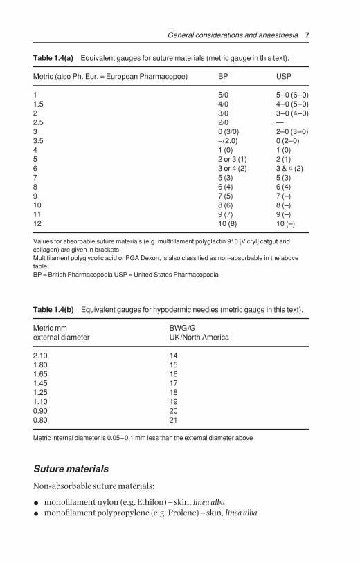
Table 1.4(a) Equivalent gauges for suture materials (metric gauge in this text).
Metric (also Ph. Eur. = European Pharmacopoe) BP USP
1 5/0 5–0 (6–0)1.5 4/0 4–0 (5–0)2 3/0 3–0 (4–0)2.5 2/0 —3 0 (3/0) 2–0 (3–0)3.5 −(2.0) 0 (2–0)4 1 (0) 1 (0)5 2 or 3 (1) 2 (1)6 3 or 4 (2) 3 & 4 (2)7 5 (3) 5 (3)8 6 (4) 6 (4)9 7 (5) 7 (–)10 8 (6) 8 (–)11 9 (7) 9 (–)12 10 (8) 10 (–)
Values for absorbable suture materials (e.g. multifilament polyglactin 910 [Vicryl] catgut andcollagen) are given in bracketsMultifilament polyglycolic acid or PGA Dexon, is also classified as non-absorbable in the abovetableBP = British Pharmacopoeia USP = United States Pharmacopoeia
Table 1.4(b) Equivalent gauges for hypodermic needles (metric gauge in this text).
Metric mm BWG/G external diameter UK/North America
2.10 141.80 151.65 161.45 171.25 181.10 190.90 200.80 21
Metric internal diameter is 0.05–0.1 mm less than the external diameter above
Suture materials
Non-absorbable suture materials:
• monofilament nylon (e.g. Ethilon) – skin, linea alba
• monofilament polypropylene (e.g. Prolene) – skin, linea alba
BSAC01 3/5/05 4:28 PM Page 7

8 Chapter 1
• pseudomonofilament polyamide polymer (e.g. Supramid®) – skin, lineaalba
• mono or multifilament surgical steel – skin, linea alba
Absorbable suture materials:
• chromic catgut – subcutis, muscle, peritoneum, bowel, bladder, uterus,penis
• multifilament polyglycolic acid or PGA (e.g. Dexon®) – bowel, muscleincluding teat muscularis
• multifilament polyglactin 910 (Vicryl®) – subcutis, muscle including teatmuscularis, bowel, bladder
• monofilament polyglyconate (e.g. Maxon™) – subcutis, bowel, teat(except skin) bladder, uterus
• monofilament polydioxanone (PDS) – bowel, muscle including teat muscularis, linea alba
• ‘soft’ gut (Softgut™) – muscle, bowel, teat muscularis
DiscussionSelection of suture material should be based on the known biological andphysical properties of the suture, wound environment and tissue response tothe suture.
Monofilament nylon remains encapsulated in body tissues when buried,but the inflammatory reaction is minimal. It has great size-to-strength ratioand tensile strength. It is somewhat stiff and is therefore not particularly easily handled, an important point when operating in sub-optimal conditionsof poor light and awkward corners, where the surgeon is bent down. Knotsecurity is only fair.
Multifilament polyamide polymer, encased in an outer tubular sheath(pseudomonofilament), has good strength and provokes little tissue reactionunless the outer sheath is broken, but it loses strength when autoclaved. It istherefore usually drawn from a sterile spool as and when required. It is veryeasily handled.
Surgical steel has the greatest tensile strength of all sutures, and retainsstrength when implanted. It has the greatest knot security and creates littleor no inflammatory reaction. Surgical steel however tends to cut tissue, haspoor handling and cannot withstand repeated bending without breaking. Itis sometimes used in tissues that heal slowly (e.g. infected linea alba).
Of the six absorbable materials listed, chromic catgut is still commonlyused, but has been replaced by synthetic absorbable material in cattle prac-tice. Catgut has relatively good handling characteristics, but also the dis-advantages of relatively rapid loss of strength in well vascularised sites (50%in the first week) and poor knot security (tendency to unwrap and loosenwhen wet). The potential minute risk of the transfer of infectious prion material into food-producing animals (e.g. cattle) and thence into the human
BSAC01 3/5/05 4:28 PM Page 8
General considerations and anaesthesia
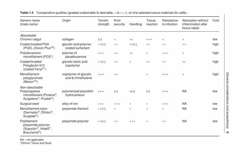
9Table 1.5 Comparative qualities (graded undesirable to desirable, + to +++), of nine selected suture materials for cattle.
Generic name Origin Tensile Knot Tissue Resistance Absorption without Cost(trade name) strength security Handling reaction to infection inflammation after
tissue repair
AbsorbableChromic catgut collagen (+) + ++ +++ + + low
Coated braided PGA glycolic acid polymer, ++(+) ++ ++(+) ++ ++ ++ high(PGS), (Dexon Plus®2) coated surfactant
Polydioxanone polymer of +++ ++ ++ + +++ + highmonofilament (PDS1) paradioxanone
Coated braided glycolic-lactic acid ++(+) ++ + ++ ++ ++ highPolyglactin 910 copolymer(coated Vicryl®1)
Monofilament copolymer of glycolic +++ ++ + +++ + highpolyglyconate acid & trimethylene(Maxon®2)
Non-absorbablePolypropylene polymerised polyolefin +++ (+) +(+) (+) +++ NA low
monofilament (Prolene®, hydrocarbonsSurgelene®, Prodek®)
Surgical steel alloy of iron +++ +++ + + +++ NA low
Monofilament nylon polyamide filament ++(+) + + + + NA low(Dermalon®, Ethilon®, Surgidek®)
Polyfilament polyamide polymer ++(+) ++ +++ + ++ NA lowpolyamide polymer (Suprylon®, Vetafil®, Braunamid®)
NA = not applicable1Ethicon 2Davis and Geck
BSAC01 3
/5/05 4
:28 PM P
age 9

10 Chapter 1
food chain has led to a ban on the use of chromic catgut in some countries(vCJD risk).
Multifilament polyglycolic acid (PGA) has greater strength which is lostevenly, provoking much less tissue reaction than chromic catgut. PGA isnon-antigenic, has a low coefficient of friction and therefore requires multiplethrows to improve knot security, but is easily handled.
Monofilament polydioxanone (PDS) is very strong, retaining its strengthfor many weeks (58% at four weeks), is easily handled, has good knot secu-rity, and has the sole disadvantage of provoking an initial tissue reactionwhich recedes during suture absorption.
‘Soft’ catgut is undoubtedly the most easily handled absorbable materialfor delicate bowel anastomoses, its quality even exceeding that of PGA, but it is not yet widely available. Plain or soft catgut is absorbed quickly andmaintains its strength for a short time. In coming years PDS and PGA arelikely to slowly replace chromic catgut, which will retain its place as a generalpurpose material. Vicryl® in its coated form is very easy to handle, has minimal tissue reaction and tissue drag. It is stable in contaminated wounds.Polyglyconate monofilament (Maxon™) has three times the strength ofVicryl® at day 21 of wound healing.
Suture needles
Suturing can only be performed efficiently with needles which are rust-free,sharp, and strong enough for insertion through the specific tissue. A selectionof needles may be conveniently maintained on a rack in a metallic sterile container.
DiscussionSuture patterns are discussed under the specific procedures. Skin under considerable or potential tension at certain sites, such as the vulval lips andperi-anal region (e.g. following replacement of prolapsed uterus, vagina orrectum), is usually sutured with sterile woven nylon tape 3–5 mm diameter.
1.4 Pre-operative assessment
IntroductionAssessment should include numerous factors apart from the physical condi-tion of the subject:
• potential duration of productive life
• insurance status
• surgical risk regarding complete recovery
• future breeding prospects
• pathology of other body systems directly or indirectly related to primarycondition
BSAC01 3/5/05 4:28 PM Page 10

General considerations and anaesthesia 11
General physical examination is essential before emergency or electivesurgery.
Laboratory testsUnder farm practice conditions laboratory tests may not be performed, butthe major parameters very simply estimated with minimal apparatus are:
• packed cell volume: microcentrifuge, microhaematocrit apparatus
• total protein: refractometer
Normal haematological and biochemical parameters of cattle are listed inTable 1.6.
In some abdominal conditions (abomasal torsion or volvulus, intestinalobstruction) estimation of plasma electrolytes (e.g. chloride) is valuable inassessing prognosis and calculating requirements for fluid replacement.
Fluid therapy is discussed in Section 1.11, p. 42.
1.5 Restraint
IntroductionRestraint is necessary for:
• administration of drugs for (a) premedication and sedation, (b) infiltrationof local analgesic drugs, and (c) induction of general anaesthesia
• examination and minor interferences carried out without sedation oranalgesia/anaesthesia
• prevention of movement in surgical intervention
Restraint may involve physical manipulation of tail, head or nares, or involveapplication of halter and ropes.
TechniquesPhysical restraint by stockman includes:
• halter
• nose grip (fingers or nose tongs)
• tail elevation
• skin grip of crural fold
Rope restraint includes:
• hock twitch
• hindlimb elevation by rope above hock and round an overhead beam
• Reuff ’s method of casting or sidelines
Many forms of cattle crush or squeeze chute are available with excellent head restraint, which are suitable for surgery of the head and cranial neck(e.g. tracheotomy) and of the perineum. (An essential feature of these crushes
BSAC01 3/5/05 4:28 PM Page 11

12 Chapter 1
or chutes is the ability to release the head rapidly should the animal collapse.)Many are unsuitable for flank laparotomy, caesarean sections or rumeno-tomy, however manufacturers will modify sides of crushes (lowering hori-zontal bar and with a greater space between vertical bars) to improve accessto the paralumbar fossa. A veterinary practice may find it advantageous tohave such a crush available for surgery on the practice premises or to be
Table 1.6 Reference ranges (haematology and plasma biochemistry) in cattle.
Units Average (%) Range (± 2SD)
Haematology
Erythrocytes ×1012/l 7.0 (5–10)Haemoglobin g/dl 11.0 (8–15)PCV (haematocrit) 1/l 35.0 28–38Fibrinogen g/l 4.0 (2–7)Leucocytes ×109/l 7.0 (4–12)Neutrophils (non-segmented bands) ×109/l 0.02 (0.5%) 0–1.12 (0–2%)Neutrophils (segmented mature) ×109/l 2.0 (28%) 0.6–4 (25–48%)Lymphocytes ×109/l 4.5 (58%) 2.5–7.5 (45–75%)Monocytes ×109/l 0.4 (4%) 0.02–0.8 (2–7%)Eosinophils ×109/l 0.65 (9%) 0–2.4 (0–20%)Basophils ×109/l 0.05 (0.5%) 0–0.2 (0–2%)Neutrophil: lymphocyte ratio — 0.45:1 —
Plasma biochemistry
Urea mmol/l 4.2 2.0–6.6Creatinine mmol/l 100 44–165Calcium mmol/l 2.5 2.0–3.4Inorganic phosphate mmol/l 1.7 1.2–2.3Sodium mmol/l 139 132–150Potassium mmol/l 4.3 3.6–5.8Chloride mmol/l 102 90–110Magnesium mmol/l 1.02 0.7–1.2Total protein g/l 67 51–91Albumin g/l 34 21–36Globulin g/l 43 30–55Glucose mmol/l 2.5 2.0–3.2Alkaline phosphatase iu/l 24 20–30AST SGOT iu/l 40 20–100ALT SGPT iu/l 10 4–50Lactate deydrogenase (LDH) iu/l 700 600–850Bilirubin mmol/l 4.1 0–6.5Cholesterol mmol/l 2.6 1.0–3.0Creatine phosphokinase mmol/l 3.0 0–50
The above values refer to healthy adult (> 3 years old) cattle, and have been compiled from varioussources. Interpretation of possible deviations from the above ranges should consider variationsdue to the laboratory technique, breed, lactational and nutritional status, and should always berelated to the presenting signs and symptoms of the individual or group. Units are given as SI units
BSAC01 3/5/05 4:28 PM Page 12
General considerations and anaesthesia 13
transported to the farm. Many crushes have poor facilities for the elevationand restraint of hind- or forelimbs for clinical examination and digitalsurgery. An exception is the Wopa crush, which is excellent for teat and feetsurgery. Suitable crushes may easily be made on the farm and tilted either bymanual means or by power.
The use of a crush/squeeze chute should never replace adequate analgesiafor surgical procedures.
1.6 Premedication and sedation
IntroductionPremedication and sedation (see Table 1.7) have five aims:
• to improve handling and restraint
• to enhance analgesic effect produced by other anaesthetic agents
• to reduce the induction and maintenance doses of general anaesthesia(GA) agents
• to reduce the possible disadvantageous side-effects of anaesthesia
• to promote smooth post-operative recovery
Very few anaesthetic drugs are approved for use in farm animals. Thoseknown to the authors include azaperone, lidocaine, methoxyflurane and thiamylal (USA). Xylazine is approved for use in cattle in Canada and the UK, and acepromazine is also approved for use in cattle in Canada.
Although not approved for use in cattle in many countries (including theUSA), several analgesic drugs (e.g. flunixin meglumine, dipyrone 50% andphenylbutazone, in addition to xylazine) are beneficial as adjunct therapyboth pre- and post-operatively in cattle with obvious somatic pain and dis-comfort. Pre-operative use of analgesics reduces the degree of operative discomfort and post-operative pain. For any medication for cattle in the USA it is the veterinarian’s responsibility to consult the Animal Medical DrugUse Clarification Act for guidelines for the extralabel use of drugs, and theFood Animal Residue Avoidance Databank (FARAD) for withdrawal times.(See Appendix 3, pp. 267, legal and professional bodies, for contact details of FARAD).
Xylazine (Rompun® [Bayer])
AdvantagesVery useful analgesic and sedative. Xylazine also causes muscle relaxation.
DisadvantagesCauses ruminal stasis, increases salivation, and effects of higher dose rate aresomewhat unpredictable as animal may or may not become recumbent.Xylazine is unsuitable as the sole agent for minor surgery when more than a
BSAC01 3/5/05 4:28 PM Page 13

14C
hapter 1
Table 1.7 Activity and dosage of selected analgesic, anti-inflammatory and sedative drugs in cattle.
Analgesic NSAID Sedative Dosage (mg/kg)i.m. i.v.
Butylscopolamine bromide/metamizole (Buscopan® Boehringer) + + — 5 ml/100 kg2
Meloxicam (Metacam® Boehringer) + + — 0.51 0.5
Carprofen (Rimadyl® LA soln, Pfizer) + + — 1.41 1.4
Xylazine (Rompun® 2% Bayer) + — + 0.05–0.3 0.03–0.1*
Diazepam (Valium®)* + — + 0.5–1.0 0.2–0.5
Flunixin meglumine (Finadyne®, Banamine®Schering-Plough) + + — — 2.2
Acetylpromazine* (ACP® Novartis) — — + 0.03–0.1 0.03–0.1
*not authorised for use in cattle in UK and EU, may only be given ‘off label’1by s.c. route, not i.v.2not authorised in lactating cattle
BSAC01 3
/5/05 4
:28 PM P
age 14

General considerations and anaesthesia 15
single painful stimulus is anticipated (e.g. unsuitable as method of analgesiain teat surgery; lancing and drainage of large flank abscess is suitable indica-tion). Xylazine is contra-indicated in the last trimester of pregnancy due to its stimulation of uterine smooth muscle (risk of abortion). It may be usedif a uterine relaxant is given before xylazine. Xylazine is contra-indicated inextreme heat, as hyperthermia may result. Avoid accidental intra-carotidinjection! Violent seizures and possibly temporary collapse are likely.
Dosage and antagonistsFor anaesthetic premedication 0.1 mg/kg xylazine i.m., 0.2 mg/kg i.m. forminor procedures in combination with local analgesia. A faster and morepredictable effect is seen following i.v. use (not authorised) at 0.1 mg/kg.
Xylazine sedation, analgesia, cardiopulmonary depression and musclerelaxation are reversible. Also xylazine overdosage (e.g. by inadvertent use ofthe equine preparation) may be antagonised by different drugs including:
• atropine (100 mg s.c.) to counteract bradycardia and hypotension
• doxapram HCl (Robins) or doxapram 4-aminopyridine (Sigma ChemicalCompany, St. Louis) respectively 1 mg/kg and 0.3 mg/kg i.v. significantlyreduces recovery period
• mixture of doxapram (1 mg/kg i.v.) and yohimbine (Sigma ChemicalCompany, St Louis) (0.125 mg/kg i.v.)
• yohimbine alone (0.2 mg/kg i.v.)
• tolazoline (4 mg/kg i.v.) – fast onset
• atipamezole (Antisedan® [Pfizer] ) 0.02–0.05 mg/kg i.v.
Doxapram acts by direct action on aortic and carotid chemoreceptors andmedullary respiratory centre, while yohimbine antagonises xylazine seda-tion by blocking central α2-adrenergic receptors.
Chloral hydrate
Long established sedative is given orally (30–60 g as 5% solution but bullrequires 120–160 g) or i.v. (5–6 g/50 kg bodyweight as 5% solution). Thesolution is irritant, and perivascular injection is likely to lead to necrosis andsevere skin slough. Infusion (total volume about 1 litre for adult cow) shouldbe made slowly via i.v. catheter over a minimal 5 minute period, since narco-sis continues to deepen after completion of injection. Chloral hydrate is not ananalgesic. Concentrations required to produce general anaesthesia causesevere, possibly fatal respiratory and circulatory depression.
Atropine sulphate
Drug reduces quantity and increases viscosity of saliva. Premedicant dose inadult cow is 60 mg s.c.
BSAC01 3/5/05 4:28 PM Page 15

16 Chapter 1
1.7 Local analgesic drugs
The four drugs of greatest value today are the hydrochloride salts of ligno-caine, procaine, bupivacaine and cinchocaine (see Tables 1.8 and 1.9).
Lignocaine
Lignocaine (Lidocaine/USP) has largely replaced procaine as it has theadvantage of:
• extreme stability
• more rapid diffusion
• longer duration of action
• useful surface analgesic activity on mucous membranes and cornea
It is however no longer authorised for cattle in the UK and EU states, as it hasno MRL.
Proprietary names include Lignodrin 2% (Vétoquinol), Lignol (Arnolds),Locaine 2%, (Animalcare), Locovetic (Bimeda).
Toxic effects are rarely encountered (e.g. inadvertent intravascular injec-tion); they include drowsiness, muscle tremors and respiratory depression,
Table 1.8 Properties of four local analgesic drugs (all hydrochloride salts).*
Generic name Lignocaine Procaine Bupivacaine Cinchocaine(trade name) (Lidocaine®) (Ethocaine®) (Marcain®) (Dibucaine®)
Main indications surface analgesia % 2–10 NS NS 0.25infiltration % 0.5–1 2–3 0.25 0.25–0.5
Nerve block % 2–3 3–5 0.5 0.5
Epidural block % 2–3 3–5 0.5–0.75 0.5
Rate of diffusion fast slow fast slow
Duration of action 60–90 mins < 60 min �8 hours �8 hours
Analgesic potency + + + +++Toxicity + + + ++Tissue irritation low low low low
Stability at boiling point ? good ? ?
Cost (low → high: + → +++) ++ + +++ ++
Other properties good safety vasodilator, — decomposes margin, no used with if mixed withvasodilator adrenalin alkalis
* several of these drugs are not authorised or licensed for use in certain countries, e.g. only procaineis authorised in UK for use in cattle (unless used in cattle not intended for human consumption)NS = not suitable
BSAC01 3/5/05 4:28 PM Page 16

General considerations and anaesthesia 17
convulsions and hypotension. Toxicity depends on the venous concentra-tion, which in turn depends largely on the rate of absorption. Concentrationsfor cattle are 2–3%, although 1% is probably adequate for infiltration, nerveblock and epidural analgesia.
Proprietary products often contain adrenaline (epinephrine) (concentra-tion 1:200 000). Adrenaline prolongs the activity of lignocaine and reducesthe possibility of toxic side-effects.
Lignocaine is also available as:
• 1% or 2% gel with chlorhexidine gluconate solution 0.25%, or hydro-benzoates in a sterile lubricant water-miscible base
• aerosol spray (lignocaine 10%) with cetylpyridinium chloride 0.1%
• 5% cream (Xylodase® [Astra]).
Procaine
Procaine (novocaine) largely replaced cocaine, and has in turn been dis-placed by lignocaine. However procaine may have economic advantagesover lignocaine. Proprietary brands include Planocaine®, Novutox® andWillcain®.
Combined with adrenaline hydrochloride, procaine absorption is slow,solutions may be sterilised by boiling and there is minimal tissue irritation.Metabolite para-amino benzoic acid inhibits action of sulphonamides.
Bupivacaine
Bupivacaine, marketed as Marcain® (Astra) with and without adrenaline(1:4 000 000) as a 0.25, 0.5 and 0.75% (plain) solution, has the followingproperties:
Table 1.9 Selected analgesics for cattle.
Generic name Proprietory name Dosage (mg/kg)i.v. i.m. oral
Ketoprofen Ketofen® 10% (Merial) 3 3 NS
Carprofen Rimadyl® (Pfizer) 1.4 NS NS
Meloxicam Metacam® (Boehringer Ingelheim) 0.5 0.51 NS
Xylazine Rompun® 2% (Bayer) 0.03–0.1 0.05–0.3 NS
Flunixin meglumine Banamine®, Finadyne®
(Schering-Plough) 2.2 NS NS
Phenylbutazone *various 2–5 NS 4–8
NS = not suitable; * not authorised for cattle use in UK/EU; 1 by s. c. route
BSAC01 3/5/05 4:28 PM Page 17

18 Chapter 1
• analgesic potency and speed of action of lignocaine
• considerably longer (× 4) duration of activity
• very well tolerated by tissues
• indicated where prolonged epidural or perineural analgesia is required
• no MRL in EU
It costs considerably more than lignocaine. The product will doubtless findmore widespread use.
Cinchocaine and Carbocaine®-V
Cinchocaine (Nupercaine™, Dibucaine®) is more toxic than procaine, butconcentrations for epidural block and surface analgesia are lower (0.5%).Other properties include:
• longer analgesia than with procaine
• drug readily decomposed by action of alkalis and therefore syringes andneedles should, if not sterile, be boiled in bicarbonate-free water
Carbocaine®-V or Mepivacain (Pharmacia and Upjohn), 2% solution is sometimes used for infiltration and epidural block, is equivalent to lignocaineand has several hours duration of activity.
1.8 Regional analgesia
Regional analgesia is the preferred method of anaesthesia for many surgicalprocedures in cattle. Its advantages over general anaesthesia (GA) (seeSection 1.9, p. 36) include:
• relatively simple technique
• general availability
• minimal apparatus, e.g. syringe, needles and drug
• little risk of toxic side-effects
• safety
Cornual block
AnatomySensitive horn corium is largely innervated by the cornual branch of thezygomatico-temporal division of the maxillary nerve (from cranial V).Caudally a few twigs of the first cervical nerve make a variable contribution toinnervation. The cornual nerve leaves the lacrimal nerve within the orbit,passes through the temporal fossa and around the lateral edge of the frontalbone, covered by fascia and thin frontalis muscle. The nerve is blocked a littlebelow the lateral ridge of the frontal crest, about halfway between the lateralcanthus of the eye and the horn (bud) base. The cornual artery and vein areclose to the site of block.
BSAC01 3/5/05 4:28 PM Page 18

General considerations and anaesthesia 19
EquipmentDisposable syringe, 10 ml for adults, 5 ml for calves, 2.4 cm 20 gauge hypo-dermic needle, 2% plain lignocaine or 5% procaine solution (2–8 ml depend-ing on size).
Technique
• insert needle, with syringe attached, midway along lateral edge of crest offrontal bone, directing needle at 30° angle through skin towards hornbase (see Figure 1.3)
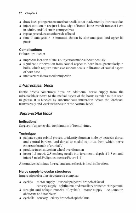
Figure 1.3 Site for cornual nerve block. A. rostral view; B. lateral view; 1 cornual branch of zygomatico-temporal nerve. Note angle of insertion of needle, and also line of cut in skin at horn base in dehorning (see Section 2.1, p. 57). X site on skull forcaptive bolt euthanasia.
1
A
X
1
B
BSAC01 3/5/05 4:28 PM Page 19
20 Chapter 1
• draw back plunger to ensure that needle is not inadvertently intravascular
• inject solution in arc just below edge of frontal bone over distance of 1 cmin adults, and 0.5 cm in young calves
• repeat procedure on other side of head
• time to analgesia 3–5 minutes, shown by skin analgesia and upper lid ptosis
ComplicationsFailures are due to:
• imprecise location of site, i.e. injection made subcutaneously
• significant innervation from caudal aspect to horn base, particularly inbulls, which require extensive subcutaneous infiltration of caudal aspectof horn base
• inadvertent intravascular injection
Infratrochlear block
Exotic breeds sometimes have an additional nerve supply from theinfratrochlear nerve to the medial aspect of the horns (similar to that seenin goats). It is blocked by subcutaneous infiltration across the forehead, transversely and level with the site of the cornual block.
Supra-orbital block
IndicationsSurgery of upper eyelid, trephination of frontal sinus.
Technique
• palpate supra-orbital process to identify foramen midway between dorsaland ventral borders, and dorsal to medial canthus, from which nerveemerges (branch of cranial V)
• produce insensitive skin wheal over foramen
• insert 1.1 metric 2.5 cm long needle into foramen to depth of 1.5 cm andinject 5 ml of 2% lignocaine (see Figure 1.4)
Alternative technique for regional anaesthesia is local infiltration.
Nerve supply to ocular structuresInnervation of ocular structures is complex:
• eyelids: motor supply – auriculopalpebral branch of facialsensory supply – ophthalmic and maxillary branches of trigeminal
• straight and oblique muscles of eyeball: motor supply – oculomotor,abducens and trochlear
• eyeball: sensory – ciliary branch of ophthalmic
BSAC01 3/5/05 4:28 PM Page 20

General considerations and anaesthesia 21
The oculomotor, trochlear, ophthalmic and maxillary branches of thetrigeminal and abducens nerve emerge from the foramen rotundum orbitale.
Retrobulbar block
Indications
• intra-ocular neoplasia (e.g. SCC)
• severe trauma (see p. 68)
Technique
• produce topical analgesia of cornea with butyn sulphate or proparacaine
• insert forefinger into lateral canthus between eyeball and conjunctival sac
• alongside finger pass 1.25 metric 7.5–10 cm curved needle throughfornix of conjunctiva until point is retrobulbar (see Figure 1.5)
• ensure that needle point does not enter optic foramen (risk of intrathecal{CSF} injection), and attempt aspiration check
H
G
F
AB
C
D
E
Figure 1.4 Supra-orbital nerve block. Oblique diagrammatic view of right side ofhead. Foramen is palpable about 3 cm dorsal to upper bony margin of orbit. Long dottedline is midline; dotted line is horn base. A. supra-orbital foramen; B. median line; C. margin of bony orbit; D. orbit; E. frontal bone; F. right ear; G. horn base; H. poll.
BSAC01 3/5/05 4:28 PM Page 21

22 Chapter 1
• inject 20–30 ml 2% lignocaine solution, which blocks nerves to ocularmuscles causing paralysis of the eyeball and analgesia
• do not attempt to anaesthetise optic nerve, since this may stimulateanimal sufficiently to cause fatality
• anaesthetise upper and lower lids by local infiltration as required
Another technique of retrobulbar injection involves placement at foursites (medial, lateral, dorsal and ventral canthi) through the conjunctiva of10 ml solution at each site, followed by infiltration of the eyelid margins (seeFigure 2.8).
Proximal paravertebral block
IndicationsLaparotomy, omentopexy, rumenotomy, caesarean section (flank incision);ruptured bladder in calves (midline incision: bilateral paravertebral).
X X
Figure 1.5 Retrobulbar block. Two methods are shown:(a) needle insertion at four points (black circles) through conjunctiva under carefuldigital guidance (4 × 8–10 ml);(b) needle insertion at lateral or medial (×) canthus, again perforating conjunctiva todeposit solution at orbital apex (20–30 ml).Use 18 gauge needle 15 cm long with marked curvature, advancing tip slowly.
BSAC01 3/5/05 4:28 PM Page 22

General considerations and anaesthesia 23
EquipmentDisposable 20 ml syringe, 3 × 1.25 metric 10 cm needles, such as HowardJones type without stilette, 2% lignocaine with adrenalin. Total volume ofsolution is 60 ml (three sites) or 80 ml (four sites).
Fat beef cattle may require needle length up to 12–15 cm to reach correctdepth.
Technique
• block dorsal and ventral branches of spinal nerves emerging from thoracic13, lumbar 1 and 2 (for most laparotomy procedures excepting caesareansection) or L1, L2 and L3 (caesarean section alone)
• ensure good head restraint and, depending on temperament of cow, standon opposite side to that to be blocked, leaning over back
• clip and scrub skin from last rib to tuber coxae along a band 15 cm wide, toleft or right of dorsal midline as appropriate
• block L2 first: identify its transverse process by counting forwards fromlast palpable process (L5) which is just cranial to sacral tuberosity of ilium
• locate point (adult Friesian cow) precisely 5 cm from midline and levelwith caudal lateral edge of L2 transverse process (see Figure 1.6)
• punch needle vigorously through skin and longissimus dorsi musculature,directed almost perpendicularly but with shaft inclined 10° medially
• advance needle firmly to contact and pass over caudal border of L2 transverse process, through intertransverse ligament (dense fibrous tissue
Figure 1.6 Paravertebral anaesthesia: horizontal diagrammatic view of left lumbarvertebrae 1–4 to show course of last thoracic (T13), first three lumbar nerves, andposition of needle. Black arrows indicate the direction in which the vertical needlepoint is ‘walked off’ the transverse process in the proximal technique. White arrowsshow the area of infiltration above and below the tips of processes 1, 2 and 4 in thedistal technique.
BSAC01 3/5/05 4:28 PM Page 23

24 Chapter 1
offering momentarily more resistance to needle point) and advance 1 cm further. Needle point now lies where dorsal and ventral branches of L2 spinal nerve have just emerged and separated adjacent to spinal foramen
• attach syringe with 20 ml solution and after applying negative pressure(check needle is not in blood vessel) inject 15 ml solution in an area about1 cm below ligament, moving shaft and needle point around very slightly
• inject remaining 5 ml solution during first stage of withdrawal of needleabove level of ligament to block aberrant dorsal cutaneous fibres
• remove needle while pushing skin down firmly to avoid development ofsubcutaneous emphysema
• block L1, (see Figure 1.6) site found by measuring distance between twolumbar transverse processes, and again inserting needle 6 cm from mid-line and directing needle point over the caudal edge of L1 transverse processes
• block cranial site (T13) by advancing needle off cranial edge of L1. Insertneedle through skin about 5 cm cranial to previous insertion point.Distance travelled by needle shaft is usually slightly more than for preced-ing sites
• in case of caesarean section, one may block L3 in similar manner to L2,but omit T13
Field of analgesiaCommencing analgesia is noted by convex curvature of spine (scoliosis) oninjection side resulting from relaxation of longissimus dorsi and flank mus-culature. Analgesia is complete within 10–15 minutes.
Field of analgesia runs slightly obliquely ventrally and caudally to midline.Innervation of the individual dermatomes overlaps so that a block of singlenerve produces a very narrow (1–4 cm wide) analgesic skin band over theflank. T13 innervates skin over middle of last 1–2 ribs (12–13), while L3block causes analgesia as far caudal as os coxae. Dorsal ramus innervates skinover upper one third of flank skin, the ventral ramus the remainder of theflank (see Figure 1.7).
• T13: dorsocranial flank, ventrally to umbilicus
• L1 (n. iliohypogastricus): dorsal midflank abdominal wall
• L2 (n. ilioinguinalis): caudal flank skin over stifle and inguinal region, scrotum and prepuce, or udder
• L3 (n. genitofemoralis): caudal flank, especially ventrally, stifle, inguinalregion, scrotum and prepuce, or udder
Variations of technique have included production of insensitive skin whealprior to insertion of a longer needle, and insertion of a stout 2.1 mm diameterneedle to infiltrate the musculature, later replaced by a finer and longer needle.
BSAC01 3/5/05 4:28 PM Page 24

General considerations and anaesthesia 25
DiscussionBlock is easier in cattle in poor body condition. Analgesic technique in exceptionally large-framed and fat cattle may require a needle 12 cm long.Successful block results in moderate convexity of the spine on the analgesicside (scoliosis), together with localised hyperthermia.
Block of L4 may sometimes cause mild ataxia. Advantages of paravertebralblock over flank infiltration include:
• minimal volume of anaesthetic solution
• absence of anaesthetic solution in surgical field
• large area of desensitisation
• rapid onset of action
Figure 1.7 Diagram of innervation of left flank: paravertebral anaesthesia.Horizontal bars indicate width of skin analgesia from block of individual nerves. Note degree of overlap of dermatomes and caudal displacement of analgesic field relative to the particular nerve root. (Modified from Dyce & Wensing, 1971.)
L1
L1
T13
L2
L2
L3
L3
L4
BSAC01 3/5/05 4:28 PM Page 25

26 Chapter 1
Distal paravertebral block
IndicationsAs for proximal paravertebral block.
EquipmentDisposable 30 ml syringe, 0.9 metric 3.75 cm, 18 gauge hypodermic needles,2% plain lignocaine; total volume about 60 ml.
Technique
• nerves to be blocked and field of analgesia see above
• insert needle 3 cm dorsal to the tip of transverse processes of L1, L2 and L4for nerves thoracic 13, lumbar 1 and 2 respectively
• inject 10 ml in a fan-shaped area
• inject a further 10 ml of lignocaine ventral to the transverse process
Flank anaesthesia (line block, T block or inverted L7 pattern)
IndicationAnaesthetic infiltration at or around incision site can produce adequate analgesia. It can also be used following unsuccessful paravertebral block. Itsadvantage is simplicity. Its disadvantages include:
• large volume of solution, local oedema and haemorrhage
• distortion of tissue layers
• poor analgesia of peritoneum
• poor muscle relaxation
• increased post-operative swelling
• increased risk of wound infection
Technique
• infiltrate subcutaneous tissues, muscularis and the sub-peritoneal layersin three distinct movements
• insert needle at point where horizontal and vertical bars of imaginary ‘T’join (see Figure 1.8). This point forms dorsal commissure of intended flankincision
• pass needle (1.1 metric 10 cm) cranially to full extent subcutaneously,and infiltrate with 2% lignocaine (plain) during slow withdrawal
• detach syringe and, without removing needle from skin, direct point caudally and advance, and likewise infiltrate during withdrawal
• repeat with infiltration of deeper tissues (total of about 60 ml in horizontalline)
• insert needle 10 cm ventral to previous point and similarly infiltrate pro-posed incision line (another 60 ml, i.e. total about 120 ml in adult cow)
Note that needle is only inserted through skin twice in entire infiltration.
BSAC01 3/5/05 4:28 PM Page 26
General considerations and anaesthesia 27
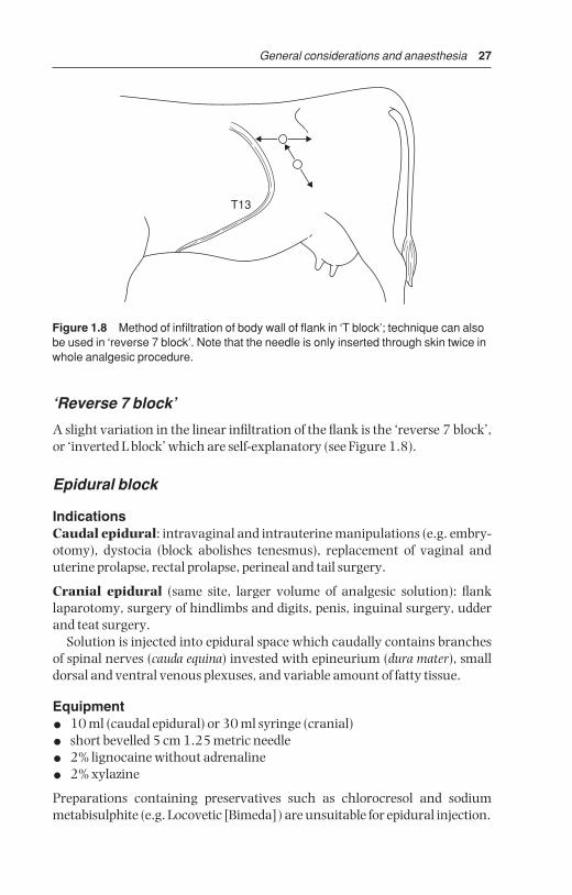
‘Reverse 7 block’
A slight variation in the linear infiltration of the flank is the ‘reverse 7 block’,or ‘inverted L block’ which are self-explanatory (see Figure 1.8).
Epidural block
IndicationsCaudal epidural: intravaginal and intrauterine manipulations (e.g. embry-otomy), dystocia (block abolishes tenesmus), replacement of vaginal anduterine prolapse, rectal prolapse, perineal and tail surgery.
Cranial epidural (same site, larger volume of analgesic solution): flanklaparotomy, surgery of hindlimbs and digits, penis, inguinal surgery, udderand teat surgery.
Solution is injected into epidural space which caudally contains branchesof spinal nerves (cauda equina) invested with epineurium (dura mater), smalldorsal and ventral venous plexuses, and variable amount of fatty tissue.
Equipment
• 10 ml (caudal epidural) or 30 ml syringe (cranial)
• short bevelled 5 cm 1.25 metric needle
• 2% lignocaine without adrenaline
• 2% xylazine
Preparations containing preservatives such as chlorocresol and sodiummetabisulphite (e.g. Locovetic [Bimeda]) are unsuitable for epidural injection.
T13
Figure 1.8 Method of infiltration of body wall of flank in ‘T block’; technique can alsobe used in ‘reverse 7 block’. Note that the needle is only inserted through skin twice inwhole analgesic procedure.
BSAC01 3/5/05 4:28 PM Page 27
28 Chapter 1
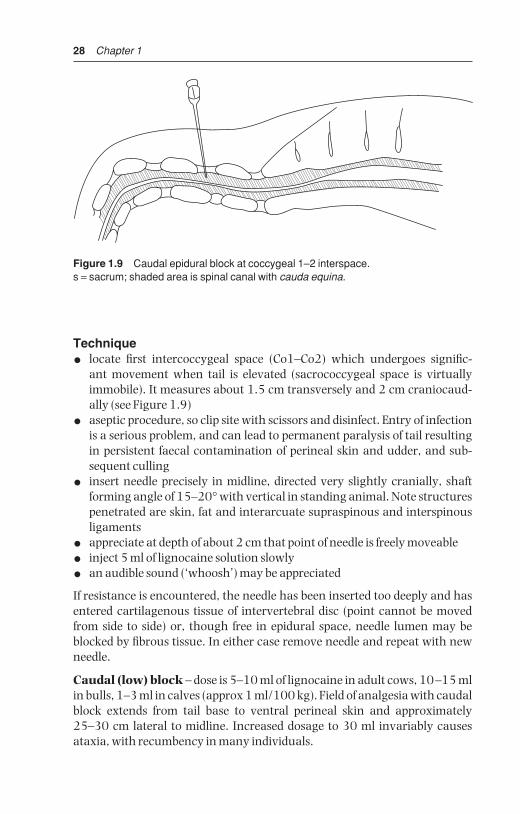
Technique
• locate first intercoccygeal space (Co1–Co2) which undergoes signific-ant movement when tail is elevated (sacrococcygeal space is virtuallyimmobile). It measures about 1.5 cm transversely and 2 cm craniocaud-ally (see Figure 1.9)
• aseptic procedure, so clip site with scissors and disinfect. Entry of infectionis a serious problem, and can lead to permanent paralysis of tail resultingin persistent faecal contamination of perineal skin and udder, and sub-sequent culling
• insert needle precisely in midline, directed very slightly cranially, shaftforming angle of 15–20° with vertical in standing animal. Note structurespenetrated are skin, fat and interarcuate supraspinous and interspinousligaments
• appreciate at depth of about 2 cm that point of needle is freely moveable
• inject 5 ml of lignocaine solution slowly
• an audible sound (‘whoosh’) may be appreciated
If resistance is encountered, the needle has been inserted too deeply and hasentered cartilagenous tissue of intervertebral disc (point cannot be movedfrom side to side) or, though free in epidural space, needle lumen may beblocked by fibrous tissue. In either case remove needle and repeat with newneedle.
Caudal (low) block – dose is 5–10 ml of lignocaine in adult cows, 10–15 mlin bulls, 1–3 ml in calves (approx 1 ml/100 kg). Field of analgesia with caudalblock extends from tail base to ventral perineal skin and approximately25–30 cm lateral to midline. Increased dosage to 30 ml invariably causesataxia, with recumbency in many individuals.
Figure 1.9 Caudal epidural block at coccygeal 1–2 interspace.s = sacrum; shaded area is spinal canal with cauda equina.
BSAC01 3/5/05 4:28 PM Page 28

General considerations and anaesthesia 29
Cranial (high caudal) block – dose is 40–80 ml in adult cattle, 5–25 ml incalves. Hindlimb function is affected by desensitisation of L6 and sacral (S) 1and 2 nerves (sciatic supply) L5 and L6 (obturator and femoral) and morecranial nerves. Dysfunction, depending on the degree of involvement, rangesfrom mild ataxia and slight spasmodic flexion and extension of stifle and hockjoints to complete posterior paralysis.
DiscussionMajor disadvantage is the risk of injury during onset (ataxia) or recoveryphase (e.g. hip dislocation). Recovery to standing takes several hours, andanimal should not be permitted to attempt to stand until tail sensation has returned. Keep in sternal recumbency with hind legs roped togetherabove hock, or sedate with xylazine or acetylpromazine to prevent attemptsto stand. Consider moving to straw-bedded box or yard.
Factors affecting extent of epidural block include volume, bodyweight,pregnancy and position of cow.
Xylocaine-lignocaine combinationsAnalgesia after epidural administration of xylazine (0.05–0.1 mg/kg body-weight of 2% solution, diluted to total volume of 5–10 ml with sterile 0.9%NaCl solution or distilled aqua; alternatively 0.03 mg/kg diluted with 2% lig-nocaine to 5 ml total volume for adult cow) lasts twice as long (four hours) asafter equivalent use of lignocaine HCI (0.2 mg/kg) alone. Very useful in cowswith chronic tenesmus (see Section 3.19, p. 138). Extent of perineal anaes-thesia is more variable than with lignocaine, but has been reported to includethe entire perineal region, including udder and flank. Side-effects includemarked transient sedation, hindlimb ataxia, bradycardia and hypotension,and can be reversed by i.v. tolazin (Priscoline HCI, 0.3 mg/kg) without affect-ing analgesia.
Pudic (internal pudendal) block
IndicationsPenile surgery distal to sigmoid flexure, examination of prolapsed penis instanding animal.
Equipment1.65 metric 12 cm needle, 30 ml syringe.2% lignocaine
TechniqueLarsen method involves block of the pudic nerve (fibres of ventral branches ofS3 and S4) and anastomotic branch of middle haemorrhoidal nerve (S3 andS4) via an ischiorectal fossa approach.
BSAC01 3/5/05 4:28 PM Page 29

30 Chapter 1
• scrub perineal region clean and insert gloved hand in rectum to locatenerve lying on sacrosciatic ligament immediately dorsal and lateral tosacrosciatic foramen, which is less than a hand’s breadth cranial to analsphincter
• note pulsation of internal pudic artery just ventral to nerve
• insert needle forward at deepest point of ischiorectal fossa, directed slightlydownwards for a distance of 6 cm (see Figure 1.10)
• check position of needle point by rectal digital control and inject 20–25 mlsolution (2–3% lignocaine) around nerve
• inject a further 10–15 ml slightly more caudally and dorsally
• inject 10–15 ml slightly cranially and ventrally, at cranial border of fora-men, for more effective block of ventral branch of the pudic nerve
• repeat procedure on other side of pelvis, reversing position of hands
Manipulation of a long needle is easier if a short stout (2.1 metric 2.4 cm) needle is inserted through skin, producing analgesic skin wheal and servingas canula for the longer needle. Alternatively caudal epidural block (5 ml)rapidly desensitises the area of intended needle insertion.
Pudic block is effective after 30–40 minutes, and persists several hours.The main advantage is that subject remains standing, while volume of drug necessary to block nerve supply to penis by epidural technique almostinvariably causes posterior paralysis. Cleanliness and experience of the pelvic
E
C C
DS4
S3
AB
Figure 1.10 Pudic (internal pudendal) nerve block. Diagram shows nerves (fromsacral 3 and 4), and injection sites A and B on medial surface of right pelvic wall andfloor of cow (pelvic viscera removed).A. is just dorsal and lateral to sacro-sciatic foramen; B. is slightly more caudal anddorsal; C. sacrum and coccygeal vertebrae 1–3; D. anus through which hand isinserted only to wrist level; E. internal pudic artery (pulsation!) lies just ventral to sitesA and B.
BSAC01 3/5/05 4:28 PM Page 30

General considerations and anaesthesia 31
landmarks are the main criteria for success with pudic block. Technical failures are common in inexperienced hands, and delay before onset of anal-gesia is a further drawback.
Block of dorsal nerve of penis
The alternative technique to pudic block for penile relaxation and analgesiainvolves analgesia of the dorsal nerve of the penis as it passes over the ischialarch.
Technique
• infiltrate skin 2.5 cm from midline adjacent to the penile body
• insert needle, advancing to contact pelvic floor and withdraw 1 cm (seeFigure 1.11)
• check that needle is not intravascular (dorsal artery of penis)
• infiltrate 20–30 ml 2% lignocaine (plain) into region
• repeat procedure on opposite side of penis
Onset of analgesia in about 20 minutes, duration one to two hours.
B
A
C
D
E
Figure 1.11 Block of dorsal nerve of penis at ischial arch.A. insertion of needle horizontally 2.5 cm from midline where penile body is palpablebelow level of tuber ischii; B. tuber ischii; C. retractor penis muscles and penis; D. point of insertion of retractor penis muscles; E. ductus (vas) deferens.
BSAC01 3/5/05 4:28 PM Page 31

32 Chapter 1
Teat block
IndicationsTeat analgesia is required for repair of teat lacerations (perforating fistula and severe lacerations), polyps, sphincter damage causing obstruction, andsupernumerary teats. Analgesia is also needed for teat endoscopy (not dis-cussed further).
Equipment20 ml syringe, 1.10 metric 2.4 cm needle, catapult elastic and large curvedartery forceps.2% lignocaine
Technique
• inject sedative drug into cow or heifer
• perform local infiltration of teat base after removing any obtruding hairsfrom udder
• insert needle subcutaneously transverse to direction of teat, and makesubcutaneous injection of 10–20 ml solution as a peripheral (ring) block(see Figure 1.12)
• accidental injection of anaesthetic into teat cistern or the circular veins atteat base is not harmful but is ineffective in producing analgesia
• analgesia develops in 5–10 minutes
• place tourniquet or Doyen intestinal clamp (with rubbers) on teat base toreduce bleeding and dripping of blood and milk
5
5
10
5
Figure 1.12 Teat ring block: 10–20 ml of 2% lignocaine are evenly distributedaround base of teat.
BSAC01 3/5/05 4:28 PM Page 32

General considerations and anaesthesia 33
DiscussionInfusion of the teat cistern is not recommended. Even cases of polyps andstenosed teat orifices prove difficult to block in this way because only themucous membrane becomes desensitised, presumably because subcutane-ous and muscularis layers are also involved in the surgical trauma.
The entire teat is anaesthetised distal to the site of injection. An alternativetechnique is by i.v. injection of any superficial teat vein distal to a tourniquet.This produces analgesia throughout the teat. This technique is virtually onlypossible in a recumbent cow.
Intravenous regional analgesia of digit
This technique is simple and effective and supersedes the cumbersome localinfiltration or nerve block procedures. It is indicated in any painful interfer-ence distal to hock and carpus, and is ideal for digital surgery. No tourniquetis required for foot surgery on a cow in a Wopa crush (see below).
EquipmentTourniquet of stout rubber tubing, metal clamp to fix tourniquet, two rolls ofmuslin bandages (or similar padding material), 20 ml syringe and 1.1 metric4 cm needle.
TechniqueCow in lateral recumbency:
• restrain animal in lateral recumbency, preferably after i.v. or i.m. injec-tion of xylazine (0.1 mg/kg) for sedation, with affected limb uppermost
• wrap rubber tourniquet firmly around limb proximal or distal to hock orcarpus (see Figure 1.13)
• in hindlimb place a rolled bandage in depression on either side of limbbetween Achilles tendon and tibia to increase pressure on underlying vessels
• clip (or shave) hair over any convenient and visible superficial limb veindistal to the tourniquet. The lateral saphenous or lateral plantar digitalvein is a suitable site in the hindlimb (see Figure 1.13)
• insert 1.10–1.65 metric (16–19 BWG) needle with syringe attached,either in proximal or distal direction, into the vein and inject 20–30 ml 2%lignocaine, with or without adrenaline, as rapidly as possible
• remove needle from vein and massage site well for one minute to preventdevelopment of subcutaneous haematoma
• in forelimb tourniquet is placed around distal radius or proximal meta-carpus and make injection into superficial vein medially, e.g. cephalicover distal radius, or medial superficial metacarpal distal to carpal jointover deep flexor tendon
BSAC01 3/5/05 4:28 PM Page 33

34 Chapter 1
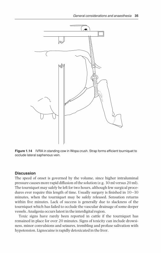
Cow standing in Wopa crush:
• elevate limb using strap and buckle, fixed overhead (strap is efficienttourniquet)
• note lateral saphenous vein prominent in proximal quarter of metatarsus(see Figure 1.14)
• push vein sideways to make it more prominent and relatively immobile
• guide loaded syringe proximally towards vein using ball of thumb tosteady syringe (see Figure 1.15)
• analgesia develops in entire limb distal to tourniquet after about five minutes and is optimal in ten minutes, persisting for at least 90 minutes if the tourniquet is left in place
A
B
C
Figure 1.13 Intravenous regional anaesthesia. Lateral aspect of left hind limb of cowshowing two possible positions for tourniquet (A) and sites for injection into lateraldigital vein (B) and dorsal common digital vein (C, lying deep, at pastern between theproximal phalanges).
BSAC01 3/5/05 4:28 PM Page 34
General considerations and anaesthesia 35
DiscussionThe speed of onset is governed by the volume, since higher intraluminal pressure causes more rapid diffusion of the solution (e.g. 30 ml versus 20 ml).The tourniquet may safely be left for two hours, although few surgical proce-dures ever require this length of time. Usually surgery is finished in 10–30minutes, when the tourniquet may be safely released. Sensation returnswithin five minutes. Lack of success is generally due to slackness of thetourniquet which has failed to occlude the vascular drainage of some deepervessels. Analgesia occurs latest in the interdigital region.
Toxic signs have rarely been reported in cattle if the tourniquet hasremained in place for over 20 minutes. Signs of toxicity can include drowsi-ness, minor convulsions and seizures, trembling and profuse salivation withhypotension. Lignocaine is rapidly detoxicated in the liver.
Figure 1.14 IVRA in standing cow in Wopa crush. Strap forms efficient tourniquet toocclude lateral saphenous vein.
BSAC01 3/5/05 4:28 PM Page 35

36 Chapter 1
1.9 General anaesthesia
IndicationsGeneral anaesthesia (GA) is rarely indicated in cattle. It is practised if theusual techniques of regional and local analgesia either cannot be adopted, orfail. Specific indications include extensive surgery of the head, neck, chestand abdomen, as well as the body wall and intra-abdominal experimentalmanipulations, (e.g. embryo transfer), as well as most long bone fractureswhen maximum relaxation is desired. GA has a relative surgical indication
Figure 1.15 Close-up view of injection of anaesthetic solution into lateral saphenousvein. Thumb pushes sideways to fix vein while simultaneously steadying syringebarrel.
BSAC01 3/5/05 4:28 PM Page 36

General considerations and anaesthesia 37
when complete asepsis is essential, such as in umbilical hernia repair incalves. For GA, food should be withheld for 6–12 hours in calves and for up to36 hours in adult cattle. Restriction of water is not indicated in calves, andshould not exceed 12 hours in adults.
Disadvantages of GARisks of GA in cattle include regurgitation, ruminal tympany, poor oxygena-tion and skeletal injury.
(a) Risk of regurgitation and subsequent aspiration of ruminal contentsand saliva into the trachea, bronchi and alveoli with potential lethalconsequences (necrotic laryngotracheitis and necrotising broncho-pneumonia with pulmonary oedema). Endotracheal intubation is there-fore essential to avoid this problem.
Factors affecting regurgitation include:
• depth of anaesthesia (see Table 1.10) – light level provokes active regurgi-tation, deep level passive regurgitation
• degree of ruminal distension or tympany
• fluidity of ruminal contents
• body and head/neck position
• body movement as in struggling and repositioning of animal
• volume of saliva
• duration of anaesthesia
(b) Risk of severe ruminal tympany (see above and below).(c) Risk of severe compromise of the effective expansion capacity of
lungs as a result of:
• increased abdominal size following development of ruminal tympanycausing pressure on diaphragm
• relatively poor oxygenation of the dependent lower lung due to inadequate circulation and pressure (ventilation-perfusion mis-match). Poorly oxygenated blood from ventral lung mixes with betteroxygenated blood from upper dorsal lung, giving lowered systemicoxygenation and increased CO2 retention (hypercapnia).
(d) Risk of skeletal injury in induction and recovery, involving possibledislocation, myositis and nerve paralyses.
(e) Expense and size of gaseous anaesthetic equipment, and appropriateexpertise in its use.
EquipmentApparatus for GA of cattle older than three to six months is similar to thatavailable for horses. Endotracheal intubation is essential in bovine GA.Equipment for volatile and gaseous agents is of circle and to-and-fro pat-tern, incorporating soda-lime canister and re-breathing bag with either an
BSAC01 3/5/05 4:28 PM Page 37

38 Chapter 1
uncalibrated or calibrated vaporiser (0–5%) to volatilise halothane (Fluothane®
[Schering-Plough, Mallard Medical]), isoflurane or sevoflurane) by means of oxygen delivered by a preset flowmeter. Minimum internal diameter of airways in such apparatus should be 4 cm.
Equipment for GA of calves with volatile or gaseous agents is similar to thatfor larger breeds of dog, e.g. Boyle’s circle absorber. The airway diameter,although theoretically inadequate, is unlikely to produce disadvantageousside-effects. Endotracheal tubes for calves should have an internal diameterof 12–16 mm, while those for adult cattle should be about 2.5 cm. Tubes of siliconised PVC are approximately one quarter the price of rubber endo-tracheal tubes (adult cattle).
Table 1.10 Main signs for assessing anaesthetic depth.
Surgical anaesthesia Excessive depth*
Cardiovascular systemHeart rate and rhythm tachycardia bradycardia, impending
arrestMuscous membrane colour pink cyanoticCapillary refill time < 2 sec > 3 sec
Respiratory systemRespiratory rate near normal shallow, irregular,
gasping, apnoeaTidal volume slightly reduced more reducedCharacter regular irregular
Ocular signsPosition and size of pupil moderately constricted, very dilated, centrally fixed
possibly rotated downPalpebral reflex present very slow or absentCorneal reflex present slow
Musculoskeletal systemMuscle tone (lower jaw, limbs) moderate poor or absent
Other signsSwallowing reflex absent absentSalivary flow present, profuse absentLacrimal secretion present absent
* Action to take in case of excessive depth:
• note time
• check patency of airway
• stop any volatile anaesthetic administration, give oxygen and artificial respiration
• check heart rate (for five seconds)
• check respiratory rate and character (for five seconds)
• check other vital signs (see above)
BSAC01 3/5/05 4:28 PM Page 38

General considerations and anaesthesia 39
List of equipment for GA by gaseous or volatile agents:
• anaesthetic apparatus – circle or to-and-fro system
• endotracheal tubes (calf-adult: 12–25 mm)
• syringe for inflation and clamping-off of cuff
• mouth gag (e.g. Drinkwater model)
• laryngoscope (e.g. Rowson pattern) optional
• nasogastric tube to act as guide, over which endotracheal tube is passed(alternative)
• halothane, isoflurane or sevoflurane and oxygen supply
• ruminal trocar and cannula
Intravenous drugsIntravenous agents for GA of cattle include:
• thiopentone sodium – give as 10% solution by rapid i.v. injection, dose1 g/100 kg 10 minutes after xylazine premedication or 1.2 g/100 kg ifunpremedicated. Perivascular injection is irritant, therefore followingsuch an accident, infiltration of 500 ml saline with hyaluronidase isessential to prevent perivascular necrosis and skin slough. Duration of GA5–8 minutes. Recover to stand in 30–60 minutes. Unsuitable for veryyoung calves.
• ketamine and xylazine – xylazine is given i.v. (0.1 mg/kg) or i.m. (0.2 mg/kg), followed 10 minutes later by i.v. ketamine (10 mg/kg). GAlasts 15–35 minutes. Recovery is fairly rapid. The two drugs may be mixedin one syringe and given i.m.
• chloral hydrate – unsuitable for GA of cattle, but remains an inexpensivesedative (see p. 15).
• ketamine and guiafenesin: 1 g of ketamine is added to a 1 litre bag orbottle of a 5% guaifenesin solution. This combination is administered toeffect at about 0.2–0.5 ml/kg/hour and produces satisfactory anaesthesiawhich can be maintained for several hours.
In any such intravenous GA technique endotracheal intubation should be carried out as soon as possible after injection. The tube should only beremoved following a demonstrable cough reflex or swallowing movement.Extubation is performed with the head lower than the trunk and with the cuffinflated until it reaches the pharynx, preventing material moving betweentube and tracheal mucosa, dribbling towards the bifurcation of the bronchiand causing a necrotic bronchotracheitis.
Immobilon™/Revivon™ – large animal productA reversible neuroleptanalgesia (narcosis) with analgesia can be attained forrestraint and surgical procedures with LA Immobilon™, the active principleof which is etorphine, combined with acetylpromazine. It is not licenced foruse in cattle, but is sometimes used in exceptional circumstances where other
BSAC01 3/5/05 4:28 PM Page 39
40 Chapter 1
methods of restraint are considered too hazardous. The drug is extensivelyused in various species in tropical Africa, but rarely now in the UK except forrestraint of dangerous animals, e.g. a bull or steer amok in public places.Warning: etorphine can be life-threatening to the operator if absorbed byany route. Extreme care should be taken. Before any use of Immobilon™ theappropriate dose of the antagonist Revivon™ (contained in the same pack)should be drawn up first into a second syringe, which should then be keptclose at hand for immediate intravenous use in the event of an accident. Asecond person should always be instructed clearly beforehand what actionshould be taken with the reversing agent, which should then be injectedbefore calling medical assistance.
IndicationsUse with a dart gun (i.e. intramuscular injection) for restraint of dangerousand uncontrollable cattle.Dosage:0.5–1 ml Immobilon™ per 50 kg bodyweight i.m. by dart syringe. Cattlebecome recumbent some minutes later, and remain immobile for about 45 minutes. Generalised muscle tremors and poor muscle relaxation are usu-ally apparent.
To reverse the drug an equal volume of Revivon™ (diprenorphine HC1)should be injected i.v. Recovery generally occurs with minimal disturbanceand noise. A second half dose of Revivon™ may be given s.c. after the initiali.v. dose if required.Operator warning (from UK data sheet of LA Immobilon™/Revivon™)To avoid accidental self-injection two sterile needles should be used, one tofill the syringe, the second to inject the patient. After the calculated dose has been withdrawn from the vial, the syringe should be removed from theneedle. The syringe should be connected to a second needle immediatelybefore delivery. Both needles should be discarded into a closed labelled con-tainer. Operator should wear rubber gloves and should not pressurise vialcontents. Veterinarian should fully brief assistant on accident procedure and administration of the reversing agent (LA Revivon™) should anyImmobilon™ be inadvertently absorbed through skin, mucous membranes(mouth, eyes) or through injection.
Immobilon is highly toxic, causing dizziness, nausea, pinpoint pupils,rapidly followed by respiratory depression, hypotension, cyanosis, loss ofconsciousness and cardiac arrest.
The reader should consult specialised textbooks (see Further Reading sec-tion, pp. 259–60) for further details of bovine GA.
1.10 Shock
A state in which there is inadequate perfusion of tissues. Shock lesions resultfrom:
BSAC01 3/5/05 4:28 PM Page 40

General considerations and anaesthesia 41
• failure of homeostatic mechanisms to maintain adequate perfusion
• homeostatic mechanisms themselves reduce perfusion by eliciting exces-sive production of various vasoactive hormones, amides, peptides andkinins
Shock may eventually become refractory to treatment. Inadequate perfusionis due to failure of blood flow, not of blood pressure. Changes are due tohypoxia and accumulated metabolites from defective perfusion. Basic patho-logy: necrosis of cells and tissue, haemorrhages and fibrin thrombi in venouscirculation (Shwartzman). Almost every organ displays lesions.
Shock in bovine practice may be due to various states, for example:
• hypovolaemic shock as a result of massive haemorrhage, namely reduc-tion of circulating blood volume to less than 80% (reduction to less than65% in critical)
• dehydration, e.g. 10–12% is severe, and over 15% is critical
• burns, e.g. third degree burns over more than 25% of body surface
• infection – septic shock, especially gram-negative bacteria liberatingendotoxins (e.g. generalised Shwartzman reaction)
• peripheral vascular disease with gangrene
• spinal anaesthesia (cranial or epidural, see Section 1.8, pp. 27–29)
• acute haemolytic conditions
A fall in blood pressure as a result of shock due to severe systemic blood losstriggers off a sequence of events in which the animal attempts to maintain anadequate blood supply to the brain and coronary vessels, but which maycompromise other tissues (see Figure 1.16). Prompt fluid therapy is often themost important corrective measure in such cattle.
Vasoconstriction in organs withalphacatecholamine receptors
(e.g. intestine, kidney, skin)
Blood supply maintainedto the cerebral andcoronary vessels
Baroreceptors in carotidand aortic vasculature
Stimulation ofsympatheticosuprarenal system
Renal tubular necrosisand intestinal mucosal ischaemia
Catecholamine release
Figure 1.16 Summary of some reactions following severe blood loss and shock.
BSAC01 3/5/05 4:28 PM Page 41

42 Chapter 1
1.11 Fluid therapy
IntroductionCorrection of fluid and electrolyte deficits may be critical in severe shock andblood loss, in conditions such as right abomasal volvulus (see Section 3.7,p. 110) and in severe, protracted toxic cases of bovine dystocia. A waterdeficit can result from anorexia, dysphagia, diarrhoea and hyperosmolality.
Fluids
Dehydration is expressed as a percentage of total reduction of body fluids andis estimated as follows:
• persistent skin fold for > 3 seconds
• prolonged capillary refill (> 4 seconds)
• sunken eyeball or enophthalmos e.g. 7 mm recession indicates about 12%dehydration
Volume of fluid required for replacement is calculated very simply: litresrequired = bodyweight in kg × % dehydration. For example, a 500 kg cowwith 10% dehydration requires 50 litres of fluid to cover her deficit.
Intravenous catheters are usually placed into the jugular vein following skinpreparation (Cathlon® IV, Johnson & Johnson, Intracath®, Becton Dickinson16 G or 19 G for calves, 10 G or 12 G for rapid infusion in cows).
Hypertonic saline (HS) (see Table 1.11)Hypertonic saline (HS) is a major advance in fluid therapy for haemorrhagicshock (massive blood loss) involving severe dehydration, hypovolaemicshock and in endotoxic shock (e.g. following correction of right abomasalvolvulus).
Technique
• i.v. dose of 7.2% sterile saline, 4 ml/kg over five minutes (i.e. 2–3 litres to a600 kg cow) via 10G (Intracath®)
• avoid perivascular injection (tissue necrosis)
• ensure immediate access to unlimited fresh water (most cows will drink20–40 litres in the next 10 minutes)
• if patient does not drink within five minutes, give 20 litres of water bystomach tube or i.v. isotonic saline
• i.v. HS may be repeated once after 24 hours
HS is useful as emergency treatment for severely diarrhoeic and (> 8%) dehydrated calves, combined with oral electrolytes or followed by i.v. isotonicsaline (dosage as above).
BSAC01 3/5/05 4:28 PM Page 42

General considerations and anaesthesia 43
Electrolytes
IntroductionIdeally, the solution is selected after determining the individual animal’sneeds by laboratory evaluation. However on the farm this is not practical. Ingeneral cattle have rather consistent acid base and electrolyte abnormalitiesassociated with surgical disease. Dehydrated cattle are twice as likely to havemetabolic alkalosis as metabolic acidosis. Alkalosis is treated by giving a solu-tion rich in chloride and potassium. Given adequate circulatory volume andelectrolytes, the kidneys can usually correct the alkalosis.
Oral rehydration can be achieved rapidly (e.g. 20 litres of dextrose and/orsaline in less than three minutes) using a stomach pump attached to anoesophageal tube, which is protected from damage by the cow’s teeth by astraight outer sleeve, one end of which has nose tongs attached for insertioninto the nostrils. Dehydration is easily corrected in adult cattle if combinedwith i.v. therapy (see below) or sometimes alone to prevent, for example, bor-derline ketosis.
Replacement rules (see Table 1.11)
• K is reduced in abomasal volvulus (see Section 3.7, p. 111) and is associ-ated with metabolic alkalosis: it should be replaced with KCl
• K is also reduced in diarrhoeic conditions: replace with K2CO3
• Na is reduced in abomasal torsion, and involves metabolic acidosis;replace with 0.9% NaCl or KCl
Table 1.11 Composition of six fluids given as replacement therapy to cattle.
Electrolyte concentration (mEq/ litre)
Na+ K+ Ca++ NH4− Cl− HCO3
− Lactate−
Normal plasma 140 4 5 0 103 25 5
0.9% NaCl 154 0 0 0 154 0 0
Ringer solution 145 4 6 0 155 0 0
Lactated Ringer 130 4 3 0 109 0 28
Hypertonic saline (7.2%) 120 17 70 0 144 0 0
Ammonium KCl solution* 0 75 0 75 150 0 0
* Whitelock, R.H., et al. (1976) Proceedings of the International Conference of ProductionDiseases of Farm Animals, 3 edn. Wageningen, The Netherlands, pp. 67–90 if CaCl2
BSAC01 3/5/05 4:28 PM Page 43

44 Chapter 1
Calculation of electrolyte quantities(a) Calculate total extracellular fluid (ECF) volume: ECF (litres) = body-
weight in kg × 0.3. For example, a 500 kg cow has 150 litres ECF(b) Deficit of Na+ and/or Cl− is estimated by laboratory biochemistry; the dif-
ference between normal and calculated values is the deficit per litre(c) The deficit of K+ is unreliable if calculated from plasma K, but twice this
calculated deficit may be safely administered if:
• renal function is satisfactory
• K+ is given as K2CO3 or KCl well diluted in other fluids, at 10–20mEq/litre and infusion rate per hour does not exceed 100 mEq K+ inadult (500 kg) cow. K+ may often be easily given in drinking waterlater (about 40 mEq/litre as K2CO3 or KCl).
1.12 Anti-microbial chemotherapy
IntroductionAnti-microbial drugs are no substitute for sound and aseptic surgical technique in sterile aseptic procedures, neither can they be expected to control deep-seated necrotic and purulent foci. These drugs should be con-sidered as adjuncts to the natural defence mechanisms of the host. The primary aim is to improve these mechanisms by proper preparation of thesurgical field, surgeon and the instruments, appropriate débridement, excision of necrotic tissue, drainage and lavage, together with the prompt re-establishment of the nutritional fluid, electrolyte and acid-base balance ofthe patient.
One example is septic arthritis where joint lavage with saline is indi-cated (see Section 7.27, p. 257) and may sometimes be replaced by more radical open joint surgery, supported by systemic and local anti-microbialchemotherapy. Antibiotic prophylaxis is not required during clean surgicalprocedures in cattle (e.g. LDA abomasopexy, entropion surgery).
Prophylactic antibiotic therapy is however indicated in extensive abdominal surgery, open fracture repair and non-sterile invasive procedures.This therapy should start before the surgical intervention, the therapeuticconcentration should already be adequate at the start of surgery and theintravenous route is preferable. The dose rates of prophylactic and therapeu-tic antimicrobials are similar.
In contaminated sites, such as caesarean hysterotomy following prolongedunsuccessful vaginal manipulation and a dead fetus, rectal tears requiringextensive suturing, or in surgical sepsis, an adequate tissue concentration ofantibiotic should be rapidly established and therapy should continue for atleast three to five days after surgery. Initial anti-microbial drug selection isusually arbitrary (broad spectrum) and may be altered following results ofsensitivity testing.
BSAC01 3/5/05 4:28 PM Page 44

General considerations and anaesthesia 45
The most common bovine pathogens and their sensitivity are shown in Table 1.12. The sensitivity may be variable depending on the area andcountry.
Reasons for a failure of sensitivity testing are listed in Table 1.13. A failureof response to anti-microbial therapy can be due to a variety of other reasons(see Table 1.14).
The problem of milk withdrawal times in lactating dairy cattle must be keptin mind (see Table 1.15). Currently ceftiofur HCL (e.g. Excenel RTU® sterilesuspension, [Pfizer] ) is a very popular systemic antimicrobial as it has a nilmilk withdrawal time.
Table 1.12 Some guidelines for the antimicrobial sensitivity of certain drugs againstcommon bovine pathogens (see footnote).
Antimicrobial drugOrganism First choice Alternative choice (s)
Arcanobacterium pyogenes Amoxicillin (c) Penicillin G (c)
Staphylococcus aureus
nonpenicillinase Penicillin G (c) amoxy/clav (c)penicillinase Oxacillin (c) amoxy/clav (c)
Clostridium spp. Penicillin G (c) Tetracycline (Cl. tetani)(s)
Escherichia coli Marbofloxacin (c) Ampicillin (c)
Fusobacterium Ceftiofur Penicillin G (c)Tetracycline (s)
Enterobacteriaceae Aminoglycosides Carbenicillin (c)Marbofloxacin (c)
Klebsiella Marbofloxacin (c) Cephalosporin (c)
Pasteurella Ceftiofur, Enrofloxacin‡ Tetracycline† (s)Trimethoprim + sulpha
(s&c)
Proteus mirabilis Ampicillin (c) Marbofloxacin (c)
other Proteus Marbofloxacin (c) Carbenicillin (c)
Salmonella Trimethoprim + sulpha (s&c) Ampicillin (c)
Streptococcus Penicillin (c) amoxy/clav (c)
Note: enormous variations exist between different laboratories and countries, also antibioticlegislation is very variable, therefore obtain advice from nearest veterinary laboratory!(c) = bactericidal; (s) = bacteriostatic; (s/c) = bactericidal in high concentrations only† widespread resistance of Pasteurella spp. to tetracycline is recognised; alternative drugs aresulfachlorpyridazine or erythromycin‡ use in USA restricted to beef cattle
BSAC01 3/5/05 4:28 PM Page 45

46 Chapter 1
In summary, in cattle anti-microbial selection should be based on theorganism’s sensitivity, predicted drug tissue levels, confidence in drug safetyand cost, and approval of extra-label use in each country.
Post-surgical analgesia may be improved by flunixin meglumine(Finadyne®, [Schering-Plough] Ketoprofen® [Rhone Merieux]), or meloxi-cam (Metacam®, Boehringer Ingelheim). Flunixin meglumine, when used atlabel dose, incurs ten days of meat withholding and three days’ milk with-holding in the United States (respectively 5 and 1 days in UK).
Table 1.13 Failure of sensitivity testing (susceptibility) to predict outcome of therapy:some possible factors.
Impaired absorptionAccelerated eliminationAdverse drug interaction(s)Inadequate penetration/drainageReduced phagocytosisDrug antagonismSuperinfectionImpaired normal host defencesUnderlying diseaseGenotypic or phenotypic drug resistanceTolerance
Table 1.14 Failure of response to antimicrobial therapy: list of possible factors.
Absence of concurrent surgical/mechanical measures, e.g. drainageInappropriate drug selection or dosageSensitivity testing: organism resistant in vitroPredisposing and management factors remaining uncorrectedMisdiagnosis, as condition not of bacterial originImpaired immune functionFluid electrolyte, acid-base imbalance, nutritional defectsInflammation, with release of mediators, cellular breakdown products, oedema, tissue
destruction, coagulation, impaired perfusion and penetrationPoor management of mixed infections involving anaerobesToxin elaboration before antibacterial concentration is obtained at infection siteDrug resistance, therapeutic, prophylactic, growth promotion useComplianceUnderlying disease or concurrent medicationAdverse reactions: toxicity
BSAC01 3/5/05 4:28 PM Page 46

General considerations and anaesthesia 47
1.13 Wound treatment
Successful surgical treatment of wounds depends on:
• thorough débridement
• meticulous haemostasis
• elimination of dead space
• proper use of instruments
• judicious insertion of drains
• proper placement of sutures
Since the healing process is basically similar in all species (see Figure 1.17),these points all apply to cattle. Bovine wounds fortunately heal without production of exuberant granulation tissue which is commonly seen in the horse.
Débridement is essential, especially when the wound (e.g. teat) is to besutured and in areas when gross contamination is commonplace.
Table 1.15 Effective therapeutic period and withdrawal times of chemotherapeuticagents in UK and USA (2004/2005). Since drugs are given in various forms andstrengths, milk discard times and withdrawal times will vary accordingly: drug labelsshould always be checked !
Effective Withdrawal times (days)therapeutic period (days) Milk discard Slaughter (meat)
Drug UK USA UK USA
Ceftiofur Na (Naxcel®) 1 0 0 0 0Ceftiofur HCl (Excenel®) 1 0 0 8 0Trimethoprim/Sulphadiazine 1 6.5 2.5 34 5Oxytetracycline 100 1 4 4 21 7–22Oxytetracycline LA 4 7 4 14 28Procaine penicillin G 1 2–5 2 3–4 10Procaine penicillin G
+ streptomycin 1 2.5 2 23 30Ampicillin 1 1–7 2 18–60 6Erythromycin 1 2 3 7 14Amoxicillin 1 1–7 NP 18–42 25Framycetin 1 2.5 NA 49 28–38Cefalexin sodium 1 0 4 19 4Florfenicol (Nuflor®) 2 NP NP 30–44 28–38Clav. acid/amoxicillin (Synulox®) 1 2.5 — 42 —Enrofloxacin (Baytril®) 1 3.5 NP 14 28
NA = not available, NP = not permittedBritish readers are referred to the current data sheets and the current NOAH Compendium. US readers should refer to the Center for Veterinary Medicine, see Appendix 3 (p. 267)
BSAC01 3/5/05 4:28 PM Page 47

48 Chapter 1
Haemostasis prevents haematoma formation in dead space which wouldpotentially offer an ideal culture medium for bacterial growth.
Dead space may be packed with sterile gauze swabs for 24–48 hours to pre-vent haematoma formation (see Section 2.7, pp. 69–70).
Drains are rarely indicated in cattle except for certain long bone fractures,the thorax or subcutaneous or deep infected wounds (e.g. laparotomy inci-sions) where:
• irrigation is required several times daily
• the outflow of infected material may be ensured without the need regu-larly to reopen the lower end of the incision
Sutures in wounds:
• do not suture infected wounds
débridementresistance to
infection
EPITHELIUM
collagenlysis
macrophagesgranulocytes
(remodelling)
HEALED WOUND
INJURY
coagulationplatelets
fibroblasts
contraction
proteoglycansynthesis
new vasculargrowth
collagensynthesis
INFLAMMATION
Figure 1.17 Basic process of wound healing.
BSAC01 3/5/05 4:28 PM Page 48

General considerations and anaesthesia 49
Irrigation of wounds:
• large volume of clean non-sterile fluid (e.g. mains water) is more valuablethan small volume of sterile physiological saline
• ensure all pockets of deeper wounds are reached by irrigation fluids
• judicious use of 3% hydrogen peroxide, or 1% chlorhexidine solutionspeeds removal of pus and tissue debris
• development of dead space may be prevented by judicious closed activedrainage (suction drainage, Redon type drain)
Skin sutures: single or interrupted mattress sutures of sheathed polyamidepolymer multifilamentous material (e.g. Vetafil®, Supramid® [Braun]),monofilament nylon or polypropylene (see Table 1.5, p. 9)
Deep sutures in infected wounds: PGA, PDS
Drains:
• Penrose
• sialastic
• polypropylene, flexible and thin-walled with multiple openings in deepertissues
1.14 Cryotherapy
IntroductionAdvantages of this technique include:
• frequently faster than knife surgery
• little or no haemorrhage
• relative absence of post-operative complications
• phenomenon of cryo-immunity may delay or prevent neoplastic regrowth
Disadvantages are few:
• objectionable odour resulting from tissue necrosis
• risk of uncontrolled freeze resulting in destruction of vital tissues
Materials, termed cryogens, are:
• liquid nitrogen (N) (boiling point −196°C)
• nitrous oxide (N2O)(−89.5°C)
• carbon dioxide (CO2)(−35.5°C)
EquipmentEquipment may be small and readily portable (special vacuum flask), or can be rather large and heavy, e.g. Frigitronics CE-8. Operator should wear
BSAC01 3/5/05 4:28 PM Page 49
50 Chapter 1
disposable protective plastic gloves and avoid any possibility of cryogen con-tacting the skin of involved personnel. General anaesthesia may be necess-ary for delicate cryotherapy, since the affected area must be kept immobileduring treatment.
Technique
• wash and dry area and apply a little paraffin jelly or vaseline (e.g. KY®
jelly) for improved initial adhesion
• if spray is to be used, mask off surrounding area with piece of plastic sheetor with thick layer of vaseline
• select suitable probe head (e.g. flat, 10 mm or 20 mm diameter) or sprayattachment (2.36–1.25 metric needle, depending on desirability of coarseor fine spray)
• if necessary attach flexible extension piece to keep apparatus vertical and ashort distance from operating site
• if available, and working near vital structures, insert thermocouples: dropof reading to −20°C is critical and indicates need to arrest cryotherapy atonce
• in absence of thermocouples and in most situations in cattle, digital assess-ment of size and position of iceball is quite reliable
• produce rapid freeze and permit slow thaw cycle; two cycles necessary forN, three for N2O or CO2
• assess extent of iceball by digital palpation. Probe is removed after naturalthaw, or after activating automatic defrosting device
• do not repeat freeze until area is completely thawed. Effectiveness of proce-dure depends on sequence of rapid freeze-slow thaw cycles
• warn owner or stockperson to anticipate tissue necrosis and slough inseven–ten days, leaving a healthy granulating surface, which should bekept clean as epithelialisation proceeds from periphery inwards
DiscussionFactors affecting degree of cryonecrosis include:
• probe temperature – liquid nitrogen is capable of freezing any tissue in thebody
• size of probe tip – the greater its diameter the greater the volume of frozentissue
• size of spray orifice – the larger the orifice, the greater the concentration ofcryogen applied to lesion, resulting in very rapid freeze and larger volumeof frozen tissue. With liquid nitrogen, spray orifice penetrates much deeperand faster than probe tip, though latter is more precise
• duration of freeze – at temperatures below −20°C for 1 minute or more virtually all living tissue undergoes cryonecrosis. The longer the freeze,the larger the iceball.
BSAC01 3/5/05 4:28 PM Page 50

General considerations and anaesthesia 51
1.15 Coccygeal venepuncture
IndicationsCollection of venous samples (volume up to 10 ml) with minimal restraintand little or no assistance. Obvious advantages over collection from jugularor subcutaneous abdominal (milk) vein (latter not recommended). Minimalrisk of infection and sporadic haematoma formation insignificant, so tailparalysis is virtually unknown.
TechniqueCollect into evacuated blood collection tubes containing edetic acid (EDTA), heparin etc., or without coagulant (Vacutainer® [Becton Dickinson,Rutherford, NJ]) via 0.9 metric 4 cm needle which is screwed into plasticholder into which collection tube is inserted. Alternatively collect via 0.8metric 2 cm needle into polypropylene syringe (5–10 ml) with negative pressure.
• restrain head in stall by chain or halter, or in squeeze chute or head gate(manual restraint rarely required)
• grasp tail in middle third and slowly elevate to almost vertical position(‘tail jack’ Figure 1.18)
Figure 1.18 Method of restraint for obtaining blood samples from coccygeal vein.
BSAC01 3/5/05 4:28 PM Page 51

52 Chapter 1
• cleanse tail site of gross faecal contamination with paper towel or cottonwool
• with free hand locate palpable vein in midline, just caudal to insertion ofskin folds of tail at level of coccygeal (Co) vertebrae 6–7
• insert needle just cranial to bony protuberance of haemal process in mid-line to depth of about 8–12 mm (in Vacutainer® system, insert needle intotube), and withdraw slightly until bloodflow starts
• if initially unsuccessful, reduce tension of tail slightly and continue atsame site, otherwise attempt at Co 5–6
• do not massage puncture site after venepuncture
Other reports claim that sites of Co 3–5 are preferable, but anatomical studiesshow the vein lies to the right of midline in the ventral sulcus of the vertebralbody at the more cranial site.
Insignificant haematoma develops in a small number of cases, but dis-appears in a few days. Procedure does not cause thrombophlebitis or totalocclusion of vein, and repeat samples are easily obtained some days or weekslater.
1.16 Hereditary defects
IntroductionUntil the 1960s congenital defects in cattle were thought to be largely herit-able. There has been a slight but perceptible change in this attitude of veter-inarians, cattle breeders and geneticists. ‘Congenital’ is not synonymouswith ‘heritable’ or ‘genetic’. There remains a great paucity of information. Alimited number of conditions can be corrected surgically, and where it islikely that the condition is inherited, steps should be taken (e.g. castration,sterilisation, or crushing of teats) to avoid breeding from such stock.
Incidence of congenital defects in cattle is 0.2–3%, with 40–50% borndead. Most defects are visible externally. Congenital defects reduce the valueof affected calves, and frequently of their normal relatives too. Economiclosses are particularly severe when congenital losses are combined in a syndrome involving embryonic and fetal mortality. Such losses are oftenincreased by longer calving intervals resulting from dystocia and subsequentinfertility. Breeding programme changes subsequent to such incidents mayrequire the introduction of less popular and profitable stock.
Close collaboration between veterinarians, farmer and geneticist is essen-tial. Good breeding records are a vital tool.
ExamplesExamples of the more common defects of each body system are:
• skeletal – single and isolated defects include spinal abnormalities such asscoliosis, kyphosis, tibial hemimelia, polydactyly, syndactyly
BSAC01 3/5/05 4:28 PM Page 52

General considerations and anaesthesia 53
• systemic skeletal defects – chondrodysplasia (dwarfism), osteopetrosis
• joint defects – arthrogryposis and congenital muscle contracture (‘ankylo-sis’), hip dysplasia, bilateral femorotibial osteoarthritis
• muscular – arthrogryposis, congenital flexed pastern and/or fetlocks,muscular hypertrophy
• spastic paresis
• CNS – internal hydrocephalus, spina bifida, Arnold Chiari malformation(herniation of cerebellar tissue through foramen magnum into cranial cer-vical spinal canal) cerebellar hypoplasia, cerebellar ataxia, spastic paresis,spastic syndrome
• skin – epitheliogenesis imperfecta, entropion
• cardiovascular – ventricular septal defect, patent ductus arteriosus
• digestive – atresia of ileum, colon, rectum and anus
• hernias – umbilical, scrotal/inguinal, schistosomus reflexus
• reproductive – testicular hypoplasia, intersex (hermaphrodite andfreemartin), ovarian hypoplasia, rectovaginal constriction ( Jerseys) andprolonged gestation
Many of the above musculoskeletal defects (e.g. muscular hypertrophy ordouble muscling in the Belgian Blue) can give rise to dystocia.
Surgical correction of several of these defects is considered elsewhere:umbilical hernia (see Section 3.13, p. 122), rectal and anal atresia (seeSection 3.18, p. 133), and spastic paresis (see Section 7.21, p. 246).
BSAC01 3/5/05 4:28 PM Page 53

CHAPTER 2
Head and neck surgery
2.1 Disbudding and dehorning
Indications
• improve stock management
• prevent potential aggressive behaviour towards other members of herdand stock personnel
• reduce traumatic damage to such persons and other individuals and stock,especially udder and skin injury resulting in eventual lowered hide value
Selection of techniqueIn the UK all calves over one week old may only be disbudded or dehornedunder anaesthesia or analgesia (Animal Anaesthetics Act 1964). In someother countries (e.g. Switzerland), regardless of age, anaesthesia is manda-tory. Under proposed animal welfare guidelines, it is suggested that dehorn-ing (not disbudding) may ultimately be outlawed. The veterinarian shouldstrive, under Herd Health Plans, to convince farmers to disbud at an earlyage.
Very young calves (< 1 week old) may be disbudded by application of a localcaustic compound (NaOH, KOH, collodion). Wear protective gloves. Clip hairfrom horn buttons. Protect surrounding skin with petrolatum, and apply thinfilm of paste. Confine calves for 30 minutes. Use of caustic preparations maybe forbidden in some countries (e.g. Switzerland).
The ideal age for disbudding is one to two weeks old, when horn buds project 5–10 mm, are easily palpable, and a disbudding iron can be usedalone (see Figure 2.1a,c). Haemorrhage is nil.
From about one to four months (horn length 3–5 cm) a Barnes dehorninggouge (see Figure 2.1b,d), Roberts dehorning trephine, or double action hoofshears may be applied, followed by a disbudding iron for haemostasis.Alternatively the Danish debudding gouge is available. The bud and peripheral
2.1 Disbudding and dehorning 542.2 Trephination of frontal sinus
(for empyema) 602.3 Entropion 632.4 Third eyelid flap 64
2.5 Neoplasia of eyelids 652.6 Ocular foreign body 672.7 Enucleation of eyeball 682.8 Tracheotomy 702.9 Oesophageal obstruction 73
BSAC02 3/5/05 4:27 PM Page 54
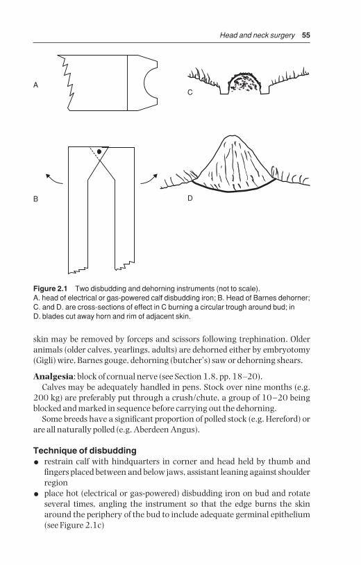
Head and neck surgery 55
skin may be removed by forceps and scissors following trephination. Olderanimals (older calves, yearlings, adults) are dehorned either by embryotomy(Gigli) wire, Barnes gouge, dehorning (butcher’s) saw or dehorning shears.
Analgesia: block of cornual nerve (see Section 1.8, pp. 18–20).Calves may be adequately handled in pens. Stock over nine months (e.g.
200 kg) are preferably put through a crush/chute, a group of 10–20 beingblocked and marked in sequence before carrying out the dehorning.
Some breeds have a significant proportion of polled stock (e.g. Hereford) orare all naturally polled (e.g. Aberdeen Angus).
Technique of disbudding
• restrain calf with hindquarters in corner and head held by thumb andfingers placed between and below jaws, assistant leaning against shoulderregion
• place hot (electrical or gas-powered) disbudding iron on bud and rotateseveral times, angling the instrument so that the edge burns the skinaround the periphery of the bud to include adequate germinal epithelium(see Figure 2.1c)
A
B
C
D
Figure 2.1 Two disbudding and dehorning instruments (not to scale).A. head of electrical or gas-powered calf disbudding iron; B. Head of Barnes dehorner;C. and D. are cross-sections of effect in C burning a circular trough around bud; in D. blades cut away horn and rim of adjacent skin.
BSAC02 3/5/05 4:27 PM Page 55

56 Chapter 2
• pressing and moving head laterally, scoop and flick off the horn bud, leaving a crater, in the middle of which is a small cartilaginous protusion,which may be left since it is not germinal epithelium
• operate on older calves by placing blades of Barnes dehorner preciselyaround base of horn bud and removing small (3–5 mm) strip of skin at sametime as bud is guillotined off. Effect haemostasis with hot disbudding iron
• alternatively remove horns as short as 5 cm rapidly with embryotomywire (more physical effort but minimal haemorrhage due to heat)
Comparison of instrumentationIn yearling and adult cattle the preferred method is embryotomy (obstetrical,Gigli) wire, disadvantages being the considerable physical effort and relatively slow speed. Advantages include neat appearance and lack of haemorrhage.
Saw method is more unsightly and control of haemorrhage takes longer.Dehorning shears (e.g. Keystone dehorner) is the most rapid method but
causes considerable haemorrhage and has a major disadvantage in that, ifanalgesia is absent or poor, sudden violent movement of the head (avoided byfirmly anchored halter!) during closure of the guillotine blades can cause ashear fracture of the frontal bone and secondary wound problems includingfrontal sinusitis. In addition the shears method raises questions of acceptableanimal welfare if analgesia is not confirmed before its application.
Dehorning of cattle without demonstrable analgesia is an unethical andunprofessional act.
Technique of dehorning
• sedation occasionally indicated
• cornual nerve block (see Section 1.8, p. 19). In adult cattle infiltrate further anaesthetic solution at caudal border of horn base
• wait five to ten minutes
• check with needle that skin adjacent to horn is painless
• obtain adequate restraint of head in gate of crush and position headstraight forward so that considerable weight can be exerted on embryotomywire during sawing movement (see Figure 2.2). Likewise, when usingsaw, ensure that operator’s position is optimal
• first cut is made with wire or saw on lateral aspect
• place wire and saw so that instrument passes through skin about 1 cmfrom skin-horn junction
• ensure that direction is correct, especially with saw, since change of direction brings greater difficulties in moving blades
• ensure that blade or wire emerges dorsally through skin lateral to midlineof poll. In Friesian/Holstein cows width of skin left in midline should be5–8 cm
• avoid interrupting sawing movement in middle of dehorning process
BSAC02 3/5/05 4:27 PM Page 56

Head and neck surgery 57
Relatively narrow diameter horns (< 5 cm) may be removed using long-handled dehorning gouge (Barnes pattern). This instrument may also be useful in removing additional protruding lips of horn where the initial procedure has been too conservative.
Haemostasis
• torsion or torsion/traction on the 2–3 major vessels in the medial aspect(ventral crescent) of the peripheral skin; they are easily identified andpicked up by artery haemostatic forceps, six to eight turns are optimal
• rubber tourniquet or string around the two horn bases (such as rubberbands made from cross-sections of car inner tubes or baling twine) appliedin pattern to exert pressure on dorsal horn border as well
• alternatively push wooden toothpick into bone canal from which con-siderable blood can spurt (remove toothpick the next day)
• cautery, e.g. hot iron, electrocautery, is often disappointingly ineffective
• liberal use of bacteriostatic (e.g. non-sterile furazolidone or sulphanilamide)or haemostatic powder (Fe salts, tannic acid, alum)
Centreline ofshears shouldbe 30–45° fromline of head
Blade ofdehorner mustbe below, or at,base of horn
Crush gate
Halter to pull headaway from crush gate
Figure 2.2 Position of cow’s head and of dehorning shears (Keystone) or saw.Note that (a) haltered head is pulled forwards away from crush gate, and to side; (b) cutting angle should be 30–45°; (c) blade or wire is placed onto skin of horn-skin junction.
BSAC02 3/5/05 4:27 PM Page 57

58 Chapter 2
• ensure dehorned cattle are checked regularly for 24 hours after surgeryfor any recurrence of bleeding which may result from local irritation andpain leading to rubbing the cut surface against a wall
Technique of cosmetic dehorningThis technique is often used in show cattle in North America. It is an uneth-ical procedure in some countries.
Cosmetic dehorning gives an improved appearance following surgery, asthe wound is closed by apposition of skin edges over horn base. The procedureis carried out aseptically with the aim of primary healing. Cosmetic dehorn-ing should decrease the risk of post-operative sinusitis.
• clip band 8 cm wide over poll and around base of each horn
• routine skin preparation
• cornual nerve block and local analgesic infiltration caudal to horn baseand in midline
• make transverse incision over poll and laterally in curved fashion passing0.5 cm from horn-skin junction, the two wounds joining lateral to hornbase and continued towards mandibular joint for 5 cm (see Figure 2.3)
• undermine skin peripherally from incision far enough to avoid skin damage when horn is removed by saw or Barnes dehorning gouge
• remove more horn if necessary (sterile bone chisel and hammer, Barnesgouge) until cut is exactly flush with frontal bone
• clean surface with sterile swabs and effect haemostasis
• undermine skin further to enable edges to be apposed across bone surfacewithout excessive tension, then check cosmetic appearance
• appose edges with interrupted sutures of monofilamentous polypropylene
• clean surface of all blood and debris, and apply antibiotic powder
• remove sutures in 14 days
Complications and discussionComplications of disbudding and dehorning include:
• side-effects caused by inadvertent i.v. injection of analgesic solution inyoung calves (excessive salivation, mild ataxia, temporary collapse)
• failure to remove the horn bud completely in calves (inadequate depth ofcauterising activity) results in regrowth or ‘scurs’
• in older cattle frontal sinusitis (pneumatisation of the horn starts at eightto nine months) and empyema caused by entry of infection and fly strike insummer and autumn. Therefore avoid dehorning in the major fly season(e.g. in UK May to late September). Also avoid feeding hay or straw fromoverhead racks to reduce risk of frontal sinusitis.
Crude surgical technique results in prolonged local irritation and increasedtendency for cut surface to be rubbed against dirty surfaces (e.g. soil, bedding,
BSAC02 3/5/05 4:27 PM Page 58

Head and neck surgery 59
walls). Most infected wounds can be cleaned easily, but infection extendinginto the frontal sinus can cause chronic discharge of pus (often A. pyogenes) topass into the maxillary region, sometimes with systemic illness (e.g. pyrexia,anorexia, loss of condition, head tilt, localised swelling and pain). Drainageby sinus trephination may then be indicated (see Section 2.2 below).
1
A
12
3
B
Figure 2.3 Incision and nerve supply for cosmetic dehorning. (A) Rostral view; (B) lateral view.1. zygomatico-temporal nerve; 2. frontal nerve; 3. infratrochlear nerve; --- skin incision.
BSAC02 3/5/05 4:27 PM Page 59

60 Chapter 2
2.2 Trephination of frontal sinus (for empyema)
IndicationFrontal sinusitis with considerable volume of pus occupying the multi-loculated structure and with chronic discharge through the horn base.
Anatomy (Figures 2.4 and 2.5)The frontal sinus is comprised of several compartments (see Figures 2.4 and 2.5). The large caudal frontal sinus is completely divided by an obliquepartition into a rostromedial and a caudolateral portion. The former has anarrow nasofrontal opening and a post-orbital diverticulum. The latter has
15
1617
14
13
10
9
11
3
2
1
5
12 4
6
8
7
X
Figure 2.4 Median section through head, left half. (From Pavaux, 1983.)1. caudal frontal sinus; 2. medial rostral frontal sinus; 3. sphenoidal sinus; 4. nasalcavity; 5. nasal septum; 6. hard palate; 7. root of tongue; 8. soft palate; 9. isthmusfaucium (oral part of pharynx, oropharynx); 10. nasal part of pharynx (nasopharynx);11. pharyngeal septum; 12. nasopharyngeal meatus; 13. laryngeal part of pharynx(laryngopharynx); 14. entrance to larynx (laryngeal aditus); 15. vestibule ofoesophagus; 16. oesophagus (cervical part); 17. cavity of larynx.X shows common site of oesophageal obstruction; long arrow shows hand passedinto pharynx, and short arrow the retrograde pressure on oesophagus. (From Pavaux;1983.)
BSAC02 3/5/05 4:27 PM Page 60

Head and neck surgery 61
the cornual diverticulum and nuchal diverticulum which ends by also exca-vating the parietal, occipital and temporal bones. Two or three small cham-bers lie level with the rostral part of the orbit.
The borders of the frontal sinus are from the rostral part of the orbit to atransverse line drawn through the midline of the orbit, laterally to the frontalcrest, and caudally to the nuchal crest (poll). A midline septum separates the two frontal sinuses. The normal small communication of the frontal sinus with the ethmoid sinus and the nasal cavity is usually occluded due tothickening of the mucosa and purulent discharge.
For clinical signs of frontal sinus empyema see section on complications of dehorning (see Section 2.1, p. 58). Some cases result from horn fracture,usually a direct result of uncontrolled movement of the head (poor anaes-thesia). Sinusitis is often confined initially to caudal part of sinus.
1
2
3
44
2
X
1
Figure 2.5 Diagram of longitudinal and rostral sections through skull to show extentof sinuses.1. frontal sinus; 2. maxillary sinus; 3. position of orbit; 4. rostral compartments offrontal sinus; X trephine sites for empyema of frontal sinus. (Modified from Dyce &Wensing, 1971.)
BSAC02 3/5/05 4:27 PM Page 61
62 Chapter 2
Technique
• restrain animal adequately in crush/chute and give sedative
• clip hair around horn base and over whole of frontal region, cleanse anddisinfect
• produce local analgesia by supra-orbital block (see Section 1.8, p. 20) orinfiltration over site of proposed trephine opening. The trephine openingmay be located 5 cm dorsal to the line joining the two supra-orbital pro-cesses and about 5 cm from midline. Further landmark: 2–3 cm abaxial atthe level of a horizontal line joining the axial parts of both orbits. Some-times a soft area of bone presents a suitable site. A ventral site is preferableif the horn sinus is still patent, permitting flushing from one opening to theother. Avoid the supra-orbital foramen and vein (see Figure 1.4)
• remove circular area of skin, subcutaneous tissue and cutaneous muscle 3 cm diameter by scalpel and forceps
• elevate periosteum with periosteal elevator and remove it with scalpel
• trephine bone over sinus using 2.5 cm diameter Galt or Horsley patterntrephine
• flush sinus cavity initially with warm water using enema pump, then withhydrogen peroxide (3%, i.e. 10 vol, diluted with equal volume of water)
• insert enema pump (Higginson’s syringe) to direct the irrigating mixtureinto the various compartments
• continue irrigation through horn sinus orifice
• irrigate finally with dilute chlorhexidine hydrochloride solution (10 ml of5% solution made up to 1 litre with water), flushing from top to bottom
• pick up any major bleeding points with artery forceps and maintaintrephine opening patent for daily flushing by stock person
• avoid feeding hay/straw from overhead rack
In chronic cases repeated lavage or a permanent through-and-throughlavage system may be needed.
The wound usually heals in three to four weeks. Parenteral medication(five to ten days with broad spectrum antibiotics) is indicated in animals with systemic signs and in all longstanding and severe cases. As trephineopening closes over, irrigation should be continued with a flexible polypro-pylene or PVC catheter attached to the syringe. It is a good idea to suture the catheter in place. Prognosis is favourable (acute cases) to guarded(chronic cases). Infection rarely extends to the opposite side of the skull or to the CNS.
DiscussionIn consideration of the possible and safe sites for trephination, note that the site for euthanasia using a captive bolt gun is midline at the junction ofdiagonal lines joining the medial canthus of the orbit to the ventral border ofthe opposite horn base (see Figure 2.3a).
BSAC02 3/5/05 4:27 PM Page 62

Head and neck surgery 63
2.3 Entropion
Incidence and signs
• low incidence, more common in some beef breeds
• involves and lower lid more frequently than upper lid
• often bilateral to varying extent
• occasionally congenital, usually acquired
• signs include mild blepharospasm, conjunctivitis, keratitis, and cornealulceration if not corrected early
• conservative non-surgical treatment often successful e.g. manual eversion,injection of saline into lid, horizontal mattress tacking suture in eyelid
Indication
• correction of congenital or acquired inversion of upper or lower lid
Technique (Hotz-Celsus procedure)
• estimate length and width of skin to be resected by pinching to produceapproximate skin fold to correct inversion of lid margin
• linear s.c. infiltration of anaesthetic solution parallel and 2 mm distant tolid margin
• scalpel incision to remove ridge of skin previously measured (see Figure 2.6)
• close wound margin with single continuous or mattress absorbable suturee.g. PDS (avoiding need for later removal)
• complications unlikely, though under-correction may necessitate secondsurgery
Left eye
dorsal
medial lateral
ventral
Figure 2.6 Entropion correction involving lower lid of left eye with skin incision 2 mmbelow lid margin.
BSAC02 3/5/05 4:27 PM Page 63
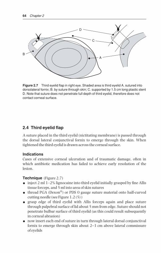
64 Chapter 2
2.4 Third eyelid flap
A suture placed in the third eyelid (nictitating membrane) is passed throughthe dorsal lateral conjunctival fornix to emerge through the skin. Whentightened the third eyelid is drawn across the corneal surface.
IndicationsCases of extensive corneal ulceration and of traumatic damage, often inwhich antibiotic medication has failed to achieve early resolution of thelesion.
Technique (Figure 2.7)
• inject 2 ml 1–2% lignocaine into third eyelid initially grasped by fine Allistissue forceps, and 5 ml into area of skin sutures
• thread PGA (Dexon®) or PDS 0 gauge suture material onto half-curvedcutting needle (see Figure 1.2 (5) )
• grasp edge of third eyelid with Allis forceps again and place suturethrough palpebral surface of lid about 5 mm from edge. Suture should notpenetrate bulbar surface of third eyelid (as this could result subsequentlyin corneal abrasion)
• now insert each end of suture in turn through lateral dorsal conjunctivalfornix to emerge through skin about 2–3 cm above lateral commissure of eyelids
B
D
C
A
Figure 2.7 Third eyelid flap in right eye. Shaded area is third eyelid A. sutured intodorsolateral fornix; B. by suture through skin; C. supported by 1.5 cm long plastic stentD. Note that suture does not penetrate full depth of third eyelid, therefore does notcontact corneal surface.
BSAC02 3/5/05 4:27 PM Page 64

Head and neck surgery 65
• insert 1 cm polypropylene stent onto suture over skin, and tie in ‘quickrelease’ fashion with sufficient tension for third eyelid to cover entire visible surface of cornea, including the lesion
• inspect eyelid suture daily (stockperson), apply any local medication andperhaps slacken off suture to inspect cornea for assessment of healing process
• leave suture in place for two to three weeks, then remove with scissors
Complications and their reasons
• early tearing out of suture from third eyelid, resulting from insufficient‘bite’
• failure to pull flap sufficiently laterally due to incorrect placement of conjunctival and skin suture, usually too medial in position
• pulling out of sutures through skin due to absence of stent
• mechanical irritation of corneal surface from suture material (suture perforated entire depth of third eyelid, or was too slack, failing to pull thirdeyelid completely across corneal surface)
DiscussionNo comparative studies are available on the success of this simple and common technique. Results are generally good as bovine cornea has greatpowers of healing.
2.5 Neoplasia of eyelids
IntroductionNeoplasms of the upper and lower lids and nictitating membrane (third eyelid) include squamous cell carcinoma (SCC) or ‘cancer eye’, and rarelyother tumours such as papillomata, and fibrosarcoma. SCC is most signific-ant in terms not only of incidence but also of economic importance and prognosis. SCC occurs more frequently on the globe (65%) than upper andlower lids (30%) or third eyelid (5%), is very invasive locally, and may meta-stasise to the local lymph nodes (parotid, atlantal or retropharyngeal and the anterior cervical chain).
Clinical signsSCC is largely confined to Hereford and Simmental breeds and their crosses,where the non-pigmented area is liable to develop neoplastic lesions underthe influence of ultraviolet radiation from sunlight. Affected cattle are usuallyfour to nine years old. About 85% of cattle with SCC lack pigment in theaffected area.
The lesion is often an obvious proliferative irregular mass which mayulcerate through the skin to cause moderate distress and blepharospasm.
BSAC02 3/5/05 4:27 PM Page 65

66 Chapter 2
Early lesions appear either as rice-grain-like plaques on the sclera or cornealsurface, or as small firm nodules in the dermis. This precursor of a greyish-white plaque at the nasal and temporal limbus develops into a papilloma and carcinoma in situ. Lid lesions often start as a dirty brown, horn-like keratomata.
TreatmentTreatment is indicated in early lesions without evidence of secondary spreadto adjacent structures (e.g. bone) or metastases to the drainage lymph nodes.
Several techniques are available and include:
(a) excisional surgery(b) cryotherapy(c) hyperthermia(d) radiotherapy (rare)(e) immunotherapy (rare)(f ) combinations of a+b, a+d or c+e
Excisional surgery of third eyelid
• in standing or recumbent animal induce analgesia by local infiltration (5 ml of 2% lignocaine) of base of eyelid after instilling topical anaestheticsolution (e.g. 0.5% proparacaine) into conjunctival sac
• draw third eyelid out by traction with forceps
• excise eyelid deep to cartilage with curved scissors
• control haemorrhage with adrenaline-soaked swab, or cryotherapy
CryotherapyCryotherapy (see Section 1.14, p. 49) is particularly advantageous since the technique avoids haemorrhage and is simple and relatively fast. Thesmall liquid nitrogen flask (Nitrospray® [Arnolds] ) is adequate for lesions upto 5 cm diameter and 1 cm deep.
• protect eye from inadvertent freezing by inserting ‘Styrofoam’ strips oracrylic between lid and corneal surface. Apply water-soluble lubricants orvaseline to skin of surrounding healthy area
• clip and wash affected area and put on disposable rubber gloves
• freeze the area twice (liquid nitrogen) or three times (nitrous oxide, carbondioxide) initially using a spray tip
• include at least 5 mm width border of clinically healthy tissue
• evert tissue lying close to cornea by grasping with towel clips or Allis tissueforceps, before applying probe head which is designed to deal with lesionsof the third eyelid
• use thermocouples if available, inserting points 5 mm from margin oflesion and stopping freeze when they indicate a temperature drop below −20°C
BSAC02 3/5/05 4:27 PM Page 66

Head and neck surgery 67
Advantages of cryotherapy over knife surgery in treatment of SCC are:
• simple, cheap and rapid method
• good post-operative analgesia
• minimal pre-operative preparation and usually no post-operative medica-tion necessary
• procedure may be repeated if there are multiple lesions
• no bleeding
Disadvantages of cryotherapy are:
• lesions > 2.5 cm diameter require relatively prolonged application of probehead for complete iceball formation
• lesions exceeding 5 cm must be treated in two stages, alternatively an initial surgical debulking procedure
• initial instrumentation cost is high, but treatment cost per lesion is then low
Other techniques
• excisional surgery: often indicated in large lesions to reduce size (‘debulk’)prior to cryotherapy
• radiofrequency hyperthermia: application of heat (50°C for 30 seconds) to various surface points of tumour and surrounding skin using probehead. Penetration is limited to 0.5–1 cm, therefore inappropriate for largemasses
• radiotherapy: radon and gold seed implants have both been successfullyused in valuable cattle. Penetration is again only 0.5–1 cm
• immunotherapy: local infiltration of mycobacterial cell wall fractionimmunostimulant (Regressin® [Ragland], USDA-approved drug for immuno-therapy). Dose rate is 0.5 ml for each centimetre of tumour diameter, i.e. 5 cm diameter mass is given 2.5 ml. It is claimed that untreated sites oftenundergo spontaneous regression
• Prognosis: recurrence possible
2.6 Ocular foreign body
IntroductionForeign bodies such as particles of chaff, burrs and thorns may lodge on the corneal surface, particularly in lateral or medial canthus, and provoke a reactive keratoconjunctivitis. Signs are obvious in recent cases withepiphora, ptosis, blepharospasm and discomfort. Chronic cases show cornealscarring and pigmented keratitis. Such material may often be removed with-out local analgesia. Suitable topical analgesics include amethocaine (e.g.Minims® Amethocaine HCL, [Smith & Nephew Pharmaceuticals] ), xylocaine(4% eyedrops – Astra®), or Proparacaine (Ophthaine®, Ophthetic®).
BSAC02 3/5/05 4:27 PM Page 67

68 Chapter 2
Technique
• hold head firmly and tilted in good light so that material is, as far as practical, in midfield of orbital fissure
• spray sterile saline through 22 gauge needle hub tangentially at foreignbody to dislodge it
• if unsuccessful then attempt dislodgement and removal with fine dis-secting forceps or with fine flat surface such as blunt surface of largescalpel blade
• assess superficial corneal damage subsequent to removal, following instillation of one to two drops of fluorescein stain (Minims® Fluoresceinsodium 1% or 2% [Smith & Nephew Pharmaceuticals] )
• insert local broad spectrum antibiotic (e.g. cloxacillin, Orbenin®, [Beecham] )four times daily for three days after removal. Ointments are more suit-able than drops. Topical 1% atropine b.i.d. or to effect to maintain pupildilatation. Corticosteroids may be contra-indicated as they hamper repairof any residual ulcer.
2.7 Enucleation of eyeball
Indications
• intra-ocular neoplasia and gross damage to bulb, usually with severe primary or secondary infection, e.g. infectious bovine keratoconjunctiv-itis (Moraxella bovis ± Neisseria spp.) associated with trauma or rupture of globe, resulting in anterior staphyloma or panophthalmitis, and risk ofascending infection up the optic stalk.
Enucleation is rarely indicated in cattle. Cosmetically satisfactory appear-ance is not so important as in equines and small animals.
Technique of transpalpebral enucleation (exenteration)
• anaesthesia: preferably GA, but alternatively standing or recumbentunder xylazine sedation and retrobulbar block (see Section 1.8, p. 21), orophthalmic nerve analgesia and local infiltration of lower lid and medialcanthus
• clip and cleanse peri-orbital area
• place continuous suture through upper and lower lids
• perform lateral canthotomy (2 cm) to aid exposure
• using traction with towel clips or Allis tissue forceps, make circumferentialincision 1 cm from skin – conjunctival junction, or as appropriate depend-ing on the distribution of non-viable or neoplastic skin (see Figure 2.8)
• continue towards orbital ridge down to, but not through, conjunctiva
• exerting some traction on eye muscles, dissect the extra-ocular musclesbluntly with Mayo scissors from lateral and medial canthus. Avoid exces-sive traction on optic nerve.
BSAC02 3/5/05 4:27 PM Page 68

Head and neck surgery 69
• grasp eyeball and use further traction to dissect it free from surroundingretrobulbar tissue (excluding conjunctival sac) and optic nerve
• leave the maximal amount of healthy retrobulbar tissue
• clamp ophthalmic vessels, optic nerve and retractor bulbi muscle withslightly curved, long-handled artery forceps (Roberts 23 cm, or Kelly 25 cm)
• ligate vessels with 7 metric chromic catgut
• remove third eyelid and Harderian gland
• check site for complete removal of all neoplastic or infected tissue
• meticulous haemostasis during enucleation is time-consuming and inmost cases not necessary
• pack the orbital space for a few minutes with sterile gauze while subcut-aneous layer of simple interrupted sutures of chromic catgut is inserted, or insert absorbable gelatinespmges
A
A
C
B
Figure 2.8 Exenteration of eye (longitudinal diagrammatic section). Dotted line (A)starts in the lids, passes through the peri-orbital structures and results in removal ofglobe, all orbital contents, eyelid margin and conjunctiva. B. optic nerve and vessels;C. muscles. Shaded areas above and below are frontal and zygomatic bones.
BSAC02 3/5/05 4:27 PM Page 69

70 Chapter 2
• place initial sutures in deepest point of wound
• remove packing and all gauze swabs before subcutaneous tissues are completely closed and irrigate with aqueous antibiotic solution (20 ml)
• appose skin edges of lids by interrupted vertical mattress sutures of mono-filament nylon, the pressure of the suture incision almost always beingadequate to stop haemorrhage
• give systemic antibiotics for five to seven days, NSAIDs for three days, andtetanus prophylaxis as required
• remove any packing after two days
• remove sutures two to three weeks later
The resulting ankyloblepharon is cosmetically acceptable. Some cowsbecome more nervous due to restricted field of vision.
ComplicationsComplications include failure or inability to remove all neoplastic tissue (SCC),massive intra-orbital haemorrhage, abscess formation, excessive dead space,and failure to appose the skin margin without excessive tension on sutures(relieving sutures may help).
2.8 Tracheostomy
IntroductionNecrotic and purulent laryngitis (caused by Fusobacterium necrophorum infec-tion and Arcanobacterium pyogenes abscessation respectively) secondary tointra- or retro-laryngeal foreign bodies, or other mechanical irritants (dust,repeated coughing due to Haemophilus somnus and other pathogens).
Indications for tracheostomy rarely involve pharyngolaryngeal neoplasia,retropharyngeal abscessation, foreign bodies in upper respiratory tract, orpersistent laryngospasm. Surgical conditions of head involving haemorrhageand potential aspiration of blood and infected tissue should be managed withan endotracheal tube in position. Tube should only be removed after return ofcough and swallow reflexes.
Clinical signsSigns indicative of need for tracheostomy, which is often an emergency pro-cedure, include progressive dyspnoea, stridor, and mild cyanosis. Some animals have fetid breath (F. necrophorum), and pharyngeal lesions whichcan be both seen and palpated. In all cases a mouth gag should be insertedand a long-bladed laryngoscope or endoscope used to aid examination of theaffected area.
AnatomyTracheal rings are readily appreciated on deep palpation of upper part ofneck. Diameter is narrow compared with equine trachea. Depth is slightly
BSAC02 3/5/05 4:27 PM Page 70
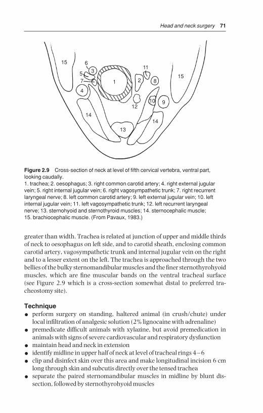
Head and neck surgery 71
greater than width. Trachea is related at junction of upper and middle thirdsof neck to oesophagus on left side, and to carotid sheath, enclosing commoncarotid artery, vagosympathetic trunk and internal jugular vein on the rightand to a lesser extent on the left. The trachea is approached through the twobellies of the bulky sternomandibular muscles and the finer sternothyrohyoidmuscles, which are fine muscular bands on the ventral tracheal surface (see Figure 2.9 which is a cross-section somewhat distal to preferred tra-cheostomy site).
Technique
• perform surgery on standing, haltered animal (in crush/chute) underlocal infiltration of analgesic solution (2% lignocaine with adrenaline)
• premedicate difficult animals with xylazine, but avoid premedication inanimals with signs of severe cardiovascular and respiratory dysfunction
• maintain head and neck in extension
• identify midline in upper half of neck at level of tracheal rings 4–6
• clip and disinfect skin over this area and make longitudinal incision 6 cmlong through skin and subcutis directly over the tensed trachea
• separate the paired sternomandibular muscles in midline by blunt dis-section, followed by sternothyrohyoid muscles
15 6
57
4
1
14
13
12
2
11
8
10 9
15
14
3
Figure 2.9 Cross-section of neck at level of fifth cervical vertebra, ventral part,looking caudally.1. trachea; 2. oesophagus; 3. right common carotid artery; 4. right external jugularvein; 5. right internal jugular vein; 6. right vagosympathetic trunk; 7. right recurrentlaryngeal nerve; 8. left common carotid artery; 9. left external jugular vein; 10. leftinternal jugular vein; 11. left vagosympathetic trunk; 12. left recurrent laryngeal nerve; 13. sternohyoid and sternothyroid muscles; 14. sternocephalic muscle; 15. brachiocephalic muscle. (From Pavaux, 1983.)
BSAC02 3/5/05 4:27 PM Page 71

72 Chapter 2
• insert self-retaining wound retractor (West or Gossett model)
• incise tracheal annular ligament and grasp adjacent tracheal ring withforceps
• resect two half-moon shaped segments of adjacent tracheal rings corres-ponding to size of the tracheal tube (see Figure 2.10). This form of trachealincision maintains the lumen of the adjacent trachea
• insert tracheal tube. Various models are available. A small size (25 mmdiameter) steel equine tube may be used in emergencies. In an immatureanimal (calf ) the maximum diameter may be 13 mm; these are commer-cially available (39 FG). Some tubes are up to 14 mm diameter (PortexBlue Line®) and have external tapes for fixation
• anchor tube to skin at a minimum of two points to prevent rotation
In emergency situation, with no other equipment available, insert a50–70 cm length of vinyl stomach tubing, 16 mm or 19 mm (external dia-meter) and suture in place around tracheotomy wound and also tape overparotid region.
In cattle any tracheotomy tube requires frequent (minimum twice daily)cleaning of both tracheal lumen and external surface. The relative quantity ofdebris and inflammatory discharge is greater than in horses.
• remove tube after alleviation of primary condition, which may requiresystemic antibiotherapy alone, or combined surgery (drainage of laryngealabscess in calves) and antibiotherapy
• do not suture the wound
• avoid hay dust in the immediate environment to reduce likelihood of iatrogenic bronchopneumonia
Tracheal ring cartilage
Ligament
Figure 2.10 Diagram of a tracheostomy incision to show portion of two adjacentrings resected to permit insertion of a tracheotomy tube.
BSAC02 3/5/05 4:27 PM Page 72
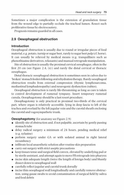
Head and neck surgery 73
Sometimes a major complication is the extension of granulation tissue from the wound edge to partially occlude the tracheal lumen. Resect suchproliferative tissue by electrocautery.
Prognosis remains guarded in all cases.
2.9 Oesophageal obstruction
IntroductionOesophageal obstruction is usually due to round or irregular pieces of foodmaterial, e.g. potato, turnip or sugar beet, rarely to sugar beet pulp (cf. horse).It can usually be relieved by medical means (e.g. tranquillizers such as phenothiazine derivatives, relaxants) and manual retrograde manipulation.
Site of obstruction is usually the proximal cervical oesophagus, often in thefirst 20 cm (see Figure 2.4, (x) ) and rarely the distal cervical or thoracicoesophagus.
Distal thoracic oesophageal obstruction is sometimes seen in calves due to‘broken’ stomach feeders following oral rehydration therapy. Rarely oesophagealobstruction results from external compression (thymic lymphosarcoma,mediastinal lymphadenopathy) and neurogenic dysfunction (rabies).
Oesophageal obstruction is rarely life-threatening as long as care is takento control development of rumenal tympany. Insert temporary rumenal canula. Oesophagotomy should be a last resort procedure.
Oesophagotomy is only practical in proximal two-thirds of the cervicalpart, where organ is relatively accessible, lying in deep fascia to left of the trachea and overlaid by the left jugular vein and the carotid sheath enclosingthe carotid and vagosympathetic trunk.
Oesophagotomy (for anatomy see Figure 2.9)
• identify site of obstruction and, if not palpable, ascertain by gently passingstomach tube
• delay radical surgery a minimum of 24 hours, pending medical relief (e.g. xylazine)
• perform surgery under GA or with sedated animal in right lateral recumbency
• infiltrate local anaesthetic solution after routine skin preparation
• carry out surgery with strict aseptic precautions
• keep tissues tense and surgical field convex, elevated by underlying pad orby sterile assistant, and attempt again to move FB retrograde into pharynx
• incise skin adequate length (twice the length of foreign body) and bluntly dissect down to oesophageal wall
• carefully reflect jugular and carotid trunk dorsally
• incise thin oesophageal wall longitudinally and carefully remove obstruc-tion, using gauze swabs to avoid contamination of surgical field by salivaand food debris
BSAC02 3/5/05 4:27 PM Page 73

74 Chapter 2
• close oesophageal wall (unless necrotic) with interrupted absorbable suturesattached to swaged-on curved round-bodied needle
• place sutures 5 mm apart through mucosa and muscularis
• oversew muscularis in second layer of simple continuous PGA sutures,including fascia for increased strength
• irrigate area well with sterile saline before routine skin closure
• insert Penrose drain along external oesophageal wall to emerge at ventral(caudal) commissure of skin wound in cases where contamination isthought likely to lead to infection and secondary healing
• remove drain after 48 hours
• do not suture oesophagus in cases of full depth mural necrosis, but leave asopen fistula
Though lying adjacent to oesophagus, it should be easy to avoid damage tothe common carotid artery (dorsolateral) and jugular vein (ventrolateral).Cattle with reversible neurogenic oesophageal dysfunction may be supportedby alimentation through a rumen fistula.
Distal oesophageal obstruction (‘broken’ stomach feeders, see above) istreated by removal via rumenotomy incision.
PrognosisIt is difficult to ensure the primary healing of oesophageal incision, butchances are increased by keeping animal on fluids for two days, followed bythree days of mash and short-cut fodder.
BSAC02 3/5/05 4:27 PM Page 74

CHAPTER 3
Abdominal surgery
3.1 Topography
Topography of the forestomachs and abomasum (see Figure 3.1) is incor-porated in descriptions of approach (e.g. left flank laparotomy, traumatic reticulitis). The complex topography of the intestinal tract is considered inthis introduction.
The intestinal tract
The small intestine includes the duodenum (cranial, descending and ascendingparts), jejunum, and ileum. The large intestine is comprised of the caecum,ascending colon (proximal loop, spiral loop [centripetal and centrifugal coils],distal loop), transverse colon, descending colon and rectum.
Position and courseThe muscular pylorus lies fairly fixed in position supported by the origin of the lesser omentum dorsally and the greater omentum ventrally, and situatedlevel with the costochondral junction of ribs 9 and 10 on the right side (seeFigures 3.2, 3.3 and 3.24). The cranial duodenal loop passes craniodorsal,
3.1 Topography 753.2 Exploratory laparotomy, left
flank 793.3 Exploratory laparotomy, right
flank 853.4 Rumenotomy 883.5 Temporary rumen fistulation 963.6 Left displacement of
abomasum 983.7 Right dilatation, displacement
and torsion (volvulus) ofabomasum 110
3.8 Other abomasal conditions 1143.9 Caecal dilatation and
dislocation 115
3.10 Intestinal intussusception 1183.11 Other forms of intestinal
obstruction 1203.12 Peritonitis 1213.13 Umbilical hernia and
abscess 1223.14 Alimentary conditions
involving neoplasia 1273.15 Vagal indigestion (Hoflund
syndrome) 1293.16 Abdominocentesis 1303.17 Liver biopsy 1323.18 Anal and rectal atresia 1333.19 Rectal prolapse 135
BSAC03 3/5/05 4:27 PM Page 75

76 Chapter 3
initially freely mobile, but with the next portion firmly adherent to the vis-ceral hepatic surface. It curves in an S-shaped manner near the bile and pan-creatic duct openings, and becomes the descending loop, which is suspendeddorsally by the mesoduodenum, and the loop passes caudally in the dorsaland right lateral part of the abdominal cavity (see Figure 3.2). Superficial anddeep parts of the greater omentum attach to the ventral surface of thedescending loop of the duodenum.
1
2
4
7 6
98
10
13
13
12
151511
11
14 14
5
320
12
17
16
Figure 3.1 Horizontal section of trunk at mid-height of thorax and thighs, lookingventrally.1–5. rumen: 1. atrium (cranial sac); 2. dorsal sac; 3. caudodorsal blind sac; 4. ventralsac; 5. caudoventral blind sac; 6. reticulum; 7. spleen; 8. liver; 9. caudal vena cava;10. omasum; 11. jejunum; 12. caecum; 13–15. ascending colon: 13. proximal loop;14. spiral loop; 15. distal loop; 16. urinary bladder; 17. uterine horns. (From Pavaux,1983.)
BSAC03 3/5/05 4:27 PM Page 76

Abdominal surgery 77
The duodenum turns craniad at the caudal flexure, where it is attached to the descending colon by the duodenocolic ligament, and becomes theascending loop to pass cranial to the left side of the mesenteric root. It turns to the right side of the root to become the jejunum.
The jejunum, 35–50 m long, comprises a mass of tight coils at the edge ofthe mesentery. The greatest intestinal mass is formed by these heaped coils ofjejunum. The mesentery of the proximal and middle sections is short, that of the distal part and that attached to the ileum are longer, forming a mobilesection which lies caudal to the supra-omental recess. The ileum comprises a convoluted proximal segment and a distal straight part. The junction ofjejunum and ileum is the point where the cranial mesenteric artery ends, andthe cranial limit of the ileocaecal fold. The ileum is attached to the caecumventrally, the orifice lying obliquely on the ventral surface of the caecum, andreadily identified in adulthood due to the fat pad overlying it.
The caecum is a mobile sac, with the blind end directed caudally. Craniallythe caecum is continuous with the proximal loop of the ascending colon. The
7 9
8
6
4
1
3
5
2
1
Figure 3.2 Distribution of greater omentum, viewed diagrammatically through avertical section of abdomen, about lumbar vertebra 3.1. parietal peritoneum; 2. insertion into left longitudinal groove of the rumen of thesuperficial layer of the greater omentum; 3. duodenum; 4. mesoduodenum (craniallythe lesser omentum); 5. deep and superficial layers of greater omentum; 6. intestinalmass in supra-epiploic recess; 7. rumen; 8. left kidney; 9. deep layer of greateromentum inserting onto right longitudinal groove of rumen. (Modified from Dyce &Wensing, 1971.)
BSAC03 3/5/05 4:27 PM Page 77
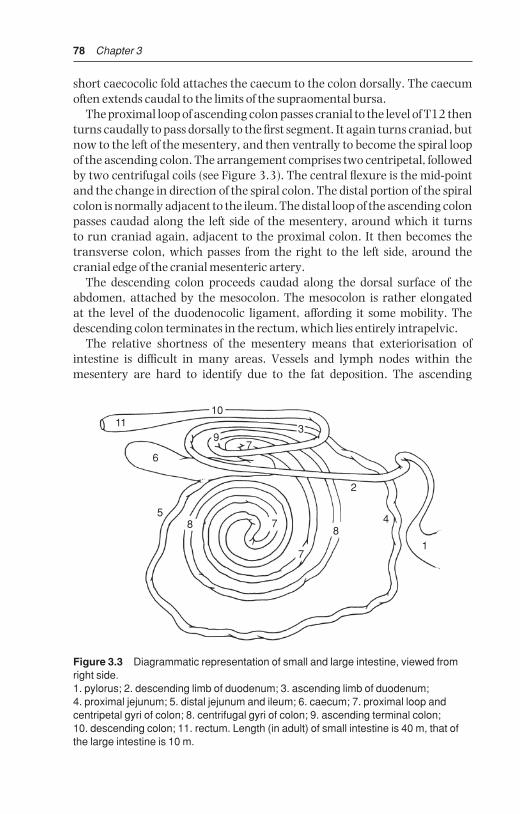
78 Chapter 3
short caecocolic fold attaches the caecum to the colon dorsally. The caecumoften extends caudal to the limits of the supraomental bursa.
The proximal loop of ascending colon passes cranial to the level of T12 thenturns caudally to pass dorsally to the first segment. It again turns craniad, butnow to the left of the mesentery, and then ventrally to become the spiral loopof the ascending colon. The arrangement comprises two centripetal, followedby two centrifugal coils (see Figure 3.3). The central flexure is the mid-pointand the change in direction of the spiral colon. The distal portion of the spiralcolon is normally adjacent to the ileum. The distal loop of the ascending colonpasses caudad along the left side of the mesentery, around which it turns to run craniad again, adjacent to the proximal colon. It then becomes thetransverse colon, which passes from the right to the left side, around the cranial edge of the cranial mesenteric artery.
The descending colon proceeds caudad along the dorsal surface of theabdomen, attached by the mesocolon. The mesocolon is rather elongated at the level of the duodenocolic ligament, affording it some mobility. Thedescending colon terminates in the rectum, which lies entirely intrapelvic.
The relative shortness of the mesentery means that exteriorisation of intestine is difficult in many areas. Vessels and lymph nodes within themesentery are hard to identify due to the fat deposition. The ascending
11
1
6
9
10
7
3
2
48
7
785
Figure 3.3 Diagrammatic representation of small and large intestine, viewed fromright side.1. pylorus; 2. descending limb of duodenum; 3. ascending limb of duodenum; 4. proximal jejunum; 5. distal jejunum and ileum; 6. caecum; 7. proximal loop andcentripetal gyri of colon; 8. centrifugal gyri of colon; 9. ascending terminal colon; 10. descending colon; 11. rectum. Length (in adult) of small intestine is 40 m, that ofthe large intestine is 10 m.
BSAC03 3/5/05 4:27 PM Page 78

Abdominal surgery 79
duodenum, proximal and distal loops of ascending colon, and the cranial portion of the descending colon lie close to one another due to the near-fusionof their mesenteries. The cranial mesenteric vessels supply the small andlarge intestine, except for parts of the duodenum and colon.
The greater omentum passes from its origin on the duodenum, pylorus andgreater curvature of the abomasum, encircles the intestinal mass, and insertson the left longitudinal groove of the rumen (superficial part), while the deeppart passes similarly ventral and to the left, to attach to the right longitudinalgroove of the rumen. These two parts are fused caudally forming the caudalfold. The lesser omentum extends from the oesophagus along the reticulargroove and omasal base to attach to the lesser curvature of the abomasum,and covers most of the parietal surface of the omasum.
Abdominal viscera (see also Fig. 3.7)
Viscera which contact left abdominal wall (see Figures 3.1, 3.2, 3.4 and 3.5):
• cranially and ventrally: reticulum
• laterally ribs 10–12 dorsally, ribs 7–8 ventrally: spleen
• laterally: rumen covered ventrally by greater omentum from left longit-udinal groove
• laterally and (sublumbar) dorsally: perirenal fat
• ventrally and caudally: sometimes coils of jejunum and ileum
Viscera which contact right abdominal wall:
• cranially and ventrally: reticulum and abomasum
• laterally and cranially (ribs 7–12): liver
• laterally (rib 7 ventrally, rib 12 dorsally); descending loop of duodenumand small intestine covered by greater omentum
Viscera which do not normally contact body wall:
• omasum at level of ribs 8–11, lying ventrally to right of midline
• caecum and ascending colon (caudal mid-abdomen), transverse anddescending colon
• left kidney (level with vertebrae 2–4), and right kidney (thoracic vertebra13–lumbar 2)
• uterus and ovaries: uterus in advanced pregnancy (gestation month seven)may contact lower left and/or right flank
3.2 Exploratory laparotomy (celiotomy), left flank
IndicationsSpecific indications are suspected left displaced abomasum (LDA) (see Sec-tion 3.6), rumenotomy, traumatic reticulitis (see Section 3.4), or caesarean
BSAC03 3/5/05 4:27 PM Page 79

80 Chapter 3
section (see Section 4.1). LDA is evident on opening into peritoneal cavity.Traumatic reticulitis may be suspected on exploration of area between cranial aspect of ruminoreticulum and the diaphragm-body wall area. In positive cases specific surgical correction is performed.
Indication is often not clearcut. Some cattle show persistent abdominalpain apparently localised to ruminal area. Left flank exploratory laparotomy
2
1
5
6
128 13
9
10
11
13
4
7
3
Figure 3.4 Cross-section of thorax through body of seventh thoracic vertebra,looking cranially. 1. body of seventh thoracic vertebra; 2. spinous process of sixth thoracic vertebra; 3. seventh rib; 4. sternum; 5. thoracic duct and aorta; 6. left lung; 7. right lung; 8. caudal vena cava; 9. liver; 10. reticulum; 11. apex of heart; 12. accessory lobe ofright lung; 13. central tendon and sternal part of diaphragm. (From Pavaux, 1983.)
BSAC03 3/5/05 4:27 PM Page 80

Abdominal surgery 81
is rarely as useful or as practical as right flank approach and is not recom-mended if a small or large intestinal surgical disorder is suspected.
Laparoscopic surgery is being developed in cattle for abdominal explorationand treatment of LDA but is currently mostly confined to referral clinics.
16
8
10
9
11
12
7
2
3
13
54
Figure 3.5 Cross-section of trunk through body of ninth thoracic vertebra, lookingcranially.1. body of ninth thoracic vertebra and head of ninth rib; 2. eighth rib; 3. seventh rib; 4. xiphoid process of sternum; 5. sternal part; 6. thoracic aorta (thoracic duct, to right,and left azygos vein to left, coursing along it dorsolaterally); 7. left lung (caudal lobe);8. right lung (caudal lobe); 9. liver; 10. caudal vena cava; 11. omasum; 12. atrium(cranial sac) of rumen; 13. reticulum. (From Pavaux, 1983.)
BSAC03 3/5/05 4:27 PM Page 81

82 Chapter 3
Technique
• paravertebral analgesia (T13, L1 and L2, see Section 1.8, pp. 22–26) orlocal infiltration (see Section 1.8, pp. 26–27)
• clip, scrub and surgically prepare a wide area of left flank including at least30 cm around proposed incision site (see Figure 3.6)
• drape with sterile cloths or rubber drape with appropriate window
• make paracostal incision 15 cm long about 5 cm behind last rib, starting10 cm below lumbar transverse processes
• incise skin in single movement and continue scalpel incision through sub-cutaneous fat and fascia to expose abdominal wall musculature
T13 L1
1 25
3
4
L2 L3 L4 L5 L6
Figure 3.6 Position of various left flank incisions (see also Figure 1.7, p. 25).1. paracostal (18–25 cm), cranial in sublumbar fossa: rumenotomy (essential to be as far cranial as possible in large-framed cow and short surgeon);2. left flank abomasopexy (Utrecht technique) or exploratory laparotomy (25 cm);3. low flank incision in recumbent cow or heifer for caesarean section, where it isanticipated that it will be difficult to bring uterine wall to flank (35 cm);4. standard caudal left flank (35–40 cm) and5. oblique flank incision (35–40 cm) for caesarean section in standing animal.
BSAC03 3/5/05 4:27 PM Page 82

Abdominal surgery 83
• insert blade of straight scissors at angle of 45° to surface and into externaloblique abdominal muscle, which is separated by blunt dissection
• make 7 cm long scalpel incision through internal oblique muscle to exposeunderlying transverse fascia
• make small incision with scissors through this fascia to reveal parietalperitoneum beneath a variable amount of loose fat
• pick up parietal peritoneum with rat-tooth forceps and make small verticalincision with scissors
• extend incision through internal oblique, transverse fascia and peritoneumwith scissors to correspond to length and direction of skin incision. Air rushesaudibly into abdominal cavity at this point creating pneumoperitoneum,and contact surface of ruminal wall (unless adhesed) drops away asabdominal wall moves laterally. (Occasionally some pneumoperitoneumis present before surgery, e.g. in traumatic reticulitis.)
Left side of abdominal cavity and part of right side may now be explored (seeFigure 3.7).
descending colon
4
body of caecum
2
3
coils of jejunum
abdominal floor (fluid)
parietal peritoneum
LEFT RUMINAL WALL left kidney
uterus & ovaries
bladder
(ureters)
lymph nodes
inguinum
rumen
spleen
reticulum
diaphragm
left lobe liver
cardiac impulse(apex beat)
1
Figure 3.7 Flow diagram of left flank exploratory examination. As in right flankapproach (see Figure 3.8) entire accessible part of abdominal cavity should be rapidlychecked in any abdominal disease. Start from left ruminal wall with palpation ofparietal peritoneum (1), then caudal abdomen (2) and the right side structures (3), before concentrating on left flank and left cranial and ventral regions (4).
BSAC03 3/5/05 4:27 PM Page 83

84 Chapter 3
Visible features
• check volume and colour of peritoneal fluid; normal colour is pale yellow.A slight pink tinge may be due to contamination of some blood from incision site. Presence of any floccules, usually purulent, is abnormal andindicates an infective focus in the visceral or parietal peritoneum. Possiblyalso an associated objectionable odour.
• run fingers over surface of both parietal and ruminal (visceral) peri-toneum adjacent to incision: surface should be smooth. Irregularities maybe in form of discrete adhesions or generalised lesion (‘sandpaper-like’)consistent with chronic peritonitis.
Palpable features
• introduce the right hand and arm to make systematic examination of theabdominal cavity (see Figure 3.7)
• pass right hand ventrally to check for possible LDA, and also cranially foradhesions between reticulum and diaphragm or liver, or (rarely) betweenrumen and abdominal wall, suggestive of traumatic reticulitis
• presence of abdominal adhesions may be of recent origin and significant, ormay be longstanding and purely an incidental finding. Recent adhesions(less than one week) tend to be broken down easily, though this may causelocalised pain. Older adhesions tend to be difficult or impossible to breakdown and pain is absent
• assess texture of peritoneal surface in different areas at early stage ofexploration before repeated movement causes iatrogenic roughening
• structures palpated on left side (see Figure 3.1) should include: rumen,reticulum, spleen, left border of liver, diaphragm, apical beat of heart, leftkidney through perirenal fat, path of ureters (normally non-palpable unlessthickened), bladder including bladder neck, uterus, left and right ovaries,and descending colon
• pass right hand and arm to right abdominal wall by directing it caudal tothe attachment of the ruminal wall to the abdominal roof, and ventral to left kidney and descending colon, thereby avoiding possible iatrogenicspread of infection from left side of abdomen
• structures now accessible include: left kidney, spiral colon, duodenum,jejunum and ileum, and caecum (see Figures 3.1–3.3).
DiscussionStructures too distant for palpation by veterinarian of average stature inadult Holstein Friesian cow include: abomasum, much of the visceral surfaceof liver and gall bladder, and parts of the omasum, some of the small intestine( jejunal loops) and large intestine (colonic coils).
BSAC03 3/5/05 4:27 PM Page 84

Abdominal surgery 85
3.3 Exploratory laparotomy, right flank
Right laparotomy site is corresponding position to that of left paralumbarfossa (incision 2 in Figure 3.6). Ensure that ventral commissure of incision isin upper half of flank, otherwise spontaneous prolapse of greater omentumand some small intestinal (jejunal) coils is almost inevitable. Take special carein peritoneal incision as descending duodenum is immediately below!
Visible featuresNote greater omentum below incision, with descending duodunum passingcaudally in the omentum.
Palpable features
• pass left hand and arm ventrally to palpate abomasum, note some mobil-ity is possible and abomasum may be grasped and pulled upwards towardsincision
• appreciate cranially visceral surface of liver (note any rounded edges,abscesses or surface irregularity) with dependent gall bladder (normal size up to 10 × 6 × 4 cm), and insert hand between liver and diaphragm topalpate cranial hepatic surface, e.g. discrete abscessation
• pass hand along lateral body wall with palm outwards and ventrally locatereticulum (note any adhesions, or foreign bodies) beyond abomasum andgreater omentum
• check contact area of reticulum with diaphragm (possible adhesions)
• note mesoduodenum is dorsal to duodenum, and deep to this area is theperirenal fat (right kidney)
• palpate, caudal to right kidney, the right surface of left kidney, both structures being slightly to right of midline due to pressure from rumen.Right kidney is dorsal to cranial part of descending duodenum, to right of mesoduodenum. Left kidney lies adjacent to middle part of descendingduodenum entirely within the supraomental recess
• pass hand caudal to caudal edge of greater omentum which runs approx-imately midway between last rib and tuber coxae
• palpate structures in this space, which include numerous coils of jejunum and ileum, as well as spiral colon, caecum, which is very vari-able in size, and dorsally ascending colon and descending colon (see Figure 3.3)
• some intestine which may be exteriorised for examination includes muchof jejunum (except cranially), apex and body of caecum, and more ventralloops of ascending spiral colon
• note, suspended from midline (palpable but cannot be exteriorised),descending colon and part of ascending colon, passing into pelvic cavitytogether with bladder, uterus and ovaries
BSAC03 3/5/05 4:27 PM Page 85

86 Chapter 3
Closure of flank laparotomy incision
• close the incision in three layers (see Figure 3.9a)
• appose peritoneum and transverse fascia with continuous suture of 4 metricPDS on 3/8 circle round-bodied needle
• commence suture at ventral commissure and tie off dorsally
• close external and internal oblique abdominal muscles separately ortogether with similar single layer
• appose skin with Ford interlocking suture of monofilament nylon orSupramid® inserted with cutting-edged semi-curved needle. A relativelyfine needle about 5 cm long requires less effort to insert through 5–7 mmthickness of skin than a larger needle
visceral surfaceof liver
gall bladder
cranial surfaceof liver
diaphragm
reticulum
23
abdominal wall
diaphragm
cardiac impulse (apex)
kidneys (left and right)
perirenal fat (left)
perirenal fat (right)
bladder
uterus andovaries
mesoduodenum
duodenum
pylorus
abomasum
descendingcolon
5
4
rectum
coils ofjejunum
root ofmesentery
body ofcaecum
spiral colon
1
6
GREATEROMENTUM
Figure 3.8 Flow diagram of right flank exploratory laparotomy. Entire accessibleabdominal cavity should be checked in any case of suspected abdominal disease. In a case of LDA the abomasum is not found on right side (step 1), but against leftabdominal wall. Nevertheless steps 2 and 3 should be followed to rule out co-existingtraumatic reticulitis or liver abscessation. In suspect cases of small or large intestinaldisease or displacement, step 4 should be followed, but exploration of remainder ofabdominal cavity should be carried out at a later stage.
BSAC03 3/5/05 4:27 PM Page 86

Abdominal surgery 87
• close ventral 5 cm with interrupted sutures which may be removed laterfor drainage of any wound infection
• note that in all three layers sutures should be just tight enough to apposethe wound edges. In the muscle layers suture should be spaced about oneevery 2 cm, while in the skin slightly more are advised
• single layer closure (see Figure 3.9b) is faster but less cosmetically pleas-ing, and any wound infection is liable to be more severe
• avoid leaving any dead space
DiscussionThe above technique achieves optimal apposition of peritoneum, muscula-ture and skin, and the cosmetic result is pleasing. Any buried deep suturesshould be of absorbable material in view of probable human consumption ofthe carcass.
Intra-abdominal and systemic antibiotherapyRoutine intra-abdominal medication is unnecessary. Peritonitis is controlledby systemic medication if the animal is not salvaged at once. Drainage ofpurulent exudate is unlikely to help control of an active process.
Intra-abdominal medication should be considered only if infection hasbeen introduced into the peritoneal cavity. Suitable drugs include antibioticaqueous solutions or oleaginous suspensions as carriers. Intramammary cerate preparations should not be inserted into the abdominal cavity or body wall. The most effective prophylactic medication is systemic injection of ceftiofur, penicillin, oxytetracycline hydrochloride or a single injection oflong-acting oxytetracycline (see Section 1.12, p. 44).
12
3
4
BA
Figure 3.9 Transverse section through flank (diagrammatic) showing two methodsof closing laparotomy wounds. (A) Recommended three layer closure; (B) single layerfigure-of-eight closure.1. peritoneum; 2. transverse fascia/muscle, 3. internal and external oblique muscles;4. skin.
BSAC03 3/5/05 4:27 PM Page 87

88 Chapter 3
Complications of exploratory laparotomy
• intra-operative collapse: occasionally a weak subject undergoing a ‘last-ditch’ exploratory laparotomy collapses during intra-abdominalexploration causing gross peritoneal contamination. Such cattle, usuallythin cows, are unsuitable subjects for surgery, and should be excluded on pre-operative clinical examination (see Section 1.4, pp. 10–11)
• wound dehiscence: results from non-sterile, excessively fine material,material traumatised by forceps before or during insertion, massivewound swelling causing tearing out of sutures, and development of a subcutaneous or subperitoneal abscess, or emphysema
Treatment of wound dehiscence: in such cases any remaining (non-functional) suture material should be removed and the wound cleansed,débrided and resutured if no infection is present, leaving a 3–10 cm opening in the ventral commissure for drainage purposes.
Cases with copious purulent discharge involving a still relatively intactwound should be opened slightly, both dorsally and ventrally, to permit vigorous irrigation with dilute chlorhexidine gluconate solution (10 ml of 5% solution made up to 1 litre with clean tap water). The incision is then leftto heal by secondary intention.
In the presence of considerable necrotic tissue, preliminary irrigation withdilute H202 (3%) may be useful. (N.B. this solution may spread infectionbetween tissue layers.) See Section 1.13.
3.4 Rumenotomy
Indications
• removal of foreign body in traumatic reticulitis or reticuloperitonitis
• gross severe rumen overload (‘grain overload’) involving acidosis follow-ing sudden ingestion of large volume of concentrates (barley etc.), andrarely, recent ingestion of toxic plant material (e.g. yew). Latter is usuallyfatal
• exploratory surgery, e.g. in chronic intermittent rumen tympany
Traumatic reticulitis
IncidenceTraumatic reticulitis is usually encountered sporadically in cattle over two years old, though occasionally farmers may experience a series of cases in a few weeks or months, associated with ingestion of particular batch of feed. Cases are more common in indoor winter feeding period on hayor silage.
BSAC03 3/5/05 4:27 PM Page 88

Abdominal surgery 89
AetiologyMost foreign bodies ingested by cattle fail to penetrate rumino-reticular walland remain in rumen (small stones etc.) or are eventually voided in faeces.Long metallic foreign bodies such as pieces of wire, needles or nails (typicallychopped wire strands from decomposing lorry or car tyres, or rusting fencing)and rarely broom bristles etc., are thrown forwards by ruminal-reticular con-tractions into the honeycombed reticulum which contracts and the foreignbody may penetrate the mucosa. Common site is the cranial and ventral reticular wall. Penetration to depth of 5–7 mm results in perforation of thevisceral peritoneum, and point then traumatises opposing parietal peritoneum,usually the diaphragm and occasionally the abdominal wall, spleen or liver(see Figure 3.10).
Pain arises from irritation to peritoneum and may be temporary, the foreign body dropping back loose into the reticular wall or lumen, either to repenetrate at another point or to pass further down the alimentary tract where further trouble is most unlikely. At site of penetration an acutelocalised inflammatory reaction with exudate becomes slowly organised as an adhesion, or abscess. In others cases the foreign body slowly advances further, persistent pain is apparent for several days, and the foreign body may eventually enter the thorax and heart, liver, spleen or abdominal wall.Sequelae of reticular foreign body penetration and their signs are multiple,potentially fatal, and are discussed on page 91.
1
5
23
4
Figure 3.10 Sagittal section at diaphragmatic area, seen from left.1. diaphragm; 2. reticulum; 3. cranial part of ventral sac of rumen; 4. rumen; 5. heart.
BSAC03 3/5/05 4:27 PM Page 89
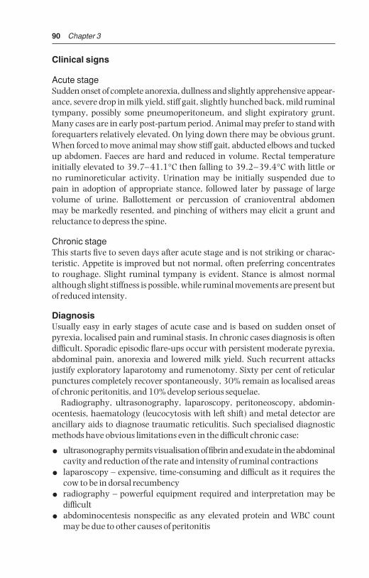
90 Chapter 3
Clinical signs
Acute stageSudden onset of complete anorexia, dullness and slightly apprehensive appear-ance, severe drop in milk yield, stiff gait, slightly hunched back, mild ruminaltympany, possibly some pneumoperitoneum, and slight expiratory grunt.Many cases are in early post-partum period. Animal may prefer to stand withforequarters relatively elevated. On lying down there may be obvious grunt.When forced to move animal may show stiff gait, abducted elbows and tuckedup abdomen. Faeces are hard and reduced in volume. Rectal temperature initially elevated to 39.7–41.1°C then falling to 39.2–39.4°C with little or no ruminoreticular activity. Urination may be initially suspended due to pain in adoption of appropriate stance, followed later by passage of large volume of urine. Ballottement or percussion of cranioventral abdomen may be markedly resented, and pinching of withers may elicit a grunt andreluctance to depress the spine.
Chronic stageThis starts five to seven days after acute stage and is not striking or charac-teristic. Appetite is improved but not normal, often preferring concentrates to roughage. Slight ruminal tympany is evident. Stance is almost normalalthough slight stiffness is possible, while ruminal movements are present butof reduced intensity.
DiagnosisUsually easy in early stages of acute case and is based on sudden onset ofpyrexia, localised pain and ruminal stasis. In chronic cases diagnosis is oftendifficult. Sporadic episodic flare-ups occur with persistent moderate pyrexia,abdominal pain, anorexia and lowered milk yield. Such recurrent attacks justify exploratory laparotomy and rumenotomy. Sixty per cent of reticularpunctures completely recover spontaneously, 30% remain as localised areasof chronic peritonitis, and 10% develop serious sequelae.
Radiography, ultrasonography, laparoscopy, peritoneoscopy, abdomin-ocentesis, haematology (leucocytosis with left shift) and metal detector areancillary aids to diagnose traumatic reticulitis. Such specialised diagnosticmethods have obvious limitations even in the difficult chronic case:
• ultrasonography permits visualisation of fibrin and exudate in the abdominalcavity and reduction of the rate and intensity of ruminal contractions
• laparoscopy – expensive, time-consuming and difficult as it requires thecow to be in dorsal recumbency
• radiography – powerful equipment required and interpretation may bedifficult
• abdominocentesis nonspecific as any elevated protein and WBC countmay be due to other causes of peritonitis
BSAC03 3/5/05 4:27 PM Page 90
Abdominal surgery 91
• metal detector – non-specific for penetration, only positive with ferrousmaterial, and false positives with cobalt and magnesium, bullets, slowrelease anthelmintics
• haematology – raised white cell count possibly due to other causes
Conservative treatmentOften, as signs are assessed over a few days, conservative medical treatment is instituted; forequarters should be elevated 45 cm with boarding or earth, and systemic antibiotic therapy given for three days. Magnetic probangs(Eisenhut model) and magnets have given disappointing therapeutic results.Many cases respond completely, some temporarily, to any conservative treatment.
Signs resulting from sequelae of traumatic reticuloperitonitis
• intrathoracic penetration: quite common, usually causing traumatic pericarditis (see Figure 3.10). Obvious illness about one week or moreafter episode of vague indigestion. Signs of congestive cardiac failure (CCF)include ventral oedema, fast pulse, distended jugular veins and localisedpain. Such cases usually die within one to two weeks. Less commonly,intrathoracic foreign body causes localised abscessation in one lung lobe with chronic suppurative pneumonia and localised thoracic pain.Occasional entry into pleural space leads to fibrinopurulent pleuritis.Rarely reaction occurs in the mediastinum, and if extensive can causepressure on heart sufficient to cause CCF (i.e. pericarditis may not be the cause of CCF)
• intra-abdominal penetration: results in hepatic, reticular, omasal or abomasal wall adhesions and a more or less localised peritonitis. Signs are vague with dullness, pyrexia and anterior abdominal pain. Splenicabscession may cause septicaemia and pyaemic spread to other organs. In rare cases abscess develops over lower body wall and penetrates skin,discharging foreign body as well as pus
• chronic reticular adhesions and abscesses: may be very extensive andinvolve vagal nerve supply to rumen. Usually animal returns to near-normal health, but some cases develop syndrome of chronic ruminal distension or Hoflund syndrome (see Section 3.15, pp. 129–130).
Technique of rumenotomyRumenotomy is the preferred treatment for acute traumatic reticuloperitonitis,and is also indicated in suspicious cases of chronic disease which are non-responsive to conservative treatment including chemotherapy, administrationof a permanent magnet, and management change.
Site is in left flank as described in exploratory laparotomy (see Section 3.2,pp. 79–84). Length of incision should be 18–25 cm, but varies somewhat
BSAC03 3/5/05 4:27 PM Page 91

92 Chapter 3
with the particular technique selected to control possible contamination byruminal contents:
• Weingart frame – incision as above
• McLintock cuff – incision length 16 cm
• suture of ruminal wall to parietal peritoneum – no critical length. Ensurein all cases that dorsal commissure of incision is about 8 cm ventral to lateral extremity of lumbar transverse processes.
Having entered abdominal cavity (see Section 3.2, pp. 79–84 for technique)and before making rumenotomy incision check several points:
• appearance of visible parietal and visceral (ruminal) peritoneum, e.g.roughening indicative of acute or chronic peritonitis
• explore right side of abdominal cavity first
• presence of excessive abdominal fluid: pass hand ventrally, and excessiveabdominal fluid is easily obtained in handfuls, i.e. repeated volumes of20–50 ml. A greyish-yellow colour with floccules of pus is evidence ofacute or chronic peritonitis
• presence of adhesions between reticulum, and diaphragm and adjacentviscera: indicative of past or present foreign body penetration. For assess-ment of age of adhesions see p. 84.
To avoid potential contamination of the abdominal cavity by ruminal contents, the ruminal lumen is either exteriorised (Weingart frame orMcLintock cuff ) or the abdominal cavity is sealed off from the rumen by temporary insertion of a continuous suture. The preferred method is theWeingart frame.
Weingart frame method (Figure 3.11)The stainless steel frame (size 27 × 18 cm) is used with two vulsellum forceps (23 cm) fixed with single hooks near the junction of blade and handle,and with six small (7 cm) tenaculum hooks. Having entered abdominal cavity:
• screw the Weingart frame into the dorsal commissure of the skin incision
• push ruminal contents inwards at intended rumenotomy incision
• grasp rumen wall dorsally and about 15 cm ventrally with the two pairs offorceps, exteriorise and fix to two rings at top and bottom of frame. Thisbrings out the rumen but does not yet prevent contamination
• place a sterile cloth or rubber drape or shroud completely around the exteriorised rumen between the frame and the abdominal wall
• incise the rumen just below the dorsal forceps
• insert one of the small ruminal hooks into the ruminal mucosa near theedge, pull back and clip the rumen onto edge of the frame at eleven o’clockposition, followed by another at one o’clock
BSAC03 3/5/05 4:27 PM Page 92

Abdominal surgery 93
• extend ruminal incision ventrally to an appropriate length for entry of thearm and attach rumen to frame with four further hooks at nine, three, thenseven and five o’clock positions. The ruminal lumen is now effectively isolated from the abdominal cavity
McLintock cuff methodThis method is equally efficient but presents greater problems with sterilisa-tion of equipment, and rubber components eventually perish.
• a special rubber cloth with everted stiff cuff surrounding the abdominalincision is placed over the flank
• exteriorise rumen and make 2.5 cm incision in upper position
• insert rubber-covered hook which is held temporarily by a non-sterileassistant
• extend incision ventrally to 10–11 cm length
Figure 3.11 Weingart frame placed to exteriorise and fix ruminal wall with six hooksand two vulsellum forceps.
BSAC03 3/5/05 4:27 PM Page 93
94 Chapter 3
• insert rim of stiff rubber cuff through the incision, whereupon the lips of the ruminal incision will grip it tightly, and pull of rumen draws rimagainst skin of flank
• place a thin rubber sheet with 15 cm elliptical hole between rumen and skin
• place another, similar sheet over the rim and double back the edge of ruminal cuff to form a seal
Suture method
• suture ruminal wall to skin by simple continuous suture of non-absorbablematerial (4 metric) on cutting needle and to rumen with Cushing-typepattern
• after suturing, check site for a good seal between rumen and skin
• incise rumen starting 2.5 cm ventral to dorsal commissure and ending 3 cm dorsal to ventral commissure
• if rumen is not relatively empty and flaccid, these sutures can possibly tearout, and in such doubtful cases suture ruminal wall to edge of parietalperitoneal incision
• for easy handling when working alone, dorsal and ventral parts of exteriorised rumen may be temporarily fixed to skin by towel clips (13 cm)for suturing purposes
Regardless of method, the next steps are similar:
• siphon off any excessive fluid with wide bore (3 cm internal diameter) plastic tubing filled with water, and remove any obstructing solid material
• pass arm cranially and ventrally over U-shaped ruminoreticular pillar andexplore reticulum methodically. Evidence of adhesions already palpatedduring intra-abdominal exploration may lead hand to a particular areaotherwise make initial rapid examination of the reticular floor, then of cranial wall
• identify and examine the cardia, oesophageal groove, and the reticulo-omasal opening as well as the medial wall: touching of reticulo-omasalorifice should provoke contraction
• remove loose reticular foreign bodies, but search specifically for pointedlongitudinal foreign body lodged in secondary reticular cells between thesecondary crests which characterise this organ
• search with fingertips as only 1 cm or less length of foreign body may protrude into lumen. In other cases it can subsequently be confirmed thatthe foreign body has passed right through the reticular wall
• it may be helpful to elevate the reticular wall with fingers to assess thepresence of adhesions out of reach on parietal surface, e.g. right side
• palpate reticular wall also for discrete abscesses
• if penetrating foreign body is found, and before its removal note depth and direction of penetration to consider the likely structures damaged atthis time. This aids prognosis. The wisdom of puncturing and draining
BSAC03 3/5/05 4:27 PM Page 94

Abdominal surgery 95
reticular or ruminal wall abscesses from the ruminoreticular lumen shouldbe carefully assessed
• use a magnet to retrieve loose ferrous material more easily
• consider placing permanent magnet into reticulum
Difficulty in reaching furthest points for exploration due to physical size of the animal or surgeon may be partially overcome by elevation of the fore-quarters, causing reticulum to drop back slightly, or indirect pressure onreticular area by upward pressure on the xiphoid region by an assistant (ortwo, using a wooden plank).
Ruminal medication may be given after exploration of the reticulum.Although awkward to obtain (unless from slaughterhouse) fresh ruminalcontents quickly normalise the flora.
Treatment of adhesions
• chronic adhesions: there is usually little or no benefit from breaking downchronic adhesions, which tend to reform very rapidly
• recent adhesions: do not break down since they may mask and surroundan abscess cavity
Closure of ruminal incisionMethod varies slightly with the method of fixation, but in all instances twolayers of inversion sutures should be placed. These should be:
• continuous Cushing inversion suture of 4 metric PDS
• continuous Lembert inversion suture of similar material
Weingart frame method
• remove the small ruminal clips and clean the peritoneal surface before andafter placing the two suture layers, and clean again before releasing largeforceps, permitting rumen to drop back into abdominal cavity
McLintock cuff method
• remove the two rubber sheets, avoiding contamination, pull out rumenand internal cuff further from body wall and apply foam-rubber edged special rumen clamp under rim of tube which is then removed
• clamp then rests on flank with ruminal edges safely fixed and the con-taminated surfaces may be easily cleaned, and the two layer suture (Cushingor Lembert type) placed in position
Suture method
• after initial cleansing of exposed ruminal surface insert two layers ofsutures and then carry out thorough cleansing of the surfaces
• débride any contaminated tissue from the body wall musculature
• remove the circumferential peritoneal suture
BSAC03 3/5/05 4:27 PM Page 95

96 Chapter 3
Intra-abdominal medication is not specifically indicated. Closure of abdominalwall has been previously described (see Section 3.3, pp. 85–88). All casesshould receive systemic antibiotic therapy for three to ten days.
Prevention of traumatic reticulitisPrevention may be difficult to achieve. However, advent of plastic bale twineand disappearance of wire has removed a major source of potential foreignbodies. Encourage at-risk farms to remove ferrous and other potentially hazardous materials from field and lane edges (e.g. during hedge-trimming).
Consider, where economically justified (especially AI centres and pedigreestock farms), use of permanent magnets (Bovivet® ruminal magnet, Kruuse)given orally, from one year old onwards. Best type is cage model in which aplastic case surrounds magnet so that most ferrous material lies within thegrooves, avoiding any contact with the reticular epithelium (Hannover modelcage magnet super 11). Magnet appears an effective prophylactic measure.
3.5 Temporary rumen fistulation
IndicationsChronic recurrent ruminal tympany usually occurs in calves of three to ninemonths. Condition causes unthriftiness resulting from reduced feed intake.Fistula affords symptomatic relief and is rapidly produced. Alternatively, self-retaining disposable calf trocar (Buff spiral model, 11 cm Kruuse) may beused for a few days, but requires to be cleaned regularly with metallic trocarto avoid blockage.
Aetiology of recurrent tympany in calvesOften caused by inadequate fibre intake and poor rumen development.Occasionally there is obstruction of thoracic oesophagus and/or cardia byexternal pressure by mediastinal lymphadenopathy, which may be sequel tochronic pneumonic pathology. Stomach tube can often be passed withoutany difficulty, so excluding possibility of mechanical stricture or stenosis.
SignsSlight but progressive loss of condition associated with more or less per-manent overdistension of rumen. Many such calves eventually recover spontaneously. Rumination is usually unaffected.
Technique
• paravertebral analgesia (T13, L1, see Section 1.8, pp. 22–24) or localinfiltration analgesia (see Section 1.7, pp. 26–27). Site is upper left para-lumbar fossa, one third of distance from last rib to external angle of ilium
• clip skin over site 10 × 7 cm and disinfect
• pass stomach tube to relieve any tympany
BSAC03 3/5/05 4:27 PM Page 96

Abdominal surgery 97
• remove oval section of skin 4 × 2 cm, and split muscularis by blunt dis-section (scissors)
• pick up peritoneum with Allis forceps, incise, and grasp
• exteriorise underlying ruminal wall with second Allis forceps
• place sutures (polyamide) between skin and rumen using interrupted horizontal mattress pattern or simple continuous stuture for initial fixation(see Figure 3.12a)
Alternatively, a screw trocar (Buff model) may be inserted throughflank for several weeks as needed. Problems can occur: lumen blockage,displacement out of rumen, peritonitis
• incise rumen (3 cm) and place four or eight simple sutures to overlaprumen and skin margin (see Figure 3.12)
• if thought necessary, convert slit into oval by removing portion of wallslightly smaller than the previously resected skin
This ruminal incision usually heals in three to five weeks following fibrous tissue proliferation and stricture. A permanent fistula requires a ruminal incision at least 6 cm long.
Chronic ruminal tympany in adult cattle
AetiologyCauses include:
• chronic reticulitis commonly with adhesion formation, with signs reflect-ing poor ruminal contractility subsequent to vagal nerve injury (seeFigure 3.22)
A B
Figure 3.12 One technique of suturing rumen to body wall to create ruminal fistula.A. four mattress sutures from ruminal wall to skin; B. eight simple sutures to overlaprumen over skin edges; Fistula of appropriate size may then be made.
BSAC03 3/5/05 4:27 PM Page 97
98 Chapter 3
• tetanus
• cancer of oesophagus, oesophageal groove or cardia (alimentary lymph-osarcoma) is rare cause
• mediastinal lymph node enlargement, nodes resting dorsal to oesophagusand effectively preventing erutcation, due to chronic systemic lymph-adenopathy, e.g. pneumonia, or actinobacillosis
• visceral actinobacillosis of oesophageal groove, reticulum or cardia, usu-ally in cattle between one and a half and three years old
• thymic lymphosarcoma in cattle aged twelve to eighteen months
DiagnosisUsually difficult. Non-response to antibiotherapy usually eliminates actinoba-cillosis. Some cases recover spontaneously, but many persist and exploratorylaparotomy and rumenotomy may sometimes be justified, i.e. valuable cattlewith considerable future breeding potential.
Treatment and prognosisApart from actinobacillosis (iodides and/or antibiotics, e.g. streptomycin)treatment of chronic ruminal tympany in adult cattle is usually unsuccessful.Creation of semi-permanent ruminal fistula generally fails to alleviate prim-ary condition. Prognosis is poor.
3.6 Left displacement of abomasum
AnatomyThe abomasum normally lies on the abdominal floor, and its relations dependon the degree of distension of rumen, reticulum and omasum, and their con-traction. The abomasum has the fundic and pyloric regions. The distinctionbetween fundus and corpus (body) is imprecise and of no clinical interest. Thenarrower pyloric part passes transversely and possibly slightly craniad alongthe right body wall, to pass into the pylorus which lies caudal and lateral tothe ventral part of the omasum beneath ribs 11–12.
AetiologyLeft displacement of abomasum (LDA), right dilatation and displacement(RDA), and abomasal volvulus probably have a common aetiology despitediffering symptomatology. LDA aetiology involves abomasal fundus hypo-motility and hypotonicity resulting in delayed emptying. Factors include:
• over-conditioning (fat) at parturition
• diet – high concentrate intake, often with high fat and/or protein, and relatively low percentage of fibre in diet
• overeating or sudden change of feed, (e.g. absence of roughage at night),other stress factors (e.g. dystocia)
• inherited factors in some strains
BSAC03 3/5/05 4:27 PM Page 98
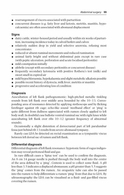
Abdominal surgery 99
• rearrangement of viscera associated with parturition
• concurrent diseases (e.g. fatty liver and ketosis, metritis, mastitis, hypo-calcaemia) are often associated with abomasal displacement
Signs
• dairy cattle, winter-housed period and usually within six weeks of parturi-tion, increasing incidence today in calved heifers and calves
• relatively sudden drop in yield and selective anorexia, refusing most concentrates
• reduced or absent ruminal movements and reduced rumination
• animal fairly bright and without abdominal pain except in rare case (with peptic ulceration, perforation and acute localised peritonitis)
• mild constipation initially
• afebrile (except with secondary peritonitis or concurrent disease)
• frequently secondary ketonuria with positive Rothera’s test (milk) andsweet smell in expired air
• mild hypochloraemia, hypokalaemia and slight metabolic alkalosis possible
• possible recent history of dystocia, milk fever, metritis or mastitis
• progressive and accelerating loss of condition
DiagnosisAuscultation of left flank pathognomonic: high-pitched metallic tinklingsounds from left flank over middle area bounded by ribs 10–13. Corres-ponding area of resonance detected by applying stethoscope and by flickingforefinger against rib cage; echo-like sound ‘steelband effect’ or ‘ping’ is quite different from dullness appreciated with rumen closely applied to leftbody wall. In doubtful case ballotte ventral ruminal sac with right knee whileauscultating left flank over ribs 10–12 (greater frequency of abnormalsounds).
Occasionally a slight distention of dorsocranial part of left paralumbarfossa just behind rib 13 results from severe abomasal tympany.
Rarely can LDA be detected on rectal examination as a tympanitic viscusbetween left dorsal sac of rumen and left flank.
Differential diagnosisDifferential diagnosis of left flank resonance: hypotonic form of vagus indiges-tion may reveal peritoneal fluid and gas
In rare difficult cases a ‘liptac test’ can be used to confirm the diagnosis. An 8 cm 14 gauge needle is pushed through the body wall into the centre of the area defined by a ‘ping’. Centesis is used to collect some fluid. A pH less than 3.5 indicates a displaced abomasum; a pH greater than 5.5 suggeststhe ‘ping’ comes from the rumen. An orogastric tube can also be passed into the rumen to help differentiate a rumen ‘ping’ from that due to LDA. Byultrasonography the LDA can be visualised as a fluid- and gas-filled viscuscovering the rumen.
BSAC03 3/5/05 4:27 PM Page 99

100 Chapter 3
Conservative techniqueIndicated in recent cases where economic factors (cost of surgery) are import-ant. Treatment is primarily replacement by rolling.
• cast cow on right side (Reuff ’s method, one person restraining head, twoon ropes)
• turn over on to left side and while turning ballotte ventral abdominal wallfirmly in an attempt to move abomasum into midline position, using kneeto push anti-clockwise
• alternatively cast cow on back in dorsal recumbency and move from 45° right lateral to 45° left lateral position, ‘shaking’ abomasum back intonormal position
• finally, in either rolling method, turn cow into left lateral recumbency andmaintain in this position for five to ten minutes to permit organ to evacu-ate excessive gas
• check absence of abomasum from left side on auscultation and finger percussion
• inject calcium and dextrose i.v.
• re-introduce concentrates slowly over one week period to cows correctedof LDA by rolling
• encourage maximal exercise in this period
Some cases of LDA corrected by rolling may at once or soon become cases ofRDA. Many others have a recurrence of LDA within 48 hours and requirerolling again or surgery. Success rate of rolling is about 20%.
Surgical technique (LDA)
Numerous methods of surgical correction have been described since the firstreports of LDA, around 1948, in Scotland.
Some options of sites and fixation techniques include:
• standing or recumbent surgery
• left or right, or bilateral flank incision
• paracostal, paramedian or midline incision
• fixation of abomasum to right flank or ventral body wall by intra-abdominalapproach
• left flank laparoscopic approach and right paramedian fixation (not discussed further)
• percutaneous fixation from abdominal cavity (Utrecht technique)
• midline percutaneous fixation through ‘blind’ suture, or (popular) toggletechnique
Advantages and disadvantages of various surgical sites are listed in Table 3.1. Personal preference varies enormously.
BSAC03 3/5/05 4:27 PM Page 100

Abdom
inal surgery101
Table 3.1 Surgical correction of left displaced abomasum: different methods.
Incision site
Anaesthesia
Ease of decompression
Ease of reposition
Area of abomasal fixation
Remarks
Personal preferences of authors(ADW)(GSJ)(AS)
Abbreviations (+) = usually spontaneous decompression and reposition following recumbencya = antepartum LDA; b = long standing LDA, c = low value cow
Right flank
paravertebral orlocal infiltration
+
++pylorus
relativelyunphysiologicalposition offixation,minimal stress
111
Left flank (Utrecht)
paravertebral orlocal infiltration
++
+fundus
technical problemsin inexperiencedhands;rumenotomy also possible
232a 3b
Right and left flank
paravertebral orlocal infiltration
++
++fundus or pylorus
2 surgeons;added timeand expense;rumenotomyalso possible
350
Midline/paramedian
sedation (xylazine +local infiltration)
(+)
(+)
fundus
possible restraintproblems; goodwound closureessential
523b
Paracostal
sedation (xylazine+ local infiltration
(+)
(+)
fundus
haemorrhage andwound healingproblems
660
Percutaneousfixation (toggle)
sedation(xylazine)
N/A
(+)
fundus
good, assistantneeded, fastcheap, but riskof misdirectedfixation (e.g.rumen)
444c
BSAC03 3
/5/05 4
:27 PM P
age 101

102 Chapter 3
Right flank approach
• paravertebral analgesia T13, L1 and L2
• skin preparation for aseptic surgery and drape
• paracostal skin incision 15–20 cm long starting 10 cm below tip of L2transverse process and 4 cm behind last rib (see Figure 3.13)
• entry into abdomen as previously described (see Section 3.2) with bluntseparation of muscle layers and incision of transverse fascia and peri-toneum, extended to permit comfortable entry of one hand (13 cm)
• make thorough manual exploration of abdominal cavity (see Figure 3.8);then insert the left hand, passing it behind the caudal edge of the greateromentum and over to the left side of the abdominal cavity, directed initi-ally towards the lower part of the wing of the ileum, ventral to descendingcolon and mesocolon and the left perirenal fat mass
• appreciate the displaced abomasum interposed between a relatively smallrumen and the left body wall (abomasum may extend two-thirds or oftenfurther up the left flank)
• check the palpable area of the abomasum for any roughness (peritonitis)and adhesions to the left flank. Peritonitis is probably due to a perforatedpeptic ulcer. It may prove difficult to reposition the abomasum, either bypushing it down or pulling on the greater omentum from the right side,before evacuating most abomasal gas
3
4
2
1
Figure 3.13 Diagram of right flank of cow to show site of laparotomy incision for rightflank abomasopexy.1. rib 13; 2. abomasal fundus and body; 3. flank incision; 4. abomasopexy site (x)sutured to body wall.
BSAC03 3/5/05 4:27 PM Page 102
Abdominal surgery 103
• decompress the abomasum by inserting wide bore needle (2.1 metric,approximately 2 mm internal diameter), 45° to abomasal wall firmlyattached to plastic tubing, into the dorsocaudal part of the abomasumwith the free end of the tubing (length approximately 2 m) held outside theanimal and evacuate air
• maintain needle firmly in place while gently pushing the viscus ventrallywith palm of hand
• anticipate that gas will be evacuated over five minutes (volume 5–15 litres)until the abomasal wall is relatively flaccid and almost out of reach, lowdown on the left side
• remove needle and tubing, carefully avoiding contamination of abdominalcavity by abomasal fluid in tubing
• place left hand onto dorsal aspect of abomasum and push it down towardsmidline
• pass left hand along right body wall with palm against rib cage to midline,midway between xiphisternum and umbilicus
• turn hand through 180° in clockwise direction, and grasp the greateromentum and abomasum now lying in midline
• hold this mass firmly and pull upwards, if necessary in a series of gentlejerking movements, and identify abomasum through right flank wound
• identify the abomasal pyloric region visually: pale pink-grey and welldelineated from adjacent insertion of greater omentum, it is firm and cannot be easily traumatised by the fingers
• if not visible through flank incision repeat midline grasping process; abomasum is easily drawn into right flank using this technique
• release abomasum and greater omentum temporarily for confirmatorymanual check of left side for absence of abomasum
• check remainder of cranial abdominal cavity (liver, reticulum, diaphragm)by rapid manual exploration
• suture greater omentum to parietal peritoneum and transversalis musclein closure of flank
• pull suture tight and tie off, ensuring that no small intestine inadvertentlybecomes trapped. Place second suture about 3 cm dorsal to the first
• alternatively suture lateral pyloric wall of abomasum and several centimetres of adjacent omentum to body wall. Needle should be about4.5 cm long, semi-curved and round-bodied, threaded with 5 metricmonofilament nylon
• close abdominal incision in routine manner (see Section 3.3, pp. 86–87)
• give systemic antibiotic therapy for one to three days.
Improvement is usually seen after two to three days with an increasedappetite for concentrate. Prognosis in uncomplicated cases is excellent(95%).
BSAC03 3/5/05 4:27 PM Page 103

104 Chapter 3
ComplicationsPost-operative complications include:
• peritonitis due to non-sterile technique or leakage of abomasal fluid causing peritonitis or abscessation in the flank wound
• recurrence resulting from breakage of the abomasal flank pexy site suturebefore development of adhesion formation
• wound dehiscence, usually ventrally
• functional stenosis possible after tacking pyloric part of abomasum insteadof greater omentum
Left flank approachThought not widely practised in the UK and North America, this (Utrecht) technique deserves greater recognition for its simplicity.
• make left paracostal incision as for exploratory laparotomy (incision 3,Fig. 3.6)
• explore abdominal cavity and identity LDA
• evacuate gas from abomasum with needle as previously described, or bysimple pressure (some surgeons prefer to place suture first, then evacuategas)
• thread 1.5 m length of non-absorbable suture material (polyamide polymer 4 gauge) onto straight triangular 8 cm needle, and insert needlethrough greater omentum and wall, but not lumen of abomasal body inseries of five to six continuous sutures, each 3 cm apart (Ford interlocking)(see Figure 3.14)
• push semi-flaccid abomasum down towards midline
• insert needle through body wall slightly right of the abdominal midline,midway between xiphisternum and umbilicus (site indicated by assistant’sfinger pressure on skin)
• ensure needle is pulled through skin to exterior by assistant, and that end of suture material is retained in artery forceps dangling beneath theabdominal wall
• with second needle threaded from distal end of same suture material pushneedle through ventral body wall 3–10 cm caudal to first point; assistantcan again indicate correct site
• assistant then ties suture on skin of ventral abdominal wall, (placing roll of bandage below suture) while surgeon ensures that abomasum is heldfirmly against ventral parietal peritoneum so preventing interposition ofgreater omentum or jejunal loops
• close abdominal flank in routine manner and give systemic antibiotics forone to three days
• remove abomaso-omental skin sutures with scissors after two weeks,when firm adhesion of greater omentum and abomasum to body wall hasformed
BSAC03 3/5/05 4:27 PM Page 104

Abdominal surgery 105
Midline approachReposition and fixation present few difficulties. Surgery may be performedunder sedation (xylazine, i.v. or i.m.) and local analgesia, or under GA. Animalshould be supported by straw bales laterally, and hind legs should be ropedaway from operative site. A stockman, restraining head, keeps forelegs awayfrom surgical field, operator’s head and hands.
• incision is midway between umbilicus and xiphisternum approximately15 cm long if visual exploration is required, or 11 cm if manipulation anddeep sutures are to be inserted by palpation alone
• incise skin, subcutaneous fat and linea alba (approximately 7 mm thick)with scalpel (see Figure 3.15)
• note small amount of subperitoneal fat overlying peritoneum, which isincised with scalpel and lengthened with straight scissors
• explore abdomen thoroughly with sweeping movement
• pass hand to left side to identify displaced abomasum or (if reposition hasalready occurred, which is likely) ventral sac of rumen
• follow greater omentum from insertion onto left longitudinal groove ofrumen over towards midline to identify body of abomasum
• briefly exteriorise abomasal body for identification
Figure 3.14 Abomasopexy from the left side using Utrecht method. Continuoussuture has been placed through greater omentum at insertion onto abomasum in leftflank. Suture is then pushed through ventral body wall just right of midline using a longstraight needle.
BSAC03 3/5/05 4:27 PM Page 105
106 Chapter 3
• suture abomasum to peritoneum and vental abdominal musculatureenclosing with each suture about 2 cm width of abomasal body (usingpolyamide polymer 4 metric or PDS)
• ensure that sutures do not penetrate lumen (danger of fistula or tearingout with risk of fatal peritonitis) but include serosa and muscularis
• continuous peritoneal suture, carefully tightening whole length beforetying off
• suture linea alba (most important supporting layer of abdominal wall) withsimple interrupted sutures of PGA 5 or 6 metric on semi-curved cuttingedge needle 5 cm long
• bury this layer with continuous layer of subcutaneous sutures of PGA 4 metric
• suture skin in horizontal everted mattress pattern with monofilament nylon
• systemic antibiotics for one to three days
• remove skin sutures after ten days.
Percutaneous fixation (toggle or bar suture)A rapid and simple technique of abomasal fixation avoiding the need forexpensive laparotomy which has found widespread acceptance over the lasttwenty years. Following diagnosis of an uncomplicated LDA the steps are:
• clip and disinfect the ventral abdomen to right of midline between xiphoidand umbilicus, marking position of right abdominal vein
• give 45–50 mg xylazine i.v. and cast cow (e.g. by Reuff ’s method) on rightside
• turn quickly into dorsal recumbency and extend and tie hind legs, placestraw bales against flanks for stability
9
8
75
4
6
1
2
3
Figure 3.15 Diagrammatic cross-section of ventral midline of abdominal wall.1. skin; 2 linea alba; 3. peritoneum; 4. external sheath of rectus muscle; 5. rectusmuscle; 6. internal sheath of rectus muscle; 7. external oblique muscle; 8. internaloblique muscle; 9. transverse muscle. (From Cox, 1981.)
BSAC03 3/5/05 4:27 PM Page 106
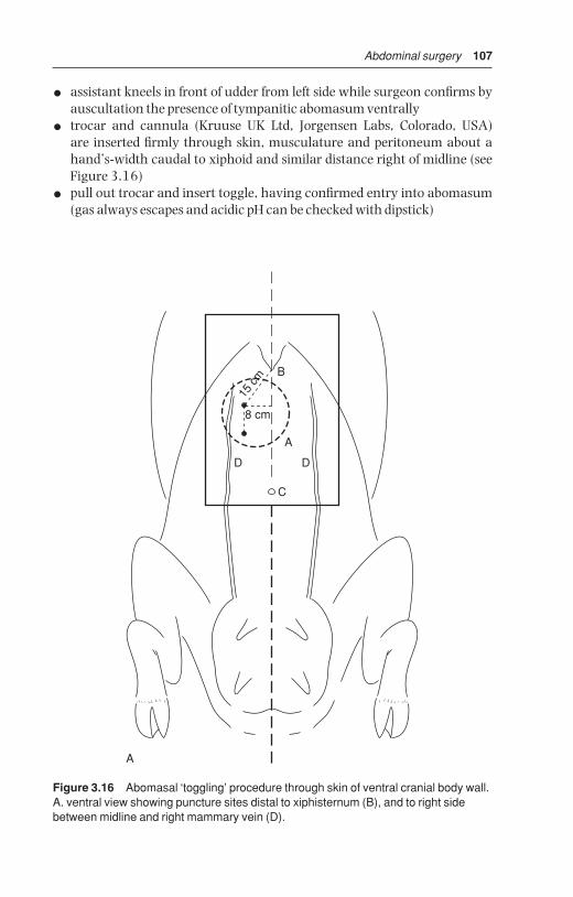
Abdominal surgery 107
• assistant kneels in front of udder from left side while surgeon confirms byauscultation the presence of tympanitic abomasum ventrally
• trocar and cannula (Kruuse UK Ltd, Jorgensen Labs, Colorado, USA) are inserted firmly through skin, musculature and peritoneum about ahand’s-width caudal to xiphoid and similar distance right of midline (seeFigure 3.16)
• pull out trocar and insert toggle, having confirmed entry into abomasum(gas always escapes and acidic pH can be checked with dipstick)
8 cm
15 c
m B
A
DD
C
A
Figure 3.16 Abomasal ‘toggling’ procedure through skin of ventral cranial body wall.A. ventral view showing puncture sites distal to xiphisternum (B), and to right sidebetween midline and right mammary vein (D).
BSAC03 3/5/05 4:27 PM Page 107

108 Chapter 3
B
C
D
Figure 3.16 (cont’d ) B. puncture by trocar and canula with first toggle in place; C. removal of trocar and insertion of toggle through canula; D. tying together ofsutures from each toggle with space for insertion of two fingers.
BSAC03 3/5/05 4:27 PM Page 108

Abdominal surgery 109
• assistant holds toggle suture with forceps
• remove cannula and rapidly repeat trocarisation and toggle insertion twofingers’-width caudal or cranial to first site
• permit most gas to escape
• tie two sutures together allowing several centimetres of play (see Figure3.16d)
• turn cow onto sternum slowly and allow to stand
• prophylactic systemic antibiotic cover
• post-operative visit about two to five days later to check abomasal positionand any concurrent disease problems.
This percutaneous procedure requires two assistants and must be performedquickly as the abomasum can rapidly deflate once the cow is on her back. If unsuccessful, a standing surgery can be done. The major hazards, apartfrom personal injury from poor restraint, are the risk of puncturing rumen,fixation of abomasum too close to the pylorus and tearing of the abomasalwall by the suture, all potentially resulting in peritonitis.
The easiest cases tend to be cows with obvious ‘pings’ in the left flank at first clinical examination, since abomasum remains gas-filled relat-ively longer. The technique clearly does not permit examination of otherabdominal viscera for concurrent disease (e.g. chronic adhesions, hepaticabscessation).
Discussion of surgical techniquesSome surgical problems and their proposed solutions are:
• inability to locate abomasum – if encountered during right flank approachit may be necessary to perform left flank incision, especially in case of inexperienced surgeon
• inability to break down left flank adhesions of abomasum to body wall – adhesions may be out of reach in right flank approach, again necessitat-ing left flank laparotomy, or they may be too firm to break down due to their longstanding and extensive nature. Beware of breaking down recentadhesions which may result in exposure of abomasal lumen through aperforated ulcer. Such complications are rare (< 5%)
• inability to replace abomasum from right flank approach – as long as midline structures are firmly grasped in palm of hand, avoiding risk ofpuncturing abomasum or of tearing omentum with finger tips, repeatedgentle traction is harmless. Check that hand is grasping structures about20 cm caudal to xiphisternum
• inability to identify pyloric region – identify caudal portion of abomasalwall adjacent to greater omentum, then pass hand dorsally, and eventu-ally area can be identified where abomasum is about 3 cm wide and where
BSAC03 3/5/05 4:27 PM Page 109

110 Chapter 3
small lobes of greater and lesser omental fat overlap the abomasal border.Abomasum is thicker and denser at this point (pyloric antrum), which isthe pylorus, located below rib 10, about half-way down body wall
3.7 Right dilatation, displacement and volvulus ofabomasum
Right dilatation and displacement of abomasum probably have commonaetiology with LDA. Incidence is lower and RDA is not so closely related to early post-partum period. RDA can result from rolling of LDA cases, inwhich dilated organ is displaced a variable distance into the right flank. Insome cases a cow may apparently have alternately LDA and RDA, the organ‘swinging’ from one flank to the other over a period of several days. Rightdilatation occasionally occurs in younger cattle and in steers.
Signs and diagnosis of RDA
• selective anorexia and gradual weight loss
• distension of low and mid right flank where auscultation reveals highpitched splashing and sometimes ringing sounds
• RDA case usually has relatively more fluid than LDA animal
• ballottement of low right flank causes loud splashing sounds but rarelyany pain
• cows may show profuse watery diarrhoea
• rectal examination may reveal a smooth distended viscus on the right side cranial to pubic brim, though only the caudal and dorsal surfaces canbe reached. At any time an RDA can develop into a right-sided volvuluswith or without severe compromise of the local vasculature. Few cases of RDA rapidly develop volvulus but, once it occurs, condition is acute and prognosis becomes guarded within some hours. Surgery is then anemergency.
Animals with RDA may make spontaneous recovery but many become progressively duller, and some develop bradycardia. Prognosis is guarded.
Conservative treatment of RDATurn animal out to grass or into yard for increased exercise and maintainaccess to bulky fodder as high priority. Some cases respond well to generalsymptomatic treatment, others to metaclopromide. Recently calved cows maybe given calcium borogluconate both i.v. and s.c.. Non-responsive cases ofRDA remain at risk of developing abomasal volvulus. Many cases of volvulushave a retrospectively elicitable history of chronic abomasal disease. Performsurgery in valuable animals as soon as possible, i.e. before volvulus occurs.
BSAC03 3/5/05 4:27 PM Page 110

Abdominal surgery 111
Signs of abomasal volvulus
• abdominal emergency: cow is in shock, collapsed, with tachycardia, mildto severe cyanosis
• weakness, possible recumbency depending on chronicity, severe abdominaldistension, especially on right side, with loud splashing sounds on ballotte-ment of right flank
• rectal examination reveals large, smooth and tense-walled viscus ven-trally on right side
• (optional) abdominocentesis may reveal large volume of reddish-brownfluid
• severe hypochloraemia and hypokalaemia, increased PCV, increased totalprotein
• metabolic alkalosis due to abomasal sequestration of acidic gastric secre-tion, later possibly metabolic acidosis
• abomasum may contain up to 50 litres of fluid
• volvulus causes severe and potentially fatal impairment of venous drainageof abomasum, and the wall becomes ischaemic, dark red, then blue andblack, at which stage rupture is likely
Differential diagnoses include caecal dilatation and dislocation, small intestinal obstruction following volvulus or invagination, functional abomasalobstruction.
In most cases mechanical movements initially involve dorsal displacementof the greater curvature followed by counter-clockwise 180°–360° torsion ofthe abomasum (see Figure 3.17). Postmortem (PM) examination often fails toreveal exactly what displacement was the primary movement. Pathologyreveals severe constriction of both venous and arterial supply at the junctionof omasum and abomasum, as well as between reticulum and omasum. Thedescending duodenum is completely occluded as a result of severe stenosisfollowing external pressure and a degree of displacement. Direction has beendescribed as clockwise or counter-clockwise when viewed from the right side.When viewed from the rear, it has a ‘spiral corkscrew-like’ counter-clockwisemovement.
Surgical treatment of RDA and abomasal volvulus
• correct right abomasal dilatation by right flank surgery after giving fluids and electrolytes (see Table 1.11) e.g. i.v. infusion of 10–50 litres0.9% NaCl with dextrose or Ringer lactate to any severely deficient cases.Early case of volvulus has metabolic alkalosis, late stage is acidotic.
• make right flank paracostal incision, starting 10 cm below lumbar trans-verse processes
• explore abdomen to determine form of displacement and any other complications
BSAC03 3/5/05 4:27 PM Page 111

112 Chapter 3
C
A
B
Figure 3.17 Three forms of right abomasal displacement and torsion and theirmanipulative correction. (From Dirksen, Gründer & Stöber, 2002.)A. left (anti-clockwise from rear view) 360° torsion and direction of manual correction;B. left 180° torsion, and correction; C. simple right-sided abomasal displacement(possibly with up to 90° rotation). (From Dirksen, Gründer & Stöber, 2002.)
BSAC03 3/5/05 4:27 PM Page 112

Abdominal surgery 113
• decompress gas from abomasum. If necessary, especially in case of volvulus, fluid can be removed from the abomasum by first placing a 4–7 cm diameter circular purse string dorsally in the serosal surface of the abomasum
• make a stab incision in the centre of the purse string and insert a stomachtube quickly in the abomasum while the purse string is tied
• tighten purse-string suture which may be oversewn by one or two mattresssutures
• examine evacuated organ carefully for signs of abomasitis (diffuse hyper-aemia) and ulceration (thickened areas, superficial fibrin deposition)
• locate pylorus for orientation
• correct displacement by pushing greater curvature cranially and ven-trally with flat palm of hand or with forearm
• confirm that position of pyloric region and descending duodenum are normal (fundus, pyloric region, pylorus and proximal duodenum all againstright abdominal wall)
• perform pyloromyotomy (optional and of controversial benefit) by lon-gitudinal 3–4 cm incision through muscular coats of the pylorus (about 1 cm deep). If lumen is inadvertently entered, several chromic catgutsutures should be placed to bury this incision
• suture greater omentum (caudal to pylorus) to body wall (see p. 103).
Surgery of abomasal volvulus may not be indicated in cows with long-standing abomasal volvulus that are unable to rise and remain standing.Such animals should be sent to emergency slaughter on humane grounds.They often fail to pass meat inspection as the carcass may not set as the animal is usually toxic. If surgery is attempted in advanced cases, fluidtherapy with i.v. lactated ringer or isotonic bicarbonate is essential beforeand during surgery (see Table 1.11), due to massive abomasal sequestrationof Cl− , K+ and H+ ions and subsequent metabolic acidosis. The prognosis isguarded or good in cases with a heart rate < 90/min, serum urea < 10 mmol/lor serum Cl− > 85 mmol/l. Cows with abomasal volvulus of chronic naturewith heart rate > 120/min and 12% dehydration have a poor prognosis for survival. The sooner abomasal volvulus is diagnosed and surgically corrected, the better the prognosis.
Surgery may be reserved for good risk standing cases and cattle where systemic antibiotic treatment has already rendered the carcass valueless for human consumption. Following the operation 20 litres of warm watercontaining 60 g NaCl and 30 g KCl should be given twice daily by stomachtube. It appears that abomasal torsion (up to 180°) must be distinguishedfrom abomasal volvulus (180°–360°). The former has in the USA a guardedprognosis, when serum Cl remains above 85 mmol/l. The latter is the formusually experienced in the UK and affected cows are often recumbent at first clinical examination (see Figure 3.17).
BSAC03 3/5/05 4:27 PM Page 113
114 Chapter 3
3.8 Other abomasal conditions
Other surgical conditions of the abomasum include:
• impaction
• abomasal tympany and volvulus in calves
• ulceration (with or without perforation)
Abomasal impaction
IntroductionImpaction may be primary, as in calves following incorrect feeding withexcessive roughage and an inadequate water intake. Impaction may also besecondary to surgical correction of abomasal volvulus, or to lymphosarcomaand adhesion formation in the cranial part of the abdominal cavity.
Treatment
• perform exploratory surgery through a right midflank incision
• break down abomasal contents manually
• give paraffin oil (5 litres, with 20 litres of water) by an orogastric tubeonce, or repeated 24 hours later
Some cases may slowly resolve. Abomasotomy through a right paramedianincision may be necessary in advanced cases.
Abomasal tympany and volvulus in calves
SignsSeen in calves typically at six weeks to three months shortly after bucket milkfeeding, signs include obvious discomfort with head extended, mild colickysigns and right flank tympany, later depression and spread of tympany toinvolve the left flank. Systemic signs rapidly become severe and calves can diewithin a few hours.
Differential diagnosis: rumen tympany (unlikely in this age group), caecaldilatation and dislocation, jejunal volvulus or intussusception
TreatmentRecumbent calves with circulatory collapse should generally be euthanised.Less severe cases:
• i.v. fluids, right paracostal or paramedian incision under local anaesthesia,in left lateral or dorsal recumbency
• exteriorise abomasum, evacuate contents by slow release of gas (tympany)or fluid
• replace organ after correction of displacement
BSAC03 3/5/05 4:27 PM Page 114

Abdominal surgery 115
Abomasal ulceration
IntroductionCommon condition in slaughterhouse statistics, both in calves and adults.Younger cows tend to be affected within four weeks of parturition. Abomasalulcers rarely cause significant clinical disease.
• type 1: non-perforating, minimal bleeding
• type 2: ulcer causing severe blood loss
• type 3: perforating ulcer with acute localised peritonitis
• type 4: perforating ulcer with diffuse peritonitis
Type 1 is often subclinical, while types 2 and 4 usually show clinical signs and may be fatal. In mature cows concurrent disease is often present such as mastitis, metritis and pneumonia. Abomasal ulceration tends to occur inrecently calved cows (corresponding to LDA incidence) and is related to stresse.g. overcrowded yards and cubicle houses.
Signs
• abdominal pain, which is not only associated with peritonitis
• calves show abomasal dilation with ulceration (painful palpation)
• melaena and pale mucous membranes (type 2)
• pyrexia and tachycardia in cases with peritonitis
• abdominocentesis (see Section 3.16, p. 130) useful to confirm peritonitise.g. acute fatal cases involving ruptured omental abscessation
Treatment
• type 2 ulcers require whole blood transfusion if PCV drops below 15 vols%
• treatment of peritonitis requires systemic antibiotics
• i.v. fluids
• exploration in left flank LDA: correction of left abomasal adhesions, rarelyis surgical resection of ulcer necessary
• correction of primary disorder
• pyloromyotomy may improve prognosis in calves with abomasal dilationand ulceration
3.9 Caecal dilatation and dislocation
Definition and anatomy (see Figures 3.3 and 3.5)The term dislocation refers to any twist, torsion, volvulus or retroflexion ofthe caecum which has a blunted round apex projecting caudally from theomental recess. Position varies with the volume of contents, floating dorsallyif gas-filled or sinking with ample fluid contents. Normal caecum cannot beidentified on rectal palpation.
BSAC03 3/5/05 4:27 PM Page 115

116 Chapter 3
Incidence and signs
• usually seen in adult dairy cows > 4 years old with signs rather similar toRDA for caecal dilatation and ressembling abomasal volvulus in caecaldislocation
• causes not understood and may involve hypocalcaemia and inhibitoryeffect of high volatile fatty acid concentrations in caecum on caecal motility
• selective anorexia (refusing concentrates) in housed or pastured cow,more frequently seen during production phase than in early lactation
• vague abdominal discomfort and pain, mild in caecal dilatation, severe incaecal dislocation
• some distension of right caudal abdominal cavity, and ballottement ofupper right flank (more dorsal and caudal than in RDA and abomasalvolvulus) produces sounds of fluid and gas
• faeces reduced in volume, dark, possibly covered with mucus
• rectal examination reveals little or no faeces and a mass like end of ‘cob’loaf of bread 15–20 cm diameter in dilatation (see Figure 3.18a,b)
• position is relatively high on right side (dilatation), or as a distended organ(retroflexed caecal body) cranially in right ventral quadrant, or painfulpalpation of ileocaecal ligament (dislocation)
• dislocation of caecum produces more severe systemic signs, but diseaseprogress is slower than in RDA and abomasal volvulus
Figure 3.18(a) Caecal torsion and retroflexion: diagrams of rectal findings.Longitudinal section through abdomen: apex of caecum and tense ileocaecal fold arepalpable in caecal torsion.
BSAC03 3/5/05 4:27 PM Page 116

Abdominal surgery 117
• haematology and biochemical parameters usually normal in caecal dilata-tion; in caecal dislocation blood biochemistry is also normal or occasionallyshows metabolic alkalosis with ↓Cl− and ↓K+ caused by intestinal stasis
• differential diagnoses: RDA, abomasal volvulus, intestinal torsion aroundmesentery
Confirmation of diagnosis depends on right flank laparotomy.
TreatmentMedical management may be indicated in some cows with dilatation.Surgical approach:
• site of incision upper right flank, and relatively more caudal than for RDA
• exteriorise caecum and examine carefully for necrosis and commencinggangrene
5
4
2
1
3
Figure 3.18(b) Transverse section through abdomen: (1) dilated body of caecum,(2) distended loops of spiral colon, and (3) sometimes also small intestine arepalpable in caecal retroflexion; (4) rumen and (5) left kidney
BSAC03 3/5/05 4:27 PM Page 117

118 Chapter 3
• correct dislocation after drainage at apex (typhlotomy, purse-string sutureplaced before insertion of scalpel blade or wide-bore needle)
• massage firmer material out from intra-abdominal portion of caecum andproximal loop of ascending colon
• inspect the caecal apex for ischaemia: the easiest surgical technique is toinvert such areas into caecal lumen and to oversew the area
• peri- and post-surgical systemic antibiotics, possibly i.v. or oral fluids
PrognosisPrognosis is good (dilatation) or guarded (dislocation) and recurrence rate is up to 20%. Typhlectomy (partial caecectomy) is rarely indicated but relatively simple to perform. Cow copes well with caecum of reduced capacity.
3.10 Intestinal intussusception
IntroductionIntussusception (invagination or telescoping of bowel) occasionally affectssmall intestine ( jejunum, ileum). Predisposing causes are unknown but thecondition is not always associated with hyperperistalsis, enteritis or diarrhoea.Any age group, from calves to older cows, may be affected, but calves appearpredisposed. Complete bowel obstruction results. Sometimes a double intus-susception (five layers of bowel wall superimposed on each side) develops.
Signs
• sudden onset of acute abdominal pain
• groaning, kicking at belly, alternately lying and standing, paddling of hindlegs
• within 12 hours acute signs are succeeded by dullness, anorexia, pre-cipitous drop in milk yield (as in traumatic reticulitis)
• initial tachycardia (heart rate 120/min) disappears, while melaena maybe replaced by total absence of faeces
• stance may be persistently abnormal after 24 hours, animal adoptingrocking-horse position, or may lie down and groan
• site is usually distal jejunum, or rarely jejunoileal junction with invagin-ated section (intussusceptum) passing into caecum or into proximal colon
Condition may last five to eight days, with slow deterioration and death frommetabolic effects of total obstruction in which plasma chloride progressivelyfalls, with haemoconcentration and dehydration.
DiagnosisDiagnosis depends on rectal palpation of several distended small bowel loops, about 5 cm diameter, or possibly a firm, painful and slightly mobile,fist-shaped mass relatively low on right side or just cranial to pelvic inlet. In
BSAC03 3/5/05 4:27 PM Page 118
Abdominal surgery 119
calf bilateral abdominal palpation may suggest a firm irregular abdominalmass. Mass may be out of reach and diagnosis then depends on right-sidedexploratory laparotomy. However, severely distended small bowel on rectalpalpation or on ultrasound may be a case of small intestinal ileus.
Differential diagnosis: intestinal volvulus, mesenteric torsion. Both haverapid clinical course, and distention of lower right flank with splashing soundson auscultation.
TreatmentIt is alleged that some cases recover spontaneously after sloughing the intus-susceptum, but this has never been seen by the authors.
• i.v. fluids e.g. hypertonic or isotonic saline
• right flank laparotomy under paravertebral analgesia (see Section 1.8, p. 23) and topical analgesia of affected bowel segment and mesentery, pos-sibly also systemic analgesic; alternatively under GA
• standing surgery preferable except in calf < 12 months
• exteriorise affected bowel, and attempt manual reduction in early case
• in case of intestinal wall devitalisation, and where manual reduction isimpossible, isolate with clamps, resect bowel segment and mesentery andperform end-to-end or less optimal side-to-side anastomosis
• note that traction on mesenteric root is very painful and may cause animal to collapse, therefore first infiltrate lignocaine into affected segmentof mesentery
• suture technique should avoid production of bowel stenosis: single layersof Cushing pattern, interrupted once, is suitable. Some surgeons mayoversew with Lembert or Cushing suture (PDS 2 metric with swaged-onneedle)
• manage differing pre- and post-stenotic luminal diameters by 45–60°angled incision line in (smaller) post-stenotic bowel
• initial single sutures in the anti-mesenteric and mesenteric borders facilit-ate manipulation
• closure of mesenteric defect is essential to prevent possible herniation
• preferred material is fine absorbable suture (e.g. 7 metric Softgut® (Davis& Geck) ) on 45 mm 3/8 circle round-bodied needle or 3.5 metric PGA
• leakage at suture line is rarely a problem
• maintain strictly aseptic procedure, otherwise massive local peritonitismay result in multiple bowel adhesions
• wash visceral peritoneal surface of bowel with copious volume of sterilephysiological saline before closing the abdominal incision
• aftercare should comprise antimicrobials for five to seven days, hay andlaxative feeds (e.g. bran) for one week
Successful surgery is evident in passage of loose dark faeces 24–48 hourslater.
BSAC03 3/5/05 4:27 PM Page 119

120 Chapter 3
Caecal invagination is a specific entity seen predominantly in youngercalves. Often reducible manually, they often recur, so amputation is pre-ferable (p. 118).
3.11 Other forms of intestinal obstruction
Intestinal obstruction is occasionally caused by other abdominal abnormalitiesincluding:
• large pedunculated fatty masses (lipomata)
• large intra-mesenteric areas of fat necrosis
• adventitious fibrous bands
• scrotal hernia, volvulus and incarceration in traumatised vas deferens
Incarceration in traumatised vas deferens (‘gut-tie’)
IntroductionObstruction, rarely strangulation, may follow passage of bowel through tearof peritoneum between vas deferens and abdominal wall, following castra-tion procedure in which excessive traction on, and recoil of, spermatic cordresults in adhesion of cord or peritoneal fold around bowel (‘gut-tie’). Jejunumis usually involved and condition develops slowly with signs related to gradual lumenal occlusion (see Figure 3.19).
Signs
• anorexia, dullness, reduced passage of faeces and distention of flank
• ballottement of flanks and palpation inconclusive
• rectal palpation often permits easy diagnosis by recognition of distendedsmall intestine and one or more abnormal cord-like structures near inguinalring (see Figure 3.19)
• diagnosis depends on exploratory laparotomy following suggestive historyand signs
• problem is usually located near internal inguinal ring and (due to anatom-ical distribution of small intestine) on right side
Treatment
• gentle rectal traction on adhesed spermatic cord stump is fairly simple to perform and may lead to rapid recovery (90% success) but can be hazardous and not recommended for inexperienced persons who shouldappreciate the risk of tearing the bowel wall
• perform right flank laparotomy in standing or left laterally recumbent animal
• section or resect the adhesed spermatic cord or vas deferens with scissorsusing blind palpation
BSAC03 3/5/05 4:27 PM Page 120

Abdominal surgery 121
• exteriorise bowel and check for viability
• resection and anastomosis required in exceptional cases
• routine closure of flank laparotomy incision (see Section 3.3 pp. 86–87).
• post-operative systemic antibiotics for three days, especially if bowel resec-tion was performed
3.12 Peritonitis
IntroductionPeritonitis is usually secondary to a diffuse or localised primary condition.Causes may be perforation of an abomasal ulcer, rupture of a reticular wall orhepatic abscess, reticular perforation of a foreign body, infection followinguterine rupture, or the introduction of infection at caesarean section. Somecases follow a breakdown of surgical asepsis.
AB
Figure 3.19 ‘Gut-tie’ involving recoiled stump of ductus (vas) deferens adhesingaround and occluding lumen of small intestine, resulting in bowel obstruction(compare with Figure 1.11).A. abnormal stump of ductus (vas) deferens; B. normal position of ductus (vas)deferens.
BSAC03 3/5/05 4:27 PM Page 121
122 Chapter 3
Signs and diagnosisEarly cases have signs of diffuse abdominal pain and may grunt spontane-ously. The acute reaction causes pyrexia (to 40.5°C), tachycardia, arched back, anorexia and reduced ruminoreticular activity. Chronic cases, in which peritoneal exudate has organised to form extensive adhesions, are generallycharacterised by chronic weight loss and unthriftiness.
Diagnosis is usually easy, based on history, classical signs, rectal palpation(adhesions possible) and in doubtful cases abdominocentesis (see Figure 3.23– possibly retain sample for antibiotic sensitivity testing).
TreatmentSpecific problems arise in treatment of peritonitis, which involves infection in a transcellular space where antibiotics cannot reach the concentrationsfound in tissues and serum. Generalised purulent peritonitis has a hopelessprognosis, and the animal should be euthanised.
Apart from appropriate antibiotics (see below), treatment in early acutecases may include:
• supportive i.v. fluids
• flunixin meglumine or corticosteroids to counter endotoxic shock and stabilise membranes
• heparin 5000 units, i.m., b.i.d. for three days in early cases
• intermittent peritoneal lavage (5–10 litres, t.i.d. or q.i.d.) with lactatedRinger solution (dorsal flank entry port, ventral midline exit). Peritoneallavage is not very effective in cattle due to blockage by fibrin clots.
Drugs of choice include:
• tetracyclines, e.g. oxytetracycline (22 mg/kg daily)
• sulphonamides (sulphadimidine, sulphadiazine, or triple sulphas).
See Section 1.12, p. 47, Table 1.15 for extra-label drug use and withdrawaltimes.
3.13 Umbilical hernia and abscess
IntroductionUmbilical herniation is a very common surgical condition in the Holstein and other cattle breeds. It may be inherited by a dominant character withincomplete penetrance, or be conditioned by environmental factors. It isunlikely to be sex linked.
Complication of herniation is frequently coexistent with umbilical abscessa-tion. Sometimes abscess may be involved in primary aetiology. Surgery maybe contra-indicated in large simple (i.e. non-infected) umbilical herniae ofanimals intended as breeding stock (e.g. bulls), because evidence suggests the condition is inherited (see Section 1.16, pp. 52–53).
BSAC03 3/5/05 4:27 PM Page 122

Abdominal surgery 123
SignsAn exceptionally large umbilical hernia is evident at birth, but the majorityare first noticed a few weeks later. The peritoneum-lined hernial sac maymeasure 3–12 cm diameter. Corresponding hernial ring is typically about1–7 cm long and 1–3 cm wide.
Contents of hernial sac are peritoneal fluid, greater omentum, and in largercases, the abomasum. Small or large intestine is sometimes involved.
DiagnosisDiagnosis is based on the history, palpation of the swelling and the adjacentventral abdominal wall for signs of intra-abdominal involvement. Pain onpalpation is suspicious of a septic process (see Figure 3.20) or incarceration.Ultrasound investigation may be considered to define the contents of anumbilical swelling and intra-abdominal involvement.
Indications for surgeryCalves under four weeks old should only be operated as an emergency, sincestress of anaesthesia may be critical to survival, and abscess capsule may notbe thick enough for safe resection.
Otherwise, indications include:
• irreducible (incarcerated or strangulated) hernial contents irrespective ofage, size and intended use of calf
• increasing hernial size
D
A
B E
C
Figure 3.20 Three sites of umbilical infection with intra-abdominal involvement.A. umbilical vein (purulent omphalophlebitis); B. umbilical artery (purulentomphaloarteritis); C. urachus (purulent urachitis); D. liver; E. bladder.
BSAC03 3/5/05 4:27 PM Page 123

124 Chapter 3
• calves aged three to six months with hernia still present, possibly enlarg-ing, since spontaneous resolution is now unlikely
Pre-operative complications:
• an encapsulated abscess makes surgery more awkward and potentiallyhazardous, but contamination of wound is not inevitable
• treat discharging umbilical sinus by repeated local irrigation and débride-ment, and possibly systemic antibiotics for three to five days. If still notresolved, discharging umbilical abscess cavity should be packed withswabs and overlying skin tightly sutured before starting surgery
Surgical technique
• reduce ruminal volume in older calves by 24 hours’ starvation to reducepost-op pressure on surgical wound, also to reduce degree of ruminal tympany during surgery, both during GA and LA
• obtain deep sedation and perform local analgesic infiltration of area with or without anterior epidural analgesia, or use GA (see Section 1.9,pp. 36–39)
• place calf in dorsal recumbency, preferably raised from ground, and positioned with straw bales laterally, and with legs fixed cranially andcaudally remote from surgical field
• in male calf irrigate and pack preputial cavity
• clip extensive area of ventral abdominal wall, scrub and disinfect threetimes
• ensure availability of sterile prosthetic mesh material for hernias wherering exceeds 10 cm diameter (see below)
• make elliptical skin incision around hernial base, continue along midlinewell cranial and caudal to limits of hernial ring
• dissect subcutaneous tissue bluntly to expose the hernial sac
• continue blunt dissection down to reveal edge of hernial ring, carefullyincise hernial sac at junction of body wall and sac and insert finger intoabdominal cavity
• resect sac, and circumferentially a thin strip of the hernial ring, as long asthere are no adhesions of gastro-intestinal organs to parietal peritoneumand umbilicus
• attempt gently to break down any adhesions between hernial sac andabdominal contents. If sac cannot be incised without damaging contents,incise longitudinally through linea alba cranial to ring and remove sactogether with herniated viscera after any necessary enterectomy
• bring together longitudinal edges of opening for suturing
• avoid undue tension by lengthening the ring cranially and caudally, converting the oval shape into a long ellipse
• if ring closure is likely to result in excessive suture tension, make longitudinal incision through external sheath of the rectus sheath, about
BSAC03 3/5/05 4:27 PM Page 124
Abdominal surgery 125
3 cm lateral to left and right of midline. Incision should not extend intolongitudinal muscle fibres or the internal rectus sheath
• close body defect wall by simple interrupted sutures of absorbable materialsuch as polyglactin 910, PGA or PDS starting at commissures of the wound
• use a near-far-far-near pattern in large hernias
• if approximation for tying knots is difficult, preplace sutures and secureends loosely with haemostatic forceps, and then use steady traction on allthe sutures finally to close the ring
• internal hernial sac may alternatively be reduced into abdomen and edgesof ring apposed in same manner but without penetrating peritoneum(closed herniorraphy), but recurrence common
• ensure sutures are well covered by subcutaneous tissues closed by PDS 5metric in continuous pattern. It is essential to bury the deep suture layerand to avoid leaving potential dead space
• appose the skin edges by vertical mattress sutures (monofilament nylon)
• eliminate dead space by resecting excessive skin tissue
• give systemic antibiotics for three to five days
• clean skin wound with warm dilute povidone-iodine solution after 24hours, or preferably abdominal wrap for 24 hours confining calf in box forone to two weeks
• give only water and a little concentrate for the first two days, after whichnormal diet is resumed
Vital points in surgical procedure include:
• do not penetrate any encapsulated infection (e.g. umbilical abscess)
• aseptic technique
• careful haemostasis
• gentle handling of relatively friable tissues
Prosthetic material repairProsthetic repair of umbilical hernias and other abdominal wall defects is seldom necessary in cattle. Indications include large hernias, cases inwhich repair has been unsuccessfully attempted by simple closure, and incisional hernias following abomasopexy or caesarean section (see Figure3.21).
Suitable materials for mesh include:
• woven plastic or polypropylene mesh – strong, inert, easily handled, andcheap, e.g. Mersilene® (Ethicon), Marlex® (Dowd Inc., Providence, RI,USA), Proxplast® (Goshen), preferably inserted doubled or absorbableVicryl® (Ethicon)
Rarely used are stainless steel gauze (very strong, inert, less easily handledand expensive), and tantalum (Fansteel Metals, Chicago) which has prop-erties as for plastic or polypropylene, but is also expensive.
BSAC03 3/5/05 4:27 PM Page 125

126 Chapter 3
TechniqueIt is frequently impossible to avoid inserting the material into the abdominalcavity, although ideally the internal hernial ring and parietal peritoneumshould be left intact, the hernial sac inverted, and the mesh should be buriedunderneath the parietal peritoneum.
• ensure that the size of mesh prepared is slightly larger in area than thedefect
• suture initially to four equidistant points of circumference e.g. caudal, cranial, left lateral, right lateral. Normally distal contact surface will begreater omentum
• trim to precise size
• place interrupted simple or vertical mattress pattern sutures into firmfibrous tissue of hernial ring (see Figure 3.21)
• bury mesh with layer of connective tissue. Mesh must not lie subcutaneously
• avoid any residual dead space by resection of excessive skin before inser-tion of interrupted vertical mattress skin sutures
A
D
2 1
B
C
3
4
Figure 3.21 Mesh implant in large umbilical hernia.A. skin and subcutaneous tissues; B. linea alba and internal rectus sheath; C. peritoneum (sac dissected away); D. mesh sutured with 1 cm overlap ontoperitoneum and internal rectus sheath/linea alba; (inset shows suggested order ofinsertion, 1 being cranial).
BSAC03 3/5/05 4:27 PM Page 126
Abdominal surgery 127
Complications of umbilical hernia repair
• seroma formation and eventually infection of this fluid with abscessation
• haematoma: should be left untreated but if subcutaneous tissues thenbecome infected, drainage must be provided at once and the cavity flushedtwice daily (with povidone-iodine), also start systemic antibiotics
• breakdown of hernial repair (dehiscence) with prolapse of omentum tooccupy subcutaneous space: early re-operation imperative to avoid incar-ceration, adhesions and early development of diffuse peritonitis
Treatment of encapsulated umbilical abscessation by surgery
• extra-capsular dissection is essential to avoid gross contamination andperitonitis
• at early stage any tract should be followed into abdominal cavity by palpa-tion and/or ultrasonography to establish direction, i.e. cranially towardsliver if umbilical vein (septic thrombophlebitis) or caudally towards bladdervertex (septic urachus, or septic arteritis) (see Figure 3.20)
• careful exploration with a sterile probe is often helpful
• paramedian laparotomy at level of umbilicus permits inspection and dis-section of such infected tracts
Infection of the umbilical vein is serious, as sepsis may extend into the liver, permitting haematogenous spread to lungs, joints (‘joint ill’) and otherorgans. Resection is then contra-indicated. If no spread has occurred, and the vein is involved in septic thrombophlebitis, marsupialisation through the ventral abdominal wall under GA may be attempted in valuable stock.
In most cases of septic thrombrophlebitis and septic urachitis, resection issimple under GA. Surgical excision of the infected urachus, arteries or veinshould be complete. The stumps of healthy tissue can, at the surgeon’s discre-tion, be oversewn with greater omentum. Sometimes small area of bladderwall continuous with pervious urachus is resected and the opening closed bycontinuous inversion suture.
Post-operative treatment: systemic antimicrobial therapy for five days.
3.14 Alimentary conditions involving neoplasia
Alimentary tract neoplasms are, with the exception below, uncommon. Someare seen in the oral cavity (fibroma, sarcoma) requiring differential diagnosisfrom actinobacillosis and actinomycosis. Intestinal neoplasms have occasion-ally been incriminated in the pathogenesis of intussusception, and are thenamenable to removal via bowel resection and anastomosis. Lipomata aresometimes the cause of vague indigestion and weight loss in adult cattle,especially the Channel Island breeds, and are rarely treatable.
BSAC03 3/5/05 4:27 PM Page 127
128 Chapter 3
Squamous cell carcinoma
Squamous cell carcinoma (SCC), has the highest incidence of any cancer in the world in certain upland areas and, since alimentary tract signs may be seen, this problem is discussed briefly. SCC develops in any part of the alimentary tract (oropharynx, oesophagus, oesophageal groove, rumen) as a proliferative, scirrhous, and often ulcerating series of masses, sometimespreceded by squamous papillomata.
Occurrence is exclusively in upland areas where older cattle (exceedingeight years, usually beef types) have had prolonged exposure for several yearsto bracken (Pteridium aquilinum). The toxic factor is ptaquiloside. There is ahistory of acute bracken poisoning in about 50% of cases of SCC.
Its relationship to papilloma virus, which produces non-infiltrating sessilewarts, is not yet precisely established. About one third of affected cattle havelesions of enzootic haematuria.
Clinical signsThese may have four forms:
• oropharyngeal – loss of condition, drooling of saliva, coughing, halitosis,enlarged submandibular lymph nodes, nasal discharge containing ingesta,and diarrhoea. A large fungating mass may involve the tongue base, pharynx and palate, or several smaller lesions may exist
• oesophageal – progressive weight loss for one to three months, cud dropping, palpable oesophageal mass, mild halitosis, drooling saliva,coughing, gurgling sounds from oesophagus, diarrhoea, resistance to passage of stomach tube and oropharyngeal papillomata
• chronic ruminal tympany – mild, or less commonly relatively sudden and severe onset with loss of condition for one to six months, profuse diarrhoea, possible resistance to passage of stomach tube due to papillo-mata in oropharynx
• wasting diarrhoea – loss of condition and chronic diarrhoea for one tonine months, initially profuse and watery, later leaving fibrous mass inruminoreticulum possibly also oropharyngeal papillomata
DiagnosisCareful inspection is made of oropharynx with mouth gag (Drinkwater pattern), torch and manual exploration. Endoscopy of oesophagus if available.
Some cases with clinical signs, including chronic tympany referable toruminoreticulum, undergo exploratory laparotomy (no significant findings)and, more usefully, rumenotomy when one or more large fungating andulcerating masses up to 12 cm diameter may be detected in or near theoesophageal groove and cardia.
Complete removal of ruminoreticular lesions with resolution of the clinicalsigns is impossible due to extensive mural infiltration. Biopsy material may
BSAC03 3/5/05 4:27 PM Page 128

Abdominal surgery 129
be taken for diagnostic pathology. Such cases are almost invariably found retrospectively to have lesions in the oropharynx.
The prognosis is hopeless and affected cattle should be slaughtered.
3.15 Vagal indigestion (Hoflund syndrome)
IntroductionVagal (vagus) indigestion is a chronic ruminoreticular condition of increas-ing abdominal distension and low-grade ruminoreticular activity, thought to be associated with dysfunction of vagal nerve branches supplying theforestomachs. Dysfunction involves hyper- or hypomotility, and often resultsin a secondary abomasal tympany.
AnatomyThe left and right vagus nerves (see Figure 3.22) form dorsal and ventraloesophageal trunks and supply direct branches to the ruminoreticular wall,including the sulcus and reticulo-omasal orifice, omasum and abomasum.Section of both trunks completely abolishes motor activity of the fore-stomachs. Section of the dorsal branch alone results in almost complete, but not inevitably permanent, paralysis of the rumen, with lesser effects on thereticulum. The effects of section of the ventral branch are less predictable andrange from little to almost complete forestomach paralysis.
1
4
3
2
56
7
Figure 3.22 Vagal trunk innervation of bovine stomachs, viewed from right side.1. dorsal sac or rumen; 2. reticulum; 3. omasum; 4. abomasum; 5. cardia; 6 & 7 dorsaland ventral trunks of vagus lying alongside oesophagus at cardia.Note that abscessation in reticular wall (x) is liable to interfere with innervation,particularly of the omentum and abomasum. Dorsal vagal trunk (6) primarilyinnervates rumen. (modified from Dyce & Wensing, 1971.)
BSAC03 3/5/05 4:27 PM Page 129

130 Chapter 3
Clinical signsSigns develop following alleged injury to the vagal nerves either in the thoracic mediastinum or in the cranial abdominal cavity, commonly due toextensive peritonitis secondary to traumatic reticulitis.
• movement of ingesta from rumenoreticulum into the omasum and abomasum is grossly disturbed due to a functional stenosis and hypo-motility of the omasum
• bradycardia often present (< 60/min)
• usually adult cows with a history of weight loss and increasing abdominaldistension often around pasturition
• frequently history of an episode of acute traumatic reticulitis
• viewed caudally, animal has a ‘ten-to-four’ or ‘papple’ (pear right, appleleft) distension, i.e. upper left flank and lower right flank
• ruminal distension possibly associated with hypomotility or atony, reducedfeed intake, and scanty faeces. Other cattle show hypermotility but abnormalrumen contents
• careful deep palpation through left flank reveals the ruminal contents tobe completely mixed and not triple-layered (gas/firm solid material/fluid)
TreatmentExploratory laparotomy and rumenotomy are indicated to check abdominalcavity between cranial surface of reticulum and diaphragm. Rumenotomymay rarely reveal lesions of actinobacillosis, which is amenable to treatment:sodium iodide i.v. streptomycin and dihydrostreptomycin (Devomycin D®,Norbrook), on days one, three, five, seven and nine or broad spectrum antibi-otics (tetracyclines) systemically.
Cases with massive adhesion formation involving vagal nerves: any treat-ment is purely palliative but fluids may be given for some days. Lancing ofperireticular abscess may temporarily or completely solve problem.
Prognosis in most adult chronic cases is poor.
3.16 Abdominocentesis
Abdominal fluid may be collected (‘belly tap’) for diagnostic purposes, generally for confirmation of a suspected peritonitis or abdominal bleeding(e.g. perforated abomasal ulcer).
Technique
• clip and surgical scrub (diluted povidone-iodine) right ventral abdominalwall cranial to umbilicus over most pendulous part (cranial quadrant) ormedial to the fold of the flank and ‘milk vein’ (caudal quadrant), in eachcase 5 cm from midline (see Figure 3.23)
BSAC03 3/5/05 4:27 PM Page 130

Abdominal surgery 131
• tail restraint with animal in crush
• insert 16 gauge 3.7–5 cm needle through skin, and slowly advance to‘pop’ through fascia and parietal peritoneum
• needle tip is palpably free in abdominal cavity
• if no fluid drips, first rotate needle and slightly change angle
• attach 5 ml sterile syringe and slowly withdraw plunger (rarely successful!)
• normal fluid is pale yellow (volume < 5 ml, odourless)
• if abdominal viscus such as omental fat is entered, another site should beselected with new needle
DiscussionFluid may be collected in EDTA tubes for a total white blood cell count, or sterile tubes for culture. Visual inspection should alone differentiate conditions such as diffuse peritonitis, haemoperitoneum and uroperitoneumfrom normal fluid.
2
13
Figure 3.23 Possible site for abdominocentesis on right side of abdominal wall inangle between right abdominal (‘milk’) vein (1) and stifle fold (2). Arrow shows site ofneedle puncture; (3) shows level of umbilicus.
BSAC03 3/5/05 4:27 PM Page 131

132 Chapter 3
Transudate: clear, colourless, low protein (< 2.5 g/litre), low cell countExudate: discoloured, turbid, high protein (> 2.5 g/litre) high cell count,
frothy on shaking
3.17 Liver biopsy
IndicationsEstimation of Cu, and trace element concentration; diagnosis of fatty liverand other hepatic pathology.
Technique
• clip and disinfect area 15 × 15 cm, centred on 11th (10th in very long-backed cattle) right intercostal space and 20 cm ventral to vertebrae.Check site by percussion for area of hepatic dullness (see Figure 3.24)
• produce local analgesia of skin and intercostal musculature by infiltrationof 5–10 ml 2% lignocaine
• make 1 cm stab incision in skin and musculature
R13R12
R11R10 5
2
1
4
3
Figure 3.24 Diagram of right side of abdominal wall of cow, showing site of hepaticdullness. Cranial boundary is usually around rib 9. In severe hepatomegaly caudalborder may be palpable behind last rib. Site for hepatic biopsy is about one third ofdistance down rib cage. (From Smart & Northcote, 1985.)1. lung; 2. liver; 3. right kidney; 4. gall bladder; 5. phrenico-costal line; X site of liverbiopsy.
BSAC03 3/5/05 4:27 PM Page 132

Abdominal surgery 133
• insert trocar and cannula (20 cm long, 6 mm external diameter, seeAppendix 3 for manufacturers) through incision, through intercostalmuscle
• perforate parietal peritoneum (palpable sensation, also pain reactionlikely in animal) to contact liver surface
• withdraw trocar and advance cannula at angle of 70° to horizontal and20° cranially (i.e. slightly downwards and forwards, aiming towards leftelbow) using slightly rotating action
• if necessary, partly withdraw cannula and repeat in slightly changeddirection: characteristic soft grinding sensation is appreciable on passagethrough liver
• attach adapter and syringe (tight fit essential), and using continuous negative pressure, twist and then withdraw cannula and contained tissue
• expel tissue (typical specimen 4 cm × 4 mm diameter) onto dry gauze swabby positive pressure, or reinsertion of trocar
• dress skin wound with dry antibiotic powder, suture unnecessary. Pro-cedure may be safely repeated at weekly intervals. Manufacturer of biopsyinstrument is given in Appendix 3, pp. 262–265.
Complications
• accidental entry into hepatic vessel – immediate bleeding from cannula.Avoid by stopping advance of cannula should any firm structure (namelyperivascular fibrous tissue) be encountered, and changing direction slightly
• accidental entry into hepatic abscess with gross peritonitis
• localised peritonitis – dirty technique
• wound infection – dirty technique
3.18 Anal and rectal atresia
IntroductionAnal and rectal atresia (imperforate anus) in the calf are rare, anal atresiabeing more frequent. Inheritance of this lethal defect in cattle is not estab-lished. Other defects e.g. taillessness and spinal dysraphia may co-exist.
DiagnosisUsually made at two to three days old unless stockman has made meticulous,neonatal examination. Absence of faeces draws attention to calf which mayhave slightly distended abdomen.
Perineum has a scar indicative of anal orifice. In anal atresia scar mayoverlie a slight bulge of the subcutaneous tissues, and becomes more pro-nounced on increased intra-abdominal pressure, applied by pushing on theflanks or by spontaneous tenesmus. Absence of such a bulge suggests thatrectal atresia may also be present.
BSAC03 3/5/05 4:27 PM Page 133

134 Chapter 3
Investigation of differential diagnosis between atresia involving anusalone and both anus and caudal rectum depends on surgical exploration.
Surgery
• operate as soon as possible under caudal epidural analgesia (1 ml 2% lignocaine plain)
• cleanse and clip area 10 cm diameter around anus
• remove 1 cm diameter circle of skin over anal scar
• retract skin edges with Allis forceps held by assistant
• in anal atresia a distended blind-ended rectum is easily located by digitalexploration in pelvic midline
• attempt to suture rectal wall to skin at this stage
• otherwise gently break down surrounding connective tissue and attemptto exteriorise the caudal portion of rectum
• place four stay sutures dorsally, ventrally and bilaterally into rectum tomaintain in position and then incise this vertically for 1–2 cm; meconiumwill spurt from lumen
• suture rectal margin to skin in simple interrupted sutures of 4 metricchromic catgut, starting with two dorsal sutures at eleven o’clock and one o’clock positions, followed by two ventrally at seven o’clock and fiveo’clock
• add additional sutures laterally
• avoid as far as possible contamination of subcutis and, more important,the pelvic cavity
• remove extra-rectal meconium with damp swabs and do not irrigatewound which could flush infection cranially
• inject systemic antibiotics (five days)
• maintain a lumen, minimum 2 cm diameter, which may require dilata-tion several weeks later as initial healing results in localised fibrosis
• milk diet for two weeks
• do not use for breeding
Rectal atresia
TreatmentCalves with additional atresia involving the rectum may prove difficult toassess, and impossible to correct without the creation of a low flank caecalfistula (preternatal anus) which, though possible experimentally, cannot be justified on economic, practical and animal welfare grounds. Cicatricialstricture is a common post-operative result.
Slight rectal atresia, with the rectum terminating 2 cm cranial to the anus,is treated by careful blunt dissection dorsally involving the mesorectum;suture to skin as for anal atresia.
BSAC03 3/5/05 4:27 PM Page 134

Abdominal surgery 135
Unfortunately in many cases the integrity of the blood supply is seriouslyimpaired by inadvertent tears and stretching of the mesorectum, causingmural necrosis followed by rupture of the rectal wall and fatal faecal peri-tonitis extending from the pelvic cavity into the abdomen. Euthanasia istherefore advisable in cases of anal and rectal atresia which involve absenceof at least 3 cm of terminal rectum.
3.19 Rectal prolapse
IntroductionRectal prolapse seen in young calves, yearling cattle and rarely adults may be:
• incomplete – prolapse of mucosal layer only, with local oedema
• complete – total eversion of caudal rectum with serosal rectal surfaces incontact
Aetiology and signs
• prime sign is marked tenesmus, usually as a result of severe localised pain
• severe enteritis involving passage of sloughed epithelial debris and blood,as in severe acute salmonellosis or coccidiosis
• sudden high protein intake with diarrhoea
• rarely due to other causes, e.g. urolithiasis (severe straining), severe ruminal tympany, high oestrogen intake (causing relaxation of ischio-rectal fossa), following a vaginal prolapse or rabies
DiagnosisDiagnosis is easily made. Degree of contusion and laceration should be estab-lished when cleaning the prolapse after inducing caudal epidural analgesia(see Section 1.8, pp. 27–29).
TreatmentThree forms can be distinguished. All may be performed under epiduralanaesthesia (1–2 ml of 2% lignocaine):
(a) Recent incomplete prolapse without mucosal injury:
• replacement and purse-string suture in subcutaneous peri-anal skin
• insert needle ventrally, emerging dorsally to expose minimum lengthof non-absorbable material (e.g. sterile nylon tape) to possible contamination
• suture should be tied in bow ventrally to permit gradual controlledslackening
• suture should permit adequate passage of faeces but prevent re-prolapse, and in a one-month-old calf should permit entry of two digits
BSAC03 3/5/05 4:27 PM Page 135

136 Chapter 3
(b) Recent incomplete prolapse with mucosal injury:
• suture tear, or if impossible, perform rectal amputation or sub-mucosal resection (see below)
(c) Complete prolapse:
• attempt replacement if not severely traumatised. Bathing with diluteEpsom salts or tannic acid solution may reduce size of oedematousmass
If replacement is impossible two procedures are available, laborious sub-mucosal resection and amputation.
Submucosal resection
• make two circular incisions around circumference of the rectum, throughmucosa to the submucosal tissue. The first incision is at the point wherethe rectum is reflected on itself, the second is about 1 cm from mucocut-aneous junction
• join two incisions by another dorsal incision at right angles, and longit-udinal in direction
• dissect and remove ‘sleeve’ of rectal mucosa between the two circular incisions
• effect haemostasis by swab pressure and ligation of large vessels
• appose mucosal edges in row of interrupted simple sutures of 4 metric PDS
• insert purse-string suture as described above
AmputationTwo techniques are available.
Technique 1 Stairstep amputation:
• initially put plastic syringe casing into lumen of rectum and insert cross-pin fixation with two hypodermic needles to stabilise the prolapsed rectumduring suturing procedure (see Figure 3.25)
• make circumferential incision cranial to necrotic area, but do not cutinner mucosa and inner submucosa
• create stairstep and amputate 3 cm caudal to initial circumferential incision
• use extra tissue (inner mucosal layer) to reduce tension on the circum-ferential suture
• remove hypodermic needles and syringe casing and insert purse-stringsuture, as above
BSAC03 3/5/05 4:27 PM Page 136
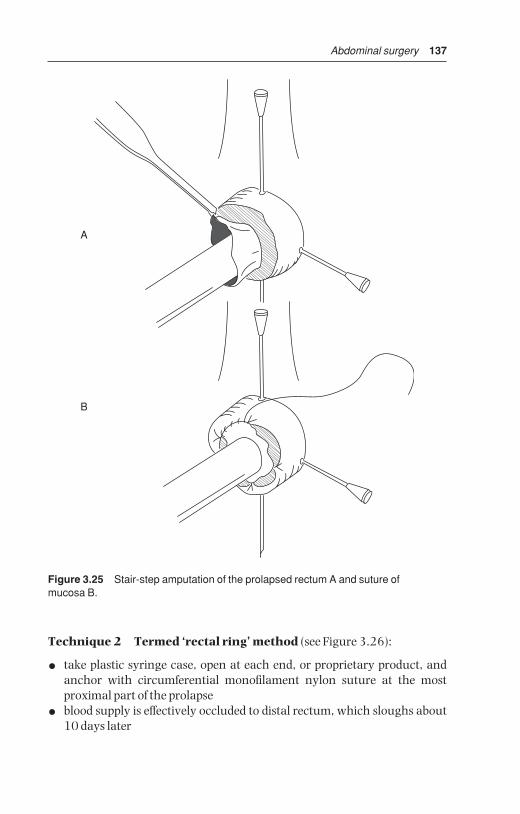
Abdominal surgery 137
Technique 2 Termed ‘rectal ring’ method (see Figure 3.26):
• take plastic syringe case, open at each end, or proprietary product, andanchor with circumferential monofilament nylon suture at the most proximal part of the prolapse
• blood supply is effectively occluded to distal rectum, which sloughs about10 days later
A
B
Figure 3.25 Stair-step amputation of the prolapsed rectum A and suture of mucosa B.
BSAC03 3/5/05 4:27 PM Page 137

138 Chapter 3
DiscussionCareful check must be made at frequent intervals, following technique 2, toensure that the plastic case has not become dislodged, and that defaecationcan proceed normally through lumen of case. The method is simple butmessy, and fails if the casing is dislodged for example by contact with pen wallor other cattle.
Post-operative tenesmus is delayed if the epidural block is made with axylazine-lignocaine mixture (see Section 1.7, p. 29) or a longer acting anal-gesic drug (e.g. Bupivacaine®).
Complications
• immediate excessive stenosis – resuture placing larger diameter hollowcasing into lumen
• excessive haemorrhage – oversew vessel including full thickness of wall
• severe continuous postoperative tenesmus – repeat long-acting epiduralblock, produce pneumoperitoneum (insufflating through needle placed in left paralumbar fossa and attached to Higginson’s syringe), slackenpurse-string suture
A
2
1
34
5
B
C
Figure 3.26 Repair of prolapsed rectum utilising syringe case or plastic tubing.A. lumen of rectum; 2. prolapsed wall. B Sutures are placed through skin-rectalmucosal junction and through holes (4) in the side of plastic tube (3). Second suturesare placed at 180° to first suture. C Sutures are pulled tight over skin as tube isinserted appropriately within rectal lumen. Sutures are then tied around thecircumference to occlude blood supply to prolapsed section (5). Prolapsed rectum and tube drop off some days later as a result of ischaemic necrosis.
BSAC03 3/5/05 4:27 PM Page 138

Abdominal surgery 139
• anal stricture due to excessive fibrosis – incise anus dorsally and ventrallythrough depth of fibrous tissue, and suture cranial commissure to caudalcommissure of wound
Prophylaxis involves attention to the underlying causes (e.g. diet) for the initial development of the rectal prolapse, especially important in cases wherethere are several affected individuals.
BSAC03 3/5/05 4:27 PM Page 139

CHAPTER 4
Female urinogenital surgery
4.1 Caesarean section (hysterotomy)
Indications
• excessive fetal size (immaturity of dam, double muscling, genetic mis-matching, and prolonged gestation)
• fetal deformity (e.g. schistosoma reflexus)
• relative or absolute narrowness of pelvic canal (immaturity of dam, traumatic pelvic deformity)
• fetal malpresentation or posture
• irreducible uterine torsion, uterine rupture, non or incomplete dilatationof cervical os
• atresia or hypoplasia of maternal vagina or vulva
• certain valuable pedigree breeding programmes where safe delivery of aviable fetus is paramount, and where management precludes the risksassociated with natural delivery
Until recently hydroallantois and hydroamnion were further indications fortwo-stage caesarean section (day one: slow drainage of uterine fluids; daytwo: caesarean section). Such cases today have a better prognosis if treatedbefore onset of recumbency with corticosteroids or prostaglandins, followedseveral days later by an i.v. oxytocin drip in refractory cases.
Contra-indications
• cattle in very poor bodily condition (cachectic)
• emphysematous fetus
• most cows with uterine infection
Caesarean section may still be preferable to embryotomy (fetotomy) in casesof general debility and prolonged dystocia despite the presence of dead fetus.
4.1 Caesarean section (hysterotomy) 140
4.2 Vaginal and cervical prolapse 146
4.3 Uterine prolapse 1494.4 Perineal laceration 1524.5 Ovariectomy 155
BSAC04 3/5/05 4:27 PM Page 140

Female urinogenital surgery 141
Advantages of caesarean section over embryotomy include:
• potential fetal survival
• usually faster and safer procedure
• feasibility where embryotomy would be impossible (cervical non-dilatation)
Surgical technique: FlankXylazine premedication is contra-indicated due to induced increase ofmyometrial tone. If permitted a uterine relaxant (e.g. clenbuterol HCl 300 µg)should be given i.m. or slowly i.v. to facilitate rotation and partial exteriorisa-tion of the uterus.
Operate if possible on standing cow or heifer under lumbar paravertebralblock (T13, L1–3, see Section 1.8, p. 23), but weak debilitated and ataxic cattle should be cast before surgery, and in such cases cranial epidural blockis a feasible alternative analgesic method (60–80 ml 2% lignocaine).Another possibility is a 30 ml caudal epidural block and inverted ‘7’ or ‘L’flank analgesia (see p. 26); tie tail to hind leg.
Left flank site is preferred to right flank unless specific indication for rightside exists (e.g. over-sized fetus in right flank, grossly distended rumen). Onright side small intestinal loops are more liable to prolapse through wound tobecome traumatised and infected.
• give caudal epidural block (e.g. 5 ml 2% lignocaine) to any cow which isstraining considerably, as tenesmus hampers precise incision of uterus,and can even provoke ruminal wall prolapse through flank incision
• clip, scrub and disinfect entire paralumbar fossa (last rib to hip) and applysterile drapes
• make 30–35 cm vertical incision in middle or caudal third of left paralum-bar fossa (see Figure 3.6(4) and effect careful haemostasis of flank vessels
• insert hand into abdomen pushing rumen forward and feeling ventrallyand caudally
• make rapid assessment of fetal position and condition of uterine wall
• attempt to bring greater curvature of gravid horn towards abdominal incision by always grasping uterine wall over protruding part of fetus (e.g.limb, hock in anterior presentation)
• attempt to exteriorise greater curvature of gravid horn
• note that, unless there is severe intra-uterine infection (e.g. grosslyemphysematous fetus), entry of uterine fluid to contaminate abdominalcavity is rarely hazardous
• grasp fetal leg (e.g. digits or point of hock) through uterine wall and maintain firmly in flank section
• incise uterine wall (see Figure 4.1) along greater curvature adjacent to thelimb and towards the tip of the horn with scalpel blade or finger embry-otomy (fetotomy) knife starting below digits and extending to hock (or car-pus in forelimb, if in posterior presentation)
BSAC04 3/5/05 4:27 PM Page 141

142 Chapter 4
• avoid incising maternal caruncles (see Figure 4.1)
• extend incision very carefully caudally until the limb can be exteriorisedfor application of obstetric chain or rope
• instruct assistant to maintain very gentle traction on rope/chain sufficientto maintain uterine wall in flank incision
• lengthen uterine incision to permit entry of hand into uterus to locate second limb, which is similarly exteriorised and a rope/chain applied
• manipulate head best by a thumb and finger grip in each orbit
• if very large fetus or uterine horn tip cannot be brought to flank wound forincision under direct vision, make blind incision over fetal extremity,which is then grasped and brought to flank in similar manner
• ensure that fetal traction is applied gently and in appropriate direction,usually initially upwards, and lengthen uterine incision, if required, withknife to avoid any tearing of uterine wall
A
B
C
Figure 4.1 Uterine incision (greater curvature) over right hind metatarsus and foot,having partially exteriorised fetus.A. uterine incision; B. exteriorised horn; C. left flank incision.
BSAC04 3/5/05 4:27 PM Page 142
Female urinogenital surgery 143
• practise careful and slow fetal manipulation during extraction especiallyin cases of schistosoma reflexus, muscle contracture and emphysematouscalves
• in case of gross fetal oversize or ankylosis the skin incision may, occasion-ally, require enlargement to 40 cm
• permit umbilical cord to rupture naturally
• after delivery hold uterine incision in flank wound and remove any looseprotruding portions of placenta, leaving remainder in situ
• do not attempt to separate placenta from maternal caruncles
• with little or no assistance available, non-crushing uterine clamps (vulsel-lum forceps) can be used to hold uterus in position
• check for a second fetus in all cases
• intra-uterine medication is unnecessary
• while fetus is being revived and umbilical cord is checked, undertake uterine repair rapidly
• wash uterine wall with saline if needed
• close uterus with continuous Cushing suture, followed by continuousLembert, or a modified Cushing (Utrecht uterine suture with buried knots)
• suture of uterine wall: start at caudal ventral commissure of wound if asingle layer closure is intended or cranially if two layers are to be inserted.Suture material is either 5 or 6 metric PGA, polyglactin or 7 metricchromic catgut (see Figure 4.2)
• in some cases uterine contraction, tone and turgidity permit only simpleapposition as sutures tear out when inversion is attempted
Evacuation of fetal and other contaminating fluids from abdominal cavity is usually unnecessary. In case of grossly infected fluids, removal must beattempted by swabs and aspiration. Give intra-abdominal antibiotic or par-enteral medication as required. Flank wound is closed in routine manner.Inject oxytocin (50 iu) parenterally to promote uterine contraction.
Post-operative careContinue parenteral antibiotics for five days especially if a dead calf was delivered, as prophylaxis against infection from retained placenta. Assesspatients in severe shock and recumbency by following parameters: generalappearance, rectal temperature, heart rate and character, colour of visiblemembranes, capillary refill time, and willingness to attempt to stand (see p. 41).
In severe cases not only will flunixin meglumine and massive antibioticmedication be required, but such animals also need large volumes of intra-venous fluids (e.g. 25 litres).
The calf should be given maternal colostrum as soon as possible by bottle(teat), or oesophageal feeder. The dam should be encouraged to stand to permit suckling as soon as possible.
Placenta is usually released and discharged within 24 hours of surgery,and is a good prognostic sign. Cases with persistent infected discharge at this
BSAC04 3/5/05 4:27 PM Page 143

144 Chapter 4
time should receive systemic antibiotics. Farmer should check rectal temper-ature and recall veterinarian if cow is febrile.
Healing of the flank wound may occur by secondary intention as a result ofintra-operative contamination, and excessive blood and fluid accumulationbetween suture layers.
Death rate following caesarean section is low (approximately 10%, evenlower if cows with poor prognosis are not operated), and is due to:
• endotoxaemic shock
• chronic severe intra-uterine haemorrhage (via vulva)
• septic metritis and peritonitis
Surgical technique: MidlineVentral midline is a good surgical approach in young beef heifers and in caseof large distended and septic uterus; it requires excellent restraint involvingmore manpower.
U
Figure 4.2 Utrecht uterine closure method with knot burial. Needle is inserted atslight angle towards incision and does not penetrate uterine lumen. Sutures areplaced sufficiently close to prevent leakage of uterine fluid. Care is taken to avoidplacenta.
BSAC04 3/5/05 4:27 PM Page 144
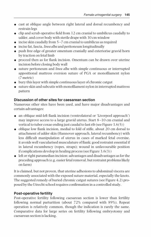
Female urinogenital surgery 145
• cast at oblique angle between right lateral and dorsal recumbency andrestrain legs
• clip and scrub operative field from 12 cm cranial to umbilicus caudally toudder, and cover body with sterile drape with 30 cm window
• incise skin caudally from 5–7 cm cranial to umbilicus as required
• incise fat, fascia, linea alba and peritoneum longitudinally
• push free edge of greater omentum cranially and exteriorise gravid hornby traction on fetal limb
• proceed then as for flank incision. Omentum can be drawn over uterineincision before closing body wall
• suture peritoneum and linea alba with simple continuous or interruptedappositional mattress eversion suture of PGA or monofilament nylon (7 metric)
• bury this layer with simple continuous layer of chromic catgut
• suture skin and subcutis with monofilament nylon in interrupted mattresspattern
Discussion of other sites for caesarean sectionNumerous other sites have been used, and have major disadvantages andcertain advantages:
• an oblique mid-left flank incision (ventrolateral or ‘Liverpool approach’)may improve access to a large gravid uterus. Start 8–10 cm cranial andvertical to tuber coxae ending just caudal to last rib (see Figure 3.6(5))
• oblique low flank incision, medial to fold of stifle, about 20 cm dorsal toattachment of udder skin (Hannover approach, lateral recumbency) withless difficult manipulation of uterus in cases of marked fetal oversize, it avoids well vascularised musculature of flank; good restraint essential ifin lateral recumbency (ropes, straps); wound in unfavourable position if complications develop in healing process (see Figure 3.6(3))
• left or right paramedian incision: advantages and disadvantages as for thepreceding approach (e.g. easier fetal removal, but restraint problems likelyon farm)
It is claimed, but not proven, that uterine adhesions to abdominal viscera arecommonly associated with the exposed suture material, especially the knots.The suggested remedy of buried chromic catgut sutures (see Figure 4.2) pro-posed by the Utrecht school requires confirmation in a controlled study.
Post-operative fertilityPost-operative fertility following caesarean section is lower than fertility following normal parturition (about 72% compared with 89%). Repeat operation is relatively common, though the indication is rarely the same.Comparative data for large series on fertility following embryotomy and caesarean section is lacking.
BSAC04 3/5/05 4:27 PM Page 145

146 Chapter 4
4.2 Vaginal and cervical prolapse
IntroductionThis condition is typically seen in late gestation or the early post-partumperiod in fat beef (especially Hereford and Santa Gertrudis) and dairy breeds(Holstein and Channel Island).
The chronic case which starts about the eighth or ninth month of gestationpresents quite different problems from the post-parturient form, and salvageafter parturition is the best solution, as it will recur in the succeeding gestation.
Predisposing factors
• excessive fat deposition in perivaginal connective tissue
• relaxation of sacrotuberous ligaments due to hormonal influence(endocrine imbalance)
• increased intra-abdominal pressure following greater abdominal size inlate pregnancy
• high roughage intake
• severe cold weather and poor conformation (large flaccid vulva)
• severe post-partum tenesmus due to vaginal injury
• inheritance is postulated in some Hereford bloodlines, but not proven
Treatment of pre-parturient chronic caseCaudal epidural analgesia is followed by thorough cleaning and replace-ment of vagina.
Moderate cases of vaginal prolapse in pregnant cows not near to term (e.g.seven months gestation) are best treated by a modified Caslick’s operation to close the dorsal vulvar commissure, which is then cut at parturition toavoid vulvar tears. Severe cases near term can be managed in two ways.
Technique 1 Perivulvar suture using Bühner’s method (see Figure4.3):
• surgical scrub of perivulvar area, and careful cleansing of exposed vagina
• place subcutaneous suture of nylon tape in deep tissues around vulva withlong vulvar needle (Gerlach pattern, Arnolds, Jorgensen)
• insert needle, threaded with 45 cm sterile nylon tape about 0.6 cm wide,approximately 3 cm below ventral vulvar commissure and directed dorsally through deep connective tissue, to emerge midway between dorsal commissure of vulva and anus
• leave one end of tape hanging from the lower incision, remove the otherend from the needle eye, and withdraw the needle
• re-insert unthreaded needle in similar manner along the other edge of thevulvar lip, to emerge adjacent to the original exit point, rethread and pulltape out ventrally (see Figure 4.3)
• tie tape so that adjustment in tension can easily be made
BSAC04 3/5/05 4:27 PM Page 146

Female urinogenital surgery 147
Vulvar lumen should permit easy entry of four fingers. Only about 1 cmlength of tape is in contact with vulvar skin dorsally and ventrally. Suturesmay be completely buried by two 1 cm long horizontal incisions sutured inmattress pattern, and placed dorsally and ventrally, if more than one monthprepartum. If inserted during late gestation, the suture is cut near the knotand removed at term, therefore it is vital to watch carefully for first signs of calving.
There is commonly vulvar oedema for several days. Local drainage of pusfrequently occurs. A sphincter-like band of connective tissue occasionallyresults and may prevent future prolapse, but occasionally can cause dystocia,necessitating dorsal episiotomy (see Section 4.4, pp. 154–155).
Technique 2 Transverse sutures:
An inferior modification of the above method involves nylon tape which isinserted in two or three deep horizontal mattress sutures across the vulvarlips. It can produce more severe local reaction, pain and irritation, leading to continued tenesmus after analgesia wears off. There is inevitably anincreased risk of sutures tearing through skin.
Anus
Vulva
Figure 4.3 Pattern of Bühner suture in vulvar lips. The Gerlach-type needle,threaded with umbilical tape, is inserted below ventral vulvar commissure, and ispassed through dense fibrous tissue to emerge above dorsal commissure. It isunthreaded, and needle alone is passed up the other side in similar manner. It isrethreaded, and tape pulled back ventrally. A quick release and adjustable knot is tied.Suture material is only visible at the commissures.
BSAC04 3/5/05 4:27 PM Page 147

148 Chapter 4
• insert needle with tape just lateral to the ‘hairline’ beside the vulvar lips
• thread suture material through or simply tie the sutures over short lengthof rubber or plastic tubing (‘quills’), reducing risk of suture tearout
Treatment of post-parturient caseTechnique 1 above is suitable. Initially in a recently calved cow, simple re-placement under a long-acting (up to eight hours’ duration, see Section 1.7,p. 28) epidural block is successful in most instances. Tenesmus may be further prevented by production of pneumoperitoneum (see Section 3.19 p. 138).
Technique 3 Modified Caslick’s operation:
After vaginal replacement the vulvar lumen is surgically occluded over itsdorsal three-quarters:
• resect vulvar mucous membrane dorsally over an area measuring about 5 cm long and 1.5 cm wide
• suture apposing surfaces with fine nylon or caprolactan (Vetafil®), sup-ported by two deeper mattress sutures of similar non-absorbable material
• remove these sutures after two to three weeks
• incise suture line shortly before imminent parturition
Technique 4 Cervicopexy (see Figure 4.4)
IntroductionA radical method for preventing further vaginal prolapse, very effective if properly performed in the valuable non-pregnant cow. Via a left-flanklaparotomy incision a suture is placed through ventral portion of cervix andprepubic tendon just lateral to midline.
Technique
• select large full-curved cutting needle and thick, non-absorbablemultifilamentous suture material (polypropylene 7 metric)
• instruct assistant to apply uterine vulsellum forceps to ventral part of cervical os per vagina, pushing forceps cranially to aid identification,which is essential for correct placement, and for avoidance of urethra andbladder
• after inserting ventral cervical suture pass double length of suture mater-ial through prepubic tendon 5 cm cranial and lateral to pecten of pubis(see Figure 4.4)
• throw knots outside abdominal cavity and carry them along suture material with thumb and fingers to avoid entrapment of small intestine
• ensure minimum of four throws to each knot
• cut suture material with scissors, leaving 3 cm ends
BSAC04 3/5/05 4:27 PM Page 148

Female urinogenital surgery 149
This modified Winkler cervicopexy is easier and safer than the original vaginal approach.
4.3 Uterine prolapse
IntroductionUterine prolapse occurs not infrequently (0.5% of calvings) following thirdstage labour, and usually involves complete inversion of the gravid cornu.Some cases follow an assisted second stage of labour. Almost unknown following caesarean section. Nearly all cases occur within fifteen hours ofparturition.
Prevalent both in multiparous dairy cows in good condition (high proteinintake prepartum), and in cases of malnutrition and chronic disease. Somecases predisposed by high oestrogen intake.
Aetiology unclear but uterine inversion and prolapse appear to be associ-ated with onset of uterine atony during third stage labour. Many cows haveconcurrent hypocalcaemia and clinical signs of milk fever.
F
E
D
CG
B
H
A
Figure 4.4 Cervicopexy through left flank laparotomy (vertical view).A. suture through ventral part of cervix (B) which is fixed and manipulated by assistantholding long-handled uterine forceps (E) in vagina; C. point of insertion of suturethrough prepubic tendon; D. pubis and ventral part of pelvis (shaded); F. rectum; G. uterus; H. flank incision and suture throw; J. bladder.
BSAC04 3/5/05 4:27 PM Page 149
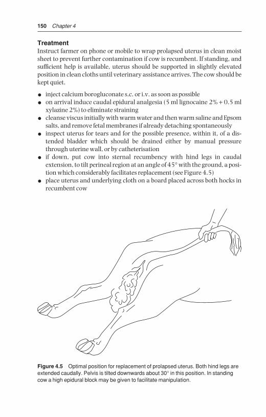
150 Chapter 4
TreatmentInstruct farmer on phone or mobile to wrap prolapsed uterus in clean moistsheet to prevent further contamination if cow is recumbent. If standing, andsufficient help is available, uterus should be supported in slightly elevatedposition in clean cloths until veterinary assistance arrives. The cow should bekept quiet.
• inject calcium borogluconate s.c. or i.v. as soon as possible
• on arrival induce caudal epidural analgesia (5 ml lignocaine 2% + 0.5 mlxylazine 2%) to eliminate straining
• cleanse viscus initially with warm water and then warm saline and Epsomsalts, and remove fetal membranes if already detaching spontaneously
• inspect uterus for tears and for the possible presence, within it, of a dis-tended bladder which should be drained either by manual pressurethrough uterine wall, or by catheterisation
• if down, put cow into sternal recumbency with hind legs in caudal extension, to tilt perineal region at an angle of 45° with the ground, a posi-tion which considerably facilitates replacement (see Figure 4.5)
• place uterus and underlying cloth on a board placed across both hocks inrecumbent cow
Figure 4.5 Optimal position for replacement of prolapsed uterus. Both hind legs areextended caudally. Pelvis is tilted downwards about 30° in this position. In standingcow a high epidural block may be given to facilitate manipulation.
BSAC04 3/5/05 4:27 PM Page 150

Female urinogenital surgery 151
• with the organ elevated, so that its lower edge is level with the ischium,start replacement by gentle pressure, using the palms of the obstetricallylubricated hands (KY® jelly) over the areas nearest to the vulvar lips,working in a circular manner around the mass
• practise gentle massage to avoid uterine perforation!
• after initial partial reposition, maintain reposed part by ensuring that theremaining prolapsed uterus is kept above vulvar lips
• progressively replace whole organ in this manner and ensure that itmoves cranial to the cervix to its normal position
• if reposition is impossible after several minutes’ manipulation locate theopening into the non-pregnant horn (usually about level with the vulva)insert the closed fist and apply firm pressure into the pelvic cavity
• check there is no residual inversion of the cornu (e.g. use empty Ca bottleas arm extension)
• if complete inversion is still impossible, fill uterine lumen with normalsaline, and then siphon off through disinfected stomach tube
• after reduction inject oxytocin (50–100 iu, i.m.) to speed involution
Limited vaginal trauma can be ignored, but a large deep laceration should besutured with catgut. Cows with extensive areas of vaginal epithelial necrosisand trauma require submucosal resection which is a haemorrhagic, slowprocedure indicated only in selected cases.
ComplicationsRecurrence of the prolapse following return of sensation to the perinealregion is uncommon. The usefulness of a Bühner pattern vulvar suture (seeSection 4.2, pp. 146–147) inserted for two to three days to avoid this hazard,is controversial. Follow-up visit advisable 12–24 hours later to check vaginaand cervix.
Other complications include haemorrhage, metritis, toxaemia, septicemia,paresis, and uterine rupture with bladder or intestinal eventration.
Chronic cases present problems; through and through sutures of tape or steel wire from vaginal lumen and lesser sciatic notch to skin, secured bybandage or sponge have been attempted (Minchiv method).
Prognosis for rebreeding is good and recurrence of prolapse at next parturi-tion is inexplicably uncommon.
Amputation of the uterus (hysterectomy)
IntroductionMajor surgery with rare indications, amputation is performed in cows in which the organ is so severely damaged (lacerations, necrosis, freezing,gangrene) that reposition would result in death, and in cases of prolongedprolapse in which replacement proves impossible. Amputation is sole
BSAC04 3/5/05 4:27 PM Page 151
152 Chapter 4
alternative to salvage. The prognosis is guarded or poor. Major problems arehaemorrhage and shock. Pre-operative whole blood or hypertonic (7.2%)saline may be advisable (see Section 1.11 p. 42). Surgery is again performedunder epidural block.
Wash and prepare operative field. Two techniques are available.
Technique 1
• insert several transfixing and circumferential sutures of nylon tape justcaudal to the cervix
• resect uterus about 5–10 cm caudal to this point, placing further haemo-static sutures throughout margin
Technique 2
• make dorsal incision through uterine wall to bifurcation
• identify and fan out left and right mesometrium
• ligate vessels in series, larger vessels individually, smaller vessels in groups(chromic catgut 7 metric)
• incise mesometrium 1 cm distal to sutures
• permit stumps to retract intra-abdominally
• insert mattress suture line just cranial to cervix
• amputate uterus 1 cm distal to mattress sutures
• return stump and vagina into pelvic cavity
• give systemic antibiotics for three days
Post-operative problemsHaemorrhage from uterine vessels, shock. Milking cows may remain pro-fitable for one to two years as long as ovariectomy (see Section 4.5, p. 155) isperformed at hysterectomy.
4.4 Perineal laceration
Classification
• first degree: involves mucosa of vulva/vestibule/vagina
• second degree: involves full thickness of vulva/vestibule/vagina wall, butnot rectal wall or anus
• third degree: involves full depth of vulva/vestibule/vaginal walls, as wellas rectovaginal tears, including anal sphincter or rectovaginal fistula
This section considers primarily third degree perineal lacerations, i.e. themost severe form.
Clinical signsInjury is almost always a result of damage from fetal head or limbs at parturi-tion. If possible injury can be anticipated, a veterinarian attending a dystocia
BSAC04 3/5/05 4:27 PM Page 152

Female urinogenital surgery 153
case may perform dorsal episiotomy (10 o’clock or 2 o’clock position) to prevent such severe lacerations. Repair of such a surgical episiotomy woundis relatively simple (see p. 154).
Surgery is essential if breeding is to be resumed, since the severe ragged andirregular tear soon becomes oedematous and grossly contaminated by faeces.Faeces in the vagina starts a chronic inflammatory process which soonspreads to involve the cervix. At oestrus infected material may result in distortion of the shape and direction of the vaginal lumen, permitting urinepooling in cranial vagina.
Delay surgery until defect is completely epithelialised, possibly four to sixweeks postpartum. Some surgeons prefer immediate surgery (not longerthan four hours postpartum), but existence of inflammation and infectionoften leads to failure.
Technique
• keep off feed for 12–24 hours
• caudal epidural analgesia in standing animal
• cleanse surrounding area and irrigate vagina with warm disinfectantsolution (povidone-iodine) and pack rectal lumen cranial to the defectwith absorbent cloths
• instruct assistant to retract the lateral borders of the cloaca to expose cranially the shelf, formed by the rectal and vaginal mucosa dorsally andlaterally, and supporting the underlying muscular layers
• incise transversely along caudal edge of rectovaginal shelf, and extendincision laterally to skin edge of original dorsal vulval commissure (seeFigure 4.6)
• completely separate the vaginal mucosa from the edge of this shelf to adepth of about 5 cm
• suture the musculofibrous bridge, starting cranially, in a transverse planeto appose left and right surfaces (e.g. 5 or 6 metric PGA)
• start suture in the vaginal lumen and also include the four edges of vaginalmucosa
• ensure sutures are placed tight, and that their distance apart is such that itis not possible to insert a digit between them
• avoid suturing skin edges caudally (contra-indicated) since it couldincrease the difficulty in defaecation
• give systemic antibiotic prophylactic cover for 5 days
Problems
• inadequate mucosal undermining (a technically difficult procedure),results in an inadequate thickness of bridge
• poor placement of sutures with excessive space, permitting faecal materialto pass into vagina
• sutures tearing out
BSAC04 3/5/05 4:27 PM Page 153
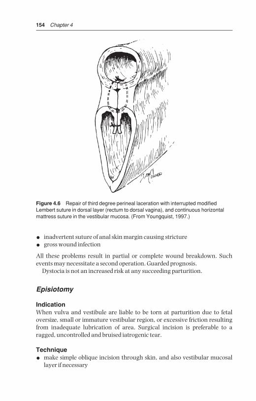
154 Chapter 4
• inadvertent suture of anal skin margin causing stricture
• gross wound infection
All these problems result in partial or complete wound breakdown. Suchevents may necessitate a second operation. Guarded prognosis.
Dystocia is not an increased risk at any succeeding parturition.
Episiotomy
IndicationWhen vulva and vestibule are liable to be torn at parturition due to fetal oversize, small or immature vestibular region, or excessive friction resultingfrom inadequate lubrication of area. Surgical incision is preferable to aragged, uncontrolled and bruised iatrogenic tear.
Technique
• make simple oblique incision through skin, and also vestibular mucosallayer if necessary
Figure 4.6 Repair of third degree perineal laceration with interrupted modifiedLembert suture in dorsal layer (rectum to dorsal vagina), and continuous horizontalmattress suture in the vestibular mucosa. (From Youngquist, 1997.)
BSAC04 3/5/05 4:27 PM Page 154

Female urinogenital surgery 155
• avoid lengthening dorsally to anal sphincter, or cranially to damage caudal branches of vaginal artery: oblique incision should be made at 10 o’clock or 2 o’clock position
• suture incision in two layers after delivery: continuous chromic catgut inmucosa, interrupted monofilament nylon in skin
• antibiotics unnecessary
Primary healing is usual.
4.5 Ovariectomy
IndicationsAlleged prolongation of lactation in mature cows as well as improvement infeed efficiency compared with non-spayed heifers. Convenient husbandrymeasure permitting spayed heifers and cows to run within herd with bullwithout risk of pregnancy. Occasionally unilateral surgery in case of ovarianpathology or persistent luteinised cyst.
Little comparative data is available regarding lactation and feed efficiencyin spayed and non-spayed cattle, and much of it is contradictory.
Not widely practised in Europe, but common in parts of both South, Middleand North America, where a single surgeon, working at a restraint chutewith a well-organised team, can spay 40–60 heifers an hour. Operated animals should be permanently identified.
TechniqueThe site for ovariectomy in a heifer (six to twelve months old) is the flank(standing) or midline caudal abdomen (recumbent). In the cow it is thevagina (colpotomy) or flank (standing).
Prior to surgery starve animal for 24 hours, no water restriction.
Flank approach
• clip left flank, wash, scrub and disinfect paralumbar fossa
• paravertebral analgesia (L1–2) or local infiltration (‘T-block’, ‘reverse 7’),(see Section 1.8, pp. 26–27)
• practise aseptic technique: instruments and hands cleansed with quater-nary ammonium disinfectant in repeated operations
• make vertical incision 10–13 cm long in left flank ventral to lumbar trans-verse processes 3–4
• separate muscles with scissors and incise peritoneum
• insert hand to enlarge peritoneal incision and locate ovaries
• remove ovaries with small effeminator (since ovarian pedicle is not anaesthetised by paravertebral block, swab soaked with local anaestheticsolution should be applied to pedicle for 1 minute before using effeminator).Important: do not drop either ovary into abdominal cavity!
BSAC04 3/5/05 4:27 PM Page 155

156 Chapter 4
• check that structure removed is entirely ovary
• suture abdominal wall routinely
• give systemic antibiotics for one to three days. Fly-repellant spray withresidual action is indicated in warm months
• remove non-absorbable sutures in 10–14 days
Vaginal approach (cows only)
• epidural anaesthesia
• make stab incision about 5 cm long through vaginal wall at 10 o’clock or2 o’clock position just caudal to cervix with sheathed knife (take care toavoid aorta and rectum)
• insert two fingers to locate one ovary, and draw it back into vagina
• apply local anaesthetic soaked swab to pedicle for 1 minute
A
B
CD
E
3 cm
F
Figure 4.7 Ovariectomy with the Willis instrument inserted through vaginal stabwound under rectal guidance. Ovary is manipulated through notch for transection ofovarian pedicle. With K-R device inner sleeve cuts ovarian pedicle while retainingovary in lumen (see smaller illustrations).A. sleeved arm in rectum; B. Willis instrument directed through vaginal fornix to ovary;C. bladder; D. uterine body; E. Willis instrument and close-up; F. K-R instrument.
BSAC04 3/5/05 4:27 PM Page 156
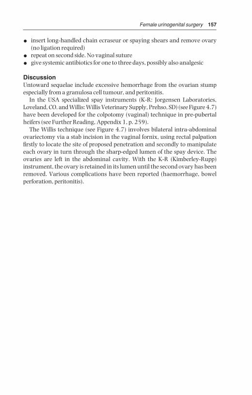
Female urinogenital surgery 157
• insert long-handled chain ecraseur or spaying shears and remove ovary(no ligation required)
• repeat on second side. No vaginal suture
• give systemic antibiotics for one to three days, possibly also analgesic
DiscussionUntoward sequelae include excessive hemorrhage from the ovarian stumpespecially from a granulosa cell tumour, and peritonitis.
In the USA specialized spay instruments (K-R: Jorgensen Laboratories,Loveland, CO. and Willis: Willis Veterinary Supply, Prehso, SD) (see Figure 4.7)have been developed for the colpotomy (vaginal) technique in pre-pubertalheifers (see Further Reading, Appendix 1, p. 259).
The Willis technique (see Figure 4.7) involves bilateral intra-abdominalovariectomy via a stab incision in the vaginal fornix, using rectal palpationfirstly to locate the site of proposed penetration and secondly to manipulateeach ovary in turn through the sharp-edged lumen of the spay device. Theovaries are left in the abdominal cavity. With the K-R (Kimberley-Rupp)instrument, the ovary is retained in its lumen until the second ovary has beenremoved. Various complications have been reported (haemorrhage, bowelperforation, peritonitis).
BSAC04 3/5/05 4:27 PM Page 157

CHAPTER 5
Teat surgery
5.1 Introduction
Intensification of dairy farming has led to earlier culling of cows with many ofthe teat problems discussed in this chapter (‘hard milker’, see Section 5.2, teatlacerations, see Section 5.5), which fail to respond to simple managementsuch as a topical spray. In many countries treatment may be uneconomic.But in the developing world with small herds the individual cow remains avaluable resource and the owner demands maximum care and attention toteat conditions.
Significant teat injuries commonly affect the orifice, and less commonly the teat sinus wall. Teat surgery is a demanding test of the skill of the veterinarian.
Restraint and anaesthesia (see Chapter 1): xylaxine alone is very effectivefor minor procedures, but it is contra-indicated in advanced gestation.Beware possible recumbency! Wopa crate with one leg lifted is ideal restraintmethod. Local anaesthesia at the teat base, with or without sedation, andwith the cow restrained in lateral recumbency, is another option.
Economic loss results from a loss in milk yield (take care with antibiotic-treated milk; possible loss of quarter if there is a necessity to dry off ).
Anatomy
• teat orifice or ostium papillare
• streak canal or ductus papillaris
• Furstenberg’s rosette
• teat sinus, pars papillaris or lactiferous sinus (teat part)
• lactiferous sinus (glandular part), milk cistern or pars glandularis
• teat canal: longitudinally folded stratified squamous epithelium
5.1 Introduction 1585.2 Stenosis of teat orifice 1605.3 Teat lumen granuloma 1615.4 Teat base membrane
obstruction 163
5.5 Traumatic lacerations of teat 1635.6 Imperforate teat 1655.7 Incompetent teat sphincter 1655.8 Teat amputation 1655.9 Discussion 166
BSAC05 3/5/05 4:27 PM Page 158

Teat surgery 159
• teat sinus: superficial columnar, deeper cuboidal epithelium; belowmucosa is layer of embryonal-type cells capable of rapid proliferation
• muscularis, subcutis and skin
5
6
4
3
3
2
2
1
1
C
D
B
B
A
A
Figure 5.1 Teat anatomy and sites of stenosis.1. teat orifice (ostium papillare); 2. teat or streak canal (ductus papillaris); 3. Furstenberg’s rosette; 4. teat sinus (pars papillaris); 5. lactiferous sinus or milkcistern (pars glandularis); 6. venous plexus.Sites of teat obstruction: A. tight streak canal; B. obstruction at Furstenberg’s rosette;C. milk cistern obstruction (proximal stenosis); D. teat sinus obstruction.
BSAC05 3/5/05 4:27 PM Page 159

160 Chapter 5
5.2 Stenosis of teat orifice, streak canal or Furstenberg’srosette
AetiologyCommon problem. Partial obstruction (‘hard milker’) due to local trauma,possibly secondary to milking machine malfunction (e.g. excessive vacuum
A
B
C
Figure 5.2 Teat instruments.A. milk catheter for passive milk drainage; B. Hug lancette for opening or dilating thestreak canal; C. Teat alligator forceps for the removal of ‘milk stones’, either free orattached to teat mucosa, through the streak canal.
BSAC05 3/5/05 4:27 PM Page 160

Teat surgery 161
pressure) or microtraumatic wounds sometimes permitting the developmentof Fusobacterium necrophorum infection (‘blackspot’).
SignsQuarter milks out slowly, or ‘valve’ is evident, and vicious cycle may developas remaining quarters tend to be overmilked, resulting in bruising and ever-sion of teat orifice and development of mastitis. Some cases are inoperable.
TreatmentControversial, and there are no controlled studies on efficacy of method.
Non-infected case (i.e. no mastitis) and non-inflammatory teat orificelesion:
• in mild cases consider simple insertion of self-retaining canula (Naylor®
teat dilator) for five days, without surgery, alternatively insert Larsen®
teat tube
• alternatively tense orifice between finger and thumb, cleanse withiodophor solution
• insert Maclean’s® teat knife, D-D (Dyakjaer-Danish), Hug® (see Figure 5.2),or Lichty® (blunt point) knife once and withdraw rapidly
• check adequacy of milk flow and if poor, re-insert knife at 90° to originalincision
• if flow is still poor, insert teat bistoury and thereby cut out plug of teat canal
• insert self-retaining teat canula for five days; initially rotate canula gentlyevery few hours to avoid development of excessive granulation tissue
• as alternative to teat canula, tie cow up and remove milk from the quarterseveral times every two to three hours for the first two days
• give intramammary antibiotics prophylactically for five days (N.B. milkwithdrawal)
• instruct stockman to roll end of teat regularly before applying machine
Infected case (i.e. with mastitis and/or severe inflammatory reaction):
• treat with intramammary antibiotics and local application of antibioticcream before attempting surgery
Treatment of choice: remove obstructing tissue in Furstenberg’s rosette or streak canal during thelotomy, or, if available, with theloresectoscope (seeFigure 5.3). Return to normal machine milking in 75% of cases
5.3 Teat lumen granuloma
IntroductionAlso known as ‘pea’ or ‘spider’, this lesion is a discrete proliferation of granu-lation tissue covered by mucosa and caused by trauma, which initially causesa submucosal haematoma projecting into teat cistern.
BSAC05 3/5/05 4:27 PM Page 161

162 Chapter 5
SignsOften asymptomatic but can eventually cause interference with free passageof milk, is rarely pedunculated, but painless.
TreatmentComplete removal is often difficult, and recurrence rate is high!
• attempted blind removal via mosquito or long alligator forceps, teat bistoury, or fixation and traction by Hudson’s spiral, often causes furthertrauma
• logical approach in case producing clinical signs is open surgery (thelo-tomy) in lateral recumbency following ring block: vertical incision in teatwall opposite mass, and resection by scalpel. Suture in three layers,mucosa with fine continuous absorbable material, then wall, finally skin. Teat wound heals well if careful, sterile technique is adopted
• milk daily using sterile teat cannula (passive milk drainage)
Figure 5.3 Theloresectoscopy. Scope is introduced into teat sinus through lateralteat wall incision. Stenotic tissue around Furstenberg’s rosette is removed with thecautery sling.
BSAC05 3/5/05 4:27 PM Page 162
Teat surgery 163
• return to machine milking of quarter on day eleven
• drying off quarter is often best option
5.4 Teat base membrane obstruction
Congenital form occurs in some heifers; acquired form is in cows, usually atstart of new lactation, aetiology unknown, possibly inflammation of basalannular membrane.
• teat lance or Hug’s knife (see Figure 5.2b) is inserted in attempt to breakdown the annular membrane
Prognosis is very poor and treatment usually hopeless.
5.5 Traumatic lacerations of teat
IntroductionFactors affecting outcome and prognosis include:
• involvement of streak canal
• direction of wound: longitudinal wounds heal better than transverse
• integrity of blood supply
• presence of infection
• amount of skin loss
TreatmentSuperficial wounds: basic surgical principles apply and all wounds must be regarded as contaminated.
• thorough gentle cleansing with non-irritant dilute antiseptic, e.g. povidone-iodine or chlorhexidine
• leave superficial wounds unsutured, but dress with bland antisepticcream, e.g. chlorhexidine gluconate (Savlon™, Schering-Plough) or pro-prietary mixture of propylene glycol and organic acids (Dermisol® cream,Pfizer) after débridement of traumatised and avascular tissue with finesharp scissors and rat-tooth forceps
• some cases benefit from taped dry dressing (e.g. Elastoplast® Smith &Nephew) to appose wound edges
Deeper wounds involving muscularis and skin:
• suture after cleansing, irrigation and débridement
• insert continuous sture into muscle layer with monofilament absorbablemetric 2
• insert skin sutures, simple interrupted of metric 2 polypropylene
• cover wound over with Opsite® (Smith & Nephew) after spraying withLeukopor® (‘New Skin’ spray)
BSAC05 3/5/05 4:27 PM Page 163

164 Chapter 5
Local antibiotics: apply only to skin suture line, never to wound surfaces,where they delay healing. Use penicillin + streptomycin (Streptopen®
Schering-Plough) or oxytetracycline hydrochloride (Terramycin® Pfizer) inpowder, cream or aerosol preparations.
Use of routine intramammary antibiotic is controversial.Passive milk drainage for ten days before resuming machine milkingProblems include excessive suture tension, which causes puckering and
skin necrosis, and poor healing resulting from inadequate débridement orcleansing of lesion.
Teat laceration involving teat sinus
IntroductionFistulae are usually acquired. Treat recent (< 12 hours) fistulae by immediatesurgery (primary wound healing). Repair older wounds by tertiary woundhealing, (e.g. 10–14 days post-trauma or during dry period). Prognosis isguarded.
Work in good operating facilities, with adequate light, cleanliness andefficient analgesia. Ensure stockman understands post-operative care.
Surgical technique
• cleanse with non-irritating antiseptic
• trim off devitalised skin, muscularis and mucosa
• appose or invert mucosal edges, but suture submucosa and musculariswith interrupted sutures of 3 metric PDS on swaged-on needle. Do notsuture mucosa!
• suture skin with simple non-absorbable (e.g. polyamide 2 metric) inter-rupted mattress sutures to produce precise apposition of edges
• use of protective bandage and self-retaining impregnated cannula for sev-eral days is controversial, and lacks comparative studies (e.g. ‘Opsite’®)
• perform passive milk drainage every one to two days (depending on quarter yield), and insert intramammary preparation every second day
• healing takes about ten days, when sutures are removed. Cases of woundbreakdown should be re-operated at the earliest possible moment in thedry period.
Causes of breakdown
• severe post-operative oedema
• poor suture technique
• excessive suture tension
• necrosis of wound edges
• infection
Local non-surgical treatment with organic acid, butter of antimony and,most recently acrylics and electrocautery, usually fails to close a teat fistula.
BSAC05 3/5/05 4:27 PM Page 164

Teat surgery 165
5.6 Imperforate teat
Aetiology is congenital in heifers, or acquired as a result of trauma in adultcattle.
Assess whether milk is present in teat sinus; surgery is only indicated inpositive case. Proceed as for teat stenosis (see Section 5.2, pp. 160–161).
5.7 Incompetent teat sphincter
Aetiology is invariably traumatic in case of a single teat, which constantly orintermittently leaks milk.
Treatment
• difficult and indicated only in chronic case (> 3 weeks) as fibrosis of granu-lation tissue repair may correct condition spontaneously
• inject minute volume of irritant drug (e.g. sterile Lugol’s iodine) withtuberculin syringe around teat orifice (e.g. 4 × 0.1 ml) to stimulate discretecircumferential fibrosis
Technique is hazardous and results unpredictable.
5.8 Teat amputation
Supernumerary teats
IntroductionCongenital and inherited condition. The teats tend to be small, are commonlycaudal, but sometimes are attached to the normal teat. Remove when one tonine months old, and never within one month of parturition (oedema, woundbreakdown, infection, mastitis). Up to three months old, removal may be performed by unqualified but trained person, but under anaesthesia. UK law requires that surgery in calves over three months old be performed by veterinary surgeon under anaesthesia. Swiss law requires all ruminantsurgical procedures to be performed by veterinarian under anaesthesia.Many countries have no legislation to cover this, and many other specific procedures.
Technique
• carefully identify supernumerary teats
• small calves: resect with scissors
• in older calf crush at base with small Burdizzo® (Emasculatome) and resectwith knife along inner edge of blades
• line of section should be cranial to caudal, not transverse, so that sub-sequent scar merges into natural folds of udder skin
• suture only if wound edges separate
BSAC05 3/5/05 4:27 PM Page 165
166 Chapter 5
Amputation due to disease
Indicated in severe purulent or gangrenous mastitis of one quarter to permitdrainage, also following irreversible teat damage.
Technique
• analgesia of teat base and application of good restraint
• place Burdizzo® at junction of middle and distal thirds of teat, amputatewith scalpel distal to jaws
• retain teat lumen by continuous sutures in wall (skin to mucosa) usinginterlocking mattress suture to maintain drainage
Haemorrhage is usually slight.Quarter will eventually dry off as a result of secondary infection and mastitis.
Amputation due to injury
IntroductionTeat amputation may also be indicated in cows with severe teat damagewhere reconstructive surgery and return to normal function cannot beexpected, e.g. loss of distal portion of teat, or long, oblique or transverse tearsinto teat canal. Amputation and closure of teat sinus is only successful in theabsence of infection.
Technique
• amputation site is 1–2 cm distal to udder-teat junction
• transect teat by scalpel, resecting the mucous membrane 1 cm below thecut surface
• invert mucosa by continuous suture in submucosa (see Figure 5.4)
• insert horizontal mattress sutures to close musculature and appose skinedges with simple sutures or metal clips (Michel)
• infuse antibiotics into the quarter before final sutures are placed.
In gangrenous mastitis, following teat amputation at the junction withudder, a cruciate incision is made into udder skin to permit optimal drainageand exposure to atmosphere to inhibit anaerobes. Irrigation with dilute H2O2is useful.
5.9 Discussion
Meticulous care to details of technique is vital in surgical repair of severe traumatic teat lesions. A multiplicity of suture patterns illustrates the unsat-isfactory results obtained in certain hands, but this is more likely to be a resultof deficiencies and breakdowns in basic surgical principles, than to any defectin the suture configuration itself.
BSAC05 3/5/05 4:27 PM Page 166
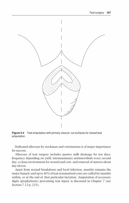
Teat surgery 167
Dedicated aftercare by stockman and veterinarian is of major importancefor success.
Aftercare of teat surgery includes passive milk drainage for ten days, frequency depending on yield, intramammary antimicrobials every secondday, a clean environment for wound and cow, and removal of sutures aboutday eleven.
Apart from wound breakdown and local infection, mastitis remains themajor hazard, and up to 40% of teat-traumatised cows are culled for mastitiswithin, or at the end of, that particular lactation. Amputation of accessorydigits (prophylactic) preventing teat injury is discussed in Chapter 7 (seeSection 7.12 p. 225).
Figure 5.4 Teat amputation with primary closure: cut surfaces for closed teatamputation.
BSAC05 3/5/05 4:27 PM Page 167

CHAPTER 6
Male urinogenital surgery
6.1 Preputial prolapse
IntroductionA mild degree of intermittent preputial prolapse or eversion is considered normal in some breeds, notably the polled Hereford and Aberdeen Angus inthe UK, together with the Brahman and Santa Gertrudis breeds (Bos indicus)in North America.
Prolapse develops during normal non-erectile movement of the peniswithin the preputial cavity. Pathological prolapse is more likely in individualsexposed to trauma.
Predisposing factors
• pendulous and long sheath
• large preputial orifice with limited ability to contract
• poor development of the preputial and retractor penis muscles in somepolled breeds (see above)
Preputial traumaPathological prolapse is more likely in individuals at range or pasture exposedto trauma, which may involve irritation from foreign bodies such as vegeta-tion, dust or earth, and which are secondary to penile injury.
Common site of injury is the mucosa adjacent to skin-prepuce junction.Frostbite can cause major problems in certain areas in the winter. All cases of preputial prolapse should have complete physical examination of the external genitalia.
Sequence of events following injury is:
• severe localised oedema and hyperaemic congestion
• extensive fibrosis with secondary infected fissures and cracks
• areas of granulation tissue develop in the cracks with secondary infection
6.1 Preputial prolapse 1686.2 Penile haematoma 1726.3 Urolithiasis 1756.4 Prevention of intromission 1816.5 Vasectomy 183
6.6 Epididymectomy 1856.7 Congenital penile
abnormalities 1876.8 Penile neoplasia 1916.9 Castration 191
BSAC06 3/5/05 4:26 PM Page 168
Male urinogenital surgery 169
• as a result, penile erection may be abnormal, with only the glans tip passing through this damaged region
Another common site of preputial trauma leading to prolapse, is thereflection of parietal prepuce on to the body of the penis. It may be sustainedduring natural or artificial service.
Conservative treatmentRecent injury to prolapsed mucosa may be treated conservatively:
• carefully cleanse the area with non-irritant dilute antiseptic solution(povidone-iodine)
• soak damaged prepuce in swabs containing 25% magnesium sulphatesolution to reduce oedema
• apply emollient dressing (e.g. zinc and castor oil ointment) and replaceprolapse
• place purse-string suture in preputial orifice or if the prepuce is too oedematous to be reduced a polyvinyl tube (2.5 cm diameter and 15 cmlong) is placed in the preputial lumen and wrapped firmly with elastic tape
• tape is applied directly to the tube and the skin on the sheath beginning at the preputial orifice and extending toward the abdominal wall
• placement of tube decreases oedema and permits escape of the urine
• systemic antibiotics and anti-inflammatory drugs for five days, as well aslocal antibiotic irrigation
• dress any prolapsed preputial mucosa with lanolin-based ointment andkeep covered to prevent further trauma
Radical surgery: circumcisionCases with secondary diffuse fibrosis and excoriation require surgery in lateralrecumbency, under sedation and ring block local analgesic infiltration, orgeneral anaesthesia (see Section 1.9, p. 36). A rubber band tourniquet maybe applied proximally to decrease haemorrhage.
Two circumcision techniques are used commonly in bulls:
• resection and anastomosis, also known as posthioplasty or reefing
• surgical amputation, resulting in the loss of equal amounts of external andinternal lining of the prolapsed portion of prepuce, performed using sutureor ring technique
Four potential post-operative complications must be recognised before sur-gery is attempted:
(a) prepuce can be made too short for breeding: this depends on the breedbut, as a general criterion, the usable prepuce must be twice as long asthe free portion of the penis
(b) surgeon should remove only the diseased prepuce
BSAC06 3/5/05 4:26 PM Page 169

170 Chapter 6
(c) circumferential scar (cicatrix) contraction can result in phimosis(d) post-operative haemorrhage can lead to dehiscence and infection
1 Resection and anastomosis technique
• close clip and shave the hair from the distal portion of the sheath andpreputial orifice
• if penile extension is possible, place towel forceps in the apical ligament oralternatively have an assistant hold the penis
• scrub and prepare the prepuce and penis
• place two marking sutures proximal and distal to the area of prepuce to beremoved to ensure that the prepuce is aligned anatomically when sutured
• examine the prepuce and make circular incision through the epitheliumof the prepuce proximal to the area to be resected
• deepen this incision until normal tissue is observed
• make a second circular incision distal to the area to be removed. In Bosindicus bulls, it usually is necessary to resect at least 15 cm of prepuce toprevent recurrence of the prolapse. In Bos taurus bulls, as much prepuce aspossible should be salvaged
• connect these two circular incisions then with two longitudinal incisions
• remove the pathological portion of prepuce by sharp dissection. It isimportant to control any haemorrhage, because even small haematomascould induce wound dehiscence
• ligate any large veins or arteries
• lavage the surgical site copiously with saline solution
• extend and retract the penis until the two incisions are in apposition thusensuring that the penis is freely movable. The two marking sutures alsoshould be aligned
• if the elastic tissue has been incised appose it using interrupted sutures
• close the epithelium with a simple interrupted pattern
• suture a 4 cm diameter Penrose drain to the free portion of the penis withthree or four interrupted sutures of catgut; place sutures superficially to avoid the urethra. The Penrose drain will direct urine away from theincision line during the healing period
• insert a gas-sterilised 15 cm section of 2.5 cm polyvinyl tube into the prepuce with the Penrose drain through it
• attach the tube to the sheath with elastic adhesive tape and completelyenclose the incision. The adhesive tape can be sutured to the skin to prevent premature dislodgement of the tube. Presence of tube decreasesoedema formation and aids in the prevention of stenosis
• inject antibiotics for a minimum of five days after surgery
• remove the bandage and tube five to seven days after surgery
• replace the tube and bandage if penile prolapse occurs. Vigorous attemptsto extend the penis and inspect the surgical site should not be made untilthree weeks after surgery
BSAC06 3/5/05 4:26 PM Page 170

Male urinogenital surgery 171
• do not remove the preputial sutures because such attempts may damagethe surgical wound
• keep the bull from breeding for at least 60 days when, if possible, the animal should be evaluated to make sure the penis is extendable with-out restriction of the prepuce. Oedema of the prepuce usually persists forten days after surgery. NSAIDs and diuretic agents can be used if oedema is excessive
2 Technique of surgical amputation
• often performed when penile extension is not possible because of phimosisor adhesions
• clip and prepare aseptically the area of the end of the sheath
• attach two Backhaus towel clamps to the distal end of the prepuce
• place a finger inside the prepuce
• insert horizontal mattress sutures full thickness around the prepuceimmediately proximal to the area of amputation: these mattress suturesshould overlap one another through both layers of the prepuce to controlbleeding; tie tightly
• amputate prepuce through an oblique line to create an oval orifice
• appose the edges of the prepuce with a simple interrupted pattern
• suture a Penrose drain to the penis and insert a tube into the prepuce andattach with elastic tape
The post-operative management is similar to the first technique. A v-shapedincision at the ventral aspect of the preputial orifice can be made to reduce therisk of phimosis.
A modification of the amputation technique has been described in whichas much as possible of the unaffected inner aspect of the prolapsed prepuce is left and only the external aspect affected by the inflammatory process isremoved. This is especially crucial in Bos taurus breeds.
3 Preputial amputation with ringPreputial amputation using the ring technique involves the insertion of aplastic ring into the preputial cavity. This technique can be advantageous tothe practitioner with limited facilities. The ring and sutures act as a tourni-quet, and the prepuce distal to the ring will necrose in about 14 days. The ringis made from a tube 5 cm long and 4 cm in diameter with 1 mm holes drilledthrough the wall spaced 5 mm apart at the midpoint. The ends of the tubeshould be smooth, and the tube should be sterilised.
• place the bull in lateral recumbency
• place the plastic ring into the preputial cavity so that the holes are locatedat the desired point of amputation
• put a ligating suture pattern around the prepuce through the holes in thering using non-absorbable suture material and tie every suture tightly
BSAC06 3/5/05 4:26 PM Page 171

172 Chapter 6
• amputate the prepuce 5 mm distal to the suture line. Minimal haemor-rhage is observed if all sutures have been tightened sufficiently
• extend the penis through the plastic ring and suture a Penrose drain to it
• return the penis, prepuce and ring to the preputial cavity and place apurse-string suture at the preputial orifice to prevent prolapse
• after two weeks remove purse-string suture and pull the ring out; necrotictissue and sutures are often still attached to the ring. The ring may sloughspontaneously during this period
• sexual rest is recommended for 60 days after treatment
Intra-preputial adhesions
IntroductionIntra-preputial adhesions present problems in exposure, evaluation andmanagement. Exposure may be very difficult and accompanied by tearing ofthe adjacent preputial mucosa.
Degree of mechanical hindrance caused by the adhesions may be hard toevaluate since, though some adhesions will be palpable, others will not.Adhesions are frequently obliquely longitudinal rather than circumferential.
Surgery
• remove mucosa overlying the adhesions between circumferential incisions
• resect all fibrotic adhesions down to the tunica albuginea (TA)
• carefully appose mucosal edges, as in the first technique (see p. 170)
Bull should be rested for two to three weeks, depending on degree of post-operative oedema, and can then be teased with increasing frequency(initially every three days, later daily) to prevent recurrence of fibrosis andadhesions.
Prognosis is guarded or poor depending on area resected.
6.2 Penile haematoma
IntroductionCondition is more accurately termed rupture of the corpus cavernosum penis(CCP) with a rent in the tunica albuginea (see Figure 6.1), through which bloodexplodes into the surrounding tissues.
Vigorous, over-enthusiastic thrusting at intromission, or possibly spontan-eously during masturbation. Penis may undergo severe downward bendingleading to tear in tunica albuginea (TA). Alleged higher incidence in hornedEnglish breeds (e.g. Hereford).
BSAC06 3/5/05 4:26 PM Page 172

Male urinogenital surgery 173
PathologySite is immediately prescrotal and usually dorsal, close to insertion of theretractor penis muscles near the distal flexure of the sigmoid. Rupture of CCPcauses immediate haematoma which may grow over a few days to a variableextent as a result of slow leakage. Lesion may be strictly localised or relativelydiffuse. Secondary preputial oedema and preputial prolapse may occur,rarely with some penile protrusion. Compression of lymphatic and venousdrainage may cause secondary preputial prolapse.
The natural course of penile haematoma is a gradual reduction of theswelling, as oedema disappears and the haematoma is slowly organised intofibrous tissue. A significant proportion of cases of penile haematoma howeverdevelop a serious complication – an abscess at the site (often Arcanobacteriumpyogenes) and a fibrous adhesion – and the prognosis is then grave.
Abscessation usually leads to a fluctuating swelling to one side of penis,separate from penile body. Avoid exploratory paracentesis, which can resultin iatrogenic infection of a sterile haematoma.
Differential diagnosis
• preputial prolapse of primary origin
• penile injury of other aetiology
• urethral rupture due to occlusion by calculus
6
1
3
4
5
7
2
Figure 6.1 Cross-section of penis just distal to distal flexure (sigmoid) to showhaematoma site.1. deep veins of penis; 2. artery of penis; 3. tunica albuginea; 4. corpus cavernosumpenis; 5. corpus cavernosum urethrae; 6. usual site of penile haematoma, with bloodfrom corpus cavernosum penis escaping through dorsal tear in tunica albuginea; 7. urethra.
BSAC06 3/5/05 4:26 PM Page 173

174 Chapter 6
Conservative treatmentSmall haematoma rapidly reduces in size, is often hard to detect ten dayslater, and requires no treatment.
A larger haematoma may resolve following medical treatment:
• remove bull from herd and ensure sexual rest for 60 days
• give systemic antibiotics for seven days
• apply hot packs, cold water sprays, or possibly ultrasound; all these methods lack controlled studies but have allegedly reduced the healingperiod, but also increase the degree of fibrosis
• after 30 days manually extend penis and check sensation of free portion.Lack of sensation indicates bilateral nerve damage and bull will neitherbreed nor ejaculate into artificial vagina
SurgeryDue to the possibility of infection entering the haematoma and causingabscessation, surgery is often advisable for all cases, except smallerhaematomata (< 20 cm) that rapidly resolve.
• place bull on systemic antibiotics as soon as possible following injury
• note that long-term systemic chemotherapy (oxytetracyclines, sulphon-amides) has been alleged adversely to affect fertility
• operate with aseptic technique under GA (preferable to sedation and localinfiltration analgesia)
• starve for 48 hours, and no water for 24 hours
• place bull in lateral recumbency
• aseptic preparation of affected area which should be draped throughoutsurgery
• incise laterally through skin of sheath into lateral aspect of CCP overhaematoma
• carefully evacuate haematoma of all clotted blood by blunt dissection withfingers and gentle curettage (Volkmann’s double curette, or spoon curette)
• exteriorise penis proximal to haematoma to inspect distal part of sigmoidflexure, where source of haemorrhage may sometimes be identified
• remove any superficial blood clots from region and bluntly dissect throughelastic tissue to identify rent
• if possible, suture tear in TA after débridement of edges with simple interrupted sutures of 5 metric PGA
• irrigate haematoma cavity with three mega crystalline penicillin G dis-solved in 20 ml normal saline, and appose wall of haematoma cavity
• similarly close subcutaneous tissues in simple continuous pattern withPGA after assistant has grasped and extended penis to maximum degree
• close skin incision with interrupted non-absorbable simple or mattresssutures
• maintain bull on systemic antibiotics for five days after surgery
BSAC06 3/5/05 4:26 PM Page 174

Male urinogenital surgery 175
• avoid insertion of drains in attempt to reduce post-operative accumula-tion of serum
Tease bull after three weeks to discourage development of scar tissue andadhesions of subcutaneous tissues to the penile body. The bull may bereturned to service after 60 days. The prognosis is guarded, but about 75%return to natural breeding with surgery.
ComplicationsTemporary seroma formation, abscessation, adhesions to sheath, nerve damage, (with desensitisation of glans penis) vascular shunt formation,recurrence of injury, and preputial injury from secondary prolapse.
6.3 Urolithiasis
IntroductionCalculus is a clinical problem in entire but more frequently in castrated malecattle. Triple phosphate calculi are found in cattle on concentrate feed; andsilica calculi (e.g. North America) in cattle on high silica pasture grass or hay.
Urolithiasis is not a problem in female cattle due to their relatively larger urethral diameter, though severe cystitis occasionally results from a bladderpacked with calculi.
Clinical signs
• lodgement at proximal or distal part of sigmoid flexure, distal 30 cm ofpenis, or over ischial arch
• initial straining with partial or complete urinary obstruction
• clinical cases are dull and feed intake is reduced, and after the initial 24 hours of obstruction straining ceases
• pressure necrosis of urethral mucosa and eventual urethral rupture withseepage of urine into subcutaneous tissues of the ventral body wall andpossibly scrotum is possible
• rupture of bladder, often in dorsal cranial part of fundus but occasionallynear bladder neck, and gradual accumulation of intra-abdominal urine isanother possibility (‘water belly’)
• subcutaneous accumulation of urine, which should be treated by inci-sional drainage, eventually results in skin slough as blood supply isimpaired, leaving moist area of granulating tissue (30–40 cm diameter)in which the penis may be identified and from which urine drips (milduraemia)
• following complete urinary obstruction, and bladder rupture, cattleremain comparatively bright; blood urea increases after three days at arate of about 10 mmol/litre/day, with death from uraemia and associatedmetabolic problems after seven to ten days.
BSAC06 3/5/05 4:26 PM Page 175
176 Chapter 6
TreatmentEmergency surgery may be undertaken in uraemic cattle, with the aim ofeconomic salvage several weeks later when there is no metabolic abnormal-ity and the surgical wounds have healed. In rare cases an attempt is made topreserve the breeding capacity of a valuable bull by urethrotomy, but surgeryis seldom successful for several reasons:
• precise location of calculus may be difficult to establish
• several calculi may be present at different sites, necessitating multiple incisions
• suture of the urethral wall is awkward, as attempts at simple apposition, withminimal stenosis, frequently result in urinary seepage through the incision
Resuturing is unlikely to be more successful:
• fibrosis and chronic stenosis are common sequelae, eventually leading torecurrence of blockage by several smaller calculi
Intra-urethral insertion of a polypropylene catheter to a site proximal to the incision and protruding through the glans, has been attempted. In thistechnique urethral sutures pass through the muscularis and outer serosallayers only. Catheter is removed five to seven days later.
Urethral obstruction caused by one or more calculi lodged in the sigmoidregion should be relieved by a midline incision in the caudal dorsal aspect ofthe scrotum, i.e. the ventral perineal region. The sigmoid, which is relativelymobile, may be exteriorised for palpation and surgery.
Ruptured bladder
Diagnosis is based on:
• rectal palpation – bladder cannot be appreciated
• abdominal ballottement for fluid thrill
• abdominal paracentesis (midline, most ventral site, 2.1–1.65 metric needle)
TreatmentAs a first step abdominal urine is drained slowly (approximately 2 litres/5 minutes) via ventral abdominal cannula attached externally to wide-boretubing and screw clips (to adjust flow rate). This drainage may start beforepreparation for laparotomy. Rapid reduction of intra-abdominal pressureimposes unnecessary cardiovascular stress as abdominal venous bed dilates.
If general condition permits, left flank laparotomy is done standing, other-wise in right lateral recumbency.
• paravertebral analgesia L1–3 (see Section 1.8, pp. 22–24)
• make vertical left flank incision 20–30 cm long in caudal paralumbarfossa
BSAC06 3/5/05 4:26 PM Page 176

Male urinogenital surgery 177
• grasp bladder with right hand and explore surface with left hand – ruptureis frequently found dorsal and cranial. Rupture more caudally, involvingbladder neck and trigone area, is extremely difficult to repair surgically
• evert bladder and examine mucosa especially of neck, to assess severity ofcystitis and to remove any calculi
• calculi detected in peritoneal cavity are harmless
• confirm patency of distal urinary tract (i.e. urethra) by flushing saline viacatheter inserted through bladder neck, around ischial arch; in case ofurethral obstruction further surgery is indicated (see above and below)
• place continuous inversion suture (Lembert or Cushing pattern) in bladder wall using 7 metric chromic catgut
• close body wall routinely (see Section 3.3, Closure of flanklaparotomyIncision p. 86), disregarding residual abdominal urine
• ensure good flank wound apposition, removing blood and urine fromflank musculature
Alternative technique for salvaging steer or bull with dorsal and cranial bladder rupture relies on spontaneous healing of this wound (see Figure 6.2).
Figure 6.2 Implantation of Foley catheter into urinary bladder for temporarydrainage in urolithiasis in male cattle. Catheter exits the abdominal cavity to the rightof the prepuce.
BSAC06 3/5/05 4:26 PM Page 177

178 Chapter 6
• perform right flank laparotomy
• pass mushroom-headed rubber (Foley) catheter from outside throughventral body wall, following stab incision through skin to grasp cathetertip with forceps within abdomen
• make stab incision in vertex of bladder
• insert catheter through stab incision and inflate balloon with 15 ml NaClto keep catheter in place
• ensure that catheter is slightly curved within the abdominal cavity toavoid possible displacement during movement such as standing up, and to allow for some growth
• anchor catheter to skin
• remove catheter only if normal urine later passes through the urethra,otherwise leave in place until salvage some weeks later
• give systemic antibiotics to all cases following ruptured bladder – poor riskcases benefit from i.v. fluids (10–20 litres)
Complications of bladder surgery
• death from circulatory failure and metabolic disturbance
• bladder wall breakdown resulting from poor surgical technique
• severe chronic haemorrhagic cystitis
• blood-borne or ascending infections leading to pyelonephritis
• urethral stenosis and renewed bladder rupture
• atony of bladder
• diffuse peritonitis
Urethrostomy
Urethrostomy, i.e. a permanent urethral fistula, is indicated in complete urethral obstruction at level of, or distal to, the ischial arch. It is a salvageoperation.
Technique
• caudal epidural analgesia
• routine skin preparation from anus to scrotal neck and about 10 cm toeach side of midline
• midline skin incision from dorsal aspect of ischium distally for 15–20 cm
• blunt dissection around distal part of root and proximal body of penis; thecrura are surrounded by the ischiocavernosus muscles which meet in amidline raphé (see Figure 6.3)
• incise penis longitudinally, precisely in midline (otherwise urethra will be bypassed), separating bulbo-urethral and ischiocavernosus muscles
BSAC06 3/5/05 4:26 PM Page 178

Male urinogenital surgery 179
• enter urethral lumen 0.5–1.5 cm deep to muscular surface, after incisingthe surrounding corpus cavernosum urethrae. Inevitably some haemor-rhage occurs from the related venous bed. Cattle with a grossly distendedbladder and a patent pelvic urethra void urine through the incision at this point
• extend incision distally 2–6 cm, depending on patient size
• suture urethral mucosa and minimal depth of fibrous tissue component oferectile tissue to skin with 5 metric PGA attached to swaged-on needle
• place initial sutures dorsally and ventrally, then suture laterally
• place indwelling catheter (flexible polypropylene with large lumen, 5–10 mm external diameter) into bladder and suture to skin throughencircling tape
• flush bladder via catheter daily with dilute povidone-iodine solution(0.1%) and remove catheter after three days
Incision site is very dorsal and avoids problem of catheter tip impinging ondistal orifices of the ducts of the bulbo-urethral glands, which lie dorsolater-ally in urethral mucosa. Size of urethrostomy should prevent significantstenosis resulting from scar tissue contraction.
8
2
1
3
4
1176
5
10
9
Figure 6.3 Horizontal section through perineal region of steer to indicate structuresin perineal urethro(s)tomy.1. corpus cavernosum; 2. tunica albuginea; 3. urethra; 4. corpus spongiosum; 5. tunica albuginea of corpus spongiosum; 6. perineal fascia; 7. retractor penismuscle; 8. dorsal artery, vein and nerve of penis; 9. deep artery of penis; 10. medialmuscles of thigh; 11. skin.
BSAC06 3/5/05 4:26 PM Page 179

180 Chapter 6
Penile amputation
Involves creation of a permanent perineal urethrostomy from the prox-imal part of the transected penis. The distal penis is resected. It is a salvageoperation.
Technique
• caudal epidural analgesia, and prepare skin as for previous technique
• skin incision 15–30 cm long, midway between caudal aspect of scrotalneck and ischium
• isolate penis from surrounding structures, pulling penis forcibly throughskin incision
• identify the fine pink retractor penis muscles, and clamp proximally withartery forceps before sectioning distally
• continue upward direction of traction on the penis and dissect bluntly,with fingers around the sigmoid flexure to facilitate easy exteriorisation ofthis portion; penis still remains attached to prepuce
• either continue traction to tear down the preputial attachment to skin at the preputial orifice, or incise the preputial tissues with scalpel and scissors just caudal to skin-preputial junction; leave preputial orifice unsutured
• transect completely exteriorised penis after ligation at a point estimated topermit about 10 cm protrusion of the penile stump through the skinwound
• ligate dorsal artery and vein of penis, situated cranially in stump, with 7 metric chromic catgut
• incise urethra proximally from cut end for 3 cm to produce a flared spatu-late end to hinder stenosis
• place PGA sutures through each side from external fibrous coat into urethral mucosa for haemostasis
• suture penis into skin edges so that stump protrudes about 5 cm at angle of30° to horizontal
• appose remaining subcutaneous tissues and skin by monofilament nylonsutures
• give systemic antibiotics for five days. Clean the penile stump twice daily for three days.
Further haemorrhage occasionally requires additional ligation.
DiscussionVentral abdominal subcutaneous accumulation of urine resulting from urethral rupture can be drained via multiple stab incisions.
Complications include slow continuing haemorrhage and development ofstenosis. In some cases a misdirected flow of urine causes soiling of the skin of the hind legs.
BSAC06 3/5/05 4:26 PM Page 180

Male urinogenital surgery 181
6.4 Prevention of intromission
IntroductionVarious methods have been developed to prevent intromission (insertion ofthe penis into the vulva and vagina of the female) in cattle. One method,penile translocation, is outlined as a possible surgical technique (see below).Several techniques may be unlawful in some countries, and unethical in others.
Other surgical techniques to prevent intromission include:
• penectomy: penis transected and exposed in perineum
• phallectomy: penis shortened by removing distal extremity, but organremains in preputial cavity
• penopexy: penis transfixed to abdominal wall ventrally or to ventral perineum via sigmoid flexure
• preputial obstruction: prosthetic device fixed in orifice, preventing extru-sion of penis, or insertion of purse-string suture at the preputial orificewith creation of a preputial fistula ventrally for the urine to escape
Reference to these specialised techniques may be found in Appendix 1 pp. 259–260.
Penile-prepuce translocation
IntroductionTranslocation (or transposition) of the penis is used in production of teaserbulls. Operation is combined with vasectomy or epididymectomy to renderthe animal sterile. Translocation avoids the hazards of possible spread ofvenereal disease by preventing intromission. It is claimed that libido is main-tained. Surgery should be done well before the breeding season in animalsideally 250–300 kg. Operation creates a new preputial opening into whichthe penis and prepuce are sutured. Surgery is performed in dorsal recum-bency under GA or deep sedation with local analgesia.
This form of surgery on normal cattle has been forbidden in the UK underthe Welfare of Livestock (Prohibited Operation Regulations) Act 1982.
Technique
• the aim being to redirect the penis 30° laterally, usually to the right, markthe proposed site on the skin in the standing bull. The place is usually at the level of the fold of the flank (see Figure 6.4)
• following anaesthesia and turning bull into dorsal recumbency, clip anddisinfect the new site and the preputial region for an aseptic procedure
• irrigate preputial cavity with a dilute (1%) povidone-iodine solution andclose orifice with purse-string suture (infection risk!)
BSAC06 3/5/05 4:26 PM Page 181
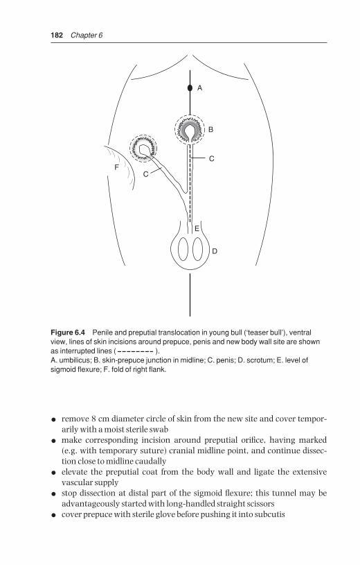
182 Chapter 6
• remove 8 cm diameter circle of skin from the new site and cover tempor-arily with a moist sterile swab
• make corresponding incision around preputial orifice, having marked(e.g. with temporary suture) cranial midline point, and continue dissec-tion close to midline caudally
• elevate the preputial coat from the body wall and ligate the extensive vascular supply
• stop dissection at distal part of the sigmoid flexure; this tunnel may beadvantageously started with long-handled straight scissors
• cover prepuce with sterile glove before pushing it into subcutis
A
B
C
CF
E
D
Figure 6.4 Penile and preputial translocation in young bull (‘teaser bull’), ventralview, lines of skin incisions around prepuce, penis and new body wall site are shownas interrupted lines ( ).A. umbilicus; B. skin-prepuce junction in midline; C. penis; D. scrotum; E. level ofsigmoid flexure; F. fold of right flank.
BSAC06 3/5/05 4:26 PM Page 182

Male urinogenital surgery 183
• carefully avoiding any torsion on the penis or prepuce and noting cranialmidline point previously marked, place preputial skin in new site andmove penis into appropriate position along abdominal wall (see Figure 6.4)
• insert a series of interrupted chromic catgut sutures in the subcutaneoustissues
• suture skin with simple interrupted sutures of monofilament nylon
• close midline skin defect similarly, but include several deeper sutures intolinea alba and rectus sheath to avoid creation of potential dead space
• five-day course of systemic antibiotics
• perform epididymectomy (see Section 6.6, pp. 185–186) or vasectomy(see below).
Three weeks after surgery several ejaculates (approximately three) should beevaluated to ensure that bull is safe for use as ‘teaser’.
6.5 Vasectomy
IntroductionA portion of ductus deferens is removed from the spermatic cord on the cranial aspect of the scrotal neck (see Figure 6.5). This technique is practised
8
6
3
5
42
17
Figure 6.5 Diagrammatic cross-section of scrotal neck (left side) showingrelationship of vas deferens, vascular supply including pampiniform plexus, andcremaster muscle, to vaginal tunics.1. vas deferens enclosed in internal tunica vaginalis; 2. vasculature (pampiniformplexus and spermatic artery) in internal tunica vaginalis; 3. cremaster muscle; 4. external tunica vaginalis; 5. internal tunica vaginalis; 6. skin; 7. scrotal septum(tunica dartos); 8. internal and external spermatic fascia.
BSAC06 3/5/05 4:26 PM Page 183

184 Chapter 6
to produce a teaser bull. Intromission is unaffected, giving the risk of trans-mission of venereal disease.
Surgery may be attempted in the standing animal (if it is not combinedwith penile translocation procedure), but is easier if bull is recumbent with the hind legs extended caudally, or in lateral recumbency with the upperhind leg held well forward, (as for equine castration). The risks of GA in bullsshould be borne in mind.
AnaesthesiaStanding method: xylazine sedation (low dose range, i.e. 0.1–0.15 mg/kg i.m.) followed by local infiltration with 10 ml 2% lignocaine injected subcutaneously around proposed incision site, carefully avoiding possible injection into pampiniform plexus.
Recumbent method: xylazine sedation (high dose range, i.e. 0.2 mg/kgi.m.) supplemented by local infiltration.
Surgical technique
• clip hair from entire upper half of the scrotum and scrotal neck
• scrub and thoroughly prepare scrotum and adjacent skin for asepticsurgery
• apply povidone-iodine twice to the scrotal skin
• make vertical incision 5 cm long on caudolateral (if standing) or craniomedial (cast) aspect of lower part of scrotal neck over tensed cordstructures. In standing position the skin must be rotated through 90°
• grasp spermatic cord between thumb and first two fingers and gentlyrotate to identify ductus deferens as very firm thick string or wire-like structure about 4 mm diameter (it is the hardest structure in the cord)
• carefully make small nick in vaginal tunic over the ductus deferens andplace hook beneath it, or grasp in Allis tissue forceps
• bring ductus through skin incision and clamp across with two pairs ofartery forceps about 5 cm apart (see Figure 6.6)
• resect intervening section of ductus with scissors and place silk ligatures(3 metric) below each forceps
• release forceps; ligature avoids possible recanalisation
• retain ductus for identification (e.g. examine for semen) and possiblehistopathology, as potentially useful evidence in eventual litigation
• close skin incision with interrupted horizontal mattress sutures ofmonofilament nylon
• repeat surgery through second incision on other side
• give systemic antibiotics for five days
• check bull for absence of semen by teasing once weekly for three weeks
• remove skin sutures after 10–14 days
BSAC06 3/5/05 4:26 PM Page 184

Male urinogenital surgery 185
6.6 Epididymectomy
IntroductionThe tail of the epididymis is resected. Resected tissue must be checked forsemen, since, inadvertently, only fat and connective tissue may be removed.Surgery can be done in standing bull under xylazine sedation and localinfiltration. Strict asepsis is essential.
C
DE
B
B
Figure 6.6 Vasectomy in recumbent bull. Vertical incision in lower scrotal neck andcord structures elevated by scissors (see also Figure 6.5).A. cord-like ductus (vas) deferens in internal tunica vaginalis, held by Allis forceps; B. non-absorbable sutures ligating ductus before resection of 5 cm; C. adjacentpampiniform plexus; D. cremaster muscle; E. fold of external tunica vaginalis incisedto expose A. and C.
BSAC06 3/5/05 4:26 PM Page 185

186 Chapter 6
Technique
• clip, wash and disinfect entire scrotal surface; dry and repeat disinfection
• push testicle distally with one hand so that epididymal tail is readilyidentifiable
• incise into epididymis at most ventral point, avoiding testicular substance(see Figure 6.7)
• grasp large portion of tail with Allis forceps and place large artery forcepstransversely proximal to this tissue, which is then resected
• place firm suture of PGA (7 metric) proximal to the artery forceps which isthen removed
• insert two to three horizontal mattress sutures to appose skin edges
• give systemic antibiotics for five days
• remove sutures in 10–14 days
• tease bull weekly for three weeks when no live sperm should be seen; bull isthen suitable for work
DiscussionAgain portions of epididymis removed at surgery should be retained as a precautionary measure should litigation materialise, based on a claim that abull has remained fertile. Recanalisation is impossible following the abovetechniques, in which the lumen of the ductus and epididymis are occluded byencircling ligatures.
Sclerosing agents have also been injected into the testicle and epididymis ofbulls to produce fibrosis and to abolish spermatogenesis and sperm transport.They are less reliable than surgical methods.
Figure 6.7 Location of head, body and tail of epididymis in relation to testicle. Arrowindicates direction and site of incision in an epididymectomy.
BSAC06 3/5/05 4:26 PM Page 186

Male urinogenital surgery 187
6.7 Congenital penile abnormalities
Corkscrew penis or spiral penis
IntroductionThis congenital defect, involving the apical ligament, occurs in bulls olderthan one year. Many affected bulls are known to have served successfully,and have sired progeny. Any breed can be affected, but polled breeds such aspolled Hereford have a higher incidence. Inheritance is questionable.
Signs
• seen just before or immediately following extrusion of the penis from thesheath
• in severe cases the penile tip is caught in distal part of preputial cavity andmay only be extruded naturally with some difficulty
• extruded penis spirals to left or right through 30° angle, viewed from theright side, and 180–270° when viewed (theoretically) from the rear
• glans penis tends to hit right perineal region of cow, about 20–30 cm fromthe vulva
Spiralling action may be variable in degree; in intermittent cases bulls may maintain a low level of fertility in the field. In normal bulls, occurrence of deviation and complete spiralling after intromission increases the contact area between the penis and vagina, and may therefore increase tactile stimuli and so promote ejaculation. In affected bulls the deviation or complete spiralling occurs prior to intromission and prevents coitus.Anatomical explanation involves slipping of the dorsal apical ligament acrossto the left and ventrally. The penile integument in affected bulls is then fully stretched over the penis and produces spiralling early in the process ofcopulation.
As long as the inheritance remains questionable, surgery should only beundertaken after consideration of the ethical position, and then reserved forvaluable bulls required for natural service.
Surgical techniquesTwo techniques are given, one using penile tunica albuginea (TA) the otherwith fascia lata from the thigh region.
Technique 1 Penile tunica albuginea (see Figure 6.8):
• GA, lateral or dorsal recumbency
• extend penis, avoiding accidental spiral rotation, and apply rubber tourni-quet at base
• grasp glans with forceps or tie with bandage, and maintain penis in fullextension
BSAC06 3/5/05 4:26 PM Page 187

188 Chapter 6
• make dorsal midline incision through penile mucosa from just cranial toreflection of prepuce forwards to just caudal to glans
• expose underlying dorsal ligament of penis
• incise longitudinally through middle of ligament into TA
• incise 3 mm laterally to left and right, parallel to previous incisions
• incise transversely and distally across distal end of previous three incisionsso creating two strips (2 mm wide) of ligament and TA
• resect about 1 cm of strip length and re-attach strips to distal end of dorsalligament with alternate sutures of 6 metric chromic catgut and 3 metricstainless steel (respectively to provoke a tissue reaction and for strength)
1
123
23
4
A
B
D
C
Figure 6.8 Surgical correction of spiral deviation of penis (‘corkscrew penis’).A. dorsal surface of penis with epithelium incised and two strips (each 10 cm × 2 mm)of apical ligament freed from tunica albuginea (TA) except proximallyB. cross-section of penis showing (from dorsal): 1. incision through penile epithelium;2. two sections of dorsal ligament (shaded); 3. incision in TA; 4. corpus cavernosumpenis and urethra ventrallyC. enlarged cross-section showing position of sutures fixing strips of apical ligament toTA; 1. sutures from strips to TA; 2. suture closing TA over strips; 3. closure of penileepitheliumD. interrupted sutures closing penile epithelium.
BSAC06 3/5/05 4:26 PM Page 188

Male urinogenital surgery 189
• strip is tacked on to underlying fibrous coat with interrupted sutures every6–7 mm, ensuring firm contact and deep insertion into TA
• close penile epithelium with 4 metric chromic catgut interrupted sutures
• irrigate wound with aqueous solution of penicillin G and release tourniquet
• give antibiotics systemically for five days
• close preputial orifice temporarily by encircling muslin bandage for somehours until bull has recovered from anaesthesia and can retain penis inpreputial cavity
• irrigate preputial cavity daily with oleaginous antibiotic preparation fromday four to day ten
• sexual rest for eight weeks, then tease bull to ascertain new position oferect penis
Technique 2 Fascia lata graft:
• general anaesthesia
• fix the bull in right lateral recumbency
First collect the fascia lata graft as follows:
• clip and prepare a liberal area from the proximal tibia to the tuber coxae forsurgery
• make an incision beginning 8 cm dorsolateral to the patella and continuedfor 20 cm toward the tuber coxae, continue the incision to the fascia lata ofthe vastus lateralis muscle
• collect a 3 cm wide and 20 cm long rectangular strip of the deep fascia lata. Remove connective tissue from the strip, and keep graft in saline;(homogenic fascia lata preserved in 70% ethyl alcohol also has been usedsuccessfully to reduce surgical time and avoid a second incision on thepatient)
• close fascial layers in a simple continuous pattern and appose skin withnon-absorbable suture material in routine fashion
• extend the penis manually, and prepare the penis and prepuce for asepticsurgery
• make a 20 cm skin incision on the dorsum of the penis starting about 2.5 cm proximal to the tip
• deepen the incision to the white fibrous apical ligament
• reflect the apical ligament laterally in both directions, exposing the TA
• do not incise the two veins on the right ventral aspect between the ligament and the TA
• place the fascia lata graft between the apical ligament and the TA on thedorsum of the penis
• insert four interrupted sutures of 2 metric polyglactin 910 through thefascia lata and into the TA under the ligament, near its origin
• then place interrupted sutures along the lateral margin of the graft at 2.5 cm intervals to stretch the implant
BSAC06 3/5/05 4:26 PM Page 189

190 Chapter 6
• trim the implant to fit the distal end of the penis and suture under tensionin an interrupted pattern
• return the apical ligament over the implant and suture it with a simpleinterrupted pattern of the same material; the thickest portion of the ligament should be on the dorsum of the penis
• close the last layer with 3 metric polyglactin 910
• insert a tube into the preputial cavity and secure to the sheath with elastictape to maintain the penis in a retracted position
• give systemic antibiotic agents for three to five days
• maintain sexual rest for 60 days
In some cases (resulting from over- or under-correction) a second operation isrequired. The prognosis remains guarded.
Persistent frenulum
IntroductionCongenital anomaly. Persistence of embryonic ectodermal lamella connectspenis to the penile part of prepuce, which usually splits after the calf is twomonths old, and in which completion is hormone-dependent, but may bedelayed until eight months. Excessive thickness of ventral bridge (= frenulum),perhaps containing blood vessels, may be an aetiological factor.
SignsBand of fibrous tissue runs from close to penile tip to near junction of penilepart of prepuce and sheath, causing marked ventral deviation of erect penis.This prevents intromission.
In the USA it is most common in Aberdeen Angus and Shorthorn bulls, buthas low incidence.
Treatment
• ligate blood vessels in frenulum and section fibrous structure with scissors
Good prognosis. Condition possibly inherited, therefore ethics of surgery aredubious.
Other congenital anomalies of bovine penis
These include:
• congenitally short penis (infantile)
• congenitally short retractor penis muscle
• congenitally tight penile adnexa
All three conditions have a low incidence. The main sign is that penis fails toreach vulva despite normal erection. No surgical correction possible.
BSAC06 3/5/05 4:26 PM Page 190

Male urinogenital surgery 191
6.8 Penile neoplasia
Penile tumours occur in two forms, papillomatosis or fibropapilloma, andmalignant squamous cell carcinoma.
Papillomatosis
Occurs in young bulls, reared in groups. Aetiologically a host-specificPapovavirus, (BPV1).
Signs
• haemorrhage from penis, often on turning out to cows or at breedingexamination
• located on free portion of penis; generally multiple and sessile, but chroniccases may have pedunculated mass
• phimosis or paraphimosis is possible
Treatment
• slow spontaneous regression can occur
• autogenous wart vaccine usually unsuccessful
• surgical removal (knife or Burdizzo®), electrocautery or cryosurgery
• unless simple procedure, patient should be in lateral recumbency to facilitate precision
• sedation with xylazine
• analgesia by local infiltration of dorsal nerves of penis (see Section 1.8, p. 31) or subcutaneous ring block
• care required not to open urethra (potential fistula formation)
• ligate or cauterise significant bleeding vessels
• close epithelial defect with 0 metric chromic catgut (not essential)
The prognosis is good.
Malignant squamous cell carcinoma
Malignant SCC is a rare condition in adult bulls, usually rapidly invasive and involving multiple ulcerating and proliferating masses. Histopathologyfor confirmation? Surgery usually impossible and prognosis correspondinglypoor.
6.9 Castration
IntroductionNeed for castration has been increasingly questioned on scientific, economicand humanitarian grounds. Two forms of castration are available: bloodlessand open surgical (scalpel).
BSAC06 3/5/05 4:26 PM Page 191

192 Chapter 6
Bloodless methods
Rubber ring methodThis method should not be used, nevertheless this technique is becomingmore common in some areas. The technique has been ethically condemnedin the UK and is illegal without anaesthetic in some countries (e.g. Germany,Switzerland). Legally in the UK a rubber ring, without anaesthetic, is onlypermitted in the first week of life.
Application of tight-fitting rubber ring to scrotal neck in calves appears tocause considerable post-operative discomfort and marked setback to growth.Some sepsis is not uncommon at site following skin necrosis. Careless applica-tion can fail to position both testes below rubber ring, resulting in induced(iatrogenic) inguinal cryptorchidism. It also carries a risk of tetanus, probablyas the duration of pressure necrosis is much greater than in other bloodlessmethods.
In the USA rubber band tubing has even been used, especially in feedlots,for the castration of large bulls. The ring is applied with the EZE® BloodlessCastrator (Wadsworth Mfg., Dublin, MT). As in the calfhood method a vascu-lar necrosis of the scrotum and testes results. The authors have no experienceof this technique which is alleged to result in ‘minimal stress’.
AnalgesiaIn the UK, legislation (Animal Anaesthetics Act 1964) makes it obligatorythat castration of calves over two months old be performed under anaesthesiaor analgesia.
In Switzerland castration without anaesthesia is forbidden.
• 10 ml syringe and 2% lignocaine (with adrenaline)
• testicle is grasped between thumb and first two fingers and drawn to ventral part of scrotal sac
• needle is introduced in horizontal or vertical position, upwards into substance of testicle, and 3 ml solution is injected
• testicle is released and 2 ml is deposited in subcutaneous position duringwithdrawal
• procedure is repeated on the second side
An alternative technique involves subcutaneous infiltration at scrotal neck,injection of a further 3–5 ml into each cord, because one author (AS) believesthat intratesticular injection is painful, minimally effective and should there-fore be avoided. Maximum dose rate of lignocaine is 4 mg/kg bodyweight.
Burdizzo® methodThe Burdizzo® instrument is 30 or 40 cm long, and has cord stops. It is used on calves one to twelve weeks old, depending on breed and development.
BSAC06 3/5/05 4:26 PM Page 192
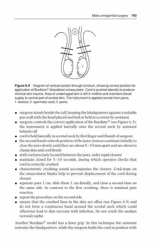
Male urinogenital surgery 193
• surgeon stands beside the calf, keeping the hindquarters against a suitablepen wall with the head placed and tied or held in a corner by assistant
• surgeon controls the correct application of the Burdizzo® (see Figure 6.5):the instrument is applied laterally onto the scrotal neck by assistantbehind calf
• cord is held laterally in scrotal neck by first finger and thumb of surgeon
• the second hand controls position of the jaws; instruct assistant initially toclose the jaws slowly until they are about 8–10 mm apart and are about toclamp skin and cord firmly
• with cord precisely located between the jaws, order rapid closure
• maintain closed for 5–10 seconds, during which operator checks thatcord is correctly crushed
• characteristic crushing sound accompanies the closure. Cord-stops on the emasculator blades help to prevent displacement of the cord duringclosure
• separate jaws 1 cm, slide them 1 cm distally, and close a second time onthe same side. In contrast to the first crushing, there is minimal pain reaction
• repeat the procedure on the second side
• ensure that the crushed lines in the skin are offset (see Figure 6.9) and do not form a continuous band around the scrotal neck which could otherwise lead to skin necrosis with infection. Do not crush the median(scrotal) raphé.
Another Burdizzo® model has a knee grip. In this technique the assistantrestrains the hindquarters, while the surgeon holds the cord in position with
3
2
1
Figure 6.9 Diagram of vertical section through scrotum, showing correct position forapplication of Burdizzo® (bloodless) emasculator. Cord is pushed laterally to produceminimal skin trauma. Area of undamaged skin is left in midline and maintains bloodsupply to ventral part of scrotal skin. The instrument is applied remote from penis.1. testicle; 2. spermatic cord; 3. penis.
BSAC06 3/5/05 4:26 PM Page 193
194 Chapter 6
one hand while closing the instrument by moving one handle against theknee-fixed second handle. A certain dexterity is required.
The effect after four weeks (young calf ) or six weeks (older calf ) is atrophy of the testicular tissue, which forms a fibrous knob or knot-like structureapproximately the diameter of the spermatic cord.
ComplicationsProblems are infrequent but can include:
• inadvertent crushing of the penis by inexperienced lay operator, as jawsare applied too high, causes urethral blockage and rupture with s.c. urineseepage. Salvage procedure is only possible solution
• gross bruising of scrotal tissues due to slackness of jaws, therefore equip-ment should be regularly overhauled and the instrument should be storedin the open position. Burdizzo® should be capable of cutting piece of stringinserted between two pieces of cigarette carton card when functioningnormally
• necrosis of scrotal skin (see above)
• oedema of caudal abdominal wall and scrotum due to slack jaws
• unilateral absence of testicular atrophy, as cord was inadvertently com-pletely or partially missed
Surgical (scalpel) methodThis technique involves removal of the testicles and haemostasis of the spermatic cord vessels by:
• traction on spermatic cord
• torsion and traction on spermatic cord
• crushing using an emasculator
• crushing and ligation of spermatic cord
Technique
• perform surgery in standing position with stockman restraining head andhindquarters and holding tail laterally
• induce local analgesia, as described above
• wash and cleanse scrotal skin with dilute (0.5%) povidone-iodine solu-tion, and preferably wear disposable gloves
• stand or crouch behind calf to make vertical incision through caudal scrotal skin into testicular substance, holding testicle tensed and pulleddistally, and continue incision along distal, i.e. ventral border of scrotum(ensuring subsequent drainage)
• scrotal incision should be slightly shorter than testicular length.Initial scrotal incision may be made with Newberry® knife (Newberry®
castrating knife, Jorgensen Labs, Loveland, CO. USA), a 24.5 cm longinstrument with steel-bladed clamp) which is placed transversely across
BSAC06 3/5/05 4:26 PM Page 194

Male urinogenital surgery 195
the base of the scrotum, closed, and immediately pulled ventrally to openboth scrotal sacs without removal of any scrotal tissue (see Figure 6.10).Both testicles may then removed with the emasculator.
• testicle prolapses through skin wound, is grasped, and vascular and non-vascular portions of the cord are identified (vascular: cranial includingpampiniform plexus and vas deferens)
• insert first finger through tunica vaginalis proximal to the epididymis, andbetween vascular and non-vascular parts.
Technique now varies depending on method of haemostasis:
• small calf (one week to two months): emasculator, torsion, traction arepossibilities in descending order of preference
• large calf (two to six months): emasculator, torsion. Traction is contra-indicated as excessively painful
• small bull: emasculator, possibly with ligation
All other methods carry a risk of severe haemorrhage.
Figure 6.10 Newberry castration technique. View from rear, showing testes pushedupwards, scrotum pulled down by hand, and Newberry knife being applied acrossscrotum for transverse incision, before being pulled quickly downwards. Dotted linesrepresent the position of the tunica vaginalis, through which the testes are then extracted.
BSAC06 3/5/05 4:26 PM Page 195

196 Chapter 6
Emasculator method
• apply after separating vascular and non-vascular parts of cord
• brief period (10 seconds) to crush non-vascular position just proximal toepididymis
• longer period (20–120 seconds depending on size of cord and the age ofthe animal), crushing proximal part of vascular portion of cord, usingtraction on distal structures to separate them from jaws of instrument
Torsion method
• break down non-vascular part of cord by traction
• twist vascular portion several (five to six) times in proximal part of cord,then use gentle traction to break cord distal to torsion point
Traction method
• break down non-vascular part, then grasp vascular portion proximally,increasing steady traction until cord ruptures and undergoes considerableelastic recoil (see Section 3.11, p. 120)
• repose any tissue protruding from wound, or resect protruding length ofductus deferens
• do not handle tissue unnecessarily
• do not become involved in calf restraint
• maintain scalpel and emasculator in bucket with antiseptic and have second bucket of antiseptic solution for washing scrotum
• local medication is unnecessary
Technique for small bullEmasculator and ligation; strict aseptic precautions and antiseptic prepara-tion of skin are essential. Use sterile emasculator and suture material.
• incise as above, caudally, into substance of scrotum, continuing incisiondistally
• apply emasculator to non-vascular part of cord, then to vascular portion,close tightly and remove testis
• leave instrument in place for minimum of 1 minute
• place circumferential ligature around cord 1 cm proximal to blade, asextra security against haemorrhage
• place artery forceps on edge of cord proximal to ligature but not acrosscord, remove emasculator, and check for haemorrhage
• release artery forceps
Ensure clean bedding. Some exercise is advisable for one week.
BSAC06 3/5/05 4:26 PM Page 196

Male urinogenital surgery 197
Complications
• infection – gross contamination at surgery, (rubber gloves!) dirty bedding
• severe swelling – infection, oedema, poor drainage due to small incision
• haemorrhage
• preputial oedema – invariably from extension of scrotal swelling
• tetanus is rare (prophylaxis in at-risk calves)
E
A
BC
D
Figure 6.11 Emasculator castration (diagrammatic). Scrotum has been inciseddistally into testis which has then been expressed from scrotum. Parietal tunicavaginalis has been sectioned by scalpel and has retracted into scrotum.A. Emasculator placed across cord structures with cutting edge of blade distal andcrushing edge proximal (‘nut’ to ‘nut’); B. pampiniform plexus and spermatic artery; C. ductus deferens; D. testicular incision; E. site for application of artery forceps toretain stump for a few seconds’ inspection if warranted.
BSAC06 3/5/05 4:26 PM Page 197

CHAPTER 7
Lameness
Systemic infections such as Foot-and-Mouth disease (FMD) and clostridialdiseases, metabolic conditions such as ketosis and hypomagnesaemia, as wellas nutritional diseases (e.g. hypovitaminosis E), are not considered in thisbook although they may lead to signs of lameness.
7.1 Incidence
Veterinary practice figures (UK) indicate an average annual incidence of4–6% lameness in dairy cows. If treatment carried out by the farmer isincluded, however, the true incidence increases to 25–30%. The incidence
7.1 Incidence 1987.2 Economic importance 1997.3 Terminology 2007.4 Interdigital necrobacillosis 2027.5 Interdigital skin hyperplasia 2047.6 Solear (solar) ulceration 2067.7 Punctured sole 2087.8 White line separation and
abscessation 2097.9 Laminitis (coriosis) 2107.10 Other digital conditions 212
(a) digital dermatitis (Mortellaro)(b) verrucose dermatitis(c) interdigital dermatitis(d) heel erosion (‘slurry heel’)(e) longitudinal (vertical) or
transverse (horizontal)sandcrack or fissure
(f ) fracture of distal phalanx(pedal bone)
7.11 Deep digital sepsis 2197.12 Digital amputation 2207.13 Other digital surgery 225
(a) tendon resection followingamputation
(b) tendon resection withoutamputation
(c) resection of distalinterphalangeal joint
(d) removal of interdigitalhyperplastic skin
7.14 Hoof deformities, overgrowth and corrective foot trimming 231
7.15 Footbaths 2347.16 Check lists for herd problems 2357.17 Infectious arthritis (‘joint ill’)
in calves 2387.18 Contracted flexor tendons 2407.19 Tarsal and carpal hygroma 2427.20 Patellar luxation 2437.21 Spastic paresis 2467.22 Hip luxation 2487.23 Stifle lameness 2507.24 Nerve paralysis of limbs 2517.25 Limb fractures 2517.26 Use of acrylics and resins in
orthopaedic problems 2557.27 Antibiotherapy of bone and
joint infections 257
BSAC07 3/5/05 4:26 PM Page 198

Lameness 199
may range from 3–100% on individual farms. Any farm with an incidenceover 15% should be considered to have a lameness problem which requiressystematic investigation (see Section 7.16). In the intensive agriculturalenterprises common in western Europe and North America:
• 95% of lame cattle are dairy breeds
• 80% of cases involve the digits
• 80% of digital lameness is located in the hindlimbs
• 50% of digital lameness involves the horny tissue and 50% the skin,mostly digital dermatitis
• 70% of the horny lesions involve the outer claw
In the UK today the three major digital lameness problems with rather similarincidence rates, are digital dermatitis, sole ulcer, and white line disease. Thelatter two may sometimes be related to a previous incident of laminitis.
Essential equipment for investigation of bovine lameness problems includes:
• facilities for good restraint and ready elevation of hind- or forelimbs ideallypurpose-built for foot work e.g. Wopa crush/chute or a rotating model
• good lighting, hosepipe, water, bucket, brush, and ropes
• left- and right-handed hoof knives
• double action hoof cutters
• hoof rasp
• hoof testers
• straight grooved probe
• grinder for large-scale (i.e. herd) trimming with electrical cut-out in caseof short-circuiting
7.2 Economic importance
Only infertility and mastitis cause greater economic losses than lameness inmost intensive dairy units in the UK, western Europe (Netherlands, Denmark,Belgium, Germany) and North America. The 2004 estimate of UK losses dueto cattle lameness is well over £100 million (US $180 million). In develop-ing countries infectious diseases and malnutrition are of more economicimportance.
Economic lossesThe direct cost of each lameness case is about £150 ($270), but if such factorsas prolonged calving interval, replacement costs and culling losses areincluded, the figure rises to approach £300 ($540) for each lame cow in anational herd of about three million dairy cows (UK). Losses are similar in USA.
Losses result from:
• reduced milk yield
• weight loss
BSAC07 3/5/05 4:26 PM Page 199

200 Chapter 7
• disposals, deaths, replacement cost
• infertility, prolonged calving interval
• veterinary expenses, drugs
• additional stockman’s time
A major loss is reduced milk yield, including discarded antibiotic-treatedmilk during therapy period. This accounts for about a quarter of all losses dueto lameness. Lameness is primarily in the early lactation period (one to threemonths postpartum) and in the immediate prepartum week. This time ofmaximum incidence results in greater economic losses than lameness in midor late lactation. Several studies have shown that higher yield cows tend tohave an increased risk of lameness, also that a reduction in yield actually pre-cedes lameness onset. No satisfactory explanation has been found for thesetwo (UK) facts. Cash loss due to a lameness successfully cured within 24hours of onset may represent 1% of lactation yield. If treatment is delayed,loss may be 20% or more of lactation yield.
Weight loss may be 10% or, in cows eventually culled due to continuingdeterioration and poor yield, may reach 25%.
Disposals as ‘lame cow culls’ in the UK are about 2–4% annually, but suchlame cows are often also affected with mastitis and/or metritis leading toinfertility. The replacement cost for culls is considerable.
Infertility attributable to lameness results from:
• failure of oestrus detection (cow often recumbent, unwilling or unable tomount neighbouring cows) or delayed return to oestrus
• anoestrus
• poor body condition postpartum (negative energy balance)
• concurrent low grade metritis
These losses are subtle, and often not fully appreciated by the dairy farmer,but are the greatest source of cash loss (see Figure 7.1).
Veterinary expenses and drugs form a small proportion of total costs. Theadditional labour costs, often ignored, are high since treatment usuallyinvolves two or three people, and each case requires several minutes’ atten-tion, often on a daily basis (see Figure 7.1).
Hoof traitsVariations in hoof traits relate to survival rates, reproductive performance,and increased yield from first to subsequent lactations (see Section 7.14 pp. 231–232).
7.3 Terminology
Terms have been introduced to give an acceptable terminology to forms oflameness common to various countries, where a variety of words have been
BSAC07 3/5/05 4:26 PM Page 200

Lameness 201
in use. The Latin terms are listed beside the English language equivalent (andillustrated in Figure 7.2).
Interdigital skin:
• digital dermatitis – dermatitis digitalis
• interdigital necrobacillosis – phlegmona interdigitalis
• interdigital skin hyperplasia – hyperplasia interdigitalis
• interdigital dermatitis – dermatitis interdigitalis
• verrucose dermatitis – dermatitis verrucosa
Horny tissues:
• solear ulceration – pododermatitis circumscripta septica
• punctured sole – pododermatitis septica (traumatica)
• white line separation – pododermatitis septica diffusa (disputed)
• laminitis – pododermatitis aseptica diffusa
• longitudinal (vertical) or transverse (horizontal) sandcrack – fissura ungulaelongitudinalis et transversalis
• heel erosion – erosio ungulae
For further details see Further Reading, pp. 259–260.
34
13
86
1325
C
B
D
E
F
A
Figure 7.1 Economic losses in dairy cattle due to lameness: A. infertility; B. reduced milk yield and sales; C. deaths, disposals and replacements; D. weight loss; E. veterinary expenses, drugs; F. cost of extra labour.
BSAC07 3/5/05 4:26 PM Page 201

202 Chapter 7
Claw zonesThe bearing surface of the claw has been divided into six zones to aid record-ing (see Figure 7.3). Thus white line separation and abscessation tends to bein zones one or two, sole ulcer in zone four, and laminitic changes predomin-antly in zones four and five.
Lameness scoring (LS)Many numerical scales (e.g. 0–3, 1–6, 1–10) have been devised and utilisedin recording the severity of lameness in an individual cow. The simplestappears adequate, viz. 0 = not lame; 1 = slightly lame; 2 = moderately lame,and 3 = severely lame/often recumbent (e.g. LS2).
7.4 Interdigital necrobacillosis
IntroductionSynonyms: phlegmona interdigitalis, ‘foul-in-the-foot’, ‘clit ill’, ‘foot rot’,interdigital pododermatitis. A peracute form, colloquially ‘superfoul’ hasbeen encountered in some countries recently.
Definition: acute inflammation of subcutaneous tissues of interdigital spaceand adjacent coronary band, spreading to dermis and epidermis.
1. Digital dermatitis 5. Heel erosion
6. Interdigital dermatitis
7. Laminitis
9. Punctured site
10. Toe ulcer
8. Interdigital hyperplasia
2. Solar ulceration
3. White line separation + abscessation
L M
4. Interdigital necrobacillosis
Figure 7.2 Important conditions of the bovine digit.1. digital dermatitis; 2. sole ulceration; 3. white line separation and abscessation; 4. interdigital necrobacillosis; 5. heel erosion; 6. interdigital dermatitis; 7. laminitis/solehaemorrhage; 8. interdigital hyperplasia; 9. punctured sole; 10. toe ulcer.
BSAC07 3/5/05 4:26 PM Page 202

Lameness 203
Signs
• mild to severe lameness (LS 1–3) of sudden onset, all ages
• interdigital swelling, later involving coronet and pastern
• toes spread apart due to interdigital swelling, initially with unbroken skinfor first 24 hours of lameness
• sometimes more proximal spread, and a secondary interdigital necrosisvery common
• little pus but characteristic foul smell and pain with split in interdigitalskin
Aetiology: interdigital microtrauma and infection with Fusobacteriumnecrophorum, Bacteroides melaninogenicus and other organisms.
Pathology: cellulitis and liquefactive necrosis of interdigital skin, withfissure formation and later, if untreated, development of granulation tissue, eventually resulting in interdigital granuloma. Advanced cases can develop digital septic arthritis and other deeper complications. The disease course is much more rapid in ‘superfoul’ where cows may have to be culled 48–72 hours after disease onset, due to the extent of destructivechanges.
Differential diagnosis: interdigital foreign body, acute laminitis, solearpenetration by foreign body, severe interdigital dermatitis, interdigitalchanges from BVD/MD, FMD, distal interphalangeal septic arthritis, distalphalangeal fracture.
6
35
4
1
2
Figure 7.3 Zones of the sole.1. white zone at toe; 2. abaxial white zone; 3. axial groove zone; 4. apex of sole; 5. junction of sole and bulb (heel); 6. bulb (heel).
BSAC07 3/5/05 4:26 PM Page 203
204 Chapter 7
Treatment
• ceftiofur, ampicillin, LA oxytetracycline, penicillin, sulphonamides (e.g.trimethoprim-sulpha) systemically
• clean affected necrotic area with disinfectant and apply a topical oxytetra-cycline or copper sulphate, or BIPP paste (bismuth subnitrate, iodoformand petrolatum)
• do not bandage, but put on to dry floor or straw bedding, preferably isolated to avoid spread of infection
• daily cleansing with disinfectant if feasible
• ‘superfoul’: early cases respond well to 6 g oxytetracycline, moreadvanced cases to tylosin, careful local débridement under analgesia andlocal antibiotic dressing. Isolation is important.
Prophylaxis
• check and improve drainage in areas where interdigital trauma can arise(e.g. gateways, tracks, stubble)
• improve dry conditions underfoot (straw yard) and increase frequency ofscraper removal of slurry from passageways
• footbaths of zinc sulphate (5–10%) copper sulphate (5%) or formalin (4%)(see Section 7.15, pp. 234–235)
• antibacterial feed additive: sulphabromomethazine in feedlot outbreak, orethylenediamine dihydroiodide for prophylaxis, though results have beenconflicting (North America)
• spread quicklime in muddy tracks or around water troughs
DiscussionFrequent form of digital lameness (e.g. 15% of total), but of relatively lessereconomic importance since the 1980s. Outbreaks of interdigital necrobacil-losis require methodical investigation of aetiology to develop appropriate prophylactic measures. Emphasis on major areas for digital trauma (fromyard to parlour to farm tracks) and for potential contamination. Review clawtrimming needs.
7.5 Interdigital skin hyperplasia
IntroductionSynonyms: hyperplasia interdigitalis, corn, interdigital granuloma, interdig-ital vegetative dermatitis, fibroma, ‘wart’.
Definition: proliferative reaction of interdigital skin and/or subcutaneoustissues to form a firm mass.
Incidence: usually sporadic, common in certain beef breeds (e.g. Hereford)and in bulls at AI centres. Occasionally follows severe interdigital disease in
BSAC07 3/5/05 4:26 PM Page 204

Lameness 205
dairy cows, then is unilateral. May start in yearling bulls, but most clinicalcases (with lameness) are in adults of four to six years.
Predisposition: inherited in some breeds (e.g. Hereford, Holstein Friesian).Severe interdigital dermatitis or sole ulcer often precedes involvement of single limb. Frequently associated with poor conformation e.g. splayed toeswith wide interdigital space.
Signs
• slight or no lameness (LS 0–1) depending on size and mechanical interfer-ence in simple case
• large lesions develop superficial digital traumatic ulceration, and contactinterdigital axial skin may undergo pressure necrosis
• both forms readily become secondarily infected with Fusobacteriumnecrophorum
• more or less symmetrical in hindlimbs of beef breeds, especially in bulls,possibly with bilateral forelimb involvement, which suggests inheritedbasis
• single abaxial hindlimb involvement suggests secondary response torecognised previous insult involving interdigital swelling and sometimessole ulcer
Pathology: skin hyperplasia with secondary ulceration. Variable degree ofhyperkeratosis (misnamed papillomatosis).
Differential diagnosis: interdigital foreign body, interdigital necrobacillo-sis, digital dermatitis.
Treatment
• none if small and asymptomatic
• local caustic (e.g. silver nitrate, copper sulphate) if small and causinglameness
• most clinical cases require resection by knife surgery electrocautery orcryosurgery: ideally in Wopa crush under IVRA (intravenous regionalanalgesia), bandage (e.g. Vetrap®) after applying sulphadimidine powder.Remove bandage after one week.
DiscussionAcquired cases develop suddenly in middle of interdigital space; while congenital cases, manifest as slight swelling in yearling and steadily enlarg-ing with adulthood, start as fold in the axial skin of the abaxial digit.Theoretically, breeding policy should be altered to reduce the inheritance risk of such cases of multilimb hyperplasia. Inheritance may be related to skinthickness, an alleged slackness of the interdigital cruciate ligaments and theamount and distribution of body fat.
BSAC07 3/5/05 4:26 PM Page 205

206 Chapter 7
7.6 Solear (solar) ulceration
IntroductionSynonyms: pododermatitis circumscripta, sole ulcer, Rusterholz ulcer.
Definition: circumscribed limited reaction of the pododerm (deep sensitivetissues) often characterised by an erosive defect typically at the sole-heeljunction of the lateral hind claw.
Incidence: high (up to 40% of digital lameness cases) and widespread indairy breeds, from calved heifers to mature cows in good body condition.
Aetiology: disputed, possibly excessive weight-bearing by lateral claw following horn overgrowth. Associated almost invariably with abnormalclaw (poor trimming), and frequently with laminitis. Primary and secondarycauses are difficult to distinguish.
Predisposition: inherited factors such as stance, which may also beacquired, and deviations from normal hoof shape (e.g. severe overgrowth),loose housing with small (narrow, short) cubicles, giving tendency for cowsto stand with hind feet in passageway, overgrown digits, acidotic rations.
Signs
• moderate degree of lameness (hesitant, wary gait, slightly arched back, LS 1) typically up to three months postpartum, masking the frequentlybilateral nature of the lesions, i.e. lateral claw of both hind legs, one morepainful than the other
• severe lameness (LS 2–3) when granulation tissue protrudes and in pres-ence of deeper purulent infection (osteomyelitis, septic arthritis)
• under-run heel horn exposes sensitive laminae
• contralateral claw: check for similar changes!
• at typical site (see Definition) granulation tissue may protrude throughundermined horn
• under-running commonly extends cranially and peripherally to abaxialwhite line
Bacteriology: none in primary condition.
Pathology: horn defect originates from damaged pododerm, which may besecondary result of laminitis (coriosis, pododermatitis aseptica diffusa), poortrimming, and heel horn deformity. Caudal border of distal phalanx andinsertion of deep digital flexor tendon (see Figure 7.4) both lie below typicalsite and are involved in complicated cases (see Section 7.11, p. 219 on deepdigital sepsis).
BSAC07 3/5/05 4:26 PM Page 206

Lameness 207
Differential diagnosis: solear abscessation due to foreign body penetration,aseptic pododermatitis (solear haemorrhage) elsewhere in weight-bearingsurface, simple heel erosion, subacute laminitis.
Treatment
• trim all feet initially or at end
• IVRA (intravenous regional analgesia, see Section 1.8, p. 33)
• remove under-run horn, trim horn of wall and heel so that weight-bearingby affected claw is minimal
• possibly apply block (hoof resin) to sound claw which should be minimallytrimmed, if weight-bearing cannot otherwise be reduced
• remove protruding granulation tissue, leaving healthy pododerm andapply tetracycline spray, and bandage (waterproof) for five days
• alternatively, put on sulphadimidine powder, bandage (Vetrap®), andspray oxytetracycline over bandage to prevent wicking by mud into bandage
• broad spectrum antibiotics in septic cases
• confine to box and straw bedding for five days
Prophylaxis
• avoid overgrown claws by emphasising need for routine trimming (seeSection 7.14, p. 232)
1
55
3 24
Figure 7.4 Typical site of sole ulcer of lateral hind claw with adjacent structures that may be affected by deep sepsis: 1. distal phalanx; 2. deep digital flexor tendon; 3. distal sesamoid (navicular bone); 4. navicular bursa; 5. distal interphalangeal joint.
BSAC07 3/5/05 4:26 PM Page 207

208 Chapter 7
• avoid factors predisposing to laminitis (see Section 7.9, p. 210) and inter-digital dermatitis (e.g. excessive moisture)
• do not breed from affected young cows
7.7 Punctured sole
IntroductionSynonym: pododermatitis septica (traumatica), septic (traumatic) pododermatitis.
Definition: diffuse or localised septic inflammation of solear corium, causingmoderate to severe lameness if purulent.
Incidence: sporadic.
Predisposition: thin solear horn from preceding laminitis (coriosis) exces-sive abrasion from rough concrete, rough tracks, any tendency to hustle herdalong inside passageways or outside tracks.
Corium (pododerm) has no fatty tissue in toe area, making entry of infec-tion into distal phalanx easier.
Signs
• sudden onset of lameness (LS 2), usually in hindlimb, with solear penetration
• site often near toe or adjacent to white line
• defect in horn extends to solear pododerm, with variable under-runningand pus production (black colour)
• localised pain
Aetiology: often iatrogenic following removal of excessive horn during trimming; also see Predisposition above. Foreign bodies (FB) include stones,often flints, nails, wire and thorns.
Bacteriology: secondary infection often mixed, including Arcanobacteriumpyogenes.
Pathology: see Definition; Secondary complication is osteomyelitis of distalphalanx or distal interphalangeal joint (see Figure 7.4).
Differential diagnosis: subacute laminitis, solear ulceration, toe ulcerinterdigital necrobacillosis.
Treatment
• primarily surgical: identify and remove FB; drain after exposure of under-run horn
• local astringent dressing
• curette distal phalanx if involved
• possibly elevate sole by block on other digit
BSAC07 3/5/05 4:26 PM Page 208

Lameness 209
• single injection of long-acting oxytetracycline if soft tissue (corium) isseverely damaged
• tetanus prophylaxis indicated in known tetanus environment
Prophylaxis
• avoid predisposing causes contributing to poor quality of solear horn, i.e.laminitis (coriosis)
• good hygiene (foreign body disposal)
• if track to pasture is hazardous due to flints, consider installation of‘friendly’ cow track
7.8 White line separation and abscessation
IntroductionSynonym: white line disease.
Definition: abaxial, or less commonly axial, wall separation from laminae atsole-wall area extending proximally, with cavity impacted with mud, faeces;or with development of abscess cavity at deepest part (abscessation).
Incidence: high and in some areas is major cause of digital lameness.
Predisposition and pathogenesis:
• abnormal horn production resulting from laminitic insult (coriosis)
• insufficient hoof trimming
• related to peripartum events some months previously
Signs
• moderate lameness (LS 1–2)
• white line wider than usual, and in early stages has series of pinpoint darkmarks, later obvious foreign material impacted in white line
• separation evident on paring, no pain
• cases of white line abscessation are lame and have pain localised to wall
• internal wall abscess, without obvious track distally, also very sensitive topincers pressure
• advanced cases have supracoronary septic sinus discharge (see Section7.11, p. 219)
Aetiology: see Predisposition.
Bacteriology: Arcanobacterium pyogenes in abscessation.
Pathology: pressure necrosis of wall laminae, possibly also of solear laminae,following under-running and septic laminitis tracking progressively moreproximally after entry of purulent micro-organisms, with absence of naturaldrainage distally due to impacted material.
BSAC07 3/5/05 4:26 PM Page 209

210 Chapter 7
Differential diagnosis: solear foreign body, laminitis, small coronary sandcrack.
Treatment
• routine trimming of all digits
• pare off wall over impacted and septic area to achieve drainage and prevent further impaction
• also remove all under-run sole (some cases have a large ‘false sole’)
• apply local antiseptic dressing (e.g. oxytetracycline spray) and firm dressing
• pare horn to normal shape of claw
• consider block on ipsilateral claw
• in septic cases give broad spectrum antibiotics for three days
Radical surgery, possibly amputation, is required in involvement of coron-ary tissues and distal interphalangeal joint (see Sections 7.11 and 7.12, pp. 219–225).
ProphylaxisAvoid predisposing factors for laminitis (coriosis) (see Section 7.9 below), andensure regular hoof trimming.
7.9 Laminitis (coriosis)
IntroductionSynonym: Pododermatitis aseptica diffusa, coriosis, ‘founder’.
Definition: diffuse acute, subacute, subclinical or chronic inflammation of pododerm, usually in several digits. Chronic cases without acute stage(subclinical) are often seen.
Incidence: sporadic acute cases, widespread subacute, subclinical andchronic cases commonly in dairy units, high incidence in recently calvedheifers and younger cows around parturition. Acute form occasionally pre-sents as outbreak in barley beef units. Common in beef feedlots.
Predisposition:
• inherited factors (proven in Jersey)
• parturition
• feeding stress (ruminal lactic acidosis, subacute ruminal acidosis orSARA) from change of dry cow concentrate diet to high productionrations, with potentially dangerous reduction of roughage intake
• exacerbation by trauma (overburdening), as in excessive standing due toreluctance to use cubicles (inexperience, bullying by herdmates)
BSAC07 3/5/05 4:26 PM Page 210

Lameness 211
Signs
• acute stage: painful hot digit, digital arterial pulsation, general depression,severe lameness, abnormal stance, possibly recumbent (LS 2–3)
• subacute: less painful but persistent stiffness, stilted gait, solear and whiteline haemorrhages (LS 1)
• chronic: stiff gait or not lame (LS 0–1), ‘slipper foot’ malformation withhorizontal lines on wall, concave dorsal wall, widened white line and evidence of old solear amd white line haemorrhages. See section onpathology below
Aetiology: see Predisposition and Pathology.
Bacteriology: none.
Pathology: blood and serum exudation in acute stage, later (chronic)grooves on hoof wall, concave profile, widened white line and flat sole.Significant sinking of distal phalax due to peripartum slackening of the connective tissue support structures has been proved; thin sole or ulcerationnear tip of distal phalanx (‘toe ulcer’) is evident as haemorrhage (‘bruising’)as toe tip has no fat layer in its corium. White line lesions may develop intowhite line disease (see Section 7.8, p. 209); sole lesions at sole-heel junctionmay develop into solear ulceration (see Section 7.6, p. 206).
Histopathology: oedema, haemorrhages and thrombosis in acute stage,fibrosis and chronic thrombosis in later stages.
Differential diagnosis: bruised sole, white line disease, punctured sole,solear ulceration all of which may be present (see Figure 7.2).
Treatment
• acute stage: give systemic NSAIDs (flunixine meglumine or meloxicam) orpossibly corticosteroids (only if non-pregnant) and diuretics
• ensure exercise (to improve local circulation and further reduce develop-ing oedema), preferably by turning on to soft ground, e.g. field
• remove any precipitating dietary causes
• feed no concentrates until acute phase is over
• in recumbent case consider digital nerve block to get heifer or cow tostand, then forced exercise
• subacute stage: as in acute case
• chronic case: hoof trimming
Prophylaxis
• avoid large amounts of prepartum concentrates (‘steaming up’ ‘lead feeding’), which should not exceed 2 kg daily
• avoid high intake of concentrate in early lactation, and aim at peak yieldabout six weeks postpartum
BSAC07 3/5/05 4:26 PM Page 211
212 Chapter 7
• ensure ready access to roughage immediately before and after concentrateintake, or consider change to complete diet feeding (TMR, total mixedrations) if problem persists
• improve buffering capacity of rumen fluid (avoid lactic acidosis or SARA)by increasing saliva production: give iodide or rock salt, grass or lucernenuts in concentrate
• consider adding 1% sodium bicarbonate to concentrate ration, whichshould be fed as three to four daily portions
• accustom down-calving heifers gradually to concrete yards and cubiclesseveral weeks beforehand, but ensure plenty of exercise in both pre- andpost-partum weeks
• avoid exposure to excessive sole wear from long stony tracks, rough concrete
• high fibre diets should be used in rearing dairy heifers in long term planning
• ensure regular claw examination and trimming
DiscussionExcess lactic acid production alters rumenal bacterial flora, and causesrelease of bacterial endotoxins involving histamine release and stagnation ofblood in laminae of digital horn, with consequent hypoxia and functionalischaemia. Ischaemic necrosis of the corium and laminae heals by fibrosis.These tissues then inevitably produce defective (soft, poor quality) horn inaberrant manner, resulting in signs seen in subacute and chronic stages.Toxic conditions (mastitis, metritis) may also contribute to development oflaminitis in some dairy cattle.
7.10 Other digital conditions
(a) Digital dermatitis (Mortellaro)
IntroductionSynonym: dermatitis digitalis, ‘hairy warts’, PDD, papillomatous dermatitis.
Definition: circumscribed superficial ulceration of skin bordering coronarymargin at heels, occasionally more dorsally. Can develop into mass of verru-cose fronds (see Verrucose dermatitis, p. 214).
Incidence: Widespread in many dairy farms in western Europe (UK,Netherlands, Germany, etc.) and North America (California, New York State,etc.), where incidence can reach 100% and prevalence 20%. Often the majorlameness problem.
BSAC07 3/5/05 4:26 PM Page 212

Lameness 213
Signs
• predisposition is not known, but disease affects adult cattle causing vari-able and often severe lameness (LS 1–3)
• white epithelial border and signs of surrounding chronic dermatitis
• caudally in skin adjacent to heel horn, also occasionally anterior interdig-ital space, coronary band or in granulation tissue on sole (see Figure 7.5)
• apparently contagious, mostly introduced into ‘clean’ farm by bought-inheifers which may not themselves show clinical lesions
• role of skin microtrauma as permitting entry of organisms is speculative.
Bacteriology: believed to involve a Treponema genus spirochaete, possiblyalso a longer filamentous organism. Other postulated organisms includeBorrelia burgdorferi, Dichelobacter nodosus and Campylobacter spp.
Differential diagnosis: possibly interdigital dermatitis, heel horn erosion,plantar eczema, but lesions are usually unmistakable.
Treatment
• treat all but most severe lesions in milking parlour
• cleanse with water spray, wait one minute, and apply topical aerosolantibiotic, e.g. oxytetracycline, daily for three days (long angled nozzlefacilitates spray direction)
• drugs have included oxytetracycline, lincomycin, erythromycin and lincomycin/spectinomycin, and tylosin
• severe proliferating masses (‘hairy warts’) should be resected through epidermis (not subcutis) under local anaesthesia (e.g. IVRA, see Section1.8, p. 35) in crush/chute.
Major sites
Minor sites
Figure 7.5 Major and minor sites of digital dermatitis.
BSAC07 3/5/05 4:26 PM Page 213

214 Chapter 7
Prevention
• keep feet as dry as possible improving cubicle bedding
• increase use of scraper or equivalent (e.g. 3 times, not twice daily) toreduce slurry
• check indoor and outdoor tracks for sites of possible microtrauma to heels
• footbath with lincomycin (1 g/litre), mixed lincomycin/spectinomycin (at33 g and 66 g respectively in total 150 litre water), tiamulin (0.5 g/litre) or tylosin (1.2 g/litre).
The efficacy of these antibiotics (none of them licensed in the UK and all forbidden in Dutch footbaths) appears to be decreasing: initially footbathedthrough such a mixture once daily for three to five days, the trend is now toalternate drugs and to use daily for a minimum one week.
• most mixtures lose therapeutic effect after about 300 cow passages (seeSection 7.15, p. 234)
• various other chemical mixtures have also been used with variable results
• isolate any newly purchased heifers for three weeks and check for overtdigital dermatitis lesions, and treat or cull if affected
• disinfect all foot paring instruments (knives, clippers, crush/chute) afterworking with an infected herd
(b) Verrucose dermatitis
IntroductionSynonyms: dermatitis verrucosa, heel warts, ‘hairy warts’.
Definition: moist proliferation of the dorsal and/or plantar/palmar skin,later developing wart-like proliferations, a common form of digital dermatitis(see Section 7.10a, p. 212) especially in parts of USA (CA, NY).
Microbiology is similar to the more ulcerative type seen commonly inEurope.
Signs treatment and prophylaxis: see Digital dermatitis p. 212. Histo-pathologically the lesion mass appears to be a papilloma.
(c) Interdigital dermatitis
IntroductionSynonyms: dermatitis interdigitalis.
Definition: inflammation of interdigital skin without extension to deeper tissues, and variable associated disturbance of horn growth.
Incidence and predisposition: widespread in certain moist housing systems and wet climates, in all age groups.
BSAC07 3/5/05 4:26 PM Page 214

Lameness 215
Signs
• mild inflammatory lesions of interdigital skin causing little or no lameness(LS 0–1)
• bulb horn clefts can lead to contusion of corium and sometimes eventuallyto solear ulceration. Lameness may be severe and chronic in such cases.
Aetiology: chronic mild irritation in moist conditions where bacterial infec-tion is important and significant.
Bacteriology: Dichelobacter nodosus consistently recovered in some areas.Fusobacterium necrophorum also present.
Pathology: dermatitis characterised by polymorphonuclear cell infiltrationof dermal structures damaged by associated bacterial invasion of germinallayer.
Hyperkeratosis and parakeratosis can follow. Disintegration of epidermismay spread to heel horn with contusion of corium and secondary ulceration.
Differential diagnosis: interdigital necrobacillosis, heel erosion, digitaldermatitis.
Treatment
• pare off any diseased horn
• single severe case: interdigital spray with oxytetracycline or copper sulphate
• multiple cases: formalin or copper sulphate footbath (see Section 7.15, pp. 234–235)
• regular foot paring
• put into dry housing and dry grazing
• consider formalin footbath for herd control
ProphylaxisDry housing and grazing, regular footbaths and foot paring.
DiscussionInterdigital dermatitis has low incidence in UK, despite frequency of heel erosion (see below). D. nodosus has (1985) been recovered from typicallesions in the USA.
Relationship of interdigital dermatitis to digital dermatitis is hotly disputed.
(d) Heel erosion (‘slurry heel’)
IntroductionSynonym: erosio ungulae.
Definition: irregular loss of bulbar horn in form of multiple blackish pit orpock-like depressions or later deeper oblique grooves, usually affecting hinddigits more severely than fore.
BSAC07 3/5/05 4:26 PM Page 215

216 Chapter 7
Incidence: widespread in winter-housed cattle from yearlings to adults, usually disappears at grass.
Predisposition: wet environment, long-term exposure to slurry, possiblesequel to interdigital dermatitis, unrelated to parturition.
SignsSlight or no lameness (LS 0–1) except in deep chronic cleft formation, whichmay damage corium, cause mild lameness, and lead to under-run heel.
Aetiology: chronic irritation, bacterial infection, interdigital dermatitis.
Bacteriology: Dichelobacter nodosus and Fusobacterium necrophorum.
Pathology: imperfect horn production and destruction. Corium traumatisedby exposure to contusion on edges of clefts. Loss of much heel horn allows footto rotate backwards and predisposes claw to sole ulcer as pedal bone increas-ingly compresses corium at ‘typical site’.
Differential diagnosis: interdigital dermatitis.
Treatment
• single case: pare away diseased and under-run horn, spray topical oxyte-tracycline gentian violet aerosol on to any exposed corium and transfer todry floor
• multiple cases: foot paring, footbaths (formalin) at twice weekly intervals
• if feasible, transfer to dry environment
• if not feasible, put lime into cubicles to disinfect and dry out heel horn
(e) Longitudinal (vertical) or transverse (horizontal)sandcrack or fissure
IntroductionSynonym: fissura ungulae longitudinalis et transversalis.
Definition: fissure of horny wall parallel to dorsal wall or parallel to coronet.An uncommon form is an axial wall crack or fissure.
Incidence: low in dairy breeds, but high incidence (prevalence 37%) foundin some Hereford beef cows in Canada.
Predisposition: transverse form predisposed by overgrowth of digital hornand severe chronic laminitic horn rings (hardship lines or grooves). Longit-udinal form predisposed by dry environment, trauma at coronary band.
Signs
• transverse form usually cosmetic blemish only, no lameness
BSAC07 3/5/05 4:26 PM Page 216

Lameness 217
• single claw form indicates local stress; multiple claws (e.g. all eight) indi-cates systemic insult e.g. parturition, diet change, altered environment
• pododerm rarely exposed
• longitudinal form may be gross and involve whole length of coronet tobearing surface
• in other longitudinal cases diagnosis is difficult when fissure is limited tosmall and extremely painful coronary lesion, which is partly obscured byhairs. Such cases have severe trauma to pododerm and early entry ofinfection.
• axial wall fissure is also hard to identify and can extend to coronary bandcausing severe pain
Aetiology: see Predisposition.
Bacteriology: no role in primary lesion.
Pathology: see above.
Differential diagnosis: transverse form – none. Longitudinal form – oftenmissed at initial superficial examination of muddy claw. Distinguish inter-digital necrobacillosis and punctured sole.
Treatment
• transverse form: pare distal section of horn, especially when it forms hingewith more proximal portion and can cause pain on underlying laminaewhen flexed upwards: shorten toe and bearing surface to avoid movementof fractured portion
• longitudinal form: remove any excessive granulation tissue (pododerm)protruding through sandcrack
• cleanse well, local astringent, local antibiotic spray, bandage and rest,possibly with block on adjacent claw especially with painful involvementof coronary band
• in severe case clean fissure well with grinder or Dremel drill, drill hole atproximal end of crack and fill with resin, and block other digit
Guarded prognosis in vertical fissures.
ProphylaxisRegular foot trimming, use of oils on at-risk cattle in dry environment.
(f) Fracture of distal phalanx (pedal bone)
IntroductionUnless involving pathological fracture (as in osteomyelitis, see Toe Ulcer p. 208) this fracture is almost invariably intra-articular (see Figure 7.6).
BSAC07 3/5/05 4:26 PM Page 217

218 Chapter 7
Incidence: uncommon, usually in one to five year olds, no breed preval-ence. Associated with trauma such as bulling activity in a concrete yard orfollowing first turnout into a rock-hard pasture, broken slatted floors, fluorosis,subclinical osteoporosis, and pathological fractures in osteomyelitis.
Signs
• sudden onset lameness (usually medial claw) in fore leg (LS 3), occasion-ally hind leg
• rarely bilateral
• medial digit of forelimb commonly involved; limb is typically carriedacross midline of body to minimise weight-bearing; occasionally cross-legged stance or leg held forward in stall or cubicle
• no digital swelling, possibly slight heat
• pain on percussion, or pincer pressure and extension (see Figure 7.6)
• flexion of digit resented
• diagnosis on medio-lateral radiograph of affected medial digit (plateinserted interdigitally), usually intra-articular
Differential diagnosis: acute laminitis, foreign body penetration, bilateralforeclaw sole ulcers, acute interdigital necrobacillosis, infected verticalfissure of hoof wall.
Treatment
• untreated animals remain lame for many weeks, as intra-articular fracture heals slowly
• block sound hoof (see Section 7.26, pp. 255–257), resulting in immediateimprovement and accelerating healing rate
B
A
C
Figure 7.6 Common sites of distal phalangeal (pedal bone) fractures:A. intra-articular to distal surface; B. occasionally involving extensor process; C. tip of distal phalanx, often secondary pathological fracture following toe ulcer andosteomyelitis.
BSAC07 3/5/05 4:26 PM Page 218

Lameness 219
• also useful to wire toe of affected digit in flexed position to wooden block onsound digit
• digital amputation rarely indicated
7.11 Deep digital sepsis
DevelopmentConditions described in preceding sections involve superficial structures (seeSections 7.5 and 7.10) and/or the sensitive pododerm (see Sections 7.6–7.9).Deep digital sepsis can result from the spread of several conditions, listedbelow:
Solear ulceration (see Figure 7.4):
• necrosis of deep flexor tendon (upward tilting of digit)
• osteomyelitis of distal phalanx and distal sesamoid
• septic navicular bursitis
• distal interphalangeal septic arthritis
• ascending septic tenosynovitis of common digital flexor sheath
Coriosis (laminitis) white line sepsis, punctured sole:
• solear abscessation
• osteomyelitis of distal phalanx and septic navicular bursitis
• distal interphalangeal septic arthritis
• septic spread to coronet, possibly partial or total secondary exungulation
Toe ulcer:
• distal phalangeal osteitis
Interdigital necrobacillosis:
• distal interphalangeal septic arthritis
• septic tenosynovitis of digital flexor tendon sheath
• retro-articular abscess of bulb
Sandcrack (fissure) at coronary band (infected):
• distal interphalangeal joint
• distal sesamoidean (navicular) bursa
• common digital flexor tendon sheath
• surrounding soft tissues including retro-articular heel bulb
Organism of greatest importance is Arcanobacterium pyogenes. Ascendinginfection usually travels primarily via synovial fluid along digital flexorsheath, eventually infecting the fetlock joint by bursting through sheath.Fetlock joint capsule, common to all the fetlock articular surfaces, is large andmobile. The pastern joint capsule has limited capacity and is relatively lessmobile, with no connection between the two digits.
BSAC07 3/5/05 4:26 PM Page 219
220 Chapter 7
SignsProgressively severe lameness (LS 3), possibly leading to recumbency,pyaemia, weight loss, poor appetite, swelling and erythema around coronetand pastern, later involving flexor tendon sheath, and extensive distal limboedema. Most cases develop a fistulous track.
Treatment
Medical and conservative surgical methodsSystemic antibiotics alone are most unlikely to control infection, since theblood supply does not extend to septic foci in the osteomyelitic bone or tendonsheath, and the antibiotic fails to penetrate these infected areas.
Conservative methods include:
• application of cast (fibreglass, resin etc). The aim is to permit infection to be walled off by fibrosis, with eventual development of ankylosis of septicjoint. The technique is simple. The disadvantages are prolonged diseasecourse, considerable pain and discomfort, and risk of pyaemia, death orcull, with condemnation of carcass
• conservative surgical drainage and irrigation of septic foci. The advantageis preservation of the digit. Several disadvantages in chronic cases include:need for good anatomical knowledge, several visits and possibly dailyirrigation, it is expensive, and eventually may fail due to spread
• surgical drainage and irrigation with curettage, often leading to facilitatedankylosis
Advantages and disadvantages are as for preceding technique, but there isless risk of failure from spread.
Radical surgical techniquesThese involve resection of diseased structures:
• amputation of digit (see Section 7.12 below)
• resection of distal sesamoidean part of deep flexor tendon (see Section7.13, p. 225)
• resection of deep flexor tendon from site proximal to fetlock joint (seeSection 7.13, p. 226).
7.12 Digital amputation
IndicationsIndications, in descending order of frequency are:
• septic arthritis of distal interphalangeal joint
• septic tenosynovitis of deep flexor
BSAC07 3/5/05 4:26 PM Page 220

Lameness 221
• osteomyelitis of distal sesamoid
• osteomyelitis of distal phalanx
• severe digital trauma, e.g. exungulation, loss of much coronary band
• sepsis of coronary band and supracoronary soft tissues.
Frequently several of the above indications are present.
Advantages
• immediate removal of potentially lethal material (reducing risk of pyaemicspread)
• relief of pain
• relatively rapid return to thriftiness and production with improved con-dition and milk yield
• simple surgical technique compared with alternatives
Disadvantages
• potential failure if case selection is poor and infection is present aboveamputation site
• persisting poor gait in some heavy cows and bulls due to altered stanceand strain on remaining digit, especially in difficult terrain
• lowered market value
• few cows with amputated digits are retained for more than eighteenmonths
TechniqueSedate animal (xylazine 0.1–0.2 mg/kg i.m. or acepromazine 0.1 mg/kgi.m.) and cast with affected digit uppermost (often lateral hind), or work with cow standing in Wopa crush (see p. 35). Produce suitable analgesia by:
• intravenous regional analgesia (IVRA) (see Section 1.8, p. 33) which ispreferred method, or ring block above fetlock
• examine digits to check that infection has not reached level of fetlock joint,and that sepsis is confined to distal part of proximal phalanx and more dis-tal structures
• clip hair from level of fetlock distally to coronet over affected side and overmedian line, i.e. interdigital space
• remove caked faeces, use stiff brush, run bandage through interdigitalspace, and give surgical scrub to area
Amputation through distal third of proximal phalanxMethod of amputation obliquely through distal third of proximal phalanx(see Figure 7.7), without preservation of skin flap, is preferred method:
BSAC07 3/5/05 4:26 PM Page 221
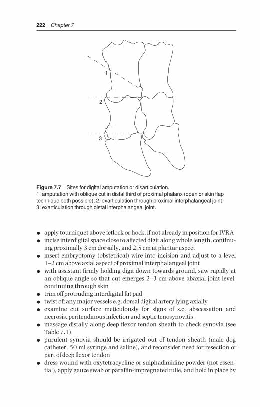
222 Chapter 7
• apply tourniquet above fetlock or hock, if not already in position for IVRA
• incise interdigital space close to affected digit along whole length, continu-ing proximally 3 cm dorsally, and 2.5 cm at plantar aspect
• insert embryotomy (obstetrical) wire into incision and adjust to a level 1–2 cm above axial aspect of proximal interphalangeal joint
• with assistant firmly holding digit down towards ground, saw rapidly atan oblique angle so that cut emerges 2–3 cm above abaxial joint level,continuing through skin
• trim off protruding interdigital fat pad
• twist off any major vessels e.g. dorsal digital artery lying axially
• examine cut surface meticulously for signs of s.c. abscessation and necrosis, peritendinous infection and septic tenosynovitis
• massage distally along deep flexor tendon sheath to check synovia (seeTable 7.1)
• purulent synovia should be irrigated out of tendon sheath (male dogcatheter, 50 ml syringe and saline), and reconsider need for resection ofpart of deep flexor tendon
• dress wound with oxytetracycline or sulphadimidine powder (not essen-tial), apply gauze swab or paraffin-impregnated tulle, and hold in place by
1
2
3
Figure 7.7 Sites for digital amputation or disarticulation.1. amputation with oblique cut in distal third of proximal phalanx (open or skin flaptechnique both possible); 2. exarticulation through proximal interphalangeal joint; 3. exarticulation through distal interphalangeal joint.
BSAC07 3/5/05 4:26 PM Page 222
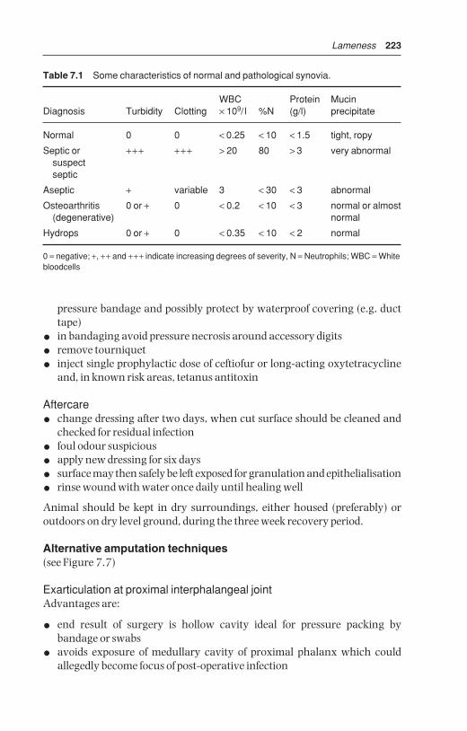
Lameness 223
pressure bandage and possibly protect by waterproof covering (e.g. ducttape)
• in bandaging avoid pressure necrosis around accessory digits
• remove tourniquet
• inject single prophylactic dose of ceftiofur or long-acting oxytetracyclineand, in known risk areas, tetanus antitoxin
Aftercare
• change dressing after two days, when cut surface should be cleaned andchecked for residual infection
• foul odour suspicious
• apply new dressing for six days
• surface may then safely be left exposed for granulation and epithelialisation
• rinse wound with water once daily until healing well
Animal should be kept in dry surroundings, either housed (preferably) or outdoors on dry level ground, during the three week recovery period.
Alternative amputation techniques(see Figure 7.7)
Exarticulation at proximal interphalangeal jointAdvantages are:
• end result of surgery is hollow cavity ideal for pressure packing by bandage or swabs
• avoids exposure of medullary cavity of proximal phalanx which couldallegedly become focus of post-operative infection
Table 7.1 Some characteristics of normal and pathological synovia.
WBC Protein Mucin Diagnosis Turbidity Clotting × 109/ l %N (g/l) precipitate
Normal 0 0 < 0.25 < 10 < 1.5 tight, ropy
Septic or +++ +++ > 20 80 > 3 very abnormalsuspect septic
Aseptic + variable 3 < 30 < 3 abnormal
Osteoarthritis 0 or + 0 < 0.2 < 10 < 3 normal or almost(degenerative) normal
Hydrops 0 or + 0 < 0.35 < 10 < 2 normal
0 = negative; +, ++ and +++ indicate increasing degrees of severity, N = Neutrophils; WBC = Whitebloodcells
BSAC07 3/5/05 4:26 PM Page 223

224 Chapter 7
Disadvantages:
• lengthy procedure
• difficult to locate joint level axially for incision to expose articulation
• liability to break scalpel blades in this awkward site
• preferable use of ‘sage knife’ (a curved solid two-edged instrument) and asmall curette
Amputation through coronary bandInvolves subsequent removal of extensor process of distal phalanx, proximalpart of middle phalanx and the distal sesamoid.
This technique is laborious, but was developed to retain potential forgrowth of a weight-bearing horny wall after surgery. Surgery preferably inrecumbent patient.
• groove is made 1 cm distal to horn-skin margin of coronary band
• obstetrical wire saw cut to remove claw, passing through distal interpha-langeal joint
• removal of extensor process of distal phalanx, transecting extensor tendonat insertion
• removal of proximal part of middle phalanx and distal sesamoid
• curettage of distal articular cartilage of proximal phalanx
• sharp dissection of any septic or otherwise discoloured soft tissue, or radical curettage
Skin flap preservationSkin flap may be preserved and placed over amputation surface, followingremoval of digit through distal one third of proximal phalanx. Advantage iscosmetic improvement and faster healing.
Disadvantages include:
• inability to inspect amputation site when dressing is changed
• suture tear out due to post-operative swelling
• risk of skin necrosis
• good case selection is essential (no phlegmon present)
Skin flap is created initially by semicircular incision from 5–6 cm above inter-digital space on dorsal and plantar aspects, passing down to the coronaryband. Ensure this flap is large and thick, and is then reflected proximally.Amputation is done in conventional way and skin flap, trimmed as needed, isthen sutured over stump.
DiscussionAverage survival period following digital amputation in commercial dairyherds ranges from 12–24 months. Exceptions survive for years. Eventual reason for disposal is further digital disease in over half of these cattle. Inmajority of cases digital amputation has no effect on yield in the lactationsubsequent to the one in which amputation is performed.
BSAC07 3/5/05 4:26 PM Page 224

Lameness 225
Amputation of accessory digits (‘dewclaws’)
IndicationProphylactic surgery on medial accessory digits of hindlimbs to prevent self-inflicted teat trauma. Ethical as well as scientific objections to techniquein Europe, therefore forbidden in many countries including UK andSwitzerland. Common routine procedure in many North American dairies.
Technique
• remove at two to eight weeks old in recumbent calf
• clean and disinfect area
• local infiltration analgesia (2 ml 2% lignocaine plain)
• push digit proximally to move away from joint space and major vessels
• resect through skin margin and dewclaw base with large scissors orBarnes dehorner, remaining superficial and so avoiding deeper digital vasculature
• suture any wounds with significant haemorrhage
• apply topical antibacterial powder, dry swab and adhesive tape dressingfor one week
7.13 Other digital surgery
(a) Resection of digital flexor tendon and their sheathsfollowing digital amputation
IndicationExtension of infection above level of proposed amputation, without involve-ment of fetlock joint, is indication for resection of part of deep and superficialflexor tendons and their sheaths.
Treatment
Resection of superficial and deep flexor tendon after amputation
• identify common digital flexor tendon sheath, and insert straight metalprobe proximally about 6 cm
• incise through skin, tendon sheath and part of annular ligament down tosuperficial flexor tendon
• remove superficial and deep tendon by cutting transversely at proximalpoint
• remove any exposed, purulent or necrotic sheath
• dress wound and leave to heal by granulation tissue
• alternatively, suture skin only
BSAC07 3/5/05 4:26 PM Page 225

226 Chapter 7
Alternative method, using approach above fetlock
• incise skin 3 cm proximal to accessory digit immediately over proximalborder of flexor digital sheath
• continue this vertical incision down to superficial flexor tendon
• transect both flexor tendons at this point and pull out tendon from amputation site
• remove any septic areas of sheath, which are less well exposed in thismethod
(b) Resection of deep and superficial flexor tendons andsheaths (see Figure 7.8)
IndicationSeptic tenosynovitis complicating deep digital sepsis.
TreatmentSometimes removal of deep digital tendon is adequate. Other possible pro-cedures are removal of deep and superficial tendons with or without partial ortotal removal of digital sheath. Precise procedure depends on extent of sepsisat surgical investigation.
Technique
• useful instruments include a blunt-ended curved tenotome and longslightly curved scissors
• tourniquet at mid-metatarsal (-carpal) region and IVRA (see Section 1.8,pp. 33–36)
• trim back accessory claws
• surgical skin preparation to mid-metatarsus (-carpus)
• skin, subcutis and horn of heel bulb are incised over deep flexor tendonfrom affected distal area (e.g. sole-heel junction), keeping incision axial toaccessory digit to point 5 cm proximal to fetlock joint
• open sheath along plantar (volar) aspect
• cut through superficial flexor tendon longitudinally over fetlock (where itencloses deep flexor)
• section deep flexor transversely, just distal to bifurcation (5 cm proximal tofetlock)
• retract tendon from incision and transect distally at insertion into distalphalanx
• check any severe involvement of superficial tendon and, if necessary,resect proximally at same level as deep flexor and at insertion to middlephalanx
• dissect free and resect any infected tendon sheath and areas of sub-cutaneous abscessation including curettage as required
BSAC07 3/5/05 4:26 PM Page 226

Lameness 227
• note that minimal amount of tissue should be resected
• dress wound with povidine-iodine-soaked gauze, pack wound, and closeproximal half (to below dewclaws) with simple interrupted skin sutures
• keep wound open distally for drainage and removal of gauze packing; bandage firmly
• apply block to sound claw, and wire toes together
• systemic antibiotics for seven to ten days
• change dressing at two, seven and fourteen days, or more frequently ifnecessary
A
D
B
E
F
C
Figure 7.8 Surgical anatomy for removal of superficial and deep digital flexortendons following proximal spread of deep infection.A. incision 12 cm long from proximal to fetlock distally into heel horn; B. sectionedfetlock ligament; C. opened common digital sheath; D. superficial flexor tendon; E. deep digital flexor tendon; F. distal cruciate ligament superficial to distal sesamoidand distal interphalangeal joint.
BSAC07 3/5/05 4:26 PM Page 227

228 Chapter 7
(c) Resection of distal interphalangeal joint and distalsesamoid
IndicationsPurulent infection of joint and osteomyelitis resulting from spread of solear ulceration, interdigital necrobacillosis and heel abscess in cases non-responsive to local débridement and several days high dosage of systemicantibiotics, and when digital amputation is either not permitted or desirable.Typically it is a neglected case of solear ulceration (abscess) with severe distalinterphalangeal joint sepsis and osteomyelitis of distal phalanx and distalsesamoid.
Technique (see Figure 7.9)
• tourniquet at mid-metatarsal (-carpal) region
• intravenous regional analgesia (see Section 1.8, pp. 33–36)
• surgical skin preparation
• carefully probe after removing all visible granulation tissue
• 3–3.5 cm circular incision through horn of sole-heel junction
• remove distal sesamoid bone
• drill out infected cartilage and subchondral bone of distal interphalangealjoint surfaces (‘apple core procedure’)
• resect any remains of insertion of deep flexor tendon with ‘sage’ knife(double-sided, slightly curved solid scalpel)
• if skin and subcutis of coronary border are not involved in sepsis, pack cavity with sterile bandage soaked in povidine-iodine
• if coronet is involved, remove skin (2 cm diameter) surrounding anyfistula or incipient abscess with low speed drill, make track 0.8–1.2 cmdiameter from sole up through coronary defect
• evaluate usefulness of irrigation tube into cavity and fixed along metatarsus
• insert bandage as above (unless twice daily irrigation is planned)
• put block on sound digit
• wire toes to prevent over-extension of operated digit
• high dosage of systemic antibiotics for ten days
• change dressing at two days, then weekly intervals, until granulation tissue has filled defect
• remove block and wire at six to twelve weeks
Intensive post-operative care is essential. If infection spreads nevertheless,digital amputation may be only alternative to cull (carcass condemnationdue to antibiotic residues!)
BSAC07 3/5/05 4:26 PM Page 228

Lameness 229
(d) Removal of interdigital hyperplastic skin (see Section 7.5)
Methods include knife surgery, electrocautery and cryosurgery (see Sec-tion 1.14, p. 49).
Technique
• give sedative (xylazine or acepromazine, see Section 1.6, pp. 13–16 andSection 7.15, pp. 204–205)
• restrain in crush/chute (e.g. Wopa crate, see Section 1.8, pp. 35–36)
• IVRA, ring block or local infiltration (poor analgesia)
• cleanse area thoroughly, clip and disinfect
A
D
B
C
Figure 7.9 Resection of distal interphalangeal joint in deep digital sepsis (right hindlateral).A. plantar skin incision (compare Figure 7.8); B. deep digital flexor tendon, resecteddistal portion; C. distal sesamoid; D. plantar margin of distal interphalangeal joint.
BSAC07 3/5/05 4:26 PM Page 229

230 Chapter 7
• apply tourniquet (unnecessary for cryosurgery) if not already in place forIVRA
• assistant should separate digits manually with length of bandage
Knife method
• grasp mass with Allis tissue forceps or Backhaus towel clamp (see Figure1.1)
• remove whole mass in wedge-shaped pattern by two incisions
• keep fine lip of skin along axial borders from which re-epithelialisation will start
• remove protruding interdigital fat, avoiding distal cruciate ligaments(which are easily palpated) and adjacent coronary band
• apply oxytetracycline or sulphadimidine powder, interdigital pressure padand figure of eight bandage
• release tourniquet
• wire toes together, especially in heavy animals, with drill holes 2.5 cmbehind point of toe to prevent separation
• no systemic antibiotics, but tetanus antitoxin may be indicated
• do not change bandage unless site becomes infected, or bandage is blood-soaked
• remove bandage after one week
Electrocautery methodAlthough a hot iron may be used, the instrument must be small and precise to reach the interdigital mass without damaging surrounding tissues. Apply electrocautery loop as in knife surgery above, ensuring integrity ofneighbouring structures especially cruciate ligaments. Advantage is reducedpost-operative haemorrhage, disadvantage is slower healing. Toes should bewired together after dressing wound with powder
Cryosurgery methodAdvantages include absence of haemorrhage and avoidance of post-operative bandaging and dressing.
• depending on size of lesion, some hyperplastic tissue may first be resected(‘debulked’) by knife
• apply probe to tissue to create ice ball (e.g. 3 cm diameter) in rapid freeze,followed by slow thaw cycle
• repeat at other sites, often totalling three, until whole area has been frozen once
• repeat freeze of each area once (with liquid nitrogen) or twice (N2O or CO2)
• do not bandage or dress the region. Wiring of toes contra-indicated
• mass will become necrotic in seven to ten days, and drop off a few dayslater leaving a granulating bed which will slowly epithelialise
BSAC07 3/5/05 4:26 PM Page 230
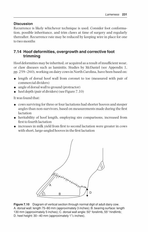
Lameness 231
DiscussionRecurrence is likely whichever technique is used. Consider foot conforma-tion, possible inheritance, and trim claws at time of surgery and regularlythereafter. Recurrence rate may be reduced by keeping wire in place for oneto two months
7.14 Hoof deformities, overgrowth and corrective foottrimming
Hoof deformities may be inherited, or acquired as a result of insufficient wear,or claw diseases such as laminitis. Studies by McDaniel (see Appendix 1, pp. 259–260), working on dairy cows in North Carolina, have been based on:
• length of dorsal hoof wall from coronet to toe (measured with pair of commercial dividers)
• angle of dorsal wall to ground (protractor)
• heel depth (pair of dividers) (see Figure 7.10)
It was found that:
• cows surviving for three or four lactations had shorter hooves and steeperangles than non-survivors, based on measurements made during the firstlactation
• heritability of hoof length, employing sire comparisons, increased fromfirst to fourth lactation
• increases in milk yield from first to second lactation were greater in cowswith short, large-angled hooves in the first lactation
A
B DC
Figure 7.10 Diagram of vertical section through normal digit of adult dairy cow.A. dorsal wall: length 75–80 mm (approximately 3 inches); B. bearing surface: length130 mm (approximately 5 inches); C. dorsal wall angle: 50° forelimb, 55° hindlimb; D. heel height: 30–40 mm (approximately 11/2 inches).
BSAC07 3/5/05 4:26 PM Page 231

232 Chapter 7
• first lactation cows with above features had shorter calving to conceptioninterval following the second lactation
• high yielding cows tend to have longer and lower-angled hooves aftertheir second parturition
• calving to conception interval within the same lactation was greater incows with longer, smaller-angled hooves than in those with shorter,steeper-angled hooves.
Hoof traits of young cows are associated with their future economic worth.Since measurements were made from progeny of bulls high on milk yield, the variations of hoof traits are associated with survival rates, reproductiveperformance and increased yield from first to later lactations.
These facts emphasise the necessity to take corrective measures, includingroutine chiropody and to review breeding policy in herds where digital over-growth is a major problem.
Corrective and preventative hoof trimming
Trimming is essential for the maintenance of healthy normal feet on mostdairy farms as well as on artificial insemination centres where dozens of bullsare kept in confined conditions. Increasingly hoof trimming is being donewell by full-time professional trimmers who have their own specialist equip-ment (e.g. purpose-built crushes/chutes, including the Wopa crate, or othermodels of turn-over devices, most mechanically powered). Trimmers oftenprefer to use powered metal disk grinders (angle grinders). Farm staff gener-ally have insufficient time to deal with the feet of 150+ herd members andinevitably lack the expertise.
Since, certainly in the UK, foot problems tend to be managed by these trimmers, it is vital that good communication is established and maintainedbetween trimmer, farmer or stock manager and the vet, who will be called into treat cows where deeper structures are affected. Good written records (ID,lameness score, diagnosis, treatment, need for revisit) should be maintainedfor stockkeeper, farm veterinarian and trimmer.
Equipment
• single or double action (Hauptner) hoof pincers or ‘nippers’
• right and left hoof knives, Swedish or German pattern
• rotary disk grinder (angle grinder, 10 cm disk)
• hoof rasp
• ropes and hobbles for restraint, hobbles preferably with Velcro® fastening
• appropriate crush/chute, standing or turn-over tilting type
• disinfectant bucket for instruments between cows
• protective goggles (horn particles and dust from grinder), possibly facemask, gloves and wrist protectors
BSAC07 3/5/05 4:26 PM Page 232

Lameness 233
• supply of prosthetic blocks and acrylic (Technovit®, Cowslip®, Shoof ®,rubber block)
Timing of trimmingIdeally at drying off, and again, assuming twice yearly job, at winter housingor not later than one month before spring turnout but often done when timeis available.
Site of trimming
• preferably under cover, but good light essential
• site should have easy route from collecting area and to outside yard forlater inspection, and be easily cleansed
Technique (see Figure 7.11)Standing behind a normal cow, an imaginary perpendicular line drawnthrough the hip joint should pass through the point of the hock and throughthe interdigital space. An abnormal stance of toe produces an inward turningof the hocks and outward pointing of the claws, indicating current or impend-ing digital problems and lameness.
• check gait (possibly lameness score) and stance when going into crush
• raise hind limb, fix, and clean off mud and slurry with sawdust, straw orcloth (not water)
• start with minimal trim of medial claw using hoof pincers to shortenlength with vertical cut (‘dumping’) to correct length or height (ideally 7.5 cm length of dorsal wall, but 8 cm acceptable in big-framed cows =length of cigarette packet) (see Figures 7.10, 7.1[1])
3
4
5 5
3
1
2
BA
Figure 7.11 Steps in routine foot trimming of dairy cow with overgrown claws.
BSAC07 3/5/05 4:26 PM Page 233

234 Chapter 7
• effect is a square-ended toe 5–7 mm thick
• remove excess horn from sole surface near toe, correcting the square orstub toe appearance, and preserving heel horn [2]
• remove any excessive axial overlapping horn flap [3], [4]
• constantly check sole surface and white line, particularly abaxially, forhaemorrhage or granulation tissue
• pare bearing surface slightly concave, with special attention to sole heelarea but retain most heel horn [5]
• repeat technique on lateral claw, which almost invariably requires moreattention
• continue with contralateral limb, and finally check forefeet
• lifting of forefeet seems more likely to lead to hindquarters collapse of cow,and is best prevented by belly band
• if sensitive corium is accidentally entered, and likely to result in lameness,consider putting block on ipsilateral claw
DiscussionMany dairy cows have grossly abnormal weight distribution of 70:30 on lat-eral and medial claws respectively. Recent (2003) studies on weight-bearinghave shown that even after the above standard (Dutch method) trimming,balance has not been restored to the (theoretical) ideal of 50:50, but merely to60:40. It is vital therefore that minimal amount of horn should be taken frommedial claw.
Pare sole of lateral claw to same level as medial claw, and try to reduce heeldepth to that of medial claw too.
Look at two heels from rear, or lay flat surface (e.g. hoof knife handle)transversely across heels to confirm their equal height
Ideal dorsal wall angle is 55° (hind) and 50° (forelimb). Ideal dorsal wallmeasures 7.5 cm (7–8 cm) from coronet to toe (see Figure 7.10).
7.15 Footbaths
IntroductionThe purpose of footbaths is:
• inhibition or destruction of bacteria (e.g. Fusobacterium necrophorum)involved in interdigital skin diseases
• washing action, cleansing of digits
• hardening of sole horn, reducing wear rate and incidence of bruising andsole penetration
• control of digital dermatitis and heel erosion
EquipmentPreferably two footbaths, in tandem, with first containing water or a milddetergent mixture, the second the active solution.
BSAC07 3/5/05 4:26 PM Page 234

Lameness 235
Bath should be made of concrete (permanent), glass-reinforced concrete,or be a sheet-steel tray (portable). A tough sponge floor mat can much reduce the necessary fluid volume. Alternatively some straw is not only ‘cowfriendly’ but tends to separate the claws so that fluid better soaks the inter-digital cleft.
Dimensions of bath: minimum 3 m long; minimum 85 cm wide, dependingon breed; 15–25 cm high, permitting fluid depth of 10–12 cm with non-slip floor.
Site the footbath in passageway or race at exit from milking parlour, preferably under cover, where dilution by rainwater can be avoided.
Bath fluid can be 2.5–5% formalin solution, alternatively 2.5% copper sulphate or zinc sulphate. Disadvantages exist. Formalin may irritate eyesand its disposal is viewed with suspicion by environmental health authoritiese.g. UK Health and Safety Executive. Never exceed 5% formalin concentra-tion, as 10% solution soon causes a severe localised chemical reaction andlameness. Ingested copper sulphate may cause acute copper poisoning.
Antibiotic footbaths for control of digital dermatitis: see Section 7.10a, p. 214.
The bath contents should be emptied into an appropriate collection facility,not to a watercourse or ground water.
TreatmentIdeally, stand lame cows (e.g. cases of interdigital necrobacillosis, interdigitalor digital dermatitis, heel erosion and possibly solear ulceration) in bath for20 minutes twice daily. This is rarely practical as it is too laborious on largerunits today.
For preventative treatment walk cattle through after four successive milkings over a two-day period, then empty, clean and refill with water for five days. Repeat weekly throughout the year, except when frozen. Note thata standard bath is inactivated (i.e. bactericidal effect is lost) after 800 cow passages (e.g. 200 cows × 4 passages, viz. one week recommended usage).Recent Dutch studies suggest the figure of 800 should be reduced to 300.
DiscussionRegular formalin footbath usage should be practised in herds with an unacceptably high incidence of interdigital disease, heel erosion and solearulceration. The lameness incidence will be reduced, and the severity of lesionswill tend to be less, reducing further the economic losses.
7.16 Check lists for herd problems
Check lists for systemic and logical examination of herd digital lamenessproblem are useful aids. Tables 7.2–7.6 cover:
BSAC07 3/5/05 4:26 PM Page 235
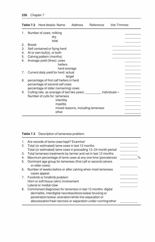
236 Chapter 7
Table 7.3 Description of lameness problem.
1. Are records of lame cows kept? Examine! __________2. Total (or estimated) lame cows in last 12 months __________
Total (or estimated) lame cows in preceding 12–24 month period __________3. Total lameness treatments by farmer and vet in last 12 months __________4. Maximum percentage of lame cows at any one time (prevalence) __________%5. Dominant age group for lameness (first calf or second calvers
or older cows) __________6. Number of weeks before or after calving when most lameness
cases appear __________7. Forelimb or hindlimb problem __________
Horn or soft tissue (skin) involvement __________Lateral or medial claw __________
8. Commonest diagnoses for lameness in last 12 months: digital dermatitis, interdigital necrobacillosis/solear bruising or penetration/solear ulceration/white line separation or abscessation/heel necrosis or separation under-running/other __________
Table 7.2 Herd details: Name Address Reference Vet/Trimmer.
1. Number of cows: milking _________dry _________total _________
2. Breed ________________3. Self contained or flying herd ________________4. AI or own bull(s), or both ________________5. Calving pattern (months) ________________6. Average yield (litres): cows _________
heifers _________herd average _________
7. Current daily yield for herd: actual _________target _________
8. percentage of first calf heifers in herd _________percentage of second calf cows _________percentage of older (remaining) cows _________
9. Culling rate, as average of last two years: _________ individuals = _________Number of culls for: lameness _________
infertility _________mastitis _________mixed reasons, including lameness _________other _________
BSAC07 3/5/05 4:26 PM Page 236

Lameness 237
Table 7.4 Herd investigation.
1. Prevalence of lameness today in milking herd _____________2. Conformation (for lame and sound cattle):
Relative weight and size ______ ______Size of digits ______ ______Angle of hock (from lateral and caudal views) ______ ______Outward angulation hind feet (°) ______ ______Angle of dorsal wall (°) ______ ______Height of heel ______ ______Overgrowth of digits (nil, moderate, severe) ______ ______Body score 1–5 (emaciated-fat) ______ ______
3. Housing system:Cowshed/cubicles/straw yard ______________Cowshed: size of standings ______________
surface characteristics (e.g. mats) ______________bedding material ______________exercise area ______________passageway surface ______________slats ______________slurry quantity and quality ______________
Cubicles: cubicle size (length, width, step height) ______________surface characteristics ______________
Concrete age and condition ______________Doorways and passageways ______________Overall cow concentration indoors (m2 per cow) ______________Distance travelled on cow tracks, surface and smoothness,
roughness and wetness ______________4. Nutrition:
Grazing, nature and conditions of herbage or crop ______________Concentrate composition (% crude protein) _______Concentrate maximum intake (kg): before calving
at peak yield _______Duration of steaming up (weeks) _______Period from calving to peak yield (weeks) _______Concentrate feeding: number of times per day where fed _______Roughage composition: % crude protein _______
% crude fibre _______Silage composition: % dry matter _______
acidity _______palatability ______________
Roughage intake (kg dry matter):maximum intake before calving peak ______________where fed ______________whether fed with concentrates ______________
Supplements (salt, sodium bicarbonate, zinc, other):in concentrates ______________fed separately ______________
Continued
BSAC07 3/5/05 4:26 PM Page 237

238 Chapter 7
• herd details
• description of lameness problem
• herd investigation
• individual cow investigation
• action recommendations
Numerous factors play interrelated roles in causing this ‘production disease’.Investigation of herd problem should be done if lameness incidence exceeds15–20%
7.17 Infectious arthritis (‘joint ill’) of calves
IntroductionJoint ill (neonatal polyarthritis) is usually caused by haematogenous spread of septic infection from the umbilicus, less commonly lungs or liver.
Table 7.5 Individual cow investigation.
1. Identification ______________2. Number of calves bred ______________
Pregnancy (mo)/number of weeks postpartum ______________Current yield/expected lactation yield (L) ______________Current concentrate consumption (Kg) ______________Body score 1–5 (emaciated-fat) ______________
3. Degree of lameness: severe/moderate/slight /non lame (3–0) ______________Affected leg(s) LF RF LH RH ______________Site: upper limb tissue primarily involved ______________
digit (interdigital or horn, lateral or medial claw) ______________Estimated duration of lameness (weeks) ______________Previous treatment (medical, surgical, trim) ______________Diagnosis: major and/or minor ______________
5. Prophylaxis:Foot-trimming: practice: none, self, visiting trimmer, vet ______________
frequency/year ______________technique (e.g. grinder) ______________
Footbath: site ______________composition ______________concentration ______________frequency of replenishment ______________usage ______________
Quality of stockmanship (including weekend and holiday staff ) ______________
BSAC07 3/5/05 4:26 PM Page 238
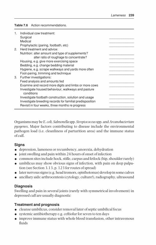
Lameness 239
Organisms may be E. coli, Salmonella spp. Streptococcus spp. and Arcanobacteriumpyogenes, Major factors contributing to disease include the environmentalpathogen load (i.e. cleanliness of parturition area) and the immune status of calf.
Signs
• depression, lameness or recumbency, anorexia, dehydration
• joint swelling and pain within 24 hours of onset of infection
• common sites include hock, stifle, carpus and fetlock (hip, shoulder rarely)
• umbilicus may show obvious signs of infection, with pain on deep palpa-tion (see Section 3.13, p. 123 for routes of spread)
• later nervous signs (e.g. head tremors, opisthotonus) develop in some calves
• ancillary aids: arthrocentesis (cytology, culture!), radiography, ultrasound
DiagnosisSwelling and pain in several joints (rarely with symmetrical involvement) indepressed calf are usually diagnostic
Treatment and prognosis
• cleanse umbilicus, consider removal later of septic umbilical focus
• systemic antibiotherapy e.g. ceftiofur for seven to ten days
• improve immune status with whole blood transfusion, other intravenousfluids
Table 7.6 Action recommendations.
1. Individual cow treatment:Surgical ______________Medical ______________Prophylactic (paring, footbath, etc) ______________
2. Herd treatment and advice:Nutrition: alter amount and type of supplements? ______________
alter ratio of roughage to concentrate? ______________Housing, e.g. give more exercising space ______________Bedding, e.g. change bedding material ______________Hygiene, e.g. scrape walkways and yards more often ______________Foot-paring, trimming and technique ______________
3. Further investigations:Feed analysis and amounts fed ______________Examine and record more digits and limbs or more cows ______________Investigate housed behaviour, walkways and pasture
conditions ______________Investigate footbath construction, solution and usage ______________Investigate breeding records for familial predisposition ______________Revisit in four weeks, three months re progress ______________
BSAC07 3/5/05 4:26 PM Page 239
240 Chapter 7
• immediate joint lavage (see Section 7.27, p. 257)
• valuable calves with severe joint destruction may benefit from radicalsurgery via arthroscopy or arthrotomy with aim of joint ankylosis
Prognosis guarded or poor unless intensive nursing is undertaken.
DiscussionMeningitis occurs in a minority of neonatal calves, usually in the first week,and is rapidly fatal. In slightly older calves septic physitis (e.g. of distal radiusor distal tibia) may complicate the initial signs, and radiography then valuable. ‘Joint ill’ is an emergency requiring rapid control and good nursing,as well as prompt review of the possible predisposing factors.
7.18 Contracted flexor tendons
IntroductionMost frequently observed congenital anomaly in dairy breeds, and rarelyacquired, congenital flexed tendons (CFT) are usually primarily in the forelegsand generally bilateral.
SignsMild cases have slight carpal flexion and intermittent knuckling of the forefetlocks, then bearing weight on the dorsum of the fetlock.
More severe cases can bear weight only on the flexed fetlocks. Advancedcases, with severe contracture, are often recumbent and, when encouragedto stand, tend to fall down immediately. Such calves are usually colostrumdeficient, dehydrated and very weak, having failed to suck since birth.
Palpation reveals excessive tension and tautness in both the super-ficial (SFT) and/or deep flexor tendons (DFT), when attempts are made to straighten the leg. No pain is evident on extension, and joint swelling isabsent (compare ‘joint ill’ see Section 7.17, p. 238).
Treatment
• in mild congenital cases check immune status and general condition (givecolostrum in first 12 hours postpartum) if doubt exists as to whether calfhas taken maternal milk
• keep with dam for first 24 hours observation in well-bedded loose box or atpasture, as exercise encourages progressive correction of the milder casesof flexion of carpus and fetlock joint
• in mild case consider elongation of claw toes with piece of wood glued to sole
• in more severe case initially correct any systemic problem and considerapplying splint on palmar aspect of limb: from pastern to midmetacarpus(fetlock flexion) or up to proximal radius (in carpal flexion)
BSAC07 3/5/05 4:26 PM Page 240

Lameness 241
• select lightweight splint material e.g. split PVC piping. Pad limb (bandage)meticulously before fitting splint. Lift calf if unable to stand up initially
• alternatively consider casting limb over padding (e.g. Lightcast), ensur-ing its easy removal for examination of flexion status after one to twoweeks
SurgeryIn severe cases of fetlock and carpal flexion (CFT) surgical correction may beattempted (see Figure 7.12).
• perform surgery in sedated calf (xylazine 0.2 mg/kg i.m.) restrained in lateral recumbency, and either IVRA or local infiltration of 2% lignocaine
• routine skin preparation along entire length of metacarpus
• make 10 cm longitudinal incision laterally over SFT and DFT in mid-metacarpal region
• carefully dissect through fascia, identifying and avoiding damage to lat-eral palmar digital nerve and adjacent vessels lying plantar to metacarpusboth medially and laterally
• elevate SDF by inserting slightly curved Mayo scissors (see Figure 1.1, p. 3) transversely between SDF and DFT and transect SDF
• check degree of fetlock flexion again and if inadequate elevate DFT andsection similarly
• finally, if necessary, section the suspensory ligament (interosseus muscle)in the proximal third of metacarpus
• close peritendinous fascia in continuous pattern with non-absorbablematerial, and skin with interrupted sutures
• apply light bandage and use splint if flexion still inadequately corrected by surgery and additional extension is needed, also in event of iatrogenicsecondary over-extension of fetlock
• in congenital carpal flexion section of the ulnaris lateralis and flexor carpiulnaris tendons is performed through a 7–8 cm incision, the distal com-missure of which lies over the accessory carpal bone
• lightly bandage limb
• prophylactic antibiotics are not needed, but analgesics are advisable forseveral days (e.g. NSAID such as flunixin meglumine)
• remove splint and/or cast after 7–10 days to assess improvement
DiscussionThe prognosis for CFT is good in calves with a mild deformity, i.e. sporadicknuckling of fetlocks. Severe fetlock and carpal deformaties often fail to becorrected despite the tenotomy procedures described above, flexion persistsand locomotion is impossible.
Scrupulous management of splints in neonatal calves ensures avoidance ofskin necrosis at potential pressure sores (see Figure 7.16).
BSAC07 3/5/05 4:26 PM Page 241

242 Chapter 7
7.19 Tarsal and carpal hygroma
IntroductionSynonyms: tarsal cellulitis, carpal/tarsal ‘bursitis’.
Definition: firm or fluctuating swelling involving pre-carpal bursa andacquired subcutaneous bursa over lateral aspect of hock.
123
4
5
7
6
distal
Figure 7.12 Tenotomy of superficial and deep flexor tendons and suspensoryligament over left metacarpus (palmar view).1. superficial part of superficial flexor; 2. deep part of superficial flexor; 3. deep flexor;4. superficial part of suspensory ligament (interosseus muscle); 5. deep part ofsuspensory ligament; 6. medial vein, artery and nerve; 7. palmar metacarpal veins.(From Dirksen, Gründer & Stöber, 2002.)
BSAC07 3/5/05 4:26 PM Page 242

Lameness 243
Incidence and aetiology: high in housed cattle on hard floors with littlebedding.
Signs
• usually no lameness, no pain, and presents purely as cosmetic blemish
• sometimes skin contusion, break in integument, with seropurulent dis-charge and invasion by Arcanobacterium pyogenes
• distension of joint capsule, heat, pain and lameness indicate further localised spread.
Differential diagnosis: precarpal abscessation, septic carpitis, septic tarsitis.
Treatment and prophylaxis
• transfer to soft bedding or turn outside
• broad spectrum antibiotics in cases involving lameness and systemic signs
• do not open cavity or inject local corticosteroids (risk of gross contamination)
• check stall dimensions relative to breed, and any behavioural abnormal-ities which can be corrected
• surgical excision of a large non-infected carpal bursa with primary woundclosure and pressure bandage is a possible and surgically hazardous pro-cedure in clinic (not on-farm) situation, as wound breakdown commonlyoccurs despite good aftercare
7.20 Patellar luxation
Three forms exist:
• dorsal patellar luxation or fixation in adults; sporadic incidence
• lateral patellar luxation; congenital, uncommon
• medial patellar luxation; congenital, rare
Dorsal patellar luxation or fixation
IntroductionTemporary or permanent fixation of patella on upper part of medial femoraltrochlear ridge.
Signs
• stiffness, later jerky action with leg extended caudally for longer than normal, followed by forward jerk (temporary fixation)
• action sometimes intermittent and limb may become fixed in rigid exten-sion, dragging claws (permanent fixation)
• position evident on patellar palpation, and manual reposition possible.
Differential diagnosis: displacement of biceps femoris muscle, spastic paresis,acute gonitis.
BSAC07 3/5/05 4:26 PM Page 243

244 Chapter 7
TreatmentSpontaneous recovery occurs in some individuals, especially cattle at grass.Complete recovery follows medial patellar desmotomy:
• sedate subject and produce local analgesia over lowest palpable point ofmedial straight patellar ligament (see Figure 7.13[11]), just proximal toinsertion into tibial tuberosity
• clip and disinfect circular area 15 cm in diameter
• make vertical incision 3 cm just cranial to cranial edge of medial patellarligament
• insert curved tenotome or bistoury (Hey-Groves pattern) through incisionin vertical manner, into triangular space bounded by middle and medialligaments and tibia
X3
11
1287
13
1314
15
16
4
5
10
6
1
29
Figure 7.13 Lateral view of left stifle joint of cow.1. femur; 2. patella; 3. and 4. medial and lateral menisci; 5. tibia; 6. fibula; 7. and 8. medial and lateral trochlear ridge; 9. patellar fibrocartilage; 10. tibial tuberosity; 11. medial straight patellar ligament (sectioned distally in patellar desmotomy); 12. and 13. middle and lateral straight patellar ligaments; 14. tendon of biceps femoris muscle; 15. lateral femoropatellar ligament; 16. lateral collateral ligament. X indicates site for femorotibial arthrocentesis.
BSAC07 3/5/05 4:26 PM Page 244

Lameness 245
• turn tenotome through 90° and section ligament by short sawing move-ment and percutaneous pressure with finger
• withdraw tenotome in vertical position, after snapping and separation ofligament is appreciated
• appose skin edges with two simple sutures
Do not use disposable scalpel blade and handle, due to risk of accidental breakage and loss of blade, into periarticular area or joint. Check success ofsurgery immediately by observing gait; recurrence has not been reported.Surgery is best performed in standing position, though access to site may bemore difficult in heavy lactating cow than in lateral recumbency.
Complications
• accidental entry into femoropatellar joint (rare)
• accidental section of middle straight patellar ligament (disastrous)
• gross iatrogenic infection of site
• severe haemorrhage
Lateral patellar luxation
IntroductionComplete or incomplete lateral displacement of patella. Femoral paralysiscaused by dystocia (oversized fetus in anterior presentation, possibly ‘hip-lock’)may be involved in some cases, therefore check skin sensation over lateralthigh. Hypoplastic lateral trochlear ridge has been postulated but remainsunproven.
Signs
• gross uni- or bilateral flexion of stifles and hocks
• limb collapses when weight is taken
• patella forms obvious bulge on lateral aspect of joint, and lateral femoraltrochlear ridge is clearly palpable
• manipulative replacement sometimes possible
Differential diagnosis:
• femoral paralysis, in which a secondarily induced lateral patellar luxationmay occur, but in which a discrete loss of skin sensation and quadricepsatrophy is evident (neonatal, following dystocia)
• quadriceps femoris muscle rupture, which is rare and accompanied byswelling
• gonitis, with evidence of increased synovial fluid and other joint signs
• distal femoral epiphyseal (supracondylar) separation with limb malalign-ment and crepitus
BSAC07 3/5/05 4:26 PM Page 245

246 Chapter 7
Treatment
• joint overlap operation: joint capsule incised medial to patella and closedby vertical mattress sutures in overlap procedure
• if patella fails to remain in trochlear groove, split fascia of thigh dorsallyfrom patella
• create new medial patellar ligament by suturing patellar cartilage andfemoral periosteum and fascia, followed by imbrication of joint capsulemedially by simple interrupted absorbable sutures
Medial patellar luxation
Introduction and signs
• rare congenital condition
• complete or incomplete, permanent or intermittent
• limb flexed with patella freely moveable
• manual reposition sometimes possible
TreatmentLateral capsular overlap procedure (see above). Prognosis is poor.
7.21 Spastic paresis
IntroductionDefinition: progressive condition characterised by contraction of gastrocne-mius and related calcanean tendons and muscle bellies, leading to severeover-extension of hock.
Predisposition: upright stance; certain breeds (e.g. Friesian, AberdeenAngus) have hereditary predisposition.
Aetiology: an over-active stretch reflex is present in the gastrocnemius, withover-stimulation or lack of inhibition of motor neurons. Electromyogram(EMG) studies indicate increased electrical activity in the gastrocnemiusmuscle, and to a lesser extent in other muscles. CSF studies have suggested anextra-pyramidal dopaminergic central disorder.
Signs
• first seen typically at two to nine months old, rarely congenital or olderanimals
• initially unilateral, later often bilateral hindlimb stiffness and increasingrigidity with heel bulbs raised off ground
• intermittent backward jerking of limb, later over-extension of hock
• raised tail head with occasional upward movement
• leg is readily and painlessly flexed manually, but immediately resumesover-extended position
BSAC07 3/5/05 4:26 PM Page 246

Lameness 247
• weight increasingly transferred to forequarters, and progressive atrophyof hindquarters
• radiographic tarsal joint changes after several months are characteristicof chronic over-extension
Differential diagnosis: dorsal luxation of patella, septic or aseptic gonitis ortarsitis, fracture dislocation of calcaneus, joint-ill, luxation of biceps femorismuscle.
TreatmentTenotomy of gastrocnemius tendon or tibial neurectomy. Tenotomy oftenonly temporarily successful in young calves (< 9 months).
Tenotomy (see Figure 7.14)
• operate on standing calf under local analgesia
• shave and disinfect area over calcanean tendon 10 cm proximal to point of hock
• incise skin 6 cm long vertically over caudal aspect of tendon
• identify gastrocnemius tendon and either section transversely, or remove2 cm portion
A
C
1D
B
23
Figure 7.14 Neurectomy and tenotomy sites for alleviation of spastic paresis.1. incision site for tibial neurectomy; 2. incision for caudal approach for gastrocnemiustenectomy; 3. incision for lateral approach for gastrocnemius tenectomy; A. sciaticnerve; B. tibial nerve; C. peroneal nerve; D. popliteal lymph node.
BSAC07 3/5/05 4:26 PM Page 247

248 Chapter 7
• section through half of transverse diameter of adjacent superficial digitalflexor tendon
• suture skin
The effect is a marked dropping of hock on weight-bearing. Some operatedcalves develop fibrous union in operated area, leading to recurrence 1.5–3months later. Guarded prognosis.
Neurectomy of tibial nerve (see Figure 7.14)Tibial nerve supplies gastrocnemius muscle.
• identify and mark on skin the groove between two heads of biceps femorisin standing calf
• operate in lateral recumbency under epidural or GA
• make aseptic surgical approach between two heads of biceps femoris muscle; popliteal lymph node is useful landmark adjacent to both tibialand peroneal nerves
• insert wound retractor
• identify the two nerves by electrical nerve stimulator (e.g. cattle goad); tibial nerve causes digital flexion and hock extension, while peronealcauses digital extension and hock flexion
• remove 2 cm length of main trunk of tibial nerve, as precise identificationof gastrocnemius branches is difficult or impossible
• suture subcutaneous tissues and skin
• encourage limited exercise for two weeks
Good prognosis follows this neurectomy.
Complications
• continuing muscle atrophy
• temporary or persistent peroneal paralysis
• wound breakdown
• gastrocnemius rupture in heavy cattle one to five days after neurectomy,possibly due to overstretching of denervated muscle
7.22 Hip luxation
IntroductionRelatively common condition in younger cows (two to five years), femoralhead usually moving in cranial and dorsal direction.
Signs
• standing animal is obviously lame
• limb appears shortened (in dorsal dislocation)
• characteristic asymmetry of greater femoral trochanters
BSAC07 3/5/05 4:26 PM Page 248
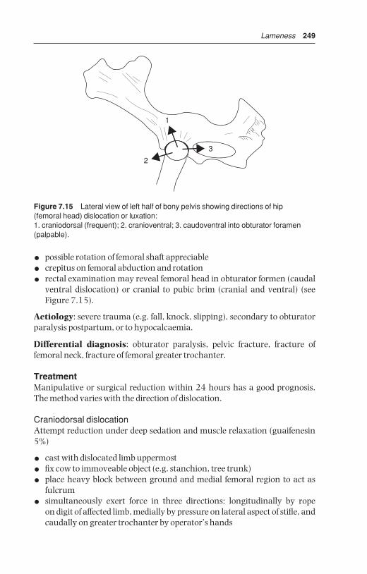
Lameness 249
• possible rotation of femoral shaft appreciable
• crepitus on femoral abduction and rotation
• rectal examination may reveal femoral head in obturator formen (caudalventral dislocation) or cranial to pubic brim (cranial and ventral) (seeFigure 7.15).
Aetiology: severe trauma (e.g. fall, knock, slipping), secondary to obturatorparalysis postpartum, or to hypocalcaemia.
Differential diagnosis: obturator paralysis, pelvic fracture, fracture offemoral neck, fracture of femoral greater trochanter.
TreatmentManipulative or surgical reduction within 24 hours has a good prognosis.The method varies with the direction of dislocation.
Craniodorsal dislocationAttempt reduction under deep sedation and muscle relaxation (guaifenesin5%)
• cast with dislocated limb uppermost
• fix cow to immoveable object (e.g. stanchion, tree trunk)
• place heavy block between ground and medial femoral region to act as fulcrum
• simultaneously exert force in three directions: longitudinally by rope on digit of affected limb, medially by pressure on lateral aspect of stifle, andcaudally on greater trochanter by operator’s hands
1
3
2
Figure 7.15 Lateral view of left half of bony pelvis showing directions of hip (femoral head) dislocation or luxation:1. craniodorsal (frequent); 2. cranioventral; 3. caudoventral into obturator foramen(palpable).
BSAC07 3/5/05 4:26 PM Page 249

250 Chapter 7
• relaxation improves with duration of anaesthesia and prolonged manipulation
• open reduction with reinforcement of the acetabular joint capsule hasbeen described with a success rate of 75% (clinic facilities).
Dislocation in other directions (see Figure 7.15)Difficult or impossible reduction. Success rate of 30% for ventral luxation isclaimed. After successful reduction keep cow recumbent for one to two daysby tying together hindlimbs above hock to avoid likelihood of immediaterecurrence, and keep in box for two months. Surgical methods of reductionand fixation often unsuccessful.
7.23 Stifle lameness
IntroductionThe stifle is often the site of non-digital joint problems in adult cattle,specifically degenerative arthritis. Bilateral spontaneous osteoarthritis maysometimes be inherited in Holstein and Guernsey cattle, possibly through a single autosomal recessive gene. Patellar abnormalities are discussed elsewhere (see Section 7.20, p. 243).
Injuries may primarily affect:
• cranial, and rarely also the caudal cruciate ligament (rupture)
• menisci, usually medial (tear)
• articular cartilage of femoral condyle, especially medial (erosion)
The intimate relationship of these structures in a major weight-bearing jointexplains the speed of secondary changes.
Signs and gross pathology of traumatic gonitis
• sudden onset of relatively severe lameness
• increased synovial fluid ( joint capsule easily appreciated) and periarti-cular swelling
• pain and crepitus in stifle on manual flexion, extension, rotation, abduc-tion, and adduction
• instability possibly evident in stifle on medial to lateral movement of os calcis with limb elevated
• synovial fluid (sterile puncture between medial and middle straight patellar ligaments) blood-tinged, some debris, no evidence of sepsis (seeTable 7.1)
• rapid development of secondary damage following primary injury: severeerosion of cartilage, eburnation of bone, meniscal cartilaginous erosion,tearing and displacement, extensive periarticular fibrosis, secondary cruciate rupture
BSAC07 3/5/05 4:26 PM Page 250

Lameness 251
TreatmentCull cases with cranial cruciate rupture except for purebred or valuable cattlewhere surgery may be attempted in a surgical clinic. In other conditionsensure absolute rest and systemic anti-inflammatory drugs for seven to tendays. Prognosis is poor.
7.24 Nerve paralysis of limbs
Five different nerves may occasionally be paralysed. The aetiology, signs,diagnosis, treatment and prognosis of obturator, peroneal, femoral, sciaticand radial paralysis are summarised in Table 7.7 page 252.
7.25 Limb fractures
IntroductionFractures can affect many bones in cattle including:
• ribs – commonest bovine fracture
• tail – likewise common, and insignificant
• vertebrae – potentially fatal
• pelvis – dystocia, involving separation of pubic symphysis in heifers;trauma (e.g. during oestrus or passing through narrow gateway) result-ing in fracture of tuber coxae
Limb fractures involving the appendicular skeleton, including epiphysealseparation in growing stock, are relatively common.
Specific features in cattle include:
• economic considerations: in many cattle salvage may be preferable to prolonged recovery period
• humane handling and recovery facilities: often sparse compared to thosefor other species, and may affect proposed treatment
Long bone fractures
Common sites in descending order of incidence: metatarsus, metacarpus,tibia, femur, radius/ulna, humerus.
Treatment
• external fixation: plaster of Paris (Gypsona®, Cellona®), polyurethaneresin on polyester-cotton fabric (Baycast®, Cuttercast®) or fibreglass(Deltalite®, Scotchcast®), fibreglass on cotton base (Crystona®) orpolyester polymer on cotton base (Hexcelite®), hanging splint with per-cutaneous fixation (Thomas splint or walking cast), see Figure 7.16
BSAC07 3/5/05 4:26 PM Page 251
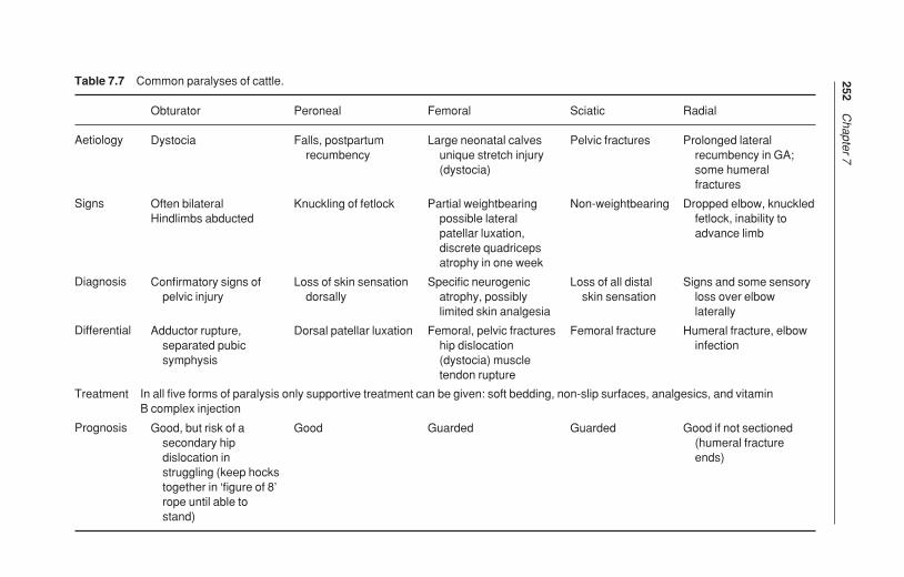
252C
hapter 7
Table 7.7 Common paralyses of cattle.
Aetiology
Signs
Diagnosis
Differential
Treatment In all five forms of paralysis only supportive treatment can be given: soft bedding, non-slip surfaces, analgesics, and vitamin B complex injection
Prognosis
Radial
Prolonged lateralrecumbency in GA;some humeralfractures
Dropped elbow, knuckledfetlock, inability toadvance limb
Signs and some sensoryloss over elbowlaterally
Humeral fracture, elbowinfection
Good if not sectioned(humeral fractureends)
Sciatic
Pelvic fractures
Non-weightbearing
Loss of all distalskin sensation
Femoral fracture
Guarded
Femoral
Large neonatal calvesunique stretch injury(dystocia)
Partial weightbearingpossible lateralpatellar luxation,discrete quadricepsatrophy in one week
Specific neurogenicatrophy, possiblylimited skin analgesia
Femoral, pelvic fractureship dislocation(dystocia) muscletendon rupture
Guarded
Peroneal
Falls, postpartumrecumbency
Knuckling of fetlock
Loss of skin sensationdorsally
Dorsal patellar luxation
Good
Obturator
Dystocia
Often bilateral Hindlimbs abducted
Confirmatory signs ofpelvic injury
Adductor rupture,separated pubicsymphysis
Good, but risk of asecondary hipdislocation instruggling (keep hockstogether in ‘figure of 8’rope until able tostand)
BSAC07 3
/5/05 4
:26 PM P
age 252

Lameness 253
• internal fixation: steel or titanium plates and screws, Kirschner-Ehmerdevice, transfixation pins with overlaid resin bridge holding pins (seeFigure 7.16), Kuntscher nail, Steinmann nail (and possibly multiple parallel nails or ‘stack pinning’).
Common problems of bovine long bone fractures:
• high incidence of comminuted fractures, often grossly displaced
• frequently compound with gross contamination
• often severe muscular contraction, increasing difficulty in manipulativereduction
For methods of internal and percutaneous fixation, reader should refer tostandard textbooks (see Further reading, pp. 259–260).
Figure 7.16 Walking cast for stabilisation of a comminuted metacarpal (MCIII/IV)fracture with two pins inserted into distal radius.
BSAC07 3/5/05 4:26 PM Page 253

254 Chapter 7
Tibial shaft fracturesThis fracture type is taken as an example of problems which are liable to bemet in attempts at treatment.
IncidenceCommon long bone fracture, occasional cases are in neonates, traumat-ised by dam, others are in growing cattle, less common in mature cows andbulls.
SignsMost fractures are in proximal or mid-diaphyseal region of tibial shaft,oblique and comminuted, with over-riding of the fracture ends. Most areclosed. Compound fractures (open) tend to have wound on medial aspect.Lameness is severe, with obvious mobility of the distal limb and marked crepitus at fracture site.
TreatmentWhile the preferred management is usually transfixation pinning (3 prox-imal, 3 distal), connected externally by methylmethacrylate side bars or fibreglass cast, such surgery usually requires referral to a specialist clinic where controlled traction, sterile facilities, general anaesthesia andradiology are available. Animals should have stall rest for some weeks. Thetransfixation pins should be removed in six to eight weeks, ideally after radiographic check to confirm fracture healing is appropriate. Minor com-plications include suppuration around the pin holes. Major problems are continuing inability to bear weight, leading to severe lateral bowing of thecontralateral hind leg, and failure of adequate callus formation.
Thomas splint cast management is not easy. The device must be ‘made tomeasure’ and well-padded over the major pressure points in the inguinum.Some cattle find it difficult to stand up initially with such a splint.
The use of a resin or fibreglass cast alone in an attempt to immobilise a mid-or proximal diaphyseal tibial fracture is doomed to failure as it is impossible toimmobilise the stifle joint. This does not apply to fractures of lower limb bones(e.g. metatarsus/metacarpus), where external immobilisation with a cast isusually the treatment of choice.
Immediate immobilisation on-farm for transport to clinicIt is imperative to minimise movement at the fracture site during transport,
as a simple fracture can so easily be converted into a compound disaster. A Robert Jones bandage, with lateral support proximally to the level of thetuber coxae in tibial fractures, is a good insurance policy against further iatro-genic injury.
BSAC07 3/5/05 4:26 PM Page 254
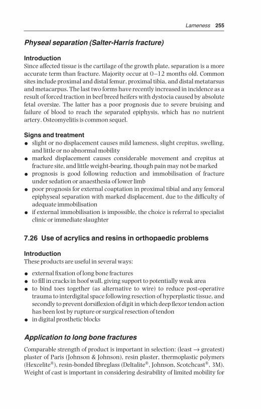
Lameness 255
Physeal separation (Salter-Harris fracture)
IntroductionSince affected tissue is the cartilage of the growth plate, separation is a moreaccurate term than fracture. Majority occur at 0–12 months old. Commonsites include proximal and distal femur, proximal tibia, and distal metatarsusand metacarpus. The last two forms have recently increased in incidence as aresult of forced traction in beef breed heifers with dystocia caused by absolutefetal oversize. The latter has a poor prognosis due to severe bruising and failure of blood to reach the separated epiphysis, which has no nutrientartery. Osteomyelitis is common sequel.
Signs and treatment
• slight or no displacement causes mild lameness, slight crepitus, swelling,and little or no abnormal mobility
• marked displacement causes considerable movement and crepitus at fracture site, and little weight-bearing, though pain may not be marked
• prognosis is good following reduction and immobilisation of fractureunder sedation or anaesthesia of lower limb
• poor prognosis for external coaptation in proximal tibial and any femoralepiphyseal separation with marked displacement, due to the difficulty ofadequate immobilisation
• if external immobilisation is impossible, the choice is referral to specialistclinic or immediate slaughter
7.26 Use of acrylics and resins in orthopaedic problems
IntroductionThese products are useful in several ways:
• external fixation of long bone fractures
• to fill in cracks in hoof wall, giving support to potentially weak area
• to bind toes together (as alternative to wire) to reduce post-operativetrauma to interdigital space following resection of hyperplastic tissue, andsecondly to prevent dorsiflexion of digit in which deep flexor tendon actionhas been lost by rupture or surgical resection of tendon
• in digital prosthetic blocks
Application to long bone fractures
Comparable strength of product is important in selection: (least → greatest)plaster of Paris (Johnson & Johnson), resin plaster, thermoplastic polymers(Hexcelite®), resin-bonded fibreglass (Deltalite®, Johnson, Scotchcast®, 3M).Weight of cast is important in considering desirability of limited mobility for
BSAC07 3/5/05 4:26 PM Page 255

256 Chapter 7
patient. Strength:weight ratio is more favourable for resin and fibreglassproducts than plaster. Products may also be used in layers, e.g. plaster of Parisoverlaid with thin layer of fibreglass, which is also water-resistant.
Padding (Soffban®, bandages) should be covered with brightly-colouredflexible bandage (Vetrap®) before being overlaid with fibreglass resin castingmaterial (Dynacast Pro®). The bright colour ensures that when it is time toremove the cast, the correct depth of cut can be seen when using the electricalcast saw.
Padding (muslin bandage 3 mm thick) should be applied in growing animals. External supports should immobilise joints immediately proximaland distal to fractured long bone, e.g. midshaft fracture of metacarpus shouldhave the cast from the proximal radius to the hoof.
Cases of recent compound fractures of metacarpus/metatarsus in smallcalves should be cleansed very thoroughly, and treated by external coaptionwith a cast or external fixator with sidebar for treatment by irrigation of theinfected focus. Cows with compound fractures of > 6 hours duration shouldbe managed with an external fixator, or be culled, depending on value of animal. Drains should never be left in place in such areas due to danger ofascending infection. Compound fractures carry a poor prognosis.
Application to digit
Surfaces should be prepared before application of resin
• clean off all loose foreign material
• trim any excessive horn from toe, bearing surface, and wall
• apply fat solvent with swabs
• groove bearing surface to increase contact area of resin (grinder very useful and efficient)
• dry hoof with electric hair drier in damp weather
• carefully observe manufacturer’s instructions regarding mixing of product (hoof compound, Technovit [Heraeus-Kulzer] ).
Other wooden blocks are produced by Demotec (Nidderau, Germany), plasticblock (e.g. Podobloc®) by Agrochemica, Bremen.
Note that nowadays a wood shoe is usually put on to resin surface (whichacts as glue), elevating digit further and saving much resin material, (e.g.Technovit). In using resin to elevate digit, material should be applied as anenveloping moccasin slipper to include part of abaxial and axial wall and toe.In one type of shoe (Cowslip®, Giltspur) which functions as a slipper – twolengths, 110 mm and 130 mm are available – the liquid resin is placed withinthe shoe before its application.
In cold weather place container of hardener in warm water before use. Also use hair drier (or even electric paint-stripper on ‘low’ setting) to cureresin.
BSAC07 3/5/05 4:26 PM Page 256

Lameness 257
Other uses of resin include the maintenance of toes in close apposition, and for protection of a vertical fissure or sandcrack. A specific indication istreatment of fracture of the distal phalanx (see Section 7.10f, pp. 217–219).The resin material is extremely hard-wearing but can shatter on suddenimpact with stone or brick. After four to six weeks it can be removed by hammer blows, or with electric grinder.
Metal and rubber shoes are also available to be nailed onto claw.
7.27 Antibiotherapy of bone and joint infections
ManagementTreatment of osteomyelitis, bone abscess and septic arthritis with systemicantibiotics presents major problems due to difficulties of penetration, especi-ally of discrete foci of purulent infection walled off from vascularised tissues.Appropriate choice (see also Table 1.12 p. 46), based on first examination, isas follows:
• streptococci: penicillin G, ceftiofur
• salmonellae: oxacillin ampicillin
• penicillinase-producing staphylococci: cephalosporin and trimethoprimand sulpha, ceftiofur
• gram negative organisms: aminoglycosides (streptomycin, kanamycin,neomycin, gentamicin)
• Arcanobacterium: amoxycillin
Treatment of acute osteomyelitis should be continued for two to three weeksafter signs of lameness have disappeared. In chronic conditions of bones andjoints specific indications for surgery include abscessation with a fistuloustrack into bone marrow, or subperiosteal sequestrum formation.
SurgeryJoint drainage procedures for cattle include:
• needle aspiration (usually inadequate)
• joint lavage through separate entry and exit portals (14 gauge needles)
• distension irrigation
• arthrotomy with and without synovectomy.
• arthroscopy
Surgery has been more effective than medical treatment for septic arthritis incattle.
Delay in treatment reduces the prognosis by allowing further periarticularfibrosis and articular damage. Systemic and intra-articular administration of antimicrobial agents is indicated. If given intra-articularly, the dose rateshould not exceed the once daily systemic dose rate. Treatment should start
BSAC07 3/5/05 4:26 PM Page 257

258 Chapter 7
before sensitivity results are available. Ceftiofur Na is often the selected drugand should be given for two weeks after the joint has returned to normal func-tion. In cases where the primary source of infection is the umbilicus, surgicalresection is indicated (see Section 3.13, p. 127).
If antibiotics and synovial aspiration do not bring improvement within 36 hours, joint lavage is indicated:
• insert two or more 14 gauge needles aseptically into different jointpouches with animal under deep sedation or light anaesthesia (e.g.ketamine) before intra-articular fibrin has developed (very acute cases)
• infuse lavage solution (polyionic) under pressure to distend joint synovialmembrane and break down adhesions
• periodically block outflow needle to promote distension
• one to four litres usually used in septic joint
• arthroscopy permits large ingress and egress portal, larger volumes ofsolution (eight to twelve litres per session), removal of fibrin and visualinspection of the articular cartilage
• when inflammatory process becomes chronic, remove fibrin clots andabnormal synovial membrane by arthroscopy
• alternatively perform arthrotomy, and protect open joint from environ-mental contamination by a sterile bandage until the incision heals by secondary intention
BSAC07 3/5/05 4:26 PM Page 258

Appendices
1 Further Reading
Anderson, D.E. ed., (March 2001) ‘Lameness’, Veterinary Clinics of NorthAmerica: Food Animal Practice, 17, 1, 1–223 (multiple topics)
Andrews, A.H. ed., (2000) The Health of Dairy Cattle: Chapter 10 ‘Internal cattle building design and cow tracks’ ( J. Hughes); Chapter 11 ‘Dairy farming systems: husbandry, economics and recording’ (D. Esslemont &M.A. Kossaibati), Oxford: Blackwell Scientific
Blowey, R.W., & Weaver A.D., (2003) Color Atlas of Diseases and Disorders ofCattle, 2nd edn. London: Mosby
Brumbaugh, G.W., (ed.) (November 2003) ‘Clinical Pharmacology update’in Veterinary Clinics of North America: Food Animal Practice, 19, 3,55–726 (multiple topics)
Cox. J.E., (1987) Surgery of the Reproductive Tract in Large Animals, 3rd edn.University of Liverpool Veterinary Field Station, Neston, Wirral (clinicalnotes, detailed surgical anatomy, line drawings)
Dirksen, G., Gründer H.D., & Stöber, M., (2002) Innere Medizin und Chirurgie des Rindes, 4th edn. Berlin: Blackwell (German ‘bible’, formerlyRosenberger)
Dyce, K.M., & Wensing, C.J., (1971) Essentials of Bovine Anatomy,Philadelphia: Lea & Febiger
Dyce, K.M., Sack, W.O., & Wensing, C.J., (1987) Textbook of VeterinaryAnatomy, Philadelphia: W.B. Saunders
Espinasse, J., Savey, M., Thorley, C.M., et al., (1984) Colour Atlas on Disordersof Cattle and Sheep Digits: International Terminology, Paris: Point Vétérinaire
Fubini, S.L., & Ducharme, N.G., eds, (2004) Farm Animal Surgery,Philadelphia: W.B. Saunders
Greenough, P.R., & Weaver, A.D., (1997) Lameness in Cattle, 3rd edn.,Philadelphia: W.B. Saunders
Hall, L.W., & Clarke, K.W., (1991) Veterinary Anaesthesia, 9th edn. London:Baillière Tindall
Hickman, J., Houlton, J., & Edwards, G.B., (1997) Atlas of Veterinary Surgery,3rd edn. Oxford: Blackwell
Kersjes, A.W., Nemeth, F., & Rutgers, L.J.E., (1985) Atlas of Large AnimalSurgery, Baltimore: Williams & Wilkins (excellent colour photographs)
BSAD01 3/5/05 4:26 PM Page 259

260 Appendices
Leipold, H.W., Huston, K., & Dennis, S.M., (1983) Bovine congenital defects,Adv. Vet. Sci. & Comp. Med., 27, 197–271
McDaniel, B.T., & Wilk, J.C., (1990) ‘Lameness in dairy cattle’ in Proceedings of British Cattle Veterinary Association 1990–1991 (from VI Symposiumon disorders of the ruminant digit, Liverpool, July 1990) 66–80.(Inheritance of conformation of bovine digit and related longevity ofdairy cows).
National Office of Animal Health Ltd. (NOAH), Compendium of Data Sheetsfor Animal Medicines 2005 (UK)
Noakes, D.E., (1997) Fertility & Obstetrics in Cattle, 2nd edn. Oxford:Blackwell Science
Noakes, D.E., Parkinson, T.S., & England, G.C.W., (2001) Arthur’s VeterinaryReproduction and Obstetrics, 8th edn. London & Philadelphia: W.B.Saunders
Pavaux, C., (1983) A Colour Atlas of Bovine Visceral Anatomy, London: WolfeSt. Jean, G., (ed.), ‘Advances in Ruminant Orthopedics’ in Veterinary
Clinics of North America: Food Animal Practice, 12, 1–298 (March1996) (multiple topics)
Toussaint Raven, E., (1985) Cattle Foot Care and Claw Trimming, Ipswich, UK.Farming Press (Dutch method of trimming)
Tyagi, R.P.S., & Singh, J., (1993) Ruminant Surgery, Delhi: CBS Publishersand Distributors (p. 484, paperback, includes camel and buffalo)
Veterinary Pharmaceuticals and Biologicals (VPB), 2001–2002 12th ed.,Veterinary Healthcare Communications, 8033 Flint St., Lexena, KS66214 (index of drugs and US manufacturers and suppliers)
Westhues, M., & Fritsch, R., (1964) Animal Anaesthesia, Vol. 1 LocalAnaesthesia, Bristol: Wright (details of nerve blocks e.g. retrobulbar p. 83pudic pp. 174–179)
Whitlock, R.H., et al., (1976) Proceedings of the International Conference of Production Diseases of Farm Animals, 3rd edn. Wageningen, TheNetherlands
Wolfe, D.F., & Moll, H.D., (1999) Large Animal Urogenital Surgery, 2nd edn.Baltimore & London: Williams & Wilkins (penile surgery, ovariectomy)
Youngquist, R.S., ed., (1997) Current therapy in large animal theriogenology,Philadelphia: W.B. Saunders (pp. 429–430 ovariectomy techniques)
2 Abbreviations
ABPI Association of British Pharmaceutical IndustryAI artificial inseminationALT/SGPT alanine aminotransferase/serum glutamin-pyruvic
transaminaseAST/SGOT aspartate aminotransferase/serum glutamic-oxaloacetic
transaminaseb.i.d. bis in die (2 times/day)
BSAD01 3/5/05 4:26 PM Page 260

Appendices 261
BP British PharmacopoeiaBVD/MD bovine viral diarrhoea/mucosal diseaseBWG British wire gaugeCCF congestive cardiac failureCCP corpus cavernosum penisCNS central nervous systemCo coccygealCSF cerebrospinal fluidECF extracellular fluidECV extracellular volumeEDTA ethylene diamine-tetra-acetic acidEMG electromyogramFARAD Federal Animal Residue Avoidance
DatabankFG French gaugeGA general anaesthesiaHS hypertonic salinei.m. intramusculari.v. intravenousL lumbarLDA left displaced abomasummEq milliequivalentMRL Maximum Residue LimitNOAH National Office of Animal HealthNSAID non steroidal anti-inflammatory drugPCV packed cell volumePDS (monofilament) dioxanonePGA polyglycolic acidPM postmortemPVC polyvinylchlorideRDA right displaced abomasumRTA right torsion of abomasumS sacralSARA sub-acute ruminal acidosiss.c. subcutaneouslySCC squamous cell carcinomaSGOT see ASTSGPT see ALTT thoracicTA tunica albugineat.i.d. ter in die (3 times/day)TMR total mixed rationsUSDA United States Department of AgricultureUSP United States PharmacopeiavCJD new variant Creutzfeldt-Jakob disease
BSAD01 3/5/05 4:26 PM Page 261

262 Appendices
3 Useful addresses
This short reference list is divided into:
• instrument makers and suppliers
• manufacturers and suppliers of suture materials, needles, dressing, etc.
• legal and professional bodies, press, and farming and breed societies
Drug houses are omitted since the list would be unwieldy, and their addressesare more readily available elsewhere.
Each list is subdivided geographically into the UK, North America, and others.
Instrument makers and suppliers
UKAlfred Cox (Surgical) Ltd., Edward Rd, Coulsden, Surrey (01668 2131)Arnolds Veterinary Products, Ltd., Cartmel Drive, Harlescott, Shrewsbury,
SY13 SJ (01743 44 1632, fax 01743 462 111)Becton Dickinson Vacutainer Systems, 21 Between Towns Road, Cowley,
Oxford (01865 748 844)Brookwick Ward & Co. Ltd., 8 Shepherds Bush Road, London W6 7PQ (0870
1118610, fax 0870 1118609), www.brookwick.comCentaur Services, Centaur House, Torbay Road, Castle Cary, Somerset BA7
7EU (01963 350005) www.centaurservices.co.uk (Trucutliver biopsy)Genusexpress (formerly Veterinary Drug Co.) (veterinary wholesalers)
Common Road, Donnington, York, YO19 (01904 487 487, fax 01904487 611)
Holborn Surgical Instrument Co., Dolphin Works, Margate Rd, Broadstairs,Kent CT10 2QQ (0843 61418)
Medichem International, P.O. Box 237, Sevenoaks, Kent, TN15 OZJ (01732763 555, fax 01732 763 530), email: [email protected]
Surgical Systems Ltd., 5 Lower Queens Road, Clevedon, North Somerset,BS21 6LX, email: [email protected] www.surgical.systemsltd.co.uk
North AmericaAmerican Medical Instrument Corp., 133–14 39th Ave, Flushing, NY
11354 (212 359 3220)Baxter Healthcare Corp., Pharmaceutical Divn., Valencia, CA (cat. no 9135S
Tru-cut biopsy needle)Becton Dickinson, Sandy, Utah, UT 84070 (catheters)Dandy Products, 3314 Route 131, Goshen OH 45122Ethicon Inc., Route 22, Somerville, NJ 08876Haver-Lockhart Laboratories, P.O. Box 390, Shawnee Mission, KS 66201Ideal Instruments, 401 North Western Ave, Chicago, IL 60612I-STAT Corporation, East Windsor, New Jersey 08520
BSAD01 3/5/05 4:26 PM Page 262

Appendices 263
Jorgensen Laboratories, 1450 Van Buren Ave, Loveland, CO 80538 (800525 5614), www.jorvet.com, email: [email protected]
Lane Manufacturing Co., Denver, CO (K-R spey instrumentation)Linde (Cryosurgery), 270 Park Ave, New York, NY 10017V. Mueller, Division of American Hospital Supply Co., 6600 W. Touhy Ave,
Chicago, IL 60648NASCO Farm & Ranch, 901 Janesville Ave., Fort Atkinson, WI 53538-0901Parks Medical Electric Inc., 19460 SW Shaw, Aloha, OR 97007Pitman Moore Surgical Instruments Division, P.O. Box 344, Washington
Crossing, NJ 08560Shamrock Scientific Speciality Systems, Inc., 34 Davis Drive, P.O. Box 143,
Bellwood IL 60104Storz Instrument Co., 3365 Tree Court Industrial Boulevard, St Louis, MO
63122United States Surgical Corporation, Norwalk, CT 06850 (Autosuture TA-90,
stapling instrumentation)US Surgical, Tyco Health Care, Norwalk, CT 06850University of Saskatchewan, Engineering Dept., Sasakatoon, Canada S7N
5B4 (liver biopsy trocar)Vet. Surgical Resources, Darling, MD 21034Willis Veterinary Supply, Chamberlain, SD 57325 (Willis ovariectomy
instrument)
OtherAesculap Werke AG, 7200 Tüttlingen, West Germany (specialist veterinary
instruments)AMICO GmbH, D7200 Tüttlingen, P.O. Box 65, Trossinger Str. 7, West
Germany (subsidiary of American Medical Instrument Corp.)Barbot-Génia, 5 rue des Clouzeaux, Parc de la Vertone 44120 (0240302417,
fax 0240031471) www.genia.fr email: [email protected] Pharmaceuticals Pvt. Ltd., 159 C.S.T. Road, Santacruz (East),
Bombay 400-098, India (veterinary instruments)Coveto, Avenue Louis Pasteur 85607 Montaigu Cedex, France (02 51 48 80
88) www.coveto.frCrepin Sarl, 29 avenue de Saint-Germain des Noyers, Z1 BP 77402, Saint-
Thibaud des Vignes, France (01 64 30 01 33, fax 01 64 30 40 73)www.crepin.fr
Equipement Vétérinaire: (anesthesie) Zone Industrielle Rue de I’Aube,51310 Esteray, France (03 26 42 50 15, fax 03 26 42 50 16) email:minerve.equipvet @online.fr
General Surgical Co., 1541 Bhagivath Palace, P.O. Box 1745, ChaudniChowk, Delhi 110006, India (veterinary instruments)
H. Hauptner, Kuller Str. 38-44, Postfach 220134, 5660 Solingen, WestGermany (02122 50075) (specialist veterinary instrument maker andretailer)
BSAD01 3/5/05 4:26 PM Page 263

264 Appendices
S. Jagdish & C., 12/21 West Patel Nagar, New Delhi 110008, India (instru-ments)
Jorgen Kruuse Denmark, DK-5290, Marslev, Denmark (459 951511)Medvet, Ludwig Bertram GmbH, Postfach 644, Spielhagenstr. 20, 3000
Hanover 1, Germany (0511 812081) (specialist bovine instruments,protective clothing)
Manufacturers and suppliers of suture materials, needles,dressings, etc.
UKArnolds Veterinary Products, Ltd., Cartmel Drive, Harlescott, Shrewsbury,
SY13 TB (01743 441 632 fax 01743 462 111)Becton Dickinson Vacutainer Systems, 21 Between Towns Road, Cowley,
Oxford (01865-748 844)Berk Pharmaceuticals Ltd, St Leonards House, Eastbourne, East Sussex BN21
3YG (01323 641144)Brookwick Ward & Co. Ltd., 8 Shepherds Bush Road, London W6 7PQ (0870
1118610, fax 0870 1118609), www.brockwick.comCentaur Services, Centaur House, Torbay Road, Castle Cary, Somerset BA7
7EU (01963 350005), www.centaurservices.co.ukCryoproducts, Wass Lane, Sotby, Market Rasen, Lincs, LN8 5LR (01507
343091, fax 01507 343092)Davis & Geck, Cyanamid of GB Ltd, Fareham Rd, Gosport, Hampshire PO13
0AS (01329 236131)Davol International Ltd, Clacton-on-Sea, EssexDuncan Flockhart & Co. Ltd. 700 Oldfield Lane North, Greenford, MX UB6
OHD (01 422 2331)eBiox Ltd, Enterprise House, 17 Chesford Grange, Woolston, Warrington,
WA14SY (0800 612 0431), email: [email protected],www.ebioxvet.co.uk
Ethicon Ltd., 14–18 Bankhead Av., Sighthill, Edinburgh (0131 453 5555,fax 0131 453 6011) (suture manufacturers)
Genusexpress (formerly Veterinary Drug Co.) (veterinary wholesalers),Common Road, Donnington, York, YO19 5RU (01904 487 487, fax01904 487 611)
Holborn Surgical Instrument Co., Dolphin Works, Margate Rd, Broadstairs,Kent CT10 2QQ (01843 61418)
Johnson & Johnson Ltd, Brunel Way, Slough, Berks SL1 1XR (01753 31234)Kruuse UK Ltd, 14a Moor Lane Industrial Estate, Sherburn-in-Elmet, N.
Yorks LS25 6ES (01977 681523, fax 01977 683537), email:[email protected], www.kruuse.com
Medichem International, P.O. Box 237, Sevenoaks, Kent, TN15 OZJ (01732763 530), email: [email protected]
BSAD01 3/5/05 4:26 PM Page 264
Appendices 265
Pal Wear Ltd, P.O. Box 144, Protection Works, Oadby, Leics LE2 5LW (dis-posable gloves, overshoes, oversleeves, trousers, coveralls, etc.)
Portek Ltd., Bleaze Farm, Old Hutton, Cumbria LA8 OLU (01539 722628, fax 01539 741282) email: [email protected] (cow blocksand glue)
Reckitt & Coleman, Pharmaceutical Division, Dansom Lane, Hull HU8 7DS(01482 26151)
Smith & Nephew Healthcare, Healthcare House, Goulton Street, Hull HU34DJ (01482 222200, fax 01482 222211)
Surgical Systems Ltd., 5 Lower Queens Road, Clevedon, North Somerset,BS21 6LX email: [email protected], www.surgi-calsystemsltd.co.uk
Teisen Products Ltd., Bradley Green, Worcs B96 6RP (01527 821488, fax01527 821665) (hoof tape, teat bandage, tar hoof dressing)
North AmericaAmerican Giltspur Inc., P.O. Box 49433, Sarasota, FL34230 (‘Cowslips’,
3 sizes)AVSC (American Veterinary Supply Company), P.O. Box 9002, Knicker-
bocker Ave., Bohemia, NY 11716American Hospital Supply Co., 6600 W. Touhy Ave, Chicago, IL 60648Baxter Healthcare Corp., Pharmaceutical Div., Valencia, CA (cat. no. 9135S
Tru-cut biopsy needle)Becton Dickinson & Co., Lincoln Park, NJ07035, also Sandy, Utah, UT84070
(catheters)Davis & Geck, American Cyanamid Company, Pearly River, New York, NY
10965Davol Inc. ( Johnson & Johnson Company) (1 800 332 2761), www.
vetsurgicalexcellence.comDispomed, 1325 DeLanuadiere Joliette, Quebec J6E 3N9, CanadaEthicon Inc., Route 22, Somerville, NJ 08876Haver Lockhart Laboratories, P.O. Box 390, Shawnee Mission, KS 66201Johnson & Johnson, 501 George St, New Brunswick, NJ 08903Jorgensen Laboratories, 1450 Van Buren Av., Loveland, CO 80538 (800
525 5614), www.jorvet.com, [email protected] Divison, Sherwood Medical, St Louis, MO 63103NASCO Farm & Ranch, 901 Janesville Ave., Fort Atkinson, WI 52538 0901Pfizer Animal Health, 812 Springdale Drive, Exton, PA 19341Provet, P.O. Box 2286, Loves Park, IL 61131 (815 877 2323)Purdue Frederick Co., 50 Washington St, Norwalk, CT (manufacturer of
Betadine surgical scrub)Sigma Chemical Co., P.O. Box 14508, St Louis, MO 63178Smith & Nephew, Memphis, TNTravenol Laboratories Inc., 1425 Lake Cook Rd, Deerfield, IL 60015
BSAD01 3/5/05 4:26 PM Page 265

266 Appendices
US Surgical, Tyco Health Care, Norwalk, CT 068503M Animal Care Products, St. Paul, MN 55108-1000 (resin-impregnated
foam padding)
OtherAlcyon, 41 rue des Plantes, 75014 Paris (01 53 90 39 39, fax 01 53 90 39
38) www.alcyon.comBarbot-Genia, 5 rue des Clouzeaux, Parc de la Vertone 44120 (02 40 03 24
17, fax 02 40 03 14 71) www.genia.fr email: [email protected]. Braun, Melsungen, Germany (supplied by Arnolds)B. Braun medical S.A.S., 204 avenue du Marechal Juin, F92107 Boulogne,
France Cedex (01 41 10 53 00 fax 01 41 10 53099)Centravet Materiel, ZA des Alleux, BP 360, 22106 Dinan, France (02 96 85
80 64, fax 02 96 85 80 65) email: materiel @centravet.frCoveto, Avenue Louis Pasteur, 85607 Montaigu Cedex, France (02 51 48 80
88) www.coveto.frEquipement Vétérinaire: (anesthesie) Zone Industrielle Rue de l’Aube, 51310
Esteray, France (03 26 42 50 15 fax 03 26 42 50 16) email:[email protected]
Demotec (Siegfried Demel) Brentostr. 21, D61130 Nidderau, Germany (496187 21200, fax 49 6187 21208); www.demotec.com (FuturaPad,Easy Bloc, claw-cutting discs)
Medvet, Ludwig Bertram GmbH, Lübeckerstr. 1, 30880 Laatzen, Germany(49) 5102 917 590, fax (49) 5102 917 599, email: [email protected],www.medvet.de (cage magnet ‘CAP-Super-11’)
Sigma Chemie GmbH, Am Bahnsteig, D8028, Taufkirchen, Germany
Legal and professional bodies, press, and farming and breed societies
UKBritish Cattle Veterinary Association, The Green, Frampton-on-Severn, Glos,
GL2 7EP (01452 740816, fax 01452 741117) www.bcva.org.uk(journal ‘Cattle Practice’)
British Veterinary Association, 7 Mansfield St., London WIG 9NG (020 76366541, fax 020 7637 0620) www.vetrecord.co.uk
Commonwealth Agricultural Bureaux International (CABI), NosworthyWay, Wallingford, Oxon OX10 8DE www: cabi-publishing.org
Dairy Farmer, CMP Information, Sovereign House, Tonbridge, Kent TN91RW (01732 377273) email: [email protected]
European Medicines Agency (EMEA): www.emea.eu.int (for authorisedproducts for ueterinary use)
Holstein UK, Scotsbridge House, Scots Hill, Rickmansworth, Herts WD3 3DBHSE Books: Video: Deal with the danger: safe cattle handling, Health and Safety
Executive, HSE Books, P.O. Box 1999, Sudbury, Suffolk CO10 2WA
BSAD01 3/5/05 4:26 PM Page 266

Appendices 267
(01787 881165, fax 01787 313995), www.hsebooks.co.uk (ISBN07176 2512 5)
National Office of Animal Health (NOAH) Ltd., 3 Crossfield Chambers,Gladbeck Way, Enfield, Middlesex EN2 7HF (020 8367 3131), email:[email protected], www.noah.co.uk
RCVS Wellcome Library & Information Service, Belgravia House, 62–64Horseferry Rd., London SW1P 2AF (020 7222 2021, fax 020 72222004, email: [email protected] www.rcvslibrary.org.uk
Veterinary Defence Society Ltd., 4 Haig Court, Parkgate Estate, Knutsford,Cheshire WA16 8XZ
Universities Federation for Animal Welfare (UFAW), The Old School, Brew-house Hill, Wheathampstead, Herts, AL4 8AN, UK (01582 831818, fax01582 831414) www.ufaw.org.uk (quarterly Animal Welfare journal)
North AmericaAmerican Association of Bovine Practitioners (AABP), P.O. Box 1755, Rome
GA 30162 1755 www.aabp.org email: [email protected] (706 2322220, fax 706 232 2232)
American College of Veterinary Surgeons, 11N. Washington St., Suite 720,Rockville, MD 20850 www.acvs.org email: [email protected] (301 61020 00, fax 301 610 0371)
American Veterinary Medical Association, 1931 N. Meacham Rd, Suite 100,Schaumburg, IL 60173 4360 www.avma.org (1 847 925 8070, fax 1847 925 1329)
AVMA Professional Liability Trust, email: richard. [email protected](800-228-7848 ext. 4669) or rodney.johnson@avmaplit (ext. 4645)
Bovine Practitioner, 3404 Live Oak Lane, Stillwater OK 74075, USACenter for Veterinary Medicine, USDHHS, 7519 Standish Place, Rockville,
MD 20855-0001 (307 827 3800 or 1 888 INFO FDA)Cornell University Online Consultant: www.vet.cornell.edu/consultant/
consult.asp from College of Veterinary Medicine, Cornell University,Ithaca, NY 14853 6401 (607 253 3000, fax 607 253 3701)
FARAD enquiries: [email protected] or [email protected] orwww.farad.org
Food Safety & Inspection Service, USDA, Washington, DC 20250www.fsis.usda.gov (202 720 7025, fax 202 205 0158)
FDA/CVM: drugs: 888-FDA-VETS (888 332 8387)Food Animal Residue Avoidance Databank (FARAD): www.farad.org; (888
USFARAD (919) 829 4431 or (916) 752 7505)Hoof Trimmers Association Inc., 4312 Wild Fox, Missoula, MT 59802-3607
(866 615 4663, fax 406 543 1823), www.hooftrimmers.org/ (quar-terly newsletter)
USDA Meat & Poultry hotline: 800 535 4555USDA National Animal Health Monitoring Systems (NAHMS), Fort Collins,
CO 80523
BSAD01 3/5/05 4:26 PM Page 267

268 Appendices
VPB Veterinary Pharmaceuticals and Biologicals, 12th Edition 2001–2Veterinary Healthcare Communications, 8033 Flint St., Lenexa, KS66214(913 492 4300), www.vetmedpub.com (drugs, manufacturers etc)
OtherEuropean College of Veterinary Surgeons (ECVS) Office, University of Zürich,
Faculty of Veterinary Medicine, Equine Hospital, Winterthurerstr 260,CH8057 Zürich, Switzerland (41 1 635 8404 or 41 1 313 0383, fax 411 313 0384) email: [email protected] www.ecvs.org (monthlyVeterinary Surgery)
Federal Veterinary Office, Schwarzenburgstr. 161, CH-Bern Switzerland(www.bvet.admin.ch)
Federation of Veterinarians of Europe (FVE), 1 Rue Defacqz, B-1000 Brussels,Belgium www.fve.org email: [email protected] (32 2 533 7020; fax 32 2537 28 28)
Société Francaise de Buiatrie, BP 11 F-31620 Castelnaud’estrefonds, France(05 62 14 04 50, fax 05 62 14 04 69) email: [email protected]@wanadoo.fr www.e-claire.fr (rubrique SFB)
Swissmedic., Erlachstr. 8, CH-3000, Bern 9, Switzerland (equivalent to FDA)(www.swissmedic.ch)
World Association for Buiatrics Secretariat, Dr. G. Szenci, Secretary General,Szent Istvan University, Faculty of Veterinary Science, P.O. Box 2, H1400Budapest, Hungary www.buiatrics.com email: [email protected]
4 Conversion factors for old and SI units
Multiplication factors
Old uniits to SI SI units to oldOld units units units SI units
RBC millions/mm3 106 10−6 × 10¹²/lPCV % 0.01 100 1/lHb g/100 ml None None g/dlWBC thousands/mm3 106 10−6 × 10⁹/lTotal serum protein g/100 ml 10 0.1 g/lAlbumin g/100 ml 10 0.1 g/lBilirubin mg/100 ml 17.1 0.0585 µmol/lCalcium mg/100 ml 0.25 4.008 mmol/lChloride mEq/l None None mmol/lCreatinine mg/100 ml 88.4 0.0113 µmol/lGlobulin g/100 ml 10 0.1 g/lGlucose mg/100 ml 0.0555 18.02 mmol/lInorganic phosphate mg/100 ml 0.323 3.1 mmol/lMagnesium mg/100 ml 0.411 2.43 mmol/lPotassium mEq/l None None mmol/lSodium mEq/l None None mmol/lUrea mg/100 ml 0.166 6.01 mmol/l
BSAD01 3/5/05 4:26 PM Page 268

Page numbers in italic refer to figures, those inbold to tables
Abbreviations 260–1Abdominal ballottement 176Abdominal fluid 132Abdominal lipomata 120Abdominal urine
drainage 176Abdominal viscera
anatomy 75–9Abdominal wall
anatomy 79–80, 82, 83, 84–6, 106ventral 101, 105–07, 106
Abdominocentesis 90, 130–2, 131, 176Abomasal fixation, techniques 109–10
complications 104, 109–10left flank 104midline (blind) 101, 105–6right flank 101, 102–03toggling 101, 106–07, 107–08Utrecht (left) 101, 104, 105
Abomasal impaction 114Abomasal sequestration 113Abomasal torsion, prognosis 113Abomasal tympany 114Abomasitis 113Abomasotomy 114Abomasum
impaction 114left displacement 98–110
abomasopexy, left 105aetiology 98anatomy 98conservative treatment 100diagnosis 99signs 99surgical problems 109surgical treatment 101, 100–10right dilatation 110–12
right displacement 110–02, 112abomasopexy, right 102, 102right volvulus 110, 111–13, 112
conservative treatment 110signs 111surgical treatment 111–13volvulus 111–13volvulus in calves 114
ulceration 115Accessory digits, amputation 225
Acetylpromazine 14Acidosis, metabolic 113Acrylic use in orthopedics 255–7Actinobacillosis 98, 130Action recommendations, lameness 239Addresses for information 262–8Adhesions
chronic abdominal 84intra-preputial 172treatment 95
Adhesions, abdominal 84treatment 95
Adverse reactions, toxicity 46Alimentary tract neoplasia 127–9Amputation technique
advantages 221, 223disadvantages 221–4
Amputation, digitsaccessory digits 167, 225advantages 221discussion 224indications 221exarticulation 219, 223
distal 224, 219proximal 223, 219
proximal phalanx 222, 219technique 221–5
Anaesthesia, general 36–40, 38Analgesia, post-surgical 46Analgesics 17Anatomy
apex of heart 80ascending colon 76, 77caecum 76, 77cervical 71colon 78, 78diaphragmatic region 89duodenum 75–7large intestine 78lungs 80, 81rumen 76, 77, 89sinuses 60–61small intestine 78thoracic 80, 81trunk 76, 77
Animal legislation 54, 181, 187, 192, 225Ankyloblepharon 70Antibiotherapy
intra-abdominal 87systemic 87
Index
BSAD02-index 3/5/05 4:26 PM Page 269

270 Index
Antibiotic footbaths 235Antimicrobial chemotherapy 44–7, 257–8 Antimicrobial sensitivity 45Antimicrobial therapy, failure 46Antiseptic compounds
properties 6, 6Antiseptic dilution 6Apple core procedure 228Arthritis, aseptic
stifle 250Arthritis, infectious
calfhood 238Arthrocentesis, femorotibial 244, 250Arthroscopy 257–8Arthrotomy 257–8Asepsis 5–6Assessment, pre-operative 10–11Atresia
anal 133–4rectal 134–5
Atropine sulphate 15Autoclaving (steam) 1–2, 2
Bacteria, sterilisation 2Backhaus towel clips 3Baroreceptors 41, 41Belly tap see AbdominocentesisBenzalkonium chloride 6Betadine 5, 6Biochemistry, normal values 12Bistoury in desmotomy 244Blackspot 161Bladder surgery
complications 178rupture 176
Blepharospasm 67Block
Carbocaine 18Cinchocaine 18cornual 18, 19dorsal nerve of penis 31epidural
caudal 27–9, 28cranial 29infection 28
infratrochlear 20internal pudendal 29, 30IVRA 33–6, 34, 36paravertebral
proximal 22–5, 23distal 23, 26
pudic 29, 30regional analgesia 18–36retrobulbar 21, 22reverse 7, 27, 27supra-orbital 20, 21teat 32–3, 32T block 27, 27
Block in digital fracture 219Blood loss, reactions to 41
severeblood valves, normal 12
Boiling 1–2, 2
Bone infection, chemotherapy 257Borrelia burgdorferi 213Bos indicus breeds 169Bos taurus breeds 171Bowel adhesions 119Boyles circle absorber 38BP gauge 7Bruising in laminitis 211Bühner suture 146–7, 147, 151Bulbo-urethral glands 179Bupivacaine, properties 16, 17–18Burdizzo emasculator
application 193, 193maintenance 194teat amputation 165
Burns 41Butorphanol 14, 17Butylscopolamine 14
Caecal dilatation prognosis 118signs 116–17treatment 117–18
Caecum dilated body 115–16, 117dislocation 115–18invagination 120rectal findings 116torsion and retroflexion 116
Caesarean section 140–4contra-indications 140indications 140post-op fertility 145surgery 141–5
flank 141–3midline 144–5other sites 145
uterine closure 143, 144uterine incision 141–2, 142Utrecht suture 143, 144
Calculi 175Calculi urinary 176–7Canula, self-retaining teat 161Carbon dioxide 49Carotid artery 71Carprofen 17Case selection for anaesthesia 10–11Caslick operation 146, 148Cast padding 256Cast strength, comparative 255–6Castration 191–6
bloodless 192analgesia 192Burdizzo 192–3complications 194intratesticular analgesia 192rubber ring 192
surgical 194–7complications 197emasculator 196–7, 197haemostasis 195–6ligation 196Newberry 194, 195
BSAD02-index 3/5/05 4:26 PM Page 270

Index 271
small bull 196torsion 196traction 196
Catecholamine 41Catgut 8–10, 9Catheter, in bladder 179Cattle crush 11–13, 11Cauda equina 28, 28Caustic compounds
disbudding 54CCF see Congestive cardiac failureCCP see Corpus cavernosum penisCeliotomy see Exploratory laparatomyCervical muscles 71, 71Cervicopexy 148–9
flank incision 149prepubic tendon 149suture 149
Check lists for lameness herds 235–8Chemotherapeutic drugs, withdrawal periods 47Chloral hydrate 15Chlorhexidine gluconate 6Chromic catgut 9Chronic vaginitis (faecal) 153Cicatrix contraction
prepuce 170Cinchocaine, properties 16Circumcision, preputial 169–70Claw dimensions, recording 231–2, 234, 237Claw zones 202Clenbuterol 141Clit ill 202Coccygeal venepuncture 50–1, 51Cold (chemical) sterilisation 1–2, 2Colon 118Collagen synthesis 48Compliance 46Compound fractures, calves 256Conformation 237Congenital defects see DefectsCongestive cardiac failure (CCF) 91Conjunctiva 69Coriosis see LaminitisCornual block 18–20, 19Corpus cavernosum penis rupture 172–5
differential diagnosis 173–4management 174 –5pathology 173surgery 174 –5
Cosmetic dehorning 58–9, 59Cow passages through footbath 235Cow survival
digit conformation 231Cremaster muscle 183Crush gate 57Cryogens 49Cryonecrosis 50Cryosurgery see CryotherapyCryotherapy 49–50, 66–7Cubicles, description 237Culls see DisposalsCyanosis, in general anaesthesia 38Cystitis 175
Dart gun 40–41Dead space, wounds 48Débridement 47Debulking interdigital hyperplasia 230Decompression of abomasum 103–04Deep flexor tendon
resection 225–7, 227, 229sepsis 219, 221
Defectscardiovascular 53CNS 53digestive 53hernial 53muscular 53penile 187–90reproductive 53skeletal 52skin 53spastic paresis 53
Dehorner, Barnes 55Dehorning 54, 55, 56–60, 57, 59
analgesia 55complications 58cosmetic 58, 59frontal sinusitis 60haemostasis 57legislation 54technique 56–9trephination 60
Dehydration 41Dermatitis digitalis 201, 213–14Dermatitis interdigitalis 201Dermatitis verrucosa 214Detomidine 14Dew claws see Accessory digitsDexon 8, 9Diaphragmatic region 89Diazepam 14Dichelobacter nodosus 213–16Digit
normal dimensions 233–4, 231Digital amputation
sites 219Digital dermatitis
footbaths 214prevention 214signs 213sites 213synonyms 212treatment 213–14
Digital sepsis deep 219–21, 229conservative treatment
cast 220curettage 221drainage 221
sandcrack 220solear ulceration 219toe ulcer 220treatment options 220–24
Digital tendon and sheathresection 226–8resection after digit amputation 225–6
Digital trimming 233
BSAD02-index 3/5/05 4:26 PM Page 271

272 Index
Digitsimportant conditions 202zones of sole 202, 203
Disbudding complications 58–9instruments 55technique 54 –6
Disk grinders 232Disposals, lameness 200Distal interphalangeal joint
amputation 221–4deep sepsis 203, 206, 208, 219–21resection 228, 229
Distal phalanxdeep sepsis 206–10, 207fracture 217–19, 218
Distal sesamoiddeep sepsis 207resection 224, 228, 229
Dorsal nerve of penis 31Doxapram 15Drain, Penrose 49Drains 47–8
woundsuse in penis 175
Drinkwater gag 39Drugs, antimicrobial
choice 44 –7effective therapeutic period 47failure 46withdrawal times 47
Drugs, local analgesic 16–18, 16Ductus (vas) deferens 31, 121Duodenum
anatomy 75–7Dutch method, claw trimming 234
Economic losslameness 199–200teat injury 158, 167
Electrocauterydehorning 54 –6interdigital skin 230
Electrolyte composition 43Electromyogram (EMG)
spastic paresis 246Emasculators
application 196Embryotomy wire
amputation technique 222Empyema
frontal sinus 60Entropion surgery 63, 63Enucleation, eyeball 68–70Epididymectomy 185–7
anatomy 186possible litigation 186
Epidural block, caudalEpiphora 67Epiphyseal separation 255Episiotomy 154–5Erosio ungulae see Heel erosionEthics of surgical procedures 54, 181, 187, 190,
192, 225
Ethylenediamine hydrobromide 204Etorphine see ImmobilonEuthanasia, skull site 62, 19Exenteration, ocular 68–70Exploration
left flank 83, 84right flank 85, 86
Exploratory laparotomyclosure 86complications 88instrumentation 2–4left flank 79–84right flank 85–6
Exenteration of eye 68–70, 69Extra-label use 46Eyelid surgery 63, 63
Failure of responses 46Failure of sensitivity testing 46Fascia lata graft technique 189–90Fat necrosis, intramesenteric 120Femoral paralysis, differential diagnosis 245,
252Fetal manipulation
caesarean surgery 143Fetlock joint sepsis 220Fibroblast contraction 48Fibroma 204Figure of eight suture 87Fissure see SandcrackFistula formation, penis 191Fixation, patellar 243–5Fixator, external 251, 253, 256Flexor tendons, surgery 240–42Fluid therapy 42–4Fluids
composition 43hypertonic saline 42
Flunixin meglumine 14, 17Fluorescein stain 68Fluids, replacement volume
calculation 42rules 43
Foley catheterisation 177–8, 177Foot rot 202–04, 202Foot trimming 233, 233–4Footbaths 234 –5, 238Forceps, different 2–4, 3Ford interlocking suture 86Foreign body
intra-abdominal penetration 91intra-thoracic penetration 91ocular 67–8reticular 89
Formalin footbath 235Foul-in-the-foot see Interdigital necrobacillosisFounder see LaminitisFractures
distal phalanx 217–19incidence 218resin block 257signs 218sites 218
inter-articular 218
BSAD02-index 3/5/05 4:26 PM Page 272
Index 273
metacarpalwalking cast 251, 253
ribs 251tail 251
Fractures 251–5long bones
external fixation 251healing problems 253internal fixation 253sites 251tibial 254
Frenulum, persistent 190Frontal sinus
anatomy 60–61, 61infection 58–9trephination 62
Furstenberg’s rosette 159, 162Further reading 259–60
Gas sterilisation 1–2, 2General anaesthesia 36–40
disadvantages 37equipment 37–9Immobilon 39–41indications 36–7intravenous agents 39
Gerlach needle 147Globe, removal of 69Gonitis, traumatic 243, 250Greater omentum 77Grinder, for trimming 199, 232Guiafenesin 39Guidelines for antimicrobials 45Gut-tie 120–21, 121
Haemostatic powders 57Haematology, normal values 12Haematoma, penile 172–5, 173Haemostasis 48Hairy warts 212–14Hand disinfection 5–6, 6Handling quality, sutures 9Hannover site
caesarean section 145Hard milker 160–61Head, anatomy 60–61Healing, wounds 48Heel height, normal 231Heel erosion 215–16
pathology 216signs 216treatment 216
Heel warts 214Hepatic biopsy 132–3Hepatic dullness 132Herd details, lameness problem 236Herd investigation 237Herd lameness prophylaxis 238Herd problems, check lists 235–8Hereditary defects 52–3Hernia, strangulated 123Hernial ring , internal 126Hip dislocation see Hip luxationHip lock presentations 245
Hip luxationsigns 248–9, 249treatment 249–50
craniodorsal 249–50other directions 250
Hoofblock 207deformities 231overgrowth 231pincers 232resin compounds 255–7traits, young cows 232
Hoof trimmingcorrective 232–4equipment 232–3preventative 232technique 233–4
Horn fracture dehorning complication 60
Hotz-Celsus entropion surgery 63
Hudson’s spiral 162Hug lancette 160, 161Hydroallantois, hydroamnion 140Hydrops ( joint) 223Hygroma, tarsal and carpal 242–3Hyperplasia interdigitalis 201Hyperthermia 66Hypertonic (7.2%) saline, composition
43Hypertonic saline
uterine amputation 152Hypodermic needles, equivalent gauge 7Hypovolaemic shock 41Hysterectomy see Uterine amputationHysterotomy see Caesarean section
Ileum, intussusception 118–19Ileus 119Immobilisation for fractures 254Immobilon 39–40Immunotherapy 66Imperforate anus 133Incisions, flank
caesarean site 82, 141–2left abomasopexy 82paracostal 82rumenotomy 82, 82–4
Infertility due to lameness 119–20, 201Inflammatory reaction 48Innervation, left flank 24, 25Instrumentation
basic 1–5, 3disbudding 54 –6, 56dehorning 56–8, 55
Intra-articular medication 257–8Interdigital dermatitis 214–16
definition 214differential diagnosis 215signs 215treatment 215
Interdigital necrobacillosis 201, 202–04diagnosis 203pathology 203
BSAD02-index 3/5/05 4:26 PM Page 273

274 Index
Interdigital necrobacillosis (cont’d )synonyms 202treatment 204
Interdigital skin hyperplasia 201, 204 –05predisposition 205signs 205surgery 229–31
cryosurgery 230electrocautery 230knife 230
synonyms 204treatment 205
Internal pudendal block 29–30, 30Intestinal ileus 119Intestinal tract
anatomy 75–8intussusception 118
diagnosis 118–19signs 118treatment 119
obstruction 120anastomosis 119
Intra-articular medication 257–8Intravenous regional anaesthesia
limb 34, 36toxicity 35
Intromission failure 187prevention of 181–3
Intubation, endotracheal 38–9Inversion of lid see EntropionIrrigation of wounds 48–9Ischaemia, abomasal volvulus 111IVRA see Intravenous regional anaesthesia
Jejunumanatomy 76, 77, 78intussuseption 118–20
Joint ill, calves 238–40Joint lavage 257–8Joint overlap procedure 246
Keratoconjunctivitis 67–8Ketamine 39Ketoprofen 17Keystone dehorning shears 56, 57Knot security, comparative 9Kuntscher nail 253K-R instrument 156
Laboratory tests 11Laceration
perineal repair 153Lactated ringer 43Lameness
economic importance 199–200, 201equipment 199examination checklists 200–02incidence 198infertility 200, 201labour costs 200, 201milk yield 200scoring 202
terminology 201weight loss 200, 201
Lameness investigation 238Lameness problems 236Laminitis 210–12
pathology 211predisposition 210prophylaxis 211signs 211toe ulcer 211treatment 211
Laparoscopic surgery 81Laparotomy
left flank for bladder rupture 176closure 86–7, 87
Laparotomy, exploratory left flank 79–84, 83right flank 85, 86
Laryngeal nerve 71Laryngitis necrotic 70Larynx 60Lavage technique 257–8Legislation 54, 165, 192, 225Lid, entropion 63–4, 63Lidocaine see LignocaineLignocaine 16–17Lignocaine, properties 16, 16–17Lipomata, abdominal 120Liquid nitrogen 49Liver biopsy 132–3
site 132trocar and canula 133
Liverpool sitecaesarean section 145
Local analgesia 16–18Longitudinal (vertical) or transverse
(horizontal) sandcrack – Fissura ungulae longitudinalis et transversalis see Sandcrack
Lymphadenopathy 73Lymphosarcoma, thymic 98
Magnet, reticular 91, 95–6Manipulative correction, LDA 112Marsupialisation 127Mastitis after teat injury 161, 164–5, 167Maxon, 8, 9McLintock cuff 93–4, 95Meat withdrawal periods 46, 47Meconium, extra-rectal 134Meloxicam 14, 17Meningitis, neonatal 240Menisci, stifle 250Mesh implant 126Mesh, polypropylene 125Metabolic balance
abomasal volvulus 111Metacarpal fracture 253Metal detector 90Milk catheter 160Milk discard times 47Milk stones 160Milk yield, reduction 160–61, 200Misdiagnosis 46
BSAD02-index 3/5/05 4:26 PM Page 274

Index 275
Monofilament suture 9Mortellaro see Digital dermatitisMuscle tone, in anaesthesia 38Muscles, abdominal wall 106
Neck, anatomy 70–71, 71Needles, suture 4Neoplasia
alimentary tract 128eyelids 65–7oropharyngeal 128papillomatosis 191penile 191squamous cell carcinoma 191
Neoplasms see NeoplasiaNerve blocks see BlockNerve supply hindlegNeurectomy, in spastic paresis 247Neuroleptanalgesia 39Newberry knife 195Nictitating membrane see Third eyelidNitrous oxide 49Nutrition 237Nylon 2
Obstruction, oesophageal 60Obturator foramen
femoral head impaction 249Obturator nerve paralysis 252Ocular muscles, anatomy 20Oesophageal obstruction 60, 73–4Oesophagotomy
anatomy 71Penrose drain 74technique 74
Oestrus detection failure 200Omentum 102–04, 127, Omphaloarteritis 123Omphalophlebitis 123Operative field, preparation 5Optic nerve 69Osteoarthritis, degenerative 223Osteomyelitis, digital
amputation advantages 221indications for amputation 221
Osteomyelitis, long bones 257Ovariectomy 152, 155–7, 156
discussion 157effeminator 155flank technique 155–6K R instrument 156–7vaginal technique 156–7Willis instrument 156–7
Overconditioning risk 98Overgrown claws 233Overlap operation
patellar laxation 246Oxytocin 143
Palpation, abdominal cavityleft side 83, 86right side 83, 86
Papovavirus neoplasia 191Papillomatosis, penile 191
Paralysis, nerve 251, 252Paramedian laparotomy
umbilical 127Paravertebral anaesthesia 22–6, 23
proximal 23–4distal 26analgesic field 24discussion 25
Patellar luxationdorsal 243–5lateral 245–6medial 246
Pathogens, sensitivity 45PDS 8, 9Pedal joint see Distal interphalangeal joint
229Penectomy 180Penetration, intrathoracic FB 91Penile abscess 173Penile damage by Burdizzo 194Penile stenosis, secondary 176Penis
amputation 180anatomy 173, 188congenital short 190corkscrew 187–90, 188
fascia lata graft 189–90signs 187surgery 187–90
haematoma 172–4, 173translocation 181–3, 182short retractor penis muscles 190spiral deviation 188tight penile adnexa 190
Penis, spiral see Penis, corkscrewPercutaneous abomasal fixation
toggle 106–07Utrecht method 104
Perfusion, inadequate 41Perineal laceration 152–4
classification 152episiotomy 152–3problems 153repair 154technique 153
Peritoneal fluid 84Peritoneoscopy 90Peritonitis 119, 121–2Persistent frenulum 190Peroneal nerve paralyis 252Pevidine 5, 6Phallectomy 180Phenylbutazone 17Phlegmona interdigitalis 201–02Physeal separation 255Pinning, transfixation 254Plastic ring
preputial amputation 171–2Pneumoperitoneum 83, 90, 138, 148Pododerm 208Pododermatitis aseptica diffusa see LaminitisPododermatitis circumscripta septica 206–08Polydioxanone 9Polyethylene 2
BSAD02-index 3/5/05 4:26 PM Page 275

276 Index
Polypropylene suture 2, 9Position
uterine replacement 150Posthioplasty 169–71Postoperative analgesia 14, 17, 46Povidone-iodine 6Precautions in cryotherapy 49–50Premedication 13–16Preputial prolapse 168–72
conservative therapy 169predisposition 168radical surgery 169–72
amputation 171amputation with ring 171–2circumcision 169–70resection and anastomosis 170–71
trauma 168–9Prepuce
adhesions 172fibrosis 160foreign bodies 168translocation 182
Preternatal anus 134Procaine 16, 17Production disease 236–8Prolapse
cervical 146–8preputial 168–72rectal 135–9uterine replacement 150vaginal 146–8
Promazine 14Prophylaxis Prosthetic blocks 233Prosthetic mesh 124 –6Proteoglycan synthesis 48Pudendal, internal, see PudicPudic nerve block 29–30, 30Punctured sole – Pododermatitis septica
(traumatica) 208–09aetiology 208synonyms 208treatment 208–09
Purse-string suture anus 135vulva 147
PVC 2Pylorus 78, 98, 101, 103–04
Quadriceps atrophy 252
Rabies 73Radial nerve paralysis 252Radiotherapy 66RDA see AbomasumReading 259–60Rectal atresia 134 –5Rectal prolapse 135–9
amputation 138stair step 136, 137rectal ring 136–7
complications 138–9submucosal resection 136
Recurrent tympany adults 97calves 96
Reefing see PosthioplastyRegional analgesia 18–36Renal tubular necrosis 41Replacement fluids 43–4, 43Resection
distal interphalangeal joint 228–9, 229distal sesamoid 228–9flexor tendons 227interdigital hyperplasia 229–31
Resins 255–7Restraint 11–13Restraint for blood sampling 51Reticular wall abscessation 89, 129Reticuloperitonitis see Traumatic reticulitisRetractor penis muscles 31Retroflexion of caecum 116, 117Reuffs method 11Reverse 7 block 27, 27Revivon 39–40Right displaced abomasum see AbomasumRing block, teat 32Ringer solution, composition 43Rolling for LDA 100Rompun see Xylazine Rumen fistulation 96
permanent 97semipermanent 97
Rumenotomy frame 93indications 88technique 91–6
Ruminal magnet 96Ruminal tympany 37, 73
calves 96chronic 97, 128
Ruminocentesis 99Rusterholz ulcer 206–08, 207
Sage knife 228Saline solution, composition 43Salter-Harris fracture 255Sandcrack 216–17
predisposition 216transverse 216–17treatment 217vertical 216–17
SARA see Sub-acute ruminal acidosisSCC see Squamous cell carcinomaSciatic nerve paralysis 252Sclerosing agents 186Scoliosis in paravertebral block 24Screw trocar 97Scrotal neck
anatomy 183Burdizzo use 193
Scrubbing up see Hand disinfectionSedation 13–16Seroflurane 38Sensitivity antimicrobial 45Sensitivity testing, failure of 46
BSAD02-index 3/5/05 4:26 PM Page 276

Index 277
Separation, physeal 255Septic arthritis 44, 238–40, 257–8Septic metritis 144Septic peritonitis 144Septic shock 41Septic tenosynovitis 219–20Sequestration, abomasal 111Shears, dehorning 57Shock 40–41Shute, see CrushSinuses, anatomy 60–61, 60, 61Siphon, for ruminal fluid 94Skin analgesia, left flank 24, 25Skin flap preservation
amputation 224Skin ‘scrub’ 6Skull, anatomy 61Slipper foot 211Slurry heel see Heel erosionSmall intestine
obstruction 121‘Softgut’ 8, 10, 119Solear (sole) ulceration 206–08
deep sepsis 207differential diagnosis 207puncture 208–09signs 206site 207synonyms 206treatment 207
Spastic paresis 246–8Splenic abscessation 91Splinting limb 240–42Squamous cell carcinoma
eye 65–7Squeeze chute see Cattle crushSteaming up 211Stenosis, rectal 138Stent, plastic 64Sterilisation methods, efficacy 1–2, 2Stifle joint anatomy 244Stifle lameness 250Stomach feeders, broken 73Subacute ruminal acidosis (SARA) 210,
212Submucosal resection
rectal 136Superficial flexor, resection 226–7, 227Superfoul 204Supernumerary teat removal 165Supramid 8, 9Supra-orbital nerve block 20–21, 21Surgical anaesthesia 38Surgical materials, sterilisation 2Survival period
post amputation 224Suture materials 6–10
absorbable 8comparison 8–10, 9equivalent gauges 7, 7filament composition 7–8non-absorbable 7–8selection 8
Suture needles 4, 5Suture, ruminal 94–5Sutures in wounds 48Sympathetico-suprarenal system 41Synovectomy 257–8Synovial fluid,
characteristics 223
Tarsal ‘bursitis’ 242–3Tarsal cellulitis see HygromaT-block 27Teat alligator forceps 160Teaser bulls 181, 182, 184Teat
amputation 165–7, 167anatomy 158–9, 159block 32imperforate 165incompetent teat sphincter 165instruments 160lumen granuloma 161–2sinus laceration 164stenosis 160–61, 159supernumerary 165traumatic laceration 162–4
teat sinus 164wound breakdown 164
Teat obstruction 160–61Teat knife
D-D 161Hug 161Lichty 161Macleans 161
Teat management 166–8Tendon surgery 227Tendons, contracted flexor 240–42Tenesmus, post-operative 138Tenotome 244Tenotomy
flexor tendons 240–41, 242suspensory ligament 241, 242
Terminology, digital 200–01, 202Theloresectoscope 162, 162Thelotomy 162Thiopentone sodium 39Third eyelid flap 64–5, 64Three layer closure 87Tibial neurectomy 247, 248Tissue reaction, sutures 9Toe ulcer 211Toggling canula and trocar 108Toggling procedure (LDA) 107–08Tolazoline 15Total mixed rations 212Tourniquet
dehorning 57Tracheal rings 72Tracheal tube 72Tracheostomy 70–73, 72
anatomy 70emergency 72introduction 70technique 71
BSAD02-index 3/5/05 4:26 PM Page 277
278 Index
Tracheotomy see TracheostomyTranslocation, penile-prepuce 181–3Transpalpebal enucleation 68Traumatic reticulitis
aetiology 89diagnosis 90–91prevention 96sequelae 91treatment
conservative 91surgical 91–6
Trephinationfrontal sinus
chronic 62technique 62
Trephine site 61Treponema in digital dermatitis 213Trimmers, claw 232Trimming method 232–4Trocar and canula 107–08Tunica albuginea surgery 188
Ultrasonography 90rumen 99
Umbilical haematoma 127Umbilical hernia 122–7
abscess 122, 124–5, 127diagnosis 123mesh repair 126prosthetic repair 125–7signs 123surgical indications 123–4
Umbilical infection 123, 239Umbilical sinus 124Umbilical tape 147Umbilical vein 123Urachitis 123Uraemia 175–7Urethral blockage post Burdizzo 194Urethrostomy 178–9Urethrostomy, perineal
anatomy 179Urine pooling 153Urine, subcutaneous 180Urolithiasis 175–6
ruptured bladder 176–7surgery 176treatment 177
Useful addresses 262–8USP gauge 7Uterine amputation 151–2Uterus, prolapse 149–51
complications 151treatment 150–51
Utrecht abomasopexy 82, 101, 104, 105
Vacutainer samples 51Vagal indigestion 97, 129–30
signs 130Vagal innervation 129Vaginal prolapse
postparturient 146, 148preparturient 146–8
Vulvar closure 147Vas deferens trauma 120Vasectomy 183–5, 183
recanalisation 184surgery 185
Vasoconstriction 41Vein, lateral saphenous 34, 36Velcro hobbles 232Venepuncture, coccygeal 51, 51Ventilation-perfusion mismatch 37Vicryl 8, 9Viscera, abdominal
body wall contact 79Volvulus
abomasal 110, 112, 114caecal 116–18
Warts 49–50, 191Water belly 175Weight-bearing
normal and abnormal 234Weingart frame 92–3, 93, 95Welfare 54White line separation and abscessation 209–10
pathology 209signs 209treatment 210
Willis instrumentation 156Winkler cervicopexy 148–9Withdrawal periods of drugs
milk discard 14, 47slaughter 14, 47
Wood blocks 256Wopa crush restraint 35Wound breakdown, umbilical 127Wound healing process 48
irrigation 49lavage 6, 6retractor 72treatment 47–9
Xylazine 13–15, 14, 17, 39Xylazine antagonist 15Xylocaine-lignocaine mixtures 29
Yohimbine 15
Zones of sole 202, 203
BSAD02-index 3/5/05 4:26 PM Page 278