Nuclear Receptor Cofactors in PPARγ-Mediated Adipogenesis and Adipocyte Energy Metabolism
Upload
independentCategory
view
1download
0
Available online at www.sciencedirect.com
www.elsevier.com/locate/ybbrc
Biochemical and Biophysical Research Communications 366 (2008) 54–59
Adipogenesis of bovine perimuscular preadipocytes
Masaaki Taniguchi a, Le Luo Guan a, Bing Zhang a, Michael V. Dodson b,Erasmus Okine a, Stephen S. Moore a,*
a Department of Agricultural, Food and Nutritional Science, University of Alberta, 410 Ag-For Centre, Edmonton, Alta., Canada T6G 2P5b Department of Animal Sciences, Washington State University, P.O. Box 646310, Pullman, WA 99164, USA
Received 7 November 2007Available online 4 December 2007
Abstract
In this study, non-transformed progeny adipofibroblasts, derived from mature adipocyte dedifferentiation, was used as a novel in vitro
model to study adipogenic gene expression in cattle. Adipofibroblasts from dedifferentiated mature perimuscular fat (PMF) tissue werecultured with differentiation stimulants until the cells exhibited morphological differentiation. Treated cells were harvested from day 2 to16 for RNA extraction, whereas control cells were cultured without addition of stimulants. Results from time course gene expressionassays by quantitative real-time PCR revealed that peroxisome proliferator-activated receptor gamma (PPAR-c), sterol regulatory ele-ment binding protein 1 (SREBP-1) and their six down-stream genes were co-expressed at day 2 post-differentiation induction. When com-pared to other adipogenesis culture systems, the adipogenic gene expression of bovine PMF adipofibroblasts culture was different,especially to the rodent model. Collectively, these results demonstrated PPAR-c and SREBP-1 cooperatively play a key role to regulatethe re-differentiation of bovine adipofibroblasts, during early conversion stages in vitro.� 2007 Elsevier Inc. All rights reserved.
Keywords: Adipocyte; Bovine; Differentiation; Fat development; Gene expression; PPAR-c; SREBP-1; Transcription factor
Understanding mechanisms regulating fat depositionand metabolism in beef cattle is important, since dynamicsof adipose physiology is directly associated with both thequality and the value of the meat [1]. Recent attempts toelucidate molecular mechanisms of fat metabolism usingadvanced molecular biological techniques have providedknowledge regarding molecular markers that are co-expressed with genetic improvement of fat related carcasstraits in the beef industry [2,3].
In mice, adipogenesis has been intensively studied usingthe 3T3-L1 preadipocyte cell line [4]. Studies on differen-tially expressed gene profiles during differentiation of3T3-L1 cells have shown that many genes were up- ordown-regulated through the course of differentiation; thesedata identified measurable molecular markers relevant toconversion of proliferative cells into lipid-assimilating adi-pocytes [5–7]. Gene families like PPAR and SREBP have
0006-291X/$ - see front matter � 2007 Elsevier Inc. All rights reserved.
doi:10.1016/j.bbrc.2007.11.110
* Corresponding author. Fax: +1 780 492 4265.E-mail address: [email protected] (S.S. Moore).
been reported to play major roles in preadipocyte differen-tiation [8–10], and these factors regulate down-streamgenes typically involved in lipid and fatty acid metabolism.Many of these markers interact functionally to direct sev-eral regulatory processes involved in adipogenesis or lipidmetabolism [11]. The expression of molecular markersand interactions among genes, during adipogenesis in live-stock species, are as yet unclear and not necessarily similarto that of 3T3-L1 cells [12]. Moreover, specific adiposedepots in ruminant animals do not respond to regulatorycompounds in a similar manner to monogastric animals[12].
Numerous studies in domestic (meat) animals utilize thestromal fraction (S–V) of cells from any given adiposedepot to identify alterations of gene expression during adi-pogenesis, and substantial knowledge has been generatedwith the S–V cell fraction. However, the S–V cell fractioncontains an ill-defined mixture of cells, making absoluteidentification of cell contribution to adipogenesis difficult[12]. An alternative cell system for adipogenesis studies is

M. Taniguchi et al. / Biochemical and Biophysical Research Communications 366 (2008) 54–59 55
derived from the mature adipocyte cell fraction. Matureadipocytes have been isolated, semi-purified by centrifuga-tion (removal of S–V cells), cultured in ceiling culture [13]and allowed to dedifferentiate [13–15]. Resultant (non-transformed) progeny cells were used for these studies.These cells are proliferative-competent, but the ability ofthe cells to undergo the conversion process to become via-ble lipid-assimilating adipocytes is unclear. The goal of thisstudy, therefore, was to investigate the molecular mecha-nisms of the conversion process in proliferative-competentbovine perimuscular fat (PMF) adipocytes by analysis ofthe expression patterns and interactions among particulartranscription factors PPAR-c and SREBP-1 with down-stream genes. Understanding of the genetic interaction pro-file (molecular markers) in these cells will provide knowl-edge about biochemical pathways involved inadipogenesis of bovine adipocytes.
Materials and methods
Cell establishment, culture and differentiation. The adipofibroblastswere obtained from the dedifferentiation and proliferation of maturebovine adipocytes, derived from perimuscular adipose depot immediatelysurrounding the sternomandibularis muscle of Angus beef steer duringroutine slaughter at the Washington State University (WSU) meats lab-oratory. Details of tissue collection and preadipocyte preparation werepreviously outlined [13]. The WSU Animal Care and Use Committeescreened the use of animals in this research, and the animal use met thestandards imposed by both the United States Department of Agricultureand Public Health Service. Cell culture and differentiation were carried outas previously described [16]. In brief, the frozen PMF preadipocytes werethawed and cultured for 2 days in DMEM supplemented with 10% FBSand 1· anitibiotic–antimycotic (Invitrogen) at 37 �C with 5% CO2. Thecell monolayer in each flask was allowed to reach confluence. Two daysafter confluence (day 0), the cells were separated to two groups: treatedand untreated (control). The treated cells were incubated with mediumincluding 1 lg/mL insulin, 0.25 lM dexamethasone (DEX, sigma) and10 mM acetic acid in DMEM supplemented with 10% FBS and 1· anti-biotic–antimycotic. The treatment was continued using the same stimu-lant-supplemented medium until day 16 when cells were fully
Table 1Oligonucleotides used for qPCR
Genes Primers (50–3 0)
SREBP-1 Forward: TACCTGCAGCTTReverse: CACCAATGGGTAC
PPAR-c Forward: GTGAAGTTCAACReverse: ATGTCCTCAATGG
A-FABP Forward: GGATGGAAAATCReverse: GCAAACGTCATCC
FASN Forward: GCATCGCTGGCTReverse: GTGTAGGCCATCA
LDLR Forward: TGGACGGATGTTReverse: TCACACCAGTTCA
ELOVL6 Forward: CTAAGCAAAGCAReverse: CCAGCAACCATGT
ACSL1 Forward: TGGCCCATATGTReverse: GGGCCTTGAGATC
SCD Forward: CCAGAGGAGGTAReverse: AGCCAGGTGACG
RPLP0 Forward: CAGCAGGTGTTTReverse: TAACCAATCTGCA
differentiated. Control cells were cultured in DMEM supplemented with10% FBS and 1· antibiotic–antimycotic for 2 days and harvested for RNAextraction. The conversion of preadipocytes into adipocytes was con-firmed by the detection of lipid droplets under microscopy using Oil-red Ostaining on day 16 cells as previously described [17]. Treated cells wereharvested on 2, 4, 8, 12, and 16 days for RNA extraction.
Total RNA extraction and complementary DNA (cDNA) synthesis.Total RNA was extracted from cells harvested on each time point by S–Vtotal RNA isolation system (Promega) with RNase-free DNaseI treat-ment. Quantity and quality of the RNA were checked using RNAnano6000 assay with Bioanalyzer 2100 (Agilent Technologies). The RNAwas amplified using MessageAmp� II aRNA Amplification kit to be ableto be utilized for future analysis of microarray hybridization (Ambion).The amplified RNA (aRNA) to produce cDNA, and the cDNA wasfurther used for measurement of RNA abundance by quantitative real-time PCR.
Quantitative real-time PCR (qPCR). The qPCR primers were designedby Primer3 [18] (Table 1). Targeted genes included SREBP-1, PPAR-c, A-FABP, ACSL1, ELOVL6, FASN, LDLR, SCD, and ribosomal protein,large, P0 (RPLP0). The RPLP0 gene was used as a reference gene in theqPCR analyses, since the gene showed the best stability in the time courseassays detected by SYBR Green I in our facility. Standards were generatedfor each gene by cloning the PCR products into pGEM-T Easy vector(Promega). The plasmid DNA clones were linearized by ScaI restrictionenzyme and their concentration was measured by NanoDrop1000 (Lab-tech International, UK). The standard curve was generated by using theplasmid DNA concentrations range was between 107 and 1013 molecules/lL using the formula [19]. The qPCR was performed with QuantitectSYBR Green PCR kit (Qiagen) using 20 lL of reaction solution con-taining 10 lL of 2· Quantitect SYBR Green PCR master mix, 20 ng ofcDNA and primers (200 nM). Each reaction was carried out with tripli-cates and run in ABI PRISM 7700 Sequence Detection System (SDS) withsoftware version 1.7 (Applied Biosystems). The thermal cycling conditionwas as follows; 95 �C for 5 min, followed by 40 cycles of denaturation at94 �C for 15 s, annealing at 60 �C for 30 s, and extension at 72 �C for 30 s.For each gene assessment, the SDS software was used to plot standardcurves of threshold cycle (CT) values derived from copy numbers using thelinearized plasmid DNA standards. The absolute number of gene copies ineach sample was then interpolated from the standard curve by using theaverage CT values for each of the replicates for each cDNA sample. Andthen, relative gene expression for each gene was calculated by ratio oftarget gene expression to that of reference. Fold change of gene expressionwas calculated by ratio of expression levels of treated cells to the expres-sion level of control cell. The up-regulated expression fold change was
Accession No.
CTCCATCA XM_879234AGCCTCT
GCACTGGA BC116098GCTTCACAACCACCA NM_174314ATTTCAA
ACTCCTAC AY343889CGAAGGT
ATCAACGA K01830CCCCTCTCCCGAACT BC148954CCTTGTA
TTGAGAGA BC119914ATCCATACTACAAACCTG AB075020
TTGAGCGACAATGG NM_001012682GGCACAC

56 M. Taniguchi et al. / Biochemical and Biophysical Research Communications 366 (2008) 54–59
calculated by dividing the results for the treated sample by the control foreach of the time point. The down-regulated expression fold change wascalculated by dividing the results for the untreated sample by the treatedsample within each of the time points, followed by the addition of anegative sign to signify a down regulation.
DNA sequencing. In order for confirmation of the products detected byqPCR, the PCR products were purified, cloned and subjected for sequenceanalysis with ABI PRISM BigDye� Terminator v3.1 Cycle Sequencing Kit(Applied Biosystems). The sequence reaction was performed in 10 lL ofvolume containing 0.5 lL of BigDye, 3.2 pmol of M13 Forward (5 0-CGCCAGGGTTTTCCCAGTCACGAC-3 0) or M13 Reverse (5 0-TTCACACAGGAAACAGCTATGAC-3 0) primer, 2.0 lL of 5·sequencing buffer, 20 ng of Plasmid DNA as a template. At least twoclones for each gene were subjected to sequencing analysis with ABI 3730genetic analyzer (Applied Biosystems) according to the manufacturer’sinstruction. Sequences were analyzed with Chromas v1.45 (http://www.technelysium.com.au/chromas.html) and BLASTed against Gen-Bank nucleotide database.
Data analysis. Data on transcript quantification was simply conductedby comparisons between means of treated PMF adipocytes in five timepoints and untreated PMF preadipocytes from day 2 culture using a t-test.Clustering analysis of gene expression changes was performed using Per-mutMatrix software with which methods for clustering and seriation werebased on average linkage criteria and multiple-fragment heuristic,respectively [20].
Results and discussion
Mature adipocytes are traditionally considered to be ter-minally differentiated cells, operational only in lipid metab-olism. However, adipocytes have been reported todedifferentiate, a process where mature adipocytes losetheir characteristics and assume a fibroblast-like appear-ance, reverting into proliferative-competent preadipocytes[21–23]. Recent studies observed the dedifferentiation ofmature adipocytes in cultures derived from bovine adiposetissues [14,15]. Fernyhough et al. [13] established cryopre-served (non-transformed) fractions of the dedifferentiated
Fig. 1. Differentiation of the bovine PMF preadipocytes. (A) PMF preadipodroplets on 16 days of culture stained by Oil-red O. Magnifications for each p
bovine adipofibroblasts used in this study. Preadipocytedifferentiation and conversion into a viable adipocyte is acomplex process accompanied by coordinated change incell morphology, hormone sensitivity, and gene expression[12,24]. In this study, the fibroblast-like bovine PMF prea-dipocyte (Fig. 1A) was confirmed to proliferate and differ-entiate to lipid filled adipocytes (Fig. 1B) with similar cellphysiology and morphology reported in rodent adipocytesmodels [9,22]. These changes result from the action of sev-eral transcription factors including PPAR-c and SREBP-1which play a regulatory role in the process [12,25].
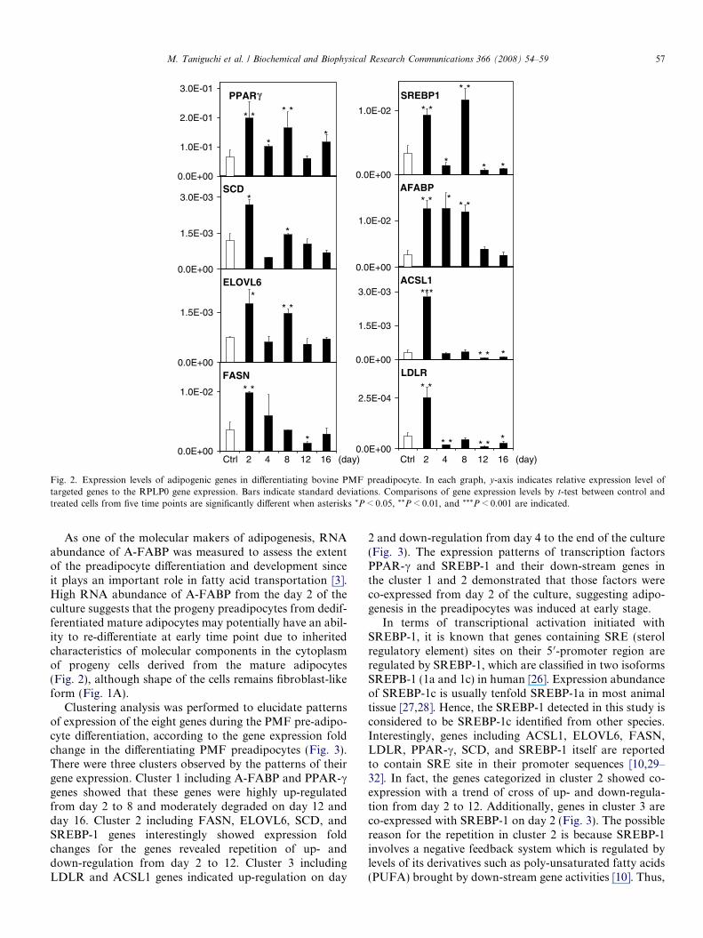
Therefore, the molecular mechanisms in the re-differen-tiating bovine PMF preadipocytes were investigated by elu-cidating expression profile of the major adipogenic factors,based on previous studies in rodent model and bovinePPAR signaling in KEGG pathway. Moreover, due to alack of understanding of the specifics of regulation by tran-scription factors in fat formation in cattle, the gene net-works of transcription factors PPAR-c and SREBP-1 andtheir down-stream genes in the process of the preadipocytedifferentiation were also explored. Fig. 2 shows that rela-tive expression level of the eight adipogenic genes toRPLP0 reference gene expression in treated cells was com-pared to control cells in time course, since RNA abundanceof RPLP0 was stable from day 2 to 16 of the culture in theqPCR assay for each gene (data not shown). It appears thatall genes showed significantly higher expression level intreated PMF adipocytes at day 2 than that of the controlcells. Furthermore, most of the genes indicated the highestgene expression level on day 2, whereas SREBP-1 geneshowed the highest expression at day 8. After day 12, mostof the genes showed down-regulations or silent, althoughonly PPAR-c gene showed up-regulation on day 16(Fig. 2).
cytes at confluent (0 days). (B) Differentiated PMF adipocytes with lipidicture are 100·.
ACSL1
0.0E+00
1.5E-03
3.0E-03
LDLR
0.0E+00
2.5E-04
SCD
γ
0.0E+00
1.5E-03
3.0E-03AFABP
0.0E+00
1.0E-02
FASN
0.0E+00
1.0E-02
ELOVL6
0.0E+00
1.5E-03
PPAR
0.0E+00
1.0E-01
2.0E-01
3.0E-01
Ctrl 2 4 8 12 16 (day) Ctrl 2 4 8 12 16 (day)
* * *
* *
*
* *
*
* *
* *
* * *
*
*
* * ** *
* ** ***
* * *
* *
*
* *
* *
SREBP1
0.0E+00
1.0E-02
Fig. 2. Expression levels of adipogenic genes in differentiating bovine PMF preadipocyte. In each graph, y-axis indicates relative expression level oftargeted genes to the RPLP0 gene expression. Bars indicate standard deviations. Comparisons of gene expression levels by t-test between control andtreated cells from five time points are significantly different when asterisks *P < 0.05, **P < 0.01, and ***P < 0.001 are indicated.
M. Taniguchi et al. / Biochemical and Biophysical Research Communications 366 (2008) 54–59 57
As one of the molecular makers of adipogenesis, RNAabundance of A-FABP was measured to assess the extentof the preadipocyte differentiation and development sinceit plays an important role in fatty acid transportation [3].High RNA abundance of A-FABP from the day 2 of theculture suggests that the progeny preadipocytes from dedif-ferentiated mature adipocytes may potentially have an abil-ity to re-differentiate at early time point due to inheritedcharacteristics of molecular components in the cytoplasmof progeny cells derived from the mature adipocytes(Fig. 2), although shape of the cells remains fibroblast-likeform (Fig. 1A).
Clustering analysis was performed to elucidate patternsof expression of the eight genes during the PMF pre-adipo-cyte differentiation, according to the gene expression foldchange in the differentiating PMF preadipocytes (Fig. 3).There were three clusters observed by the patterns of theirgene expression. Cluster 1 including A-FABP and PPAR-cgenes showed that these genes were highly up-regulatedfrom day 2 to 8 and moderately degraded on day 12 andday 16. Cluster 2 including FASN, ELOVL6, SCD, andSREBP-1 genes interestingly showed expression foldchanges for the genes revealed repetition of up- anddown-regulation from day 2 to 12. Cluster 3 includingLDLR and ACSL1 genes indicated up-regulation on day
2 and down-regulation from day 4 to the end of the culture(Fig. 3). The expression patterns of transcription factorsPPAR-c and SREBP-1 and their down-stream genes inthe cluster 1 and 2 demonstrated that those factors wereco-expressed from day 2 of the culture, suggesting adipo-genesis in the preadipocytes was induced at early stage.
In terms of transcriptional activation initiated withSREBP-1, it is known that genes containing SRE (sterolregulatory element) sites on their 5 0-promoter region areregulated by SREBP-1, which are classified in two isoformsSREPB-1 (1a and 1c) in human [26]. Expression abundanceof SREBP-1c is usually tenfold SREBP-1a in most animaltissue [27,28]. Hence, the SREBP-1 detected in this study isconsidered to be SREBP-1c identified from other species.Interestingly, genes including ACSL1, ELOVL6, FASN,LDLR, PPAR-c, SCD, and SREBP-1 itself are reportedto contain SRE site in their promoter sequences [10,29–32]. In fact, the genes categorized in cluster 2 showed co-expression with a trend of cross of up- and down-regula-tion from day 2 to 12. Additionally, genes in cluster 3 areco-expressed with SREBP-1 on day 2 (Fig. 3). The possiblereason for the repetition in cluster 2 is because SREBP-1involves a negative feedback system which is regulated bylevels of its derivatives such as poly-unsaturated fatty acids(PUFA) brought by down-stream gene activities [10]. Thus,

Fig. 3. Expression pattern of adipogenic genes by clustering analysis.Data measurements were examined by qPCR and the results were shownas fold change over the expression level of control. As shown by the colorscale bar, increasing green and red signal intensities indicate genes thatdecrease and increase in expression of treated PMF adipocytes during thetime course.
58 M. Taniguchi et al. / Biochemical and Biophysical Research Communications 366 (2008) 54–59
it seems that the genes in cluster 2 were up-regulated on thefirst day to produce PUFA by co-expression of SREBP-1,FASN, EVOLV6, and SCD, while the same genes weredown-regulated at the following time point, when increasedPUFA blocked transcription activity of SREBP-1. Unlikethe genes in cluster 2, transcription activation of two genes(LDLR and ACSL1) in cluster 3 may be repressed by theother effect.
As shown in the PPAR signaling pathway in bos taurus
from KEGG (http://www.genome.jp/dbget-bin/get_path-way?org_name=bta&mapno=03320), fatty acid bindingproteins trigger interactions mediated by PPAR-c toinduce various biological pathways including lipid metabo-lism, adipocyte differentiation and gluconeogenesis. In themolecular pathway map, PPAR-c stimulates transcriptionof A-FABP, ACSL1, FASN, and SCD genes. In additionto the PPAR signaling indicated in the KEGG, previousresults demonstrated in rodent models support that thegene interaction shown in this study was regulated by tran-scription activation of SREBP-1 and PPAR-c [10,33].However, the timing of the transactivation was earlier thanthat of the other in vitro models [24].
Although CCAAT/enhancer binding protein alpha (C/EBP-a) has been also known as another important tran-scription factor in regulation of adipocyte differentiationin various species [34–36], the expression of this gene wasunder detectable level in the bovine PMF system (datanot shown). Tan et al. [37] also reported that C/EBP-a gene
was not expressed in an in vitro study of differentiationusing bovine bone marrow-derived preadipocytes. Thesesuggest the transcriptional regulation of adipogenic genesin bovine preadipocytes is different from that of porcineand rodent preadipocytes [35,36,38], although the resultsshowed PPAR-c played a pivotal role in the regulation ofgene transcription and cellular differentiation as indicatedin the other animal species [39].
In summary, our results suggest that the bovine adipofi-broblast cell culture system is viable for use in adipogenesisstudies, that there are specific regulatory genes expressedduring the adipogenesis process of these cells, and thatgenetic interactions exist among specific adipogenic factorsevaluated. This information is lacking in the bovine. Glo-bal gene expression profiling during adipogenesis in thesebovine preadipocytes using high-throughput technologysuch as DNA microarray, is on-going, and will supplydetailed molecular biochemical pathway underlying bovinepreadipocyte differentiation.
Acknowledgment
This study is supported by Alberta Agricultural FindingConsortium (AARI 2006F039).
References
[1] H.D. Ritchie, S.R. Rust, R.A. Merkel, W.G. Bergen, Getting rid ofexcess fat is not an easy task, Feedstuffs (1993) 13.
[2] Y.H. Wang, A. Reverter, H. Mannen, M. Taniguchi, G.S. Harper, K.Oyama, K.A. Byrne, A. Oka, S. Tsuji, S.A. Lehnert, Transcriptionalprofiling of muscle tissue in growing Japanese Black cattle to identifygenes involved with the development of intramuscular fat, Aus. J.Exp. Agric. 45 (2005) 809–820.
[3] Y.H. Wang, K.A. Byrne, A. Reverter, G.S. Harper, M. Taniguchi,S.M. McWilliams, H. Mannen, K. Oyama, S.A. Lehnert, Transcrip-tional profiling of skeletal muscle tissue from two breeds of cattle,Mamm. Genome 16 (2005) 201–210.
[4] F.M. Gregoire, C.M. Smas, H.S. Sul, Understanding adipocytedifferentiation, Physiol. Rev. 78 (1998) 783–809.
[5] X. Guo, K. Liao, Analysis of gene expression profile during 3T3-L1preadipocyte differentiation, Gene 251 (2000) 45–53.
[6] G.R. Burton, R. Nagarajan, C.A. Peterson, R.E. McGehee Jr.,Microarray analysis of differentiation-specific gene expression during3T3-L1 adipogenesis, Gene 329 (2004) 167–185.
[7] C. Hansen, A. Fu, C. Li, W.T. Dixon, R. Christopherson, S.S.Moore, Global gene expression patterns spanning 3T3-L1 preadipo-cyte differentiation, Can. J. Anim. Sci. 84 (2004) 367–376.
[8] M.S. Brown, J.L. Goldstein, A proteolytic pathway that controls thecholesterol content of membranes, cells, and blood, Proc. Natl. Acad.Sci. USA 96 (1999) 11041–11048.
[9] J.M. Ntambi, Y.C. Kim, Adipocyte differentiation and gene expres-sion, J. Nutrit. 130 (2000) 3122–3126.
[10] H. Shimano, Sterol regulatory element-binding proteins (SREBPs):transcriptional regulators of lipid synthetic genes, Prog. Lipid Res. 40(2001) 439–452.
[11] J.B. Kim, H.M. Wright, M. Wright, B.M. Spiegelman, ADD1/SREBP1 activates PPARgamma through the production of endog-enous ligand, Proc. Natl. Acad. Sci. USA 95 (1998) 4333–4337.
[12] M.E. Fernyhough, E. Okine, G.J. Hausman, J.L. Vierck, M.V.Dodson, Invited review: PPAR-gamma and GLUT-4 expression asdifferentiation markers for preadipocyte conversion to become anadipocyte, Domest. Anim. Endocrinol. 33 (2007) 367–378.

M. Taniguchi et al. / Biochemical and Biophysical Research Communications 366 (2008) 54–59 59
[13] M.E. Fernyhough, J.L. Vierck, G.J. Hausman, P.S. Mir, E.K. Okine,M.V. Dodson, Primary adipocyte culture: adipocyte purificationmethods may lead to a new understanding of adipose tissue growthand development, Cytotechnology 46 (2005) 163–172.
[14] M.V. Dodson, M.E. Fernyhough, J.L. Vierck, G.J. Hausman,Adipocytes may not be a terminally differentiated cell type: implica-tions for animal production, Anim. Sci. 80 (2005) 239–240.
[15] M.E. Fernyhough, D.L. Helterline, J.L. Vierck, G.J. Hausman, R.A.Hill, M.V. Dodson, Dedifferentiation of mature adipocytes to formadipofibroblasts: more than just a possibility, Adipocytes 1 (2005) 17–24.
[16] K. Tahara, H. Aso, T. Yamasaki, M.T. Rose, A. Takasuga, Y.Sugimoto, T. Yamaguchi, S. Takano, Cloning and expression of typeXII collagen isoforms during bovine adipogenesis, Differentiation 72(2004) 113–122.
[17] A. Sen, Y.R. Lea-Currie, D. Sujkowska, D.M. Franklin, W.O.Wilkinson, Y.D. Halvorsen, J.M. Gimbre, Adipogenic potential ofhuman adipose derived stromal cells from multiple donors isheterogeneous, J. Cell. Biochem. 81 (2001) 312–319.
[18] S. Rozen, H. Skaletsky, Primer3 on the WWW for general users andfor biologist programmers, Methods Mol. Biol. 132 (2000) 365–386.
[19] M.W. Pfaffl, T.M. Georgieva, I.P. Georgiev, E. Ontsouka, M.Hageleit, J.W. Blum, Real-time RT-PCR quantification of insulin-like growth factor (IGF)-1, IGF-1 receptor, IGF-2, IGF-2 receptor,insulin receptor, growth hormone receptor, IGF-binding proteins 1, 2and 3 in the bovine species, Domest. Anim. Endocrinol. 22 (2002) 91–102.
[20] G. Caraux, S. Pinloche, Permutmatrix: a graphical environment toarrange gene expression profiles in optimal linear order, Bioinfor-matics 21 (2005) 1280–1281.
[21] H. Sugihara, N. Yonemitsu, S. Miyabara, K. Yun, Primary culturesof unilocular fat cells: characteristics of growth in vitro and changesin differentiation properties, Differentiation 31 (1986) 42–49.
[22] H. Sugihara, N. Yonemitsu, S. Miyabara, S. Toda, Proliferation ofunilocular fat cells in primary culture, J. Lipid Res. 28 (1987) 1038–1045.
[23] H. Sugihara, S. Funatsumaru, N. Yonemitsu, S. Miyabara, S. Toda,Y. Hikichi, A simple culture of fat cells from mature fat tissuefragments, J. Lipid Res. 30 (1989) 1987–1995.
[24] T.A. Kokta, M.V. Dodson, A. Gertler, R.A. Hill, Intercellularsignaling between adipose tissue and muscle tissue, Domest. Anim.Endocrinol. 27 (2004) 303–331.
[25] B.M. Spiegelman, J.S. Flier, Adipogenesis and obesity: rounding outthe big picture, Cell 87 (1996) 377–389.
[26] I. Shimomura, H. Shimano, J.D. Horton, J.L. Goldstein, M.S.Brown, Differential expression of exons 1a and 1c in mRNAs forsterol regulatory element binding protein-1 in human and mouseorgans and cultured cells, J. Clin. Invest. 99 (1997) 838–845.
[27] J.D. Horton, Sterol regulatory element-binding proteins: transcrip-tional activators of lipid synthesis, Biochem. Soc. Trans. 30 (2002)1091–1095.
[28] J.D. Horton, J.L. Goldstein, M.S. Brown, SREBPs: activators of thecomplete program of cholesterol and fatty acid synthesis in the liver,J. Clin. Invest. 109 (2002) 1125–1131.
[29] M.R. Briggs, C. Yokoyama, X. Wang, M.S. Brown, J.L. Goldstein,Nuclear protein that binds sterol regulatory element of low densitylipoprotein receptor promoter, J. Biol. Chem. 268 (1993) 14490–14496.
[30] L. Fajas, K. Schoonjans, L. Gelman, J.B. Kim, J. Najib, G. Martin,J.C. Fruchart, M. Briggs, B.M. Spiegelman, J. Auwerx, Regulation ofperoxisome proliferators-activated receptor gamma expression byadipocyte differentiation and determination factor 1/sterol regulatoryelement binding protein 1: implications for adipocyte differentiationand metabolism, Mol. Cell Biol. 19 (1999) 5495–5503.
[31] M.M. Magana, T.F. Osborne, Two tandem binding sites for sterolregulatory element binding proteins are required for sterol regulationof fatty acid synthase promoter, J. Biol. Chem. 271 (1996) 32689–32694.
[32] M. Taniguchi, T. Utsugi, K. Oyama, H. Mannen, M. Kobayashi, Y.Tanabe, A. Ogino, S. Tsuji, Genotyping of stearoyl-CoA desaturase isassociated with fatty acid composition in Japanese Black cattle,Mamm. Genome 14 (2004) 142–148.
[33] D.M. Mutch, W. Wahli, G. Williamson, Nutrigenomics and nutri-genetics: the emerging faces of nutrition, FASEB J. 12 (2005) 1602–1616.
[34] M.D. Lane, Q.Q. Tang, M.S. Jiang, Role of the CCAAT enhancerbinding proteins (C/EBPs) in adipocyte differentiation, Biochem.Biophys. Res. Commun. 266 (1999) 677–683.
[35] G.J. Hausman, The influence of dexamethasone and insulin onexpression of CCAAT/enhancer binding protein isoforms duringpreadipocyte differentiation in porcine stromal–vascular cell cultures:evidence for very early expression of C/EBPa, J. Anim. Sci. 78 (2000)1227–1235.
[36] J.K. Hamm, B.H. Park, S.R. Farmer, A role for C/EBPb inregulating peroxisome proliferators-activated receptor c activityduring adipogenesis in 3T3-L1 preadipocytes, J. Biol. Chem. 276(2001) 18464–18471.
[37] S.H. Tan, A. Reverter, Y.H. Wang, K.A. Byrne, S.M. McWilliam,S.A. Lehnert, Gene expression profiling of bovine in vitro adipogen-esis using a cDNA microarray, Funct. Integr. Genomics 6 (2006) 235–249.
[38] Z. Wu, P. Puigserver, B.M. Spiegelman, Transcriptional activation ofadipogenesis, Curr. Opin. Cell Biol. 11 (1999) 689–694.
[39] K.L. Houseknecht, B.M. Cole, P.J. Steele, Peroxisome proliferators-activated receptor gamma (PPARc) and its ligands: a review, Domest.Anim. Endocrinol. 22 (2002) 1–23.
Copyright © 2022 FDOKUMEN