T2-weighted fast spin-echo magnetic resonance imaging of extraocular muscles
7
T2-weighted fast spin-echo magnetic resonance imaging of extraocular muscles Joseph L. Demer, MD, PhD, a,b,c,d and Anita Dushyanth, MS a PURPOSE Magnetic resonance imaging (MRI) can provide unique information about extraocular muscle (EOM) structure and function. Previous high-resolution motility imaging studies used T1 weighting, which provides intrinsic contrast of dark-appearing EOMs against bright orbital fat and is suitable for intravenous contrast. However, time-consuming T1 sequences are subject to motion artifacts. We evaluated an alternative T2-weighted fast spin-echo pulse sequence that emphasizes tissue-free fluid. METHODS We prospectively used high-resolution, surface coil technique for orbital MRI at 1.5T in 21 orthotropic and 113 living strabismic subjects and 2 monkey cadavers by using T2 fast spin-echo (T2FSE) weighting (long repetition time, short echo time). T2FSE was compared with T1 in 17 subjects, and with T1 in 506 different living subjects, and 12 cadavers. RESULTS For 2 mm thick coronal MRIs of 312 mm resolution spanning the entire orbit, T1 acqui- sition required 218 seconds, whereas T2FSE required 150 seconds (31% faster). T2-defined the globe border better, and provided intrinsic contrast between EOMs and their pulleys. Although both T1 and T2 demonstrated motor nerves to EOMs in living subjects, only T1 was satisfactory with injected contrast and in cadavers. CONCLUSIONS For motility imaging, T2FSE is faster than T1 MRI and demonstrates superior tissue de- tails of EOMs and other orbital tissues. T2FSE of the orbits can be performed by the use of widely available standard equipment. We suggest that T2FSE be the preferred method for clinical imaging of EOM structure, function, and innervation, although T1 may be more appropriate when intravenous contrast must be used. ( J AAPOS 2011;15:17-23) M agnetic resonance imaging (MRI) has become increasingly used in pediatric ophthalmology and strabismus. Technical improvements in MRI now afford the opportunity for detailed study of the functional anatomy of the extraocular muscles (EOMs), as- sociated orbital connective tissues, and nerves in living sub- jects patients, 1 and cranial nerves can be imaged against the surrounding cerebrospinal fluid as they exit the brainstem. 2 Orbital MRI for evaluation of ocular motility uses tech- niques different from those appropriate to brain MRI. The positions, sizes, and shapes of the EOMs are strongly dependent on eye position, and MRI quality depends strongly on minimization of physiologic motion artifacts that do not occur in the brain. The proximity of the orbits to air-filled sinuses and their corresponding MRI signal artifacts reduces and often even reverses the imaging ad- vantages that high-field imaging affords in brain. 3 Intrave- nous contrast injection is often unnecessary for orbital MRI in the evaluation of strabismus. Yet, for clinical util- ity, orbital MRI resolution must be greater than that usu- ally used in brain MRI. A typical whole-head MRI with 5 mm plane thickness and a 20 cm square field of view has 781 mm in-plane resolution, giving 3 mL of voxel vol- ume. A typical orbital MRI with surface coil technique, 2 mm plane thickness, and an 8 cm square field of view has 312 mm in-plane resolution, giving 0.2 mL of voxel vol- ume. The high-resolution orbital MRI thus has 15-fold higher voxel resolution than the brain MRI. A further implication of high-resolution orbital technique is that its field of view is large enough only for one orbit at a time, rather than both as is imaged with the use of typical brain technique; twice as many sets of images are therefore required to image both orbits at high resolution. The demanding technical requirements of high-resolution orbital MRI may not be generally appreciated. Numerous MRI parameters may be varied to image different features of tissues, including different pulse sequences. 4 The authors have performed preliminary eval- uations of a wide range of MRI pulse sequences and proto- cols, including volumetric and high field (3T) acquisitions, during the last 2 decades as new approaches have emerged. Author affiliations: Departments of a Ophthalmology, b Neurology, c Bioengineering, and d Neuroscience Interdepartmental Programs, Jules Stein Eye Institute, University of California, Los Angeles Submitted July 31, 2010. Revision accepted December 31, 2010. Presented as a poster at the 35th Annual Meeting of the American Association for Pediatric Ophthalmology and Strabismus, San Francisco, California, April 17-21, 2009. Grant Support: Supported by grants from the National Eye Institute: EY08313 and EY00331. Also supported by a Research to Prevent Blindness Walt and Lilly Disney Award. Reprint requests: Joseph L. Demer, MD, PhD, Jules Stein Eye Institute, 100 Stein Plaza, UCLA, Los Angeles, CA 90095-7002 (email: [email protected]). Copyright Ó 2011 by the American Association for Pediatric Ophthalmology and Strabismus. 1091-8531/$36.00 doi:10.1016/j.jaapos.2010.12.006 Journal of AAPOS 17
Transcript of T2-weighted fast spin-echo magnetic resonance imaging of extraocular muscles

T2-weighted fast spin-echo magnetic resonanceimaging of extraocular musclesJoseph L. Demer, MD, PhD,a,b,c,d and Anita Dushyanth, MSa
PURPOSE Magnetic resonance imaging (MRI) can provide unique information about extraocular
Author affiliations: DepartmentdNeuroscience InterdepartmentalCalifornia, Los AngelesSubmitted July 31, 2010.Revision accepted December 31Presented as a poster at the 35
Pediatric Ophthalmology and StrGrant Support: Supported by
EY00331. Also supported by a RAward.Reprint requests: Joseph L. De
UCLA, Los Angeles, CA 90095Copyright � 2011 by the Am
Strabismus.1091-8531/$36.00doi:10.1016/j.jaapos.2010.12
Journal of AAPOS
muscle (EOM) structure and function. Previous high-resolution motility imaging studiesused T1 weighting, which provides intrinsic contrast of dark-appearing EOMs againstbright orbital fat and is suitable for intravenous contrast. However, time-consuming T1sequences are subject to motion artifacts. We evaluated an alternative T2-weighted fastspin-echo pulse sequence that emphasizes tissue-free fluid.
METHODS We prospectively used high-resolution, surface coil technique for orbital MRI at 1.5T in
21 orthotropic and 113 living strabismic subjects and 2 monkey cadavers by using T2fast spin-echo (T2FSE) weighting (long repetition time, short echo time). T2FSE wascompared with T1 in 17 subjects, and with T1 in 506 different living subjects, and 12cadavers.RESULTS For 2 mm thick coronal MRIs of 312 mm resolution spanning the entire orbit, T1 acqui-
sition required 218 seconds, whereas T2FSE required 150 seconds (31% faster).T2-defined the globe border better, and provided intrinsic contrast between EOMs andtheir pulleys. Although both T1 and T2 demonstrated motor nerves to EOMs in livingsubjects, only T1 was satisfactory with injected contrast and in cadavers.CONCLUSIONS For motility imaging, T2FSE is faster than T1 MRI and demonstrates superior tissue de-
tails of EOMs and other orbital tissues. T2FSE of the orbits can be performed by the use ofwidely available standard equipment. We suggest that T2FSE be the preferred method forclinical imaging of EOM structure, function, and innervation, although T1 may be moreappropriate when intravenous contrast must be used. ( J AAPOS 2011;15:17-23)Magnetic resonance imaging (MRI) has becomeincreasingly used in pediatric ophthalmologyand strabismus. Technical improvements in
MRI now afford the opportunity for detailed study of thefunctional anatomy of the extraocular muscles (EOMs), as-sociated orbital connective tissues, and nerves in living sub-jects patients,1 and cranial nerves can be imaged against thesurrounding cerebrospinal fluid as they exit the brainstem.2
Orbital MRI for evaluation of ocular motility uses tech-niques different from those appropriate to brain MRI. Thepositions, sizes, and shapes of the EOMs are stronglydependent on eye position, and MRI quality dependsstrongly on minimization of physiologic motion artifactsthat do not occur in the brain. The proximity of the orbits
s of aOphthalmology, bNeurology, cBioengineering, andPrograms, Jules Stein Eye Institute, University of
, 2010.th Annual Meeting of the American Association forabismus, San Francisco, California, April 17-21, 2009.grants from the National Eye Institute: EY08313 andesearch to Prevent Blindness Walt and Lilly Disney
mer,MD, PhD, Jules Stein Eye Institute, 100 Stein Plaza,-7002 (email: [email protected]).erican Association for Pediatric Ophthalmology and
.006
to air-filled sinuses and their corresponding MRI signalartifacts reduces and often even reverses the imaging ad-vantages that high-field imaging affords in brain.3 Intrave-nous contrast injection is often unnecessary for orbitalMRI in the evaluation of strabismus. Yet, for clinical util-ity, orbital MRI resolution must be greater than that usu-ally used in brain MRI. A typical whole-head MRI with5 mm plane thickness and a 20 cm square field of viewhas 781 mm in-plane resolution, giving 3 mL of voxel vol-ume. A typical orbital MRI with surface coil technique,2 mm plane thickness, and an 8 cm square field of viewhas 312 mm in-plane resolution, giving 0.2 mL of voxel vol-ume. The high-resolution orbital MRI thus has 15-foldhigher voxel resolution than the brain MRI. A furtherimplication of high-resolution orbital technique is that itsfield of view is large enough only for one orbit at a time,rather than both as is imaged with the use of typical braintechnique; twice as many sets of images are thereforerequired to image both orbits at high resolution. Thedemanding technical requirements of high-resolutionorbital MRI may not be generally appreciated.
Numerous MRI parameters may be varied to imagedifferent features of tissues, including different pulsesequences.4 The authors have performed preliminary eval-uations of a wide range of MRI pulse sequences and proto-cols, including volumetric and high field (3T) acquisitions,during the last 2 decades as new approaches have emerged.
17

18 Demer and Dushyanth Volume 15 Number 1 / February 2011
Despite the advantages of some of these methods for brainimaging, the techniques described here have proved supe-rior for orbital MRI in pediatric ophthalmology and stra-bismus. T1 weighting was formerly recommended forhigh-resolution orbital MRI because with this image se-quence the orbital fat is bright, against which the globeand EOMs appear with high contrast in dark signal.5 AT2 image heavily weights free fluid, such as aqueous andvitreous humors, and cerebrospinal fluid. T2-weightedfast spin-echo (T2FSE) has been developed for acquiringimages rapidly (Clark RA, Demer JL. Differential lateralrectus [LR] compartmental contraction: A novel mecha-nism accounts for impaired ocular counter-rolling [OCR)in superior oblique [SO] palsy. J AAPOS 2011;15:e000Abstract 055).3 This method is also known as “rapid acqui-sition with relaxation enhancement.”4
Such a time advantage could be of great practical impor-tance because motion artifacts are now the main limitationon surface coil orbital MRI resolution. It is possible, how-ever, that T2FSE might not show the same features in theorbit as the recommended T1MRI. Although both T1 andT2FSE MRI display orbital fat as bright and EOMs asdark, the vitreous appears dark with T1 and bright withT2FSE. Free fluid, such as in aqueous, vitreous, and cere-brospinal fluid, is bright on T2FSE. These qualitative fea-tures might have differing clinical implications in the orbit.We conducted the present study to evaluate the advantagesand limitations of T2FSE for EOM imaging.
Subjects and Methods
From June 2007 through December 2010, a total of 21
orthotropic and 106 strabismic living humans underwent high-
resolution orbital MRI under a prospective protocol for study
of strabismus. Retrospective comparison was made with
T1-weighted MRI obtained in the same study from 1990 to
2010 in 506 living humans, 10 human cadavers, and 21 monkey
cadavers. The imaging goals, use of quasicoronal imaging planes,
target fixation, gaze directions, and goal of limiting scan duration
on scan duration remained the same throughout the study. The
aim remained to optimize the resolution possible with the exist-
ing hardware.
As technical capabilities improved over the years, image plane
thickness was reduced, quasisagittal imaging was added, and the
total number of image planes acquired per sequence was
increased, and finally, with T2FSE the acquisition time was
further reduced. As a result, most of the early MRI studies in
the project include views exactly comparable with the most recent
studies; they differ, of course, in resolution. The direct compari-
sons illustrated in figures were all from the period 2007 to 2010
and used the same scanner, the same target device, and the same
surface coil array. The T1 protocol did not change during this
period. A total of 15 subjects were prospectively scanned with
the use of both T1-weighted and T2FSE sequences. Orthotropic
volunteers were recruited by advertising; the patients were
recruited from referral strabismus practices. In all subjects, writ-
ten informed consent was obtained prospectively according to
a protocol approved by the institutional review board for the pro-
tection of human subjects and in conformity with the Health
Insurance Portability and Accountability Act. All subjects under-
went complete ophthalmological examination, with particular
attention paid to ocular motility.
Human cadaver specimens were obtained by anatomical dona-
tion in conformity with applicable laws. Macaque monkey speci-
mens were obtained by tissue sharing from investigators whose
protocols for the living animals had been approved by applicable
animal welfare committees. After formalin fixation, 10 human and
19 monkey cadavers underwent MRI with T1, and 2 monkey
cadavers with both T1 and T2FSE, with the use of dual 75 mm
phased-array surface coils (General Electric, Milwaukee, WI).
Cadaver specimens were warmed to physiological temperature
in a water bath immediately before MRI.
Imaging was performed using a 1.5 T General Electric Signa
scanner. Orbital MRI in living subjects was performed by the
use of an array of surface coils embedded in a transparent face-
mask (Medical Advances, Milwaukee, WI), and fixation targets
to avoid eye motion artifacts.5,6 The head was stabilized in the
supine position by tightly fastening the surface coil mask to the
face using headbands and fixing the mask to the scanner gantry
by the use of foam cushions and tape. These measures avoided
head rotation during scanning. An adjustable array of
illuminated fixation targets was secured in front of each orbit
with the center target in subjective central position for each eye,
and in selected cases in secondary and tertiary gaze positions. In
living humans, nonoverlapping images of 2 mm thickness in
a matrix of 256 � 256 were obtained over a field of view of 6–8
cm for a resolution in plane of 234–312 mm, respectively. In
cadavers, some images were obtained with a field of view of
4 cm for a resolution in plane of 156 mm. Axial scout images
were obtained, as well as quasicoronal images perpendicular
to the long axis of the orbit, and quasisagittal images parallel to
the long axis of the orbit.
All living subjects underwent MRI using a T2FSE protocol
summarized in Table 1. Selected subjects also underwent addi-
tional T1-weighted imaging for comparison purposes, with the
use of published parameters.1,5 After initial T1 and T2FSE MRI
was performed in some subjects, the paramagnetic MRI contrast
agent gadodiamide (0.1 mmol/kg) was given intravenously
before T1 MRI was repeated.7
Digital MRI images were transferred to Macintosh computers
(Apple Computer, Inc, Cupertino, CA), converted into 8-bit
tagged image file format (ie, TIFF), and examined with the pro-
gram NIH Image (W. Rasband, National Institutes of Health;
available from zippy.nimh.nih.gov or on disk from NTIS, 5285
Port Royal Road, Springfield, VA 22161, part number PB95-
500195GEI).
After MRI, cadaver orbits imaged with T1 and T2FSE were re-
moved en bloc with periorbita intact. As previously described, the
whole orbits were sectioned in the quasicoronal plane into 10 mm
thick sections by a microtome (HM325, Carl-Zeiss, Thornwood,
NY) after tissue processing, including formalin fixation,
decalcification, dehydration, and embedding in paraffin.8-11
Selected sections were stained with Masson trichrome stain to
distinguish collagen (blue), muscle (red), and nerve (purple).12
Journal of AAPOS

Table 1. Comparison of MRI scan parameters
Parameter T1 T2FSE
Typical field of view, cm 8 � 8 8 � 8Typical in-plane resolution, mm 312 312Image plane thickness, mm 2 2Repetition time (TR), ms 416 4116Echo time (TE), ms 13 87Excitations (acquisitions) 2 2Bandwidth, Hz/pixel 122 195Echo train length, echoes/TR 1 13Total imaging time, seconds* 218 150Reduction in imaging time, % 0 31
T2FSE, T2 fast spin-echo.*Imaging time is for a typical set of 18 quasicoronal image planes.
FIG 1. Axial MRI in 2 comparable esotropic patients. T1 image abovedemonstrates dark signal from slower realignment of proton spin axesin the vitreous. T2FSE image below demonstrates bright signal fromslower dephasing of proton spin axes in the vitreous. Horizontal rectusEOMs are well demonstrated with both methods. T1 demonstrates irisand ciliary body better than T2FSE.
Volume 15 Number 1 / February 2011 Demer and Dushyanth 19
Stained sections were digitally photographed with a Nikon
Eclipse microscope (Melville, NY).
The authors have extensive experience with high-resolution
orbital MRI. Images were evaluated qualitatively by the authors
for resolution of details of the globe, EOMs, orbital connective
tissues, and nerves. This included contrast of these anatomical
features against surrounding structures such as orbital fat, and
pathological changes within the features. Imaging acquisition
time was evaluated as total time required to acquire each image
set after initiation of the imaging pulse series.
Results
Imaging Quality
In general, T2FSE produced images of the globes andorbits that were comparable, if not better than, MRI withT1 weighting. The typical level of detail is illustrated inFigure 1, containing axial images of 2 different patientswith left abducens paralysis. In each case, atrophy of the in-volved lateral rectus muscle is obvious, as well as lateralbowing and inflection of the lateral rectus path. In theT1 image, the cornea, lens, choroids, and ciliary body areclearly distinguishable against the dark vitreous humor(Figure 1, top). In the T2FSE image, the dark-appearinglens is vividly contrasted against the bright vitreous humor,but the uveal tract provides no contrast against the vitreous.The optic nerve is readily visible in both T1 and T2FSEimages.
Imaging Time
With two excitations, acquisition of 18 quasicoronal MRIplanes 2 mm thick with an 8 cm square field of view re-quired 218 seconds for T1 but only 150 seconds forT2FSE. This example represents a 31% time advantagefor T2FSE imaging (Table 1). Although TR and TEwere varied slightly for each study to minimize total imag-ing time, this example was typical of the lower acquisitionfrom T2FSE than T1 imaging.
Nerves and Blood Vessels
Quasicoronal images perpendicular to the long axis ofthe orbit are optimal for demonstration of rectus and thesuperior oblique EOMs in cross section. This imaging
Journal of AAPOS
plane is also optimal to demonstrate small intraorbital mo-tor nerves to the EOMs. As illustrated in Figure 2, poste-rior to the globe, T1 and T2FSE images appear almostidentical. Both techniques demonstrate EOM cross sec-tions in the deep orbit, orbital nerves, and blood vesselsalmost identically by their dark signals against the brightorbital fat. One feature favors T2FSE in the deep orbit:T2FSE provides vivid contrast of the bright-appearingcerebrospinal fluid against the optic nerve within itsdural sheath (Figure 2, bottom row).
Intrinsic Tissue Contrast
Although both T1 and T2FSE provided excellent delinea-tion of tissue boundaries demarcated by orbital fat, onlyT2FSE demonstrated additional boundaries delineatedby variations in tissue water content. Figure 3 shows com-parable quasicoronal images of the right orbit of the samepatient, who has ethmoid sinusitis. The T1 image clearlydemonstrates the EOMs and orbital connective tissues,but the periorbital tissues are almost uniformly dark(Figure 3 left). With T2FSE, water-containing sinus
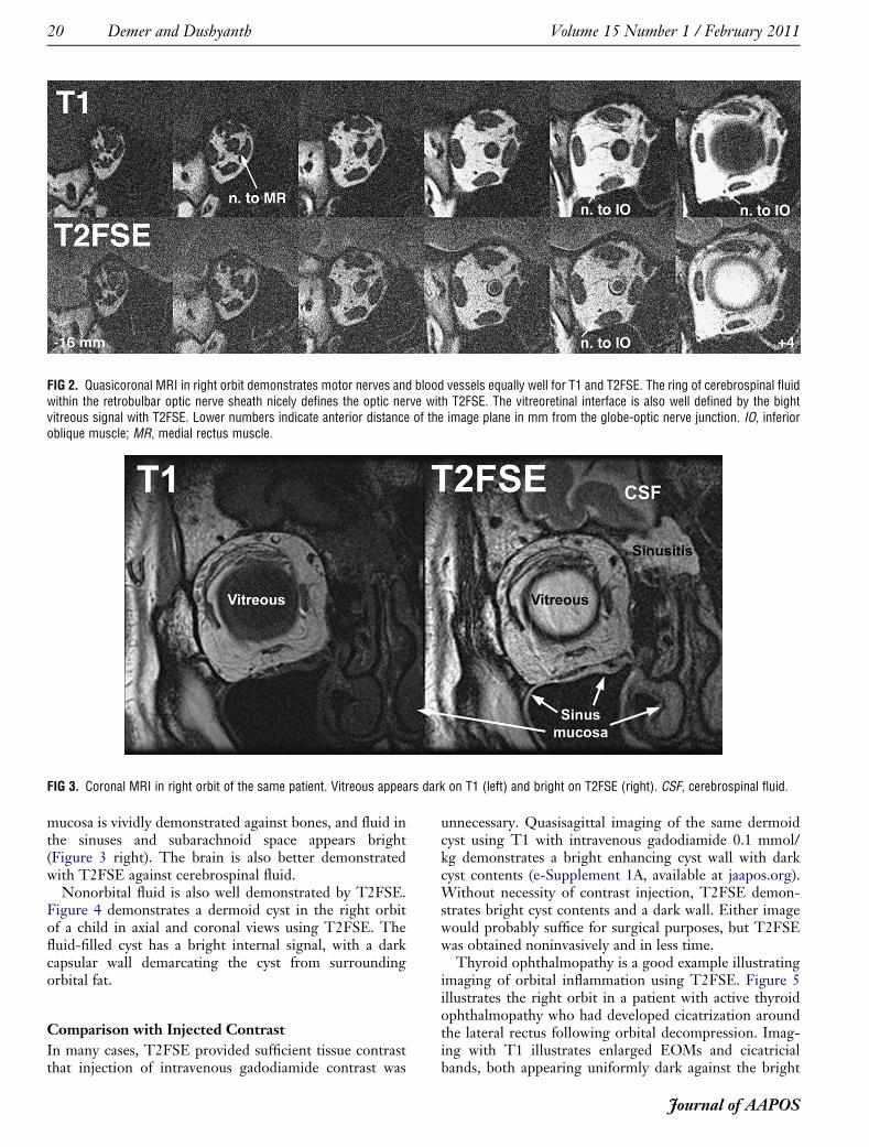
FIG 2. Quasicoronal MRI in right orbit demonstrates motor nerves and blood vessels equally well for T1 and T2FSE. The ring of cerebrospinal fluidwithin the retrobulbar optic nerve sheath nicely defines the optic nerve with T2FSE. The vitreoretinal interface is also well defined by the bightvitreous signal with T2FSE. Lower numbers indicate anterior distance of the image plane in mm from the globe-optic nerve junction. IO, inferioroblique muscle; MR, medial rectus muscle.
FIG 3. Coronal MRI in right orbit of the same patient. Vitreous appears dark on T1 (left) and bright on T2FSE (right). CSF, cerebrospinal fluid.
20 Demer and Dushyanth Volume 15 Number 1 / February 2011
mucosa is vividly demonstrated against bones, and fluid inthe sinuses and subarachnoid space appears bright(Figure 3 right). The brain is also better demonstratedwith T2FSE against cerebrospinal fluid.
Nonorbital fluid is also well demonstrated by T2FSE.Figure 4 demonstrates a dermoid cyst in the right orbitof a child in axial and coronal views using T2FSE. Thefluid-filled cyst has a bright internal signal, with a darkcapsular wall demarcating the cyst from surroundingorbital fat.
Comparison with Injected Contrast
In many cases, T2FSE provided sufficient tissue contrastthat injection of intravenous gadodiamide contrast was
unnecessary. Quasisagittal imaging of the same dermoidcyst using T1 with intravenous gadodiamide 0.1 mmol/kg demonstrates a bright enhancing cyst wall with darkcyst contents (e-Supplement 1A, available at jaapos.org).Without necessity of contrast injection, T2FSE demon-strates bright cyst contents and a dark wall. Either imagewould probably suffice for surgical purposes, but T2FSEwas obtained noninvasively and in less time.
Thyroid ophthalmopathy is a good example illustratingimaging of orbital inflammation using T2FSE. Figure 5illustrates the right orbit in a patient with active thyroidophthalmopathy who had developed cicatrization aroundthe lateral rectus following orbital decompression. Imag-ing with T1 illustrates enlarged EOMs and cicatricialbands, both appearing uniformly dark against the bright
Journal of AAPOS

FIG 4. Axial (A) and quasicoronal (B) MRI in right orbit of child with an orbital dermoid cyst. Signal in the dermoid is bright, similar to that of theorbital fat and slightly darker than the vitreous.
Volume 15 Number 1 / February 2011 Demer and Dushyanth 21
orbital fat (Figure 5, middle column). After gadodiamidecontrast injection, the enlarged EOMs enhance in a non-uniform manner suggestive of inflammation, but thepoorly vascular cicatrix around the lateral rectus remainsdark (Figure 5, left column). Contrast enhancement ofEOMs during T1 imaging reduces their visibility againstthe adjacent bright orbital fat. With T2FSE in the absenceof injected contrast medium, both the cicatrix and EOMsexhibit excellent contrast against the surrounding brightorbital fat (Figure 5, right column). The EOMs are moredistinct against surrounding orbital fat with T2FSE thanwith contrast-enhanced T1, yet T2FSE demonstratesvariations in signal within EOMs about as well as doesthe former. This signal variation within EOMs in thyroidophthalmopathy probably indicates active myositis.
Demonstration of Pulleys
In the anterior orbit, the thin rectus EOMs have been diffi-cult to distinguish from their surrounding connective tissuepulleys when T1 MRI is used, unless intravenous gadodia-mide contrast is injected that preferentially enhances thehighly vascular EOMswithin their collagenous pulleys.13,14
This is illustrated in e-Supplement 2 (available at jaapos.org), which demonstrates in coronal views with axial refor-matting the horizontal rectus pulleys of the left orbit a nor-mal subject in adduction and abduction. Without contrastinjection, T2FSE can also distinguish rectus pulleys fromanterior EOMs, presumably because EOMs contain morefluid than pulleys. E-Supplement 3 (available at jaapos.org) demonstrates the horizontal rectus pulleys of anesotropic patient in coronal views with axial reformattingin adduction and abduction. However, T2FSE did notprovide as much contrast of EOMs against their pulleys asdid gadodiamide-enhanced T1 MRI.
Journal of AAPOS
Cadaver Imaging
Although not performed clinically, imaging of cadavericorbits permits histological validation of MRI findings.Two formalin-fixed monkey orbits were imaged by bothT1 and T2FSE before en bloc embedding and serial sec-tioning for staining with Masson trichrome. As illustratedin e-Supplement 4 (available at jaapos.org), T1 imagingprovides excellent detail of EOM and nerve borders delin-eated by their dark signal against the bright orbital fat, andsimilarly demonstrated the superior ophthalmic vein.Although T2FSE also demonstrated the nerves and supe-rior ophthalmic vein, there was poor contrast of EOMsagainst surrounding orbital fat. Poor EOM contrastmade T2FSE unsuitable for MRI of cadaveric orbits.Cadaveric MRI using T1 weighting provided near micro-scopic resolution, as previously described.15
Discussion
Orbital MRI has broadened our understanding of the anat-omy and physiology of the EOMs and their associated con-nective tissues.16,17 In living humans, it is now possible toimage with MRI at near-microscopic resolution the physi-ologic changes associated with conjugate eye movements,vergence, and accommodation.18 It has been possible forsome years to use clinically available equipment to imagethe size, contractility, and position of each EOM. How-ever, it takes time to acquire high-resolution MRI.
In this work we demonstrate that orbital MRI by usingthe T2FSE sequence is approximately one-third fasterthan imaging with previously recommended T1 weighting,but in living people T2FSE demonstrates as well as T1nearly all details of EOMs, orbital connective tissues, andEOM innervation. Furthermore, T2FSE demonstratesfluid-filled features such as cysts and edematous tissues

FIG 5. Quasicoronal MRI of right orbit with thyroid ophthalmopathy, post orbital decompression that caused cicatrization around the lateral rectusmuscle. Detail with T2FSE is comparable with T1 with intravenous gadodiamide contrast, which shortens both T1 and T2. Contrast enhancementoccurs in tissues in proportion to blood flow. T2FSE also gives bright signal in EOM regions that enhance with contrast in T1 imaging, which mayobviate the need for contrast injection for demonstration of extraocular muscle inflammation.
22 Demer and Dushyanth Volume 15 Number 1 / February 2011
without contrast injection. Although not as effective indemonstrating the rectus pulleys as gadodiamide-enhanced T1 MRI, T2FSE may still demonstrate thesepulleys adequately in many cases without the invasive ma-neuver of intravenous contrast injection. Although T2FSEis generally preferred for orbital MRI in living people,T2FSE is not suitable for cadaveric orbital imaging. Thismay be attributable to postmortem alterations in free fluid,and will not be a relevant limitation in clinical imaging.
All modern clinical MRI installations are capable of per-forming T2FSE imaging without hardware additions ormodifications. Faster orbital imaging with T2FSE notonly saves costly scanner time and reduces the burden onpatient cooperation, but, in addition, shorter imagingtime reduces motion artifacts and thus generally improvesimage quality. OrbitalMRI in strabismus is frequently used
to demonstrate functional effects of gaze direction onEOMs and associated connective tissues. This functionalapplication of MRI necessitates repetition of MRI acquisi-tion in up to 3 separate planes for each orbit separately ineach of several controlled gaze directions, so that it is com-mon to perform 10 to 20 image acquisitions in the samestudy. Even accounting for overall patient setup time,a 68-second reduction in time for each of these 10 to 20 ac-quisitions adds up to a significant practical benefit. Patientmotion during scanning is the resolution-limiting factor inmodern surface coil MRI of the orbit. Use of surface coilsfor orbital MRI offers improvements in signal and resolu-tion that are additive with use of T2FSE sequences.
Wider use of T2FSE for orbital imaging would improvethe availability and convenience of orbital MRI. Most clin-ical MRI facilities do not perform T2FSE imaging of the
Journal of AAPOS

Volume 15 Number 1 / February 2011 Demer and Dushyanth 23
orbits unless the physician ordering the scan specifies thistechnique. We suggest that T2FSE be the preferredmethod for clinical imaging of EOM structure, function,and innervation, although T1 is more appropriate whenintravenous contrast must be used. Strabismologists maywish to promote the use of T2FSE when they refer theirpatients for high-resolution orbital MRI.
References
1. Demer JL. A 12 year, prospective study of extraocular muscle imagingin complex strabismus. J AAPOS 2003;6:337-47.
2. Seitz J, Held P, Strotzer M, et al. MR imaging of cranial nerve lesionsusing six different high-resolution T1 and T2(*)-weighted 3D and 2Dsequences. Acta Radiol 2002;43:349-53.
3. Kuhl CK, Traber F, Schild HH. Whole-body high-field-strength(3.0-T) MR imaging in clinical practice. Part I. Technical consider-ations and clinical applications. Radiology 2008;246:675-96.
4. Mayr R, Gottschall J, Gruber H, Neuhuber W. Internal structure ofcat extraocular muscle. Anat Embryol 1975;148:25-34.
5. Demer JL, Miller JM. Orbital imaging in strabismus surgery. In:Rosenbaum AL, Santiago AP, eds. Clinical Strabismus Management:Principles and Techniques. Philadelphia: WB Saunders; 1999:84-98.
6. Clark RA, Miller JM, Demer JL. Three-dimensional location ofhuman rectus pulleys by path inflections in secondary gaze positions.Invest Ophthalmol Vis Sci 2000;41:3787-97.
7. Oh SY, Poukens V, Cohen MS, Demer JL. Structure-function corre-lation of laminar vascularity in human rectus extraocular muscles.Invest Ophthalmol Vis Sci 2001;42:17-22.
An Eye on the Arts—T
“A little girl came home from school with a drthe kitchen, where her mother was preparing
“‘Mom, guess what?’ she squealed, waving“Her mother never looked up.“‘What?’ she said, tending to the pots.“‘Guess what?’ the child repeated, waving“‘What?’ the mother said, tending to the p“‘Mom, you’re not listening.’“‘Sweetie, yes I am.’“‘Mom,’ the child said, ‘you’re not listenin
—Mitch Albom (from Have a Little Faith, H
Journal of AAPOS
8. Demer JL, Oh SY, Poukens V. Evidence for active control of rectusextraocular muscle pulleys. Invest Ophthalmol Vis Sci 2000;41:1280-90.
9. Kono R, Poukens V, Demer JL. Quantitative analysis of the structureof the human extraocular muscle pulley system. Invest OphthalmolVis Sci 2002;43:2923-32.
10. Kono R, Poukens V, Demer JL. Superior oblique muscle layers inmonkeys and humans. Invest Ophthalmol Vis Sci 2005;46:2790-99.
11. Miller JM, Demer JL, Poukens V, Pavlowski DS, Nguyen HN,Rossi EA. Extraocular connective tissue architecture. J Vis 2003;3:240-51.
12. SheehanDC,Hrapchak BB. Theory and Practice ofHistotechnology.St. Louis: Mosby; 1973. 95-116.
13. Demer JL, Clark RA.Magnetic resonance imaging of human extraoc-ular muscles during static ocular counter-rolling. J Neurophysiol2005;94:3292-302.
14. KonoR,ClarkRA,Demer JL. Active pulleys:Magnetic resonance im-aging of rectus muscle paths in tertiary gazes. Invest Ophthalmol VisSci 2002;43:2179-88.
15. Karim S, ClarkRA, PoukensV,Demer JL.Quantitativemagnetic res-onance imaging and histology demonstrates systematic variation inhuman intraorbital optic nerve size. Invest Ophthalmol Vis Sci2004;45:1047-51.
16. Demer JL. Pivotal role of orbital connective tissues in binocular align-ment and strabismus. The Friedenwald lecture. Invest OphthalmolVis Sci 2004;45:729-38.
17. Demer JL. Anatomy of Strabismus. In: Taylor D, Hoyt C, eds.Pediatric Ophthalmology and Strabismus. London: Elsevier; 2005;849-61.
18. Demer JL,KonoR,WrightW.Magnetic resonance imaging of humanextraocular muscles in convergence. J Neurophysiol 2003;89:2072-85.
he Arts on the Eye
awing she’d made in class. She danced intodinner.the drawing.
the drawing.lates.
g with your eyes.’”
yperion, New York, 2009)