Electromyographic Analysis of Trunk Muscle Activation During ...
Upload
khangminh22Category
view
3download
0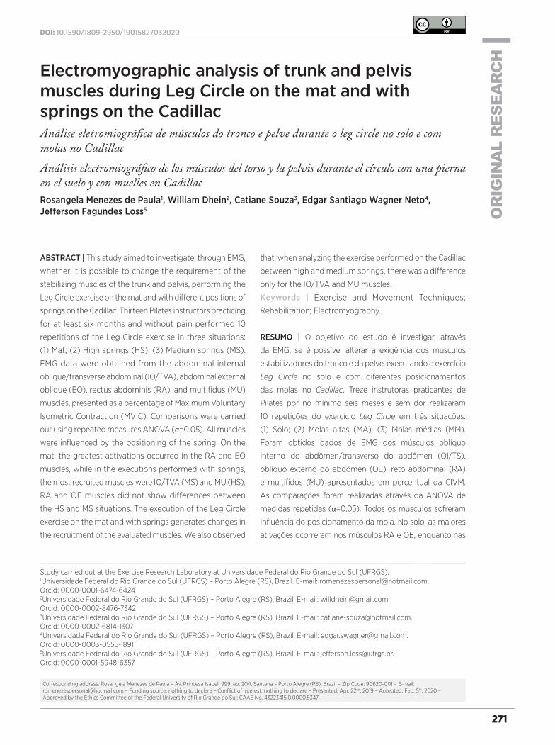
271
OR
IGIN
AL
RES
EAR
CH
DOI: 10.1590/1809-2950/19015827032020
Study carried out at the Exercise Research Laboratory at Universidade Federal do Rio Grande do Sul (UFRGS).1Universidade Federal do Rio Grande do Sul (UFRGS) – Porto Alegre (RS), Brazil. E-mail: [email protected]. Orcid: 0000-0001-6474-64242Universidade Federal do Rio Grande do Sul (UFRGS) – Porto Alegre (RS), Brazil. E-mail: [email protected]. Orcid: 0000-0002-8476-73423Universidade Federal do Rio Grande do Sul (UFRGS) – Porto Alegre (RS), Brazil. E-mail: [email protected]. Orcid: 0000-0002-6814-13074Universidade Federal do Rio Grande do Sul (UFRGS) – Porto Alegre (RS), Brazil. E-mail: [email protected]. Orcid: 0000-0003-0555-18915Universidade Federal do Rio Grande do Sul (UFRGS) – Porto Alegre (RS), Brazil. E-mail: [email protected]. Orcid: 0000-0001-5948-6357
271
Corresponding address: Rosangela Menezes de Paula – Av. Princesa Isabel, 999, ap. 204, Santana – Porto Alegre (RS), Brazil – Zip Code: 90620-001 – E-mail: [email protected] – Funding source: nothing to declare – Conflict of interest: nothing to declare – Presented: Apr. 22nd, 2019 – Accepted: Feb. 5th, 2020 – Approved by the Ethics Committee of the Federal University of Rio Grande do Sul: CAAE No. 43223415.0.0000.5347
Electromyographic analysis of trunk and pelvis muscles during Leg Circle on the mat and with springs on the CadillacAnálise eletromiográfica de músculos do tronco e pelve durante o leg circle no solo e com molas no CadillacAnálisis electromiográfico de los músculos del torso y la pelvis durante el círculo con una pierna en el suelo y con muelles en CadillacRosangela Menezes de Paula1, William Dhein2, Catiane Souza3, Edgar Santiago Wagner Neto4, Jefferson Fagundes Loss5
ABSTRACT | This study aimed to investigate, through EMG,
whether it is possible to change the requirement of the
stabilizing muscles of the trunk and pelvis, performing the
Leg Circle exercise on the mat and with different positions of
springs on the Cadillac. Thirteen Pilates instructors practicing
for at least six months and without pain performed 10
repetitions of the Leg Circle exercise in three situations:
(1) Mat; (2) High springs (HS); (3) Medium springs (MS).
EMG data were obtained from the abdominal internal
oblique/transverse abdominal (IO/TVA), abdominal external
oblique (EO), rectus abdominis (RA), and multifidus (MU)
muscles, presented as a percentage of Maximum Voluntary
Isometric Contraction (MVIC). Comparisons were carried
out using repeated measures ANOVA (α=0.05). All muscles
were influenced by the positioning of the spring. On the
mat, the greatest activations occurred in the RA and EO
muscles, while in the executions performed with springs,
the most recruited muscles were IO/TVA (MS) and MU (HS).
RA and OE muscles did not show differences between
the HS and MS situations. The execution of the Leg Circle
exercise on the mat and with springs generates changes in
the recruitment of the evaluated muscles. We also observed
that, when analyzing the exercise performed on the Cadillac
between high and medium springs, there was a difference
only for the IO/TVA and MU muscles.
Keywords | Exercise and Movement Techniques;
Rehabilitation; Electromyography.
RESUMO | O objetivo do estudo é investigar, através
da EMG, se é possível alterar a exigência dos músculos
estabilizadores do tronco e da pelve, executando o exercício
Leg Circle no solo e com diferentes posicionamentos
das molas no Cadillac. Treze instrutoras praticantes de
Pilates por no mínimo seis meses e sem dor realizaram
10 repetições do exercício Leg Circle em três situações:
(1) Solo; (2) Molas altas (MA); (3) Molas médias (MM).
Foram obtidos dados de EMG dos músculos oblíquo
interno do abdômen/transverso do abdômen (OI/TS),
oblíquo externo do abdômen (OE), reto abdominal (RA)
e multífidos (MU) apresentados em percentual da CIVM.
As comparações foram realizadas através da ANOVA de
medidas repetidas (α=0,05). Todos os músculos sofreram
influência do posicionamento da mola. No solo, as maiores
ativações ocorreram nos músculos RA e OE, enquanto nas
Fisioter Pesqui. 2020;27(3):271-276
272
execuções realizadas com molas, os músculos mais recrutados
foram OI/TS (MM) e MU (MA). Os músculos RA e OE não obtiveram
diferenças entre as situações MA e MM. A execução do exercício
Leg Circle no solo e com molas gera alterações no recrutamento
dos músculos avaliados. Observa-se também que, ao analisar o
exercício realizado no Cadillac entre molas altas e molas médias,
houve diferença apenas para os músculos OI/TS e MU.
Descritores | Técnicas de Exercício e de Movimento; Reabilitação;
Eletromiografia.
RESUMEN | El objetivo del estudio el investigar, através de la EMG,
si es posible alterar la exigencia de los músculos estabilizadores
del tronco y de la pelvis, ejecutando el ejercicio Leg Circle en
el suelo y con diferentes posicionamientos de los muelles en el
Cadillac. Trece instructores profesionales de Pilates durante al
menos seis meses y sin dolor realizó 10 repeticiones del ejercicio
Leg Circle en tres situaciones: (1) El suelo; (2) Muelles altos (MA);
(3) Muelles medianos (MM). Se obtuvieron datos de EMG de los
músculos oblicuos internos del abdomen / transverso del abdomen
(OI / TS), oblicuo externo del abdomen (OE), recto abdominal
(RA) y multífidos (MU) presentados en porcentaje de la CIVM.
Las comparaciones se realizaron a través de la ANOVA de medidas
repetidas (α=0,05). Todos los músculos sufrieron influencia del
posicionamiento del muelle. En el suelo, las mayores activaciones
ocurrieron en los músculos RA y OE, mientras que en las ejecuciones
realizadas con muelles, los músculos más reclutados fueron OI / TS
(MM) y MU (MA). Los músculos RA y OE no obtuvieron diferencias
entre las situaciones MA y MM. La ejecución del ejercicio Leg Circle
en el suelo y con muelles genera cambios en el reclutamiento de
los músculos evaluados. Se observa también que, al analizar el
ejercicio realizado en el Cadillac entre muelles altos y muelles
medianos, hubo diferencia sólo para los músculos OI / TS y MU.
Palabras clave | Técnicas de Ejercicio y de Movimiento;
Rehabilitación; Electromiografía.
INTRODUCTION
The Pilates method became popular as a physical activity and as a rehabilitation tool, seeking to integrate the mind and body of practitioners1-3. Maintaining the stability of the trunk and pelvis while performing the exercises is one of the main characteristics of this method1,4.
While classic training programs, such as strength training, use the increment of loads for training progression5, the Pilates method increases the challenges of instability, by variations in the execution of exercises or even in the loads of the springs as the practitioner evolves in training6. Nevertheless, the increase in progression within the method has been done subjectively7.
One of the exercises frequently used to challenge the stability of the hip/pelvis, in addition to promoting the functional and anatomical integrity of the hip joint, is the Leg Circle or Hip Circle. This exercise can be performed either on the mat, without any accessory, or on the Cadillac, where the individual is placed in supine position and the springs are placed on the head of the device and on the lower limbs, in the feet area6,8.
The fixation height, different deformation constants, or absence of springs, during the exercise, aim to help or hinder the maintenance of this stability. It has been shown in other exercises that different spring positions alter the muscular responses6,7,9. However, as far as can be investigated, there is no information regarding the effects
of different positioning of the springs on the stability of the Leg Circle exercise. Therefore, this study aims to investigate, by surface electromyography (EMG), whether it is possible to change the requirement of the stabilizing muscles of the trunk and pelvis, performing the Leg Circle exercise on the mat and with different positions of the springs on the Cadillac.
METHODOLOGY
Sample
The sample estimate was performed using G*Power 3.1.9.2, adopting the following criteria: 0.40 effect size; 5% probability of error; 80% statistical power; 0.5 correlation between measurements; assuming sphericity (E=1) for the family of repeated measures ANOVA statistical tests. The calculation defined the sample size in 10 subjects. Due to possible sample losses, 13 Pilates instructors were evaluated. The participants had an average age of 31.3 years (±5.3), height of 1.61cm (±0.05), and weight of 61.2kg (±6.1). Participants were selected intentionally, with the following eligibility criteria: being a Pilates instructor for at least six months; practicing at least once a week for at least six months; not having acute or chronic low back pain, pain in the lower limbs, or in the abdominal region.
Paula et al. EMG during Leg Circle on the mat and with springs on the Cadillac
273
Procedures
All collections were performed at the Exercise Research Laboratory of the Federal University of Rio Grande do Sul, scheduled according to the availability of the participants. Initially, the participants signed the Free and Informed Consent Form, which contained the objectives and procedures of the study. For each sample, an initial evaluation was performed, where data on weight, height, and age were obtained. This was followed by a warm-up, stretching, and familiarization with the proposed exercises. After this initial stage, the participant was prepared for data collection.
Initially, trichotomy and cleaning of the skin with alcohol on the evaluated muscles were performed. Disposable surface electrodes (Miotec DoubleTrace – 100; Ag/AgCl, 10mm diameter, self-adhesive, in bipolar configuration),were unilaterally fixedto the right of the Powerhouse muscles1, namely: Rectus Abdominal (RA), Abdominal External Oblique (EO), Abdominal Internal Oblique/Transverse Abdominal (IO/TVA), and Multifidus (MU). After placing the electrodes, the tests of maximum voluntary isometric contraction (MVIC) of each muscle were performed. The MVICs of each of the muscles lasted 5 seconds with verbal encouragement, and two repetitions with an interval of two minutes were performed. The positioning of the electrodes and the conduction of the MVIC followed recommendations
in the literature9-11. The acquisition of electromyography data was obtained using the electromyograph of the brand BTS FREEEMG 1000 with a sampling rate of 1000Hz. The software used to acquire the electromyographic data was BTS Smart Capture.
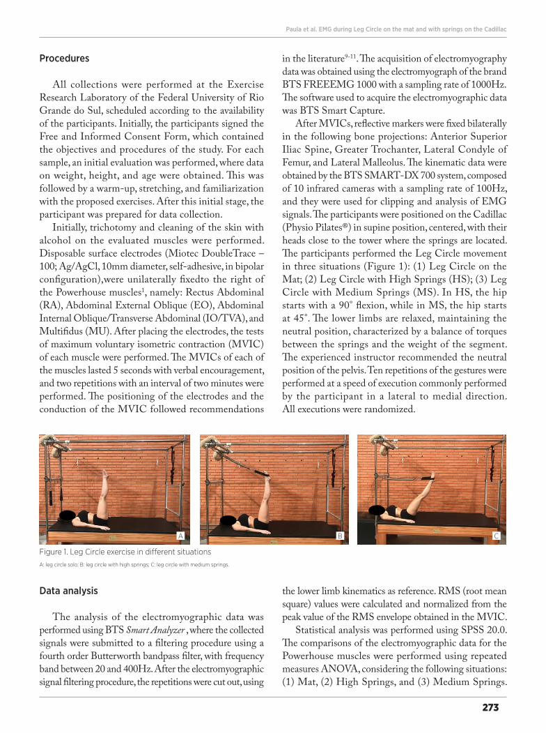
After MVICs, reflective markers were fixed bilaterally in the following bone projections: Anterior Superior Iliac Spine, Greater Trochanter, Lateral Condyle of Femur, and Lateral Malleolus. The kinematic data were obtained by the BTS SMART-DX 700 system, composed of 10 infrared cameras with a sampling rate of 100Hz, and they were used for clipping and analysis of EMG signals. The participants were positioned on the Cadillac (Physio Pilates®) in supine position, centered, with their heads close to the tower where the springs are located. The participants performed the Leg Circle movement in three situations (Figure 1): (1) Leg Circle on the Mat; (2) Leg Circle with High Springs (HS); (3) Leg Circle with Medium Springs (MS). In HS, the hip starts with a 90° flexion, while in MS, the hip starts at 45°. The lower limbs are relaxed, maintaining the neutral position, characterized by a balance of torques between the springs and the weight of the segment. The experienced instructor recommended the neutral position of the pelvis. Ten repetitions of the gestures were performed at a speed of execution commonly performed by the participant in a lateral to medial direction. All executions were randomized.
A B C
Figure 1. Leg Circle exercise in different situationsA: leg circle solo; B: leg circle with high springs; C: leg circle with medium springs.
Data analysis
The analysis of the electromyographic data was performed using BTS Smart Analyzer , where the collected signals were submitted to a filtering procedure using a fourth order Butterworth bandpass filter, with frequency band between 20 and 400Hz. After the electromyographic signal filtering procedure, the repetitions were cut out, using
the lower limb kinematics as reference. RMS (root mean square) values were calculated and normalized from the peak value of the RMS envelope obtained in the MVIC.
Statistical analysis was performed using SPSS 20.0. The comparisons of the electromyographic data for the Powerhouse muscles were performed using repeated measures ANOVA, considering the following situations: (1) Mat, (2) High Springs, and (3) Medium Springs.
Fisioter Pesqui. 2020;27(3):271-276
274
In case of statistical differences in the comparisons, the Bonferroni post hoc test was used. The significance level adopted was α<0.05.
RESULTS
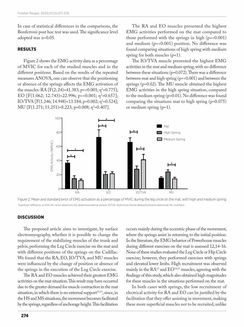
Figure 2 shows the EMG activity data as a percentage of MVIC for each of the studied muscles and in the different positions. Based on the results of the repeated measures ANOVA, one can observe that the positioning or absence of the springs affects the EMG activation of the muscles: RA [F(2; 24)=41.383; p=<0.001; ƞ2=0.775]; EO [F(1.062; 12.743)=22.996; p=<0.001; ƞ2=0.657]; IO/TVA [F(1.246; 14.948)=13.184; p=0.002; ƞ2=0.524]; MU [F(1.271; 15.251)=8.223; p=0.008; ƞ2=0.407].
The RA and EO muscles presented the highest EMG activities performed on the mat compared to those performed with the springs in high (p=<0.001) and medium (p=<0.001) position. No difference was found comparing situations of high spring with medium spring for both muscles (p=1).
The IO/TVA muscle presented the highest EMG activities in the mat and medium spring, with no difference between these situations (p=0.072). There was a difference between mat and high spring (p=<0.001) and between the springs (p=0.02). The MU muscle obtained the highest EMG activities in the high spring situation, compared to the medium spring (p=0.01). No difference was found comparing the situations mat to high spring (p=0.075) or medium spring (p=1).
Mat
High Spring
Medium Spring
EMG
Act
ivity
(%
MV
IC)
MUIO/TVAEORA
100
80
60
40
20
0
Figure 2. Mean and standard error of EMG activation as a percentage of MVIC during the leg circle on the mat, with high and medium spring*Significant difference: p<0.05; RA: rectus abdominis; EO: abdominal external oblique; IO/TVA: abdominal internal oblique/transverse abdominal; MU: multifidus.
DISCUSSION
The proposed article aims to investigate, by surface electromyography, whether it is possible to change the requirement of the stabilizing muscles of the trunk and pelvis, performing the Leg Circle exercise on the mat and with different positions of the springs on the Cadillac. We found that the RA, EO, IO/TVA, and MU muscles were influenced by the change of position or absence of the springs in the execution of the Leg Circle exercise.
The RA and EO muscles achieved their greatest EMG activities on the mat situation. This result may have occurred due to the greater demand for muscle contraction in the mat situation, in which there is no external support12,13, since, in the HS and MS situations, the movement becomes facilitated by the springs, regardless of anchorage height. This facilitation
occurs mainly during the eccentric phase of the movement, where the springs assist in returning to the initial position. In the literature, the EMG behavior of Powerhouse muscles during different exercises on the mat is assessed 12,14-16. None of these studies evaluated the Leg Circle or Hip Circle exercise; however, they performed exercises with springs and elevated lower limbs. High recruitment was observed mainly in the RA12 and EO14,15 muscles, agreeing with the findings of this study, which also obtained high magnitudes for these muscles in the situations performed on the mat.
In both cases with springs, the low recruitment of electrical activity for RA and EO can be justified by the facilitation that they offer assisting in movement, making these more superficial muscles not to be recruited, unlike
Paula et al. EMG during Leg Circle on the mat and with springs on the Cadillac
275
IO and MU muscles, which are deeper. While in the HS situation the demand for the posterior chain is greater, with the recruitment of MU and muscles that act on the hip7, in the MS situation, this requirement becomes lower, causing the recruitment of the MU muscle to decrease. Since RA and EO do not alter their EMG activity in the situation with external spring support, the IO muscle increases its recruitment to compensate for the alteration of the posterior chain, reinforcing its importance in stabilizing the lumbar-pelvic complex13,17,18. In the literature, the comparison between high and low springs has already been verified9 in the hip extension and flexion exercise. The EO muscle showed greater EMG activity in the low springs situation, disagreeing with this study, which found no differences between the HS and MS situation for this muscle. In this study, this difference was found only for IO, which was not assessed in the mentioned study9. We speculate that the result may come from the stabilizing function of the lumbar-pelvic region performed by the deeper muscles of the abdomen, such as the internal oblique4, as well as from the spring position being medium in our study and not low as in the literature9.
Despite the expressive results and their applicability, this study has some limitations, such as: the muscles that act in the movement of the hip were not analyzed. This statement can answer questions that have been speculated based on literature studies. Thus, the relationship between Powerhouse and hip musculature can be investigated in future studies. We can also emphasize that the samples can influence the execution mode and result, because they present different levels of muscular strength and/or standardization of the springs used during the study. Despite not individualizing the resistance exerted by the springs, methodological care was taken to standardize the 45° (MS) and 90° (HS) angle of the pelvis and hips. In the HS situation, there is a difficulty of execution for some samples due to the resistance exerted by the springs, which can generate compensations.
Regarding the practical application of the study, Pilates instructors who aim to prescribe exercises with greater recruitment of the most superficial muscles, such as RA and EO, should take as a choice the exercises performed on the mat, while, when targeting deeper muscles, IO and MU, the choice should be medium and high spring exercises.
Within the Pilates method, there is a vast repertoire of exercises with countless possibilities. Some exercises are more effective than others for working on trunk stability and finding greater activation of the Powerhouse.
To achieve the proposed objectives, it is necessary that the exercises are carried out effectively. With this study, the qualified professionals will be able, at the time of assembling their repertoire, to make the best choice in the prescription of the exercises, considering the degree of difficulty, position of the springs, form of execution, and activation of the Powerhouse.
CONCLUSION
The execution of the Leg Circle exercise on the mat and on the Cadillac generates changes in the recruitment of the stabilized muscles of the trunk and pelvis. In the mat situation, the RA and EO muscles are more activated than in spring situations. The IO/TVA muscle has greater activity on the mat and with a medium spring. In the MU muscle, the greatest activation occurred in the HS situation. Instructors who aim to prescribe exercises with greater recruitment of the more superficial muscles, such as RA and EO, should choose exercises performed on the mat, while, when targeting deeper muscles, IO and MU, the choice should be exercises with medium and high spring.
REFERENCES
1. Muscolino JE, Cipriani S. Pilates and the “powerhouse”—I. J Bodyw Mov Ther. 2004;8(1):15-24. doi: 10.1016/S1360-8592(03)00057-3
2. Bryan M, Hawson S. The benefits of pilates exercise in orthopaedic rehabilitation. Tech Orthop. 2003;18(1):126-9. doi: 10.1097/00013611-200303000-00018
3. Latey P. The pilates method: history and philosophy. J Bodyw Mov Ther. 2001;5(4):275-82. doi: 10.1054/jbmt.2001.0237
4. Barbosa AC, Vieira ER, Silva AF, Coelho AC, Martins FM, Fonseca DS, et al. Pilates experience vs. muscle activation during abdominal drawing-in maneuver. J Bodyw Mov Ther. 2018;22(2):467-70. doi: 10.1016/j.jbmt.2017.05.002
5. Cadore EL, Brentano MA, Kruel LFM. Efeitos da atividade física na densidade mineral óssea e na remodelação do tecido ósseo. Rev Bras Med Esporte. 2005;11(6):373-9. doi: 10.1590/S1517-86922005000600013
6. Melo MO, Gomes LE, Silva YO, Bonezi A, Loss JF. Análise do torque de resistência e da força muscular resultante durante exercício de extensão de quadril no pilates e suas implicações na prescrição e progressão. Rev Bras Fisioter. 2011;15(1):23-30. doi: 10.1590/S1413-35552011000100006
7. Silva YO, Melo MO, Gomes LE, Bonezi A, Loss JF. Análise da resistência externa e da atividade eletromiográfica do movimento de extensão de quadril realizado segundo o método pilates. Rev Bras Fisioter. 2009;13(1):82-8. doi: 10.1590/S1413-35552009005000010
Fisioter Pesqui. 2020;27(3):271-276
276
8. Aparicio E, Pérez J. El auténtico método pilates: el arte del control. Barcelona: Martínez Roca; 2006.
9. Loss JF, Melo MO, Rosa CH, Santos AB, La Torre M, Silva YO. Atividade elétrica dos músculos oblíquos externos e multífidos durante o exercício de flexoextensão do quadril realizado no Cadillac com diferentes regulagens de mola e posições do indivíduo. Rev Bras Fisioter. 2010;14(6):510-7. doi: 10.1590/S1413-35552010000600010
10. Hermens HJ, Freriks B, Disselhorst-Klug C, Rau G. Development of recommendations for SEMG sensors and sensor placement procedures. J Electromyogr Kinesiol. 2000;10(5):361-74. doi: 10.1016/S1050-6411(00)00027-4
11. Konrad P. The ABC of EMG: a practical introduction to kinesiological electromyography. Seattle: Herman & Wallace; 2005.
12. Dias JM, Oliveira Menacho M, Mazuquin BF, Obara K, Mostagi FQRC, Lima TB, et al. Comparison of the electromyographic activity of the anterior trunk during the execution of two Pilates exercises – teaser and longspine – for healthy people. J Electromyogr Kinesiol. 2014;24(5):689-97. doi: 10.1016/j.jelekin.2014.06.005
13. Marques NR, Morcelli MH, Hallal CZ, Gonçalves M. EMG activity of trunk stabilizer muscles during Centering Principle
of Pilates Method. J Bodyw Mov Ther. 2013;17(2):185-91. doi: 10.1016/j.jbmt.2012.06.002
14. Pereira IL, Queiroz B, Loss J, Amorim C, Sacco ICN. Trunk muscle EMG during intermediate pilates mat exercises in beginner healthy and chronic low back pain individuals. J Manipulative Physiol Ther. 2017;40(5):350-7. doi: 10.1016/j.jmpt.2017.02.010
15. Silva GB, Morgan MM, Carvalho WRG, Silva E, Freitas WZ, Silva FF, Souza RA. Electromyographic activity of rectus abdominis muscles during dynamic Pilates abdominal exercises. J Bodyw Mov Ther. 2015;19(4):629-35. doi: 10.1016/j.jbmt.2014.11.010
16. Souza EF, Cantergi D, Mendonça A, Kennedy C, Loss JF. Análise eletromiográfica dos músculos reto femoral e reto abdominal durante a execução dos exercícios hundred e teaser do método pilates. Rev Bras Med Esporte. 2012;18(2):105-8.
17. Queiroz BC, Cagliari MF, Amorim CF, Sacco IC. Muscle activation during four pilates core stability exercises in quadruped position. Arch Phys Med Rehabil. 2010;91(1):86-92. doi: 10.1016/j.apmr.2009.09.016
18. Gouveia KMC, Gouveia EC. O músculo transverso abdominal e sua função de estabilização da coluna lombar. Fisioter Mov. 2008;21(3):45-50.
Copyright © 2022 FDOKUMEN