Haemoptysis in Pulmonary Arterial Hypertension Associated ...
Upload
khangminh22Category
view
0download
0
Journal of
Cardiovascular
Development and Disease
Article
Common Arterial Trunk Associated with FunctionallyUniventricular Heart: Anatomical Study and Review of the Literature
Sami Chatila 1, Lucile Houyel 1,2,*, Manon Hily 1,2 and Damien Bonnet 1,2
�����������������
Citation: Chatila, S.; Houyel, L.; Hily,
M.; Bonnet, D. Common Arterial
Trunk Associated with Functionally
Univentricular Heart: Anatomical
Study and Review of the Literature. J.
Cardiovasc. Dev. Dis. 2021, 8, 175.
https://doi.org/10.3390/jcdd8120175
Academic Editor:
Mathilda Mommersteeg
Received: 27 October 2021
Accepted: 2 December 2021
Published: 6 December 2021
Publisher’s Note: MDPI stays neutral
with regard to jurisdictional claims in
published maps and institutional affil-
iations.
Copyright: © 2021 by the authors.
Licensee MDPI, Basel, Switzerland.
This article is an open access article
distributed under the terms and
conditions of the Creative Commons
Attribution (CC BY) license (https://
creativecommons.org/licenses/by/
4.0/).
1 Congenital and Pediatric Cardiology Unit, M3C-Necker, Hôpital Necker-Enfants Malades, AP-HP,75015 Paris, France; [email protected] (S.C.); [email protected] (M.H.); [email protected] (D.B.)
2 Université de Paris, 75006 Paris, France* Correspondence: [email protected]
Abstract: Common arterial trunk (CAT) is a rare congenital heart disease that is commonly includedinto the spectrum of conotruncal heart defects. CAT is rarely associated with functionally univen-tricular hearts, and only few cases have been described so far. Here, we describe the anatomicalcharacteristics of CAT associated with a univentricular heart diagnosed in children and fetuses re-ferred to our institution, and we completed the anatomical description of this rare condition throughan extensive review of the literature. The complete cohort ultimately gathered 32 cases describedin the literature completed by seven cases from our unit (seven fetuses and one child). Four typesof univentricular hearts associated with CAT were observed: tricuspid atresia or hypoplastic rightventricle in 16 cases, mitral atresia or hypoplastic left ventricle in 12 cases, double-inlet left ventricle in2 cases, and unbalanced atrioventricular septal defect in 9 cases. Our study questions the diagnosis ofCAT as the exclusive consequence of an anomaly of the wedging process, following the convergencebetween the embryonic atrioventricular canal and the common outflow tract. We confirm that someforms of CAT can be considered to be due to an arrest of cardiac development at the stages precedingthe convergence.
Keywords: common arterial trunk; univentricular heart; tricuspid atresia; mitral atresia; double inletleft ventricle; atrioventricular septal defect
1. Introduction
Common arterial trunk (CAT) is a rare birth defect with an estimated incidence ofone birth in 10,000 [1]. This congenital disorder is part of the so-called “conotruncal”heart diseases spectrum, also called cardiac neural crest and second heart field defects,characterized by anomalies in the development and position of the outflow tract, and bythe presence of a ventricular septal defect (VSD) related to malalignment or the absenceof the development of the outlet septum (outlet VSD) [2]. Associations between CAT anddefects of the inlet or apical segments of the heart have been described in the literature,including malformations of the atrioventricular junction and various degrees of ventricularhypoplasia. These anomalies often correspond to a hypoplasia of an atrioventricularvalve or to the absence of an atrioventricular connection (mitral or tricuspid atresia).Hitherto, however, these cases have only been described through case reports or shortseries. The purpose of this study was (1) to describe a series of one patient, five fetalheart specimens, and one fetus with these rare associations of CAT and functionally oranatomically univentricular heart, and (2) to carry out an extensive review of the literature.
2. Anatomical Definitions2.1. Common Arterial Trunk
Common arterial trunk is defined as “a congenital cardiovascular malformation inwhich a single arterial trunk arises from the heart, giving origin sequentially to the coronaryarteries, one or more pulmonary arteries, and the systemic arterial circulation” [3]. There is
J. Cardiovasc. Dev. Dis. 2021, 8, 175. https://doi.org/10.3390/jcdd8120175 https://www.mdpi.com/journal/jcdd
J. Cardiovasc. Dev. Dis. 2021, 8, 175 2 of 18
therefore a common ventriculo–arterial junction, with a common arterial valve, above alarge outlet juxta–arterial VSD. The different types of CAT are classified according to thedominance of either the systemic of pulmonary components of the common trunk [2–5].
• CAT with aortic dominance and confluent pulmonary arteries (Type 1–2 of the modi-fied Van Praagh classification).
• CAT with aortic dominance and discontinuous pulmonary arteries (Type 3 of themodified Van Praagh classification). One pulmonary artery originates from the CAT,the other is supplied by the arterial duct or collateral arteries.
• CAT with pulmonary dominance and interruption of the aortic arch (IAA, usuallytype B of Celoria and Patton [6], between the left carotid artery and the left subclavianartery), or coarctation of the aorta (Type 4 of the modified Van Praagh classification).
2.2. Functionally Univentricular Heart
According to the International Society of Nomenclature for Pediatric and CongenitalHeart Disease (ICD-11 IPCCC), the term functionally univentricular heart describes aspectrum of congenital cardiac malformations in which the ventricular mass may notreadily lend itself to partitioning that commits one ventricular pump to the systemiccirculation, and another to the pulmonary circulation [3]. In addition, “a heart may befunctionally univentricular because of its anatomy or because of the lack of feasibility orlack of advisability of surgically partitioning the ventricular mass”.
Common lesions in this category typically include double-inlet ventricles, tricuspidatresia, mitral atresia, and hypoplastic left heart syndrome. Unbalanced atrioventricularseptal defects (AVSD) with hypoplasia of one ventricle may also be considered functionallyuniventricular hearts.
3. Methods3.1. Data Collection
The data presented here are the result of a systematic review of the available literature,to which we added the analysis of five fetal heart specimens from the M3C-Necker anatom-ical collection, one fetal echocardiography, and the follow-up of a patient hospitalizedin the pediatric cardiology department of Necker–Enfants malades University Hospital.Data for the living patient were obtained from the Necker–Enfants malades and ImagineInstitute congenital heart disease database using Dr Warehouse®, a full-text clinical datawarehouse (CDW) for cohort identification and data extraction [7].
3.2. Literature Review
Articles of interest were selected online using the PubMed database. All articleswere gathered by a keyword search and by cross referencing previously obtained articles.Articles of interest were found using the following keyword associations: “Univentricularheart”, “Single ventricle”, “Hypoplastic ventricle”, “Ventricular hypoplasia”, “Doubleinlet ventricle”, “Fontan”, “Tricuspid atresia”, “Mitral atresia”, “Complete atrioventricularcanal”, “Unbalanced atrioventricular septal defect”, “Common arterial trunk”, “Arterialtrunk “, and “Persistent arterial trunk”. No limitation was included regarding publicationdate, studied population, or language. SC and LH reviewed the search results indepen-dently to select the reports based on the inclusion and exclusion criteria. Chest X-ray,echocardiography, angiography, and CT images were interpreted when available, com-pared to the authors’ interpretation, and exploited when textual data were unavailable.The literature review was conducted in accordance with the Preferred Reporting Items forSystematic Reviews and Meta-Analyses (PRISMA) guidelines [8].
Thirty-nine articles whose title and abstract matched the association between a func-tionally or anatomically univentricular heart and a CAT were collected. Eleven articleswere excluded from this list: seven of them were not available online, and four of thempresented insufficient data for proper interpretation (Figure 1). Among the selected arti-cles [9–36], only one included a series of five cases [32], the others were case reports of one

J. Cardiovasc. Dev. Dis. 2021, 8, 175 3 of 18
or two patients (Table 1). Among the functionally univentricular hearts, we decided toinclude only hearts with severe right or left ventricular hypoplasia, including unbalancedAVSDs. Three cases of complete AVSD with balanced ventricles who were not eligible for abiventricular repair due to abnormal valve attachments in the LV-to-aorta pathway wereexcluded from the cohort [32].
1
Figure 1. PRISMA (Preferred Reporting Items for Systematic Reviews and Meta-Analyses) flow diagram of the study selection.

J. Cardiovasc. Dev. Dis. 2021, 8, 175 4 of 18
Table 1. Anatomical characteristics of cases 1 to 32.
AuthorsCase Number
ChromosomalAbnormality FUVH Type VSD CAT Type/
Origin
TruncalValve
NbLeaflets
AorticArch Anomalies
Fujimoto et al.[9]
Case 1Hypoplastic RV Large outlet 4 4 Left IAA type B, left coronary
ostium from right sinus
Zeevi et al. [10]Case 2 Hypoplastic RV No VSD 1 from LV 2 Left
No ASD, mitral stenosis,atretic right coronary
ostium with retrogradesinusoid filling
Gonzalez-Lopez et al.
[11]Case 3
Tricuspid atresia Outlet 1 from RV 3 Left LUPV in retro aorticinnominate vein
Rao et al. [12]Case 4 Tricuspid atresia Outlet 1 3 Left
Hypoplastic thymus,right thumb and
hemivertebrae, narrowPA branches
Roldan et al.[13]
Case 5Tricuspid atresia Large outlet 2 US US
Malec et al.[14]
Case 6Tricuspid atresia Outlet 1 US Left Restrictive ASD
Numata et al.[15]
Case 7Tricuspid atresia Outlet 1 US US
Sreeram et al.[16]
Case 8Tricuspid atresia Outlet 1 US Left
Alva et al. [17]Case 9 Di George Tricuspid atresia Outlet 1 from LV US Right
Areias et al.[18]
Case 10
Tricuspidatresia Outlet 3 3 Right LPA from left-sided duct
Sharma et al.[19]
Case 11Tricuspid atresia Large outlet 1 from LV 3 Right Two OS ASD
Diogenes et al.[20]
Case 12Tricuspid atresia Large outlet 2 2 Left Large ASD, L-JAA
Hoashi et al.[21]
Case 13Hypoplastic LV Large outlet 2 4 Right RUPLV in right SCV
Marathe et al.[22]
Case 14Hypoplastic LV No VSD 4 from RV 3 Left IAA type B
Murdison et al.[23]
Case 15Hypoplastic LV No VSD 1 From RV 3 Left
Subarterial conus, only 2brachiocephalic arteries,
RCA origin fromposterior cusp
Imai et al. [24]Case 16 Hypoplastic LV US 2 US Double LSVC
Michelfelderet al. [25]Case 17
Di George Mitral atresia No VSD 1 From RV 3 Left Restrictive ASD,Subarterial conus
Rice et al. [26]Case 18 Mitral atresia Outlet 1 From RV 4 Left LSCV to CS, Subarterial
conus

J. Cardiovasc. Dev. Dis. 2021, 8, 175 5 of 18
Table 1. Cont.
AuthorsCase Number
ChromosomalAbnormality FUVH Type VSD CAT Type/
Origin
TruncalValve
NbLeaflets
AorticArch Anomalies
Jacobs et al.[27]
Case 19Mitral atresia Tiny outlet 1 US US
Alves et al.[28]
Case 20Mitral atresia No VSD 4 from RV 3 Left
Subarterial conus, IAAType A, LCA ostiumsupracommissural
Cree et al. [29]Case 21 Mitral atresia No VSD 2 from RV 3
Subarterial conus, singlecoronary artery from
inominate artery
Shaddy et al.[30]
Case 22Double inlet LV US 1 US Left Situs Solitus {SDD}, LSCV
to LA, L-JAA
Paris et al. [31]Case 23 Double inlet LV Large outlet 2 3 Left Situs Solitus {SDD},
dextrocardia, L-JAA
He et al. [32]Cases 24 and
25
Di George uAVSD (hypo LV)Large inlet
extending tooutlet
2 3 Right LSCV
Di George uAVSD (hypo LV)
Large inletextending to
muscularseptum
2 4 Right
Panwar et al.[33]
Cases 26 and27
uAVSD (hypoLV) Restrictiveinlet 1 from RV US US
uAVSD (hypoLV) Restrictiveinlet 1 from RV US Right
Tripathi et al.[34]
Cases 28 and29
uAVSD(hypoLV)
Restrictiveinlet 1 from RV 3 Right
uAVSD (hypoLV)
Restrictiveinlet withmuscularextension
1 from RV US Left LSCV
Kumar et al.[35]
Case 30uAVSD (hypoLV) Inlet 2 from RV 3 Right
Situs inversus,dextrocardia, common
atrium
Shapiro et al.[36]
Cases 31 and32
uAVSD (hypoRV) Muscular(BVF) 1 US Left PA trunk stenosis
uAVSD(hypoRV)
Muscular(BVF) 1 from LV 4 Left
AoA, aortic arch; ASD, atrial septal defect; BVF, bulboventricular foramen; CAT, common arterial trunk; CS, coronary sinus; FUVH,functionally univentricular heart; IAA, interrupted aortic arch; LCA, left coronary artery; LSCV, left superior caval vein; L-JAA, leftjuxtaposition of atrial appendages; LUPV, left upper pulmonary vein; LV, left ventricle; OS, Ostium secundum; RCA, right coronary artery;RV, right ventricle; SV, single ventricle; uAVSD, unbalanced atrioventricular septal defect; US, unspecified; VSD, ventricular septal defect.
3.3. Heart Specimens
Five fetal heart specimens (Cases 33 to 37, Table 2) were extracted from the M3C-Necker anatomic collection of fetal hearts by MH, and were analyzed by LH, using thesegmental analysis method as described by Van Praagh [37].

J. Cardiovasc. Dev. Dis. 2021, 8, 175 6 of 18
Table 2. Anatomic characteristics of cases 33 to 39.
Case Number FUVH Type VSD CAT Type/Origin
TruncalValve
Nb LeafletsAortic Arch Anomalies
33 (HS) Hypoplastic RV Outlet VSD 1 from RV 4 Left Subarterial conus
34 (HS) Hypoplastic RV Outlet VSD 1 3 Left Very large ASD
35 (HS) Mitral atresia No VSD 2 from RV 3 Left
Subarterial conus,supra commissural
RCA ostium,narrow LCA
ostium
36 (HS) Mitralhypoplasia Large muscular VSD 4 from RV 3 Left
Subarterial conus,narrow LCA
ostium, LCSV toCS, Hypoplastic
horizontal Ao
37 (HS) Hypoplastic LV No VSD 4 from RV 3 LeftSubarterial conus,
Type A IAA, LSCVto CS, TAPVR in CS
38 (fetus) Hypoplastic RV Outlet 1 from LV US US
39 (patient) Tricuspidatresia Large outlet 1 3 Left LSCV to CS
Abbreviations: Ao, Aorta; ASD, atrial septal defect; CAT, common arterial trunk; CS, coronary sinus; HS, heart specimen; IAA, interruptedaortic arch; LCA, left coronary artery; LSCV, left superior caval vein; LV, left ventricle; RCA, right coronary artery; RV, right ventricle;TAPVR, total anomalous pulmonary venous return; FUVH, univentricular heart; VSD, ventricular septal defect.
3.4. Fetal Echocardiography
A diagnosis of CAT type 1 associated with severe tricuspid and RV hypoplasiawas made at 25 weeks of gestation (WG) on a fetal echocardiography (Case 38, Table 2).Six weeks later, the pregnancy was ongoing, without any pejorative event.
3.5. Case Report
A prenatal diagnosis of tricuspid atresia associated with a CAT type 1 was made at20 WG (Case 39, Table 2). Pregnancy was obtained after in vitro fertilization and intracyto-plasmic sperm injection. No chromosome 22q11 microdeletion was identified by FISH andthe CGH array on the amniotic fluid was normal.
A 3010 g male child was delivered at term. He adapted well to extrauterine life.Initial echocardiography found atrial situs solitus, levocardia, D-loop ventricles, normalpulmonary venous returns, a left superior caval vein draining in the coronary sinus, a largeostium secundum type atrial septal defect with a right to left shunt, and a hypoplasticright ventricle (RV) with absent right atrioventricular connection. The left ventricle (LV)was fully developed with a normal size mitral valve with mild regurgitation. A large CAToverrode the ventricular septum above a large outlet juxta-arterial VSD (Figure 2). Fromthe CAT originated successively the coronary arteries, a short pulmonary arterial trunkwith two normal size pulmonary arteries, and a left aortic arch. There was no arterial duct.The patient was discharged after leaving the maternity ward. Furosemide therapy wasinitiated at 2 weeks of age because of clinical signs of excessive pulmonary blood flow.

J. Cardiovasc. Dev. Dis. 2021, 8, 175 7 of 18
J. Cardiovasc. Dev. Dis. 2021, 8, x FOR PEER REVIEW 6 of 18
pulmonary venous returns, a left superior caval vein draining in the coronary sinus, a
large ostium secundum type atrial septal defect with a right to left shunt, and a hypo-
plastic right ventricle (RV) with absent right atrioventricular connection. The left ventri-
cle (LV) was fully developed with a normal size mitral valve with mild regurgitation. A
large CAT overrode the ventricular septum above a large outlet juxta-arterial VSD (Fig-
ure 2). From the CAT originated successively the coronary arteries, a short pulmonary
arterial trunk with two normal size pulmonary arteries, and a left aortic arch. There was
no arterial duct. The patient was discharged after leaving the maternity ward. Furo-
semide therapy was initiated at 2 weeks of age because of clinical signs of excessive
pulmonary blood flow.
Figure 2. Case 39 (Table 2) with tricuspid atresia and common arterial trunk type 1: echocardiographic views. 2A:
Four-chamber apical view. Red arrow: absence of right atrioventricular connection (tricuspid atresia). CS, dilated coro-
nary sinus; LA, left atrium; LV, left ventricle; RA, right atrium; RV, right ventricle. 2B: Five-chamber apical view. The
common arterial trunk overrides the ventricular septum, above a large outlet ventricular septal defect (yellow arrow).
CAT, common arterial trunk; LV, left ventricle; RV, right ventricle. 2C: Subcostal view. Common arterial trunk type 1.
Yellow arrow, outlet VSD; Large outlet VSD. Ao: Aorta; MPT: Main pulmonary trunk.
At the age of 1 month, the patient was admitted to our department for surgical
management. The first intervention consisted of a disconnection of the pulmonary arter-
ies and the creation of a 4mm Gore-Tex systemic-to-pulmonary shunt (modified
Blalock-Taussig-Thomas (BTT) anastomosis). The child was extubated, weaned from in-
otropic support on the fourth postoperative day, and discharged from the hospital on the
ninth postoperative day.
At the age of 5 months, heart catheterization was performed. The mean right and
left mean pulmonary pressures were 10 mmHg. The BTT shunt was patent, and angio-
graphies showed normal size left and right pulmonary arteries. Bilateral partial ca-
vopulmonary connection was performed, with a section of azygos and hemi-azygos
veins. The aortic oxygen saturation at the end of the procedure was 90%. The child was
quickly extubated and the inotropes were weaned. The postoperative course was com-
plicated by a chylothorax treated with a fat-free diet for 6 weeks.
At 3 years of age, catheterization showed mean pulmonary artery pressure of
14mmHg with a good function of the left ventricle (end-diastolic left ventricular pressure
10mmHg). The child underwent a non-fenestrated total cavopulmonary connection with
extracardiac conduit and pulmonary bifurcation repair with a Gore-Tex patch. He was
quickly extubated and weaned from inotropic support. The postoperative course was
complicated by a second chylothorax, which regressed after a 6 week fat free diet. He was
discharged home at day 5, under diuretic treatment and Aspirin.
Figure 2. Case 39 (Table 2) with tricuspid atresia and common arterial trunk type 1: echocardiographic views. (A): Four-chamber apical view. Red arrow: absence of right atrioventricular connection (tricuspid atresia). CS, dilated coronary sinus;LA, left atrium; LV, left ventricle; RA, right atrium; RV, right ventricle. (B): Five-chamber apical view. The common arterialtrunk overrides the ventricular septum, above a large outlet ventricular septal defect (yellow arrow). CAT, common arterialtrunk; LV, left ventricle; RV, right ventricle. (C): Subcostal view. Common arterial trunk type 1. Yellow arrow, outlet VSD;Large outlet VSD. Ao: Aorta; MPT: Main pulmonary trunk.
At the age of 1 month, the patient was admitted to our department for surgicalmanagement. The first intervention consisted of a disconnection of the pulmonary arteriesand the creation of a 4 mm Gore-Tex systemic-to-pulmonary shunt (modified Blalock-Taussig-Thomas (BTT) anastomosis). The child was extubated, weaned from inotropicsupport on the fourth postoperative day, and discharged from the hospital on the ninthpostoperative day.
At the age of 5 months, heart catheterization was performed. The mean right and leftmean pulmonary pressures were 10 mmHg. The BTT shunt was patent, and angiographiesshowed normal size left and right pulmonary arteries. Bilateral partial cavopulmonaryconnection was performed, with a section of azygos and hemi-azygos veins. The aortic oxy-gen saturation at the end of the procedure was 90%. The child was quickly extubated andthe inotropes were weaned. The postoperative course was complicated by a chylothoraxtreated with a fat-free diet for 6 weeks.
At 3 years of age, catheterization showed mean pulmonary artery pressure of 14 mmHgwith a good function of the left ventricle (end-diastolic left ventricular pressure 10 mmHg).The child underwent a non-fenestrated total cavopulmonary connection with extracardiacconduit and pulmonary bifurcation repair with a Gore-Tex patch. He was quickly extu-bated and weaned from inotropic support. The postoperative course was complicated by asecond chylothorax, which regressed after a 6 week fat free diet. He was discharged homeat day 5, under diuretic treatment and Aspirin.
At the last follow-up visit at the age of 3.5 years, the patient was still in good condition.Oxygen saturation was normal. He was treated with aspirin and was schooled normally.
3.6. Statistical Analysis
Statistical analysis was performed with Statview 5.0. The statistics were descriptiveand expressed as percentages. Fisher’s exact test for was used for comparison between thetwo major groups of hypoplastic RV and hypoplastic LV. A value of p < 0.05 was consideredstatistically significant.

J. Cardiovasc. Dev. Dis. 2021, 8, 175 8 of 18
4. Results
The anatomical description of each case has been summarized in Table 1 (review ofthe literature, cases 1 to 32) and Table 2 (cases 33 to 39). The distribution of the anatomicalcharacteristics of the cohort is summarized in Table 3.
Table 3. Distribution of the anatomical characteristics of the cohort.
TricuspidAtresia and
HypoRVN = 16
MitralAtresia and
HypoLVN = 12
DILVN = 2
UnbalancedAVSDN = 9
HypoplasticRV, Global
N = 20
HypoplasticLV, Global
N = 19p
Systemic venousreturn
LSCV to CS 1 3 0 0 1 3LSCV to LA 0 0 1 0 1 0
LSCV(unspecified) 0 1 0 2 0 3
Pulmonary venousreturn
PAPVR 1 1 0 0 1 1TAPVR 0 1 0 0 0 1
Ventricular septaldefect
No VSD 1 7 0 0 1 (5%) 7 (37%) <0.02Outlet 15 3 1 0 16 (89%) 3 (27%) <0.002Inlet 0 0 0 7 0 7
Muscular 0 1 0 2 2 1Unspecified 0 1 1 0 1 1
Type CAT1 12 4 1 6 15 82 2 4 1 3 3 73 1 0 0 0 1 04 1 4 0 0 1 4
From RV 2 9 0 5 2 (10%) 14 (74%) 0.0001From LV 3 0 0 1 3 1
Truncal valveBicuspid 2 0 0 0 2 0Tricuspid 6 8 1 3 7 11
Quadricuspid 2 2 0 2 3 3Unspecified 6 2 1 4 7 6
Subarterial conus 1 (tiny) 8 0 0 1 (5%) 8 (42%) <0.01
Coronary arteryanomalies 2 5 0 0 2 5
Aortic archLeft 10 9 2 3 14 12
Right 3 1 0 5 3 6IAA type A 0 2 0 0 0 2IAA type B 1 1 0 0 1 1
AoA hypoplasia 0 1 0 0 0 1Double AoA 0 1 0 0 0 1Unspecified 3 1 0 1 2 2
L-JAA 1 0 2 0 3 0
AoA, aortic arch; CAT, common arterial trunk; CS, coronary sinus; IAA, interrupted aortic arch; LA, left atrium; L-JAA, left juxtaposition ofthe atrial appendages; LSCV, left superior caval vein; PAPVR, partial anomalous pulmonary venous return; RV, right ventricle; TAPVR,total anomalous pulmonary venous return; VSD, ventricular septal defect.
J. Cardiovasc. Dev. Dis. 2021, 8, 175 9 of 18
Among these 39 cases, four types of functionally or anatomically univentricular heartswere found in association with CAT:
4.1. Tricuspid Atresia and Hypoplastic Right Ventricle
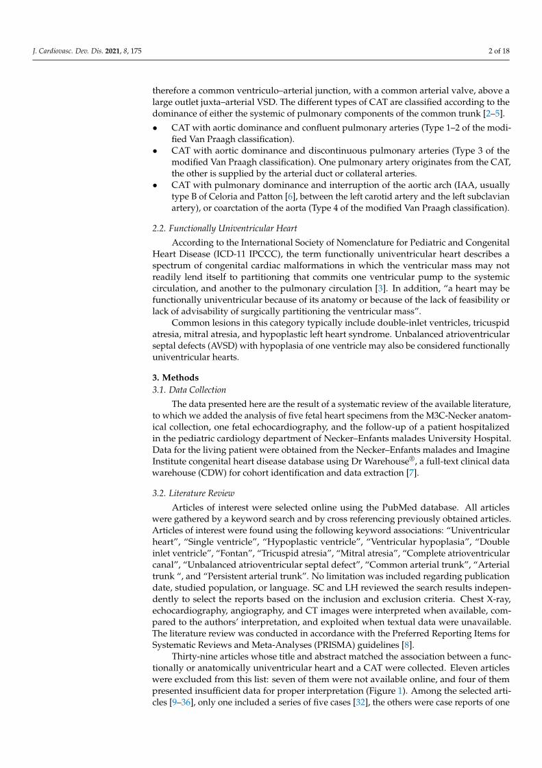
Tricuspid atresia and hypoplastic RV were found in 16/39 cases (41%). Tricuspidatresia was observed in 11 cases and RV hypoplasia in 5. One of the cases was associatedwith Di George syndrome. There was no situs anomaly or heterotaxy syndrome. Systemicvenous returns were always normal, except for one case of persistent left superior cavalvein in the coronary sinus (Case 39). One case was associated with a partial anomalouspulmonary venous return (APVR) of the left upper pulmonary vein in the innominate vein(Case 3). The VSD was always of the outlet type (Figure 3), adjacent to the truncal valve(Table 3), except in one patient without VSD (Case 2). CAT was most often type 1 (12 cases,Figure 3), type 2 in two cases, type 3 in one case, and type 4 in one case with IAA type B.A right aortic arch was described in three cases, including one with associated Di Georgesyndrome (Cases 9 to 11). In two cases there was a coronary anomaly: atresia of the rightcoronary ostium with RV perfusion by myocardial sinusoids in Case 2, and left coronaryorifice within the right sinus of Valsalva in Case 1. One case with tricuspid atresia had aleft juxtaposition of the atrial appendages (Case 12).
J. Cardiovasc. Dev. Dis. 2021, 8, x FOR PEER REVIEW 9 of 18
Figure 3. Heart specimen with common arterial trunk type 1 and hypoplastic right ventricle (Table
3, Case 33). The truncal valve is quadricuspid. Yellow arrow, outlet ventricular septal defect; CAT,
common arterial trunk; LPA, left pulmonary artery; LV, left ventricle; RPA, right pulmonary artery;
RV, right ventricle; TV, tricuspid valve.
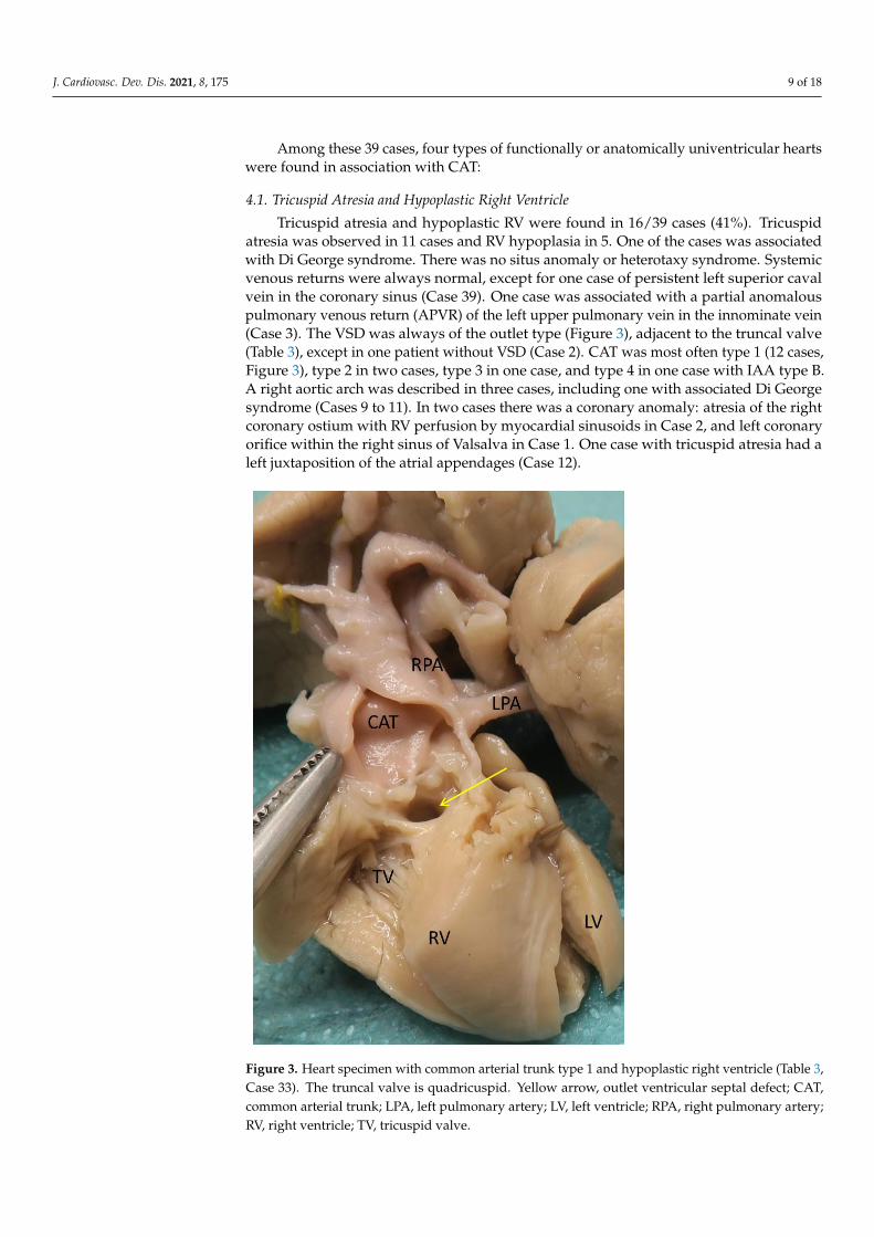
4.2. Mitral Atresia and Left Ventricle Hypoplasia
Mitral atresia or LV hypoplasia was found in 12/39 cases (31%). Mitral atresia was
observed in six cases and LV hypoplasia in six. One case was associated with Di George
syndrome (Case 17). None of the cases had situs or looping anomalies. Systemic venous
returns were normal except for four cases of persistent left superior caval vein, draining
in the coronary sinus in three (Cases 18,36,37), and the site of drainage being not specified
in one (Case 16). Two cases of AVPR were found: one had total APVR to the coronary
sinus (Case 37) and the other was partial (right upper pulmonary vein in the right supe-
rior caval vein, Case 13). An intact ventricular septum was found in seven cases, and a
subarterial conus was described in eight cases (Figure 4). There was an outlet VSD in
Figure 3. Heart specimen with common arterial trunk type 1 and hypoplastic right ventricle (Table 3,Case 33). The truncal valve is quadricuspid. Yellow arrow, outlet ventricular septal defect; CAT,common arterial trunk; LPA, left pulmonary artery; LV, left ventricle; RPA, right pulmonary artery;RV, right ventricle; TV, tricuspid valve.
J. Cardiovasc. Dev. Dis. 2021, 8, 175 10 of 18
4.2. Mitral Atresia and Left Ventricle Hypoplasia
Mitral atresia or LV hypoplasia was found in 12/39 cases (31%). Mitral atresia wasobserved in six cases and LV hypoplasia in six. One case was associated with Di Georgesyndrome (Case 17). None of the cases had situs or looping anomalies. Systemic venousreturns were normal except for four cases of persistent left superior caval vein, draining inthe coronary sinus in three (Cases 18,36,37), and the site of drainage being not specified inone (Case 16). Two cases of AVPR were found: one had total APVR to the coronary sinus(Case 37) and the other was partial (right upper pulmonary vein in the right superior cavalvein, Case 13). An intact ventricular septum was found in seven cases, and a subarterialconus was described in eight cases (Figure 4). There was an outlet VSD in three cases,a mid-muscular VSD in one case, and the VSD type was not described in one case. Thetruncal valve was described as originating from the RV in nine cases and overriding theventricular septum in three cases. The CAT was type 1 or 2 in the majority of cases (8/12,66%) and type 4 in four cases. In those four last cases, one had hypoplasia of the transverseaorta, two had IAA type A (Figure 5), and one IAA type B. One of these 12 cases had adouble aortic arch, and one a right aortic arch, both unrelated to Di George syndrome.An abnormal coronary origin was present in five cases (two cases with hypoplasia of theleft coronary orifice, one case with high take-off of the left coronary artery, just above thecommissure between the “right and left coronary cusps”, one case with right coronaryorifice within the posterior sinus, and one case with a single coronary artery originatingfrom the innominate artery).
J. Cardiovasc. Dev. Dis. 2021, 8, x FOR PEER REVIEW 10 of 18
three cases, a mid-muscular VSD in one case, and the VSD type was not described in one
case. The truncal valve was described as originating from the RV in nine cases and over-
riding the ventricular septum in three cases. The CAT was type 1 or 2 in the majority of
cases (8/12, 66%) and type 4 in four cases. In those four last cases, one had hypoplasia of
the transverse aorta, two had IAA type A (Figure 5), and one IAA type B. One of these 12
cases had a double aortic arch, and one a right aortic arch, both unrelated to Di George
syndrome. An abnormal coronary origin was present in five cases (two cases with hy-
poplasia of the left coronary orifice, one case with high take-off of the left coronary artery,
just above the commissure between the “right and left coronary cusps”, one case with
right coronary orifice within the posterior sinus, and one case with a single coronary ar-
tery originating from the innominate artery).
Figure 4. Heart specimen with common arterial trunk type 4, aortic coarctation and hypoplastic left
ventricle (Table 3, Case 35). 4A: view from the right ventricle. The common arterial trunk (CAT) is
entirely above the right ventricle and there is a subarterial conus (asterisk). 4B: view from the left
ventricle. There is a large mid-muscular ventricular septal defect (yellow arrow).
Figure 4. Heart specimen with common arterial trunk type 4, aortic coarctation and hypoplastic leftventricle (Table 3, Case 35). (A): view from the right ventricle. The common arterial trunk (CAT) isentirely above the right ventricle and there is a subarterial conus (asterisk). (B): view from the leftventricle. There is a large mid-muscular ventricular septal defect (yellow arrow).

J. Cardiovasc. Dev. Dis. 2021, 8, 175 11 of 18J. Cardiovasc. Dev. Dis. 2021, 8, x FOR PEER REVIEW 11 of 18
Figure 5. Heart specimen with common arterial trunk type 4, interruption of the aortic arch type A,
left aortic arch, and hypoplastic left ventricle (Table 3, Case 36). View from the right ventricle. The
common arterial trunk (CAT) is entirely above the right ventricle and there is a subarterial conus
(asterisk). AD, arterial duct; BCAT, brachiocephalic arterial trunk; LCA: Left carotid artery; LPA,
left pulmonary artery; LSCA, left subclavian artery; RPA, right pulmonary artery; RV, right ven-
tricle.
4.3. Double Inlet Left Ventricle:
This is an extremely rare association as only two cases were described in the litera-
ture (Cases 22 and 23). Both cases were in situs solitus with D-loop ventricles. Situs of
other organs was not described. Left juxtaposition of the atrial appendages was present
in both cases and dextrocardia in 1. A superior left caval vein drained in the left atrium in
Case 22. The CAT was type 1 and 2, and emerged from the left ventricle in both. There
was a muscular VSD in Case 22, and a large outlet VSD in Case 23.
Figure 5. Heart specimen with common arterial trunk type 4, interruption of the aortic arch type A,left aortic arch, and hypoplastic left ventricle (Table 3, Case 36). View from the right ventricle. Thecommon arterial trunk (CAT) is entirely above the right ventricle and there is a subarterial conus(asterisk). AD, arterial duct; BCAT, brachiocephalic arterial trunk; LCA: Left carotid artery; LPA, leftpulmonary artery; LSCA, left subclavian artery; RPA, right pulmonary artery; RV, right ventricle.
4.3. Double Inlet Left Ventricle:
This is an extremely rare association as only two cases were described in the literature(Cases 22 and 23). Both cases were in situs solitus with D-loop ventricles. Situs of otherorgans was not described. Left juxtaposition of the atrial appendages was present in bothcases and dextrocardia in 1. A superior left caval vein drained in the left atrium in Case 22.The CAT was type 1 and 2, and emerged from the left ventricle in both. There was amuscular VSD in Case 22, and a large outlet VSD in Case 23.

J. Cardiovasc. Dev. Dis. 2021, 8, 175 12 of 18
4.4. Unbalanced Complete AVSD
We found nine cases of unbalanced complete AVSD associated with CAT reportedin the literature (23% of all cases). Of these, seven had a hypoplastic left ventricle, whilethe other two had right ventricular hypoplasia, both of them having Di George syndrome(Cases 24 and 25). Case 30 was associated with atrial situs inversus, dextrocardia, andcommon atrium, without heterotaxy. Two of the cases were associated with a persistentsuperior left caval vein, without a description of its connection. The VSD was of the inlettype in seven, restrictive in four of them, and muscular (bulbo-ventricular foramen) in two.The CAT was type 1 in six cases and type 2 in three cases. When the right ventricle wasdominant, CAT originated exclusively from the RV in 5/7 cases. A patient with type 2 CATalso had left pulmonary artery hypoplasia. The aortic arch was right-sided in five cases,including both cases associated with Di George’s syndrome.
4.5. Global RV and LV Hypoplasia
We decided to group together those cases of the four groups above with RV hypoplasia(n = 20) and those with LV hypoplasia (n = 19) (Table 3). The ventricular septum was intactin only one case of Group 1 (5%) vs. seven in Group 2 (37%, p < 0.02). When present andwhen its type was specified, the VSD was of the outlet type in 16/18 cases of Group 1(89%) vs. 3/11 patients of Group 2 (27%, p = 0.002). The common arterial trunk originatedexclusively from the RV in two cases of Group 1 (10%) vs. 14 of Group 2 (74%, p = 0.0001).The last significant difference between the two groups was that a subarterial conus wasfound almost exclusively in Group 2 (5% vs. 42%, p < 0.01).
5. Discussion
So-called “conotruncal” defects consist of a spectrum of outflow tract anomaliesdue to an abnormal rotation and/or septation of the outflow tract. The differences in“conotruncal” heart defect phenotypes can be explained by the degree of rotation of theaortic valve and the position of the outlet septum relative to the outflow tract of the twoventricles [38–40]. The persistence of the common arterial trunk corresponds to a veryearly interruption of the mechanism of rotation and the septation of the outflow tract [41].The absence of colonization of the outflow endocardial cushions by neural crest cells leadsto a lack of fusion of the outflow tract cushions, explaining the non-separation of thetwo great vessels and the presence of a common truncal valve. The lack of addition ofmyocardial cells from the anterior second heart field to the developing outflow tract resultsin an absence of development of the outlet septum, leading to a large outlet juxta-arterialVSD, and in the lack of elongation of the outflow tract, preventing normal wedging ofthe aortic valve between mitral and tricuspid valve, with an outflow tract that is shorterthan normal [42–44]. Comparatively, a lack of rotation of the developing outflow tract withnormal arterial septation leads to the various malalignment defects (double outlet rightventricle (DORV) with subaortic, doubly committed or subpulmonary VSD, tetralogy ofFallot and variants, overriding aorta, IAA type B). All these defects include an outlet VSDdue to the malalignment and absence of fusion of the outlet septum with the rest of theventricular septum [40].
Common arterial trunk is most often an anomaly of the outflow tract only, the mostcommon association being aortic arch anomalies and 22q11.2 deletion syndrome. Theincidence of 22q11.2 deletion is estimated to be 32%, and is higher in types 3 and 4 andwhen there are associated aortic arch anomalies such as a right aortic arch or aberrantsubclavian artery [45]. Association to anomalies of other segments of the heart, suchas atrioventricular septal defect, or ventricular hypoplasia, is exceptional [46,47]. Rareassociations with anomalous pulmonary venous return have also been reported [48]. Thisseries confirms the rarity of anatomically or functionally univentricular hearts in associationwith CAT.

J. Cardiovasc. Dev. Dis. 2021, 8, 175 13 of 18
5.1. Common Arterial Trunk Associated with Functionally Univentricular Heart has SpecificAnatomic Characteristics Compared to Isolated CAT
Contrarily to the usual CAT where an outlet juxta–arterial VSD is an integral part ofthe phenotype except in very rare cases [49], an intact ventricular septum was found in8/39 cases (20.5%) in our series. The absence of VSD was almost unique to left ventricularhypoplasia, except for one case with hypoplasia of RV and the tricuspid valve (Table 3).In all other cases with a hypoplastic RV, the VSD was of the usual type, outlet juxta–arterial, located between the two limbs of the septal band and adjacent to the truncalvalve, except in the two cases with unbalanced AVSD and hypoplastic RV in whom theVSD was muscular [32]. Conversely, only three cases with a hypoplastic LV had an outletVSD, described as tiny in one. In addition, a subarterial conus—exceptional in classicalCAT—was found in 8/19 cases (42%) with a hypoplastic LV (6 of them had no VSD), whilea small subarterial conus was found in only one case with hypoplastic RV and tricuspidhypoplasia (Table 3). Interestingly, in all those nine cases with a subarterial conus, the CATarose exclusively above the RV (Tables 1 and 2).
The presence in these nine cases of a CAT emerging entirely from the RV above asubarterial conus suggests an anatomy similar to the double outlet right ventricle, corre-sponding to group II of Van Praagh’s classification [50]. This type of double outlet rightventricle is the consequence of an arrest of heart development at the early looping stage,before the convergence of the inlet and outlet segments of the heart, and is associated with a“non-committed” muscular or inlet type of VSD, often with hypoplasia of the left ventricleand hypoplasia or atresia of the mitral valve. In our cohort, this anatomy suggestive ofdouble outlet right ventricle is associated with the common ventriculo–arterial junctiondue to a failure of aorticopulmonary septation and arterial valve formation, while theoutlet septum is well formed and therefore results in the presence of a complete subarterialinfundibulum. This association would plead in favor of a different mechanism for CATmorphogenesis from what is observed in the isolated “conotruncal” type of CAT, wherethere is no subarterial conus and an outlet juxta–arterial VSD.
There were no differences in our cohort regarding the anatomic type of CAT and thenumber of truncal leaflets compared to usual forms of CAT. The truncal valve was tricuspidin 69% of cases, quadricuspid in 23%, and bicuspid in 8%. Common arterial trunk wastype 1 or 2 in 85%, type 3 in only 1 case (2%), and type 4 in 5 (13%). These figures arecomparable to those described in other series [49].
Coronary anomalies are frequent in CAT, and are up to 87% in anatomic series [39].These are mostly anomalies of the position, size, and shape of the coronary orifices, morefrequently the left coronary orifice [39]. In our series, the rate of coronary anomalies wasmuch lower (7/39, 18%). All were anomalies of the orifices, except in two cases. The firstone, described by Zeevi and al. [10], had a coronary abnormality very similar to thoseencountered in pulmonary atresia with an intact ventricular septum. It is indeed the onlycase in this series with a hypoplastic RV and no VSD. The CAT originated exclusivelyabove the left ventricle, the tricuspid valve was hypoplastic but patent, with suprasystemicRV pressure, and the right coronary orifice was atretic with multiple ventriculo–coronaryconnections. The second one, with mitral atresia, no VSD and the CAT exclusively abovethe right ventricle, had a single coronary artery originating from the innominate artery [29].
5.2. The Phenotype of Functionally Univentricular Hearts Associated with CAT Is Different fromTheir Classical Forms Associated with Separate Ventriculo–Arterial Junctions
In the study, we chose to include all types of functionally or anatomically univentricu-lar hearts that were found in association with CAT. In the whole series, there were almostexactly as many cases with hypoplastic RV as those with hypoplastic LV. However, the LVwas hypoplastic in the majority of cases with unbalanced AVSD (78%).
Tricuspid atresia associated with common arterial trunk was included in the clas-sification of tricuspid atresia by Tandon and Edwards in 1974, despite the rarity of thisassociation [51]. This classification, modified by Rao in 1980 [52], is based on the type ofventriculo–arterial connection, and on the presence or absence of pulmonary stenosis or

J. Cardiovasc. Dev. Dis. 2021, 8, 175 14 of 18
atresia. A striking difference between the intracardiac anatomy of our cases with CAT andtricuspid atresia or hypoplasia, and those with classic forms of tricuspid atresia is that theVSD in our cases is not the persisting primary interventricular foramen or bulbo-ventricularforamen with muscular borders, but an outlet juxta–arterial VSD, like in usual “conotrun-cal” forms of CAT. The fact that the VSD in tricuspid atresia almost always has muscularborders could thus be explained not only by the persistence of the primary interventricularforamen [53], but also by the septation of the outflow tract itself, which involves fusionand muscularization of the outflow tract endocardial cushions to produce the subarterialconus. This does not seem to be the case in mitral hypoplasia or atresia, as the VSD is ofthe outlet type in only 16.7% of cases.
It is of note that the association of tricuspid atresia and CAT could be considered asan arrest in heart development at a very early stage. Indeed, the embryonic outflow tractlies entirely above the RV until the convergence between the embryonic atrioventricularcanal and the common outflow tract, leading to correct alignment between the atrial andventricular septal, and the beginning of the rotation of the aortic valve towards the leftventricle [53]. However, in this series, only two cases with hypoplastic RV had a CATlocated entirely above the hypoplastic RV (Cases 3 and 33). Therefore, there might bea variation in the degree of convergence, which influences the final position of the CATrelative to the ventricular septum.
Surprisingly, the association between the double inlet left ventricle and CAT is veryrare, with only two cases described in the literature. The double inlet left ventricle corre-sponds to a very early interruption in heart morphogenesis, before the establishment of theright atrioventricular junction and the convergence stage. Indeed, the primitive ventricleis the morphologically left ventricle. If cardiac development is interrupted immediatelyafter cardiac looping (early looping stage), there will be a malalignment of the atrial andventricular septa and the two atrioventricular valves will open in the morphologicallyleft ventricle. The segmental analysis is most often {S,L,L}, but the two cases describedin our cohort were {S,D,D}. The VSD in double-inlet left ventricle is muscular, tends tobe restrictive, and corresponds to the primitive interventricular communication or bulbo–ventricular foramen [53]. However, in one of the two cases of our cohort the VSD was ofthe outlet type.
The morphogenesis of double inlet left ventricle with CAT could thus correspondto a global arrest of cardiac development at a very early stage. This could be supportedin our two cases by the presence of the left juxtaposition of the atrial appendages, withdextrocardia in one of them. However, while an arrest of development of the ventricles atthis stage might explain the double inlet left ventricle and also tricuspid atresia, the factthat there are almost always two distinct great arteries in these congenital cardiac defectssuggests that the outflow tract develops independently from the ventricular segment in themajority of cases. Total arrest of cardiac development at the early looping stage, includingaortopulmonary septation, appears to be extremely rare, for a still unknown reason.
Complete unbalanced ASVD associated with CAT is a complex and rare heart diseaserelated to both an outflow tract septation defect (related to cardiac neural crest and anteriorsecond heart field) and an atrioventricular septation defect (related to posterior secondheart field). This dual origin has also been evoked in the rare association of CAT andanomalous pulmonary venous return [48].
Embryologically, it is possible to distinguish two types of AVSD: “early” AVSD asso-ciated with heterotaxy syndrome, resulting from persistence of the embryonic atrioven-tricular canal, and the later, isolated AVSD, which results from a defective atrioventricularseptation related to a lack of growth of the vestibular spine, derived from the posteriorheart field [54–56]. It must be underlined that none of our cases displayed heterotaxyfeatures, and only one case with unbalanced AVSD had atrial situs inversus, ruling outdisturbed laterality as a major determinant of the association between CAT and a func-tionally univentricular heart. This is in accordance with the very low rate of commonarterial trunk associated with laterality defects in the National Birth Defects Prevention

J. Cardiovasc. Dev. Dis. 2021, 8, 175 15 of 18
Study: 0.8% in overall laterality defects and 1.1% in heterotaxy [57]. A few cases of CATin the setting of heterotaxy with right isomerism have been reported, with or withoutventricular hypoplasia [47,58,59]. Interestingly, Pitx2abc null mice mutants display rightatrial isomerism associated with a common arterial trunk, indicating that altered left-rightsignaling at the venous pole can be associated with an abnormal signaling in the cardiacneural crest, leading to CAT [60].
6. Conclusions
Our study questions the phenotype “CAT” as a defect affecting exclusively the devel-opment and septation of the outflow tract. We can hypothesize that in the rare occurrenceof association with abnormalities in other cardiac segments, the visible phenotype of CATis not exclusively the consequence of an anomaly of wedging (“late” CAT), but could alsobe due to a disturbance or interruption of heart development at earlier stages, prior toconvergence between the embryonic atrioventricular canal and the common outflow tract(“early” CAT). In this case, the “early” CAT would be homoplastic to the “late” CAT, inthe sense that they share the same phenotype regarding the anatomy of the unique vesselarising from the heart, although the developmental defect that underlies this phenotype isdifferent. In other words, CAT can be considered not only as a specific congenital heartdefect, but as one of the abnormal phenotypes of the outflow tract of the heart with alargely predominant association with outlet VSD, normal size left and right ventricles andnormal convergence (usual “late” forms of CAT), and at a much lower frequency withearlier and more complex intracardiac anatomies (“early” forms of CAT). Finally, we foundno heterotaxy in this series, confirming that the prevalence of the phenotype “CAT” inheterotaxy is indeed very low [56].
The rare associations of CAT with a variety of underlying early anomalies of cardiacdevelopment leading to functionally univentricular heart illustrate the fact that somecardiac defects involving a segment of the heart (segmental phenotypes) can be observed inassociation with different anomalies of the other segments. Identifying CAT as a congenitalheart defect belonging to the group of outflow tract malformations proceeds of the idea ofdevelopmental or phylogenetic relationships between these defects—they belong to thesame path of abnormal development, the same clade. This is the usual cladistic approach.Identifying CAT as a physical attribute in the setting of a non-limited spectrum of cardiacdefects, that is considering it as one type of ventriculo–arterial connection among othersand not only as a congenital heart disease by itself, and naming it CAT based on phenotypicsimilarities, is a phenetic approach. This approach does not necessarily reflect geneticsimilarity or evolutionary relatedness, and is based only on observable characteristics,here the total absence of septation of the outflow tract (CAT), which can occur with alltypes of atrioventricular connections and all degrees of development of the ventricles.This new approach can be applied to all varieties of segmental phenotypes in congenitalheart diseases, opening new perspectives in the comprehension and analysis of thesecongenital anomalies.
Author Contributions: Conceptualization, D.B.; methodology, D.B.; software, S.C. and L.H.; valida-tion, D.B. and L.H.; formal analysis, S.C. and L.H.; investigation, S.C. and L.H.; resources, M.H. andL.H.; data curation, S.C., M.H., and L.H.; writing—original draft preparation, S.C.; writing—reviewand editing, L.H. and D.B.; visualization, D.B. and L.H.; supervision, D.B. and L.H.; project adminis-tration, S.C. and D.B. All authors have read and agreed to the published version of the manuscript.
Funding: This research received no external funding.
Institutional Review Board Statement: The study was conducted according to the guidelines of theDeclaration of Helsinki, and approved by the Institutional Review Board of AP-HP (protocol codeMR-004, n◦ 20211026121154, 26/10/2021).
Informed Consent Statement: Informed consent was obtained from all subjects involved in the study.

J. Cardiovasc. Dev. Dis. 2021, 8, 175 16 of 18
Data Availability Statement: The data presented in this study are available on request from thecorresponding author.
Conflicts of Interest: The authors declare no conflict of interest.
References1. Hoffman, J.I.; Kaplan, S. The incidence of congenital heart disease. J. Am. Coll. Cardiol. 2002, 39, 1890–1900. [CrossRef]2. Van Praagh, R. Truncus Arteriosus: What Is It Really and How Should It Be Classified? Eur. J. Cardio-Thorac. Surg. 1987, 1, 65–70.
[CrossRef]3. Jacobs, J.P.; Franklin, R.C.; Béland, M.J.; Spicer, D.E.; Colan, S.D.; Walters, H.L.; Bailliard, F.; Houyel, L.; Louis, J.D.S.; Lopez, L.;
et al. Nomenclature for Pediatric and Congenital Cardiac Care: Unification of Clinical and Administrative Nomenclature—The2021 International Paediatric and Congenital Cardiac Code (IPCCC) and the Eleventh Revision of the International Classificationof Diseases (ICD-11). Cardiol. Young 2021, 31, 1057–1188.
4. Jacobs, M.L. Congenital Heart Surgery Nomenclature and Database Project: Truncus Arteriosus. Ann. Thorac. Surg. 2000, 69,50–55. [CrossRef]
5. Russell, H.M.; Jacobs, M.L.; Anderson, R.H.; Mavroudis, C.; Spicer, D.; Corcrain, E.; Backer, C.L. A Simplified Categorization forCommon Arterial Trunk. J. Thorac. Cardiovasc. Surg. 2011, 141, 645–653. [CrossRef]
6. Celoria, G.C.; Patton, R.B. Congenital absence of the aortic arch. Am. Heart J. 1959, 58, 407–413. [CrossRef]7. Garcelon, N.; Neuraz, A.; Benoit, V.; Salomon, R.; Burgun, A. Improving a Full-Text Search Engine: The Importance of Negation
Detection and Family History Context to Identify Cases in a Biomedical Data Warehouse. J. Am. Med. Inform. Assoc. 2017, 24,607–613. [CrossRef] [PubMed]
8. Page, M.J.; McKenzie, J.E.; Bossuyt, P.M.; Boutron, I.; Hoffmann, T.C.; Mulrow, C.D.; Shamseer, L.; Tetzlaff, J.M.; Akl, E.A.;Brennan, S.E.; et al. The PRISMA 2020 statement: An updated guideline for reporting systematic reviews. BMJ 2021, 372, n71.[CrossRef] [PubMed]
9. Fujimoto, Y.; Tachi, M.; Suehiro, S.; Ito, M.; Oda, T. A Case of Staged Norwood Procedure for a Unique Form of Truncus Arteriosus,Interrupted Aortic Arch with Hypoplastic Tricuspid Valve and Right Ventricle and Anomalous Left Coronary Artery Orifice:First Reported Case. Gen. Thorac. Cardiovasc. Surg. 2017, 65, 209–212. [CrossRef] [PubMed]
10. Zeevi, B.; Dembo, L.; Berant, M. Rare Variant of Truncus Arteriosus with Intact Ventricular Septum and Hypoplastic RightVentricle. Heart 1992, 68, 214–215. [CrossRef]
11. González-López, M.-T.; Crucean, A.; Seale, A.; McGuirk, S. Truncus Arteriosus, Tricuspid Atresia and Partial AnomalousPulmonary Venous Drainage: A Unique Form of Univentricular Heart. Interact. Cardiovasc. Thorac. Surg. 2015, 21, 252–253.[CrossRef]
12. Rao, P.S.; Levy, J.M.; Nikicicz, E.; Gilbert-Barness, E.F. Tricuspid Atresia: Association with Persistent Truncus Arteriosus. Am.Heart J. 1991, 122, 829–835. [CrossRef]
13. Roldan, S.; Pieles, G.; Caputo, M.; Morgan, G.; Stoica, S.; Parry, A. Tricuspid Atresia with Truncus Arteriosus: Successful SurgicalTreatment. Ann. Thorac. Surg. 2014, 98, 721–723. [CrossRef]
14. Malec, E.; Mroczek, T.; Pajak, J.; Kordon, Z. Operative Treatment of Truncus Arteriosus Communis Coexisting with TricuspidAtresia. Ann. Thorac. Surg. 2000, 69, 278–280. [CrossRef]
15. Numata, S.; Uemura, H.; Kagisaki, K.; Yagihara, T. Tricuspid Atresia with Common Arterial Trunk: Successful Treatment UsingFontan Procedure. Interact. Cardiovasc. Thorac. Surg. 2004, 3, 161–162. [CrossRef]
16. Sreeram, N.; Alvarado, O.; Peart, I. Tricuspid Atresia with Common Arterial Trunk: Surgical Palliation in a Neonate. Int. J. Cardiol.1991, 32, 251–253. [CrossRef]
17. Alva, C.; David, F.; Hernández, M.; Argüero, R.; Ortegón, J.; Martínez, A.; López, D.; Jiménez, S.; Sánchez, A. Tricuspid AtresiaAssociated with Common Arterial Trunk and 22q11 Chromosome Deletion. Arch. Cardiol. Mex. 2003, 73, 271–274. [PubMed]
18. Areias, J.C.; Lopes, J.M. Common Arterial Trunk Associated with Absence of One Atrioventricular Connexion. Int. J. Cardiol.1987, 17, 329–332. [CrossRef]
19. Sharma, D.; Mehta, A.B.; Bharati, S.; Lev, M. Tricuspid Atresia with Persistent Truncus Arteriosus. Chest 1981, 79, 363–365.[CrossRef] [PubMed]
20. Diógenes, T.C.; Atik, E.; Aiello, V.D. Common Arterial Trunk Associated with Absence of Right Atrioventricular Connexion.Int. J. Cardiol. 1990, 27, 385–388. [CrossRef]
21. Hoashi, T.; Bove, E.L.; Ohye, R.G. Successful Staged Fontan Completion for Truncus Arteriosus with Hypoplastic Left Ventricle.Ann. Thorac. Surg. 2010, 89, 635–637. [CrossRef] [PubMed]
22. Marathe, S.P.; Naganur, S.H.; Menon, S.; Orr, Y.; Cooper, S.G.; Winlaw, D.S. An Unusual Combination of Truncus Arteriosus,Interrupted Aortic Arch, and Hypoplastic Left Ventricle. World J. Pediatr. Congenit. Heart Surg. 2018, 9, 714–717. [CrossRef][PubMed]
23. Murdison, K.A.; McLean, D.A.; Carpenter, B.; Duncan, W.J. Truncus Arteriosus Communis Associated with Mitral Valve and LeftVentricular Hypoplasia without Ventricular Septal Defect: Unique Combination. Pediatr. Cardiol. 1996, 17, 322–326. [CrossRef][PubMed]
24. Imai, K.; Tsukuda, K.; Sakazaki, H.; Fujiwara, K. Persistent Truncus Arteriosus with Double Aortic Arch and Mitral Stenosis.Pediatr. Cardiol. 2013, 34, 2024–2026. [CrossRef]

J. Cardiovasc. Dev. Dis. 2021, 8, 175 17 of 18
25. Michelfelder, E.C.; Zales, V.R.; Jacobs, M.L. Surgical Palliation of Truncus Arteriosus with Mitral Atresia and Hypoplastic LeftVentricle. Ann. Thorac. Surg. 1998, 65, 260–263. [CrossRef]
26. Rice, M.J.; Andrilenas, K.; Reller, M.D.; McDonald, R.W. Truncus Arteriosus Associated with Mitral Atresia and a HypoplasticLeft Ventricle. Pediatr. Cardiol. 1991, 12, 128–130. [CrossRef] [PubMed]
27. Jacobs, M.L.; Pourmoghadam, K.K. Fontan Procedure for Truncus Arteriosus with Functionally Univentricular Heart. Ann.Thorac. Surg. 2010, 90, 1746. [CrossRef] [PubMed]
28. Alves, P.M.; Ferrari, A.H. Common Arterial Trunk Arising Exclusively from the Right Ventricle with Hypoplastic Left Ventricleand Intact Ventricular Septum. Int. J. Cardiol. 1987, 16, 99–102. [CrossRef]
29. Cree, I.C. Truncus Arteriosus and a Single Ventricle. Heart 1956, 18, 553–556. [CrossRef] [PubMed]30. Shaddy, R.E.; McGough, E.C. Successful Diagnosis and Surgical Treatment of Single Ventricle, Truncus Arteriosus.
Ann. Thorac. Surg. 1989, 48, 298–300. [CrossRef]31. Paris, Y.M.; Bhan, I.; Marx, G.R.; Rhodes, J. Truncus Arteriosus with a Single Left Ventricle: Case Report of a Previously
Unrecognized Entity. Am. Heart J. 1997, 133, 377–380. [CrossRef]32. He, D.; Olivieri, L.J.; Jonas, R.A.; Sinha, P. Palliation of Truncus Arteriosus Associated with Complete Atrioventricular Canal—
Results of Single Ventricle Palliation. World J. Pediatr. Congenit. Heart Surg. 2015, 6, 663–666. [CrossRef] [PubMed]33. Panwar, S.; Bradley, S.M.; Kavarana, M.N. Truncus Arteriosus and Unbalanced Complete Atrioventricular Septal Defect:
Pulmonary Protection in the Neonate. Ann. Thorac. Surg. 2012, 94, e151–e153. [CrossRef] [PubMed]34. Tripathi, R.R.; Sridhar, A.; Chidambaram, S. Unusual Combination of Hypoplastic Left Ventricle, Atrioventricular Septal Defect
with Restrictive Ventricular Septal Defect, and Common Arterial Trunk. World J. Pediatr. Congenit. Heart Surg. 2012, 3, 396–398.[CrossRef] [PubMed]
35. Kumar, P.; Devi, A.; Ghosh, G. An Infant with Truncus Arteriosus with Situs Inversus with Single Atrium: A Case Report.J. Cardiol. Cases 2017, 15, 107–109. [CrossRef] [PubMed]
36. Shapiro, S.R.; Ruckman, R.N.; Kapur, S.; Chandra, R.; Galioto, F.M.; Perry, L.W.; Scott, L.P. Single Ventricle with Truncus Arteriosusin Siblings. Am. Heart J. 1981, 102, 456–459. [CrossRef]
37. Van Praagh, R.J.A.S. The Segmental Approach to Diagnosis in Congenital Heart Disease. Birth Defects Orig. Artic. Ser. 1972, 8,4–23.
38. Van Praagh, R. What Determines Whether the Great Arteries Are Normally or Abnormally Related? Am. J. Cardiol. 2016, 118,1390–1398. [CrossRef]
39. Houyel, L.; Bajolle, F.; Capderou, A.; Laux, D.; Parisot, P.; Bonnet, D. The Pattern of the Coronary Arterial Orifices in Heartswith Congenital Malformations of the Outflow Tracts: A Marker of Rotation of the Outflow Tract during Cardiac Development?J. Anat. 2013, 222, 349–357. [CrossRef]
40. Mostefa-Kara, M.; Bonnet, D.; Belli, E.; Fadel, E.; Houyel, L. Anatomy of the Ventricular Septal Defect in Outflow Tract Defects:Similarities and Differences. J. Thorac. Cardiovasc. Surg. 2015, 149, 682–688.e1. [CrossRef]
41. Kirby, M.L.; Gale, T.F.; Stewart, D.E. Neural crest cells contribute to normal aortopulmonary septation. Science 1983, 220,1059–1061. [CrossRef] [PubMed]
42. Gittenberger-de Groot, A.C.; Bartelings, M.M.; Bogers, A.J.J.C.; Boot, M.J.; Poelmann, R.E. The Embryology of the CommonArterial Trunk. Prog. Pediatr. Cardiol. 2002, 15, 1–8. [CrossRef]
43. Yelbuz, T.M.; Waldo, K.L.; Kumiski, D.H.; Stadt, H.A.; Wolfe, R.R.; Leatherbury, L.; Kirby, M.L. Shortened outflow tract leads toaltered cardiac looping after neural crest ablation. Circulation 2002, 106, 504–510. [CrossRef] [PubMed]
44. Waldo, K.L.; Hutson, M.R.; Ward, C.C.; Zdanowicz, M.; Stadt, H.A.; Kumiski, D.; Abu-Issa, R.; Kirby, M.L. Secondary Heart FieldContributes Myocardium and Smooth Muscle to the Arterial Pole of the Developing Heart. Dev. Biol. 2005, 281, 78–90. [CrossRef][PubMed]
45. Momma, K. Cardiovascular anomalies associated with chromosome 22q11.2 deletion syndrome. Am. J. Cardiol. 2010, 105,1617–1624. [CrossRef]
46. Freedom, R.M. Unusual forms of common arterial trunk. Prog. Pediatr. Cardiol. 2002, 15, 19–22. [CrossRef]47. Adachi, I.; Ho, S.Y.; Bartelings, M.M.; McCarthy, K.P.; Seale, A.; Uemura, H. Common arterial trunk with atrioventricular septal
defect: New observations pertinent to repair. Ann. Thorac. Surg. 2009, 87, 1495–1499. [CrossRef]48. Bajolle, F.; Zaffran, S.; Losay, J.; Ou, P.; Buckingham, M.; Bonnet, D. Conotruncal defects associated with anomalous pulmonary
venous connections. Arch. Cardiovasc. Dis. 2009, 102, 105–110. [CrossRef]49. Calder, L.; Van Praagh, R.; Van Praagh, S.; Sears, W.P.; Corwin, R.; Levy, A.; Keith, J.D.; Paul, M.H. Truncus arteriosus communis.
Clinical, angiocardiographic, and pathologic findings in 100 patients. Am. Heart J. 1976, 92, 23–38. [CrossRef]50. Van Praagh, S.; Davidoff, A.; Chin, A.; Shiel, F.S.; Reynolds, J.; Vanpraagh, R. Double Outlet Right Ventricle: Anatomic Types and
Developmental Implications Based on a Study of 101 Autopsied Cases. Coeur 1982, 13, 389–440.51. Tandon, R.; Edwards, J.E. Tricuspid atresia: A re-evaluation and classification. J. Thorac. Cardiovasc. Surg. 1974, 67, 530. [CrossRef]52. Rao, P.S. A unified classification for tricuspid atresia. Am. Heart J. 1980, 99, 799–804. [CrossRef]53. Anderson, R.H.; Spicer, D.E.; Mohun, T.J.; Hikspoors, J.P.; Lamers, W.H. Remodeling of the embryonic interventricular communi-
cation in regard to the description and classification of ventricular septal defects. Anat. Rec. 2019, 302, 19–31. [CrossRef]

J. Cardiovasc. Dev. Dis. 2021, 8, 175 18 of 18
54. Burnicka-Turek, O.; Steimle, J.; Huang, W.; Felker, L.; Kamp, A.; Kweon, J.; Peterson, M.; Reeves, R.H.; Maslen, C.L.; Gruber, P.J.;et al. Cilia gene mutations cause atrioventricular septal defects by multiple mechanisms. Hum. Mol. Genet. 2016, 25, 3011–3028.[CrossRef]
55. Burns, T.; Yang, Y.; Hiriart, E.; Wessels, A. The dorsal mesenchymal protrusion and the pathogenesis of atrioventricular septaldefects. J. Cardiovasc. Dev. Dis. 2016, 3, 29. [CrossRef] [PubMed]
56. Goddeeris, M.M.; Rho, S.; Petiet, A.; Davenport, C.L.; Johnson, G.A.; Meyers, E.N.; Klingensmith, J. Intracardiac septation requireshedgehog-dependant cellular contributions from outside the heart. Development 2008, 135, 1887–1895. [CrossRef] [PubMed]
57. Lin, A.E.; Krikov, S.; Riehle-Colarusso, T.; Frías, J.L.; Belmont, J.; Anderka, M.; Geva, T.; Getz, K.D.; Botto, L.D.; National BirthDefects Prevention Study. Laterality defects in the national birth defects prevention study (1998–2007): Birth prevalence anddescriptive epidemiology. Am. J. Med. Genet. Part A 2014, 164, 2581–2591. [CrossRef] [PubMed]
58. Deshpande, J.; Desai, M.; Kinare, S. Persistent truncus arteriosus—An autopsy study of 16 cases. Int. J. Cardiol. 1992, 37, 395–399.[CrossRef]
59. Gumbiner, C.H.; McManus, B.M.; Latson, L.A. Associated occurrence of persistent truncus arteriosus and asplenia. Pediatr. Cardiol.1991, 12, 192–195. [CrossRef]
60. Franco, D.; Campione, M. The role of Pitx2 during cardiac development. Linking left–right Signaling and congenital heartdiseases. Trends Cardiovasc. Med. 2003, 13, 157–163. [CrossRef]
Copyright © 2022 FDOKUMEN