On The Dynamic Stability of Functionally Graded Material ...
Upload
khangminh22Category
view
0download
0
Molecular and Cellular Endocrinology 177 (2001) 145–159
Functionally relevant polymorphisms in the human nuclearvitamin D receptor gene
G. Kerr Whitfield, Lenore S. Remus, Peter W. Jurutka, Heike Zitzer, Anish K. Oza,Hope T.L. Dang, Carol A. Haussler, Michael A. Galligan, Michelle L. Thatcher,
Carlos Encinas Dominguez, Mark R. Haussler *Department of Biochemistry, College of Medicine, 1501 N. Campbell A�e., Uni�ersity of Arizona, Tucson, AZ 85724, USA
Abstract
The functional significance of two unlinked human vitamin D receptor (hVDR) gene polymorphisms was evaluated in twentyhuman fibroblast cell lines. Genotypes at both a Fok I restriction site (F/f ) in exon II and a singlet (A) repeat in exon IX (L/S)were determined, and relative transcription activities of endogenous hVDR proteins were measured using a transfected,1,25-dihydroxyvitamin D3-responsive reporter gene. Observed activities ranged from 2–100-fold induction by hormone, withhigher activity being displayed by the F and the L biallelic forms. Only when genotypes at both sites were consideredsimultaneously did statistically significant differences emerge. Moreover, the correlation between hVDR activity and genotypesegregated further into clearly defined high and low activity groups with similar genotypic distributions. These results not onlydemonstrate functional relevance for both the F/f and L/S common polymorphisms in hVDR, but also provide novel evidencefor a third genetic variable impacting receptor potency. © 2001 Elsevier Science Ireland Ltd. All rights reserved.
Keywords: Vitamin D receptor; Gene polymorphisms; Pharmacogenomics
www.elsevier.com/locate/mce
1. Introduction
The biological actions of 1,25-dihydroxyvitamin D3
[1,25(OH)2D3] are mediated largely, if not entirely, bythe vitamin D receptor (VDR), a member of the super-family of nuclear hormone receptors (Whitfield et al.,1999). This protein is found in tissues known to play arole in calcium homeostasis, and also in numerousother tissues, where it appears to regulate a variety ofprocesses, including cell proliferation and differentia-tion (Haussler et al., 1998). The significance of thenuclear VDR in calcium homeostasis, as well as incertain differentiation and proliferation processes inskin and uterus, has been confirmed by gene knockoutstudies in mice (Li et al., 1998; Kato et al., 1999).
A simplified diagram that illustrates how nuclearhVDR mediates transcriptional activation by the1,25(OH)2D3 hormone is shown in Fig. 1A. The keyfeatures of this model are: (i) liganding of nuclear VDR
by 1,25(OH)2D3; (ii) recruitment by 1,25(OH)2D3-VDRof its retinoid X receptor (RXR) heteropartner that, inturn, facilitates high-affinity interaction of the dimericcomplex with vitamin D responsive elements (VDREs)upstream of target genes; (iii) attraction by VDR ofbasal transcription factor IIB (TFIIB), the rate-limitingcomponent of the transcription preinitiation complex;and (iv) recruitment by the heterodimer of a number oftranscription coactivators, some with histone acetyltransferase (HAT) activity to modify nucleosome/chro-matin organization, such as SRC-1 (Gill et al., 1998),and others like the DRIPs (Rachez et al., 1999) thattarget the VDR supercomplex to the TATA-box/TBPand RNA polymerase II transcription initiation ma-chinery. The net result of this 1,25(OH)2D3-triggeredresponse is the regulation of genes coding for proteinsthat carry out intestinal calcium absorption, bone re-modeling, cell differentiation, etc. (Jurutka et al., 2001).
A modular diagram of the functional domains withinthe hVDR protein is presented in Fig. 1B. The detailsof the hVDR subdomain arrangement (see figure leg-end) basically follow the general pattern for the sub-
* Corresponding author: Tel.: +1-520-6266033; fax: +1-520-6269015.
E-mail address: [email protected] (M.R. Haussler).
0303-7207/01/$ - see front matter © 2001 Elsevier Science Ireland Ltd. All rights reserved.PII: S 0 3 0 3 -7207 (01 )00406 -3

G. Kerr Whitfield et al. / Molecular and Cellular Endocrinology 177 (2001) 145–159146
family of nuclear receptors that heterodimerize withRXR, such as the all-trans retinoic acid receptors (RARs)and the thyroid hormone receptors (TRs) (Whitfield etal., 1999). For the purposes of the present communica-tion, the most relevant regions of hVDR are the hor-mone-binding domain, encoded by exons VI–IX of thehuman gene (see also Fig. 2), the DNA binding domain/zinc fingers, encoded by exons II–IV, and a set ofdiscontinuous transactivation domains, including re-gions at the N-terminus (for TFIIB docking) (Jurutka etal., 2000), and in helices 3 and 12 (for coactivatorrecruitment). Since transactivation is the ultimate bio-chemical action of the liganded VDR and depends on allof the other capabilities of the receptor (ligand binding,nuclear localization, heterodimerization and VDRE/DNA binding), the present study focuses on this parame-ter of receptor activity in order to probe for functionalsignificance of hVDR gene polymorphisms.
The chromosomal gene for VDR has been cloned(Miyamoto et al., 1997), and several common geneticvariants have been described in humans, most of whichare identified by a biallelic variation in a restrictionendonuclease site (Fig. 2). Genetic variation in the 3�region of the hVDR gene is observed in specific intronicsites for Bsm I (Morrison et al., 1992) and Apa I (Faracoet al., 1989), a silent Taq I site in exon IX (Morrison etal., 1992), as well as in a singlet(A) repeat in the portionof exon IX encoding the 3� UTR (Ingles et al., 1997a) (seeFig. 2, right). All of these variations near the 3� end ofthe gene are in linkage disequilibrium (Morrison et al.,1992; Verbeek et al., 1997), although this linkage isweaker in some ethnic groups such as African-Americans(Ingles et al., 1997a). Interestingly, none of these poly-morphisms affect the structure of the VDR protein itself,although the singlet(A) repeat in the 3� UTR is expressedin the mature mRNA for hVDR. Singlet(A) variants areclassified according to length by the number of consec-utive A’s in the repeat, with �17 A’s scored as ‘long’(L), and �15 A’s considered ‘short’ (S).
Another polymorphic site has been found in exon IInear the center of the hVDR gene (Saijo et al., 1991). Thissite, which is genetically unlinked to the above Bsm/Apa/Taq/singlet(A) cluster, is unique among common hVDRvariants described so far, in that it results in an alterationof the hVDR protein structure (Fig. 2, bottom center).Presence of the Fok I site (designated f ) predicts that a427-residue VDR protein will be produced beginning atMet-1 (M1 according to the numbering scheme of Bakeret al. (1988), whereas absence of this site (denoted F)dictates translation from Met-4 (M4), producing aprotein of 424 amino acids (Arai et al., 1997).
In an initial report (Morrison et al., 1994), allelicvariation in the chromosomal gene for the vitamin Dreceptor was proposed to represent a major part of thegenetic predisposition for low bone mineral density(BMD), and perhaps for osteoporosis and/or skeletal
Fig. 1. (A) Model for transcriptional activation by 1,25(OH)2D3 asmediated by a heterodimer of VDR and RXR bound to VDREsupstream of target genes in vitamin-D responsive cells. As the pri-mary receptor, VDR is activated by 1,25(OH)2D3 binding, but theRXR coreceptor apparently can remain unliganded. The receptorheterodimer associates with direct repeat-type responsive elementsupstream of the target genes and the liganded heterocomplex at-tracts various coactivators, some with histone acetylase (HAT) ac-tivity. VDR itself recruits basal transcription factor IIB (TFIIB).Finally, the ensemble of protein-protein-DNA interactions pro-motes transcriptional initiation of a battery of target genes, leadingto the pleiotrophic effects of the 1,25(OH)2D3 hormone (Haussleret al., 1998). (B) Schematic illustration of functional domains in theVDR protein. The DNA binding domain, with two zinc fingermotifs, is located near the N-terminus and also contains residuesthat promote nuclear localization of the receptor. The central andC-terminal region of the receptor contain subdomains that mediateligand binding (Rochel et al., 2000). The heterodimerization (withRXR) and transactivation functions of hVDR appear to be medi-ated by widely separated regions of the receptor. Heterodimeriza-tion is supported by heptad repeats in the helix 7–10 region(Nakajima et al., 1994), an E1 domain (Whitfield et al., 1995), aswell as residues in the T-box and first zinc finger (Hsieh et al.,1995). Transactivation regions include: (i) the extreme N-terminus,which possesses a TFIIB docking site (Jurutka et al., 2000); (ii) acentrally located domain corresponding to helix 3 in the rat TR�,hRAR� and hVDR ligand binding domain crystals (Renaud et al.,1995; Wagner et al., 1995; Kraichely et al., 1999; Rochel et al.,2000); and (iii) the extreme C-terminus, corresponding to helix 12in the rat TR�, hRAR� and hVDR crystals (Renaud et al., 1995;Wagner et al., 1995; Jurutka et al., 1997; Rochel et al., 2000). ‘A’designates the A-box, which contains important DNA-bindingamino acids (Hsieh et al., 1999). The residue 159–201 segment,which is unconserved in VDRs, is encoded by a novel exon (V),not seen in other nuclear receptors for which the gene structure isknown (Haussler et al., 1998).

G. Kerr Whitfield et al. / Molecular and Cellular Endocrinology 177 (2001) 145–159 147
Fig. 2. The human VDR gene, with key features relevant to polymorphic variation in VDR expression and activity. The fifteen known exons aredepicted at the top (Miyamoto et al., 1997; Crofts et al., 1998). The 5� untranslated region of hVDR mRNAs is observed to be alternatively splicedin all tested human tissues (Crofts et al., 1998). The predominant hVDR mRNA in tissues tested to date contains a 5� UTR consisting of exonIA spliced to IC; several other less abundant spliced forms have been described, implying the existence of at least three promoters for the hVDRgene, depicted by arrows above exons IF, IA and ID, respectively, (Crofts et al., 1998). Exons II and III encode the translation start site, a shortN-terminal domain, and the two zinc finger motifs of the DNA binding domain (one in each exon). The overlapping ligand-binding and strongheterodimerization domains are encoded by exons VI–IX, with exon IX also containing the entire 3� UTR. At right are shown four linkedpolymorphic sites in or somewhat 5� to exon IX. The present study focuses on the singlet(A) repeat, which lies about 1 kb upstream of thepolyadenylation site and exists in either a long (L=17–24 A’s) or a short (S=10–15 A’s) form (Ingles et al., 1997a). An additional site of interestto the current study is the dimorphic translational start site (Saijo et al., 1991), the two forms of which (F or f, illustrated at bottom center) areunlinked to the L/S variants (Gross et al., 1996).
fractures, although these associations have been dis-puted by other studies [reviewed in (Wood and Fleet,1998)]. More recently, correlations have been reportedbetween VDR allelic variants and risk of prostate can-cer (Ingles et al., 1997b; Watanabe et al., 1999), breastcancer (Ingles et al., 1997c; Ruggiero et al., 1998;Curran et al., 1999), sporadic primary hyperparathy-roidism (Correa et al., 1999), and sarcoidosis (Niimi etal., 1999). However, conflicting reports have appearedthat minimize or even contradict these associations(Cheng and Tsai, 1999; Correa et al., 1999). Likewise,direct testing of hVDR alleles for activity has yieldedsomewhat variable results, although, when a differenceis found, the b and F hVDR alleles appear to be moreactive than the B or f alleles (see Section 4).
One caveat in most of the above-cited studies is thatcorrelations were sought between a single, specific poly-morphism, or between the Bsm-Apa-Taq linkage group,and the physiological parameter of interest. Very fewstudies have attempted to control for hVDR genotypeat both the Bsm/Apa/Taq/singlet(A) cluster and theFok I site. In one example (Ferrari et al., 1998), acorrelation between Fok I alleles and BMD could notbe demonstrated, but ‘cross-genotyping’ with Bsm Ialleles revealed a potentially important positive associa-tion in prepubertal girls between the ffBB hVDR geno-type and low BMD (Ferrari et al., 1998).
Another caveat in the above cited studies is that adirect influence of allelic variation on VDR expressionor activity was not demonstrated, leaving open thepossibility that the observed correlation might be due
to linkage to another nearby site or even to a differentgene. In the only two extant studies in which thepotential relationship between genotype and activity ofthe hVDR protein was evaluated (Verbeek et al., 1997;Gross et al., 1998), no functional influence of specificalleles was observed, but again, only a single polymor-phic site was examined in isolation.
In the present communication, we report an evalua-tion of a panel of twenty human fibroblast lines. Thecurrent protocol includes simultaneous consideration ofthe hVDR genotypes at both the singlet(A) and the FokI loci, which are then correlated with activity of theendogenous VDR in the corresponding cell line. Fromthese data, we conclude that (a) biallelic variants at theFok I and the singlet(A) sites, in combination, affecttranscriptional activation by the endogenous hVDR inthe tested human fibroblasts; (b) the singlet(A) L alleleis more active than the S allele; and (c) a third, un-known genetic variable appears to influence VDRactivity.
2. Materials and methods
2.1. Plasmid DNAs used for transfection and in �itrotranscription
The 1,25(OH)2D3-responsive reporter plasmid,(CT4)4TKGH, contains four copies of the rat osteocal-cin VDRE (Terpening et al., 1991) linked upstream ofthe thymidine kinase promoter-GH reporter gene

G. Kerr Whitfield et al. / Molecular and Cellular Endocrinology 177 (2001) 145–159148
(Nichols Institute, San Juan Capistrano, CA). ThehVDR expression vector, pSG5-hVDR, expressing theF/M4 isoform of hVDR, has been described earlier(Hsieh et al., 1991). This construct was adapted forexpression of the f/M1 hVDR isoform by inserting theappropriate DNA codons via in vitro site-directed mu-tagenesis (Jurutka et al., 2000). For monitoring theefficiency of transfection, a commercial plasmid ex-pressing �-galactosidase (CMV-�gal) was obtainedfrom Promega Corp. (Madison, WI).
2.2. Cell lines
Cell lines DNF-BJ, DWF-CV and DWF-TW wereprovided courtesy of C. Bloch at the Children’s Hospi-tal, Denver, CO. Patients DWF-CV and DWF-TW arereported to have features of William’s Syndrome. Othercell lines were obtained from the American Type Cul-ture Collection, Manassas, VA, with patients Ber Lin,Be Sal and Ran Nor reported to have late-onsetosteoporosis.
2.3. Transfection of cultured cells and transcriptionalacti�ation assay
Human fibroblast cell lines were cultured inDMEM:Ham’s F12 medium supplemented with 10%fetal bovine serum (FBS), 100 U/ml penicillin and 100�g/ml streptomycin. Cells were transfected by electro-poration (see Fig. 3, top left). Briefly, cells were col-lected by trypsinization, pelleted at low speed and
resuspended at 5×10⁶ cells per ml in 1X HeBS buffer(20 mM HEPES, pH 7.1, 137 mM NaCl, 5 mM KCl,0.7 mM Na2HPO4, 6 mM dextrose). Suspended cells(0.8 ml) were then combined with 40 �g (CT4)4 TKGHreporter plasmid, 10 �g of CMV-�gal plasmid, and 450�g carrier DNA (pTZ18U plasmid), and adjusted to atotal volume of 1 ml in 1X HeBS buffer in a 0.4 cmelectrode gap electroporation cuvette. Each cuvette wasthen subjected to electroporation in a Bio-Rad GenePulser II apparatus (with capacitance extender attach-ment) at settings of 200 V and 950 �F. After 10 min ofincubation at room temperature, the electroporatedcells were suspended in culture medium and then di-vided into six 60 mm culture dishes and incubated at37°C for 72 h in the presence of 10−8 M 1,25(OH)2D3
(three plates) or ethanol vehicle (also in triplicateplates). The levels of growth hormone secreted into theculture medium were then assessed by radioim-munoassay using a commercial kit (Nichols Institute)according to the manufacturer’s protocol. To normalizeresults for the efficiency of transfection in each plate,�-galactosidase levels were assayed in cell lysates(freeze-thaw method) using reagents and instructionsfrom a commercial kit (Promega Corp.). These stepsare represented as a flow chart in Fig. 3 (left).
For the experiment depicted in Fig. 7, ROS 2/3 cells(8×10⁵ cells per 60 mm dish) were transfected bycalcium phosphate coprecipitation as described earlier(Jurutka et al., 2000) using 10 �g of a reporter plasmidcontaining 1100 bp of the natural rat osteocalcin pro-moter linked to the human growth hormone gene [de-
Fig. 3. Protocol for VDR genotype/phenotype analysis of human fibroblast cell lines. Cultured cells (top center) were transfected, incubated andassayed for relative transcriptional activity as depicted schematically at the left and detailed in Section 2. Results were expressed as fold-inductionby 1,25(OH)2D3. Cells of each line were also harvested for genomic DNA isolation and genotyping by PCR as shown at the upper right. Fordetermination of singlet(A) repeat length, multiple PCR products from exon IX of each line were sequenced individually (see sample gel at farright). PCR products of exon II were digested with the Fok I restriction enzyme and resolved by electrophoresis (see sample gel at center-right).Final results for each cell line thus included the genotype at both the 3� UTR singlet(A) site (L/S), and the translation initiation site (F/f ), alongwith relative transactivation activity of the endogenous VDR (see sample summary graph at bottom center).

G. Kerr Whitfield et al. / Molecular and Cellular Endocrinology 177 (2001) 145–159 149
noted BGP-TKGH (Terpening et al., 1991)], along with1.0 �g of pSG5-hVDR expressing either F/M4 or f/M1hVDR. Sixteen hours post-transfection, the cells werewashed, and refed (DMEM:Ham’s F-12 medium sup-plemented with 10% FBS, 100 U/ml penicillin, 100�g/ml streptomycin) and treated with 10−8 M1,25(OH)2D3 or ethanol vehicle. After 24 h, the level ofsecreted growth hormone was assayed in the culturemedium from each plate as described above.
2.4. Genotyping of human fibroblasts
DNA was prepared from cultured human fibroblasts(107 cells) using the QIAmp tissue kit (Qiagen Inc.,Valencia, CA) according to the manufacturer’s instruc-tions. For F/f genotyping, isolated genomic DNA (500ng) was combined with 100 ng each of primers 2a and2b (Gross et al., 1996), along with 5 ml of 10X buffer(Perkin Elmer, Norwalk, CT) plus 1.5 mM MgCl2, 2.5mM each of dATP, dCTP, dTTP and dGTP, and 0.25ml Taq DNA polymerase (Roche Molecular Biochemi-cals, Indianapolis, IN). PCR conditions were: 20 cyclesat 94°C for 30 s, 74°C for 30 s (with −0.1°C per cycle)and 72°C for 60 s. This was followed by 25 cycles at94°C for 30 s, 63°C for 30 s and 72°C for 60 s.Approximately, 200 ng of unpurified PCR product wasthen incubated with 1 �l Fok I enzyme (New EnglandBiolabs, Beverly, MA) and 1 �l 10X buffer in a totalvolume of 10 �l for 1.5 h at 37°C. The digestionmixture was electrophoresed on a 4% NuSieve (3:1)Agarose gel in TBE buffer (90 mM Tris–borate, pH8,2 mM EDTA) to determine whether the PCR productwas completely digested (indicating the ff genotype),partially digested (Ff ) or completely undigested (FF).
For L/S genotyping, isolated genomic DNA (500 ng)was combined with 5� and 3� primers (100 ng each)(Ingles et al., 1997b) using the same PCR profile de-scribed above. PCR products at approximately 400–425 bp were resolved on 0.8% Agarose gels, excisedfrom the gel and isolated into 20 �l of Tris–HCl, pH8.5, using a QIAEX II extraction kit (Qiagen Inc.)according to the protocol of the manufacturer. Theisolated PCR products (7.5 �l) were then cloned intothe T-vector and transformed into the XL-1 Blue strainof E. coli using a T-vector kit (Promega Corp.). Plas-mid DNA was isolated from transformed bacteria bystandard methods and sequenced (‘A’ reaction only)using a T7 Sequenase kit (Amersham Pharmacia Bio-tech, Piscataway, NJ). Typical results are depicted inFig. 3 (right).
2.5. GST coprecipitation assays
The ability of either F/M4 or f/M1 hVDRs to inter-act with human TFIIB was assessed as described earlier(Jurutka et al., 2000). Briefly, TFIIB-glutathione-S-
transferase (GST) fusion protein was expressed frompGEX-2T-hTFIIB (Baniahmad et al., 1993) and GSTalone was expressed from pGEX-4T, both in E. colistrain DH5�. Each protein was then coupled to glu-tathione Sepharose. For the GST ‘pull-down’ assays,pSG5-hVDR vectors expressing either F/M4 or f/M1hVDRs were used to generate [35S] methionine-labeledproteins utilizing the TNT Coupled Reticulocyte Lysatekit (Promega Corp.). The desired 35S-labeled proteinwas then incubated with the beads in the absence orpresence of 1,25(OH)2D3 (10−6 M). Next, the unboundproteins were washed from the beads with 4×1 mlwash buffer [0.15 M KCl, 10 mM Tris–HCl, pH 7.6, 1mM EDTA, 0.3 mM ZnCl2, 1 mM dithiothreitol, 0.1%Tween-20, 1 mg/ml BSA, and the following proteaseinhibitors, obtained from Roche Molecular Biochemi-cals (Indianapolis, IN): 0.5 mg/ml Pefabloc SC, 15�g/ml aprotinin, 1 �g/ml pepstatin A, 1 �g/ml leu-peptin]. The bound proteins were extracted from thebeads into 40 �l loading buffer (2% SDS, 5% �-mercap-toethanol, 125 mM Tris–HCl, pH 6.8, and 20% glyc-erol), boiled for 3 min, separated by SDS-PAGE andvisualized via autoradiography.
3. Results
3.1. Genotyping of human fibroblast lines
As described in Section 2 and depicted schematicallyin Fig. 3, genomic DNA samples were extracted fromtwenty human fibroblast cell lines, subjected to PCRusing two independent sets of primers, and analyzed fortheir hVDR genotype at the polymorphic sites in exonII (F/f ) and exon IX (L/S). F/f genotypes were deter-mined by digestion of the PCR products from each linewith the restriction enzyme Fok I (Gross et al., 1996).Two independent PCR reactions were performed foreach line, and digestion experiments included a ff andFf line as positive controls to monitor activity of theFok I restriction endonuclease. A typical digestion pat-tern is shown at the center-right of Fig. 3, displayingthe undigested PCR product (265 bp), partially digestedDNA from a heterozygote, and completely digestedDNA (69 and 196 bp fragments) from an ff subject.The frequencies of the F and f alleles in the presentsample group were 62.5 and 37.5%, respectively. Thisdistribution of hVDR alleles is similar to that charac-terized for Caucasian populations in other studies(Gross et al., 1996; Eccleshall et al., 1998; Gennari etal., 1999).
L/S genotypes were determined by sequencing ofPCR products to ascertain the exact length of thesinglet(A) repeat (Ingles et al., 1997b). As reported byothers (Ingles et al., 1997a), we observed (Fig. 4) multi-ple alleles at this locus which segregate into a ‘long’ (L)

G. Kerr Whitfield et al. / Molecular and Cellular Endocrinology 177 (2001) 145–159150
Fig. 4. Compilation of sequencing results from L/S genotyping(approximately 4–5 sequences per cell line). Variations of 1–2 A’swere observed in separate sequencing reactions from the same cellline, presumably because of errors in PCR (Ingles et al., 1997a) or inthe bacterial replication of these repeated sequences. Allele frequen-cies in the present sample of 20 cell lines were 40% S and 60% L,nearly identical to published results of 41% S for Caucasians living inthe USA (Ingles et al., 1997a).
tion with a 1,25(OH)2D3-responsive reporter plasmidand incubation in the presence or absence of 10−8 M1,25(OH)2D3, expressed as fold-induction by the hor-mone. The assay conditions, including quantitativemonitoring of transfection efficiency using a �-galac-tosidase vector, were designed to permit comparison ofendogenous VDR activity between cell lines. The data(Fig. 5) reveal a striking spectrum of activities, rangingfrom only a 1.75-fold induction of the growth hormonereporter within the 72-h treatment period to a 100-foldeffect of hormone, with a mean of 28�24 (S.D.) foldinduction. The lowest activity (1.75-fold induction by1,25(OH)2D3) was observed in a fibroblast line derivedfrom a 5-week-old embryo. As denoted in Fig. 5, asubset of the tested fibroblast cell lines are from pa-tients with either osteoporosis (*), in which subjects areusually normocalcemic, but can have low blood cal-cium, or with William’s Syndrome (§), a conditionfrequently presenting with hypercalcemia. Interestingly,one of the William’s patients (DWF-CV) contains en-dogenous VDR with the highest activity (100-fold in-duction by 1,25(OH)2D3), while the other patientdisplays a near-average (23-fold) induction by1,25(OH)2D3. Conversely, one of the osteoporosis pa-tients (Ran Nor) yielded a very low VDR activity in theassay (6-fold induction), while the two other os-teoporotic individuals (Be Sal, Ber Lin) displayed in-ductions by hormone that were only slightly belowaverage (18- and 22-fold, respectively). No other associ-ations were noted between patient status (e.g., age, sexor medical condition) and VDR activity (see Section 4).
3.3. Correlations between either Fok I or L/S genotypeand acti�ity of endogenous VDR
Given the current interest in the Fok I and L/ShVDR polymorphisms, correlations were next soughtbetween the genotypes of each cell line at these two lociand the relative activities of the corresponding endoge-nous VDRs. Accordingly, all twenty cell lines examinedwere grouped into ff (4 lines), Ff (7 lines) or FF (9lines). The average fold-inductions by 1,25(OH)2D3 foreach group are displayed (�S.E.M.) in Fig. 6A. Al-though no clear trend is evident, it is notable that theFF group displays the highest average fold-induction bythe hormonal ligand. These F/f groupings were thensubdivided into sets with the LS genotype (Fig. 6C, leftpanel) and the LL genotype (Fig. 6C, right panel),thereby controlling for the L/S genotype when evaluat-ing the activity of endogenous F/f hVDR. Again, inboth groups of data, the FF cells display the highestactivity. However, none of the differences betweengroupings in either Fig. 6A or 6C achieve statisticalsignificance (at the 95% confidence interval), pre-sumably because of the small number of cell lines ineach grouping or the possible existence of a novel
group with 17–24 A’s in the repeat, and a ‘short’ group(S) with 10–15 A’s in the repeat. The frequencies of theL and S hVDR alleles in the present panel of cell lineswere 60 and 40%, respectively, similar to that earlierpublished for Caucasian populations (Ingles et al.,1997a).
3.2. Relati�e VDR transcriptional acti�ities of humanfibroblast cell lines
Fig. 5 shows the hVDR-mediated transcription re-sults from 20 human fibroblast cell lines after transfec-
Fig. 5. Relative transcriptional activity of endogenous VDR in 20human fibroblast lines. Results from all cell lines (average of �6experiments, each in triplicate (�S.E.M.) are arranged in order ofincreasing fold-induction by 1,25(OH)2D3. Cell lines DWF-TW andDWF-CV (§) were obtained from patients with features of William’sSyndrome, and patients Ran Nor, Be Sal and Ber Lin (*) hadlate-onset osteoporosis. All other lines were taken from patients withno known disorders of bone or calcium metabolism. Genotypes ofeach line are given at the top.

G. Kerr Whitfield et al. / Molecular and Cellular Endocrinology 177 (2001) 145–159 151
Fig. 6. Correlation of transcriptional activity with hVDR genotype.(A) All 20 cells lines are grouped by F/f genotype. Each groupingconsists of the indicated number of lines, and the fold-inductionvalues for these lines (taken from Fig. 5) are represented as theaverage�S.E.M. (B) As in (A), but all 20 cell lines are grouped byL/S genotype. (C; left panel) The ten cell lines with genotype LS aregrouped by F/f genotype, and the average fold-induction by1,25(OH)2D3�S.E.M. is shown. (C; right panel) The seven lines withan LL genotype are similarly grouped by F/f genotype and plotted.The three SS hVDR cell lines are omitted from this analysis, sincethey do not form a complete set of F/f groupings ( ffSS is missing).(D; left panel) The seven cell lines with genotype Ff are grouped byL/S genotype. (D; right panel) The nine lines with a FF genotype aresimilarly grouped by L/S genotype and plotted. The four ff cell linesare omitted, as they do not form a complete set of L/S groupings(again, ffSS is missing).
3.4. The F hVDR isoform is more acti�e in transfectedcells
Although the above F/f groupings did not show arigorous association between hVDR genotype and fold-induction by 1,25(OH)2D3 (Fig. 6A and C), recentresults from other laboratories (Arai et al., 1997; Colinet al., 2000) have demonstrated an apparent higheractivity for the F isoform of hVDR relative to the fisoform. The results presented in Fig. 6 are generallyconsistent with this conclusion. In addition, Fig. 7Adepicts an in vitro experiment in which the F and fhVDR isoforms were expressed from a pSG5-hVDRconstruct and directly tested for transcriptional activity.The original pSG5-hVDR vector expresses the F/M4isoform; a cDNA insert expressing the f/M1 was engi-neered into this same vector via site-directed mutagene-sis [see Section 2 and (Jurutka et al., 2000)]. These twovectors were separately transfected into the VDR-defi-cient rat osteosarcoma line, ROS 2/3, along with theBGP-TKGH reporter plasmid. The results of this anal-ysis (Fig. 7A) show a significant (P�0.001) differencein fold-induction by 1,25(OH)2D3, with the F hVDRconstruct displaying a greater response to 1,25(OH)2D3
(4.2-fold) than the f allele construct (2.6-fold).
Fig. 7. Relative activities of F/M4 hVDR and f/M1 hVDR, expressedfrom an engineered construct. (A) An f/M1 hVDR cDNA, differingat the translational start site, was created from an existing F/M4cDNA by site-directed mutagenesis. Both cDNAs, cloned in thevector pSG5, were used to express F/M4 and f/M1 hVDRs inVDR-deficient ROS 2/3 cells (Jurutka et al., 2000). Assays for relativetranscriptional activity were performed as described above for en-dogenous VDR in human fibroblast cell lines, except that the BGP-TKGH reporter vector was used (see Section 2). The results shown(triplicate assays�S.E.M.) are representative of at least three inde-pendent experiments. (B) Using the same cDNAs as in (A), 35S-la-beled F/M4 and f/M1 hVDRs were expressed in a coupled in vitrotranscription/translation system (see Section 2) and assayed for theirability to bind a human TFIIB-glutathione S-transferase fusionprotein that had been immobilized on Sepharose beads (Jurutka etal., 2000). Washed beads were denatured and subjected to elec-trophoresis on 5–15% SDS-PAGE gels (see Section 2), and 35S-la-beled protein bands were visualized by autoradiography (left panel ofB). The right panel of B displays autoradiograms of 35S-labeledproteins used for the pull-down assays (5% of total input).
genetic variable in hVDR activity. Nevertheless, the FFgroupings show higher fold-inductions than the corre-sponding Ff groups, with the exception of lines with theSS hVDR genotype (see Fig. 6D), appearing to confirman effect of the F/f polymorphism and suggesting thatthe F allele is more active.
When 1,25(OH)2D3-stimulated transcription activitiesin the twenty lines are grouped by L/S genotype (Fig.6B), a much clearer, but still not statistically significant,trend emerges, with SS having the lowest fold-induc-tion, LL possessing the highest, and the LS genotypeexhibiting intermediate activity. This trend persistswhen the groupings are subdivided into those withconstant Ff or FF genotypic backgrounds (Fig. 6D),leading to the tentative conclusion that the L hVDRallele is more active than the S allele. The SS and ffsubsets were not included in Fig. 6C and Fig. 6D,because of the low number of samples of both SS andff homozygotes (3 and 4, respectively), as well as thecomplete lack of the ffSS hVDR genotype in the cur-rent series of fibroblasts lines.

G. Kerr Whitfield et al. / Molecular and Cellular Endocrinology 177 (2001) 145–159152
3.5. Relati�e ability of F/f hVDR isoforms to interactwith TFIIB, in �itro
It has earlier been reported by our group (Jurutka etal., 2000) that F hVDR interacts more efficiently withTFIIB than does the f hVDR isoform, thus providing aplausible mechanism for the greater transactivation po-tency of the F hVDR. Fig. 7B illustrates a typicalexperiment, utilizing the GST pull-down technique tocompare the abilities of in vitro-synthesized, 35S-labeledF/M4 and f/M1 hVDR isoreceptors to interact with animmobilized TFIIB fusion protein. Even in the face ofa higher input of 35S-labeled f/M1 protein, the F/M4protein shows a reproducibly greater (approximately2-fold) ability to interact with TFIIB, when comparedwith the f/M1 protein under the same conditions (Ju-rutka et al., 2000). Whether this difference in activityreflects that occurring under in vivo conditions is notknown; however, these results provide a reasonablemechanism by which to explain the enhanced transacti-vation ability of the F/M4 hVDR isoform, in vitro, andare consistent with a proposed bioactivity for F hVDRthat is also greater than that of f hVDR, in vivo [(Grosset al., 1996; Arai et al., 1997; Harris et al., 1997; Tao etal., 1998; Videman et al., 1998; Correa et al., 1999;Ferrari et al., 1999; Gennari et al., 1999; Kurabayashiet al., 1999; Lucotte et al., 1999; Colin et al., 2000; Sosaet al., 2000), see Section 4].
3.6. Correlation between genotype at both polymorphicloci and transacti�ation by endogenous VDRs
Considering the lack of genetic linkage between theFok I and L/S polymorphisms (Gross et al., 1996;Ferrari et al., 1998; Cheng and Tsai, 1999), plus the factthat both loci appear to affect function of the endoge-nous receptor, in vivo (Fig. 6), as well as evidenceindicating that F hVDR is more active than f, in vitro,we attempted next to correlate the combined genotypesat both loci with hVDR transactivation ability. In orderto condense genotypic information from both sites intoa single variable, an ‘allele score’ was devised based onwhich allelic variants appear more active in the litera-ture and in the present experiments. Since the F geno-type is more active than f both in vivo and in vitro,each F hVDR allele was assigned a value of 1, while falleles were scored as zero. Likewise, because the datain Fig. 6, panels B and D, indicate the L hVDR allelesto be more active than the S alleles, L and S allelesreceived scores of 1 and 0, respectively. Since the hVDRgene resides on an autosome (chromosome 12) (Szpireret al., 1991), possible total allele scores range from 0 to4 for both sexes. After grouping all twenty cell linesaccording to this formula, the average fold-inductionby 1,25(OH)2D3 was plotted versus the allele score (Fig.8A). A striking trend emerges from this analysis, with
each increasing increment in allele score yielding ahigher average fold-induction by 1,25(OH)2D3. A quali-tatively similar trend was seen if 1,25(OH)2D3-stimu-lated values for reporter gene production were plottedinstead of fold-induction values (data not shown).Thus, the dramatic escalation of hVDR functional ac-tivity appears to correlate to the combined hVDRgenotypic allele score at the F/f and L/S loci. Impor-tantly, the difference between the two groups with allelescores of 2 and 4 achieves statistical significance by thetwo-tailed Student’s t-test (P=0.035).
Fig. 8B depicts the identical data set analyzed in A,but with each cell line plotted as an individual point.When a linear regression line is calculated for all 20lines, the allele score shows a moderately strong, andstatistically significant, positive correlation with trans-activation (correlation coefficient=0.595; P=0.012).If, however, the cell lines are divided into a high group(above the n=20 regression line) and low group (belowthe n=20 line), the respective correlation coefficientsare markedly improved, to 0.958 for the high group(n=8, P �0.001) and 0.858 for the low group (n=12,P�0.001). This apparent segregation of values intohigh and low groups argues for the existence of a new,third variable, other than the Fok I or 3� cluster ofpolymorphisms containing L/S, in determining hVDRfunctional activity.
4. Discussion
A goal of the present study was to examine endoge-nous hVDR transcriptional activity in relation tohVDR genotype at unlinked polymorphic sites in bothexons II and IX. The results reveal a strong correlationbetween genotype and VDR activity that emerges whenboth polymorphic sites are simultaneously considered(Fig. 8). A corollary of this conclusion is that variationat both polymorphic sites is important to hVDR func-tional activity, in vivo, but that considering each siteseparately may not reveal significant effects. Thus, thecurrent data may explain why many attempts to corre-late hVDR activity with genotype at a single locus havebeen unsuccessful.
The allelic distributions found in the present sampleare similar to genotype frequencies in the publishedliterature. The 62.5% frequency reported here for the Fallele is comparable to the 63.5% published for Italianwomen (Gennari et al., 1999), the 62% for Frenchwomen (Eccleshall et al., 1998) and the 61% for Mexi-can-American Caucasian women (Gross et al., 1996),but lower than the incidences of the F allele (68.5%)observed for Japanese women (Arai et al., 1997), or the80.5% reported for African-American women (Harris etal., 1997). Concerning the singlet(A) L/S alleles, thefrequency of the L allele in the present sample, 60%,

G. Kerr Whitfield et al. / Molecular and Cellular Endocrinology 177 (2001) 145–159 153
Fig. 8. Correlation of transcriptional activity with hVDR genotype at both L/S and F/f loci. (A) Results from Fig. 5 were grouped according toan ‘allele score’, computed as the sum of F and L alleles in each cell line. As indicated earlier, no cell lines with the ffSS hVDR genotype areincluded in the cell lines studied; hence, no cell line with a score of zero could be evaluated. The average of the group with a score of two issignificantly different from the average of the group with a score of 4 (P=0.035) as assessed by the two-tailed Student’s t-test. (B) The data from(A) were plotted as individual points instead of the average of each group, with each point representing a single cell line. The linear regressionfit for all 20 points is represented by a dotted line. The regression line for an apparent ‘high’ group (�, n=8) is shown as a dashed line, whilethe remaining lines appear to form a ‘low’ group (�, n=12), with its regression fit illustrated as a solid line.
again resembles published results for Caucasians (59%),but is less than that noted for Hispanics (69%), African-Americans (71%), Japanese-Americans (91%), and Chi-nese (91%) (Ingles et al., 1997a).
The assignment of F as the more active hVDR alleleis based not only on the current analysis of fibroblastlines, but also on in vitro data collected with F and fproteins expressed in transfected cells [Fig. 7 and (Araiet al., 1997; Jurutka et al., 2000)]. Another group (Colinet al., 2000) has also studied F versus f hVDR proteins,and reported a lower ED50 for 1,25(OH)2D3 with the Fallele. These data indicating a more active F hVDRallele are consistent with a number of epidemiologicalstudies which suggest that the F allele, when comparedwith the f allele, is associated with increased BMD(Gross et al., 1996; Arai et al., 1997; Harris et al., 1997;Tao et al., 1998; Ferrari et al., 1999; Lucotte et al.,1999), higher rates of bone turnover (Kurabayashi etal., 1999), lower risk for primary hyperparathyroidism(Correa et al., 1999; Sosa et al., 2000), lower risk forintervertebral disc degeneration (Videman et al., 1998)and lower incidence of vertebral fracture (Gennari etal., 1999). However, it should be acknowledged that notall studies have found these associations. For instance,one group (Eccleshall et al., 1998) did not observe acorrelation between hVDR genotype and BMD in alarge cohort of French women. Also, another group(Gross et al., 1998) was unable to correlate any hVDR-related functional parameter with F/f genotype in eithercells transfected with vectors expressing F versus fhVDRs, or in a small panel of human fibroblast lines,although perhaps because of methodological limita-
tions, small differences in activity may have escapeddetection. Additionally, as discussed above, the factthat only the F/f genotype was considered in the aboveinvestigations implies that the L/S genotype could havebeen a significant confounder in these studies.
The assignment of L hVDR as more active than S isbased exclusively on observations with the presentpanel of fibroblast cell lines. L and S hVDR alleles donot produce different proteins, and, therefore, cannotbe tested in the same fashion as F/f isoforms. As aprovocative test of this assignment, we instead enter-tained the alternative hypothesis that S hVDR allelesare more active, and replotted the data in Fig. 8A.However, the plot of this modified allele score (F+S)versus transcriptional activity appeared to show aninverse correlation between hVDR allele score andbioactivity (data not shown). This exercise, plus thepresent data (Fig. 6 and Fig. 8), strongly support thepremise that L is the more active hVDR allele. Sincethis polymorphism occurs in exon IX, but is expressedonly in the 3� UTR of hVDR mRNA, the workinghypothesis presented herein states that the L allele mayproduce receptor mRNA that is more stable and/or istranslated more efficiently into hVDR protein than theS allele.
Pertinent to the hypothesis above, the mRNA stabil-ities of allelic variants in or adjacent to exon IX [i.e.,considering one or more sites in the Bsm/Taq/Apa/sin-glet(A) cluster] have been evaluated in recent studiesusing various strategies (Morrison et al., 1994;Mocharla et al., 1997; Verbeek et al., 1997; Carling etal., 1998; Gross et al., 1998; Durrin et al., 1999).

G. Kerr Whitfield et al. / Molecular and Cellular Endocrinology 177 (2001) 145–159154
Mirroring the epidemiological investigations, thesestudies have yielded conflicting results. In one set ofexperiments (Verbeek et al., 1997), lymphocytes het-erozygous for the Taq polymorphism were examined. Itwas found that, whereas mRNA stabilities were similarfor both alleles, the mRNA from the t allele (linked toS) was consistently 30% less abundant. This result wasinterpreted as suggesting a possible difference in tran-scriptional regulation between the two allelic formsstudied, although no mechanism for this effect wasproposed (Verbeek et al., 1997). In contrast, anotherstudy (Carling et al., 1998) examining pituitary ade-nomas from 42 patients showed that B and t alleleswere associated with higher hVDR mRNA levels,reaching statistical significance when homozygous BBor tt lines were compared with bb or TT homozygotes.Results similar to those of Carling et al. were obtainedby others (Morrison et al., 1994), using a heterologoussystem in which 3.2 kb of 3� UTR from two subjectshomozygous for either BAtS or baTL were linked to aluciferase reporter gene. In transfected COS-7 cells, theBAtS construct displayed higher luciferase activity, sug-gesting to these authors that either transcriptional ac-tivity of the construct itself, or mRNA stability of itstranscript, were more favorably affected by attachmentof the BAtS as opposed to the baTL 3� UTR (Morrisonet al., 1994). Finally, three further groups reported nosignificant effect of 3� UTR allelic variants on hVDRmRNA. These studies examined B versus b mRNAabundance in blood monocytes (Mocharla et al., 1997),B versus b hVDR protein and mRNA abundance incultured skin fibroblasts (Gross et al., 1998), and stabil-ity of globin mRNAs attached to L or S 3� UTRs intransfected NIH3T3 cells (Durrin et al., 1999). In par-ticular, the last study, in which methodology similar tothat of Morrison et al. was used, strongly suggests thatthe baTL and BAtS 3� UTRs do not confer differentmRNA stabilities, at least when attached to a het-erologous (rabbit � globin) mRNA (Durrin et al.,1999).
Drawing conclusions from the above investigationswith respect to the L/S polymorphism must be donecautiously, since only two of the six studies cited above(Morrison et al., 1994; Durrin et al., 1999) actuallydetermined L/S genotype in their subjects. Neverthe-less, given the reasonably tight linkage between Bsm Iand singlet(A) polymorphisms (Ingles et al., 1997a), theabove discussed results, when taken together, do sug-gest that mRNA stability may not be a major mecha-nism distinguishing the activity of L versus S alleles.
The possibility remains, however, that the L allele insome fashion produces more VDR protein from a givenunit of mRNA. While there is a paucity of data tosupport or refute such a conclusion, it is notable thatligand binding assays (Gross et al., 1998) seem toindicate a trend toward higher VDR abundance (ex-
pressed as Nmax) in bb versus BB fibroblast lines, al-though these differences were not statisticallysignificant. Should it be the case that L alleles (linked tob) produce more hVDR protein, what could be themechanism for such an effect? Recent observationsregarding mammalian and yeast poly(A) bindingproteins (PABPs) indicate that binding of PABP tomRNA enhances translatability of mRNAs via an in-teraction with other proteins that interact with the 5�end of the message (Munroe and Jacobson, 1990; Le etal., 1997). Usually, multiple PABP monomers bind topoly(A)+ RNAs, with each monomer occupying ap-proximately 27 adenylate residues (Baer and Kornberg,1980). Intriguingly, further studies with human PABPsuggest that as few as 11 consecutive A’s can bind toPABP, with 25 A’s giving maximum affinity (Deo et al.,1999). Thus, one could speculate that: (a) PABP may becapable of binding to the singlet(A) repeat in the hVDRgene; and (b) its ability to bind may be enhanced inlong (L) alleles (17–24 A’s) versus short (S) alleles(10–15 A’s). Greater association of PABP with L alle-les would then lead to more efficacious translation via amore potent interaction with translation factors such asEF-4B (Le et al., 1997).
Regardless of the potential mechanism, the endoge-nous VDR activities, as measured in the present experi-ments using a 1,25(OH)2D3-responsive reporterplasmid, showed a surprising range of activities, from1.75- to 100-fold induction by 1,25(OH)2D3 It is as-serted that the current results are valid when comparingcell lines within the studied group, particularly becausethe data were normalized for transfection efficiency asmonitored by inclusion of an expression plasmid for�-galactosidase. It is possible, nonetheless, that thepresence of four closely-spaced vitamin D responsiveelements in the (CT4)4TKGH reporter construct usedcould exaggerate small differences in activity that maybe much more subtle under in vivo conditions, espe-cially considering that natural promoters often possessa single, or at most, two vitamin D responsive elements(Haussler et al., 1998). Indeed, the results with theBGP-TKGH, which contains a single VDRE element,revealed more modest transactivation levels in trans-fected cells (see Fig. 7A) and a less dramatic differencebetween F versus f hVDR activity.
The absence in the current sample set of a cell linewith an allele score of zero (i.e. ffSS) is notable becauseit did not allow an evaluation of the activity of thisgenotype. The obvious explanation for this absence isthe fact that, since hVDR ff and SS homozygotes arerelatively uncommon among Caucasians (19 and 15%,respectively), the combination of ffSS would, therefore,represent a rare genotype (estimated frequency about3% in Caucasians). Given the observed correlation be-tween allele score and fold-induction by 1,25(OH)2D3
(Fig. 8B), it is predicted that cells with this genotype

G. Kerr Whitfield et al. / Molecular and Cellular Endocrinology 177 (2001) 145–159 155
would have very low relative transcriptional activity. Arecent epidemiological study (Hutchinson et al., 2000),in which a large cohort with malignant melanoma wasgenotyped at the T/t and F/f loci, supports this conclu-sion. It was found that fftt (recall that t is often linkedto S) had significantly thicker tumors (P=0.001).These results intimate that the fftt allele combinationmight be associated with less active hVDR, as VDR hasbeen reported to have antiproliferative effects thatmight be expected to counter the malignant phenotype(Haussler et al., 1998). Clearly, more fibroblast linesmust be examined to answer these questions, and it willbe interesting to determine if ffSS hVDR cell linesactually possess very low hVDR activity. Should this bethe case, the provocative possibility could be raised thatthis rare ffSS hVDR genotype might be so disadvanta-geous with respect to calcium and bone metabolismthat it has been sharply reduced in the gene pool.
The two fibroblast lines in the present panel frompatients with William’s Syndrome exhibited very differ-ent hVDR activities, with the endogenous VDR in theDWF-CV line displaying extremely high (100-fold) in-duction by 1,25(OH)2D3, and the DWF-TW line dis-playing near-average activity (23-fold induction). Theextremely high activity of the DWF-CV line (threestandard deviations above the mean of 28�24 fold-in-duction) suggests a possible association with the hyper-calcemia often seen in patients with William’sSyndrome. However, because the great majority ofWilliam’s Syndrome cases involve a chromosomal dele-tion at 7q11.23 (Ewart et al., 1993), and the hVDRgene resides on chromosome 12, this syndrome wouldappear to be unrelated to VDR action (as seems to bethe case with patient DWF-TW). However, it is con-ceivable that patient DWF-CV, whose fibroblasts ex-hibit extremely elevated VDR activity, may representan atypical William’s case that does involve increasedsensitivity to 1,25(OH)2D3, a mechanism that has al-ready been speculated for isolated cases resemblingWilliams’s Syndrome (Ghirri et al., 1999).
Concerning the three osteoporotic patients in thecurrent sample set, two fibroblast lines showed normalhVDR activity, but a third (Ran Nor, from a 69-year-old male) displayed very low induction of the reportergene (6.2-fold versus the average of 28-fold). The lowactivity of hVDR in the Ran Nor cell sample could, intheory, be related to the low bone density of osteoporo-sis. However, osteoporosis is a multifactorial diseaseand, therefore, a very large study would be required totest any relationship between its etiology and hVDRalleles.
The lowest induction of the tested reporter constructby 1,25(OH)2D3 (1.75-fold) was observed in the trans-fected cell line HS 144.We, taken from a 5-week-oldembryo. These cells represent one of two prenatal celllines in the present panel; thus, one possible explana-
tion for the very low hVDR activity is that cells fromthis early gestational stage may not yet be differentiatedsufficiently to express VDR at levels seen postnatally.However, the other fetal cell line, HE-SK (exact fetalage unknown), displayed a slightly above average in-duction of 35.4-fold. Further arguing against the aboveinterpretation is the observation from this laboratorythat, with the exception of tissues like intestine that arephenotypically responsive to vitamin D in the adult,VDR expression has actually been shown to diminish inrat and chick tissues such as muscle and liver when theymature beyond the embryonic stage (M.R. Hausslerand K. Yamaoka, unpublished data). Other groupshave also observed VDR expression in various embry-onic and fetal tissues (Takeuchi et al., 1994; Johnson etal., 1995; Delvin et al., 1996; Johnson et al., 1996;Veenstra et al., 1998; Segura et al., 1999). Finally, giventhat the HS 144.We line has an allele score of 1(genotype FfSS), its observed fold-induction of 1.75resides between a predicted value of 5.3-fold stimula-tion by 1,25(OH)2D3 using the regression line for all 19other lines, and a prediction of negligible stimulation(i.e. close to 1.0-fold) for the ‘low group’ regression line(both values calculated from plots similar to that ofFig. 8B, but omitting the HS 144.We data point). Thus,the observed HS 144.We hVDR transcriptional activityis not outside the predicted range for its allele score.Regardless, the data do not rule out early developmen-tal-stage variations in human VDR expression, a topicthat deserves further study.
The presence of two distinct groupings of hVDRactivity versus allele score at the F/f plus L/S loci inFig. 8B argues in favor of the existence of anothervariable that influences innate hVDR activity, at leastin fibroblasts. There are a number of potential variablesto consider, including such parameters as age and sexof the patients from whom the cells were taken. Ananalysis of these variables in the current sample setreveals that, although the gender distribution betweenthe high and low groups of Fig. 8B is similar (60 and50% male, respectively), the average age is somewhathigher in the low group, but this difference is withoutstatistical significance (20.9 versus 4.8 years, P=0.11).Thus, based on the current data, it is contended thatage and sex of the cell donors would not be satisfactoryexplanations for the existence of a high and a lowactivity group.
Instead, the hypothesis put forth in the present com-munication (Fig. 9) is that there exists one or moreadditional polymorphic variations in the hVDR genebeyond those at the F/f locus and in the 3� cluster(Apa/Bsm/Taq and L/S) that affect(s) hVDR activity.The coding exons of the hVDR gene have been studiedrather extensively, and have yielded numerous pointmutations causing hereditary vitamin D resistant rickets(Hawa et al., 1996; Lin et al., 1996; Whitfield et al.,
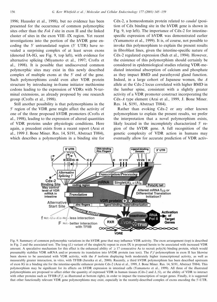
G. Kerr Whitfield et al. / Molecular and Cellular Endocrinology 177 (2001) 145–159156
1996; Haussler et al., 1998), but no evidence has beenpresented for the occurrence of common polymorphicsites other than the Fok I site in exon II and the linkedcluster of sites in the exon VIII–IX region. Yet recentinvestigations into the portion of the hVDR gene en-coding the 5� untranslated region (5� UTR) have re-vealed a surprising complex of at least seven exons(denoted IA-IG, see Fig. 9, top left), with evidence foralternative splicing (Miyamoto et al., 1997; Crofts etal., 1998). It is possible that undiscovered commonpolymorphic sites may exist in this newly describedcomplex of multiple exons at the 5� end of the gene.Such polymorphisms could even alter VDR proteinstructure by introducing in-frame initiator methioninecodons leading to the expression of VDRs with N-ter-minal extensions, as already proposed by one researchgroup (Crofts et al., 1998).
Still another possibility is that polymorphisms in the5� region of the VDR gene might affect the activity ofone of the three proposed hVDR promoters (Crofts etal., 1998), leading to the expression of altered quantitiesof VDR proteins under physiologic conditions. Hereagain, a precedent exists from a recent report (Arai etal., 1999 J. Bone Miner. Res. 14, S191, Abstract T084),which describes a polymorphism in a binding site for
Cdx-2, a homeodomain protein related to caudal (posi-tion of Cdx binding site in the hVDR gene is shown inFig. 9, top left). The importance of Cdx-2 for intestine-specific expression of hVDR was demonstrated earlier(Yamamoto et al., 1999). It is, of course, not possible toinvoke this polymorphism to explain the present resultsin fibroblast lines, given the intestine-specific nature ofCdx-2 regulated expression (Suh et al., 1994). However,the existence of this polymorphism should certainly beconsidered in epidemiological studies relating VDR-me-diated intestinal absorption of calcium and phosphateas they impact BMD and parathyroid gland function.Indeed, in a large cohort of Japanese women, the Aallele at the Cdx-2 locus correlated with higher BMD inthe lumbar spine, consistent with a slightly greateractivity of a VDR promoter construct incorporating theCdx-A type element (Arai et al., 1999, J. Bone Miner.Res. 14, S191, Abstract T084).
Rather than evoking Cdx-2 or any other knownpolymorphism to explain the present results, we preferthe interpretation that a novel polymorphism exists,likely located in the incompletely characterized 5� re-gion of the hVDR gene. A full recognition of thegenetic complexity of VDR action in humans mayeventually allow for accurate prediction of VDR activ-
Fig. 9. Summary of common polymorphic variations in the hVDR gene that may influence VDR activity. The exon arrangement (top) is describedin Fig. 2 and the associated text. The long (L) variant of the singlet(A) repeat in exon IX is proposed herein to be associated with increased VDRamount. A speculative mechanism for this effect is the enhanced ability of �17 consecutive As to recruit poly(A) binding protein, which wouldpresumably stabilize VDR mRNA and/or promote its translation into hVDR protein (see text). The F/f polymorphism in exon II has likewisebeen shown to be associated with VDR activity, with the F isoform displaying both moderately higher transcriptional activity, as well asmeasurably greater interaction, in vitro, with TFIIB (Jurutka et al., 2000). Recently, a third hVDR polymorphism has been described upstreamof exon IG in a binding site for the intestine-specific enhancer protein Cdx-2 (Arai et al., 1999, J. Bone Miner. Res. 14, S191, Abstract T084). Thispolymorphism may be significant for its effects on hVDR expression in intestinal cells (Yamamoto et al., 1999). All three of the illustratedpolymorphisms are proposed to affect either the quantity of expressed VDR in human tissues (Cdx-2 and L/S), or the ability of VDR to interactwith other proteins such as TFIIB (F/f, as illustrated at bottom right), in order to impact the transcription of target genes. Finally, it is suggestedthat other functionally relevant VDR gene polymorphisms may exist, especially in the recently-described complex of exons encoding the 5�-UTR.

G. Kerr Whitfield et al. / Molecular and Cellular Endocrinology 177 (2001) 145–159 157
ity in individual patients based on genotype, along withan enhanced ability to assess disease risk, as well asresponse to pharmacologic agents related to VDRaction.
Acknowledgements
This work was supported by National Institutes ofHealth grants to Mark R. Haussler.
References
Arai, H., Miyamoto, K.-I., Taketani, Y., Yamamoto, H., Iemori, Y.,Morita, K., Tonai, T., Nishisho, T., Mori, S., Takeda, E., 1997. Avitamin D receptor gene polymorphism in the translation initia-tion codon: effect on protein activity and relation to bone mineraldensity in Japanese women. J. Bone Miner. Res. 12, 915–921.
Baer, B.W., Kornberg, R.D., 1980. Repeating structure of cytoplas-mic poly(A)-ribonucleoprotein. Proc. Natl. Acad. Sci. USA 77,1890–1892.
Baker, A.R., McDonnell, D.P., Hughes, M.R., Crisp, T.M., Mangels-dorf, D.J., Haussler, M.R., Pike, J.W., Shine, J., O’Malley, B.W.,1988. Cloning and expression of full-length cDNA encoding hu-man vitamin D receptor. Proc. Natl. Acad. Sci. USA 85, 3294–3298.
Baniahmad, A., Ha, I., Reinberg, D., Tsai, S., Tsai, M.-J., O’Malley,B.W., 1993. Interaction of human thyroid-hormone receptor-betawith transcription factor TFIIB may mediate target gene derepres-sion and activation by thyroid-hormone. Proc. Natl. Acad. Sci.USA 90, 8832–8836.
Carling, T., Rastad, J., Akerstrom, G., Westin, G., 1998. Vitamin Dreceptor (VDR) and parathyroid hormone messenger ribonucleicacid levels correspond to polymorphic VDR alleles in humanparathyroid tumors. J. Clin. Endocrinol. Metab. 83, 2255–2259.
Cheng, W.C., Tsai, K.S., 1999. The vitamin D receptor start codonpolymorphism (Fok1) and bone mineral density in premenopausalwomen in Taiwan. Osteoporosis Int. 9, 545–549.
Colin, E.M., Weel, A.E., Uitterlinden, A.G., Buurman, C.J., Birken-hager, J.C., Pols, H.A., Van Leeuwen, J.P., 2000. Consequencesof vitamin D receptor gene polymorphisms for growth inhibitionof cultured human peripheral blood mononuclear cells by 1,25-di-hydroxyvitamin D3. Clin. Endocrinol. (Oxford) 52, 211–216.
Correa, P., Rastad, J., Schwarz, P., Westin, G., Kindmark, A.,Lundgren, E., Akerstrom, G., Carling, T., 1999. The vitamin Dreceptor (VDR) start codon polymorphism in primary hyper-parathyroidism and parathyroid VDR messenger ribonucleic acidlevels. J. Clin. Endocrinol. Metab. 84, 1690–1694.
Crofts, L.A., Hancock, M.S., Morrison, N.A., Eisman, J.A., 1998.Multiple promoters direct the tissue-specific expression of novelN-terminal variant human vitamin D receptor gene transcripts.Proc. Natl. Acad. Sci. USA 95, 10529–10534.
Curran, J.E., Vaughan, T., Lea, R.A., Weinstein, S.R., Morrison,N.A., Griffiths, L.R., 1999. Association of a vitamin D receptorpolymorphism with sporadic breast cancer development. Int. J.Cancer 83, 723–726.
Delvin, E.E., Lopez, V., Levy, E., Menard, D., 1996. Calcitrioldifferentially modulates mRNA encoding calcitriol receptors andcalcium-binding protein 9 kDa in human fetal jejunum. Biochem.Biophys. Res. Commun. 224, 544–548.
Deo, R.C., Bonanno, J.B., Sonenberg, N., Burley, S.K., 1999. Recog-nition of polyadenylate RNA by the poly(A)-binding protein. Cell98, 835–845.
Durrin, L.K., Haile, R.W., Ingles, S.A., Coetzee, G.A., 1999. VitaminD receptor 3�-untranslated region polymorphisms: lack of effecton mRNA stability. Biochim. Biophys. Acta 1453, 311–320.
Eccleshall, T.R., Garnero, P., Gross, C., Delmas, P.D., Feldman, D.,1998. Lack of correlation between start codon polymorphism ofthe vitamin D receptor gene and bone mineral density in pre-menopausal French women: the OFELY study. J. Bone Miner.Res. 13, 31–35.
Ewart, A.K., Morris, C.A., Atkinson, D., Jin, W., Sternes, K.,Spallone, P., Stock, A.D., Leppert, M., Keating, M.T., 1993.Hemizygosity at the elastin locus in a developmental disorder,Williams syndrome. Nat. Genet. 5, 11–16.
Faraco, J.H., Morrison, N.A., Baker, A., Shine, J., Frossard, P.M.,1989. ApaI dimorphism at the human vitamin D receptor genelocus. Nucl. Acids Res. 17, 2150.
Ferrari, S., Rizzoli, R., Manen, D., Slosman, D., Bonjour, J.P., 1998.Vitamin D receptor gene start codon polymorphisms (Fok I) andbone mineral density: interaction with age, dietary calcium, and3�-end region polymorphisms. J. Bone Miner. Res. 13, 925–930.
Ferrari, S., Manen, D., Bonjour, J.P., Slosman, D., Rizzoli, R., 1999.Bone mineral mass and calcium and phosphate metabolism inyoung men: relationships with vitamin D receptor allelic polymor-phisms. J. Clin. Endocrinol. Metab. 84, 2043–2048.
Gennari, L., Becherini, L., Mansani, R., Masi, L., Falchetti, A.,Morelli, A., Colli, E., Gonnelli, S., Cepollaro, C., Brandi, M.L.,1999. Fok I polymorphism at translation initiation site of thevitamin D receptor gene predicts bone mineral density and verte-bral fractures in postmenopausal Italian women. J. Bone Miner.Res. 14, 1379–1386.
Ghirri, P., Bottone, U., Coccoli, L., Bernardini, M., Vuerich, M.,Cuttano, A., Riparbelli, C., Pellegrinetti, G., Boldrini, A., 1999.Symptomatic hypercalcemia in the first months of life: calcium-regulating hormones and treatment. J Endocrinol. Invest. 22,349–353.
Gill, R.K., Atkins, L.M., Hollis, B.W., Bell, N.H., 1998. Mapping thedomains of the interaction of the vitamin D receptor and steroidreceptor coactivator-1. Mol. Endocrinol. 12, 57–65.
Gross, C., Eccleshall, T.R., Malloy, P.J., Villa, M.L., Marcus, R.,Feldman, D., 1996. The presence of a polymorphism at thetranslation initiation site of the vitamin D receptor gene is associ-ated with low bone mineral density in postmenopausal Mexican-American women. J. Bone Miner. Res. 11, 1850–1855.
Gross, C., Krishnan, A.V., Malloy, P.J., Eccleshall, T.R., Zhao,X.Y., Feldman, D., 1998. The vitamin D receptor gene startcodon polymorphism: a functional analysis of Fok I variants. J.Bone Miner. Res. 13, 1691–1699.
Harris, S.S., Eccleshall, T.R., Gross, C., Dawson-Hughes, B., Feld-man, D., 1997. The vitamin D receptor start codon polymorphism(Fok I) and bone mineral density in premenopausal Americanblack and white women. J. Bone Miner. Res. 12, 1043–1048.
Haussler, M.R., Whitfield, G.K., Haussler, C.A., Hsieh, J.-C.,Thompson, P.D., Selznick, S.H., Encinas Dominguez, C., Ju-rutka, P.W., 1998. The nuclear vitamin D receptor: biological andmolecular regulatory properties revealed. J. Bone Miner. Res. 13,325–349.
Hawa, N.S., Cockerill, F.J., Vadher, S., Hewison, M., Rut, A.K.,Pike, J.W., O’Riordan, J.L., Farrow, S.M., 1996. Identification ofa novel mutation in hereditary vitamin D resistant rickets causingexon skipping. Clin. Endocrinol. (Oxf) 45, 85–92.
Hsieh, J.-C., Jurutka, P.W., Galligan, M.A., Terpening, C.M., Haus-sler, C.A., Samuels, D.S., Shimizu, Y., Shimizu, N., Haussler,M.R., 1991. Human vitamin D receptor is selectively phosphory-lated by protein kinase C on serine 51, a residue crucial to itstrans-activation function. Proc. Natl. Acad. Sci. USA 88, 9315–9319.
Hsieh, J.-C., Jurutka, P.W., Selznick, S.H., Reeder, M.C., Haussler,C.A., Whitfield, G.K., Haussler, M.R., 1995. The T-box near the

G. Kerr Whitfield et al. / Molecular and Cellular Endocrinology 177 (2001) 145–159158
zinc fingers of the human vitamin D receptor is required forheterodimeric DNA binding and transactivation. Biochem. Bio-phys. Res. Commun. 215, 1–7.
Hsieh, J.-C., Whitfield, G.K., Oza, A.K., Dang, H.T.L., Price, J.N.,Galligan, M.A., Jurutka, P.W., Thompson, P.D., Haussler, C.A.,Haussler, M.R., 1999. Characterization of unique DNA bindingand transcriptional activation functions in the carboxyl-terminalextension of the zinc finger region in the human vitamin Dreceptor. Biochemistry 38, 16347–16358.
Hutchinson, P.E., Osborne, J.E., Lear, J.T., Smith, A.G., Bowers,P.W., Morris, P.N., Jones, P.W., York, C., Strange, R.C., Fryer,A.A., 2000. Vitamin D receptor polymorphisms are associatedwith altered prognosis in patients with malignant melanoma. Clin.Cancer Res. 6, 498–504.
Ingles, S.A., Haile, R.W., Henderson, B.E., Kolonel, L.N., Nakaichi,G., Shi, C.Y., Yu, M.C., Ross, R.K., Coetzee, G.A., 1997a.Strength of linkage disequilibrium between two vitamin D recep-tor markers in five ethnic groups: implications for associationstudies. Cancer Epidemiol. Biomarkers Prev. 6, 93–98.
Ingles, S.A., Ross, R.K., Yu, M.C., Irvine, R.A., La Pera, G., Haile,R.W., Coetzee, G.A., 1997b. Association of prostate cancer riskwith genetic polymorphisms in vitamin D receptor and androgenreceptor. J. Natl. Cancer Inst. 89, 166–170.
Ingles, S.A., Haile, R., Henderson, B., Kolonel, L., Coetzee, G.,1997c. Association of vitamin D receptor genetic polymorphismwith breast cancer risk in African-American and Hispanic women.In: Norman, A.W., et al. (Eds.), Vitamin D: Chemistry, Biologyand Clinical Applications of the Steroid Hormone. University ofCalifornia, Printing and Reprographics, Riverside, pp. 813–814.
Johnson, J.A., Grande, J.P., Roche, P.C., Sweeney, W.E. Jr, Avner,E.D., Kumar, R., 1995. 1�,25-dihydroxyvitamin D3 receptor on-togenesis in fetal renal development. Am. J. Physiol. 269, F419–428.
Johnson, J.A., Grande, J.P., Roche, P.C., Kumar, R., 1996. On-togeny of the 1,25-dihydroxyvitamin D3 receptor in fetal rat bone.J. Bone Miner. Res. 11, 56–61.
Jurutka, P.W., Hsieh, J.-C., Remus, L.S., Whitfield, G.K., Thomp-son, P.D., Haussler, C.A., Blanco, J.C.G., Ozato, K., Haussler,M.R., 1997. Mutations in the 1,25-dihydroxyvitamin D3 receptoridentifying C-terminal amino acids required for transcriptionalactivation that are functionally dissociated from hormone bind-ing, heterodimeric DNA binding and interaction with basal tran-scription factor IIB, in vitro. J. Biol. Chem. 272, 14592–14599.
Jurutka, P.W., Remus, L.S., Whitfield, G.K., Thompson, P.D.,Hsieh, J.C., Zitzer, H., Tavakkoli, P., Galligan, M.A., Dang,H.T.L., Haussler, C.A., Haussler, M.R., 2000. The polymorphicN terminus in human vitamin D receptor isoforms influencestranscriptional activity by modulating interaction with transcrip-tion factor IIB. Mol. Endocrinol. 14, 401–420.
Jurutka, P.W., Whitfield, G.K., Hsieh, J.-C., Thompson, P.D., Haus-sler, C.A., Haussler, M.R., 2001. Molecular nature of the vitaminD receptor and its role in regulation of gene expression. Rev.Endocrinol. Met. Disorders, 2, 203–216.
Kato, S., Takeyama, K., Kitanaka, S., Murayama, A., Sekine, K.,Yoshizawa, T., 1999. In vivo function of VDR in gene expression-VDR knock-out mice. J. Steroid Biochem. Mol. Biol. 69, 247–251.
Kraichely, D.M., Collins, J.J. III, DeLisle, R.K., MacDonald, P.N.,1999. The autonomous transactivation domain in helix H3 of thevitamin D receptor is required for transactivation and coactivatorinteraction. J. Biol. Chem. 274, 14352–14358.
Kurabayashi, T., Tomita, M., Matsushita, H., Yahata, T., Honda,A., Takakuwa, K., Tanaka, K., 1999. Association of vitamin Dand estrogen receptor gene polymorphism with the effect ofhormone replacement therapy on bone mineral density inJapanese women. Am. J. Obstet. Gynecol. 180, 1115–1120.
Le, H., Tanguay, R.L., Balasta, M.L., Wei, C.C., Browning, K.S.,Metz, A.M., Goss, D.J., Gallie, D.R., 1997. Translation initiationfactors eIF-iso4G and eIF-4B interact with the poly(A)-bindingprotein and increase its RNA binding activity. J. Biol. Chem. 272,16247–16255.
Li, Y.C., Amling, M., Pirro, A.E., Priemel, M., Meuse, J., Baron, R.,Delling, G., Demay, M.B., 1998. Normalization of mineral ionhomeostasis by dietary means prevents hyperparathyroidism, rick-ets, and osteomalacia, but not alopecia in vitamin D receptor-ab-lated mice. Endocrinology 139, 4391–4396.
Lin, N.U.-T., Malloy, P.J., Sakati, N., Al-Ashwal, A., Feldman, D.,1996. A novel mutation in the deoxyribonucleic acid-bindingdomain of the vitamin D receptor causes hereditary 1,25-dihy-droxyvitamin D-resistant rickets. J. Clin. Endocrinol. Metab. 81,2564–2569.
Lucotte, G., Mercier, G., Burckel, A., 1999. The vitamin D receptorFokI start codon polymorphism and bone mineral density inosteoporotic postmenopausal French women. Clin. Genet. 56,221–224.
Miyamoto, K.-i., Kesterson, R.A., Yamamoto, H., Taketani, Y.,Nishiwaki, E., Tatsumi, S., Inoue, Y., Morita, K., Takeda, E.,Pike, J.W., 1997. Structural organization of the human vitamin Dreceptor chromosomal gene and its promoter. Mol. Endocrinol.11, 1165–1179.
Mocharla, H., Butch, A.W., Pappas, A.A., Flick, J.T., Weinstein,R.S., De Togni, P., Jilka, R.L., Roberson, P.K., Parfitt, A.M.,Manolagas, S.C., 1997. Quantification of vitamin D receptormRNA by competitive polymerase chain reaction in PBMC: lackof correspondence with common allelic variants. J. Bone Miner.Res. 12, 726–733.
Morrison, N.A., Yeoman, R., Kelly, P.J., Eisman, J.A., 1992. Contri-bution of trans-acting factor alleles to normal physiological vari-ability: vitamin D receptor gene polymorphisms and circulatingosteocalcin. Proc. Natl. Acad. Sci. USA 89, 6665–6669.
Morrison, N.A., Qi, J.C., Tokita, A., Kelly, P.J., Crofts, L., Nguyen,T.V., Sambrook, P.N., Eisman, J.A., 1994. Prediction of bonedensity from vitamin D receptor alleles. Nature 367, 284–287.
Munroe, D., Jacobson, A., 1990. Tales of poly(A): a review. Gene 91,151–158.
Nakajima, S., Hsieh, J.-C., MacDonald, P.N., Galligan, M.A., Haus-sler, C.A., Whitfield, G.K., Haussler, M.R., 1994. The C-terminalregion of the vitamin D receptor is essential to form a complexwith a receptor auxiliary factor required for high affinity bindingto the vitamin D responsive element. Mol. Endocrinol. 8, 159–172.
Niimi, T., Tomita, H., Sato, S., Kawaguchi, H., Akita, K., Maeda,H., Sugiura, Y., Ueda, R., 1999. Vitamin D receptor gene poly-morphism in patients with sarcoidosis. Am. J. Respir. Crit. CareMed. 160, 1107–1109.
Rachez, C., Lemon, B.D., Suldan, Z., Bromleigh, V., Gamble, M.,Naar, A.M., Erdjument-Bromage, H., Tempst, P., Freedman,L.P., 1999. Ligand-dependent transcription activation by nuclearreceptors requires the DRIP complex. Nature 398, 824–828.
Renaud, J.-P., Rochel, N., Ruff, M., Vivat, V., Chambon, P., Grone-meyer, H., Moras, D., 1995. Crystal structure of the RAR-�ligand-binding domain bound to all-trans retinoic acid. Nature378, 681–689.
Rochel, N., Wurtz, J.M., Mitschler, A., Klaholz, B., Moras, D., 2000.The crystal structure of the nuclear receptor for vitamin D boundto its natural ligand. Mol. Cell 5, 173–179.
Ruggiero, M., Pacini, S., Aterini, S., Fallai, C., Ruggiero, C., Pacini,P., 1998. Vitamin D receptor gene polymorphism is associatedwith metastatic breast cancer. Oncol. Res. 10, 43–46.
Saijo, T., Ito, M., Takeda, E., Mahbubul Huq, A.H.M., Naito, E.,Yokota, I., Sone, T., Pike, J.W., Kuroda, Y., 1991. A uniquemutation in the vitamin D receptor gene in three Japanese pa-tients with vitamin D-dependent rickets type II: utility of single

G. Kerr Whitfield et al. / Molecular and Cellular Endocrinology 177 (2001) 145–159 159
stranded conformation polymorphism analysis for heterozygouscarrier detection. Am. J. Hum. Genet. 49, 668–673.
Segura, C., Alonso, M., Fraga, C., Garcia-Caballero, T., Dieguez, C.,Perez-Fernandez, R., 1999. Vitamin D receptor ontogenesis in ratliver. Histochem. Cell Biol. 112, 163–167.
Sosa, M., Torres, A., Martin, N., Salido, E., Liminana, J.M., Barrios,Y., De Miguel, E., Betancor, P., 2000. The distribution of twodifferent vitamin D receptor polymorphisms (Bsm I and startcodon) in primary hyperparathyroidism. J. Intern. Med. 247,124–130.
Suh, E., Chen, L., Taylor, J., Traber, P.G., 1994. A homeodomainprotein related to caudal regulates intestine-specific gene tran-scription. Mol. Cell. Biol. 14, 7340–7351.
Szpirer, J., Szpirer, C., Riviere, M., Levan, G., Marynen, P., Cassi-man, J.J., Wiese, R., DeLuca, H.F., 1991. The Sp1 transcriptionfactor gene (SP1) and the 1,25-dihydroxyvitamin D3 receptor gene(VDR) are colocalized on human chromosome arm 12q and ratchromosome 7. Genomics 11, 168–173.
Takeuchi, A., Okano, T., Sekimoto, H., Kobayashi, T., 1994. Theenzymatic formation of 1alpha,25-dihydroxyvitamin D3 from 25-hydroxyvitamin D3 in the liver of fetal rats. Comp. Biochem.Physiol. C Pharmacol. Toxicol. Endocrinol. 109, 1–7.
Tao, C., Yu, T., Garnett, S., Briody, J., Knight, J., Woodhead, H.,Cowell, C.T., 1998. Vitamin D receptor alleles predict growth andbone density in girls. Arch. Dis. Child. 79, 488–493.
Terpening, C.M., Haussler, C.A., Jurutka, P.W., Galligan, M.A.,Komm, B.S., Haussler, M.R., 1991. The vitamin D-responsiveelement in the rat bone gla protein is an imperfect direct repeatthat cooperates with other cis-elements in 1,25-dihydroxyvitaminD3-mediated transcriptional activation. Mol. Endocrinol. 5, 373–385.
Veenstra, T.D., Prufer, K., Koenigsberger, C., Brimijoin, S.W.,Grande, J.P., Kumar, R., 1998. 1,25-Dihydroxyvitamin D3 recep-tors in the central nervous system of the rat embryo. Brain Res.804, 193–205.
Verbeek, W., Gombart, A.F., Shiohara, M., Campbell, M., Koeffler,H.P., 1997. Vitamin D receptor: no evidence for allele-specificmRNA stability in cells which are heterozygous for the Taq I
restriction enzyme polymorphism. Biochem. Biophys. Res. Com-mun. 238, 77–80.
Videman, T., Leppavuori, J., Kaprio, J., Battie, M.C., Gibbons, L.E.,Peltonen, L., Koskenvuo, M., 1998. Intragenic polymorphisms ofthe vitamin D receptor gene associated with intervertebral discdegeneration. Spine 23, 2477–2485.
Wagner, R.L., Apriletti, J.W., McGrath, M.E., West, B.L., Baxter,J.D., Fletterick, R.J., 1995. A structural role for hormone in thethyroid hormone receptor. Nature 378, 690–697.
Watanabe, M., Fukutome, K., Murata, M., Uemura, H., Kubota, Y.,Kawamura, J., Yatani, R., 1999. Significance of vitamin D recep-tor gene polymorphism for prostate cancer risk in Japanese.Anticancer Res. 19, 4511–4514.
Whitfield, G.K., Hsieh, J.-C., Nakajima, S., MacDonald, P.N.,Thompson, P.D., Jurutka, P.W., Haussler, C.A., Haussler, M.R.,1995. A highly conserved region in the hormone binding domainof the human vitamin D receptor contains residues vital forheterodimerization with retinoid X receptor and for transcrip-tional activation. Mol. Endocrinol. 9, 1166–1179.
Whitfield, G.K., Selznick, S.H., Haussler, C.A., Hsieh, J.-C., Galli-gan, M.A., Jurutka, P.W., Thompson, P.D., Lee, S.M., Zerwekh,J.E., Haussler, M.R., 1996. Vitamin D receptors from patientswith resistance to 1,25-dihydroxyvitamin D3: point mutationsconfer reduced transactivation in response to ligand and impairedinteraction with the retinoid X receptor heterodimeric partner.Mol. Endocrinol. 10, 1617–1631.
Whitfield, G.K., Jurutka, P.W., Haussler, C.A., Haussler, M.R.,1999. Steroid hormone receptors: evolution, ligands and molecu-lar basis of biologic function. J. Cell. Biochem. Suppl. 32/33,110–122.
Wood, R.J., Fleet, J.C., 1998. The genetics of osteoporosis: vitamin Dreceptor polymorphisms. Ann. Rev. Nutr. 18, 233–258.
Yamamoto, H., Miyamoto, K., Li, B., Taketani, Y., Kitano, M.,Inoue, Y., Morita, K., Pike, J.W., Takeda, E., 1999. The caudal-related homeodomain protein Cdx-2 regulates vitamin D receptorgene expression in the small intestine. J. Bone Miner. Res. 14,240–247.
.
Copyright © 2022 FDOKUMEN