The MRC OX-44 antigen marks a functionally relevant subset among rat thymocytes
13
THE MRC OX-44 ANTIGEN MARKS A FUNCTIONALLY RELEVANT SUBSET AMONG RAT THYMOCYTES BY DAVID J. PATERSON, JON R. GREEN, WILFRED A. JEFFERIES, MICHAEL PUKLAVEC, AND ALAN F. WILLIAMS From the Medical Research Council Cellular Immunology Unit, Sir William Dunn School of Pathology, University of Oxford, Oxford OX1 3RE, United Kingdom The immunocompetent T lymphocytes that are found in the peripheral lymphoid tissue can be subdivided into three sets on the basis of cell surface marker antigens . The first set to be identified includes the precursors for cytotoxic T cells that are specific for, or restricted by, class I MHC antigens and these are marked by expression of the CD8 antigen (1-3) . Complementing this set are the rest of the peripheral T cells that express the CD4 antigen and include T helper cells and precursors for cytotoxic cells whose specificity involves the class II MHC antigens (3-6) . The CD4' cells can be further subdivided into one set that includes cells providing help for B cells and another that contains the cells that initiate the MLR and provide help for CTL (7) . In the thymus 10-15% of the cells have phenotypes apparently similar to peripheral T cells in that they express either CD4 or CD8 antigens (but not both) and contain cells that can function as precursors to cytotoxic T cells (CD4 - CD8+) or T helper cells (CD4+ CD8- ) (8-11) . These cells are found largely in the medulla, whereas the great majority of the remaining cells express both CD4 and CD8 antigens and are found largely in the cortex (12, 13) . Functional activity from the CD4+ CD8+ cells has not been identified in studies in the mouse (9), and these cells have the additional unusual feature of expressing very low levels of class I MHC antigens (14, 15) . The CD4 - CD8- set constitutes only 1-2% of thymocytes but these cells can reconstitute all other populations either in vivo when injected intravenously into irradiated recipients (16), or in vitro when added to fetal thymic lobes in culture (17) . The lineage relationships between the various populations of cells in the thymus are largely unknown and new markers are needed if these are to be resolved . We now describe an antibody called MRC OX-44 that labels all leukocytes in the periphery but only 10-15% in the thymus . Functional studies lead to the working hypothesis that the OX-44+ cells include all thymocytes that are on the pathway to immunocompetent T lymphocytes . Volume 165 January 1987 1-13 Materials and Methods Animals. Strains used were HO (RT1`, leukocyte-common antigen [L-CA]' 1 .2), HO .B2 (RT1"), DA (RTIa), and (HO X HO .B2) F, . All were from the Specific Pathogen- free Unit at the Medical Research Council Cellular Immunology Unit, Oxford . HO 'Abbreviations used in this paper: L-CA, leukocyte-common antigen ; RAM, rabbit anti-mouse F(ab') 2 ; TDL, thoracic duct lymphocyte . J. Exp . MED. © The Rockefeller University Press - 0022-1007/87/01/0001/13 $1 .00 1 on April 23, 2016 jem.rupress.org Downloaded from Published January 1, 1987
-
Upload
independent -
Category
Documents
-
view
1 -
download
0
Transcript of The MRC OX-44 antigen marks a functionally relevant subset among rat thymocytes

THE MRC OX-44 ANTIGEN MARKS A FUNCTIONALLYRELEVANT SUBSET AMONG RAT THYMOCYTES
BY DAVIDJ. PATERSON, JON R. GREEN, WILFRED A. JEFFERIES,MICHAEL PUKLAVEC, AND ALAN F. WILLIAMS
From the Medical Research Council Cellular Immunology Unit, Sir William Dunn School ofPathology, University of Oxford, Oxford OX1 3RE, United Kingdom
The immunocompetent T lymphocytes that are found in the peripherallymphoid tissue can be subdivided into three sets on the basis of cell surfacemarker antigens . The first set to be identified includes the precursors forcytotoxic T cells that are specific for, or restricted by, class I MHC antigens andthese are marked by expression of the CD8 antigen (1-3). Complementing thisset are the rest of the peripheral T cells that express the CD4 antigen and includeT helper cells and precursors for cytotoxic cells whose specificity involves theclass II MHC antigens (3-6). The CD4' cells can be further subdivided into oneset that includes cells providing help for B cells and another that contains thecells that initiate the MLR and provide help for CTL (7).
In the thymus 10-15% of the cells have phenotypes apparently similar toperipheral T cells in that they express either CD4 or CD8 antigens (but not both)and contain cells that can function as precursors to cytotoxic T cells (CD4-CD8+) or T helper cells (CD4+ CD8-) (8-11) . These cells are found largely inthe medulla, whereas the great majority of the remaining cells express both CD4and CD8antigens and are found largely in the cortex (12, 13) . Functional activityfrom the CD4+ CD8+ cells has not been identified in studies in the mouse (9),and these cells have the additional unusual feature of expressing very low levelsof class I MHC antigens (14, 15). The CD4- CD8- set constitutes only 1-2% ofthymocytes but these cells can reconstitute all other populations either in vivowhen injected intravenously into irradiated recipients (16), or in vitro whenadded to fetal thymic lobes in culture (17) .The lineage relationships between the various populations of cells in the thymus
are largely unknown and new markers are needed if these are to be resolved .We now describe an antibody called MRC OX-44 that labels all leukocytes inthe periphery but only 10-15% in the thymus . Functional studies lead to theworking hypothesis that the OX-44+ cells include all thymocytes that are on thepathway to immunocompetent T lymphocytes .
Volume 165 January 1987
1-13
Materials and MethodsAnimals. Strains used were HO (RT1`, leukocyte-common antigen [L-CA]' 1 .2),
HO.B2 (RT1"), DA (RTIa), and (HO X HO .B2) F, . All were from the Specific Pathogen-free Unit at the Medical Research Council Cellular Immunology Unit, Oxford . HO
'Abbreviations used in this paper:
L-CA, leukocyte-common antigen; RAM, rabbit anti-mouseF(ab') 2 ; TDL, thoracic duct lymphocyte .
J. Exp. MED. © The Rockefeller University Press - 0022-1007/87/01/0001/13 $1 .00
1
on April 23, 2016
jem.rupress.org
Dow
nloaded from
Published January 1, 1987

2 FUNCTIONS OF RAT THYMOCYTE SUBSETS
(RT1`, L-CA 1 .1) rats were maintained under conventional conditions . Chimeric rats wereprepared by irradiating HO.B2 rats (9-12 wk) with 950 rad of 7-irradiation (from a' s'Cssource at 87 rad/min) followed by reconstitution with 5-10 X 10' bone marrow cells i .v .per rat from the F, hybrid 5-6 h later .
Antibodies.
A hybridoma secreting the MRC OX-44 mAb (IgG,) was derived from afusion between BALB/C mouse spleen cells and the mouse myeloma NSO/1 followingmethods as in Galfre and Milstein (18) . The mouse had been immunized with T lympho-blasts from an MLR between purified rat T helper cells and irradiated semiallogeneic ratspleen, as described in Jefferies et al . (19) . Rabbit anti-mouse F(ab')2 (RAM) and itsfluorescein isothiocyanate (RAM-FITC) and horseradish peroxidase (RAM-peroxidase)conjugates were prepared as previously described (20, 21) . Other mAbs used were MRCOX-1, anti-L-CA common determinant (22) ; MRC OX-8, anti-CD8 (2) ; MRC OX-18,anti-class I MHC common determinant (15) ; MRC OX-19, anti-rat CD-5 (Lyt-1) (23) ;MRC : OX-27, anti-class I MHC RT1` haplotype (19) ; MRC OX-35 and MRC OX-38,noncompeting anti-CD4 mAbs (19) ; MRC OX-42, anti-rat C3b receptor (24) . ED2, anti-rat tissue macrophages (25), was the kind gift of Dr . C . D . Dijkstra, Free University,Amsterdam, The Netherlands ; NDS 58, anti-RT-7 .1 (anti-L-CA1 .2) (26), was the kindgift of Dr . M. R. Newton, Department of Zoology, University of Oxford ; 8G6.1, anti-RT-7 .2 (anti-L-CA 1 .1) alloantigen (27) was the kind gift of Dr . D . L . Greiner, UniversityofConnecticut Health Center, Farmington CT; Bu20a, reactive with bromodeoxyuridine2was the kind gift of Dr . J.-P . Magaud, Department of Haematology, John RadcliffeHospital, Oxford . MRC OX-21, anti-human C3b 1NA, was used as a control mAb.
Cells .
Thoracic duct lymphocytes (TDL) were collected overnight into ice-cold Dul-becco's A + B medium (DAB) containing 20 U/ml heparin . Thymus, spleen, and cervicallymph nodes were removed aseptically, and either teased apart with forceps or pushedthrough a square metal mesh of 0.09-mm2 pore size, filtered through lens tissue, andwashed twice in DAB/0.2% BSA.Bone marrow cells were removed from femur and tibia into ice-cold DAB/0.2% BSA
by syringing . Cells were filtered, washed twice, and injected in a volume of 1 ml ofDAB/0.2% BSA for the preparation of radiation bone marrow chimeras .
For intrathymic injections, thymocytes were prepared in Hepes/RPMI 1640 with 2.5%FCS .
Immunoprecipitation . HO TDL were ' 25 1 surface-labeled using the lactoperoxi-dase/glucose oxidase method (28) and a 1 % NP-40 detergent extract was prepared .Material that could bind to OX-44 antibody was precipitated and visualized on 12% SDS-PAGE gels by autoradiography using methods exactly as described by Jefferies et al . (29) .
In Vitro Proliferation Assays .
For the primary semiallogeneic MLR, varying numbersof responder HO thymocytes were cultured with 5 X 10 5 irradiated (HO X HO.B2)F,spleen cells (1,800 rad) for 4 d in RPMI 1640 containing 2.5 X 10⁻⁵ M 2-mercaptoethanol,antibiotics, and 5% DA serum .
For the Con A stimulation assay, HO thymocytes were incubated for 3 d with 10jug/ml - ' Con A, 5 X 10⁵ HO-irradiated spleen cells (1,800 rad), and 25 A.1 of a Con Asupernatant (produced by incubating 5 X 10⁶ HO spleen cells/ml for 24 h with 5 wg/ml- 'Con A in the above medium with FCS in place of DA serum) .
Culture volumes were 0.2 ml in sterile 96-well U-bottomed microtiter plates at 37°Cin a humidified incubator with 5% C02 . 0.5 uCi of [ sH]thymidine (Amersham Interna-tional, Amersham, United Kingdom) was added 18 h before cells were collected ontoglass-fiber filters for determination of radioactivity incorporated .
Staining ofThymus In Situ.
Thymi were removed from HO (4-8 wks) or chimeric rats,dissected, immediately embedded in OCT (R . A . Lamb, London), and frozen in 2-methylbutane cooled by liquid nitrogen . Cryostat sections (5 /m) were cut, dried in astream of warm air, and fixed in ethanol for 10 min at 4'C . Sections were stained by theimmunoperoxidase technique (21), and counterstained with Harris's hematoxylin .
For staining sections with the Bu20a mAb the method of Magaud et al . 2 was used .
2 Magaud, J.-P ., I . Sargeant, J . Clark, and D . Y . Mason . 1986 . Immunocytochemica l labeling withmonoclonal antibromodeoxyuridine . Manuscript in preparation .
on April 23, 2016
jem.rupress.org
Dow
nloaded from
Published January 1, 1987

PATERSON ET AL .
3
Briefly, sections were fixed in acetone for 10 min at 4 ° C, air dried, incubated in 95%formamide for 35 min at 67° C, washed in PBS, and then stained as usual.
Rosette Depletion .
This was carried out as described by Mason (30) . Briefly, thymocyteswere labeled with mAb(s), washed, and mixed with SRBC coated with RAM IgG for 20min at VC.C. Rosettes formed between labeled thymocytes andSRBC-RAM were removed,and the remaining cells were treated with ammonium chloride in Tris buffer to lyseerythrocytes and then washed . The success of depletion was assessed by labeling pre- andpostdepletion samples with RAM-FITC, and analyzing on the FACS .
Thymus Repopulation Assay.
The assay of Goldschneider et al . (27) was used . HO L-CA1 .2 rats (5-7 wk old) were given 600 rad and injected intrathymically with HO L-CA 1 .1 thymocytes 6-8 h later . Donor cells, present in 20 ul of PBS, were injected intofour to six sites per thymus at ^"3-5 A,1 per site . Donor reconstitution was assessed byanalyzing host thymi on the FACS 14 d later .
Results
Expression and Nature of the OX-44 Antigen.
The OX-44 mAb resulted froman immunization of mice with rat T blasts, in which mAbs were first detected bybinding to T blasts and then screened with the FACS on other lymphoid cellpopulation to detect any interesting labeling patterns. Fig. 1 a shows that all Tblasts were labeled by the antibody ; this was also the case for lymph node cellsalthough two modes of labeling intensity were seen (Fig . 1 b) . The more weaklylabeled cells were T lymphocytes as shown by the fact that the low intensity peakwas shifted to denser labeling when OX-19 mAb, which labels T cells but notmost B cells, was added with OX-44 antibody (Fig . 1 e) . A large fraction of bonemarrow cells were labeled (Fig . 1 c) and these include the lymphoid and neutro-phil populations. The antibody did not label erythrocytes (data not shown) andthe negative cells in the bone marrow may be of the erythroid lineage. Thelabeling of thymocytes was of most interest (Fig. 1 d) because in this case only12% of cells showed detectable labeling . These included the larger cells amongthe thymocytes as assessed by their scatter profiles in the FACS (Fig . If-h) . In22 different labeling experiments of HO rats aged 6-8 wk the mean labelingwith OX-44 mAb was 12.2% ± SD 1 .9% .
In other studies (data not shown) it was found that OX-44 labeled all nucleatedperitoneal cells and dendritic cells obtained from lymph draining the gut. Theresults of all the labeling studies indicate that OX-44 may label all cells of thelymphoid and myeloid lineages with the exception of the majority of thymocytes .
Preliminary experiments with immunoperoxidase labeling on tissue sectionsfrom liver, kidney, and brain show some OX-44 staining . Part of this seemed tobe due to a nonspecific nuclear crossreaction, whereas the rest appeared tocorrespond to the presence of leukocytes in these tissues (data not shown) .
Immunoprecipitation studies identified the OX-44 antigen as a broad band ofabout 43,000 mol wt in the unreduced or reduced state (Fig . 1 i) . The diffusenature of the band obtained after SDS-PAGE was not due to technical problemsbecause other antigens run on the same gels gave much sharper bands (notshown) . The heterogeneity could be due to diversity in carbohydrate attachedto the OX-44 antigen .
Thymocytes Labeled by the OX-44 mAb.
The relationship of the OX-44+ andOX-44- thymocytes to subsets defined by other mAbs was studied by FACSanalysis on unfractionated thymocytes or cells obtained after rosette depletionof thymocytes labeled with various mAbs . Fig. 2, a and b, shows labeling of
on April 23, 2016
jem.rupress.org
Dow
nloaded from
Published January 1, 1987

4
FUNCTIONS OF RAT THYMOCYTE SUBSETS
FIGURE 1 .
Distribution and nature of the OX-44 antigen. (a-h) Cells were incubated withmAb(s) for 60 min at 4°C, washed, labeled with RAM-FITC, and analyzed on the FACS II(B-D FACS Systems, Becton Dickinson & Co ., Sunnyvale, CA). Profiles (a-d) show MRC 0X-44 labeling of T lymphoblasts, lymph node cells (LNC) (with negative control), bone marrow(BM), and thymocytes . (e) Labeling of LNCwhen OX-44 and OX-19 mAbs were used togetherin the first step . (fand g) Forward light scatter (FSC) profiles of whole, MRC OX-44-, andMRC OX-44+ thymocytes . FLI, fluorescence . (i) Immunoprecipitation of the MRC OX-44antigen : material precipitated from "'I surface-labeled TDL lysate was run reduced (RED)and nonreduced (NON-RED) on 12% SDS-PAGE and visualized by autoradiography . Lane 1,control mAb; lane 2, MRC OX-44.
on April 23, 2016
jem.rupress.org
Dow
nloaded from
Published January 1, 1987

PATERSON ET AL .
FIGURE 2.
Analysis of thymocyte subpopulations. FACS analysis of(a) unseparated, (b) MRCOX-44-, and (c) CD4- CD8- thymocytes was carried out using single or multiple mAb labelingin the first step . Thymocyte populations in panels b and c were prepared by rosette depletion .Figures indicate the percentage of positive cells to the right ofthe marker . Thymocyte subsetscan be calculated from such analyses, as shown in Table I .
undepleted thymocytes and of OX-44- cells with mAbs against rat CD4, CD8,class I, and MRC OX-44 antigen either alone or in combination with each otherwhereas Fig. 2 c shows labeling of CD4- CD8- cells .The results for unfractionated thymocytes (Fig . 2 a showed that a majority of
OX-44' cells were negative for either CD4 or CD8 antigen, or both, inasmuchas when cells were incubated with OX-44 plus antibodies against CD4 or CD8,then the small negative peaks seen with labeling of mAbs against CD4 or CD8alone were substantially reduced. The CD4-, CD8- cells were almost all OX-44'as shown by labeling with combinations of antibodies (Fig . 2, a and b) and (Fig .2 c) . In contrast, not all cells singly positive for CD4 or CD8 were OX-44' becausesmall negative peaks remained in labeling of thymocytes with anti-CD4 or CD8plus OX-44 (Fig . 2 a) and in labeling of OX-44- cells with the anti-CD4 or CD8antibodies (Fig . 2 b) . The significance of these small peaks is proven by the findingthat OX-44- cells were 99 .9% labeled by anti-CD4 and CD8 mixed together(Fig . 2 b) . Most CD4+ CD8+ cells were OX-44-, but calculations of subsets fromlabeling data as in Fig. 2 a always led to the conclusion that a small set of CD4+CD8+ OX-44' cells was present. The average percentages from seven experi-ments for subsets defined by CD4, CD8, and OX-44 mAbs are given in Table I .
on April 23, 2016
jem.rupress.org
Dow
nloaded from
Published January 1, 1987

6
FUNCTIONS OF RAT THYMOCYTE SUBSETS
240
200
1600
120V
80
40
* Mean ± SD from seven experiments such as the one shown in Fig. 2.
TABLE I
Thymocyte Subsets Calculated from FRCS Analysis
Subset phenotype
Percent of total population*CD4- CD8- OX-44+
1 .3 ± 0.2CD4+ CD8+ OX-44+
2.2 ± 1 .3CD4+ CD8+ OX-44-85 .3 ± 1 .3CD4+ CD8- OX-44+
4.8 ± 0.7CD4+ CD8- OX-44-2 .0 ± 0.6CD4- CD8+ OX-44+
2.2 ± 0.7CD4- CD8+ OX-44-1 .9 ± 0.6
FIGURE 3.
In vitro proliferative responses of thymocyte populations . The proliferative re-sponses of FACS-purified (O) MRC OX-44+ and (0) MRC OX-44- thymocytes are shown inthe (a) Con A response and (b) primary MLR. MRC OX-44- thymocytes were 99.8% pure,whereas the MRC OX-44+ thymocytes were 94% pure . Cells labeled with MRC OX-44 andRAM-FITC but not run through the FACS were used as the unfractionated thymocyte control(A) . (c) The Con A response of (O) CD4- CD8- and (A) unfractionated thymocytes. CD4-CD8- thymocytes were prepared by rosette depletion followed by cell sorting, and were 99.8%pure . Points are means ± I SD using four or six replica wells.
Evidence for all these sets can be directly seen from the labeling profiles in Fig.2 except for the CD4+ CD8+ OX-44+ set .mAbs against class I MHC antigens are known to label a subfraction of
thymocytes (15) and the OX-44+ set was found to overlap with class I+ cells. Fig.2a shows labeling of thymocytes with OX-27 (anti-class I) mAb, and comparisonof this with Fig. 2 b shows that removal of OX-44+ cells resulted in the loss ofcells that were heavily labeled with OX-27 mAb. The CD4- CD8- cells were allclass I+ (Fig . 2c), as were all cells singly positive for CD4 or CD8 (not shown) .The class I - fractions are all found in the CD4+ CD8+ set but a minority of CD4+CD8+ cells are weakly positive for class 1.
Proliferation of Thymocyte Subsets In Vitro.
The Con A stimulation assay (Fig .3a) and MLR (Fig . 3b) were used to assess the proliferative capacity in vitro ofvarious thymocyte subsets isolated using the FACS or by rosette depletion . Inneither assay did OX-44- cells show any proliferation while OX-44+ cells werepotent responders . The enrichment of activity shown by OX-44+ cells in com-parison with unseparated thymocytes was consistent with the total activity ofunseparated cells being due to the OX-44+ cells. It should be noted that OX-44-cells failed to respond despite the fact that, in the Con A stimulation assay,optimal conditions were used including the presence of irradiated syngeneicspleen cells as asource ofdendritic cells and T helper cells, as well as a supernatant
on April 23, 2016
jem.rupress.org
Dow
nloaded from
Published January 1, 1987
PATERSON ET AL .
7
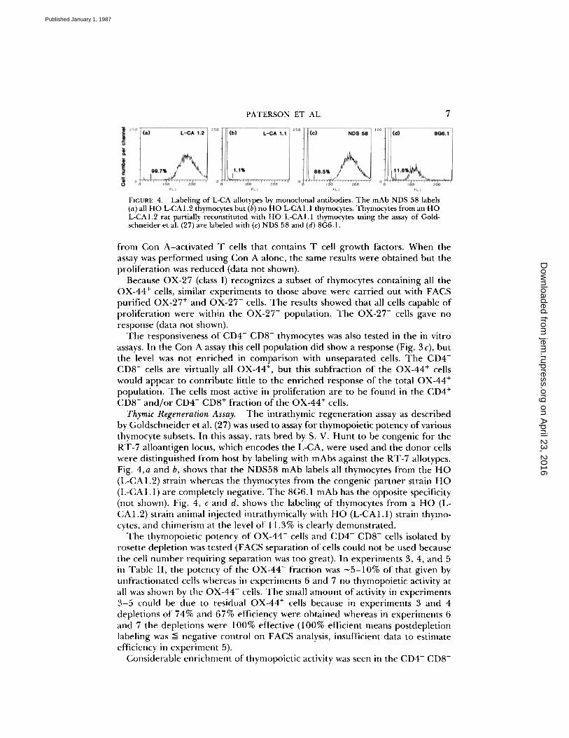
FIGURE 4.
Labeling of L-CA allotypes by monoclonal antibodies. The mAb NDS 58 labels(a) all HO L-CAL2 thymocytes but (b) no HO L-CALL thymocytes. Thymocytes from an HOL-CA 1 .2 rat partially reconstituted with HO L-CAI . I thymocytes using the assay of Gold-schneider et al . (27) are labeled with (c) NDS 58 and (d) 8G6. L
from Con A-activated T cells that contains T cell growth factors. When theassay was performed using Con A alone, the same results were obtained but theproliferation was reduced (data not shown).Because OX-27 (class I) recognizes a subset of thymocytes containing all the
OX-44+ cells, similar experiments to those above were carried out with FACSpurified OX-27' and OX-27- cells. The results showed that all cells capable ofproliferation were within the OX-27+ population . The OX-27- cells gave noresponse (data not shown) .The responsiveness of CD4- CD8- thymocytes was also tested in the in vitro
assays . In the Con A assay this cell population did show a response (Fig . 3 c), butthe level was not enriched in comparison with unseparated cells . The CD4-CD8- cells are virtually all OX-44+, but this subfraction of the OX-44+ cellswould appear to contribute little to the enriched response of the total OX-44+population . The cells most active in proliferation are to be found in the CD4+CD8- and/or CD4- CD8+ fraction of the OX-44+ cells .
Thymic Regeneration Assay.
The intrathymic regeneration assay as describedby Goldschneider et al . (27) was used to assay for thymopoietic potency of variousthymocyte subsets. In this assay, rats bred by S . V . Hunt to be congenic for theRT-7 alloantigen locus, which encodes the L-CA, were used and the donor cellswere distinguished from host by labeling with mAbs against the RT-7 allotypes.Fig. 4,a and b, shows that the NDS58 mAb labels all thymocytes from the HO(I:CA 1 .2) strain whereas the thymocytes from the congenic partner strain HO(L-CA 1 .1) are completely negative . The 8G6.1 mAb has the opposite specificity(not shown) . Fig. 4, c and d, shows the labeling of thymocytes from a HO (L-CA1.2) strain animal injected intrathymically with HO (L-CA1 .1) strain thymo-cytes, and chimerism at the level of 11 .3% is clearly demonstrated .The thymopoietic potency of OX-44- cells and CD4- CD8- cells isolated by
rosette depletion was tested (FACS separation of cells could not be used becausethe cell number requiring separation was too great) . In experiments 3, 4, and 5in Table 11, the potency of the OX-44- fraction was ^-5-10% of that given byunfractionated cells whereas in experiments 6 and 7 no thymopoietic activity atall was shown by the OX-44- cells . The small amount of activity in experiments3-5 could be due to residual OX-44+ cells because in experiments 3 and 4depletions of 74% and 67% efficiency were obtained whereas in experiments 6and 7 the depletions were 100% effective (100% efficient means postdepletionlabeling was : negative control on FACS analysis, insufficient data to estimateefficiency in experiment 5) .
Considerable enrichment of thymopoietic activity was seen in the CD4- CD8-
on April 23, 2016
jem.rupress.org
Dow
nloaded from
Published January 1, 1987

8
FUNCTIONS OF RAT THYMOCYTE SUBSETS
]'he assay of Goldschneider et al . (27) was used to assess the ability of different thymocyte subsets to repopulate a thymus in vivo.140 congenic strains were used . After 600 rad of irradiation, 1..-CA 1 .2 recipients were injected intrathymically with LCALI donorcells, killed 2 wk later, and the percentage of donor thymocytes assessed by FAGS analysis . Table summarizes data from allexperiments performed.
* Donor populations prepared by rosette depletion, see text for details of purities.$ Percent donor cells calculated by subtracting NDS 58 labelling from OX-1 labelling (OX-1 always gave 99 .7-99 .9% labelling) .4 F:ach figure represents one animal .
cells (almost all OX-44+, Fig. 2c). This result is in accord with data in the mouse(16) and, in addition, showed that the poor activity of the OX-44- cells couldnot be explained by artifacts in the isolation of cells by rosette depletion . TheCD4` CD8- cells were 10-20 times more potent than the unfractionated thy-mocytes compared with a theoretical expected enrichment of 70-fold if all theactivity were in this subset . The low experimental value may be due to some lossof functional viability in the rosetted cells compared with unfractionated cells,or to the possibility that the thymopoietic activity is due to a subset within theCD4- CD8- cells that is recovered with lower efficiency than the bulk CD4`CD8` population . A third possibility is that some cells in the OX-44' set otherthan the CD4- CD8- OX-44' cells may have thymopoietic activity . It is not likelythat the CD4- CD8- set contained many cells of lineage other than T lymphoidinasmuch as almost all cells were weakly OX-19+ (rat CD5 or Ly-1) (23) andmAbs recognizing class 11 MHC, surface Ig, or C3b receptor only gave ^-5%labeling (data not shown) .
Localization of the OX-44' Cells.
This was studied in tissue sections using theimmunoperoxidase technique with the results shown in Fig. 5 . Unlabeled cellscould not be seen in the medulla where positive cells with both lymphoid anddendritic morphology were seen (Fig . 5a). In the cortex most lymphoid cellswere OX-44-. This is consistent with the fact that most CD4+ CD8' cells areOX-44- and that these cells are known to exist in the cortex, whereas the medullamostly contains the cells positive for CD4 or CD8 but not both (data for CD8labeling is shown in Fig. 5 c) . Small numbers of OX-44' cells are scatteredthroughout the cortex : some have irregular morphology, shown by the star ;
on April 23, 2016
jem.rupress.org
Dow
nloaded from
Published January 1, 1987

PATERSON ET AL .
9
others are small round cells as shown by arrows in Fig. 5 b. All peripheralleukocytes appear to be OX-44+ and thus the sections were stained with variousmAbs against macrophages to see if these might account for all the OX-44+cortical cells. The OX-42 mAb, which labels many rat macrophages and may bethe equivalent of mouse Mac-1, labeled few cells in the thymus sections (Fig . 5d)but the ED-2 mAb stained considerable numbers of cortical (but not medullary)macrophages (Fig . 5e) in accord with the data of Dijkstra et al . (25) . However,ED-2+ cells did not appear to account for the small rounded cortical cells .The OX-44+ cells include those that express large amounts of class I MHC
antigens, and the localization of class I+ cells was studied in chimeric animals.Class I on leukocytes cannot be visualized in the normal thymus owing to stainingof epithelial cells (Fig . 5f ), and thus expression of leukocyte class I was studiedin HO.B2 (RT1°) animals reconstituted with HOM (RT1°) X HO (RT1`) bonemarrow cells. The donor-derived class I could then be detected with the OX-27mAb which is specific for RT1` while all class I was detected with the OX-18mAb which recognizes a common determinant. The results for OX-27 labelingin sections from the chimeric thymus (Fig . 5g) were very similar to those seenwith OX-44 labeling in the chimeric (Fig . 5h) or normal thymuses (Fig . 5a).This experiment additionally established that most of the class I in the thymiccortex is derived from epithelial cells in that staining of the chimeric thymuseswith the class I-common OX-18 antibody gave the extensive pattern of stainingas shown for normal thymus in Fig. 5f(OX-18 data not shown) .
Extensive cell division is known to occur in the thymic cortex . This wasconfirmed in the present series of experiments by injecting animals intravenouslywith 5-bromo-2' deoxyuridine (10 mg/kg body wt), killing them after 30 min,and detecting cells that had incorporated the metabolite with the mAb BOW.A thymus section from such an animal stained with Bu20a is shown in Fig. 5i :there is considerable labeling of cells throughout the cortex, with the highestdensity in the outer cortical layer, whereas few medullary cells were labeled.Analysis of cells isolated from such a thymus showed that 8.1 % of unfractionatedthymocytes were Bu20a+, compared with 7.8% for OX-44- thymocytes . Thissuggests that most dividing cells in the cortex are CD4+ CD8+ , which agrees withthe data of Scollay et al . (31) in the mouse. The OX-44- cells cannot be stimulatedto further division with Con A plus growth factors; thus the DNA synthesis inthis set seems likely to be occurring in the final divisions of a cell series undergoingterminal differentiation. The situation may be similar to that seen in the avianerythroid series where a set of cell divisions occur along with production ofhemoglobin and other erythroid components to produce a nucleated cell thatdoes not divide again (32) .
DiscussionThe OX-44 mAb labels a small fraction of thymocytes that mediates functions
that are characteristic of the most undifferentiated thymocytes as well as thethymic cells that are most like mature T cells . An anti-rat mAb with similarlabeling and molecular properties was described by Matsuura et al . (33) butminimal functional data were reported . An antigen similar to OX-44 has notbeen described in other species.From the current studies we derive the working hypothesis that the OX-44+
on April 23, 2016
jem.rupress.org
Dow
nloaded from
Published January 1, 1987

lO
FUNCTIONS OF RAT THYMOCYTE SUBSETS
FIGURE 5 .
Monoclonal antibody labeling of thymus . HO thymus cryostat sections (5 pm)were stained with various mAbs using the immunoperoxidase technique . (a) MRC OX-44 . m,
on April 23, 2016
jem.rupress.org
Dow
nloaded from
Published January 1, 1987

PATERSON ET AL .
11
cells may include all the cells that are on the differentiation pathway to immu-nocompetent T cells while the OX-44- compartment represents cells that havefailed some test in differentiation and are fated to die in situ . The followingfactors lead to this view : (a) The OX-44- cells were negative for proliferationin the MLR or by Con A stimulation and had no thymopoiesic activity . Further-more, it seems unlikely that other functions, for example, for T help or cytotoxicactivity, would be mediated by OX-44- cells because the cells that mediate thesefunctions would be expected to divide vigorously with Con A stimulation plusaccessory cells and growth factors. Rat thymic T helpers are known to have theCD4+ CD8- phenotype (8). (b) The OX-44 antigen is a leukocyte-commonantigen apart from its absence from most thymocytes . Other antigens are knownto be expressed intermittently in a cell lineage (e.g ., rat W3/13 antigen is onstem cells and plasma cells but not on pre-B or resting B cells), but in these casesthe antigen does not label all peripheral leukocytes as is the case for OX-44.(c) The OX-44 labeling appears to be a more extreme case of a similar patternshown by class I MHC antigen, which is also heavily expressed on all leukocytesexcept for the CD4' CD8' thymocytes which are weak or negative . It could bethat in thymic differentiation cells that are fated to die first switch off OX-44expression and then class I.
Regardless ofthis working hypothesis, the OX-44 antigen allows the definitionof new thymic subsets for further study. CD4- CD8- OX-44+ cells constitute themuch-studied "double negative" cells, whereas the CD4+ CD8- OX-44+ andCD4- CD8+ OX-44+ cells correspond closely to mature phenotype T cells . Incontrast, the CD4+ CD8+ OX-44+, CD4+ CD8- OX-44-, CD4- CD8+ OX-44-sets have not been previously described and are potentially intermediate cells inthe thymic differentiation pathways . In accord with this view is the finding thatCD4- CD8+ OX-44- cells are a prominent population in thymuses regeneratingafter irradiation (manuscript in preparation) . Further studies on these subsetsmay help in delineation of pathways for thymic differentiation.
Summary
A monoclonal antibody called MRC OX-44 is described that labels all myeloidcells and peripheral lymphoid cells but only 12% of thymocytes . The OX-44+thymic cells include most if not all cells found in the medulla but only a smallfraction of the cortical cells . Together with CD4 and CD8 antigens, seven subsetsof thymic cell were defined and it was notable that most CD4- CD8- cells wereOX-44+ whereas almost all CD4+ CD8+ cells were OX-44-. In functional tests,the OX-44+ cells accounted for all proliferation by thymocytes when stimulatedby allogeneic spleen cells or concanavalin A plus growth factors and OX-44-cells were completely negative in these assays . Also, in tests for thymopoiesis
medulla; c, cortex . (b) MRC OX-44, high power of cortex . Arrows indicate labeled smallmononuclear cells ; star indicates area of irregular non-thymocyte labeling . Other mAbs usedare (c) anti-CD8, (d) OX-42, (e) ED2, and (f) MRC OX-27. Thymus from an MHC class Ichimaeric animal is labeled with (g) MRC OX-27 and (h) MRC OX-44. (i) Bu20a labeling ofthymus from an animal injected with 5-bromo-2' deoxyuridine . (j) Control labeling by 0X-21 plus RAM-peroxidase. Bar in panel b represents 20 gym ; all other sections are at the samemagnification with bar in panel j, representing 80 gm .
on April 23, 2016
jem.rupress.org
Dow
nloaded from
Published January 1, 1987

12
FUNCTIONS OF RAT THYMOCYTE SUBSETS
after intra-thymic injection of cells, all activity was OX-44+ . It seems possiblethat the OX-44+ set may include all functionally relevant cells in the rat thymus .
We thank Mr . Steve Simmonds for expert technical assistance, Mr . S . Buckingham forphotography, and Mrs . C . Griffin for typing the manuscript .
Receivedfor publication 28 July 1986 .
References1 . Cantor, H., and E . A . Boyse . 1976 . Regulation of cellular and humoral immune
responses by T-cell subclasses . Cold Spring Harbor Symp. Quant. Biol. 41 :23 .2 . Brideau, R . J., P . B . Carter, W. R . McMaster, D. W. Mason, and A . F . Williams .
1980 . Two subsets of rat T lymphocytes defined with monoclonal antibodies . Eur . J .Immunol . 10 :609 .
3 . Swain, S . L . 1983 . T cell subsets and the recognition of MHC class . Immunol . Rev .74:129 .
4 . White, R . A . H., D . W. Mason, A. F . Williams, G . Galfre, and C. Milstein . 1978 . T-lymphocyte heterogeneity in the rat : separation of functional subpopulations using amonoclonal antibody . J. Exp . Med. 148:664 .
5 . Reinherz, E . L ., P . C . Kung, G . Goldstein, and S . F . Schlossman . 1979 . Separationof functional subsets of human T cells by a monoclonal antibody . Proc . Natl . Acad .Sci. USA . 76:4061 .
6 . Dialynas, D . P ., D . B . Wilde, P . Marrack, A. Pierres, K . A . Wall, W . Havran, G .Otten, M . R . Loken, M. Pierres, J . Kappler, and F . W . Fitch . 1983 . Characterizatio nof the murine antigenic determinant, designated L3T4a, recognised by monoclonalantibody GK 1 .5 : expression of L3T4a by functional T cell clones appears to correlateprimarily with class 11 MHC antigen-reactivity . Immunol. Rev . 74 :29 .
7 . Arthur, R . P ., and D. Mason . 1986 . T cells that help B cell responses to solubleantigen are distinguishable from those producing interleukin 2 on mitogenic orallogeneic stimulation . J. Exp . Med . 163:774 .
8 . Bunce, J . V., and D . W. Mason . 1981 . The tolerization of rat thymocytes toxenogeneic erythrocytes : kinetics ofinduction and recovery . Eur. J . Immunol. 11 :889 .
9 . Ceredig, R., A . L . Glasebrook, and H. R . MacDonald . 1982 . Phenotypi c and func-tional properties of murine thymocytes. 1 . Precursors of cytolytic T lymphocytes andinterleukin 2-producing cells are all contained within a subpopulation of "mature"thymocytes as analyzed by monoclonal antibodies and flow microfluorometry . J . Exp.Med . 155:358 .
10 . Lopez-Botet, M ., and L . Moretta . 1985 . Functional characterization of human thy-mocytes : a limiting dilution analysis of precursors with proliferative and cytolyticactivities .J. Immunol. 134:2299 .
11 . Mathieson, B . J ., and B . J . Fowlkes . 1984 . Cel l surface antigen expression onthymocytes : development and phenotypic differentiation of intrathymic subsets.Immunol. Rev. 82:141 .
12 . Van Ewijk, W., P . L . van Soest, and G . J . van den Engh . 1981 . Fluorescence analysisand anatomical distribution of mouse T lymphocyte subsets defined by monoclonalantibodies to the antigens Thy-1, Lyt-1, Lyt-2, and T-200 . J . Immunol . 127 :2594 .
13 . Bhan, A . K ., E . L . Reinherz, S . Poppema, R . T . McCluskey, and S . F . Schlossman .1980 . Location of T cell and major histocompatibility complex antigens in the humanthymus . J . Exp. Med. 152:771 .
14 . Scollay, R., S . Jacobs, L . Jerabek, E . Butcher, and 1 . Weissman . 1980 . T cellmaturation : thymocyte and thymus migrant subpopulations defined with monoclonalantibodies to MHC region antigens . J. Immunol. 124:2845 .
on April 23, 2016
jem.rupress.org
Dow
nloaded from
Published January 1, 1987

PATERSON ET AL .
1 3
15 . Fukumoto, T., W . R . McMaster, and A. F . Williams . 1982 . Mouse monoclonalantibodies against rat major histocompatibility antigens: two la antigens and expres-sion of la and class I antigens in rat thymus . Eur. J. Immunol. 12:237 .
16 . Fowlkes, B . J ., L . Edison, B . J . Mathieson, and T. M . Chused . 1985 . Early Tlymphocytes : differentiation in vivo of adult intrathymic precursor cells . J. Exp.Med.162:802 .
17 . Kingston, R., E . J . Jenkinson, and J . J . T . Owen. 1985 . A single stem cell canrecolonize an embryonic thymus, producing phenotypically distinct T-cell popula-tions . Nature (Lond.). 317:811 .
18 . Galfre, G., and C . Milstein . 1981 . Preparation of monoclonal antibodies : strategiesand procedures . Methods Enzymol. 73 :3 .
19 . Jefferies, W. A., J . R . Green, and A . F . Williams . 1985 . Authenti c T helper CD4(W3/25) antigen on rat peritoneal macrophages . J. Exp. Med. 162:117 .
20 . Mason, D. W ., and A. F . Williams . 1980 . The kinetics of antibody binding tomembrane antigens in solution and at the cell surface . Biochem . J. 187 :1 .
21 . Barclay, A . N . 1981 . The localization of populations of lymphocytes defined bymonoclonal antibodies in rat lymphoid tissues . Immunology . 42 :593 .
22 . Sunderland, C . A ., W. R . McMaster, and A . F . Williams . 1979 . Purification withmonoclonal antibody of a predominant leukocyte-common antigen and glycoproteinfrom rat thymocytes . Eur. J . Immunol. 9:155 .
23 . Dallman, M. J ., M . L . Thomas, and J . R . Green . 1984 . MRC OX-19: a monoclonalantibody that labels rat T lymphocytes and augments in vitro proliferative responses .Eur. J. Immunol. 14:260 .
24 . Robinson, A. P ., T . M . White, and D . W. Mason . 1986 . Macrophage heterogeneityin the rat as delineated by two monoclonal antibodies MRC OX-41 and MRC 0X-42, the latter recognizing complement receptor type 3 . Immunology . 57 :239 .
25 . Dijkstra, C . D ., E . A . Dopp, P . Joling, and G . Kraal . 1985 . The heterogeneity ofmononuclear phagocytes in lymphoid organs : distinct macrophage subpopulations inthe rat recognized by monoclonal antibodies ED 1, ED2 and ED3. Immunology . 54:589 .
26 . Newton, M. R., K . J . Wood, J . W. Fabre, and P . J . Morris . 1985 . A monoclonalantibody recognizing a polymorphism in the rat leukocyte common antigen : distri-bution and biochemical characterization of the antigen recognized . Transplant . Proc.17:1867 .
27 . Goldschneider, I ., K . L . Komschlies, and D . L . Greiner . 1986 . Studies of thymocy-topoiesis in rats and mice . I . Kinetics of appearance of thymocytes using a directintrathymic adoptive transfer assay for thymocyte precursors . J. Exp. Med. 163:1 .
28 . Trowbridge, 1 . S ., P . Ralph, and M . J . Bevan . 1975 . Difference s in the surfaceproteins of mouse B and T cells . Proc. Natl . Acad . Sci . USA. 72:157 .
29 . Jefferies, W. A., M . R . Brandon, A . F . Williams, and S . V . Hunt . 1985 . Analysis oflymphopoietic stem cells with a monoclonal antibody to the rat transferrin receptor .Immunology . 54 :333 .
30 . Mason, D . W. 1981 . Subsets of T cells in the rat mediating lethal graft-versus-hostdisease . Transplantation. 32:222 .
31 . Scollay, R ., P . Bartlett, and K . Shortman . 1984 . T cell development in the adultmurine thymus : changes in the expression of the surface antigens Ly2, L3T4 andB2A2 during development from early precursor cells to emigrants . Immunol. Rev.82:79 .
32 . Williams, A . F . 1972 . DNA synthesis in purified population of avian erythroid cells .J. Cell . Sci . 10:27 .
33 . Matsuura, A., Y . Ishii, H . Iwaki, and K. Kikuchi . 1985 . A novel cell-surface antigenexpressed on most leukocytes and a minor cortisone-resistant population of thymo-cytes in rats: characterization by monoclonal antibodies . Microbiol. Immunol. 29:873 .
on April 23, 2016
jem.rupress.org
Dow
nloaded from
Published January 1, 1987