The MRC OX-44 antigen marks a functionally relevant subset among rat thymocytes
Upload
univ-paris5Category
view
1download
0
321
J. Exp. Med.
The Rockefeller University Press • 0022-1007/2000/01/321/14 $5.00Volume 191, Number 2, January 17, 2000 321–334http://www.jem.org
Activation of the p38 Mitogen-activated Protein Kinase Pathway Arrests Cell Cycle Progression and Differentiationof Immature Thymocytes In Vivo
By Nicole L. Diehl,
*
Hervé Enslen,
‡§
Karen A. Fortner,
*
Chris Merritt,
*
Nate Stetson,
*
Colette Charland,
*
Richard A. Flavell,
i
¶
Roger J. Davis,
‡§
and Mercedes Rincón
*
From the
*
Immunobiology Program, Department of Medicine, University of Vermont, Burlington, Vermont 05405; the
‡
Program in Molecular Medicine, Department of Biochemistry and Molecular Biology, University of Massachusetts Medical School, and the
§
Howard Hughes Medical Institute, Worcester, Massachusetts 01605; and the
i
Section of Immunobiology, Yale University School of Medicine, and the
¶
Howard Hughes Medical Institute, New Haven Connecticut 06520
Abstract
The development of T cells in the thymus is coordinated by cell-specific gene expression pro-grams that involve multiple transcription factors and signaling pathways. Here, we show thatthe p38 mitogen-activated protein (MAP) kinase signaling pathway is strictly regulated duringthe differentiation of CD4
2
CD8
2
thymocytes. Persistent activation of p38 MAP kinase blocksfetal thymocyte development at the CD25
1
CD44
2
stage in vivo, and results in the lack of Tcells in the peripheral immune system of adult mice. Inactivation of p38 MAP kinase is re-quired for further differentiation of these cells into CD4
1
CD8
1
thymocytes. The arrest of cellcycle in mitosis is partially responsible for the blockade of differentiation. Therefore, the p38MAP kinase pathway is a critical regulatory element of differentiation and proliferation duringthe early stages of in vivo thymocyte development.
Key words: transgenic mice • thymocyte development • mitosis • apoptosis • T cells
Introduction
The generation of T cells in the thymus is mediated by acomplex biological mechanism that combines differentia-tion, proliferation, death, selection, and lineage commit-ment. These processes require TCR signals together withsignals delivered by cell–cell interactions and soluble factorsprovided by the thymic environment. Stem cells seed thethymus and differentiate into immature CD4
2
CD8
2
dou-ble negative (DN)
1
thymocytes, which represent a minorpopulation (3–5%) in the adult thymus. DN thymocytesexpress CD8 on the cell surface first, progressively acquirethe expression of CD4 (CD8
1
CD4
low
), and become CD4
1
CD8
1
thymocytes. The immature CD8
1
CD4
low
populationis clearly detected during fetal thymocyte development (em-bryonic days E16–18), but is almost undetectable in the adultthymus. Based on the expression of the cell surface markersCD25 (IL-2 receptor
a
chain), CD44, and c-Kit, the differ-entiation of DN thymocytes follows the sequence c-Kit
1
CD25
2
CD44
1
(most immature), c-Kit
1
CD25
1
CD44
1
,c-Kit
2
CD25
1
CD44
2
, and c-Kit
2
CD25
2
CD44
2
(for areview, see reference 1). Downmodulation of CD25 fromthe cell surface is associated with the expression of the pre-TCR, which is formed by the association of the TCR
b
chain with gp33/pre-T
a
. Signals mediated by the pre-TCRcomplex induce the differentiation of DN thymocytes intoCD4
1
CD8
1
double positive (DP) cells. DP cells undergoboth positive and negative selection, which involve signalsdelivered by the interaction of the TCR with the corre-sponding MHC, and additional signals provided by stromalcells. As a result of these processes, and the downmodulationof either CD4 or CD8 coreceptors, DP thymocytes differen-tiate into mature single CD4
1
or CD8
1
thymocytes.Impairments of the appropriate signals occurring during
the generation of T cells in the thymus can lead to immunedisorders. Defective deletion of autoreactive T cells in the
N.L. Diehl and H. Enslen contributed equally to this paper.Address correspondence to Mercedes Rincón, Immunobiology Pro-
gram, Department of Medicine, Given Medical Building D-305, Univer-sity of Vermont, Burlington, VT 05405. Phone: 802-656-0937; Fax:802-656-3854; E-mail: [email protected]
1
Abbreviations used in this paper:
BrdU, bromodeoxyuridine; DN, dou-ble negative; DP, double positive; ERK, extracellular signal regulatory ki-nase; FTOC, fetal thymocyte development organ culture; GST, glu-tathione
S
-transferase; JNK, c-Jun NH
2
-terminal kinase; HSA, heat stableantigen; MAP, mitogen-activated kinase; MKK, MAP kinase kinase;RAG, recombination activating gene; TUNEL, terminal deoxynucleoti-dyl transferase–mediated FITC-dUTP nick end labeling.
on Novem
ber 3, 2012jem
.rupress.orgD
ownloaded from
Published January 17, 2000

322
Activation of p38 MAP Kinase Arrests Thymocyte Development
thymus during negative selection results in the presence ofthese cells in the peripheral immune system and can lead toautoimmune diseases. The failure to successfully undergopositive selection may be reflected by the absence of anti-gen-specific T cells in the peripheral immune system. Moreimportantly, impairment of early DN thymocyte develop-ment can result in a severe T cell immunodeficiency.
Thymocyte development is coordinated by cell-specificgene expression programs. Several transcription factors andsignaling pathways provide critical checkpoints (for reviews,see references 1–4). The mitogen-activated protein (MAP)kinase signaling cascades have been implicated in cell growth,differentiation, and death (5). This family of kinases alsoplays an important role in thymocyte selection. The extra-cellular signal regulatory kinase (ERK) MAP kinase signal-ing pathway is required for positive selection and lineagecommitment, but is not necessary for negative selection (6).c-Jun NH
2
-terminal kinase (JNK) and p38 MAP kinasepathways have been implicated in negative selection of DPthymocytes (7, 8). ERK activation is also required for dif-ferentiation of DN into DP thymocytes (9). The roles of JNKand p38 MAP kinases in early thymocyte differentiationhave not yet been determined.
p38 MAP kinase is activated by phosphorylation on Thrand Tyr residues by the MAP kinase kinases MKK3, MKK4,and MKK6 (10–13). Intrathymic signals appear to activatethe p38 MAP kinase pathway in thymocytes from recom-bination-activating gene (RAG)-2–deficient mice (14), sug-gesting that this pathway could also play a role in early thy-mocyte development. Previous studies have established thatthe p38 MAP kinase pathway can be regulated by severalcytokines, including TNF-
a
, IL-1, CSF-1, GM-CSF, IL-3,IL-2, and IL-7 (15–20). Some of these cytokines, notablyTNF-
a
, IL-1, and IL-7, are involved in differentiation andcell expansion of DN thymocytes. Here, we show that thep38 MAP kinase pathway is strictly regulated during differ-entiation of DN thymocytes. The persistent activation ofp38 MAP kinase in vivo arrests cell cycle progression anddifferentiation of immature fetal thymocytes at the CD8
1
CD4
low
CD25
1
CD44
2
stage, leading to a lack of T cells inthe peripheral immune system. Therefore, the p38 MAPkinase plays an essential regulatory role in early thymocytedevelopment.
Materials and Methods
Generation of Transgenic Mice.
The constitutively activated MKK6cDNA was generated by replacing Ser
207
and Thr
211
by Glu (MKK6[Glu]) (13). The dn p38 MAP kinase mutant was generated byreplacing Thr
180
and Tyr
182
by Ala and Phe, respectively (16). TheFlag epitope tag was inserted between codons 1 and 2 of the MKK6and p38 cDNAs, which were subcloned downstream of theproximal
lck
MKK6(Glu) promoter (6, 21, 22). The DNA frag-ments containing the proximal
lck
promoter, the MKK6(Glu)gene, and the
b
-globin intron and polyadenylation signals, or theproximal
lck
promoter, dn p38 gene, and the human growth hor-mone intron and polyadenylation sequence were injected intofertilized (C57BL/6
3
C
3
H)F2 eggs. Transgenic mice were gen-
erated as described previously (23). Transgene expression was an-alyzed by slot blot using a 500-bp fragment of the proximal
lck
promoter (MKK6[Glu]) or a 500-bp fragment from the humangrowth hormone sequence (dn p38). The founders were back-crossed into B10.BR mice (The Jackson Laboratory) to establishstable transgenic lines.
Flow Cytometry Analysis.
The distribution of populations inthe thymus, spleen, and lymph nodes was examined by cell sur-face staining and flow cytometry (EPICS; Coulter). The followingantibodies and conjugates were used: PE-conjugated anti-CD4mAb, a red
613
-conjugated anti-CD8 mAb, red
613
-streptavidin(GIBCO BRL); FITC-conjugated anti-CD25, biotinylated anti-TCR (H57), biotinylated anti-CD69, biotinylated anti–heat sta-ble antigen (HSA) or anti-CD25, red
670
-streptavidin (PharMin-gen); PE–anti-CD44 (Caltag); and Quantum red–anti-CD4 andQuantum red–anti-CD8 (Sigma Chemical Co.). Isolation ofCD8
1
CD4
low
CD25
1
CD44
2
and CD8
1
CD4
low
CD25
2
CD44
2
populations was performed by staining with the correspondingmAbs and cell sorting (EPICS; Coulter). Staining was performedin the presence of Fc Block (PharMingen) in all conditions.
Intracellular staining for cyclin A and p27 was performed asdescribed (24). The cells were stained for cell surface expressionof CD4 and CD8, fixed with 1% paraformaldehyde, and permea-bilized with cold 0.1% (wt/vol) saponin, 1% bovine serum albu-min (BSA) fraction V (Sigma Chemical Co.) in PBS. The cellswere incubated sequentially with either a rabbit anti–mouse cy-clin A or goat anti–mouse p27 (Santa Cruz Biotechnology), fol-lowed by either FITC-conjugated anti–rabbit IgG or FITC-con-jugated anti–goat IgG, respectively. To examine intracellular stainingfor TCR
b
chain, the thymocytes were stained for surface CD4,CD8, CD25, and CD44 and fixed with 1% (vol/vol) methanol-free formaldehyde in PBS for 15 min at 4
8
C. Fixed thymocyteswere then permeabilized with cold 0.03% (wt/vol) saponin inPBS/1% BSA and stained using an FITC-conjugated anti–TCR-
b
mAb (H57; PharMingen) or an FITC–hamster Ig as an isotypecontrol. Both cell surface and intracellular staining were per-formed in the presence of Fc Block (PharMingen).
Histological Analysis.
Tissues were fixed in 1% paraformalde-hyde, embedded in TissueTek, sectioned, and stained with he-matoxylin and eosin. Freshly isolated or treated thymocyteswere cytospun, fixed in methanol for 7 min, and stained withGiemsa.
Cell Cycle Analysis.
Total thymocytes (10
6
cells) were resus-pended in low salt staining solution (3 g/ml polyethylene glycolPEG 8000, 50
m
g/ml propidium iodide, 180 U/ml RNase A, 0.1%Triton X-100, 4 mM sodium citrate) and incubated at 4
8
C for 30min. An equal volume of high salt solution (3 g/ml polyethyleneglycol PEG 8000, 50
m
g/ml propidium iodide, 180 U/ml RNase A,0.1% Triton X-100, 400 mM sodium chloride) was added. Propid-ium iodide incorporation was analyzed by flow cytometry.
Bromodeoxyuridine Staining.
Bromodeoxyuridine (BrdU) incor-poration was examined as described previously (25). Mice wereadministered three intraperitoneal injections of 1 mg of BrdU inPBS at 4-h intervals on day 1. On day 2, an additional intraperi-toneal injection was administered 1 h before killing the mouse.Thymocytes were fixed in 70% ethanol, washed with PBS, fixedagain in 1% paraformaldehyde, washed, and incubated for 30 minat 37
8
C in 0.15 M NaCl, 4.2 mM MgCl
2
, and 100 U/ml DNase.Cells were then stained with FITC-conjugated anti-BrdU (Bec-ton Dickinson) at room temperature, washed, and analyzed byflow cytometry.
TUNEL Assay.
To determine incidence of apoptosis, totalthymocytes were fixed in 1% paraformaldehyde, permeabilized in
on Novem
ber 3, 2012jem
.rupress.orgD
ownloaded from
Published January 17, 2000

323
Diehl et al.
70% ethanol, and assayed for apoptosis via terminal deoxynucleo-tidyl transferase–mediated FITC-dUTP nick end labeling (TUNEL),as recommended by the manufacturer (PharMingen).
p38 and JNK MAP Kinase Assays.
Cells were lysed with buffer A(20 mM Tris, pH 7.5, 10% glycerol, 1% Triton X-100, 0.137 MNaCl, 25 mM
b
-glycerophosphate, 2 mM EDTA, 0.5 mMdithiothreitol, 1 mM sodium orthovanadate, 2 mM sodium pyro-phosphate, 10
m
g/ml leupeptin, 1 mM PMSF) as described (26, 27).Endogenous p38 MAP kinase and JNK were immunoprecipi-tated using anti-p38 polyclonal antibody (16) or anti-JNK poly-clonal antibody (Santa Cruz Biotechnology), respectively. Theseantibodies were prebound to protein A–sepharose. The immuno-precipitates were washed twice with buffer A and twice with ki-nase buffer (25 mM Hepes, pH 7.4, 25 mM
b
-glycerophosphate,25 mM magnesium chloride, 0.5 mM dithiothreitol, 0.1 mM so-dium orthovanadate). The protein kinase reactions were initiatedby addition of 1
m
g of recombinant substrate protein (glutathione
S
-transferase [GST]-ATF2 for p38 and GST-c-Jun for JNK) and50
m
M [
g
-
32
P]ATP (10 Ci/mmol). The reactions were termi-nated after 30 min at 30
8
C by addition of Laemmli sample buffer.Phosphorylation of the substrate protein was examined afterSDS-PAGE by autoradiography and PhosphorImager analysis(Molecular Dynamics).
p38 MAP kinase activation was also determined by intracellu-lar staining. Total thymocytes were stained with anti-CD4, anti-CD8, anti-CD25, and anti-CD44 mAbs, in the presence of FcBlock (PharMingen). Stained cells were fixed for 20 min in 4%paraformaldehyde, and incubated in permeabilization buffer (1%FCS, 0.1% saponin/PBS) in the presence of an FITC–antiphos-pho-p38 MAP kinase mAb (Santa Cruz Biotechnology). Thecells were examined by flow cytometry.
Western Blot Analysis.
Proteins were fractionated by SDS-PAGE,electrophoretically transferred to an Immobilon-P membrane(Millipore), and probed with an anti-p38 polyclonal antibody(Santa Cruz Biotechnology) to detect endogenous p38 MAP ki-nase, or anti-Flag mAb (Sigma Chemical Co.) to detect MKK6(Glu). Immunocomplexes were detected using chemiluminescence(LumiGLO™; Kirkegaard & Perry).
Results
Increased p38 MAP Kinase Activity in Immature Thymocytes.
To investigate the role of the p38 MAP kinase pathway inthymocyte development, we first examined p38 MAP ki-nase activity in freshly isolated DN, DP, and mature CD4
1
thymocytes from wild-type mice. Consistent with a previ-ous report (14), we found that p38 MAP kinase was highlyactivated in DN thymocytes (Fig. 1 A). The level of p38MAP kinase activity in DP thymocytes was lower than theactivity found in DN thymocytes. Only low levels of p38MAP kinase activity were detected in mature CD4
1
thy-mocytes (Fig. 1 A). These results suggested that inactivationof p38 MAP kinase may be important for thymocyte matu-ration. To address this possibility, we have developed amouse model in which p38 MAP kinase is persistently acti-vated during early thymocyte development.
Abnormal Thymocyte Cellularity in the MKK6(Glu) Trans-genic Mice.
MKK6 is an upstream activator of p38 MAPkinase (11–13) and does not activate other MAP kinasefamily members such as JNK or ERK. MKK6 is activatedby dual phosphorylation within subdomain VIII. The re-
placement of Ser
207
and Thr
211
with Glu generates an acti-vated MKK6 (MKK6[Glu]) that is able to phosphorylatep38 MAP kinase in the absence of prior stimulation (13).Here, we generated transgenic mice that express MKK6(Glu)in thymocytes using the proximal
lck
promoter, whichdrives a high level of expression in all thymocyte popula-tions. Thus, thymocytes in these mice maintain p38 MAPkinase constitutively activated.
We examined the expression of MKK6(Glu) protein inthree transgenic mouse lines by immunoblot analysis usingan antibody that recognizes the Flag epitope incorporatedinto the transgene. The MKK6(Glu) was expressed in thy-mocytes from all three transgenic mouse lines. The level ofexpression obtained in line 6 was dramatically higher (10–15-fold) than the other two lines (Fig. 1 B). A low level oftransgene expression was also observed in the spleen (Fig. 1 B).The expression of constitutively activated MKK6 resultedin increased p38 MAP kinase activity in thymocytes fromthe MKK(Glu) transgenic mice (Fig. 1 C), but did not af-fect JNK activity (Fig. 1 C), demonstrating the specific ef-fect of MKK6 on the p38 MAP kinase pathway in vivo.The thymus from 2–3-mo-old mice exhibited reduced cel-lularity in each of three transgenic lines compared with neg-ative littermate control mice (Fig. 1 D). Consistent withthe level of MKK6(Glu) expression and p38 MAP kinaseactivity (Fig. 1, B and C), the most dramatic effect was ob-served in transgenic mice from line 6 (Fig. 1 D).
Interestingly, despite the reduced cell number, the size ofthymi from 2–3-mo-old MKK6(Glu) transgenic mice (line6) was slightly larger than thymi of negative littermate con-trol mice (Fig. 1 E). The weights of thymi from 2–3-mo-old MKK6(Glu) transgenic mice were approximately two-fold greater than those of age-matched negative littermatecontrols (Fig. 1 F), although the number of thymocytes wasreduced (Fig. 1 G). The difference in thymus size betweentransgenic and control mice increased with age. At 5–5.5mo, the thymi of the MKK6(Glu) transgenic mice weredramatically enlarged (Fig. 1 E). The weight of the thymi(Fig. 1 F) and the number of thymocytes (Fig. 1 G) were, re-spectively, 8–10-fold and 3-fold greater than the weightand the number of thymocytes in age-matched negative lit-termate control mice. The MKK6(Glu) transgenic mice(line 6) died between 5.5 and 6 mo of age. 1 or 2 wk be-fore death, the mice exhibited signs of respiratory distresscharacterized by a reduced respiratory rate and gasping.Death ensued, apparently due to either compression of thetrachea or extinction of lung expansion (Fig. 1 E, right).Death did not appear to be secondary to extrinsic compressionof the heart, as there was no evidence of hepatic congestion,ascites, or peripheral edema characteristic of heart failure.
Absence of T Cells in the Peripheral Lymphoid Organs of theMKK6(Glu) Transgenic Mice.
In contrast to the dramati-cally enlarged thymus, the spleen and lymph nodes of theMKK6(Glu) transgenic mice were significantly smaller(Fig. 2 A). Very few cells could be obtained from lymphnodes from the MKK6(Glu) transgenic mice (100-fold re-duction; Fig. 2 B). The number of cells in the spleen fromthese mice was also significantly reduced (Fig. 2 B). Histo-
on Novem
ber 3, 2012jem
.rupress.orgD
ownloaded from
Published January 17, 2000

324
Activation of p38 MAP Kinase Arrests Thymocyte Development
Figure 1. Progressive enlargement of the thymus in the MKK6(Glu) transgenic mice. (A) p38 MAP kinase activity in thymocyte populations fromwild-type mice. Whole cell extracts (4 3 105) of freshly isolated total thymocytes (T.T), DN, DP, and mature single CD41 thymocytes from wild-typemice were assayed for p38 MAP kinase activity using the substrate GST-ATF2. (B) Expression of MKK6(Glu) in the MKK6(Glu) transgenic mice. Theexpression of endogenous p38 MAP kinase and the MKK6(Glu) transgene (MKK6) was analyzed in thymocytes and spleen cells from negative littermatecontrol mice (NLC) and mice from three different MKK6(Glu) transgenic (Tg1) lines (lines 45, 3, and 6) by Western blot analysis. Blots were probedwith either an anti-Flag antibody (MKK6) or an anti-p38 MAP kinase polyclonal antibody (p38). (C) Activation of p38 MAP kinase by the expression ofMKK6(Glu) in thymocytes. Total thymocytes (5 3 105 cells) from MKK6(Glu) transgenic mice and negative littermate control mice were lysed, andwhole extracts were assayed for p38 activity using the substrate GST-ATF2 or for JNK activity using the substrate GST-c-Jun. (D) Diminished thy-mocyte number in the MKK6(Glu) transgenic mice. Data represent the percentage of the total number of cells in the thymus from the MKK6(Glu) trans-genic mice from different lines compared with the total number of thymocytes in negative littermate control mice. (E) Enlarged thymus in theMKK6(Glu) transgenic mice. Thymi from 3- and 5.5-mo-old (3 m and 5 m, respectively) negative littermate control and MKK6(Glu) transgenic (line 6)mice (left two panels). Thymus (thick arrow) and lungs (thin arrow) from 5.5-mo-old (5 m) negative littermate control and MKK6(Glu) transgenic mice(right two panels). (F) Weight of thymi from 2.5–3.5- and 4.5–5.5-mo-old (3 m and 5 m, respectively) negative littermate control and MKK6(Glu) trans-genic mice. Values represent average weight (n 5 4). (G) Progressive accumulation of thymocytes in the MKK6(Glu) transgenic mice. Values represent theaverage (n 5 4) of the number of total thymocytes in 3- and 5-mo-old negative littermate control and MKK6(Glu) transgenic mice.
on Novem
ber 3, 2012jem
.rupress.orgD
ownloaded from
Published January 17, 2000

325 Diehl et al.
logical analysis confirmed that the spleen from the MKK6(Glu) transgenic mice contained only a few cells in the Tcell zone, but B cell zones were not affected (Fig. 2 C).Lymph nodes from the MKK6(Glu) mice contained stro-mal cells, but were virtually devoid of lymphocytes (Fig.2 C). Cell surface staining and analysis by flow cytometrydemonstrated that CD41 and CD81 T cells were almostabsent in spleen and lymph nodes from the MKK6(Glu)transgenic mice (Fig. 2 D). B cells were the major popula-tion present in these organs from the MKK6(Glu) trans-genic mice (data not shown). These results indicated thatthe expression of constitutively activated MKK6 in thy-mocytes caused a T lymphocyte immunodeficiency.
Immature Thymocyte Development Arrested by the PersistentActivation of p38 MAP Kinase. The absence of T cells inthe peripheral immune organs (Fig. 2) and the progressiveenlargement of the thymus (Fig. 1 E) suggested an impair-ment of thymocyte development. Histological analysis sup-
ported this hypothesis (Fig. 3 A). Thymi from the MKK6(Glu) transgenic mice contained uniform cortical areas (im-mature thymocytes) and lacked defined medulla (maturethymocytes) (Fig. 3 A), indicating that the expression ofMKK6(Glu) resulted in an accumulation of immature thy-mocytes. To determine the specific stage of differentiationof the thymocytes, we examined the expression of CD4 andCD8 cell surface markers. The DP population defined bya high cell surface expression of both CD4 and CD8(CD81CD41) was not detected in the MKK6(Glu) trans-genic mice (Fig. 3 B). Surprisingly, most thymocytes fromthe MKK6(Glu) transgenic mice were CD81CD4low (Fig.3 B). The immature CD81CD4low population is a transientstage clearly identified during fetal thymic differentiation ofDN thymocytes into DP thymocytes, although very fewCD81CD4low cells can be detected in adult thymus in wild-type mice. These results suggest that the expression ofMKK6(Glu) may arrest immature thymocyte differentiation.
Figure 2. Expression of activated MKK6 in thymocytes results in the lack of T cells in peripheral lymphoid organs. (A) Small lymph nodes and spleenin the MKK6(Glu) transgenic mice. Lymph nodes (LN) and spleen from 5.5-mo-old negative littermate control (NLC) and MKK6(Glu) transgenic(Tg1) mice. (B) Decreased numbers of cells in lymph nodes and spleen. Values represent the average (n 5 4) of the number of total lymph node andspleen cells in negative littermate control and MKK6(Glu) transgenic (line 6) mice (4–5.5 mo of age). (C) Lack of T cell area in peripheral lymphoid or-gans. Hematoxylin and eosin–stained tissue sections from spleen and lymph nodes of negative littermate control and MKK6(Glu) transgenic mice. B cell(B) and T cell (T) areas are labeled. (D) Lack of CD41 and CD81 T cells. Cells from lymph nodes and spleen from negative littermate control orMKK6(Glu) transgenic mice were isolated, stained for CD4 and CD8, and analyzed by flow cytometry. Numbers represent the percentage of cells ineach quadrant.
on Novem
ber 3, 2012jem
.rupress.orgD
ownloaded from
Published January 17, 2000

326 Activation of p38 MAP Kinase Arrests Thymocyte Development
Figure 3. Expression of activated MKK6 blocks dif-ferentiation of immature thymocytes. (A) Lack of thy-mic medulla in the MKK6(Glu) transgenic mice. He-matoxylin and eosin–stained thymus sections fromnegative littermate control (NLC) and MKK6(Glu)transgenic (Tg1) mice. (B) CD81CD4low thymocytesconstitute the major population in the thymus from theMKK6(Glu) transgenic mice. Total thymocytes fromnegative littermate control and the MMK6(Glu) trans-genic mice were stained for CD4 and CD8 and ana-lyzed by flow cytometry. (C) CD81CD4lowCD251
CD442 thymocytes constitute the major thymocytepopulation in the MKK6(Glu) transgenic mice. Totalthymocytes from control and the MKK6(Glu) trans-genic mice were stained for CD4, CD8, CD25, andCD44. CD25 and CD44 expression was examined ineach gated thymocyte population. Numbers representthe percentage of cells in each quadrant. (D) Expressionof thymocyte maturation markers on MKK6(Glu) thy-mocytes. Histograms represent the cell surface expres-
sion of TCR-b, CD69, and HSA on CD42CD82 DN, CD41CD81 DP, and CD81 single-positive thymocytes from negative littermate control mice;CD81CD4low thymocytes from the MKK6(Glu) transgenic mice; and CD81CD4low fetal thymocytes from day 16 negative littermate control embryos(Fetal NLC) analyzed by flow cytometry. (E) Expression of activated MKK6 blocks fetal thymocyte development. Fetal thymi were isolated from neg-ative littermate control and the MKK6(Glu) transgenic embryos at E15, E16, or E19. Total fetal thymocytes were stained for CD4 and CD8 and analyzedby flow cytometry. (F) Elevated p38 MAP kinase activity in CD251CD442 thymocytes. Fetal thymocytes from wild-type E18 embryos were isolated,pooled, and stained for CD4, CD8, CD25, and CD44. CD81CD4lowCD251CD442 and CD252CD442 populations were purified by cell sorting, lysed,and assayed for p38 MAP kinase activity using GST-ATF2 as a substrate as described in the legend to Fig. 1 B.
on Novem
ber 3, 2012jem
.rupress.orgD
ownloaded from
Published January 17, 2000

327 Diehl et al.
The expression of CD25 and CD44 characterizes thespecific differentiation stages within the DN population(Fig. 3 C). The expression of CD25 during thymocyte de-velopment is restricted to this immature stage. Interestingly,CD81CD4low thymocytes from the MKK6(Glu) transgenicmice expressed high levels of CD25, but did not expressCD44 (Fig. 3 C). Thus, the expression of activated MKK6blocked thymocyte differentiation and led to an accumula-tion of immature CD81CD4lowCD251CD442 thymocytes.The high level of HSA characteristic of immature cells andthe lack of expression of CD69 (mature thymocytes) (Fig.3 D) confirm the immature phenotype of the thymocytesin the MKK6(Glu) transgenic mice. The high level of HSApresent on thymocytes from the MKK6(Glu) transgenicmice was comparable to the level expressed in E16 fetalthymocytes from control mice (Fig. 3 D), supporting theimmature phenotype of the thymocytes from the MKK6(Glu) transgenic mice. Low but significant levels of TCR-bexpression were detected in the CD81CD4low populationfrom the MKK6(Glu) transgenic mice (Fig. 3 D), suggest-ing that the pre-TCR could already be present on theseimmature thymocytes.
To further demonstrate that activated MKK6 arrestedthymocyte development at an early stage, we examined invivo fetal thymic development during embryogenesis.CD81CD4low thymocytes constituted the predominant pop-ulation at E16–17, whereas CD81CD4low thymocytes hadalready differentiated into DP thymocytes in E19 wild-typeembryos (Fig. 3 E). In contrast, at E19 only CD81CD4low
thymocytes were found in the MKK6(Glu) embryos (Fig.3 E). In control mice, the differentiation of CD81CD4low
fetal thymocytes into DP fetal thymocytes is associated witha remarkable increase in cell number (z10-fold per day).Consistent with the arrest at the CD81CD4low stage, thenumber of fetal thymocytes at E19 was dramatically lower(three- to fourfold) in the MKK6(Glu) embryos comparedwith the number found in control fetal thymi (data notshown). The number of total thymocytes in the MKK6(Glu) transgenic mice was consistently lower than thenumber of thymocytes in control mice during the first 2–3mo of age. These results indicated that the early arrest of thy-mocyte differentiation at the CD81CD4lowCD251CD442
stage in the MKK6(Glu) transgenic mice was associatedwith an inhibition of the expansion of these immature cells.The increased number of thymocytes observed in MKK6(Glu) transgenic mice by 5–6 mo of age may be the resultof the continuous entry of new cohorts of bone marrow–derived precursor thymocytes and their limited expansionbefore the CD251CD442 stage.
Our data demonstrated that a persistent activation of p38MAP kinase arrested the differentiation and expansion of im-mature CD81CD4lowCD251CD442 thymocytes and sug-gested that p38 MAP kinase must be inactivated during thetransition from the CD251CD442 to the CD252CD442
stage. To test this hypothesis, we examined p38 MAP ki-nase activity in CD81CD4lowCD251CD442 and CD81
CD4lowCD252CD442 thymocytes from wild-type mice.p38 MAP kinase activity was substantially higher in the
CD251CD442 subpopulation compared with the activityfound in the CD252CD442 cells (Fig. 3 F). Therefore, p38MAP kinase was inactivated during the differentiation ofCD251CD442 into CD252CD442 thymocytes, and the per-sistent activation of this pathway in the MKK6(Glu) trans-genic mice prevented this differentiation step.
Inhibition of Mitotic Progression of CD251CD442 Thymo-cytes by Activation of p38 MAP Kinase. Two subpopulations,defined by cell size, have been described within the CD251
CD442 immature thymocyte subset (28). The CD251CD442
“E” (expected size) stage, characterized by a small cell size,precedes the CD251CD442 “L” (large size) stage, charac-terized by large cells (Fig. 4 A). The CD251CD442 L andE stages also define cells before (E) or after (L) a successfulb chain rearrangement and expression of the pre-TCR.Progression from E to L stage is inhibited in mice deficientin specific components of the pre-TCR (e.g., RAG, pre-Ta, TCR-b [28]). Histological analysis showed that the cellspresent in the thymus from the MKK6(Glu) transgenicmice were larger than thymocytes from negative littermatecontrol mice (Fig. 3 A). This size difference was confirmedby flow cytometry analysis of forward and side scatter (Fig.4 B), suggesting that thymocytes from the MKK6(Glu)transgenic mice could be arrested at the L stage. The lowlevel of cell surface expression of TCR-b detected onCD81CD4lowCD251CD442 thymocytes from the MKK6(Glu) transgenic mice (Fig. 3 D) suggested that successfulb chain rearrangement could occur in these cells. To furtherdemonstrate the presence of functional TCR-b, we ex-amined the intracellular expression of TCR-b by flowcytometry. In correlation with previous studies, two popu-lations of CD251CD442 thymocytes could be clearly distin-guished based on the intracellular expression of the b chainin negative littermate control mice (Fig. 4 C). Most CD81
CD4lowCD251CD442 thymocytes from the MKK6 (Glu)transgenic mice expressed the TCR-b chain, indicatingthat b rearrangement had successfully occurred in thesecells (Fig. 4 C). These results indicated that the activationof p38 MAP kinase resulted in an accumulation of imma-ture CD251CD442 L thymocytes.
A substantial percentage of the CD251CD442 L thymo-cytes from wild-type mice has been described to be cyclingcells (28). Therefore, we examined the cell cycle status ofthymocytes from the MKK6(Glu) transgenic mice by mea-suring DNA content using propidium iodide. Only a smallnumber of thymocytes was in S and G2/M phases in nega-tive littermate control mice (Fig. 4 D). In contrast, thethymi from MKK6(Glu) transgenic mice contained an ele-vated proportion of cells in the S and G2/M phases and alow percentage of cells in G0/G1 phase (Fig. 4 D). The in-creased percentage of thymocytes with .2N DNA contentwas observed in both younger (2–3 mo) and older (5–6mo) MKK6(Glu) transgenic mice.
Despite the elevated number of cells with .2N DNAcontent, the total number of thymocytes during fetal devel-opment through the first 3–4 mo after birth of the MKK6(Glu) transgenic mice was significantly lower than the num-ber of thymocytes found in negative littermate control mice.
on Novem
ber 3, 2012jem
.rupress.orgD
ownloaded from
Published January 17, 2000

328 Activation of p38 MAP Kinase Arrests Thymocyte Development
Figure 4. Persistent activation of p38 MAP kinase arrests cell cycle in MKK6(Glu)transgenic mice. (A) Description of the E (expected) and L (large) subsets within theCD251CD442 subpopulation. (B) Increased thymocyte size in the MKK6(Glu)transgenic mice. Forward and side scatter of thymocytes from negative littermatecontrol (NLC) and MKK6(Glu) transgenic (Tg1) mice were determined by flowcytometry. (C) MKK6(Glu) transgenic thymocytes express TCR b chain protein.Histograms represent intracellular expression of TCR-b in total thymocytes from theMKK6(Glu) transgenic mice or DN CD251CD442 thymocytes from negative litter-mate control mice. Hamster IgG was used as an isotype-matching control. (D) In-creased number of cells in S/G2/M in the thymus from the MKK6(Glu) transgenicmice. The cell cycle in total thymocytes from negative littermate control andMKK6(Glu) transgenic mice was examined by propidium iodide staining and flowcytometry. Histograms represent the mean fluorescence intensity (MFI) of propi-dium iodide (PI) incorporation. Numbers represent the percentage of cells in eachphase. (E) Analysis of apoptosis in thymocytes from MKK6(Glu) transgenic mice.Apoptosis of freshly isolated total thymocytes from negative littermate control and
MKK6(Glu) transgenic mice was examined by TUNEL assay. Histograms represent the mean fluorescence intensity of the incorporation of FITC-dUTP. Numbers represent the percentage of dUTP1 cells. (F) Normal proliferation rate of MKK6(Glu) thymocytes. In vivo BrdU incorporation in thy-mocytes from negative littermate control and MKK6(Glu) transgenic mice was determined by intracellular staining using an anti-BrdU mAb. Numbersrepresent the percentage of cells that have incorporated BrdU. (G) Accumulation of mitotic thymocytes in the MKK6(Glu) transgenic mice. Total thy-mocytes from negative littermate control and MKK6(Glu) transgenic mice were cytospun and stained with Giemsa. Two fields of the same preparationare shown for the transgenic mice. Cells in mitosis (m) or interphase (i) are labeled. (H) Increased expression of cyclin A in the MKK6(Glu) transgenicthymocytes. p27 and cyclin A expression in thymocytes from negative littermate control and MKK6(Glu) transgenic mice were determined by intracel-lular staining using unconjugated anti-p27 and anti-cyclin A antisera followed by staining with the corresponding conjugated secondary antibody (redline, open histograms). Thymocytes stained with the secondary antibody alone are included as a control (gray line, filled histograms). The experimentshown represent the results from four independent experiments.
on Novem
ber 3, 2012jem
.rupress.orgD
ownloaded from
Published January 17, 2000

329 Diehl et al.
These results could be explained by (a) an accelerated entryof G0/G1 cells into S phase (increased rate of proliferation)accompanied by an increased cell death, or (b) a partial in-hibition of cell cycle progression from G2/M into G0/G1
phase and the progressive accumulation of new bone mar-row–derived precursor thymocytes.
Using TUNEL assay, we examined apoptosis of freshlyisolated thymocytes from negative littermate control andMKK6(Glu) transgenic mice. No significant numbers ofapoptotic thymocytes were observed in either negativecontrol or MKK6(Glu) transgenic mice (Fig. 4 E), suggest-ing that the constitutive activation of p38 MAP kinase didnot cause increased cell death of thymocytes in vivo. Todetermine whether thymocytes from the MKK6(Glu) trans-genic mice had a higher rate of proliferation, we examinedthe fraction of cells synthesizing DNA by BrdU incorpora-tion in vivo. As described previously, a large percentage ofthymocytes from control mice incorporated BrdU, indicat-ing that they have undergone S phase transition during the24-h labeling period (Fig. 4 F). The percentage of BrdU1
thymocytes in the MKK6(Glu) transgenic mice was com-parable to the percentage found in control mice (Fig. 4 F),indicating that the number of cells that had progressedthrough S phase was similar. Together, these results indicatethat the accumulation of cells with .2N DNA content andthe corresponding reduction of the number of cells in G0/G1 phase in the MKK6(Glu) transgenic mice were notcaused by an accelerated entry of G0/G1 cells into S phase,suggesting that cell cycle progression could be partially in-hibited at G2/M phase.
Therefore, the large CD81CD4lowCD251CD442 thy-mocytes with .2N DNA content that accumulate in theMKK6(Glu) transgenic mice could be mitotic cells, sug-gesting that activation of p38 MAP kinase inhibited mitoticprogression of immature CD251CD442 thymocytes. Incorrelation with this hypothesis, it has been shown that ac-tivation of p38 MAP kinase, but not ERK or JNK, can ar-rest the cell cycle in meiosis (M phase) in Xenopus embryos(29). We examined whether activated MKK6 could inhibitthe progression of mitosis in immature thymocytes by Gi-emsa staining, since propidium iodide staining does not dis-criminate G2 from M phases. Most thymocytes (90–95%)from negative littermate control mice were in interphase(G0/G1, S, or G2), as indicated by the compact nucleipresent in these cells (Fig. 4 G). In contrast, a significantproportion of the thymocytes from the MKK6(Glu) trans-genic mice were in early/late prophase of mitosis, indicatedby the visualization of chromosomes and increased cell size(Fig. 4 G). In addition, intracellular staining and flow cy-tometry analysis showed that the level of cyclin A normallyexpressed during mitosis was increased in thymocytes fromthe MKK6(Glu) transgenic mice compared with controlthymocytes (Fig. 4 H). The level of cyclin-dependent ki-nase inhibitor p27 levels was not affected (Fig. 4 H). Theseresults supported the hypothesis that the activation of p38MAP kinase inhibited progression of mitosis in CD81CD4low
CD251CD442 thymocytes that progressively accumulatein the thymus.
Inhibition of p38 MAP Kinase Restores Cell Cycle Progres-sion and Differentiation of CD251CD442 Thymocytes. Dif-ferentiation and proliferation are closely regulated duringthe development of T cells in the thymus. Differentiationof CD252CD441 thymocytes into CD251CD441 andCD251CD442 thymocytes is associated with some prolif-eration, but the principal expansion of cells occurs duringthe differentiation of CD251CD442 thymocytes into CD252
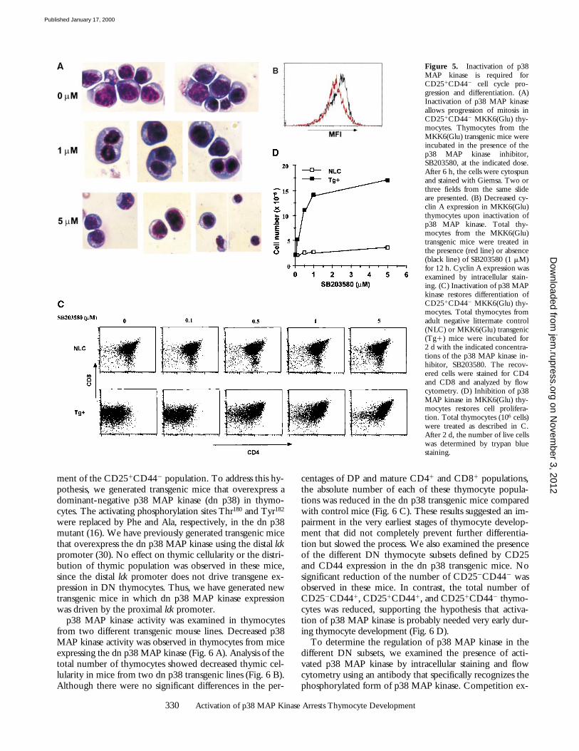
CD442 thymocytes (for a review, see reference 1). How-ever, it is unclear which of these processes, cell cycle pro-gression or differentiation, initiates the transition betweentwo differentiation stages. Our results indicated that activa-tion of p38 MAP kinase prevented cell cycle progressionand differentiation of CD251CD442 thymocytes. To ad-dress which of these two biological functions is the cause ofthe arrest of thymocyte development, we examined the ef-fect of p38 MAP kinase inhibition on thymocytes from theMKK6(Glu) transgenic mice. Thymocytes from adult MKK6(Glu) transgenic mice were incubated in vitro with me-dium alone or in the presence of different concentrations ofSB203580, a pyridinyl imidazol drug that selectively inhib-its p38 MAP kinase (18). After 6 h, the nuclear morphol-ogy of MKK6(Glu) thymocytes was examined by Giemsastaining. As shown above, in the absence of the inhibitor alarge proportion of these cells remained in prophase (Fig.5 A). However, after treatment with 1 mM SB203580, asignificant number of thymocytes in late anaphase/early te-lophase was observed (Fig. 5 A). Cells in late telophase andsmall cells in interphase were also detected upon incubationwith 5 mM SB203580 (Fig. 5 A). No SB203580 effect wasobserved in control thymocytes (data not shown). Down-regulation of cyclin A expression was also observed in theMKK6(Glu) thymocytes treated with SB203580 (Fig. 5 B).These results indicated that inhibition of p38 MAP kinaseis required for progression of mitosis in CD251CD442
thymocytes.We examined whether the presence of SB203580 allowed
further differentiation of CD251CD442 thymocytes. Nosignificant differences in the expression of differentiationcell markers were observed between 12 and 24 h of treat-ment with SB203580 (data not shown). However, after 2 dof treatment, immature CD251CD442 thymocytes differ-entiated into DP thymocytes in a dose-dependent manner(Fig. 5 C). The presence of SB203580 did not affect thedistribution of negative littermate control thymocytes (Fig.5 C). The differentiation of CD81CD4low thymocytes fromthe MKK6(Glu) transgenic mice into DP thymocytes inthe presence of SB203580 was associated with cell expan-sion (Fig. 5 D). Therefore, inactivation of p38 MAP kinasemust occur for CD251CD442 thymocytes to progressthrough mitosis, expand, and ultimately differentiate intoCD252CD442 and DP thymocytes.
Activation of the p38 MAP Kinase Pathway Is Important forthe Earliest Stages of Thymocyte Development. The presenceof high levels of p38 MAP kinase activity in immatureCD251CD442 thymocytes compared with the activity inCD252CD442 thymocytes (Fig. 3 F) suggested that activa-tion of p38 MAP kinase may be important for develop-
on Novem
ber 3, 2012jem
.rupress.orgD
ownloaded from
Published January 17, 2000
330 Activation of p38 MAP Kinase Arrests Thymocyte Development
ment of the CD251CD442 population. To address this hy-pothesis, we generated transgenic mice that overexpress adominant-negative p38 MAP kinase (dn p38) in thymo-cytes. The activating phosphorylation sites Thr180 and Tyr182
were replaced by Phe and Ala, respectively, in the dn p38mutant (16). We have previously generated transgenic micethat overexpress the dn p38 MAP kinase using the distal lckpromoter (30). No effect on thymic cellularity or the distri-bution of thymic population was observed in these mice,since the distal lck promoter does not drive transgene ex-pression in DN thymocytes. Thus, we have generated newtransgenic mice in which dn p38 MAP kinase expressionwas driven by the proximal lck promoter.
p38 MAP kinase activity was examined in thymocytesfrom two different transgenic mouse lines. Decreased p38MAP kinase activity was observed in thymocytes from miceexpressing the dn p38 MAP kinase (Fig. 6 A). Analysis of thetotal number of thymocytes showed decreased thymic cel-lularity in mice from two dn p38 transgenic lines (Fig. 6 B).Although there were no significant differences in the per-
centages of DP and mature CD41 and CD81 populations,the absolute number of each of these thymocyte popula-tions was reduced in the dn p38 transgenic mice comparedwith control mice (Fig. 6 C). These results suggested an im-pairment in the very earliest stages of thymocyte develop-ment that did not completely prevent further differentia-tion but slowed the process. We also examined the presenceof the different DN thymocyte subsets defined by CD25and CD44 expression in the dn p38 transgenic mice. Nosignificant reduction of the number of CD252CD442 wasobserved in these mice. In contrast, the total number ofCD252CD441, CD251CD441, and CD251CD442 thymo-cytes was reduced, supporting the hypothesis that activa-tion of p38 MAP kinase is probably needed very early dur-ing thymocyte development (Fig. 6 D).
To determine the regulation of p38 MAP kinase in thedifferent DN subsets, we examined the presence of acti-vated p38 MAP kinase by intracellular staining and flowcytometry using an antibody that specifically recognizes thephosphorylated form of p38 MAP kinase. Competition ex-
Figure 5. Inactivation of p38MAP kinase is required forCD251CD442 cell cycle pro-gression and differentiation. (A)Inactivation of p38 MAP kinaseallows progression of mitosis inCD251CD442 MKK6(Glu) thy-mocytes. Thymocytes from theMKK6(Glu) transgenic mice wereincubated in the presence of thep38 MAP kinase inhibitor,SB203580, at the indicated dose.After 6 h, the cells were cytospunand stained with Giemsa. Two orthree fields from the same slideare presented. (B) Decreased cy-clin A expression in MKK6(Glu)thymocytes upon inactivation ofp38 MAP kinase. Total thy-mocytes from the MKK6(Glu)transgenic mice were treated inthe presence (red line) or absence(black line) of SB203580 (1 mM)for 12 h. Cyclin A expression wasexamined by intracellular stain-ing. (C) Inactivation of p38 MAPkinase restores differentiation ofCD251CD442 MKK6(Glu) thy-mocytes. Total thymocytes fromadult negative littermate control(NLC) or MKK6(Glu) transgenic(Tg1) mice were incubated for2 d with the indicated concentra-tions of the p38 MAP kinase in-hibitor, SB203580. The recov-ered cells were stained for CD4and CD8 and analyzed by flowcytometry. (D) Inhibition of p38MAP kinase in MKK6(Glu) thy-mocytes restores cell prolifera-tion. Total thymocytes (106 cells)were treated as described in C.After 2 d, the number of live cellswas determined by trypan bluestaining.
on Novem
ber 3, 2012jem
.rupress.orgD
ownloaded from
Published January 17, 2000

331 Diehl et al.
periments using a phospho-p38 peptide demonstrated thespecificity of this antibody (data not shown). In correlationwith the increase of p38 MAP kinase activity (threefold)determined by the conventional immunocomplex kinaseassay (Fig. 3 F), intracellular staining and flow cytometryanalysis revealed increased levels (threefold) of activated(phosphorylated) p38 MAP kinase in DN CD251CD442
thymocytes compared with DN CD252CD442 thymo-cytes (Fig. 6 E). Interestingly, p38 MAP kinase activity wasdramatically augmented in the DN CD251CD441 popula-tion compared with DN CD252CD441 thymocytes (Fig.6 E), indicating that p38 MAP kinase is strictly regulatedduring the differentiation of DN thymocytes.
Together, our results indicate that activation of the p38MAP kinase pathway is important for differentiation and/orsurvival of immature CD252CD441 thymocytes into CD251
CD441 and CD251CD442 thymocytes, but that inactiva-tion of p38 MAP kinase is required to proceed from theCD251CD442 to the CD252CD442 stage (Fig. 6 E).
DiscussionReprogramming of gene expression patterns has been
implicated as a mechanism for the morphologic and func-tional commitment of cells to specific lineages during thedevelopment of T cells in the thymus. Transcription factorsand signaling pathways play critical roles in the control ofthe differentiation, proliferation, and death of thymocytes.Here, we have shown that the p38 MAP kinase pathway isvery strictly regulated during the early stages of thymocytedevelopment. The level of p38 MAP kinase activity is crit-ical for both the differentiation and expansion of immaturethymocytes.
Using the MKK6(Glu) transgenic mice, we have shownthat persistent activation of p38 MAP kinase in thymocytesin vivo results in a progressive increase of thymus size. Theenlarged thymus might theoretically be the result of a thy-moma induced by the expression of activated MKK6. Sev-eral lines of evidence presented in this study argue againstthis possibility. First, splenomegaly and lymphadenopathy
Figure 6. Strict regulation of the p38 MAP ki-nase pathway during immature thymocyte develop-ment. (A) Expression of dn p38 in thymocytes in-hibits endogenous p38 MAP kinase. Whole cellextracts of freshly isolated thymocytes from negativelittermate control (NLC) and dn p38 transgenic(TG1) mice from lines 2 and 10 were assayed forp38 MAP kinase activity using the substrate GST-ATF2. (B) Reduced total thymocyte number in thedn p38 transgenic mice. Thymocyte numbers fromlines 2 and 10 of dn p38 transgenic mice are shownas a percentage of the thymocyte number from neg-ative littermate control mice. Values represent theaverage percentage (n 5 4). (C) Reduced numbersof DP, DN, and single CD41 and CD81 thy-mocytes in the dn p38 transgenic mice (line 10).Total thymocytes were isolated from control and dnp38 transgenic mice, stained for CD4 and CD8, andexamined by flow cytometry. Values represent theaverage percentage of thymocyte number in eachpopulation versus the number of thymocytes of thecorresponding population in control mice (n 5 3).(D) Reduced number of DN thymocyte popula-tions in the dn p38 transgenic mice. Total thy-mocytes were isolated from negative littermate con-
trol and dn p38 transgenic mice, stained for CD4, CD8, CD25, and CD44, and examined by flow cytometry. Values represent the average of theabsolute number of thymocytes in each DN subpopulation, based on the total number of thymocytes (n 5 3). (E) Activation and inactivation of p38MAP kinase during earliest stage of thymocyte development. Total thymocytes from wild-type mice were stained for CD4, CD8, CD44, and CD25.Cell surface staining was followed by intracellular staining for the active form of p38 MAP kinase using an FITC–antiphospho-p38 MAP kinase mAb.Histograms (left) represent the presence of activated p38 MAP kinase in the different DN subpopulations (right) and DP cells. Vertical line represents thenegative control. Numbers express the mean fluorescence intensity of phospho-p38 MAP kinase positive cells. The results are representative of five inde-pendent experiments.
on Novem
ber 3, 2012jem
.rupress.orgD
ownloaded from
Published January 17, 2000

332 Activation of p38 MAP Kinase Arrests Thymocyte Development
are frequently associated with the development of thy-moma. However, the spleen and lymph nodes from thesemice are dramatically reduced in size and cell number anddo not contain T cells. No metastases are found in otherorgans of the MKK6(Glu) transgenic animals. Second, thy-mocytes from the MKK6(Glu) transgenic mice do not ex-hibit increased proliferation either in vivo or in vitro. Pro-liferation of MKK6(Glu) thymocytes in vitro is observedonly when p38 MAP kinase is inactivated by the presenceof a specific inhibitor, SB203580. Third, despite the in-creased size of the thymus, the total number of thymocytesis reduced in embryos and young MKK6(Glu) transgenicmice. Increased thymocyte cell number was observed onlyafter these mice reached 4–5 mo of age. The discrepancybetween the size of the thymus and the total number ofthymocytes can be explained by the difference in thy-mocyte size. Flow cytometry and histological analysis (Figs.3 A and 4 B) demonstrate that thymocytes from the MKK6(Glu) transgenic mice are larger than thymocytes from con-trol mice and would therefore occupy more space. Finally,analysis of the TCR repertoire by examining differentTCR Vb (Vb5, Vb6, Vb8, and Vb11) expression demon-strates that the thymocytes that accumulate in the MKK6(Glu) transgenic mice do not represent a monoclonal popu-lation (data not shown). Together, these observations makeit unlikely that the accumulation of thymocytes and progres-sive enlargement of the thymus in the MKK6(Glu) trans-genic mice are the result of development of a thymoma.
Instead, our studies demonstrate that the persistent acti-vation of the p38 MAP kinase pathway arrests the differen-tiation of immature thymocytes early in development. Sev-eral lines of evidence confirm the immature phenotype ofthe CD81CD4low population present in the thymus of theMKK6(Glu) transgenic mice. First, the high level of HSAand low level of CD69 and TCR-b expression detected onthymocytes from the MKK6(Glu) transgenic mice arecharacteristic of immature thymocytes (Fig. 3 D). Second,MKK6(Glu) CD81CD4low thymocytes display the CD251
CD442 phenotype characteristic of a transient stage of im-mature thymocytes before differentiation into DP thy-mocytes (Fig. 3 C). Most importantly, we have demonstratedthat the arrest of thymocyte differentiation in the MKK6(Glu) transgenic mice occurs during fetal development atapproximately E16–17 (Fig. 3 E). Finally, inhibition of p38MAP kinase in vitro restores differentiation of the MKK6(Glu) CD81CD4lowCD251CD442 thymocytes into DP thy-mocytes (Fig. 5 C).
Recently, Sugawara et al. (8) have shown that the p38MAP kinase inhibitor SB203580 inhibits deletion of DPthymocytes by anti-CD3 mAb in fetal thymocyte develop-ment organ culture (FTOC) in vitro. However, they alsoshow that the presence of SB203580 results in a decreasedpercentage of DP thymocytes and increased percentage ofCD81, CD41, and DN thymocytes in FTOC (8). Thus, theinterpretation of these results is unclear. A differential effectof SB203580 in stromal cells and thymocytes may explainthese apparently contradictory results. In the same study, itwas suggested that the expression of MKK6 induces nega-
tive selection of DP thymocytes. However, the presence ofthe CD81CD4low population, increased percentage of DNthymocytes, and absence of CD41 thymocytes in wild-typeMKK6 retrovirus-infected FTOC (8) do not support thismodel. In contrast, these results suggest an inhibition of thedifferentiation of immature fetal thymocytes into DP thy-mocytes by the expression of wild-type MKK6. Thus, theexpression of wild-type MKK6 in FTOC in vitro maymimic the phenotype obtained in mice expressing constitu-tively activated MKK6 in vivo.
The E and L subsets have been defined within the DNCD251CD442 population in the adult thymus based on theirsize, TCR-b expression, and cell cycle status (28). Differentia-tion of thymocyte development is partially or completely in-hibited at the CD251CD442 E stage (normal size thymocytes)in mice deficient for components of the pre-TCR complex(e.g., pre-Ta, TCR-b, CD3e, RAG, T cell factor [TCF]/lymphoid enhancer-binding factor [LEF]–deficient mice) orthe pre-TCR signaling pathways (e.g., lck, ZAP-70, SH2 do-main–containing leukocyte phosphoprotein of 76 kD [SLP-76]–deficient mice) (1, 31–36). Thus, pre-TCR–mediatedsignals appear to be critical for the progression from E to Lstage. In our study, we show that activation of the p38 MAPkinase pathway does not prevent the expression of the TCR-band progression into the L stage. However, a persistent activa-tion of p38 MAP kinase completely arrests the differentiationof CD251CD442 L thymocytes into CD252CD442 and DPthymocytes. Therefore, endogenous p38 MAP kinase must beinactivated before differentiation to the CD252CD442 stage.The analysis of p38 MAP kinase activity in these populationsconfirms this model (Figs. 3 F and 6 E). The p38 MAP kinasepathway therefore is the first signaling pathway that has beendescribed to play a critical role at the CD251CD442 L check-point during thymocyte development.
The specific nuclear morphology (prophase), large size,and low rate of proliferation in CD251CD442 thymocytesfrom the MKK6(Glu) transgenic mice in vivo indicate thatMKK6(Glu) CD251CD442 thymocytes are arrested in mito-sis. Moreover, progression of MKK6(Glu) thymocytes towardstelophase, cytokinesis, and G1 phases is restored upon inactiva-tion of p38 MAP kinase in the presence of SB203580. There-fore, activation of p38 MAP kinase in vivo prevents progres-sion through the late phases of mitosis in CD251CD442
thymocytes. The study by Takenaka et al. (29) showing thatmicroinjection of activated p38 MAP kinase into blastomeresof Xenopus embryos causes miotic arrest supports our results inimmature thymocytes. Recently, it has been proposed that ac-tivation of the p38 MAP kinase pathway can inhibit progres-sion of both G1 and mitosis in NIH-3T3 fibroblasts (37).
The inhibition of cell cycle progression of CD251CD442
thymocytes from the MKK6(Glu) transgenic mice appearscontradictory to the increased thymocyte number observedin 5–6-mo-old MKK6(Glu) transgenic mice. Althoughonly a few cells (50–100/d) from the bone marrow repop-ulate the thymus in control mice (38), these immature thy-mocytes (predominantly CD252CD441 thymocytes) canproliferate during their differentiation into CD251CD441
and CD251CD442 thymocytes. The major expansion of cell
on Novem
ber 3, 2012jem
.rupress.orgD
ownloaded from
Published January 17, 2000

333 Diehl et al.
numbers appears to occur during the differentiation ofCD251CD442 into CD252CD442 thymocytes. The con-tinuous entry of a new cohort of precursor cells into thethymus and the differentiation and expansion of CD252
CD441 and CD251CD441 thymocytes can explain the slowbut progressive accumulation of thymocytes with time inthe MKK6(Glu) transgenic mice. Although the number offetal thymocytes in E15 MKK6(Glu) embryos was similarto the number in age-matched control embryos, four- tofivefold fewer thymocytes were found in E19 MKK6(Glu)embryos compared with control embryos. Only 1.5–2-foldfewer cells were found in 2–3-mo-old MKK6(Glu) trans-genic mice, and 2–3-fold more thymocytes were found in5–6-mo MKK6(Glu) mice compared with age-matchedcontrol mice.
Proinflammatory cytokines (e.g., TNF-a and IL-1), he-matopoietic factors (e.g., GM-CSF and IL-3), and environ-mental stress can induce p38 MAP kinase activity in variouscell types (15–20). Interestingly, in correlation with the el-evated p38 MAP kinase activity in the CD251CD441 thy-mocytes, it has been shown that TNF-a and IL-1 producedby stromal cells can stimulate the expression of surface CD25on immature CD252CD441 thymocytes. Furthermore, thesetwo cytokines are required for the differentiation of imma-ture fetal thymocytes into DP thymocytes (39). It is possi-ble that p38 MAP kinase activity in immature thymocytesmay be regulated by stromal cell–derived cytokines.
Previous studies have demonstrated the involvement ofthe p38 MAP kinase pathway in proliferation and celldeath. Our studies show the importance of the p38 MAPkinase pathway in the control of differentiation and cell cy-cle progression of CD251CD442 thymocytes in vivo. Pro-liferation is closely associated with differentiation duringthymocyte development, and it remains unclear which ofthese processes is the initial event in each developmentalstep. We have demonstrated that the inhibition of p38 MAPkinase in MKK6(Glu) thymocytes in vitro rapidly (severalhours) restores cell cycle progression, whereas a longer pe-riod of time (24–48 h) is required to observe significantchanges on the cell surface phenotype. Therefore, we pro-pose that inhibition of cell cycle progression by the p38MAP kinase prevents further differentiation of immatureCD251CD442 thymocytes.
Our study demonstrates that p38 MAP kinase is strictlyregulated during the early stages of thymocyte develop-ment. Although an initial activation of p38 MAP kinase ap-pears to be important for thymic cellularity, persistent acti-vation of this pathway completely blocks the generation ofT cells and results in T cell immunodeficiency. The regula-tion of the p38 MAP kinase pathway is critical in thy-mocyte development and generation of mature T cells.
We thank C. Hughes and D. Butkus for generating the MKK6(Glu)transgenic mice; C. Irvin and D. Schneider for the diagnosis of thecause of mouse death; M.S.-S. Su (Vertex Pharmaceuticals, Cambridge,MA) for kindly providing SB203580; and R. Perlmutter (University ofWashington, Seattle, WA) for the proximal lck promoter construct.
This work was supported in part by National Institutes of Healthgrant R29 AI42138 (to M. Rincón), and National Cancer Insti-
tute grants CA58396 and CA72009 (to R.J. Davis). R.J. Davisand R.A. Flavell are Investigators of the Howard Hughes MedicalInstitute.
Submitted: 3 August 1999Revised: 28 October 1999Accepted: 2 November 1999
References1. Rodewald, H.-R., and H.J. Fehling. 1998. Molecular and cel-
lular events in early thymocyte development. Adv. Immmunol.69:1–113.
2. Glimcher, L.H., and H. Singh. 1999. Transcription factors inlymphocyte development—T and B cells get together. Cell.96:13–23.
3. Clevers, H., and P. Ferrier. 1998. Transcriptional controlduring T-cell development. Curr. Opin. Immunol. 10:166–171.
4. Killeen, N., B.A. Irving, S. Pippig, and K. Zingler. 1998.Signaling checkpoints during the development of T lympho-cytes. Curr. Opin. Immunol. 10:360–367.
5. Ip, Y.T., and R.J. Davis. 1998. Signal transduction by thec-Jun N-terminal kinase (JNK)—from inflammation to de-velopment. Curr. Opin. Cell Biol. 10:205–219.
6. Alberola-Ila, J., K.A. Forbush, R. Seger, E.G. Krebs, andR.M. Perlmutter. 1995. Selective requirement for MAP kinaseactivation in thymocyte differentiation. Nature. 373:620–623.
7. Rincón, M., A. Whitmarsh, D.D. Yang, L. Weiss, B. Déri-jard, P. Jayaraj, R.J. Davis, and R.A. Flavell. 1998. The JNKpathway regulates the in vivo deletion of immatureCD41CD81 thymocytes. J. Exp. Med. 188:1817–1830.
8. Sugawara, T., T. Moriguchi, E. Nishida, and Y. Takahama.1998. Differential roles of ERK and p38 MAP kinase path-ways in positive and negative selection of T lymphocytes. Im-munity. 9:565–574.
9. Crompton, T., K.C. Gilmour, and M.J. Owen. 1996. TheMAP kinase pathway controls differentiation from double-negative to double-positive thymocyte. Cell. 86:243–251.
10. Dérijard, B., J. Rainjeaud, T. Barret, I.-H. Wu, J. Han, R.J.Ulevitch, and R.J. Davis. 1995. Independent human MAPkinase signal transduction pathways defined by MEK andMKK isoforms. Science. 267:683–685.
11. Han, J., J.-D. Lee, Y. Jiang, Z. Li, L. Feng, and R.J. Ulevitch.1996. Characterization of the structure and function of a novelMAP kinases kinase (MKK6). J. Biol. Chem. 271:2886–2891.
12. Moriguchi, T., F. Toyoshima, Y. Gotoh, A. Iwamatsu, K.Irie, E. Mori, N. Kuroyanagi, M. Hagiwara, K. Matsumoto,and E. Nishida. 1996. Purification and identification of a ma-jor activator for p38 from osmotically shocked cells. Activa-tion of mitogen-activated protein kinase kinase 6 by osmoticshock, tumor necrosis factor-a and H2O2. J. Biol. Chem. 271:26981–26988.
13. Raingeaud, J., A.J. Whitmarsh, T. Barret, B. Dérijard, and R.J.Davis. 1996. MKK3- and MKK6-regulated gene expression ismediated by the p38 mitogen-activated protein kinase signaltransduction pathway. Mol. Cell. Biol. 16:1247–1255.
14. Sen, J., R. Kapeller, R. Fragoso, R. Sen, L.I. Zon, and S.J.Burakoff. 1996. Intrathymic signals in thymocytes are medi-ated by p38 mitogen-activated protein kinase. J. Immunol.156:4535–4538.
15. Freshney, N.W., L. Rawlinson, F. Guesdon, E. Jones, S.Cowley, J. Hsuan, and J. Saklatvala. 1994. Interleukin-1 acti-vates a novel protein kinase cascade that results in the phos-
on Novem
ber 3, 2012jem
.rupress.orgD
ownloaded from
Published January 17, 2000

334 Activation of p38 MAP Kinase Arrests Thymocyte Development
phorylation of the Hsp27. Cell. 78:1039–1049.16. Raingeaud, J., S. Gupta, J. Roger, M. Dickens, J. Han, R.J.
Ulevitch, and R.J. Davis. 1995. Pro-inflammatory cytokinesand environmental stress cause p38 MAP kinase activation bydual phosphorylation on tyrosine and threonine. J. Biol.Chem. 270:7420–7426.
17. Han, J., J.-D. Lee, L. Bibbs, and R.J. Ulevitch. 1994. A MAPkinase targeted by endotoxin and hyperosmolarity in mam-malian cells. Science. 265:808–811.
18. Lee, J.C., T. Laydon, P.C. McDonnell, T.F. Gallagher, S.Kumar, D. Gree, D. McNulty, M.J. Blumenthal, J.R. Heys,S.W. Landvatter, et al. 1994. A protein kinase involved in theregulation of inflammatory cytokine biosynthesis. Nature.372:739–746.
19. Rouse, J., P. Cohen, S. Trigon, M. Morange, A. Alonso-Llamazares, D. Zamanillo, T. Hunt, and A.R. Nebreda.1994. A novel kinase cascade triggered by stress and heatshock that stimulates MAPKAP kinase-2 and phosphoryla-tion of the small heat shock proteins. Cell. 78:1027–1037.
20. Foltz, I.N., J.C. Lee, P.R. Young, and J.W. Schrader. 1997.Hemopoietic growth factors with the exception of interleu-kin-4 activate the p38 mitogen-activated protein kinase path-way. J. Biol. Chem. 272:3296–3301.
21. Wildin, R.S., A.M. Garvin, S. Pawar, D.B. Lewis, K.M.Abraham, K.A. Forbush, S.F. Ziegler, J.M. Allen, and R.M.Perlmutter. 1991. Developmental regulation of lck gene ex-pression in T lymphocytes. J. Exp. Med. 173:383–393.
22. Swan, K.A., J. Alberola-Ila, J.A. Gross, M.W. Appleby, K.A.Forbush, J.F. Thomas, and R.M. Perlmutter. 1995. Involve-ment of p21ras distinguishes positive and negative selection inthymocytes. EMBO (Eur. Mol. Biol. Organ.) J. 14:276–285.
23. Hogan, B., F. Constantini, and E. Lacy. 1986. Manipulatingthe Mouse Embryo. Cold Spring Harbor Laboratory, ColdSpring Harbor, NY.
24. Cibotti, R., J.A. Punk, K.S. Dash, S.O. Sharrow, and A.Singer. 1997. Surface molecules that drive T cell develop-ment in vitro in the absence of thymic epithelium and in theabsence of lineage-specific signals. Immunity. 6:245–255.
25. Lucas, B., F. Vasseur, and C. Penit. 1995. Stochastic corecep-tor shut-off is restricted to the CD4 lineage maturation path-way. J. Exp. Med. 181:1623–1633.
26. Rincón, M., B. Dérijard, C.-W. Chow, R.J. Davis, andR.A. Flavell. 1997. Reprogramming the signaling require-ment for AP-1 (activator protein-1) activation during differ-entiation of precursor CD41 T cells to effector Th1 and Th2cells. Genes Funct. 1:51–68.
27. Dérijard, B., M. Hibi, I.-H. Wu, T. Barret, B. Su, T. Deng,M. Karin, and R.J. Davis. 1994. JNK1: a protein kinase stim-
ulated by UV light and Ha-Ras that binds and phosphorylatesthe c-Jun activation domain. Cell. 76:1025–1037.
28. Hoffman, E.S., L. Passoni, T. Crompton, T.M.J. Leu, D.G.Schatz, A. Koff, M. Owen, and A.C. Hayday. 1996. Produc-tive T-cell receptor b-chain gene rearrangement: coincidentregulation of cell cycle and clonality during development invivo. Genes Dev. 10:948–962.
29. Takenaka, K., T. Moriguchi, and E. Nishida. 1998. Activa-tion of the protein kinase p38 in the spindle assembly check-point and mitotic arrest. Science. 280:599–602.
30. Rincón, M., H. Enslen, J. Raingeaud, M. Recht, T. Zapton,M.S.-S. Su, L.A. Penix, R.J. Davis, and R.A. Flavell. 1998.Interferon-g expression by Th1 effector T cells mediated bythe p38 MAP kinase signaling pathway. EMBO (Eur. Mol.Biol. Organ.) J. 17:2817–2829.
31. Brickner, A.G., D.L. Gossage, M.R. Dusing, and D.A. Wigin-ton. 1995. Identification of a murine homolog of the humanadenosine deaminase thymic enhancer. Gene. 167:261–266.
32. Oosterwegel, M.A., M. van de Wetering, D. Dooijes, L.Klomp, A. Winoto, K. Georgopoulos, F. Meijlink, and H.Clevers. 1991. Cloning of murine TCF-1, a T cell–specifictranscription factor interacting with functional motifs in theCD3e and T cell receptor a enhancers. J. Exp. Med. 173:1133–1142.
33. Sawada, S., and D.R. Littman. 1991. Identification and char-acterization of a T-cell specific enhancer adjacent to the mu-rine CD4 gene. Mol. Cell. Biol. 11:5027–5035.
34. Travis, A., A. Amsterdam, C. Belanger, and R. Grosschedl.1991. LEF-1, a gene encoding a lymphoid-specific proteinwith an HMG domain, regulates T-cell receptor a enhancerfunction. Genes Dev. 5:880–894.
35. van de Wetering, M., J. Gastrop, V. Korinek, and H. Clev-ers. 1996. Extensive alternative splicing and dual promoterusage generate Tcf-1 protein isoforms with differential tran-scription control properties. Mol. Cell. Biol. 16:745–752.
36. Waterman, M.L., W.H. Fischer, and K.A. Jones. 1991. A thy-mus specific member of the HMG protein family regulates thehuman T-cell receptor a enhancer. Genes Dev. 5:656–669.
37. Ellinger-Ziegelbauer, H. 1999. Cell cycle arrest and reversionof Ras-induced transformation by a conditionally activatedform of mitogen-activated protein kinase kinase kinase 3.Mol. Cell. Biol. 19:3857–3868.
38. Scollay, R., J. Smith, and V. Stauffer. 1986. Dynamics ofearly T cells: prothymocyte migration and proliferation in theadult mouse thymus. Immunol. Rev. 91:129–157.
39. Züñiga-Pflucker, J.C., J. Di, and M.J. Lenardo. 1998. Re-quirement for TNFa and IL-1a in fetal thymocyte commit-ment and differentiation. Science. 268:1906–1909.
on Novem
ber 3, 2012jem
.rupress.orgD
ownloaded from
Published January 17, 2000
Copyright © 2022 FDOKUMEN