Trophic Factor Withdrawal: p38 Mitogen-Activated Protein Kinase Activates NHE1, Which Induces...
13
MOLECULAR AND CELLULAR BIOLOGY, 0270-7306/01/$04.000 DOI: 10.1128/MCB.21.22.7545–7557.2001 Nov. 2001, p. 7545–7557 Vol. 21, No. 22 Copyright © 2001, American Society for Microbiology. All Rights Reserved. Trophic Factor Withdrawal: p38 Mitogen-Activated Protein Kinase Activates NHE1, Which Induces Intracellular Alkalinization ANNETTE R. KHALED, 1 ANDREA N. MOOR, 2 AIQUN LI, 3 KYUNGJAE KIM, 1 DOUGLAS K. FERRIS, 4 KATHRIN MUEGGE, 1 ROBERT J. FISHER, 3 LARRY FLIEGEL, 2 AND SCOTT K. DURUM 1 * Laboratory of Molecular Immunoregulation, 1 Intramural Research Support Program, SAIC Frederick, 3 and Laboratory of Leukocyte Biology, 4 Center for Cancer Research, National Cancer Institute-Frederick Cancer Research and Development Center, Frederick, Maryland 21702, and Department of Biochemistry, University of Alberta, Edmonton, Alberta, Canada 2 Received 28 February 2001/Returned for modification 27 April 2001/Accepted 6 August 2001 Trophic factor withdrawal induces cell death by mechanisms that are incompletely understood. Previously we reported that withdrawal of interleukin-7 (IL-7) or IL-3 produced a rapid intracellular alkalinization, disrupting mitochondrial metabolism and activating the death protein Bax. We now observe that this novel alkalinization pathway is mediated by the pH regulator NHE1, as shown by the requirement for sodium, blocking by pharmacological inhibitors or use of an NHE1-deficient cell line, and the altered phosphorylation of NHE1. Alkalinization also required the stress-activated p38 mitogen-activated protein kinase (MAPK). Inhibition of p38 MAPK activity with pharmacological inhibitors or expression of a dominant negative kinase prevented alkalinization. Activated p38 MAPK directly phosphorylated the C terminus of NHE1 within a 40-amino-acid region. Analysis by mass spectroscopy identified four phosphorylation sites on NHE1, Thr 717, Ser 722, Ser 725, and Ser 728. Thus, loss of trophic cytokine signaling induced the p38 MAPK pathway, which phosphorylated NHE1 at specific sites, inducing intracellular alkalinization. The requirement for cytokines in hematopoiesis is partly attributable to a trophic activity, the protection of cells from programmed cell death (17, 44). Interleukin-7 (IL-7), a prod- uct of the thymic epithelium, protects lymphocyte progenitor cells from apoptotic death during T-cell development (19, 55). Survival of pro-T cells isolated from the thymus requires the presence of IL-7 (25), whereas disruption of the gene for IL-7 (51) or its receptor (35) dramatically reduces thymic cellularity (29). IL-7 also has trophic activities on cells of the developing brain (30). IL-3 has similar trophic activities on early hemato- poietic precursors of the myeloid, lymphoid, and erythoid lin- eages. In IL3-dependent cell lines, withdrawal leads to apo- ptotic cell death (21). The trophic action of cytokines has been partly attributed to the Bcl-2 family of proteins, which are important intracellular regulators of apoptosis (25, 37). Overexpression of the anti- apoptotic protein Bcl-2 in IL-7R / mice partially restored T-cell numbers (1), but complete restoration of a normal phe- notype was not achieved (8, 13). Recent studies with IL-3- dependent cell lines have shown that the up-regulation of Bcl-2 and Bcl-X L and the down-regulation of proapoptotic proteins such as Bad are involved in survival (21); however, as with IL7, the trophic action of IL-3 involves more than the balance of Bcl-2 family members, since the overexpression of Bcl-2 ex- tended life for only 1 day following IL-3 withdrawal (32). The intracellular signaling pathways triggered by cytokine receptor engagement are yet to be fully defined. However, it is known that hematopoietic cytokine receptors can induce dis- tinct members of the mitogen-activated protein kinase (MAPK) family, contributing to the processes of proliferation and sur- vival (10, 11; E. Rajnavolgyi, N. Benbernou, K. Muegge, and S. K. Durum, unpublished data [for similar results with IL-7]). Three of the MAPK signaling units have been characterized in detail: the extracellular signal-regulated kinases (ERKs), the c-Jun amino-terminal kinases (JNK) or stress-activated protein kinases (SAPK), and the p38 MAPKs (p38) (20). Mitogens, inflammatory cytokines, and growth factors are known to activate various MAPK signaling pathways, whereas cellular stresses such as UV light, heat, or osmotic shock selectively induce the JNK/SAPK and p38 MAPK pathways. A common feature of all the MAPKs is that they are activated by the phosphorylation of both threonine and tyrosine residues by a dual-specificity serine-threonine MAPK (18). In turn, MAPKs frequently phosphorylate their substrates at serine or thre- onine residues adjacent to prolines (15, 28). Although the MAPKs have been implicated in the regulation of apoptotic death, for example following nerve growth factor withdrawal (26, 59), the spontaneous apoptosis of neutrophils (2), and loss of IL-7 signaling in a dependent cell line (Rajnavolgyi et al., unpublished) it is not known how these MAPKs contribute to the apoptotic process and their relevant substrates have not been identified. The dysregulation of intracellular pH is part of the trigger for apoptotic cell death following cytokine withdrawal (23, 24) or ceramide treatment (3). Control of cytosolic pH in eukary- otic cells involves various proton pumps, proton channels, and ion transporters that drive H or H equivalents and HCO 3 ions into and out of the cell. Among the better-characterized ion transporters or exchangers are the Na /H exchanger (NHE), which has multiple isoforms with NHE1 being the most prevalent, the Na -dependent and Na -independent HCO 3 transporters or anion exchangers (AE), the CL /OH exchanger (CHE), and a lactate-proton cotransporter (36). * Corresponding author. Mailing address: Section of Cytokines and Immunity, National Cancer Institute, Bldg. 560, Rm. 31-71, Frederick, MD 21702-1201. Phone: (301) 846-1545. Fax: (301) 846-6720. E-mail: [email protected]. 7545
Transcript of Trophic Factor Withdrawal: p38 Mitogen-Activated Protein Kinase Activates NHE1, Which Induces...

MOLECULAR AND CELLULAR BIOLOGY,0270-7306/01/$04.00�0 DOI: 10.1128/MCB.21.22.7545–7557.2001
Nov. 2001, p. 7545–7557 Vol. 21, No. 22
Copyright © 2001, American Society for Microbiology. All Rights Reserved.
Trophic Factor Withdrawal: p38 Mitogen-Activated Protein KinaseActivates NHE1, Which Induces Intracellular Alkalinization
ANNETTE R. KHALED,1 ANDREA N. MOOR,2 AIQUN LI,3 KYUNGJAE KIM,1 DOUGLAS K. FERRIS,4
KATHRIN MUEGGE,1 ROBERT J. FISHER,3 LARRY FLIEGEL,2 AND SCOTT K. DURUM1*
Laboratory of Molecular Immunoregulation,1 Intramural Research Support Program, SAIC Frederick,3 and Laboratoryof Leukocyte Biology,4 Center for Cancer Research, National Cancer Institute-Frederick Cancer Research
and Development Center, Frederick, Maryland 21702, and Department of Biochemistry,University of Alberta, Edmonton, Alberta, Canada2
Received 28 February 2001/Returned for modification 27 April 2001/Accepted 6 August 2001
Trophic factor withdrawal induces cell death by mechanisms that are incompletely understood. Previouslywe reported that withdrawal of interleukin-7 (IL-7) or IL-3 produced a rapid intracellular alkalinization,disrupting mitochondrial metabolism and activating the death protein Bax. We now observe that this novelalkalinization pathway is mediated by the pH regulator NHE1, as shown by the requirement for sodium,blocking by pharmacological inhibitors or use of an NHE1-deficient cell line, and the altered phosphorylationof NHE1. Alkalinization also required the stress-activated p38 mitogen-activated protein kinase (MAPK).Inhibition of p38 MAPK activity with pharmacological inhibitors or expression of a dominant negative kinaseprevented alkalinization. Activated p38 MAPK directly phosphorylated the C terminus of NHE1 within a40-amino-acid region. Analysis by mass spectroscopy identified four phosphorylation sites on NHE1, Thr 717,Ser 722, Ser 725, and Ser 728. Thus, loss of trophic cytokine signaling induced the p38 MAPK pathway, whichphosphorylated NHE1 at specific sites, inducing intracellular alkalinization.
The requirement for cytokines in hematopoiesis is partlyattributable to a trophic activity, the protection of cells fromprogrammed cell death (17, 44). Interleukin-7 (IL-7), a prod-uct of the thymic epithelium, protects lymphocyte progenitorcells from apoptotic death during T-cell development (19, 55).Survival of pro-T cells isolated from the thymus requires thepresence of IL-7 (25), whereas disruption of the gene for IL-7(51) or its receptor (35) dramatically reduces thymic cellularity(29). IL-7 also has trophic activities on cells of the developingbrain (30). IL-3 has similar trophic activities on early hemato-poietic precursors of the myeloid, lymphoid, and erythoid lin-eages. In IL3-dependent cell lines, withdrawal leads to apo-ptotic cell death (21).
The trophic action of cytokines has been partly attributed tothe Bcl-2 family of proteins, which are important intracellularregulators of apoptosis (25, 37). Overexpression of the anti-apoptotic protein Bcl-2 in IL-7R��/� mice partially restoredT-cell numbers (1), but complete restoration of a normal phe-notype was not achieved (8, 13). Recent studies with IL-3-dependent cell lines have shown that the up-regulation of Bcl-2and Bcl-XL and the down-regulation of proapoptotic proteinssuch as Bad are involved in survival (21); however, as with IL7,the trophic action of IL-3 involves more than the balance ofBcl-2 family members, since the overexpression of Bcl-2 ex-tended life for only 1 day following IL-3 withdrawal (32).
The intracellular signaling pathways triggered by cytokinereceptor engagement are yet to be fully defined. However, it isknown that hematopoietic cytokine receptors can induce dis-tinct members of the mitogen-activated protein kinase (MAPK)
family, contributing to the processes of proliferation and sur-vival (10, 11; E. Rajnavolgyi, N. Benbernou, K. Muegge, andS. K. Durum, unpublished data [for similar results with IL-7]).Three of the MAPK signaling units have been characterized indetail: the extracellular signal-regulated kinases (ERKs), thec-Jun amino-terminal kinases (JNK) or stress-activated proteinkinases (SAPK), and the p38 MAPKs (p38) (20). Mitogens,inflammatory cytokines, and growth factors are known toactivate various MAPK signaling pathways, whereas cellularstresses such as UV light, heat, or osmotic shock selectivelyinduce the JNK/SAPK and p38 MAPK pathways. A commonfeature of all the MAPKs is that they are activated by thephosphorylation of both threonine and tyrosine residues by adual-specificity serine-threonine MAPK (18). In turn, MAPKsfrequently phosphorylate their substrates at serine or thre-onine residues adjacent to prolines (15, 28). Although theMAPKs have been implicated in the regulation of apoptoticdeath, for example following nerve growth factor withdrawal(26, 59), the spontaneous apoptosis of neutrophils (2), and lossof IL-7 signaling in a dependent cell line (Rajnavolgyi et al.,unpublished) it is not known how these MAPKs contribute tothe apoptotic process and their relevant substrates have notbeen identified.
The dysregulation of intracellular pH is part of the triggerfor apoptotic cell death following cytokine withdrawal (23, 24)or ceramide treatment (3). Control of cytosolic pH in eukary-otic cells involves various proton pumps, proton channels, andion transporters that drive H� or H� equivalents and HCO3
�
ions into and out of the cell. Among the better-characterizedion transporters or exchangers are the Na�/H� exchanger(NHE), which has multiple isoforms with NHE1 being themost prevalent, the Na�-dependent and Na�-independentHCO3
� transporters or anion exchangers (AE), the CL�/OH�
exchanger (CHE), and a lactate-proton cotransporter (36).
* Corresponding author. Mailing address: Section of Cytokines andImmunity, National Cancer Institute, Bldg. 560, Rm. 31-71, Frederick,MD 21702-1201. Phone: (301) 846-1545. Fax: (301) 846-6720. E-mail:[email protected].
7545

Through the use of specific inhibitors, the roles of these com-plex membrane proteins have been examined; however, muchremains to be understood regarding the mechanisms of theiraction and regulation.
To dissect the complex physiological events that occur ontrophic factor withdrawal, two cytokine-dependent cell lines,one requiring IL-7 and one requiring IL-3, were used. Ourprevious studies showed that following cytokine withdrawal,a novel intracellular alkalinization occurred and triggered adeath pathway involving mitochondrial translocation of theproapoptotic protein Bax (23) and inhibition of mitochondrialADP transport, ATP synthesis, and subsequent mitochondrialbioenergetics (24). In the present study we examine the mech-anism of this cytokine withdrawal-induced alkalinization andshow that it is caused by a specific membrane ion exchanger,which in turn is phosphorylated by a stress-activated MAPK.
MATERIALS AND METHODS
Cells and reagents. The IL-3-dependent murine pro-B-cell line FL5.12A (akind gift from James A. McCubrey, East Carolina University, Greenville, S.C.)was maintained in RPMI 1640 supplemented with 10% fetal bovine serum,2-�-mercaptoethanol (1,000�; Life Technologies), and 0.4 ng of recombinantmouse IL-3 (PeproTech Inc.) per ml. The IL-7-dependent cell line D1 wasestablished from the CD4� CD8� subset sorted from p53�/� mouse thymocytesinitially propagated in IL-7 and stem cell factor and maintained as previouslydescribed (23). Chinese hamster ovary cells (CHO) and the NHE�/�-deficientCHO cells (39) were maintained in Dulbecco Modified Eagle Medium DMEM(Life Technologies) with 10% fetal bovine serum.
FL5.12A cells were transiently transfected by electroporation (0.310 V, 960 �Fcapacitance) using a Bio-Rad GenePulser. Prior to pulsing, 5 � 107 cells in0.4-cm cuvettes (Bio-Rad) were preincubated for 10 min with either 40 �g ofpEGFP-C3 (Clontech) or 20 �g of pEGFP-C3 and 20 �g of pCMV-Flag-p38(agf) (a kind gift from Roger Davis, University of Massachusetts Medical Center,Worcester, Mass.). To select for cells transfected with plasmid DNA, greenfluorescent protein-containing (GFP�) cells were first sorted by fluorescence-activated cell sorting, and cell populations containing at least 98% GFP� cellswere cultured with or without IL-3 for 48 h (after which detection of GFPfluorescence declined). Viability and cell number were evaluated by trypan bluestaining (Life Technologies). For detection of DNA fragmentation, cells werestained with a 2-�g/ml solution of propidium iodide in a detergent buffer (48)and analyzed by flow cytometry. CHO cells were transfected with 3 �g ofpCMV-Flag-p38 (agf) using Fugene 6 (Roche) as specified by the manufacturer.Transfection efficiencies averaged 90%.
To measure intracellular pH, BCECF-AM (Molecular Probes) was used at1 �M. Cells were treated as described in the figure legends with the followingreagents: 1 mM ouabain (Sigma), 10 �g of cycloheximide (Sigma) per ml, 1 �gof antimycin A (Sigma) per ml, 200 �M 5-(N,N-dimethyl)amiloride (DMA)(Sigma), 200 �M 5-(N-ethyl-N-isopropyl)amiloride (EIPA) (Sigma), 1,000 and200 �M 4,4�(diisothiocyanato)stilbene-2-2�-disulfonic acid (DIDS) (Sigma),1,000 and 200 �M 4-acetamido-4�-isothiocyanato-stilbene-2-2�-disulfonic acid(SITS) (Sigma), 100 �g of anisomycin (Sigma) per ml, 20 �M PD169316, 20 �MSB202190, 20 �M SB203580, 20 �M SB202474, and 20 �M PD98059 (all fromCalbiochem). The NaCl buffer contained 140 mM NaCl, 1 mM MgCl2, 1 mMCaCl2, 1 mM KCl, 10 mM glucose, and 20 mM HEPES. For the buffer lackingNa�, 140 mM choline chloride replaced NaCl. Bicarbonate-free RPMI 1640(Sigma) was used in the experiments requiring a solution lacking HCO3
� ions.Measurement of intracellular pH. FL5.12A or D1 cells (106 cells/ml) deprived
of IL-3 or IL-7, respectively, for various times were treated, after being washedin a HEPES buffer (25 mM HEPES, 140 mM NaCl, 5 mM KCl, 0.8 mM MgCl2,1.8 mM CaCl2, 5.0 mM glucose), with 1 �M BCECF-AM for 30 min at 37°C, andintracellular pH was determined by flow cytometry with excitation set at 488 nmand emissions filtered through 525-nm band pass (green fluorescence) and610-nm long-pass (red fluorescence) fitters (16). Dead cells (less than 10%) wereexcluded by forward- and side-scatter gating. A pH calibration curve was gen-erated by preloading cells with 1 �M BCECF-AM for 30 min followed byincubation for 30 min in a high-K� HEPES buffer (25 mM HEPES, 145 mM KCl,0.8 mM MgCl2, 1.8 mM CaCl2, 5.5 mM glucose) at different pH values from 6.8to 8.0 in the presence of the permeabilizing agent nigericin (10 �M) (23). Afterincubation, nigericin was absorbed with excess bovine serum albumin.
Detection of MAPKs by Western blot analyses. Whole-cell lysates were madeby lysing cells (107) in a modified RIPA buffer containing phosphatase inhibitors(150 mM NaCl, 80 mM NaF, 50 mM Tris-HCl, 5 mM EDTA, 1 mM EGTA, 25mM sodium pyrophosphate, 1 mM sodium orthovanadate, 1.0% [wt/vol] NP-40,0.5% [wt/vol] deoxycholate, 0.1% [wt/vol] sodium dodecyl sulfate [SDS]) (31). Aprotease inhibitor cocktail (Complete Mini; Roche) was added to the lysis buffer.Cell lysates were centrifuged at 20,000 � g and 4°C, and the recovered super-natant was used for protein analysis.
For detection of phosphorylated and nonphosphorylated forms of p38 MAPK,ATF-2, ERK1/2, SAPK/JNK, and p90RSK proteins by Western blot analysis,protein samples were heated to 90°C for 5 min in 2� Tris-glycine SDS loadingbuffer (Invitrogen) with 2.5% mercaptoethanol. Cell equivalent samples (20 �lcontaining approximately 40 �g of protein) were then separated by SDS-poly-acrylamide gel electrophoresis on either 8 or 12% Tris-glycine gels (Invitrogen)run under denaturing conditions using Tris-glycine SDS running buffer (Invitro-gen). Proteins were transferred to 0.2 �M polyvinylidene difluoride membranes(Invitrogen) using Tris-glycine transfer buffer (Invitrogen). The blots were probedwith a rabbit polyclonal antiserum specific for phospho-p38 MAPK protein (D-8;Santa Cruz) and p38 MAPK protein (Santa Cruz), phospho-ATF-2 (Cell Sig-naling Technologies), phospho-ERK1/2 and ERK1/2 (Cell Signaling Technolo-gies), phosphoJNK/SAPK (Cell Signaling Technologies), and phospho-p90RSK(Cell Signaling Technologies), as specified by the manufacturers, followed byanti-rabbit secondary antibodies conjugated to horseradish peroxidase (HRP)(Santa Cruz), and then developed by enhanced chemiluminescence (ECL)(Pierce) and visualized on BioMax ML film (Kodak).
NHE1 phosphorylation and detection of complex with p38 MAPK. To measureNHE1 phosphorylation, 2 � 107 cells were preincubated for 30 min at 37°C in aphosphate-free buffer (130 mM NaCl, 5 mM KCl, 1.8 mM CaCl2, 1 mM MgSO4,5 mM glucose, 20 mM HEPES, 2 mM glutamine, and 1 g of bovine serumalbumin per liter). The cells were then incubated, with or without cytokine, for3 h (FL5.12A cells) or 5 h (D1 cells) with 1 mCi of [32P]orthophosphate per ml(stock, 10 mCi/ml) in the phosphate-free buffer at 37°C. Cell lysates were madeas described above using the modified RIPA buffer containing phosphataseinhibitors (31). Protein lysates were immunoprecipitated overnight using ananti-NHE1 antibody against the C terminus (A7) (31) and protein A-Sepharosebeads (Amersham), treated to reduce nonspecific binding. After extensive wash-ing of beads, the recovered protein was analyzed by SDS-polyacrylamide gelelectrophoresis on 8% Tris-glycine SDS gels (Invitrogen). Autoradiography wasperformed, and the results were visualized on BioMax MR film (Kodak).
To detect complex formation between NHE1 and p38 MAPK, 5 � 107 cellswere first chemically cross-linked with 10 mM disuccinimidyl suberate (Pierce)for 15 min and then lysed in the modified RIPA buffer. Samples were immuno-precipitated overnight, after being precleared with rabbit immunoglobulin (Ig)and protein A-Sepharose, with the anti-NHE1 antibody (A7) as well as anantibody to Mapkap 2 (Santa Cruz) and rabbit Ig (Santa Cruz). The immuno-precipitates were washed in RIPA buffer, run on a 10% Tris-glycine SDS gel(Invitrogen), and blotted for p38 MAPK (Santa Cruz) as previously described.
NHE1 protein detection. To detect NHE1 protein in cell lysates by Westernblotting, whole-cell lysates, made from 2 � 107 cells using the modified RIPAbuffer previously described, were precleared with mouse nonimmune sera (SantaCruz) and protein A-Sepharose and immunoprecipiated with the anti-NHE (A7)antibody and protein A-Sepharose beads, and the recovered protein was ana-lyzed on 8% Tris-glycine SDS gels (Invitrogen), transferred as described above,blotted using a commercially available anti-NHE1 mouse monoclonal antibody(diluted 1:1,000) (Chemicon) followed by the appropriate secondary mouseantibody conjugated to HRP (Santa Cruz), and then developed by ECL (Pierce)and visualized on BioMax ML film.
Kinase assays. To determine whether in vivo-activated kinases could phos-phorylate NHE1, FL5.12A cells, (2 � 107) were cultured with or without IL-3 for1 or 3 h and lysates were made with an extraction buffer containing 50 mMsodium pyrophosphate, 5 mM sodium fluoride, 5 mM sodium chloride, 5 mMEDTA, 5 mM EGTA, 0.1 mM sodium orthovanadate, 0.1% Triton X-100, 10mM HEPES (pH 7.2), and protease inhibitors (31). Equal amounts of proteinwere used for an in-gel kinase assay in which cell lysates were separated in a 10%gel containing 1 mg of either glutathione S-transferase (GST) or a carboxy-terminal wild-type (WT) NHE-GST fusion protein (described in Fig. 7C) per ml.Protocols for the in-gel kinase assay and the purification of the NHE-GST fusionproteins have been previously described (31). NHE-GST fusion proteins, WTand deletion mutants, were based on the rabbit NHE1 sequence, which is almostidentical to the human NHE1 sequence.
The p38 MAPK and SAPK/JNK kinase assays were performed with a com-mercially available kits (Cell Signaling Technologies) as specified by the manu-facturer. Lysates extracted as described above were immunoprecipitated over-
7546 KHALED ET AL. MOL. CELL. BIOL.

night with an antibody specific for phospho-p38 MAPK conjugated to beads orwith c-Jun fusion beads for phosphorylated SAPK/JNK. Phosphorylated ATF-2or c-Jun were detected by Western blotting with a specific antibody as describedin the kit instructions (Cell Signaling Technologies). For detection of the kinase-phosphorylated NHE-GST fusion proteins, [�-32P]ATP was incorporated in thekinase assay mix and samples were analyzed on 12% Tris-glycine SDS gels byautoradiography. Gel Code blue stain reagent (Pierce) was used to stain the totalconcentration of NHE-GST fusion proteins run in the 12% Tris-glycine SDSgels.
Phosphothreonine proline protein detection. To detect proteins containing thephosphorylated threonine-proline motif in cell lysates by Western blotting,whole-cell lysates, made from 2 � 107 cells, were first precleared with nonim-mune mouse sera and protein A-Sepharose and then immunoprecipiated with amouse antibody targeting phosphothreonine proline (Cell Signaling Technolo-gies); alternatively, to detect such proteins associated with NHE1, lysates werefirst immunoprecipitated with the anti-NHE1 (A7) antibody as described above.Using protein A-Sepharose beads to bind protein-antibody complexes, the re-covered protein was analyzed on 8% Tris-glycine SDS gels, transferred, blottedas previously described above using first the anti-phosphothreonine antibody(diluted 1:1,000), then reblotted with a commercially available anti-NHE1 mousemonoclonal antibody (diluted 1:1,000) (Chemicon), as indicated in the figurelegends, followed by the appropriate secondary mouse antibody conjugated toHRP (Santa Cruz), and then developed by ECL (Pierce) and visualized onBioMax ML film.
In-gel tryptic digestion or formic acid cleavage of gel-separated NHE-GSTfusion proteins. For mass spectrometry analysis, the WT NHE-GST fusion pro-tein was phosphorylated in vitro by immunoprecipitated phospho-p38 MAPK asdescribed above. Phosphorylated (or unphosphorylated) fusion protein was runon a 12% Tris–glycine–SDS gel and stained with Gel Code blue stain reagent,and the stained gel bands were excised, cleaned, dehydrated, dried, and digestedwith trypsin by standard procedures (57). Tryptic digestion was carried out at37°C for 16 to 20 h. The extracted peptides were pooled and dried with aSpeedVac. A 6-�l volume of 50% acetonitrile in water was added to reconstitutethe peptide solution for mass spectrometry analysis. Alternatively, approximately200 �l of freshly diluted 2% formic acid was added to the dry protein gel bandin a microcentrifuge tube. After complete rehydration of the gel, the mixture wasincubated at 108°C in a Thermolyne Dri-Bath. The resulting supernatant wasthen transferred to a fresh microcentrifuge tube and dried for further analysis bymass spectrometry as described below.
Phosphatase treatment of peptides. A 2-�l volume of the reconstituted trypsindigest or formic acid cleavage peptide products was dried under vacuum, mixedwith 2 �l of 0.5-U �l of calf intestine phosphatase (New England Biolabs, Bev-erly, Mass.) in 50 mM NH4HCO3, and incubated at 37°C for 1.5 h. The reactionmixture was SpeedVac dried and reconstituted in 1.5 �l of 50% acetonitrile inwater for mass spectrometry analysis.
Mass spectrometry. A MALDI-TOF mass spectrometer with delayed extrac-tion (Voyager-DE PRO; Perseptive Biosystems, Framingham, Mass.) was used.The matrix solution used was �-cyano-4-hydroxycinnamic acid in methanol (Agi-lent Technology). Aliquots of 0.5 �l of the peptide mixture and 0.5 �l of thematrix solution were mixed on the sample plate and air dried prior to analysis.All spectra were taken in reflectron mode. Masses were calibrated internally withpeptides from trypsin autolysis or from NHE1.
RESULTS
Intracellular alkalinization occurs following trophic factorwithdrawal (23, 24) or ceramide treatment (3). The mechanismof this alkalinization was examined using two cytokine-depen-dent cell lines, D1, a thymocyte cell line that is dependent onIL-7 for survival, and FL5.12A, a pro-B-cell line that is depen-dent on IL-3 for survival. Withdrawal of the required cytokinefrom either cell line results in cell death by 48 and 72 h, re-spectively.
To distinguish among several membrane ion exchangers thatcould potentially cause the observed alkalinization, we firstevaluated the requirement for sodium or bicarbonate. Intra-cellular pH was measured using the pH-sensitive fluorochromeBCECF, specifically reacts to pH within the cytosolic compart-ment (56). Figure 1 demonstrates the requirement for sodium
in alkalinization following either IL-7 or IL-3 withdrawal (Fig.1A and C), whereas removal of bicarbonate did not affect al-kalinization (Fig. 1B and D). The requirement for sodium andindependence of bicarbonate tentatively implicated the NHE.
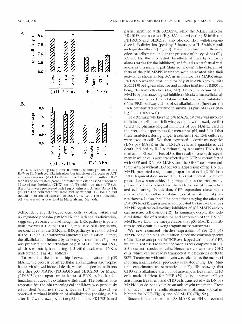
We next disrupted the sodium gradient, generated by theplasma membrane Na�/K� ATPase, with ouabain. As shownin Fig. 2, ouabain prevented intracellular alkalinization duringeither IL-7 (Fig. 2A) or IL-3 (Fig. 2B) withdrawal. In contrast,inhibition of de novo protein synthesis with cycloheximide orinhibition of de novo ATP synthesis with antimycin A did notprevent the alkalinization process (Fig. 2). We further evalu-ated the sodium flux across the plasma membrane followingcytokine withdrawal by using 22Na and observed that 22Naefflux (export versus import) increased during cytokine with-drawal and that this increase could be reversed by the additionof ouabain (data not shown). Hence, sodium and the mainte-nance of a sodium gradient across the plasma membrane arecomponents in the process of alkalinization induced by cyto-kine withdrawal.
Regulators of intracellular pH include the NHEs as well asthe sodium-dependent and -independent AEs. We tested sev-eral specific inhibitors of the ion exchangers for their effect onalkalinization induced by cytokine withdrawal. In Fig. 3 areshown the effect of either DMA or EIPA (NHE inhibitors) ver-sus DIDS and SITS (AE inhibitors) in culture with the D1 orFL5.12A cells with or without cytokines. Only the NHE inhib-itors, DMA and EIPA, prevented the pH rise induced by with-drawal of IL-7 (Fig. 3A) or IL-3 (Fig. 3C). Surprisingly, thesame NHE inhibitors induced alkalinization when added tocells in the presence of either IL-7 (Fig. 3A) or IL-3 (Fig. 3C).This appears to result from overactivation of AE when NHE isblocked (data not shown), suggesting that the presence of afunctional NHE is required for the regulation of HCO3
�-dependent transporters, as has been shown in the CA1 neu-rons of NHE1 mutant mice (60). The use of the AE inhibitorsDIDS (which blocks sodium-independent AE) and SITS(which blocks sodium-dependent AE), had no effect on alka-linization induced by either IL-7 withdrawal (Fig. 3B) or IL-3withdrawal (Fig. 3D). Likewise, these inhibitors did not alterintracellular pH levels in the presence of either cytokine (Fig.3B, 3D). Therefore, our results identify NHE as the mediatorof alkalinization following cytokine withdrawal based on so-dium dependence, requirement for a sodium gradient, and theeffect of inhibitors of NHE.
Since various signaling pathways regulate NHE, includingthose involving the MAPKs, we next examined the pathwayleading to activation of NHE. Using anisomycin, which ac-tivates stress-signaling pathways, we observed the induction ofintracellular alkalinization in the presence of IL-3 (Fig. 4A)and an accelerated induction of alkalinization during with-drawal of IL-3 (Fig. 4A). Similar results were obtained withIL-7-dependent D1 cells (data not shown). This suggested thatactivation of stress pathways involving MAPKs could poten-tially mediate alkalinization following the withdrawal of tro-phic cytokines.
To determine which of the MAPK pathways could be in-volved in alkalinization, we evaluated the phosphorylation sta-tus of different MAPKs during IL-3 or IL-7 withdrawal. In Fig.4B are shown the results of Western blot analyses for thephosphorylated (activated) and nonphosphorylated forms of
VOL. 21, 2001 ALKALINIZATION IS MEDIATED BY NHE1 AND p38 MAPK 7547

p38 MAPK and its substrate ATF-2, ERK1/2 and its substratep90RSK, and JNK. The levels of phospho-p38 MAPK in-creased following IL-3 withdrawal (Fig. 4B, top) at time pointscorresponding to the alkalinization period (Fig. 1C and D).Although a representative phospho-p38 blot is shown (Fig.4B), these results were reproduced in numerous experiments.The levels of a downstream substrate of p38 MAPK, phospho-ATF-2, also increased in parallel to those of phospho-p38 (Fig.4B, top), suggesting increased activation of the p38 MAPKpathway following IL-3 withdrawal. However, since ATF-2 isnot a specific p38 MAPK substrate, we further confirmed thatp38 MAPK activity is increased during IL-3 withdrawal byperforming a p38 MAPK in vitro kinase assay with immuno-precipitated p38 MAPK and an ATF-2 peptide as substrate(Fig. 4B, second panel from the top). We also examined thephosphorylation of MKK3/6, upstream activators of p38MAPK, and found elevated phosphorylated protein levels dur-ing the withdrawal of IL-3 (data not shown). The phosphory-lation of p38 MAPK was transient, in that 3 to 4 h after IL-3withdrawal the level of phospho-p38 MAPK declined (Fig. 4B,top) and was undetectable by 5 h (data not shown); this declinecoincided with the decline in intracellular pH. As a positive
control, anisomycin-treated cells showed increased phospho-p38 MAPK (Fig. 4B, top).
In contrast to the rise in phospho-p38 MAPK, the oppositepattern was observed in levels of phospho-ERK1/2, which rap-idly declined following IL-3 withdrawal, as did the levels of itssubstrate, phospho-p90RSK (Fig. 4B, second panel from thebottom). Therefore, IL-3 stimulation (not withdrawal) activat-ed phospho-ERK1/2 and its downstream substrate, p90RSK.The levels of phospho-JNK, like ERK1/2, were stimulated byIL-3 and decreased following IL-3 withdrawal; however, theeffect of anisomycin was stimulatory, resembling that of phos-pho-p38 rather than ERK1/2 (Fig. 4B, bottom). JNK activityduring trophic factor withdrawal was further assayed using anin vitro kinase assay with c-Jun as a substrate, and no signifi-cant kinase activity was detected (data not shown).
Similar to the IL-3-dependent FL5.12A cells, up-regulationof phospho-p38 MAPK occurred following IL-7 withdrawal inthe D1 thymocyte cell line and was concurrent with alkaliniza-tion (4 to 6 h post-withdrawal) (data not shown). However,phospho-ERK1/2 was not detected in either the presence orabsence of IL-7, as previously observed in this cell line (E.Rajnavolgyi et al., unpublished results). Therefore, in both IL-
FIG. 1. Intracellular alkalinization following IL-7 or IL-3 withdrawal is dependent on sodium but not bicarbonate. (A) D1 cells were deprivedof IL-7 for 0 to 6 h and incubated in either Na�-containing buffer or Na�-free buffer. D1 cells were maintained in Na�-containing buffer with IL-7as a handling control. (B) D1 cells were deprived of IL-7 for 0 to 6 h and incubated in either HCO3
�-containing medium or HCO3�-free medium.
D1 cells were maintained in HCO3�-containing medium with IL-7 as a handling control. (C) FL5.12A cells, deprived of IL-3 for 0 to 4 h, were
incubated in either Na�containing buffer or Na�-free buffer. FL5.12A cells were maintained in Na�-containing buffer with IL-3 as a handling con-trol. (D) FL5.12A cells, deprived of IL-3 for 0 to 4 h, were incubated in either HCO3-containing medium or HCO3
�-free medium. FL5.12A cellswere maintained in HCO3
�containing medium with IL-3 as a handling control. Intracellular pH was assayed using the pH-sensitive fluorescent probeBCECF, and pH values were derived from a pH calibration curve generated through the use of nigericin as described in Materials and Methods.
7548 KHALED ET AL. MOL. CELL. BIOL.
3-dependent and IL-7-dependent cells, cytokine withdrawalup-regulated phospho-p38 MAPK and induced alkalinization,suggesting a connection. Although the ERK pathway is poten-tially involved in IL3 (but not IL-7)-mediated NHE regulation,we conclude that the ERK and JNK pathways are not involvedin the IL-3 or IL-7 withdrawal-induced alkalinization. Hence,the alkalinization induced by anisomycin treatment (Fig. 4A)was probably due to activation of p38 MAPK and not JNK,which is especially true during IL-3 withdrawal, when JNK isundetectable (Fig. 4B, bottom).
To examine the relationship between activation of p38MAPK, the process of intracellular alkalinization and trophicfactor withdrawal-induced death, we tested specific inhibitorsof either p38 MAPK (PD169316 and SB202190) or MEK1(PD98059), the upstream activator of ERK, to block alka-linization induced by cytokine withdrawal. The optimal doseresponse for the pharmacological inhibitors was previouslyestablished (data not shown). During IL-7 withdrawal, weobserved maximal inhibition of alkalinization (peaking at 5 hafter IL-7 withdrawal) with the p38 inhibitor, PD169316, and
partial inhibition with SB202190, while the MEK1 inhibitor,PD98059, had no effect (Fig. 5A). Likewise, the p38 inhibitorsPD169316 and SB202190 also blocked IL-3 withdrawal-in-duced alkalinization (peaking 3 hours post-IL-3-withdrawal)with greater efficacy (Fig. 5B). These inhibitors had little or noeffect on cells maintained in the presence of the cytokines (Fig.5A and B). We also tested the effects of dimethyl sulfoxidealone (carrier for the inhibitors) and found no artifactual vari-ations in intracellular pH (data not shown). The different ef-fects of the p38 MAPK inhibitors were correlated with theiractivity, as shown in Fig. 5C, in an in vitro p38 MAPK assay.PD169316 was the best inhibitor of p38 MAPK activity, withSB202190 being less effective and another inhibitor, SB203580,being the least effective (Fig. 5C). Hence, inhibition of p38MAPK by pharmacological inhibitors blocked intracellular al-kalinization induced by cytokine withdrawal, while inhibitionof the ERK pathway did not block alkalinization (however, theERK pathway did contribute to survival as part of IL-3 signal-ing [data not shown]).
To determine whether the p38 MAPK pathway was involvedin inducing cell death following cytokine withdrawal, we firsttested the pharmacological inhibitors of p38 MAPK, used inthe preceding experiments for measuring pH, and found thatthese inhibitors, during longer treatments (i.e., 15-h cultures),were toxic to cells. We then expressed a dominant negative(DN) p38 MAPK in the FL5.12A cells and quantitated celldeath, induced by IL-3 withdrawal, by measuring DNA frag-mentation. Shown in Fig. 5D is the result of one such experi-ment in which cells were transfected with GFP or cotransfectedwith GFP and DN p38 MAPK and the GFP� cells were cul-tured with or without IL-3 for 48 h. Expression of the DN p38MAPK protected a significant proportion of cells (20%) fromDNA fragmentation induced by IL-3 withdrawal. Completeprotection was not achieved, perhaps due to the transient ex-pression of the construct and the added stress of transfectionand cell sorting. In addition, GFP expression alone had amodest effect on cell survival during cytokine withdrawal (datanot shown). It also should be noted that assaying the effects ofDN p38 MAPK expression is complicated by the fact that p38MAPK regulates cell cycling; inhibition of p38 MAPK activitycan increase cell division (12). In summary, despite the tech-nical difficulties of transfection and expression of the DN p38MAPK, we favor the interpretation that p38 MAPK contrib-utes to cell death following trophic factor withdrawal.
We next examined whether expression of the DN p38MAPK could inhibit alkalinization. Since the emission spectraof the fluorescent probe BCECF overlapped with that of GFP,we could not use the same approach as was employed in Fig.5D to select transfected cells. Hence, we chose to use CHOcells, which can be readily transfected at efficiencies of 80 to90%. Treatment with anisomycin was selected as the means ofinducing alkalinization (previously evaluated in Fig. 4A). Mul-tiple experiments are summarized in Fig. 5E, showing thatCHO cells alkalinize after 1 h of anisomycin treatment. CHOcells made deficient for NHE (39) do not increase pH onanisomycin treatment, and CHO cells transfected with DN p38MAPK also do not alkalinize on anisomycin treatment. Thesefindings confirm the results obtained with pharmacological in-hibitors for NHE (Fig. 3) and p38 MAPK (Fig. 5A).
Since inhibition of either p38 MAPK or NHE prevented
FIG. 2. Disrupting the plasma membrane sodium gradient blocksIL-7- or IL-3-induced alkalinization, but inhibition of protein or ATPsynthesis does not. (A) D1 cells were incubated with or without IL-7for 5 h and not treated (None) or treated with either 1 mM ouabain or10 �g of cycloheximide (CHX) per ml. To inhibit de novo ATP syn-thesis, cells were pretreated with 1 �g of antimycin A (Anti A) for 1 h.(B) FL5.12A cells were incubated with or without IL-3 for 3 h andtreated or not treated as described above for D1 cells. The intracellularpH was assayed as described in Materials and Methods.
VOL. 21, 2001 ALKALINIZATION IS MEDIATED BY NHE1 AND p38 MAPK 7549

intracellular alkalinization following IL-7 or IL-3 withdrawal,we examined the phosphorylation of NHE for effects of cyto-kine withdrawal and MAPKs. Figure 6 displays the results,representative of multiple experiments, in which cells wereradiolabeled with [32P]orthophosphate and lysed and NHE1was immunoprecipitated with a specific antibody, confirmingthe presence of this isoform. As a control for the immuno-precipitation procedure, the lower panels show equivalentamounts of unlabeled NHE1 protein obtained using a differentanti-NHE1 antibody for detection from that used for immu-noprecipitation (Fig. 6). As shown, NHE1 was phosphorylatedin either the presence or absence of either cytokine (data forIL-3-dependent cells are displayed in Fig. 6A). The totalamount of NHE1 protein per cell was much greater in theIL-3-dependent FL5.12A cells than the IL-7-dependent D1cells, as seen in a comparative Western blot for NHE1 (datanot shown), so the subsequent phosphorylation studies areshown in the former cell type only. The p38 inhibitor PD169316dramatically inhibited the phosphorylation of NHE1 followingIL-3 withdrawal (Fig. 6B) but not in the presence of the cyto-kine. The MEK1 inhibitor PD98059 did not decrease NHE1phosphorylation induced by cytokine withdrawal (data not
shown), although it did inhibit the phosphorylation of NHE1 inthe presence of IL-3 (Fig. 6C). This suggests that two differentphosphorylation pathways act on NHE1: in the presence ofIL-3, the ERK MAPK pathway results in normal NHE1 func-tion, whereas in the absence of IL-3, the p38 MAPK pathwayinduces NHE1 to alkalinize the cytosol.
Having shown that p38 MAPK inhibitors blocked NHE1phosphorylation following IL-3 withdrawal (Fig. 5A and B), weexplored the possibility that p38 MAPK directly phosphory-lated NHE1. To test whether p38 MAPK physically interactedwith NHE1 in vivo, it was necessary to treat FL5.12A cells witha chemical crosslinker (disuccinimidyl suberate) to observe anyassociations. Cells treated with cross-linker were lysed, andNHE1 was immunoprecipitated and blotted for coassociationwith p38 MAPK. As shown in Fig. 6D, NHE1 and p38 MAPKdid coimmunoprecipitate, and the association increased fol-lowing IL-3 withdrawal. Mapkap2 is shown as a positive con-trol for a substrate known to associate with p38 MAPK,whereas normal rabbit Ig served as a negative control for im-munoprecipitation.
Further evidence that p38MAPK could directly phosphory-late NHE1 was obtained by performing an in-gel kinase assay
FIG. 3. Inhibitors of the NHE (but not the anion exchangers) prevent intracellular alkalinization following IL-7 or IL-3 withdrawal. (A) D1 cellswere cultured in the presence or absence of IL-7 for 5 h and either not treated (None) or treated with 200 �M DMA or EIPA (inhibitors of theNHE). (B) D1 cells were cultured in the presence or absence of IL-7 for 5 h and either not treated (None) or treated with 200 or 1,000 �M DIDSor 1,000 �M SITS (inhibitors of the anion exchangers). (C) FL5.12A cells, incubated with or without IL-3 for 3 h, were either not treated (None)or treated with 200 �M DMA or EIPA (inhibitors of the NHE). (D) FL5.12A cells, incubated with or without IL-3, were either not treated (None)or treated with 200 or 1,000 �M DIDS or 1000 �M SITS (inhibitors of the anion exchangers). The intracellular pH was assayed as described inMaterials and Methods.
7550 KHALED ET AL. MOL. CELL. BIOL.

with an NHE-GST fusion protein as the substrate. The resultsdemonstrated that lysates from FL512.A cells undergoing IL-3withdrawal contained a kinase of approximately 40 kDa thatwas capable of phosphorylating the C terminal of NHE1 (datanot shown). This kinase was then identified as p38 MAPK byimmunoprecipitating phospho-p38 MAPK, which then phos-phorylated the NHE-GST fusuin protein in an in-gel assay, asshown in Fig. 7A (left panel). Additionally, to determine if anyother kinases in the MAPK pathway could phosphorylateNHE1 during IL-3 withdrawal, we tested an antibody specificfor the motif, phosphorylated threonine-proline (p-TP), that iscreated on its substrates by MAPKs (28). Only the immuno-precipitated phospho-p38 MAPK phosphorylated the NHE-GST fusion protein, as shown in Fig. 7A (left panel), with a1.8-fold increase in activity during IL-3 withdrawal. Therefore,in vitro, p38 MAPK, activated by IL-3 withdrawal, is capable ofdirectly phosphorylating NHE1, and no other substrates of theMAPK pathway containing active p-TP sites (i.e., Mapkap2)are involved.
Anisomycin induced alkalinization, as shown in Fig. 4A.Since anisomycin activated both p38 MAPK and JNK (Fig.4B), we next determined if JNK could phosphorylate the NHE-GST fusion protein in an in vitro kinase assay. Results shownin Fig. 7A (right panel) demonstrate that JNK, precipitatedfrom cells treated or not treated with IL-3 and treated with ani-somycin, does not phosphorylate NHE1 as does p38 MAPK.The levels of c-Jun phosphorylation are shown to indicate JNKactivity. The slight phosphorylation of NHE1 by the JNK prep-aration is probably due to contamination by p38 MAPK, sinceelimination of p38 MAPK by preimmunoprecipitation de-creased this activity.
To determine the phosphorylation site for p38 MAPK in theNHE1 C-terminal region, NHE-GST fusion proteins based onthe rabbit NHE1 sequence were made containing various de-letions. The results of these experiments are shown in Fig. 7Band summarized in Fig. 7C. Phospho-p38 MAPK phosphory-lated the WT NHE-GST fusion protein containing amino acids637 to 815. To test whether p38 MAPK phosphorylated the
FIG. 4. IL-3 withdrawal and anisomycin treatment both activate p38 MAPK and induce alkalinization. (A) Treatment with anisomycin (a p38MAPK activator) induces alkalinization in the presence (top) or absence (bottom) of IL-3. FL5.12A cells were treated with 100 �g of anisomycinper ml. The intracellular pH was measured as described in Materials and Methods. (B) IL-3 withdrawal activates the phosphorylation of p38MAPK and ATF-2 but not ERK1/2, p90RSK, or JNK. FL5.12A cells were incubated without IL-3 for 0 to 3 h, as well as stimulated with anisomycin,and levels of phosphorylated p38 and phosphorylated ATF-2 (top panel), phosphorylated ERK1/2 or phosphorylated p90RSK (second panel frombottom), and phosphorylated JNK (bottom panel) were evaluated from whole-cell lysates. Nonphosphorylated p38 and ERK were included asloading controls. In addition, p38 MAPK was immunoprecipitated from protein lysates, made from cells incubated without IL-3 for 0 to 3 h, andused in an in vitro kinase assay to phosphorylate an ATF-2 peptide (second panel from top). SDS-polyacrylamide gel electrophoresis and Westernblot analysis were performed as described in Materials and Methods.
VOL. 21, 2001 ALKALINIZATION IS MEDIATED BY NHE1 AND p38 MAPK 7551

same site on NHE1 as shown for p90RSK (46), this residue,serine 703 (numbering from the human NHE1 sequence), waschanged to alanine. This mutation did not block phosphoryla-tion by phospho-p38 MAPK (Fig. 7B), implicating a different
target site(s) for p38 MAPK on NHE1 than the proposedtarget of p90RSK. Deleting amino acids 702 to 815 or 661 to793 eliminated phosphorylation of NHE1 (Fig.7B). This firstgroup of NHE-GST deletion proteins defined the target site(s)on NHE1 for p38 MAPK phosphorylation to a region fromamino acids 703 to 793 (Fig. 7C). In a second group of exper-iments, testing NHE-GST deletion proteins with stops atamino acids 744, 762, and 777, the location of the site for p38MAPK phosphorylation of NHE was further narrowed to aregion between amino acids 703 and 743 (Fig. 7B and C).Furthermore, NHE-GST fusion proteins spanning the regionsfrom amino acids 635 to 663 (the proposed sites for calmodulin
FIG. 5. Inhibitors of p38 MAPK block alkalinization induced byIL-7 or IL-3 withdrawal and enhance survival. (A) D1 cells were cul-tured in the presence or absence of IL-7 for 5 h and either not treated(None) or treated with 20 �M PD169316, 20 �M SB202190 (inhibitorsof p38 MAPK), or 20 �M PD98059 (inhibitor of MEK1). (B) FL5.12Acells were incubated with or without IL-3 for 3 h and either not treated(None) or treated with 20 �M PD169316, 20 �M SB202190 (inhibitorsof p38 MAPK), or 20 �M PD98059 (inhibitor of MEK1). IntracellularpH was measured as described in Materials and Methods. (C) Theefficacy of the different MAPK inhibitors (20 �M) was tested in a p38MAPK in vitro kinase assay using p38 MAPK immunoprecipiatatedfrom anisomycintreated FL5.12A cells. Of the three p38 MAPK inhib-itors assayed, PD169316 was the most effective in blocking p38 MAPKactivity (shown at two concentrations, 20 and 10 �M), SB202190 wasthe second best, and SB203580 had minimal effect; the ERK inhibitor(PD98059) or the nonspecific inhibitor (SB202474) had no effect at the20 �M dose. (D) GFP� cells or GFP� cells cotransfected with aDN-p38 MAPK were cultured in the absence of IL-3 for 48 h, and thepercentage of cells with DNA fragmentation was measured by pro-pidium iodide staining and flow cytometry analysis. Expression of theDN-p38 MAPK decreased DNA fragmentation by 20% during IL-3withdrawal. GFP� cells cultured in the presence of IL-3 were includedas a positive control for survival. (E) Loss of NHE or expression of aDN-p38 MAPK in CHO cells prevents alkalinization after anisomycintreatment. CHO cells were made deficient for NHE as previouslydescribed (39) and/or transfected with DN-p38 MAPK and evaluated24 h later for pH changes after 1 h of anisomycin treatment by incor-poration of BCECF as described in Materials and Methods.
7552 KHALED ET AL. MOL. CELL. BIOL.

binding [54]) were not phosphorylated by p38 MAPK (Fig.7C).
To characterize the p38 MAPK phosphorylation site further,we sought the p-TP motif that many of its substrates, likeMapKap2, display. However, immunoprecipitation and West-ern blot analysis did not reveal the p-TP motif on NHE (Fig.7D). The 40-amino-acid region defined by deletion analysiscontained multiple threonine and serine residues. To deter-mine which of these sites were the targets for p38 MAPKphosphorylation, we performed mass spectroscopy on the WTNHE-GST fusion protein phosphorylated in vitro by immuno-precipiated p38 MAPK. For comparison, the unphosphory-lated NHE-GST fusion protein was also tested. Mass spec-trometry analysis of NHE-GST peptides digested with eithertrypsin or formic acid revealed discrete phosphorylation siteson NHE1 between amino acids 711 and 739. Tryptic peptidefragments, corresponding to amino acids 711 to 739, containedone to three phosphates. Peptide fragments from formic acidcleavage, spanning amino acids 713 to 718, contained onephosphate, and those spanning amino acids 720 to 729 con-tained one to three phosphates. Treatment with calf intestinalphosphatase removed the phosphates from all these peptides,confirming the phosphorylated state. Therefore, we concluded,from both the deletion analysis and mass spectrometry, that
there are four targets on NHE1 for in vitro phosphorylation byp38 MAPK-Thr 717, Ser 722, Ser 725, and Ser 728 (Fig. 7E).Two of the serines, Ser 722 and Ser 725, are located adjacentto prolines, representing common MAPK motifs. It remains tobe determined how phosphorylation by p38 MAPK of thesesites on NHE1 induces the intracellular alkalinization that istriggered by cytokine withdrawal.
DISCUSSION
Loss of cytokine receptor signaling induces apoptotic celldeath in dependent cell lines. To understand this process, wehave defined early events (i.e., pH rise) following cytokinewithdrawal that lead to sequelae such as mitochondrial hy-perpolarization, loss of bioenergetics, and the mitochondrialtranslocation of Bax, culminating in cell death (23, 24). Herewe show that the rapid alkalinization of the cytosol was medi-ated by the membrane exchanger NHE1 through activation bythe stress kinase p38 MAPK. Phosphorylation of p38 MAPKincreased within the first hours of IL-3 or IL-7 withdrawal,while levels of other MAPKs, ERK or JNK, decreased. ADN-p38 MAPK could protect cells from DNA fragmentationinduced by IL-3 withdrawal and alkalinization induced by ani-
FIG. 6. Phosphorylation of NHE1 occurs via different MAPKs in the presence and absence of IL-3. (A) FL5.12A cells were incubated for 3 hwith [32P]orthophosphate in the presence (�) or absence (�) of IL-3. (B) 32P-labeled FL5.12A cells were incubated for 3 h with PD169316 with(�) or without (�) IL-3. (C) 32P-labeled FL5.12A cells were incubated with or without PD98059 in the presence (�) of IL-3. Cells were lysed,and NHE1 was immunoprecipitated and electrophoresed on SDS–8% polyacrylamide gels and autoradiography was performed as described inMaterials and Methods. To confirm the specificity of the NHE antibody (A7) used for immunoprecipitation, parallel cell lysates (not labeled with[32P]orthophosphate) were immunoprecipitated with the anti-NHE antibody (A7), electrophoresed on SDS–8% polyacrylamide gels, analyzed byWestern bloting using a different anti-NHE1 antibody, and shown in the lower panels. (D) In vivo coimmunoprecipitation of NHE and p38 MAPK.Protein lysates extracted from cells cultured with or without IL-3, and chemically cross-linked with DSS, were immunoprecipitated with either anantibody specific for NHE1, an antibody for Mapkap2 (positive control), or rabbit Ig (negative control). The lysates were blotted for p38 MAPKas described in Materials and Methods.
VOL. 21, 2001 ALKALINIZATION IS MEDIATED BY NHE1 AND p38 MAPK 7553

somycin treatment. p38 MAPK directly phosphorylated NHEbetween amino acids 704 and 743, on one threonine (Thr 717)and three serines (Ser 722, Ser 725, and Ser 728), as defined bymass spectrometry analysis. Therefore, our data suggest a mod-
el in which cytokine withdrawal activates the p38 MAPK path-way leading directly to the phosphorylation and activation ofNHE1, the membrane ion exchanger responsible for intracel-lular alkalinization, a critical trigger in the apoptotic process.
FIG. 7. Kinase assays demonstrate that p38 MAPK directly phosphorylates NHE1 at a site(s) between amino acids 703 and 793. The numberingsystem used corresponds to the rabbit NHE1 sequence. (A) Protein lysates from FL5.12A cells cultured with or without IL-3 for 2 h wereimmunoprecipitated with either an antibody specific for phospho-p38 MAPK (p38) or an antibody specific for the motif, p-TP, and the ability ofthese kinases to phosphorylated the WT NHE-GST fusion protein was evaluated by an in-gel kinase assay as described in Materials and Methods.Only p38 phosphorylated the WT NHE-GST fusion protein, an activity which increased 1.8-fold during IL-3 withdrawal (left panel). Protein lysatesfrom FL5.12A cells were also immunoprecipitated with c-Jun fusion beads to capture JNK, which was tested in an in vitro kinase assay for its abilityto phosphorylate the WT NHEGST fusion protein. Unlike p38 MAPK, JNK did not phosphorylate NHE (right panel). (B) Protein lysates extractedfrom anisomycin-activated FL5.12A cells were immunoprecipitated with antibody to phospho-p38 MAPK and a kinase assay performed asdescribed in Materials and Methods using the WT NHE-GST fusion protein and seven mutant NHE-GST fusion proteins as substrates. GST alonewas included as a negative control. (C) The results of the kinase assay from panel B are summarized in table form. In addition, phospho-p38 MAPKdid not phosphorylate NHE-GST fusion proteins containing amino acids 635 to 663. Possible phosphorylation sites by p38 MAPK on NHE arelocated in the region from amino acids 703 to 743, which excludes the proposed p90RSK phosphorylation site (Serine 703, numbering from humanNHE1 sequence). (D) FL5.12A cells, cultured without IL-3 for 0, 1, 2, and 3 h or treated with anisomycin (�), were lysed, and the preclearedprotein extracts were immunoprecipitated with the A7 antibody specific for NHE1 and then run on an 8% Tris-glycine SDS gel and analyzed byWestern blotting with the p-TP-specific antibody (left panel). The same blots were then reprobed with a commercially available anti-NHE1antibody (right panel). NHE1 did not contain an activated p-TP motif. (E) Mass spectrometry analysis, as described in Materials and Methods,identified four sites at Thr 717, Ser 722, Ser 725, and Ser 728 as in vitro targets for p38 MAPK activity. Phosphorylation of one or more of thesesites by p38 MAPK modulates alkalinization mediated by NHE during trophic factor withdrawal.
7554 KHALED ET AL. MOL. CELL. BIOL.

Although NHE is normally involved in maintaining physio-logical pH, there are other reports of NHE-mediated intracel-lular alkalinization. Stimulation of platelets with agonists likethrombin lead to a rapid rise in cytosolic pH, induced byincreased transport activity of NHE, of approximately 0.4 pHunit (38). Thymocytes that underwent spontaneous apoptosiswere also highly alkaline (pH 7.6), and depletion of sodiumcould prevent the death process, as did inhibitors of the mem-brane ion exchangers (50). Our own studies show that alkalin-ization contributes to cell death in several ways. (i) Alkaliniza-tion activates Bax, inducing its mitochondrial translocation(23), also shown to occur during ceramide treatment of U937cells (3). (ii) Alkalinization inhibits the import of ADP intomitochondria, which severely impairs the synthesis of ATP,and heightens the mitochondrial membrane potential (24).
The NHE, expressed by many cell types, is regulated bydiverse stimuli. One means by which intracellular pH can bemodulated is by cellular adherence mediated through integrins(42). However, pH changes induced by cytokine loss wereindependent of adherence (data not shown). Fluxes of ionsthrough the NHE are driven by the sodium gradient across theplasma membrane and do not directly require metabolic en-ergy, although ATP can influence it (33). A number of studiesdemonstrated that the NHE is regulated by the association ofcalmodulin, which causes autoinhibition of the transporter (4);deletion of the calmodulin binding domain in NHE renders theexchanger constitutively activated, perhaps by altering its setpoint for “pH sensing” (53, 54). In our studies, we did not iden-tify any sites in the calmodulin binding domain of NHE as tar-gets for phosphorylation by p38 MAPK. Hence, modificationof calmodulin binding by p38 MAPK activity does not appearto account for alkalinization during cytokine withdrawal.
Phosphorylation of the NHE plays an essential role in itsactivation. NHE is constitutively phosphorylated in restingcells, and mitogenic stimulation leads to an increase in phos-phorylation on serine residues in a time course that parallelsthe rise in intracellular pH (40). These results and others sug-gested that phosphorylation of the NHE directly triggered itsactivation (5, 41). Recently, MAPKs were examined as poten-tial mediators of growth factor-induced activation of NHE1.Inhibition by either a dominant negative ERK or the ERK in-hibitor PD98059 prevented growth factor-induced activation ofthe NHE (6, 31). However, it did not appear that ERKs weresolely responsible for phosphorylating the NHE but, rather,that a downstream substrate of the ERK pathway, p90 RSK,could also in some instances be involved (31, 46). Therefore,ERKs, perhaps via p90RSK, are in the pathway from stimulisuch as mitogens, serum, or growth factors leading to the ac-tivation of NHE. We found that IL-3 induced a similar path-way, in that IL-3 induced high levels of ERKs and p90RSK(Fig. 4B), and addition of the ERK inhibitor PD98059 de-creased IL-3-stimulated NHE phosphorylation (Fig. 6C). How-ever, the withdrawal-induced phosphorylation of NHE was notan ERK-mediated event but instead required the activation ofa different kinase, p38 MAPK (Fig. 5 and 6B).
That intracellular alkalinization could activate the stresspathway was suggested in a study in which treatment of U937cells with permeant weak bases increased the activity of JNKand p38 MAPK (43). The authors proposed that the activationof NHE and that of stress kinases were parallel pathways.
However, our results clearly show that following cytokine with-drawal, activation of p38 MAPK is upstream of NHE phos-phorylation (Fig. 6B) and alkalinization (Fig. 5A and E).
In another study, p38 MAPK was proposed to have an in-hibitory effect on NHE (27) (in contrast to our findings, inwhich p38 MAPK has a stimulatory effect). In that reportvascular smooth muscle cells were stimulated with angiotensinII, which activated both ERK and p38 MAPK, and ERK inturn activated NHE; it was suggested that p38 MAPK inhibitedthe function of ERK and therefore inhibited NHE. However,these results differ from the cellular responses to the two tro-phic cytokines studied here. The IL-7-dependent cell line usedhere lacks ERK (Rajnavolgyi et al., unpublished), and IL-7 waspreviously shown to not activate ERK in other cells (9, 14),whereas IL-3 withdrawal rapidly down-regulates ERK (andup-regulates p38 MAPK). Withdrawal of either IL-7 or IL-3activated the p38 MAPK (independent of other MAPKs), andthe pH rose. There are previous accounts of trophic-factor with-drawal causing a shift in the balance of p38 MAPK and ERK,with ERK activity decreasing as p38 MAPK activity increased(7, 59). Thus, our data show that following trophic factor with-drawal (and in the absence of ERK), p38 MAPK is an exceed-ingly potent inducer of NHE phosphorylation and activity.
The direct phosphorylation of NHE by MAPKs has beendemonstrated by others in vitro. We initially detected in celllysates a kinase of approximately 40 kDa that phosphorylatedNHE in an in-gel kinase assay (data not shown). By immuno-precipitation, we confirmed that the kinase activity seen in theprotein lysates from IL-3-deprived cells was p38 MAPK andnot one of the other MAPK family member (i.e., ERK orJNK), nor was it a downstream substrate of p38 MAPK such asMapkap2 (45) (data not shown and Fig. 7D). As seen in ourresults, p38 MAPK phosphorylated NHE at multiple sites in aregion between amino acids 704 and 743 (Fig. 7B and C). Inaddition, p38 MAPK did not appear to phosphorylate the pro-posed p90 RSK target site, Serine 703 (human) (46). This sug-gests that whereas the RSK site on NHE activates NHE toaccelerate the adjustment of acidic pH to physiological levels,the p38 site(s) activates NHE to create alkaline levels. Usingmass spectrometry, we identified four in vitro phosphorylationsites for p38 MAPK on NHE at Thr 717, Ser 722, Ser 725, andSer 728. We speculate that phosphorylation of the NHE in thisregion could create either a docking site for another protein,leading to deregulation and alkalinization, or repress an auto-inhibitory regulatory domain.
One further feature of the cytokine withdrawal-induced al-kalinization is its transience. Previously we reported that lossof IL-7 or IL-3 signaling led to a pH rise lasting 2 to 3 h (23,24). We do not yet know the mechanism that returns the in-tracellular pH to neutrality. Since p38 MAPK is an upstreammediator of the alkalinization process, one of many possiblemechanisms of normalization would be for alkaline pH to in-hibit p38 MAPK activity. A recent study identified the dockingsites in MAPKs and MAPK-interacting proteins and showedthat such sites contain defined motifs characterized by thepresence of charged amino acids (47). We and others haveshown that charged amino acids in interacting peptides can bea basis for the pH- sensitivity of proteins (23, 34).
The upstream chain of events that activate the p38 MAPKpathway following cytokine withdrawal remain to be identified.
VOL. 21, 2001 ALKALINIZATION IS MEDIATED BY NHE1 AND p38 MAPK 7555

The possibilities include G proteins, which have already beenshown to regulate intracellular pH (52); RhoA, which activatesthe NHE (49); and other GTPases and signaling proteins thatlie upstream of p38 MAPK activation (61). Another possibilityis that cessation of Jak/STAT signaling following cytokinewithdrawal may contribute to the loss of ERK and a compen-satory activation of p38 MAPK observed during apoptoticdeath (58). It is also possible that induction of a dephosphor-ylation activity, as seen during cytomegalovirus infection, mod-ulates p38 MAPK function (22). Whatever the initiating mech-anism, the activation of p38 MAPK following IL-7 and IL-3withdrawal, phosphorylation of specific sites, and subsequentactivation of the NHE represents a novel pathway that linkstrophic factor deprivation to a death-promoting physiologicalconsequence, intracellular alkalinization.
ACKNOWLEDGMENT
This project has been funded in whole or in part with federal fundsfrom the National Cancer Institute, National Institutes of Health,under contract NO1-CO-56000.
REFERENCES
1. Akashi, K., M. Kondo, U. von Freeden-Jeffry, R. Murray, and I. L. Weiss-man. 1997. Bcl-2 rescues T lymphopoiesis in interleukin-7 receptor-deficientmice. Cell 89:1033–1041.
2. Aoshiba, K., S. Yasui, M. Hayashi, J. Tamaoki, and A. Nagal. 1999. Role ofp38-mitogen-activated protein kinase in spontaneous apoptosis of humanneutrophils. J. Immunol. 162:1692–1700.
3. Belaud-Rotreau, M. A., N. Leducq, F. M. P. de Gannes, P. Diolez, L. Lacoste,F. Lacombe, P. Bernard, and F. Belloc. 2000. Early transitory rise in intra-cellular pH leads to Bax conformation change during ceramideinduced ap-optosis. Apoptosis 5:551–560.
4. Bertrand, B., S. Wakabayashi, T. Ikeda, J. Pouyssegur, and M. Shigekawa.1994. The Na�/H� exchanger isoform 1 (NHE1) is a novel member of thecalmodulin-binding proteins. Identification and characterization of calmod-ulin-binding sites. J. Biol. Chem. 269:13703–13709.
5. Bianchini, L., M. Woodside, C. Sardet, J. Pouyssegur, A. Takai, and S.Grinstein. 1991. Okadaic acid, a phosphatase inhibitor, induces activationand phosphorylation of the Na�/H� antiport. J. Biol. Chem. 266:15406–15413.
6. Bianchini, L., G. L’Allemain, and J. Pouyssegur. 1997. The p42/p44 mitogen-activated protein kinase cascade is determinant in mediating activation ofthe Na�/H� exchanger (NHE1 isoform) in response to growth factors.J. Biol. Chem. 272:271–279.
7. Birkenkamp, K. U., W. H. Dokter, M. T. Esselink, L. J. Jonk, W. Kruijer,and E. Vellenga. 1999. A dual function for p38 MAP kinase in hematopoieticcells: involvement in apoptosis and cell activation. Leukemia 13:1037–1045.
8. Blom, B., H. Spits, and P. Krimpenfort. 1997. The role of the commongamma chain of the IL-2, IL-4, IL-7 and IL-15 receptors in development oflymphocytes. Constitutive expression of bcl-2 does not rescue the develop-mental defects in common gamma deficient mice, p. 3–11. In S. Smit Sibinga,P. Das, and D. Loewenberg, (ed.), Cytokines and growth factors in bloodtransfusion. Kluwer Academic Publishers, Dordrecht, The Netherlands
9. Crawley, J. B., J. Willcocks, and B. M. Foxwell. 1996. Interleukin-7 inducesT cell proliferation in the absence of Erk/MAP kinase activity. Eur. J. Im-munol. 26:2717–2723.
10. Crawley, J. B., L. Rawlinson, F. V. Lali, T. H. Page, J. Saklatvala, and B. M.Foxwell. 1997. T cell proliferation in response to interleukins 2 and 7 re-quires p38MAP kinase activation. J. Biol. Chem. 272:15023–15027.
11. de Groot, R. P., P. J. Coffer, and L. Koenderman. 1998. Regulation ofproliferation, differentiation and survival by the IL-3/IL-5/GM-CSF receptorfamily. Cell Signalling 10:619–628.
12. Diehl, N. L., H. Ensien, K. A. Fortner, C. Merritt, N. Stetson, C. Charland,R. A. Flavell, R. J. Davis, and M. Rincon. 2000. Activation of the p38mitogen-activated protein kinase pathway arrests cell cycle progression anddifferentiation of immature thymocytes in vivo. J. Exp. Med. 191:321–334.
13. Di Santo, J. P., and H. R. Rodewald. 1998. In vivo roles of receptor tyrosinekinases and cytokine receptors in early thymocyte development. Curr. Opin.Immunol. 10:196–207.
14. Dorsch, M., H. Hock, and T. Diamantstein. 1994. Gene transfer of the IL2receptor beta chain into an IL-7-dependent pre-B cell line permits IL-2-driven proliferation: tyrosine phosphorylation of Shc is induced by IL-2 butnot IL-7. Eur. J. Immunol. 24:2049–2054.
15. English, J., G. Pearson, J. Wilsbacher, J. Swantek, M. Karandikar, S. Xu,
and M. H. Cobb. 1999. New insights into the control of MAP kinase path-ways. Exp. Cell Res. 253:255–270.
16. Franck, P., N. Petitipain, M. Cheriet, M. Dardennes, F. Maachi, B. Schutz,L. Poisson, and P. Nabet. 1996. Measurement of intracellular pH in culturedcells by flow cytometry with BCECF-AM. J. Biotechnol. 46:187–195.
17. Goldsmith, M. A., A. Mikami, Y. You, K. D. Liu, L. Thomas, P. Pharr, andG. D. Longmore. 1998. Absence of cytokine receptor-dependent specificity inred blood cell differentiation in vivo. Proc. Natl. Acad. Sci. USA 95:7006–7011.
18. Herlaar, E., and Z. Brown. 1999. p38 MAPK signaling cascades in inflam-matory disease. Mol. Med. Today 5:439–447.
19. Hofmeister, R., A. R. Khaled, N. Benbernou, E. Rajnavolgyi, K. Muegge, andS. K. Durum. 1999. Interleukin-7: physiological roles and mechanisms ofaction. Cytokine Growth Factor Rev. 10:41–60.
20. Ichijo, H. 1999. From receptors to stress-activated MAP kinases. Oncogene18:6087–6093.
21. Johnson, D. E. 1998. Regulation of survival pathways by IL-3 and inductionof apoptosis following IL-3 withdrawal. Front. Biosci. 3:d313–d324.
22. Johnson, R. A. 2000. Activation of the mitogen-activated protein kinase p38by human cytomegalovirus infection trhough two distinct pathways: a novelmechanism for activation of p38. J. Virol. 74:1158–1167.
23. Khaled, A. R., K. Kim, R. Hofmeister, K. Muegge, and S. K. Durum. 1999.Withdrawal of IL-7 induces Bax translocation from cytosol to mitochondriathrough a rise in intracellular pH. Proc. Natl. Acad. Sci. USA 96:14476–14481.
24. Khaled A. R., D. A. Reynolds, H. A. Young, C. B. Thompson, K. Muegge, andS. K. Durum. 2001. IL-3 withdrawal induces an early increase in mitochon-drial membrane potential unrelated to the Bcl-2 family: roles of intracellularpH, ADP transport and FOF1-ATPase. J. Biol. Chem. 276:6453–6462.
25. Kim, K., C. K. Lee, T. J. Sayers, K. Muegge, and S. K. Durum. 1998. Thetrophic action of IL-7 on pro-T cells: inhibition of apoptosis of pro-T1, -T2,and -T3 cells correlates with Bcl-2 and Bax levels and is independent of Fasand p53 pathways. J. Immunol. 160:5735–5741.
26. Kummer, J. L., P. K. Rao, and K. A. Heidenreich. 1997. Apoptosis inducedby withdrawal of trophic factors is mediated by p38 mitogen-activated pro-tein kinase. J. Biol. Chem. 272:20490–20494.
27. Kusuhara, M., E. Takahashi, T. E. Peterson, J. Abe, M. Ishida, J. Han, R.Ulevitch, and B. C. Berk. 1998. p38 Kinase is a negative regulator of angio-tensin II signal transduction in vascular smooth muscle cells: effects onNa�/H� exchange and ERK1/2. Circ. Res. 83:824–831.
28. Lewis, T. S., P. S. Shapiro, and N. G. Ahn. 1998. Signal transduction throughMAP kinase cascades. Adv. Cancer Res. 74:49–139.
29. Maraskovsky, E., M. Teepe, P. J. Morrissey, S. Braddy, R. E. Miller, D. H.Lynch, and J. J. Peschon. 1996. Impaired survival and proliferation in IL-7receptor-deficient peripheral T cells. J. Immunol. 157:5315–5323.
30. Michaelson, M. D., M. F. Mehler, H. Xu, R. E. Gross, and J. A. Kessler.1996. Interleukin-7 is trophic for embryonic neurons and is expressed indeveloping brain. Dev. Biol. 179:251–263.
31. Moor, A. N., and L. Fliegel. 1999. Protein kinase-mediated regulation of theNa(�)/H(�) exchanger in the rat myocardium by mitogen-activated proteinkinase-dependent pathways. J. Biol. Chem. 274:22985–22992.
32. Nunez, B., L. London, D. Hockenbery, M. Alexander, J. P. McKearn, andS. J. Korsmeyer. 1990. Deregulated Bcl-2 expression selectively prolongssurvival of growth factor-deprived hemopoietic cell lines. J. Immunol. 144:3602–3610.
33. Orlowski, J., and S. Grinstein. 1997. Na�H� exchangers of mammaliancells. J. Biol. Chem. 272:22373–22376.
34. Papa, S., F. Zanotti, T. Cocco, C. Perrucci, C. Candita, and M. Minuto. 1996.Identification of functional domains and critical residues in the adenosinet-riphosphatase inhibitor protein of mitochondrial F0F1 ATP synthase. Eur.J. Biochem. 240:461–467.
35. Peschon, J. J., P. J. Morrissey, K. H. Grabstein, F. J. Ramsdell, E. Maras-kovsky, B. C. Gliniak, L. S. Park, S. F. Ziegler, D. E. Williams, and C. B.Ware. 1994. Early lymphocyte expansion is severely impaired in interleukin7 receptor-deficient mice. J. Exp. Med. 180:1955–1960.
36. Puceat, M. 1999. pHi regulatory ion transporters: an update on structure,regulation and cell function. Cell. Mol. Life Sci. 55:1216–1229.
37. Rinaudo, M. S., K. Su, L. A. Falk, S. Haider, and R. A. Mufson. 1995. Humaninterleukin-3 receptor modulates bcl-2 mRNA and protein levels throughprotein kinase C in TF-1 cells. Blood 86:80–88.
38. Rosskopf, D. 1999. Sodium-hydrogen exchange and platelet function. J.Thromb. Thrombolysis 8:15–24.
39. Rotin, D., D. Norwood, S. Grinstein, and I. Tannock. 1989. Requirement ofthe Na�/H� exchanger for tumor growth. Cancer Res. 49:205–211.
40. Sardet, C., L. Counillon, A. Franchi, and J. Pouyssegur. 1990. Growthfactors induce phosphorylation of the Na�H� antiporter, glycoprotein of110 kD. Science 247:723–726.
41. Sardet, C., P. Fafournoux, and J. Pouyssegur. 1991. Alpha-thrombin, epi-dermal growth factor, and okadaic acid activate the Na�H� exchanger,NHE-1, by phosphorylating a set of common sites. J. Biol. Chem. 266:19166–19171.
42. Schwartz, M. A., G. Both, and C. Leohene. 1989. Effect of cell spreading on
7556 KHALED ET AL. MOL. CELL. BIOL.

cytoplasmic pH in normal and transformed fibroblasts. Proc. Natl. Acad. Sci.USA 86:4525–4529.
43. Shrode, L. D., E. A. Rubie, J. R. Woodgett, and S. Grinstein. 1997. Cytosolicalkalinization increases stress-activated protein kinase/c-Jun NH2-terminalkinase (SAPK/JNK) activity and p38 mitogen-activated protein kinase activ-ity by a calcium-independent mechanism. J. Biol. Chem. 272:13653–13659.
44. Socolofsky, M., H. F. Lodish, and G. Q. Daley. 1998. Control of hematopoi-etic differentiation: lack of specificity in signalling by cytokine receptors.Proc. Natl. Acad. Sci. USA 95:6573–6575.
45. Stokoe, D., D. G. Campbell, S. Nakielny, H. Hidaka, S. J. Leevers, C.Marshall, and P. Cohen. 1992. MAPKAP kinase-2: a novel protein kinaseactivated by mitogen-activated protein kinase. EMBO J. 11:3985–3994.
46. Takahashi, E., J. Abe, B. Gaills, R. Aebersold, D. J. Spring, E. G. Krebs, andB. C. Berk. 1999. p90(RSK) is a serum-stimulated Na�/H� exchanger iso-form-1 kinase. Regulatory phosphorylation of serine 703 of Na�/H� ex-changer isoform-1. J. Biol. Chem. 274:20206–20214.
47. Tanoue, T., M. Adachi, T. Moriguchi, and E. Nishida. 2000. A conserveddocking motif in MAP kinases common to substrates, activators and regu-lators. Nat. Cell Biol. 2:110–116.
48. Telford, W. G., L. E. King, and P. J. Fraker. 1991. Evaluation of glucocor-ticoid-induced DNA fragmentation in mouse thymocytes by flow cytometry.Cell Prolif. 24:447–449.
49. Tominaga, T., and D. L. Barber. 1998. Na-H exchange acts downstream ofRhoA to regulate integrin-induced cell adhesion and spreading. Mol. Biol.Cell 9:2287–2303.
50. Tsao, N., and H. Y. Lei. 1996. Activation of the Na(�)/H(�) antiporter,Na�/HCO3(�)/CO3(2�) cotransporter, or Cl(�)/HCO3(�) exchanger inspontaneous thymocyte apoptosis. J. Immunol. 157:1107–1116.
51. von-Freeden-Jeffry, U., P. Vieira, L. A. Lucian, T. McNeil, S. E. Burdach, andR. Murray. 1995. Lymphopenia in interleukin (IL)-7 gene-deleted miceidentifies IL-7 as a nonredundant cytokine. J. Exp. Med. 181:1519–1526.
52. Voyno-Yasenetskaya, T. A. 1998. G proteins and Na�/H� exchange. Biol.Signals Recept. 7:118–124.
53. Wakabayashi, S., B. Bertrand, T. Ikeda, J. Pouyssegur, and M. Shigekawa.1994. Mutation of calmodulin-binding site renders the Na�/H� exchanger(NHE1) highly H(�)-sensitive and Ca2� regulationdefective. J. Biol. Chem.269:13710–13715.
54. Wakabayashi, S., T. Ikeda, T. Iwamoto, J. Pouyssegur, and M. Shigekawa.1997. Calmodulin-binding autoinhibitory domain controls “pH-sensing” inthe Na�/H� exchanger NHE1 through sequence-specific interaction. Bio-chemistry 36:12854–12861.
55. Watson, J. D., P. J. Morrissey, A. E. Namen, P. J. Conlon, and M. B. Wid-mer. 1989. Effect of IL-7 on the growth of fetal thymocytes in culture.J. Immunol. 143:1215–1222.
56. Weinllch, M., C. Thelss, C. T. Lin, and R. K. Kinne. 1998. BCECF in singlecultured cells: inhomogeneous distribution but homogeneous response.J. Exp. Biol. 201:57–62.
57. Wilm, M., A. Shevchenko, T. Houthaeve, S. Breit, L. Schweigerer, T. Fotsis,and M. Mann. 1996. Femtomole sequencing of proteins from polyacrylamidegels by nano-electrospray mass spectrometry. Nature. 379:466–469.
58. Winston, L. A. and T. Hunter. 1996. Intracellular signaling: putting JAKs onthe kinase MAP. Curr. Biol. 6:668–671.
59. Xia, Z., M. Dickens, J. Raingeaud, R. J. Davis, and M. E. Greenberg. 1995.Opposing effects of ERK and JNK-p38 MAP kinases on apoptosis. Science270:1326–1331.
60. Yao, H., E. Ma, X. Q. Gu, and G. G. Haddad. 1999. Intracellular pH regu-lation of CA1 neurons in Na(�)/H(�) isoform 1 mutant mice. J. Clin. In-vestig. 104:637–645.
61. Zhang, S., J. Han, M. A. Sells, J. Chernoff, U. G. Knaus, R. J. Ulevitch, andG. M. Bokooch. 1995. Rho family GTPases regulate p38 mitogenactivatedprotein kinase through the downstream mediator Pak1. J. Biol. Chem. 270:23934–23936.
VOL. 21, 2001 ALKALINIZATION IS MEDIATED BY NHE1 AND p38 MAPK 7557