Peroxynitrite-induced p38 MAPK pro-apoptotic signaling in enterocytes
Upload
independentCategory
view
0download
0
John N. Lorenz, Jop H. van Berlo, Robert N. Willette and Jeffery D. MolkentinMannix Auger-Messier, Federica Accornero, Sanjeewa A. Goonasekera, Orlando F. Bueno,
Cardiomyopathy Double-Null Mice InducesDusp1/4Unrestrained p38 MAPK Activation in
Print ISSN: 0009-7330. Online ISSN: 1524-4571 Copyright © 2012 American Heart Association, Inc. All rights reserved.is published by the American Heart Association, 7272 Greenville Avenue, Dallas, TX 75231Circulation Research
doi: 10.1161/CIRCRESAHA.112.2729632013;112:48-56; originally published online September 19, 2012;Circ Res.
http://circres.ahajournals.org/content/112/1/48World Wide Web at:
The online version of this article, along with updated information and services, is located on the
http://circres.ahajournals.org/content/suppl/2012/09/19/CIRCRESAHA.112.272963.DC1.htmlData Supplement (unedited) at:
http://circres.ahajournals.org//subscriptions/
is online at: Circulation Research Information about subscribing to Subscriptions:
http://www.lww.com/reprints Information about reprints can be found online at: Reprints:
document. Permissions and Rights Question and Answer about this process is available in the
located, click Request Permissions in the middle column of the Web page under Services. Further informationEditorial Office. Once the online version of the published article for which permission is being requested is
can be obtained via RightsLink, a service of the Copyright Clearance Center, not theCirculation Researchin Requests for permissions to reproduce figures, tables, or portions of articles originally publishedPermissions:
at Universite de Sherbrooke on January 8, 2013http://circres.ahajournals.org/Downloaded from

48
Integrative Physiology
The greater mitogen-activated protein kinase (MAPK) signaling cascade consists of a sequence of successively
acting kinases that result in the dual phosphorylation and activation of terminal kinases p38, c-Jun N-terminal kinases (JNKs), and extracellular signal-regulated kinases (ERKs).1 The major upstream activators of ERK1/2 are two MAP kinase kinases, MEK1 and MEK2, whereas p38 kinases are directly activated by MKK6 and MKK3, and JNKs are directly activated by MKK4 and MKK7.1 Upstream of the MAP kinase kinases, multiple MAP kinase kinase kinases form a complex network that either directly sense stress stimulation or directly are regulated by effectors such as low-molecular-weight G-proteins (Ras, Rac, Rho, and others) and G-protein-coupled receptors.1,2 In the heart, MAPK signaling pathways regulate
hypertrophic growth, dilated cardiomyopathy with ventricular remodeling, cellular apoptosis, fibrosis, contractility, and cellular proliferation.2,3 With respect to p38 MAPK, overexpression of an activated MKK3 mutant protein in the heart promoted dilated cardiomyopathy and early lethality in transgenic mice, whereas activated MKK6 induced restrictive cardiomyopathy with hypertrophy that also led to early lethality.4 However, deletion of the primary p38 gene in the heart, p38α, also predisposed to disease because mice subjected to transverse aortic constriction showed a worse phenotype.5 Cardiac-specific overexpression of dominant-negative p38α or dominant-negative MKK3 rendered the heart more susceptible to cardiac hypertrophy and ventricular remodeling with pressure overload stimulation.6 Thus, p38 appears to have both
Original received September 13, 2012; revision received September 13, 2012; accepted September 19, 2012. In August 2012, the average time from submission to first decision for all original research papers submitted to Circulation Research was 11.48 days.
From the Department of Pediatrics (M.A.-M., F.A., S.A.G., O.F.B., J.H.v.B., J.D.M.) and Department of Systems Biology (J.N.L.) University of Cincinnati, Cincinnati Children’s Hospital Medical Center, Cincinnati, OH; Howard Hughes Medical Institute, Cincinnati, OH (J.D.M.); and GlaxoSmithKline, King of Prussia, PA (R.N.W.).
*These authors contributed equally to this study.The online-only Data Supplement is available with this article at http://circres.ahajournals.org/lookup/suppl/doi:10.1161/CIRCRESAHA.
112.272963/-/DC1.Correspondence to Jeffery D. Molkentin, Howard Hughes Medical Institute, Cincinnati Children’s Hospital Medical Center, 240 Albert Sabin Way,
Cincinnati, OH 45229. E-mail: [email protected]© 2012 American Heart Association, Inc.
Circulation Research is available at http://circres.ahajournals.org DOI: 10.1161/CIRCRESAHA.112.272963
Rationale: Mitogen-activated protein kinases (MAPKs) are activated in the heart by disease-inducing and stress-inducing stimuli, where they participate in hypertrophy, remodeling, contractility, and heart failure. A family of dual-specificity phosphatases (DUSPs) directly inactivates each of the MAPK terminal effectors, potentially serving a cardioprotective role.
Objective: To determine the role of DUSP1 and DUSP4 in regulating p38 MAPK function in the heart and the effect on disease.
Methods and Results: Here, we generated mice and mouse embryonic fibroblasts lacking both Dusp1 and Dusp4 genes. Although single nulls showed no molecular effects, combined disruption of Dusp1/4 promoted unrestrained p38 MAPK activity in both mouse embryonic fibroblasts and the heart, with no change in the phosphorylation of c-Jun N-terminal kinases or extracellular signal-regulated kinases at baseline or with stress stimulation. Single disruption of either Dusp1 or Dusp4 did not result in cardiac pathology, although Dusp1/4 double-null mice exhibited cardiomyopathy and increased mortality with aging. Pharmacological inhibition of p38 MAPK with SB731445 ameliorated cardiomyopathy in Dusp1/4 double-null mice, indicating that DUSP1/4 function primarily through p38 MAPK in affecting disease. At the cellular level, unrestrained p38 MAPK activity diminished cardiac contractility and Ca2+ handling, which was acutely reversed with a p38 inhibitory compound. Poor function in Dusp1/4 double-null mice also was partially rescued by phospholamban deletion.
Conclusions: Our data demonstrate that Dusp1 and Dusp4 are cardioprotective genes that play a critical role in the heart by dampening p38 MAPK signaling that would otherwise reduce contractility and induce cardiomyopathy. (Circ Res. 2013;112:48-56.)
Key Words: dilated cardiomyopathy ◼ genetically altered mice ◼ mitogen-activated protein kinases ◼ signal transduction
Unrestrained p38 MAPK Activation in Dusp1/4 Double-Null Mice Induces Cardiomyopathy
Mannix Auger-Messier,* Federica Accornero,* Sanjeewa A. Goonasekera, Orlando F. Bueno, John N. Lorenz, Jop H. van Berlo, Robert N. Willette, Jeffery D. Molkentin
at Universite de Sherbrooke on January 8, 2013http://circres.ahajournals.org/Downloaded from

Auger-Messier et al p38 Activation Induces Cardiomyopathy 49
pathologic and compensatory functions in the heart, although a nonoverexpression approach is needed to better examine its gain-of-function effects and to determine if p38 really could be a relevant target for treating heart disease.
The duration, extent, and subcellular compartment for p38, JNK, and ERK1/2 phosphorylation are critical determinants of the physiological response to any given mitogenic or stress stimulation. The terminal MAPKs (ERK1/2, JNK, and p38) are either activated or inactivated through the phosphorylation status of the threonine and adjacent tyrosine residue within the activation loop of these kinases.1–3 A specialized family of phos-phatases has evolved that can dephosphorylate both serine/thre-onine and tyrosine residues within the activation loop, known as the dual-specificity protein phosphatases (DUSPs). There are 10 Dusp genes in the mouse genome that are specialized for the MAPKs and, hence, have been referred to as MAPK phospha-tases (MKPs).7 A unique feature of most MKPs/DUSPs is their regulation at the level of transcription after stress or mitogen stimulation, providing a negative feedback loop to dampen the extent and duration of MAPK signaling with a typical lag of 15 to 45 minutes.7 Once expressed, DUSPs are constitutively active and capable of direct binding to the activation loop in MAPKs, resulting in dephosphorylation and their inactivation. Each of the 10 MKP/DUSP family members differs with respect to subcellular localization, tissue expression pattern, and exact specificity for ERK1/2 vs JNK1/2 vs p38.7 DUSP1 (MKP-1) and DUSP4 (MKP-2) are each induced by stress stimulation in the heart or cultured myocytes with agonist treatment, where they then reside mostly within the nucleus (although some cytoplasmic localization is observed) and have the highest de-gree of action against p38 MAPK, followed by JNK and then ERK1/2.7–12 Although the function of the Dusp6 gene has been investigated in the heart, where it serves as an exclusive regula-tor of ERK1/2 signaling, with effects on hypertrophic growth and myocyte proliferation,13,14 the function of the p38 inactivat-ing DUSPs has not been evaluated, nor have their roles in heart disease been characterized.
MethodsDusp1-null mice were described previously.15 The Dusp4 gene was targeted in embryonic stem cell using homologous recombination, af-ter which gene-deleted mice were created using standard techniques. Mouse embryonic fibroblasts (MEFs) were generated from Dusp1/4
double-null embryos harvested at embryonic day 12.5 and cultured in 10% fetal bovine serum containing Dulbecco modified Eagle’s medi-um. Echocardiography was performed with a Hewlett Packard SONOS 5500 with a 15-mHz probe and images were collected in M-mode. Cardiac pressure overload was induced by transverse aortic constric-tion (TAC) in young adult mice as described previously.16 Myocytes were isolated from adult hearts and cultured for either Western blot analysis of MAPK phosphorylation or assessment of cellular shorten-ing and Ca2+ handling as described previously.17 Results are shown as mean ± SEM, and significance between groups was evaluated by ANOVA or t test when appropriate. Detailed Materials are available in the Online Data Supplement.
ResultsGeneration of Dusp1/4 Double-Null MiceDUSP1, DUSP4, and DUSP10 are the major regulators of p38 MAPK dephosphorylation to allow inactivation and re-cycling of this kinase. Both DUSP1 and DUSP4 are induced by hypertrophic agonists in cultured cardiomyocytes or dur-ing heart failure, in which they contribute to MAPK inac-tivation.10,11,18 Consistent with these previous observations, we observed an increase in DUSP1 and DUSP4 mRNA in the mouse heart after 7 days of hypertrophic pressure over-load stimulation; however, by 8 weeks of stimulation when the heart is transitioning into failure, only DUSP4 remained high (Figure 1A). DUSP10, although expressed in the heart, was constitutively present and not subject to induction with hypertrophy (Figure 1A). To begin to address the physiologic
Non-standard Abbreviations and Acronyms
DUSP dual-specificity phosphatase
ERK extracellular signal-regulated kinase
FS fractional shortening
JNK c-Jun N-terminal kinase
LVEDd left ventricular end diastolic dimension
MAPK mitogen-activated protein kinase
MEF mouse embryonic fibroblast
MK2 MAPK-activated protein kinase 2
MKK mitogen-activated protein kinase kinase
MKP MAPK phosphatase
TAC transverse aortic constriction
Wt wild-type
DUSP10
0
1
2
3
4
ShamTAC
8wksmR
NA
(fo
ld in
crea
se)
DUSP1DUSP4
2d 7d
#*
#
HindIIISacI
HindIII
Neo TK4.4 Kb 1.8 Kb
HindIIIExon 1 Exon 4
HindIIIHindIII
Dusp4
T.V.
HindIII
B
A
NeoHindIIIHindIII
Dusp4targeted Exon 1 Exon 4
HindIII
Dusp1/4-/-
DUSP4
DUSP1
L7
WtCycles:C
Figure 1. Gene targeting of stress-responsive Dusp4 in the heart. A, Normalized SYBR green quantitative polymerase chain reaction (PCR) for dual-specificity phosphatase (DUSP) 1, DUSP4, and DUSP10 encoding mRNA from heart of mice subjected to sham or transverse aortic constriction (TAC) procedure for the indicated period of time in days or weeks (*P<0.01 vs sham; #P<0.05 vs sham; n=3–6 per time point). B, Schematic of the Dusp4 genetic locus and the targeting vector (T.V.) used to create Dusp4 gene-deleted mice. C, Reverse-transcriptase PCR for DUSP1, DUSP4, and L7 (control) from hearts of control (wild-type [Wt]) and Dusp1/4 double-null (Dusp1/4−/−) mice at 1 month of age.
at Universite de Sherbrooke on January 8, 2013http://circres.ahajournals.org/Downloaded from

50 Circulation Research January 4, 2012
relevance of DUSP function in the heart in regulating p38 MAPK during disease, we inactivated the Dusp4 gene by targeting this locus in embryonic stem cells for the genera-tion of gene-deleted mice (Figure 1B). Reverse-transcriptase polymerase chain reaction confirmed the deletion of the gene product in the heart (Figure 1C). Given redundancy in func-tion of Dusp4 with Dusp1, we crossed these 2 gene-deleted mice together, and for control purposes we also analyzed DUSP1 mRNA from these hearts, which confirmed gene de-letion as previously shown (Figure 1C).15
Loss of Dusp1/4 Only Alters p38 MAPK ActivityPart of the rationale for generating Dusp1/4 double-null mice was based on our inability to consistently measure a difference in p38, JNK, or ERK1/2 phosphorylation in single Dusp1 or Dusp4 gene-targeted mouse hearts or MEFs in culture (data not shown). Again, this is likely attributable to redundancy and compensation among the Dusp gene family members, of which Dusp1 and Dusp4 appear to function most similarly with respect to specificity for p38.7 Dusp1/4
double-null mice were viable and overtly normal, and fibroblasts from these mice proliferated normally (data not shown). Interestingly, Dusp1/4−/− MEFs cultured in serum-containing medium showed a remarkable and specific defect in p38 MAPK dephosphorylation at baseline, whereas none of the other MAPK family members were affected (Figure 2A). Specifically, phospho-p38 was dramatically increased at all times in Dusp1/4−/− MEFs, with a corresponding downregulation of its upstream activator MKK6 (Figure 2A). As previously reported, activated p38 MAPK negatively regulates MKK6 mRNA stability, and thus decreases MKK6 expression in various cell types.19 Downstream of p38, there was a constitutive increase in phosphorylation of MAPK-activated protein kinase 2 (MK2) at T222 and T334 in the absence of Dusp1/4, which are direct p38 MAPK target sites not affected by JNK or ERK1/2 (Figure 2A). Dusp1/4−/− MEFs cultured in serum showed no baseline changes in the phosphorylation status of ERK1/2 or JNK or their upstream activating kinases (Figure 2A).
Figure 2. Combined deletion of Dusp1 and Dusp4 results in unrestrained p38 mitogen-activated protein kinase (MAPK) phosphorylation. A, Western blot analysis of key MAPK pathway enzymes from serum-cultured mouse embryonic fibroblasts (MEFs) derived from wild-type (Wt) or Dusp1/4−/− embryos. Arrowheads indicate changed band intensities detected from Dusp1/4−/− cells. B, Western blot analysis of MAPK phosphorylation from Wt or Dusp1/4−/− MEFs stimulated with anisomycin (1 µg/mL) for the indicated period of time under serum-starved conditions. *Expanded p38 phosphorylation. C, Western blot analysis of the indicated proteins and phospho-proteins from hearts of adult mice subjected to sham or transverse aortic constriction (TAC) procedure (15 minutes of stimulation) in Wt or Dusp1/4−/− mice previously fed vehicle or SB731445-formulated chow. Asterisks and arrowheads in the gel picture or bottom of picture show bands that are differentially regulated by loss of Dusp1/4, whereas the 2 arrowheads on the right show the migration of MK2. D, Western blot analysis of phosphorylated and total p38 MAPK from the hearts of 2-week-old Wt or Dusp1/4−/− mice injected with anisomycin systemically (10 mg/kg) for the indicated period of time. *Expanded p38 phosphorylation. E, Western blot analysis of phosphorylated and total p38 MAPK from the hearts of 2-month-old Wt or Dusp1/4−/− mice subjected to sham or TAC procedure for the indicated periods of time in minutes or hours. *Expanded p38 phosphorylation.
MKK6
p-p38
p38
pT222-MK2
MK2
pT334-MK2
MEK1/2
p-ERK1/2
A
Dusp1/4-/-
C
D
p-p38p38
pT334-MK2
MK2
p-ERK1/2
ERK1/2
p-JNK1/2
JNK1/2
TAC TAC TAC TAC– – – –SB SBVeh.Veh.
Wt Dusp1/4-/-
*** *
ERK1/2
MKK4
p-JNK1/2
JNK1/2
GAPDH
p-p38p38
p-JNK1/2
p-ERK1/2
Wt Dusp1/4-/-
*Aniso(min):
B
p-p38
p38
Aniso:
Wt Dusp1/4-/-
**
Wt
p-p38
p38
TAC:
Dusp1/4-/-
* * * *
E
at Universite de Sherbrooke on January 8, 2013http://circres.ahajournals.org/Downloaded from

Auger-Messier et al p38 Activation Induces Cardiomyopathy 51
Typically, MAPKs are activated within seconds to minutes of stress or mitogen stimulation, and thereafter the DUSPs are transcriptionally induced within 15 to 45 minutes to mediate inactivation of the MAPKs.7 To analyze the kinetics of p38 MAPK inactivation over time, we used serum-free cultures of Wt vs Dusp1/4−/− MEFs treated with the stress agent anisomycin (Figure 2B). This agent induced a transient activation of p38 MAPK in Wt MEFs at 10 and 30 minutes, but levels were back to baseline by 180 minutes. By comparison, Dusp1/4−/− MEFs showed no inactivation at 180 minutes, with even slightly greater total activation at all time points analyzed (Figure 2B). No effect was observed in JNK or ERK1/2 phosphorylation status, again suggesting that loss of Dusp1/4 genes only impacted p38 MAPK in MEFs (Figure 2B). We next investigated MAPK responsiveness in the heart at baseline or with acute TAC stimulation for 15 minutes in the presence or absence of the p38 inhibitor SB731445 administered to the mice starting 2 days before surgery (Figure 2C). The data show greater p38 and MK2 activation at baseline in the hearts of Dusp1/4−/− mice, with even greater activation after TAC stimulation compared with Wt controls (Figure 2C, arrowheads). Hearts from Dusp1/4−/− mice showed no changes in ERK1/2 or JNK1/2 phosphorylation at baseline or with TAC stimulation (Figure 2C). We also analyzed hearts from mice subjected to sham or TAC procedures for p38 and MK activation in either the cytoplasm or the nucleus (Online Figure I). The data again show greater p38 and MK2 phosphorylation in hearts from Dusp1/4−/− mice, both at baseline and with TAC stimulation, as well as greater MK2 nuclear content in the absence of Dusp1/4, again suggesting greater activation (Online Figure I). We also injected 2-week-old Wt and Dusp1/4−/− mice with anisomycin to induce a systemic stress response and, at select times thereafter, mice were euthanized and the hearts were removed for analysis of p38 MAPK phosphorylation (Figure 2D). Similar to the results observed in MEFs, anisomycin induced a transient increase in p38 MAPK phosphorylation in the hearts of Wt mice at 5 to 60 minutes, which was inactivated by 180 minutes. In contrast, hearts from Dusp1/4−/− mice showed no inactivation up to 180 minutes and, in fact, activation attained a higher level as time progressed (Figure 2D). We also repeated this same type of analysis in Wt vs Dusp1/4−/− mice subjected to pressure overload by TAC for varying lengths of time to gauge the temporal aspects of regulation within the heart more thoroughly (Figure 2E). Hearts from Wt mice showed variable and intermittent activation of p38 at different time periods of pressure overload, as shown previously.20 In contrast, hearts from Dusp1/4−/− mice subjected to TAC showed uniform and maximal p38 phosphorylation at every time point analyzed, with no periods of inactivation (Figure 2E). Taken together, these results suggest that loss of Dusp1/4 from the heart or cultured MEFs removes a break on p38 MAPK signaling, rendering this pathway unrestrained and with greater net activity.
Dusp1/4−/− Mice Show CardiomyopathyWhereas Dusp1/4−/− mice were overtly normal at birth and young adulthood, we did observe a significant decrease in survival with aging, such that by 8 months, 30% of the
double-null mice had died compared with no lethality in Wt or single Dusp1-null or Dusp4-null mice (Figure 3A). We suspected that some of these mice may have died of heart failure, because careful analysis by echocardiography showed progressive dilation of the ventricular chambers in the double-knockout (DKO) mice, reductions in fractional shortening, and secondary increases in heart weight normalized to body weight (Figure 3B–3D), although no increase in TUNEL was observed suggesting the failure was not associated with
A
020406080
100
0 50 100 150 200 250
WTDusp1-/-
Dusp4-/-
Dusp1/4-/-
Days
Sur
viva
l (%
)
456
LVE
Dd
(mm
) ***
1 mo.2.5 mo.8 mo.
0
10
20
30
40
50
Wt Dusp1-/- Dusp4-/- DKO
1 mo.2.5 mo.8 mo.
***
FS
(%
) *
B
C
F
G
Wt DKO
0
20
40
60
80
100
0 1 2 3 4 5 6 7
Wt TACDKO TAC
Days
Sur
viva
l (%
)
01234
Wt Dusp1-/- Dusp4-/- DKO
**
0123456789
Hw
/Bw
(m
g/g)
Wt Dusp1-/- Dusp4-/- DKO
**1 mo.
2.5 mo.8 mo.
D
0
2
4
6
8
Wt DKOLeng
th/w
idth
rat
io *E
p-p38p38
pT222-MK2
p-JNK1/2
pT334-MK2
JNK1/2
p-ERK1/2
ERK1/2
α-actinin
H
Days
Figure 3. Dusp1/4−/− mice have spontaneous development of cardiomyopathy with aging. A, Kaplan-Meier survival curve of wild-type (Wt; black square, n=10), Dusp1−/− (triangle, n=10), Dusp4−/− (circle, n=10), and Dusp1/4−/− (grey square, n=71) mice. *P<0.01 vs other genotypes. B and C, Assessment of fractional shortening (FS) and left ventricular end dimension in diastole (LVEDd) by echocardiography in Wt (n=8), Dusp1−/− (n=8), Dusp4−/− (n=8), and Dusp1/4−/− (double knockout [DKO], n=17) mice at 1, 2.5, and 8 months of age. *P<0.01 vs Wt). D, Heart weight (Hw) normalized to body weight (Bw) in Wt, Dusp1−/−, Dusp4−/−, and Dusp1/4−/− DKO mice at 1, 2.5, and 8 months of age (n=10 per genotype/age). *P<0.01 vs Wt. E, Length/width ratio from Wt and Dusp1/4−/− DKO isolated adult cardiomyocytes (*P<0.01 vs Wt; n=200 cells/genotype). F, Gross hematoxylin and eosin-stained histological section through hearts of the indicated mice at 8 months of age. G, Kaplan-Meier survival curve over 7 days after transverse aortic constriction (TAC) stimulation in adult Wt or DKO mice (N=12 or greater mice per group at onset). H, Western blot analysis of MAPK pathway enzymes from Wt or Dusp1/4−/− isolated adult cardiomyocytes cultured in the absence of serum for 16 hours. Arrows indicate increased phospho-specific bands detected from Dusp1/4−/− cells.
at Universite de Sherbrooke on January 8, 2013http://circres.ahajournals.org/Downloaded from

52 Circulation Research January 4, 2012
apoptosis (Online Figure II). As a whole, Dusp1 and Dusp4 single-null mice showed no cardiac abnormalities up to 8 months of age, except for a small, albeit significant, reduction in fractional shortening in Dusp4−/− mice at 8 months of age (Figure 3B–D). The large increase in heart weight observed in Dusp1/4−/− mice at 8 months of age was characterized mostly by myocyte thinning, as measured from isolated adult myocytes from these hearts (Figure 3E), as well as frank dilation of the left ventricle at the whole-organ level (Figure 3F). Dusp1/4 DKO mice also were highly susceptible to lethality after TAC stimulation, showing their propensity toward rapid failure (Figure 3G), and the few DKO mice that survived this procedure to 14 days showed much greater cardiac hypertrophy than Wt controls (Online Figure III). These phenotypic characteristics likely were attributable to unrestrained p38 MAPK activity, at least partially consistent with the cardiac phenotype described previously for transgenic mice overexpressing activated MKK3/MKK6.4 Isolation and culturing (serum-free) of adult myocytes from hearts of wild-type vs Dusp1/4−/− mice showed again that only p38 MAPK was hyperphosphorylated in double-null cells, with activation of the p38-specific downstream kinase MK2 (Figure 3H). No changes were observed in JNK or ERK1/2 phosphoryla-tion, again suggesting that DUSP1/DUSP4 are the primary regulators of p38 MAPK dephosphorylation in the heart, and their absence permits unrestrained activity that is associated with cardiomyopathy.
Inhibition of p38 Protects Dusp1/4−/− Mice From CardiomyopathyDUSP1 and DUSP4 also can dephosphorylate JNK, albeit less efficiently than p38 MAPK. Moreover, there are other DUSPs with greater specificity for JNK over p38 that likely would compensate when Dusp1/Dusp4 are deleted. Our analysis of Dusp1/4−/− MEFs and hearts failed to show any effect on JNK phosphorylation status, suggesting that the phenotype observed in the DKO mice was attributable solely to unre-strained p38 activity. However, to better establish specificity for p38 MAPK, we treated Dusp1/4−/− mice with a selective and orally active p38 inhibitor, SB731445, which was formu-lated in mouse chow at a dosage of 50 mg/kg per day. Mice achieved a blood and heart tissue level of ≈350 ng/mL (or per gram for heart). In Wt control mice administered SB731445, we observed an increase in MKK6 phosphorylation in both heart and skeletal muscle, as well as an increase in total MKK6 protein, suggesting that p38 was inhibited because it normally reduces MKK6 expression through feedback, as discussed previously (Figure 4A). SB731445 binds to the ATP pocket in p38 and reduces its ability to phosphorylate substrates, but it does not prevent p38 itself from being phosphorylated and, hence, in skeletal muscle p38 was hyperphosphorylated be-cause of greater MKK6 activity (Figure 4A). MK2 was hypo-phosphorylated (faster migration on a gel) in skeletal muscle and, to a lesser extent, in cardiac muscle, again suggesting that the inhibitor was blocking p38 activity (Figure 4A). Wt and Dusp1/4−/− mice were administered SB731445 chow be-ginning at 8 weeks of age for 2 months of treatment (6 mice per group). Mice were subjected to echocardiography every 2 weeks and euthanized at 4 months of age for measurement of
heart weights (echo started 4 weeks before treatment began). p38 inhibition with SB731445 prevented the increase in heart weight, prevented ventricular dilation, and prevented loss of fractional shortening in Dusp1/4−/− mice compared with vehicle-treated DKO mice over the next 2 months of treat-ment (Figure 4B–E). Moreover, the known lethality in DKO mice with acute TAC stimulation was partially protected by SB731445 (Figure 4F). Inhibitor treatment of Wt mice had no effect in any of the parameters evaluated here. These results suggest that unrestrained p38 activity is the primary reason for dilated cardiomyopathy and hypertrophy in Dusp1/4−/− mice,
1
2
3
2 4 6 8 10 12 14 16Age (Week)
LVE
Ds
(mm
)
3
4
5
2 4 6 8 10 12 14 16Age (Week)
LVE
Dd
(mm
)
Wt Con.-chowWt SB-chowDKO Con.-chowDKO SB-chow
Wt Con.-chowWt SB-chowDKO Con.-chowDKO SB-chow
78
Heart Quadriceps
p-MKK6MKK6
p-p38
p38
p-MK2
MK2
SB-chow SB-chowCon
.
Con
.
*
A
B
C
D
* *
* * * *
Treatment
Treatment
20
30
40
2 4 6 8 10 12 14 16Age (Week)
FS
(%
)
01234567
Hw
/Bw
(m
g/g)
6 6 6 6
#*
E
* * ** *
Treatment
0
20
40
60
80
100
0 1 2 3 4 5 6 7
DKO Con.-chowDKO SB-chow
Sur
viva
l (%
)
Days
F
Figure 4. Pharmacological inhibition of p38 mitogen-activated protein kinase (MAPK) prevents cardiomyopathy in Dusp1/4−/− mice. A, Western blot analysis of key p38 MAPK pathway enzymes from heart or quadriceps of 2-month-old wild-type (Wt) mice fed with control (Con) or p38 inhibitor SB731445-formulated chow for 1 week (50 mg/kg per day; SB-chow). Arrowheads indicate accelerated migration of MAPK-activated protein kinase 2 (MK2) characteristic of its unphosphorylated form, as well as the increase in mitogen-activated protein kinase kinase (MKK) 6 and phospho-p38. B, Heart weight (Hw) normalized to body weight (Bw) in 16-week-old Wt and Dusp1/4−/− double knockout (DKO) mice fed with either control chow (Con chow) or SB chow from 8 to 16 weeks of age (*P<0.01 vs Wt Con chow; #P<0.01 vs DKO Con chow). The number of animals is indicated in the bars. C–E, Assessment of left ventricular end dimension in diastole (LVEDd), left ventricular end dimension in systole (LVEDs), and fractional shortening (FS) by echocardiography in Wt and Dusp1/4−/− DKO mice fed with either control chow (Con chow) or SB chow from 8 to 16 weeks of age, although echo assessment began 4 weeks before treatment (*P<0.01 vs other genotypes). F, Kaplan-Meier survival curve over 7 days after transverse aortic constriction (TAC) stimulation in adult DKO mice with previous and continuous vehicle or SB731445-containing chow treatment (N=12 or more mice per group at onset).
at Universite de Sherbrooke on January 8, 2013http://circres.ahajournals.org/Downloaded from

Auger-Messier et al p38 Activation Induces Cardiomyopathy 53
again suggesting that other MAPKs, such as JNK and ERK1/2, are not appreciably involved.
p38 Negatively Regulates Cardiac Contractility in Dusp1/4−/− MiceTo probe more deeply into the mechanism of cardiomyopathy associated with unrestrained p38 activity in Dusp1/4−/− mice, we assessed cardiac contractility at the whole organ and cel-lular levels. p38 activity was previously suggested to reduce
the sensitivity of myofilaments to Ca2+, thus acting as a nega-tive inotrope.21 Consistent with these results, 2-month-old Dusp1/4−/− mice showed reduced systolic contractile perfor-mance with a Millar pressure-transducing catheter (Figure 5A). Dusp1/4−/− mice also showed decreased relaxation, collectively suggesting that increased p38 activity affected systolic and diastolic performances (Figure 5B). However, a parallel group of mice also was acutely treated with SB731445 chow for 2 weeks and assessed for contractile changes, which showed no rescue in systolic performance but showed a sig-nificant rescue in relaxation and diastolic performance (Figure 5A–B). These results suggest that p38 activity is acutely in-volved in cardiomyocyte contractility.
Isolation of adult myocytes from 2-month-old Wt control and Dusp1/4−/− hearts also revealed a reduction in cellular contractile performance from DKO mice (Figures 5C and 5G). Associated with this reduction in myocyte contractility was a reduction in peak Ca2+ release, a reduction in sarcoplasmic reticulum Ca2+ content, and prolonged relaxation times (Figure 5C–F). Acute administration of the p38 inhibitor SB239063 for 10 minutes reversed all these effects in the DKO adult cardiomyocytes, suggesting that acute phosphorylation events by p38 directly dampened contractility, although it is not clear if such effects are through phosphorylation of myofilament proteins, Ca2+ handling proteins, or both. These results provide additional mechanistic insight into how enhanced p38 activity negatively impacts the heart and how DUSP1/4 are protective by maintaining contractile performance.
To gain greater insight into the mechanism of how p38 affects contractility and secondary propensity to cardiomy-opathy, we crossed Dusp1/4−/− mice with mice lacking Pln (phospholamban). The rationale here is that if deletion of Pln complemented (rescued) the contractile deficits in Dusp1/4−/− mice, then it would suggest a mechanism primarily involving sarcoplasmic reticulum Ca2+ handling but not myofilament Ca2+ sensitivity. The mice were analyzed at 4 months of age and show that DKO/Pln null mice had a restoration in frac-tional shortening and prevention of ventricular dilation com-pared with Dusp1/4 DKO–only mice (Figure 6A–C). These results suggest that loss of Pln, which enhances sarcoplasmic reticulum Ca2+ cycling, can partially restore function and pre-vent aspects of cardiomyopathy in DKO hearts, further sug-gesting that p38 regulates Ca2+ cycling as a mechanism for mediating decreased contractility when activated.
DiscussionOf the 10 Dusp genes, 5 have been reported to prefer p38 and JNK for dephosphorylation over ERK1/2, including Dusp1, Dusp4, Dusp8, Dusp10, and Dusp16.7 However, some literature is in disagreement and differences in substrate specificity have been reported. For example, DUSP4 (MKP-2) was suggested to primarily regulate ERK1/2, then JNK, but not p38 in cultured cells, although in vivo regulation of ERK1/2 was not observed.22 Before the generation of gene-deleted mice (and null MEFs), it was often difficult to ascertain the true specificity of a given DUSP protein for a MAPK subfamily member in vitro because of promiscuity associated with reconstitution assays that fail to mimic true physiologic concentrations and subcompartmentation, as it would occur in
0
2000
4000
6000
8000
10000
Max
dP
/dt (
mm
Hg/
sec)
WtWt + SBDKODKO + SB
-12000
-10000
-8000
-6000
-4000-2000
0
Min
dP
/dt (
mm
Hg/
sec)
Stimulation 0.5 Hz
0.1
∆
1s
1s
1.5
µm
Fur
a-2
ratio
Cel
l len
gth
∆(µ
m)
* *
*
A B
C
#
Wt Wt + SB DKO DKO + SB
Stimulation 0.5 Hz
*
*
##
*
*#
#
0
0.1
0.2
0.3
0.4
0.5
0100200300400500600700
Wt DKO
ControlSB
Wt DKO
∆ra
tio (
F34
0/F
380)
ControlSB
ControlSB
Dec
ay ti
me
(ms)
012345
Wt DKO
FS
(%
)
D E
F
∆ra
tio (
F34
0/F
380)
0.3
0
0.6
0.9
Wt DKO
ControlSB G
SR
Ca2
+ lo
adP
eak
Ca2
+tr
ansi
ent
45 42 72 62 45 42 72 62
18 22 34 26 24 28 43 37
Figure 5. Inhibition of p38 mitogen-activated kinase (MAPK) restores decreased contractility in Dusp1/4−/− mice and isolated cardiomyocytes. A and B, Invasive hemodynamic assessment of contraction (maximum pressure difference over time interval [dP/dt]) and relaxation (minimum dP/dt) velocities in anesthetized, close-chested wild-type (Wt) and Dusp1/4−/− double knockout (DKO) mice fed for 2 weeks with either control chow (Con chow) or p38 inhibitor SB731445-formulated chow (50 mg/kg per day; SB chow). N=5 or more mice per group. *P<0.01 vs Wt Con chow; #P<0.01 vs DKO Con chow. C, Representative traces of fura-2 F340/F380 fluorescence ratio (black trace) and myocytes shortening (red trace) recordings from Wt or Dusp1/4−/− DKO isolated adult cardiomyocytes in the absence or presence of p38 inhibitor SB239063 (50 µmol/L; SB). D–G, Average maximal peak amplitude of electrically evoked Ca2+ transients, time constant of Ca2+ decay, average maximal Ca2+ response to a 10-mmol/L caffeine bolus, and percentage of shortening in isolated adult cardiomyocytes from the indicated genotypes in absence (control) or presence of p38 inhibitor SB239063 (50 µmol/L; SB). At least 4 animals were used, and the total number of cells analyzed is indicated in the bars (*P<0.05 vs Wt control; #P<0.05 vs DKO control).
at Universite de Sherbrooke on January 8, 2013http://circres.ahajournals.org/Downloaded from

54 Circulation Research January 4, 2012
a cell. Analysis in cultured cells also was confusing because it often relied on overexpression or partial gene knockdown approaches, which again did not mimic known physiologic concentrations of the DUSPs. Hence, the potentially only reliable means of assessing DUSP “physiologic” specificity is using gene-deleted cells or tissues to then probe for upregulation in selected MAPK members. Moreover, regulation even varies by tissue or cell type, depending on the levels of each DUSP expressed, the levels of other interacting proteins that support a complex between DUSP-MAPKs, and the levels of the individual MAPKs themselves and even their nuclear versus cytoplasmic localization. Thus, every tissue or cell type could have a unique profile of MAPK subfamily member regulation by an individual DUSP, depending on what other DUSPs are expressed and the relative content of each MAPK subfamily member. We have generated or obtained gene-deleted mice for 7 of the 10 Dusp genes, and only DUSP1, DUSP4, and DUSP10 appear to dominantly regulate p38 MAPK dephosphorylation in the heart and MEFs (results shown here and data not shown). DUSP8 appears to be mostly specific for JNK in the heart (data not shown), and DUSP6 is entirely specific for ERK1/2 in the heart and MEFs.13
Single disruption of Dusp1, Dusp4, or Dusp10 did not ap-preciably impact baseline or inducible p38 MAPK phosphory-lation status attributable to compensation, although Dusp1/4 double-null MEFs and hearts from these DKO mice showed a dramatic and specific effect on only p38 MAPK phosphoryla-tion, suggesting that these 2 DUSPs are the primary regula-tors of p38 in vivo, or suggesting that no remaining DUSPs are present in the heart with sufficient activity or specificity
for p38. However, our loss-of-function results do not prove that DUSP1/4 cannot additionally affect JNK, because other DUSPs are still present that could potentially compensate for the loss of DUSP1/4, especially if they had preference for JNK over p38 (ie, DUSP8). The critical aspect underlying our results is that loss of DUSP1/4 appears to account for the ma-jority of DUSP activity toward p38 MAPK in fibroblasts and the heart, because the remaining 8 Dusp genes are not suffi-cient to fully inactivate p38 MAPK.
Another issue to consider is how MAPK activity is assessed in interpreting the effects of DUSP deletion. Although the field relies heavily on direct assessment of ERK1/2, JNK, and p38 phosphorylation using phosphospecific antibodies, it is still arguably better to examine direct downstream targets of each MAPK. However, we have determined that this is not a trivial order, because MK2 appears to be the only truly specific target of any of the MAPK family members (at least for p38 activ-ity in heart). We surveyed a number of reported ERK-specific phosphorylation sites in different target proteins, such as Elk-1, but none was affected in hearts of our Dusp1/4 DKO mice, nor was it affected in Erk1/2 double heart-specific deleted mice that we generated previously.23 c-Jun also is thought to be a specific target of JNK1/2, but p38 also can phosphorylate this target. However, c-Jun phosphorylation levels also were not changed in the hearts of our Dusp1/4 DKO mice, which is still consistent with DUSP1/4 only dominantly regulating p38 MAPK.
DUSP1 and DUSP4 both are thought to be primarily lo-calized to the nucleus, thus it is difficult to envision how a specific alteration in nuclear p38 phosphorylation would lead to acute changes in myocyte contractility. However, DUSP1/4 both show some degree of localization and function within the cytosol. For example, some DUSP1 was observed within the cytosol of arterial smooth muscle cells, and some DUSP4 was similarly observed in the cytosol of cultured endothelial cells.24,25 Another issue related to subcellular compartments was suggested by the observation that p38 within the nucleus can activate MK2 and mediate its nuclear extrusion into the cytosol.26 Wang et al27 showed that the failure and hypertro-phic cardiomyopathic phenotype observed in activated MKK3 transgenic mice was reduced in mice lacking the gene encod-ing MK2. However, activated p38 promoted greater MK2 nuclear occupancy, not its extrusion, suggesting that in the adult heart of Dusp1/4−/− mice MK2 might have deleterious functions in the nucleus that are p38-dependent.
Dusp1/4−/− mice have development of cardiomyopathy with aging, such that by 8 months the ventricular chambers have dilated and function is significantly diminished, the start of which can be observed as early as 2.5 months of age. In addition, 2-month-old Dusp1/4−/− mice show exaggerated hypertrophic enlargement after 2 weeks of pressure overload stimulation compared with a normal hypertrophic response in the single-null mice and Wt mice. These results clearly indicate that Dusp1/4 are cardioprotective genes, and because the only identifiable molecular effect consistent with the known function of the Dusp gene family is an increase in p38 MAPK activity, we assume that unrestrained p38 activity underlies the cardiomyopathic phenotype in these DKO mice. Proof for this assumption came from the use of SB731445 over 2 months
05
10152025303540
Wt DKO DKO/PLN-KO
1.52.02.53.03.5
FS
(%
)LV
ED
d (m
m)
*
*
#
#
11 11 8
A
B
0
1
2
3
4
5
00.51.01.5
Wt DKO DKO/PLN-KO
Wt DKO DKO/PLN-KO
LVE
Ds
(mm
)
* #
11
11
11
11 8
8
C
Figure 6. Deletion of Pln prevents cardiomyopathy in Dusp1/4 double knockout (DKO) mice. Assessment of fractional shortening (A; FS %), left ventricular end dimension in diastole (B; LVEDd), and left ventricular end dimension in systole (C; LVEDs) in the indicated groups of mice at 4 months of age. Number of mice analyzed is shown in the bars. *P<0.05 vs wild-type (Wt); #P<0.05 vs Dusp1/4 DKO mice.
at Universite de Sherbrooke on January 8, 2013http://circres.ahajournals.org/Downloaded from

Auger-Messier et al p38 Activation Induces Cardiomyopathy 55
in Dusp1/4−/− mice, which prevented cardiac disease. These results suggest that prolonged p38 MAPK activity in the heart induces pathology, consistent with the phenotype of transgenic mice expressing an activated mutant of MKK6 specifically in cardiomyocytes of the heart, which showed restrictive hypertrophy and lethality, whereas activated MKK3 transgenic mice showed dilated cardiomyopathy with lethality.4 However, unlike MKK3/6 transgenic mice that presumably show near-maximal p38 activity at all times, combined loss Dusp1/4 does not induce p38 activity; it merely removes a necessary “brake,” thus allowing prolongation in activation as long as a “physiologic” stimulus is present. Hence, our results suggest that increased p38 activity in the heart is profoundly pathologic and could be a novel target for inhibition in treating patients with heart failure or select disease states that lead to hypertrophy and fibrosis, such as muscular dystrophy. A hamster model of muscular dystrophy and associated heart disease showed less fibrosis and better cardiac function with systemic p38 MAPK inhibitor treatment.28 Similar cardioprotection also was observed in diabetic mice treated with a p38 MAPK inhibitor29 or in mice after myocardial infarction injury.30 In contrast, we also performed a study in 8-month-old Dusp1/4−/− mice treated with SB731445 for 8 weeks and failed to observe any reversal in pathology, suggesting that once cardiomyopathy is established, inhibition of p38 may not be effective (data not shown).
One potential mechanism whereby prolonged p38 MAPK activity could lead to cardiac pathology is through a reduction in contractile performance. Previously, p38 MAPK activation with adenoviral-based overexpression of an activated MKK3 mutant showed reduced contractile performance of adult rat cardiac myocytes, which was reversed with a p38 inhibitory compound.4 Whereas Liao et al. 4 never directly identified a mechanism of action whereby p38 negatively impacted con-tractility, they failed to observe a change in Ca2+ handling, suggesting that the effect was at the level of the myofilament. Detergent-skinned papillary muscle strips from transgenic mice expressing an activated MKK6 mutant protein in the heart showed a reduction in maximum tension and ATPase activity, suggesting that p38 was dampening contractility at the level of myofilament proteins.31 We similarly observed a reduction in myocyte contractility with enhanced p38 activ-ity attributable to Dusp1/4 deletion, which was reversed with SB239063 in minutes, suggesting that regulation of contrac-tility was attributable to immediate phosphorylation of ≥1 proteins. Crossing into the Pln-null background, which par-tially prevented disease and enhanced cardiac contractility in Dusp1/4 DKO mice, suggests that part of the regulation by p38 is through affecting ≥1 Ca2+ handling proteins, presum-ably at the level of sarcoplasmic reticulum.
Hence, Dusp1/4−/− mice most likely have a chronic reduc-tion in cardiac contractility because of attenuated Ca2+ cy-cling, which then likely requires neuroendocrine enhancement to maintain cardiac output, leading to a secondary hastening of disease. Sustained neuroendocrine drive is known to cause hypertrophy or augment heart failure.32 More importantly, be-cause p38 MAPK is activated in a semisustained manner in failing human hearts,33 and because p38 inhibitors appear to
slightly but significantly augment cardiac contractile perfor-mance in diseased myocytes, drugs that block p38 could be of further benefit in heart failure patients.32 Finally, in addition to alterations in contractility and associated Ca2+ handling, it is likely that unrestrained activation of p38 is pathologic and leads to dilated or hypertrophic cardiomyopathy for additional reasons, such as enhanced fibrosis and remodeling.4,28,34,35
AcknowledgmentsWe thank Dr Evangelia G. Kranias for supplying the Pln gene- targeted mice (University of Cincinnati, Cincinnati, OH).
Sources of FundingThis work was supported by grants from the National Institutes of Health (to Dr Molkentin and Dr Lorenz). Dr Molkentin also was supported by the Howard Hughes Medical Institute. Dr van Belo and Dr Accornero were supported by local affiliate grants from the American Heart Association. Dr Auger-Messier was supported by a Heart and Stroke Foundation of Canada postdoctoral fellowship. Dr van Berlo also was supported by a Rubicon fellowship from the Netherlands Organization for Scientific Research, and Dr Accornero also was supported by a 1-year fellowship from the Italian Intesa SanPaolo SpA.
DisclosuresNone.
References 1. Garrington TP, Johnson GL. Organization and regulation of mitogen-
activated protein kinase signaling pathways. Curr Opin Cell Biol. 1999;11:211–218.
2. Baines CP, Molkentin JD. STRESS signaling pathways that modulate cardiac myocyte apoptosis. J Mol Cell Cardiol. 2005;38:47–62.
3. Rose BA, Force T, Wang Y. Mitogen-activated protein kinase signaling in the heart: angels versus demons in a heart-breaking tale. Physiol Rev. 2010;90:1507–1546.
4. Liao P, Georgakopoulos D, Kovacs A, Zheng M, Lerner D, Pu H, Saffitz J, Chien K, Xiao RP, Kass DA, Wang Y. The in vivo role of p38 MAP kinases in cardiac remodeling and restrictive cardiomyopathy. Proc Natl Acad Sci USA. 2001;98:12283–12288.
5. Nishida K, Yamaguchi O, Hirotani S, et al. p38alpha mitogen-activated protein kinase plays a critical role in cardiomyocyte survival but not in cardiac hypertrophic growth in response to pressure overload. Mol Cell Biol. 2004;24:10611–10620.
6. Braz JC, Bueno OF, Liang Q, Wilkins BJ, Dai YS, Parsons S, Braunwart J, Glascock BJ, Klevitsky R, Kimball TF, Hewett TE, Molkentin JD. Targeted inhibition of p38 MAPK promotes hypertrophic cardiomyopa-thy through upregulation of calcineurin-NFAT signaling. J Clin Invest. 2003;111:1475–1486.
7. Dickinson RJ, Keyse SM. Diverse physiological functions for dual-spec-ificity MAP kinase phosphatases. J Cell Sci. 2006;119:4607–4615.
8. Franklin CC, Kraft AS. Conditional expression of the mitogen-activated protein kinase (MAPK) phosphatase MKP-1 preferentially inhibits p38 MAPK and stress-activated protein kinase in U937 cells. J Biol Chem. 1997;272:16917–16923.
9. Fischer TA, Singh K, O’Hara DS, Kaye DM, Kelly RA. Role of AT1 and AT2 receptors in regulation of MAPKs and MKP-1 by ANG II in adult cardiac myocytes. Am J Physiol. 1998;275:H906–H916.
10. Lim HW, New L, Han J, Molkentin JD. Calcineurin enhances MAPK phosphatase-1 expression and p38 MAPK inactivation in cardiac myo-cytes. J Biol Chem. 2001;276:15913–15919.
11. Communal C, Colucci WS, Remondino A, Sawyer DB, Port JD, Wichman SE, Bristow MR, Singh K. Reciprocal modulation of mitogen-activated protein kinases and mitogen-activated protein ki-nase phosphatase 1 and 2 in failing human myocardium. J Card Fail. 2002;8:86–92.
12. Bueno OF, De Windt LJ, Lim HW, Tymitz KM, Witt SA, Kimball TR, Molkentin JD. The dual-specificity phosphatase MKP-1 lim-its the cardiac hypertrophic response in vitro and in vivo. Circ Res. 2001;88:88–96.
at Universite de Sherbrooke on January 8, 2013http://circres.ahajournals.org/Downloaded from

56 Circulation Research January 4, 2012
13. Maillet M, Purcell NH, Sargent MA, York AJ, Bueno OF, Molkentin JD. DUSP6 (MKP3) null mice show enhanced ERK1/2 phosphoryla-tion at baseline and increased myocyte proliferation in the heart affect-ing disease susceptibility. J Biol Chem. 2008;283:31246–31255.
14. Purcell NH, Wilkins BJ, York A, Saba-El-Leil MK, Meloche S, Robbins J, Molkentin JD. Genetic inhibition of cardiac ERK1/2 promotes stress-induced apoptosis and heart failure but has no effect on hypertrophy in vivo. Proc Natl Acad Sci USA. 2007;104:14074–14079.
15. Dorfman K, Carrasco D, Gruda M, Ryan C, Lira SA, Bravo R. Disruption of the erp/mkp-1 gene does not affect mouse development: normal MAP kinase activity in ERP/MKP-1-deficient fibroblasts. Oncogene. 1996;13:925–931.
16. Wilkins BJ, Dai YS, Bueno OF, Parsons SA, Xu J, Plank DM, Jones F, Kimball TR, Molkentin JD. Calcineurin/NFAT coupling participates in pathological, but not physiological, cardiac hypertrophy. Circ Res. 2004;94:110–118.
17. Goonasekera SA, Hammer K, Auger-Messier M, et al. Decreased car-diac L-type Ca²? channel activity induces hypertrophy and heart failure in mice. J Clin Invest. 2012;122:280–290.
18. Fischer TA, Singh K, O’Hara DS, Kaye DM, Kelly RA. Role of AT1 and AT2 receptors in regulation of MAPKs and MKP-1 by ANG II in adult cardiac myocytes. Am J Physiol. 1998;275:H906–H916.
19. Ambrosino C, Mace G, Galban S, Fritsch C, Vintersten K, Black E, Gorospe M, Nebreda AR. Negative feedback regulation of MKK6 mRNA stability by p38alpha mitogen-activated protein kinase. Mol Cell Biol. 2003;23:370–381.
20. Hoshijima M, Chien KR. Mixed signals in heart failure: cancer rules. J Clin Invest. 2002;109:849–855.
21. Liao P, Wang SQ, Wang S, Zheng M, Zheng M, Zhang SJ, Cheng H, Wang Y, Xiao RP. p38 Mitogen-activated protein kinase mediates a nega-tive inotropic effect in cardiac myocytes. Circ Res. 2002;90:190–196.
22. Kondoh K, Nishida E. Regulation of MAP kinases by MAP kinase phos-phatases. Biochim Biophys Acta. 2007;1773:1227–1237.
23. Kehat I, Davis J, Tiburcy M, Accornero F, Saba-El-Leil MK, Maillet M, York AJ, Lorenz JN, Zimmermann WH, Meloche S, Molkentin JD. Extracellular signal-regulated kinases 1 and 2 regulate the bal-ance between eccentric and concentric cardiac growth. Circ Res. 2011;108:176–183.
24. Lai K, Wang H, Lee WS, Jain MK, Lee ME, Haber E. Mitogen-activated protein kinase phosphatase-1 in rat arterial smooth muscle cell prolifera-tion. J Clin Invest. 1996;98:1560–1567.
25. Robinson CJ, Sloss CM, Plevin R. Inactivation of JNK activity by mito-gen-activated protein kinase phosphatase-2 in EAhy926 endothelial cells is dependent upon agonist-specific JNK translocation to the nucleus. Cell Signal. 2001;13:29–41.
26. Engel K, Kotlyarov A, Gaestel M. Leptomycin B-sensitive nuclear ex-port of MAPKAP kinase 2 is regulated by phosphorylation. EMBO J. 1998;17:3363–3371.
27. Streicher JM, Ren S, Herschman H, Wang Y. MAPK-activated protein kinase-2 in cardiac hypertrophy and cyclooxygenase-2 regulation in heart. Circ Res. 2010;106:1434–1443.
28. Kyoi S, Otani H, Matsuhisa S, Akita Y, Tatsumi K, Enoki C, Fujiwara H, Imamura H, Kamihata H, Iwasaka T. Opposing effect of p38 MAP kinase and JNK inhibitors on the development of heart failure in the cardiomyopathic hamster. Cardiovasc Res. 2006;69:888–898.
29. Westermann D, Rutschow S, Van Linthout S, Linderer A, Bücker-Gärtner C, Sobirey M, Riad A, Pauschinger M, Schultheiss HP, Tschöpe C. Inhibition of p38 mitogen-activated protein kinase attenuates left ventricular dysfunction by mediating pro-inflammatory cardiac cy-tokine levels in a mouse model of diabetes mellitus. Diabetologia. 2006;49:2507–2513.
30. Liu YH, Wang D, Rhaleb NE, Yang XP, Xu J, Sankey SS, Rudolph AE, Carretero OA. Inhibition of p38 mitogen-activated protein kinase pro-tects the heart against cardiac remodeling in mice with heart failure re-sulting from myocardial infarction. J Card Fail. 2005;11:74–81.
31. Vahebi S, Ota A, Li M, Warren CM, de Tombe PP, Wang Y, Solaro RJ. p38-MAPK induced dephosphorylation of alpha-tropomyosin is associ-ated with depression of myocardial sarcomeric tension and ATPase ac-tivity. Circ Res. 2007;100:408–415.
32. Dorn GW 2nd, Molkentin JD. Manipulating cardiac contractility in heart failure: data from mice and men. Circulation. 2004;109:150–158.
33. Haq S, Choukroun G, Lim H, Tymitz KM, del Monte F, Gwathmey J, Grazette L, Michael A, Hajjar R, Force T, Molkentin JD. Differential activation of signal transduction pathways in human hearts with hypertrophy versus advanced heart failure. Circulation. 2001;103:670–677.
34. Bassi R, Heads R, Marber MS, Clark JE. Targeting p38-MAPK in the ischaemic heart: kill or cure? Curr Opin Pharmacol. 2008;8:141–146.
35. Xu L, Kappler CS, Menick DR. The role of p38 in the regulation of Na+-Ca2+ exchanger expression in adult cardiomyocytes. J Mol Cell Cardiol. 2005;38:735–743.
What Is Known?
• Constitutive activation of p38 in the heart using cardiac-specific transgenesis induces cardiac dysfunction.
• Dual-specificity phosphatase (DUSP) proteins dephosphorylate mitogen-activated protein kinase (MAPK), leading to their inactivation.
• p38 activity is associated with decreased cardiac contractility.
What New Information Does This Article Contribute?
• DUSP1 and DUSP4 are cardioprotective factors.• DUSP1 and DUSP4 protect the heart by specifically inactivating p38,
allowing this kinase to maintain a dynamic range of activity.• DUSP1 and DUSP4 are the primary regulators of p38 activity in the
heart.• Unrestrained activation of p38 activity by removing the brake of
DUSP1/4 leads to cardiomyopathy that can be prevented with a p38 pharmacologic inhibitor.
• Deletion of Pln prevents loss of contractile performance and ventricu-lar dilation in Dusp1/4-null mice, suggesting that p38 affects con-tractility by altering calcium handling at the level of the sarcoplasmic reticulum.
This study was designed to examine the function of Dusp genes as negative regulators of the MAPK superfamily to better un-derstand which kinases might be pathologic and the best tar-gets for pharmacotherapy. We show that deletion of Dusp1 and Dusp4 together in gene-targeted mice leads to cardiomyopathy with aging and enhances decompensation and disease with pressure overload stimulation by leading to prolongation and maintenance of p38 MAPK activity. Our results suggest that p38 MAPK inhibitors may be useful in the treatment of heart failure patients.
Novelty and Significance
at Universite de Sherbrooke on January 8, 2013http://circres.ahajournals.org/Downloaded from
1
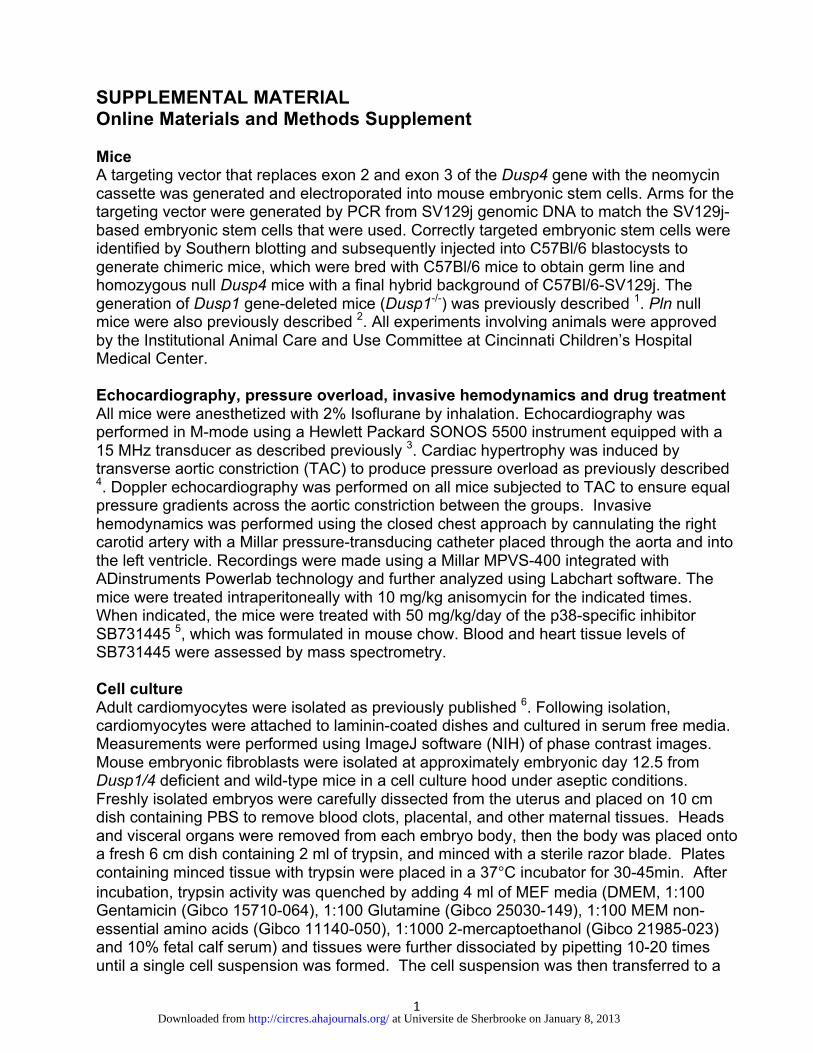
SUPPLEMENTAL MATERIAL Online Materials and Methods Supplement Mice A targeting vector that replaces exon 2 and exon 3 of the Dusp4 gene with the neomycin cassette was generated and electroporated into mouse embryonic stem cells. Arms for the targeting vector were generated by PCR from SV129j genomic DNA to match the SV129j-based embryonic stem cells that were used. Correctly targeted embryonic stem cells were identified by Southern blotting and subsequently injected into C57Bl/6 blastocysts to generate chimeric mice, which were bred with C57Bl/6 mice to obtain germ line and homozygous null Dusp4 mice with a final hybrid background of C57Bl/6-SV129j. The generation of Dusp1 gene-deleted mice (Dusp1-/-) was previously described 1. Pln null mice were also previously described 2. All experiments involving animals were approved by the Institutional Animal Care and Use Committee at Cincinnati Children’s Hospital Medical Center. Echocardiography, pressure overload, invasive hemodynamics and drug treatment All mice were anesthetized with 2% Isoflurane by inhalation. Echocardiography was performed in M-mode using a Hewlett Packard SONOS 5500 instrument equipped with a 15 MHz transducer as described previously 3. Cardiac hypertrophy was induced by transverse aortic constriction (TAC) to produce pressure overload as previously described 4. Doppler echocardiography was performed on all mice subjected to TAC to ensure equal pressure gradients across the aortic constriction between the groups. Invasive hemodynamics was performed using the closed chest approach by cannulating the right carotid artery with a Millar pressure-transducing catheter placed through the aorta and into the left ventricle. Recordings were made using a Millar MPVS-400 integrated with ADinstruments Powerlab technology and further analyzed using Labchart software. The mice were treated intraperitoneally with 10 mg/kg anisomycin for the indicated times. When indicated, the mice were treated with 50 mg/kg/day of the p38-specific inhibitor SB731445 5, which was formulated in mouse chow. Blood and heart tissue levels of SB731445 were assessed by mass spectrometry. Cell culture Adult cardiomyocytes were isolated as previously published 6. Following isolation, cardiomyocytes were attached to laminin-coated dishes and cultured in serum free media. Measurements were performed using ImageJ software (NIH) of phase contrast images. Mouse embryonic fibroblasts were isolated at approximately embryonic day 12.5 from Dusp1/4 deficient and wild-type mice in a cell culture hood under aseptic conditions. Freshly isolated embryos were carefully dissected from the uterus and placed on 10 cm dish containing PBS to remove blood clots, placental, and other maternal tissues. Heads and visceral organs were removed from each embryo body, then the body was placed onto a fresh 6 cm dish containing 2 ml of trypsin, and minced with a sterile razor blade. Plates containing minced tissue with trypsin were placed in a 37°C incubator for 30-45min. After incubation, trypsin activity was quenched by adding 4 ml of MEF media (DMEM, 1:100 Gentamicin (Gibco 15710-064), 1:100 Glutamine (Gibco 25030-149), 1:100 MEM non-essential amino acids (Gibco 11140-050), 1:1000 2-mercaptoethanol (Gibco 21985-023) and 10% fetal calf serum) and tissues were further dissociated by pipetting 10-20 times until a single cell suspension was formed. The cell suspension was then transferred to a
at Universite de Sherbrooke on January 8, 2013http://circres.ahajournals.org/Downloaded from

2
10 cm plate and MEF media was added to a final volume of 10 ml. Plates were incubated in 37°C incubator and cells were allowed to grow to confluency (3-4 days approximately). Passage 2 cells were harvested via trypsin and frozen down at 2x10⁶ cells per vial. Cells were split every 4 days until they reached senescence crisis (at 3 months approximately). When indicated, the MEFs were stimulated with 1 µg/ml of anisomycin for the indicated amount of time. Isolation of adult cardiomyocytes for contractility and Ca2+ measurements. Adult cardiomyocytes were isolated from Wt and DKO mice using a standard isolation procedure via perfusing whole hearts with a Tyrode’s solution containing liberase blendzyme (Roche) at 37°C (10-14 minutes total) as previously described 7. After perfusion, the ventricles were disassociated into individual myocytes, filtered and Ca2+ was reintroduced in incremental steps. Myocytes were then incubated with 2 µM Fura-2 acetoxymethyl ester (Invitrogen) and pluronic acid for 15 minutes in M199 media with 2,3-butanedione monoxime (BDM) at room temperature. After loading, the cells were washed and resuspended in Ringer’s solution. Electrical and caffeine (10 mM) stimulated Ca2+ transients were measured using a DeltaRam spectrofluorophotometer (Photon Technology International), operated at an emission wavelength of 510 nm, with excitation wavelengths of 340 and 380 nm. The stimulating frequency for Ca2+ transient measurements was 0.5 Hz. Baseline amplitude in the presence and absence of 50 µM SB239063 compound (calculated by change in base to peak in 340 nm/380 nm ratio) of the Ca2+ signal was acquired, and data were analyzed using Felix and Clampfit software. Contractility measurements were acquired using video edge detection as previously described 8. Myocytes were incubated for a minimum of 10 minutes in vehicle or SB compound before measurements were made. Western blotting Western blot analysis of mouse heart homogenates and cell cultures was performed as previously described 9. Antibodies used were phospho-ERK1/2 (Cell Signaling; 9101), ERK1/2 (Cell Signaling; 9102), phospho-p38 (Cell Signaling; 9211), p38 (Cell Signaling; 9212), phospho T222-MK2 (Cell Signaling; 3316), phospho T334-MK2 (Cell Signaling; 3007), MK2 (Cell Signaling; 3042), MEK1/2 (Cell Signaling; 9122), MKK4 (Cell Signaling; 9152), phospho-JNK1/2 (Promega; V7932), JNK1/2 (Cell Signaling; 9252), GAPDH (Fitzgerald; RDI-TRK5G4-6C5), phospho-MKK6 (Cell Signaling; 9231), MKK6 (Cell Signaling; 9264), α-actinin (Cell Signaling; 4051). mRNA expression analysis and histology RNA was extracted from ventricles using the RNeasy Kit according to manufacturer’s instructions (Qiagen). Reverse transcription was performed using the High Capacity cDNA Reverse Transcription Kit (Applied Biosystems). Gene expression was analyzed by real-time qPCR using SYBR green (Applied Biosystems). The expression of DUSP1 and DUSP4 was analyzed by PCR using the primers 5’-ctccaaggaggatatgaagcg-3’ (DUSP1-for), 5’-ctccagcatccttgatggagtc-3’ (DUSP1-rev), 5’-gtggaaatcctccctttcctctac-3’ (DUSP4-for), and 5’-gatgtcggccttgtggttgtcttc-3’ (DUSP4-rev). Standard histological methods were employed with H&E staining to show gross anatomical features of the heart, as well as without staining for assessment of TUNEL according to the CardioTACS (Trevigen). Statistical analysis
at Universite de Sherbrooke on January 8, 2013http://circres.ahajournals.org/Downloaded from

3
Data are represented as means ± SEM. A two-sample Student t test was used to compare means between 2 groups. 1-way ANOVA with Bonferroni correction was used for groups of 3 or more. P values less than 0.05 were considered significant. References 1. Dorfman K, Carrasco D, Gruda M, Ryan C, Lira SA, Bravo R. Disruption of the erp/mkp-1 gene does not affect mouse development: normal MAP kinase activity in ERP/MKP-1-deficient fibroblasts. Oncogene. 1996;13:925–931. 2. Luo W, Grupp IL, Harrer J, Ponniah S, Grupp G, Duffy JJ, Doetschman T, Kranias EG. Targeted ablation of the phospholamban gene is associated with markedly enhanced myocardial contractility and loss of beta-agonist stimulation. Circ Res. 1994;75:401-409. 3. Oka T, Maillet M, Watt AJ, Schwartz RJ, Aronow BJ, Duncan SA, Molkentin JD. Cardiac-specific deletion of Gata4 reveals its requirement for hypertrophy, compensation, and myocyte viability. Circ Res. 2006;98:837-845. 4. Wilkins BJ, Dai YS, Bueno OF, Parsons SA, Xu J, Plank DM, Jones F, Kimball TR, Molkentin JD. Calcineurin/NFAT coupling participates in pathological, but not physiological, cardiac hypertrophy. Circ Res. 2004;94:110-118. 5. Page TH, Brown A, Timms EM, Foxwell BMJ, Ray KP. Inhibitors of p38 suppress cytokine production in rheumatoid arthritis synovial membranes. Arth & Rheum. 2010;62:3221-3231. 6. Nakayama H, Bodi I, Correll RN, Chen X, Lorenz J, Houser SR, Robbins J, Schwartz A, Molkentin JD. alpha1G-dependent T-type Ca2+ current antagonizes cardiac hypertrophy through a NOS3-dependent mechanism in mice. J Clin Invest. 2009;119:3787-3796. 7. Goonasekera SA, Hammer K, Auger-Messier M, Bodi I, Chen X, Zhang H, Reiken S, Elrod JW, Correll RN, York AJ, Sargent MA, Hofmann F, Moosmang S, Marks AR, Houser SR, Bers DM, Molkentin JD. Decreased cardiac L-type Ca²⁺ channel activity induces hypertrophy and heart failure in mice. J Clin Invest. 2012;122:280-290. 8. Elrod JW, Wong R, Mishra S, Vagnozzi RJ, Sakthievel B, Goonasekera SA, Karch J, Gabel S, Farber J, Force T, Brown JH, Murphy E, Molkentin JD. Cyclophilin D controls mitochondrial pore-dependent Ca(2+) exchange, metabolic flexibility, and propensity for heart failure in mice. J Clin Invest. 2010;120:3680-3687. 9. Wilkins BJ, Dai YS, Bueno OF, Parsons SA, Xu J, Plank DM, Jones F, Kimball TR, Molkentin JD. Calcineurin/NFAT coupling participates in pathological, but not physiological, cardiac hypertrophy. Circ Res. 2004;94:110-118.
at Universite de Sherbrooke on January 8, 2013http://circres.ahajournals.org/Downloaded from

TAC TAC TAC TAC– – – –DKO DKOWtWt
nucleus cytoplasm
p-p38p38
Lamin A/Cα-actinin
* *
pT334-MK2
MK2^ ^ ^
^ ^ ^
^ ^ ^
^ ^ ^
Online Figure I. Western blot analysis of the indicated proteins and phospho-proteins from hearts of adult mice subjected to sham or transverse aorticconstriction (TAC, 15 minutes of stimulation) in Wt or Dusp1/4-/- animals.Asterisks and arrow heads in the blots show bands that are differentiallyregulated by loss of Dusp1/4, while the open up-arrows show regulation andgreater nuclear occupancy of MK2 associated with deletion of Dusp1/4. LaminA/C western shows purity of the nuclear extract, while α-actinin shows purity ofthe cytoplasmic extract. The results again suggest that loss of Dusp1/4upregulates p38 activity in the heart at baseline, and even greater activationwith TAC stimulation for 15 minutes.
at Universite de Sherbrooke on January 8, 2013http://circres.ahajournals.org/Downloaded from

05
10152025303540
Wt DKO
2.5 mo.8 mo.
TUN
EL+
/105
nucl
ei
4 4 4 5
Online Figure II. Quantitation of TUNEL levels in histological sections fromhearts of Wt and Dusp1/4 DKO mice at 2.5 and 8 months of age. At least 4full sections were analyzed for each heart, and the total number of heartsanalyzed is shown in the bars for each genotype or time point. Nodifferences were observed, as the Dusp1/4-/- hearts were largely devoid ofsignificant ongoing cell death.
at Universite de Sherbrooke on January 8, 2013http://circres.ahajournals.org/Downloaded from

3456789
10
Wt Dusp1-/- Dusp4-/- DKO Wt Dusp1-/- Dusp4-/- DKO
ShamTAC 14days
19 11
* * * **#
Hw
/Bw
(mg/
g)
9 7 7 8 10 9
Online Figure III. Quantitation of heart weight to body weight (Hw/Bw) ratios in 2month-old Wt, Dusp1-/-, Dusp4-/-, and Dusp1/4-/- (DKO) mice subjected to Sham or TACsurgery for 14 days (*p<0.01 vs. Sham; #p<0.01 vs. other genotypes with sameprocedure). The number of animals is indicated in the bars. Double null mice (DKO)that survived the TAC procedure for 14 days showed much greater increases in heartweights.
at Universite de Sherbrooke on January 8, 2013http://circres.ahajournals.org/Downloaded from
Copyright © 2022 FDOKUMEN