Nitrosative stress elicited by nNOSµ delocalization inhibits muscle force in dystrophin-null mice
18
Nitrosative stress elicited by nNOSμ delocalization inhibits muscle force in dystrophin-null mice Dejia Li, Yongping Yue, Yi Lai, Chady H Hakim, and Dongsheng Duan * Department of Molecular Microbiology and Immunology, School of Medicine, The University of Missouri, Missouri, USA Abstract The mechanism of force reduction is not completely understood in Duchenne muscular dystrophy (DMD), a dystrophin-deficient lethal disease. Nitric oxide regulates muscle force. Interestingly, neuronal nitric oxide synthase μ (nNOSμ), a major source of muscle nitric oxide, is lost from the sarcolemma in DMD muscle. We hypothesize that nNOSμ delocalization contributes to force reduction in DMD. To test this hypothesis, we generated dystrophin/nNOSμ double knockout mice. Genetic elimination of nNOSμ significantly enhanced force in dystrophin-null mice. Pharmacological inhibition of nNOS yielded similar results. To further test our hypothesis, we studied δ-sarcoglycan-null mice, a model of limb-girdle muscular dystrophy. These mice had minimal sarcolemmal nNOSμ delocalization and muscle force was less compromised. Annihilation of nNOSμ did not improve their force either. To determine whether nNOSμ delocalization itself inhibited force, we corrected muscle disease in dystrophin-null mice with micro-dystrophins that either restored or did not restore sarcolemmal nNOSμ. Similar muscle force was obtained irrespective of nNOSμ localization. Additional studies suggest that nNOSμ delocalization selectively inhibits muscle force in dystrophin-null mice via nitrosative stress. In summary, we have demonstrated for the first time that nitrosative stress elicited by nNOSμ delocalization is an important mechanism underlying force loss in DMD. Keywords dystrophin; Duchenne muscular dystrophy; nitrosative stress; nitrotyrosination; nNOS; δ- sarcoglycan; mdx; mdx4cv; ryanodine receptor 1; S-nitrosylation Introduction Duchenne muscular dystrophy (DMD) is a lethal muscle disease caused by dystrophin gene mutation [1]. Dystrophin is a sub-sarcolemmal protein that connects the extracellular matrix to the cytoskeleton. The absence of dystrophin weakens the sarcolemmal integrity. This mechanical defect has been suggested as the primary pathogenic underpinning for DMD [2], yet the full-spectrum of DMD pathophysiology remains elusive [3]. Copyright © 2010 Pathological Society of Great Britain and Ireland. Published by John Wiley & Sons, Ltd. * Correspondence to: Dongsheng Duan, PhD, Professor, Department of Molecular Microbiology and Immunology, One Hospital Drive, Columbia, MO 65212, USA. [email protected]. No conflicts of interest were declared. Author contribution statement DD and LD designed the study. CH, DD, LD, YL, and YY carried out experiments. DD, LD, YL, and YY analysed data. DD wrote the paper. NIH Public Access Author Manuscript J Pathol. Author manuscript; available in PMC 2012 January 1. Published in final edited form as: J Pathol. 2011 January ; 223(1): 88–98. doi:10.1002/path.2799. NIH-PA Author Manuscript NIH-PA Author Manuscript NIH-PA Author Manuscript
-
Upload
independent -
Category
Documents
-
view
2 -
download
0
Transcript of Nitrosative stress elicited by nNOSµ delocalization inhibits muscle force in dystrophin-null mice

Nitrosative stress elicited by nNOSμ delocalization inhibitsmuscle force in dystrophin-null mice
Dejia Li, Yongping Yue, Yi Lai, Chady H Hakim, and Dongsheng Duan*
Department of Molecular Microbiology and Immunology, School of Medicine, The University ofMissouri, Missouri, USA
AbstractThe mechanism of force reduction is not completely understood in Duchenne muscular dystrophy(DMD), a dystrophin-deficient lethal disease. Nitric oxide regulates muscle force. Interestingly,neuronal nitric oxide synthase μ (nNOSμ), a major source of muscle nitric oxide, is lost from thesarcolemma in DMD muscle. We hypothesize that nNOSμ delocalization contributes to forcereduction in DMD. To test this hypothesis, we generated dystrophin/nNOSμ double knockoutmice. Genetic elimination of nNOSμ significantly enhanced force in dystrophin-null mice.Pharmacological inhibition of nNOS yielded similar results. To further test our hypothesis, westudied δ-sarcoglycan-null mice, a model of limb-girdle muscular dystrophy. These mice hadminimal sarcolemmal nNOSμ delocalization and muscle force was less compromised.Annihilation of nNOSμ did not improve their force either. To determine whether nNOSμdelocalization itself inhibited force, we corrected muscle disease in dystrophin-null mice withmicro-dystrophins that either restored or did not restore sarcolemmal nNOSμ. Similar muscleforce was obtained irrespective of nNOSμ localization. Additional studies suggest that nNOSμdelocalization selectively inhibits muscle force in dystrophin-null mice via nitrosative stress. Insummary, we have demonstrated for the first time that nitrosative stress elicited by nNOSμdelocalization is an important mechanism underlying force loss in DMD.
Keywordsdystrophin; Duchenne muscular dystrophy; nitrosative stress; nitrotyrosination; nNOS; δ-sarcoglycan; mdx; mdx4cv; ryanodine receptor 1; S-nitrosylation
IntroductionDuchenne muscular dystrophy (DMD) is a lethal muscle disease caused by dystrophin genemutation [1]. Dystrophin is a sub-sarcolemmal protein that connects the extracellular matrixto the cytoskeleton. The absence of dystrophin weakens the sarcolemmal integrity. Thismechanical defect has been suggested as the primary pathogenic underpinning for DMD [2],yet the full-spectrum of DMD pathophysiology remains elusive [3].
Copyright © 2010 Pathological Society of Great Britain and Ireland. Published by John Wiley & Sons, Ltd.*Correspondence to: Dongsheng Duan, PhD, Professor, Department of Molecular Microbiology and Immunology, One Hospital Drive,Columbia, MO 65212, USA. [email protected] conflicts of interest were declared.Author contribution statementDD and LD designed the study. CH, DD, LD, YL, and YY carried out experiments. DD, LD, YL, and YY analysed data. DD wrotethe paper.
NIH Public AccessAuthor ManuscriptJ Pathol. Author manuscript; available in PMC 2012 January 1.
Published in final edited form as:J Pathol. 2011 January ; 223(1): 88–98. doi:10.1002/path.2799.
NIH
-PA Author Manuscript
NIH
-PA Author Manuscript
NIH
-PA Author Manuscript

Besides providing a mechanical support to the sarcolemma, dystrophin helps recruit muscle-specific neuronal nitric oxide synthase μ (nNOSμ) to the membrane [4]. In DMD, nNOSμdelocalizes from the sarcolemma and the relative cytosolic NOS activity is elevated [5–12].When activated by calcium, nNOS generates nitric oxide (NO), a major muscle oxidant anda critical muscle force regulator [13–15]. Currently, it is not clear whether the relativeincrease of cytosolic NOS activity affects force generation in DMD. Herein, we hypothesizethat nNOSμ delocalization contributes to muscle weakening in DMD.
To test this hypothesis, we crossed nNOSμ knockout mice with dystrophin-deficient mdx4cvmice and generated nNOSμ/dystrophin double-deficient (n-dko) mice [9,16–18]. In supportof our hypothesis, the specific muscle forces were significantly improved when nNOSμ waseliminated. Administration of the nNOS inhibitor 7-nitroindazole (7-NI) to dystrophin-deficient mdx mice also increased specific muscle force. To further understand therelationship between nNOSμ delocalization and force loss in dystrophic muscle, weeliminated nNOSμ from δ-sarcoglycan-null mice, a model for limb girdle musculardystrophy 2F. In contrast to dystrophin-deficient mice, sarcolemmal nNOSμ was largelypreserved and muscle strength was less compromised in δ-sarcoglycan-null mice [19].Importantly, muscle force was not improved in δ-sarcoglycan/nNOSμ double-deficient mice(δ/n-dko). To determine whether delocalized nNOSμ inhibited force in a non-dystrophicmuscle, we corrected muscle disease in mdx mice with transgenic micro-dystrophinexpression. Sarcolemmal nNOSμ expression was restored in the R16–17/ΔC, but not theΔR4–23/ΔC, strain. However, both strains yielded identical muscle forces. Collectively, ourdata suggest that nNOSμ delocalization selectively weakened the force of dystrophicmuscle. To elucidate the underlying mechanism(s), we compared muscle pathology,utrophin expression, and markers for nitrosative stress in mdx4cv and n-dko mice. Therewas a nominal difference in muscle pathology [9,18]. Utrophin levels were not up-regulatedin n-dko mice either. However, protein nitrotyrosination and ryanodine receptor 1 (RyR1) S-nitrosylation were greatly mitigated in n-dko mice. In summary, we have revealed nNOSμdelocalization as a previously unappreciated mechanism underlying force loss in DMD. Inthis model, delocalized nNOSμ is activated by high cytosolic free calcium in dystrophicmuscle. This leads to supra-physiological levels of reactive nitrogen species (RNS)production. Muscle force is weakened by RNS-mediated nitrosative modification of RyR1and other yet to be defined contractile proteins.
Materials and methodsAnimals
All animal experiments were approved by the institutional animal care and use committeeand were in accordance with NIH guidelines. The breeding pairs for C57Bl/6 (BL6), C57Bl/10 (BL10), mdx4cv, mdx, δ-SG KO, and nNOSμ KO mice were purchased from TheJackson Laboratory (Bar Harbor, ME, USA). The experimental n-dko and δ/n-dko micewere generated by crossing nNOSμ KO mice with mdx4cv and δ-SG KO mice, respectively.The founder lines of human micro-dystrophin transgenic mice were generated at theUniversity of Missouri transgenic core. The micro-gene expression was controlled by theskeletal specific human skeletal α-actin promoter and the bovine growth hormone pA [20].The experimental microgene transgenic mice were obtained after backcrossing with mdxmice for at least five generations. Only male mice were used in the study. Mouse age andsample size for each experimental group are detailed in Table 1.
To inhibit nNOS activity, 7-NI (5 mg/ml, dissolved in peanut oil; Sigma, St Louis, MO,USA) was injected into 7-month-old mdx mice intraperitoneally at a dose of 50 mg/kg daily.Control mdx mice were injected with the same volume of peanut oil. After 1 month oftreatment, mice were euthanized for functional assay.
Li et al. Page 2
J Pathol. Author manuscript; available in PMC 2012 January 1.
NIH
-PA Author Manuscript
NIH
-PA Author Manuscript
NIH
-PA Author Manuscript

Western blotWhole muscle lysate and membrane enriched microsomal preparations were preparedaccording to published protocols [4,19,21,22]. Full-length wild-type dystrophin was detectedwith a mouse monoclonal antibody against the dystrophin C-terminal domain (Dys-2, 1:100; Novocastra, Newcastle, UK). Human micro-dystrophin was detected with a humandystrophin N-terminal specific monoclonal antibody (Dys-3, 1: 100; Novocastra). Utrophinwas detected with a mouse monoclonal antibody against utrophin amino acid residues 768–874 (1: 200; BD Biosciences, San Diego, CA, USA). nNOSμ was detected with a rabbitpolyclonal antibody against the N-terminal end of nNOS (1: 1000; Upstate, Lake Placid,NY, USA). The size of the band suggests that it is nNOSμ rather than nNOSβ. α-Sarcoglycan was detected with a mouse monoclonal antibody (1: 1000; Vector Laboratories,Burlingame, CA, USA). 3-Nitrotyrosine was detected with a mouse monoclonal antibody (1:3000; Caymen Chemicals, Ann Arbor, MI, USA).
To detect total cellular RyR1, the sarcoplasmic reticulum membrane fraction was preparedas described by Saito et al in the presence of 1% protease inhibitor (Roche, Indianapolis, IN,USA) [23]. To detect S-nitrosylated RyR1, the sarcoplasmic reticulum membrane fractionwas further purified using a resin-assisted capture method as reported by Forrester et al [24].RyR1 was detected with a mouse monoclonal antibody (1: 1000; Affinity Bioreagents,Golden, CO, USA). The loading controls included a monoclonal antibody against α-tubulin(1: 3000; Sigma), a monoclonal antibody against α-actin (1: 3000; Sigma), and rapid bluestaining of duplicated gels (Geno Technology, St Louis, MO, USA).
Immunofluorescence staining and in situ NOS activity stainingImmunofluorescence staining was performed essentially as described previously [19,22].Full-length and micro-dystrophin were revealed with Dys-2 (1: 30) and Dys-3 (1: 20),respectively. Utrophin was revealed with a mouse monoclonal antibody against the utrophinN-terminal domain (1: 20; Vector Laboratories). nNOS was revealed with a rabbitpolyclonal antibody against an epitope near the C-terminal end of nNOS (1: 2000; SantaCruz Biotechnology, Santa Cruz, CA, USA). NOS activity staining was performed oncryosections according to our published protocol [4]. The histochemical staining revealedthe NADPH diaphorase activity of NOS.
Histopathology and serum creatine kinase (CK) assayCentral nucleation quantification was performed as described previously [25]. Serum CKlevel was determined using a CK liqui-UV test kit from Stanbio Laboratory (Boerne, TX,USA) as described before [22].
Muscle force measurementTwitch and tetanic forces of the extensor digitorium longus (EDL) muscle were measured exvivo using a 305B dual-mode servomotor transducer (Aurora Scientific Inc, Aurora, Ontario,Canada) according to our published protocol [19,22,25]. Twitch and tetanic forces of thetibialis anterior (TA) muscle were measured in situ with a 305C-LR dual-mode servomotortransducer (Aurora Scientific Inc) according to a published protocol [26]. The specificmuscle force was calculated by dividing the absolute muscle force by the muscle cross-sectional area (CSA) (kN/m2). Muscle CSA was calculated according to the followingequation: CSA = (muscle mass, in g)/[(muscle density, in g/cm3) × (length ratio) × (optimalmuscle length, in cm)]. 1.06 g/cm3 is used as the muscle density [27]. The length ratio refersto the ratio of optimal fiber length to optimal muscle length. The length ratios for the EDLand TA muscles are 0.44 and 0.6, respectively [28,29].
Li et al. Page 3
J Pathol. Author manuscript; available in PMC 2012 January 1.
NIH
-PA Author Manuscript
NIH
-PA Author Manuscript
NIH
-PA Author Manuscript

Statistical analysisData are presented as mean ± standard error of the mean. Statistical analysis was performedwith SPSS software (SPSS, Chicago, IL, USA). Statistical significance among multiplegroups was determined by one-way ANOVA followed by Bonferroni post-hoc analysis. Fortwo-group comparison, statistical significance was determined by the Student t -test.Difference was considered significant when p < 0.05.
ResultsSpecific forces of dystrophin-deficient mice are enhanced by nNOSμ elimination or nNOSinhibition
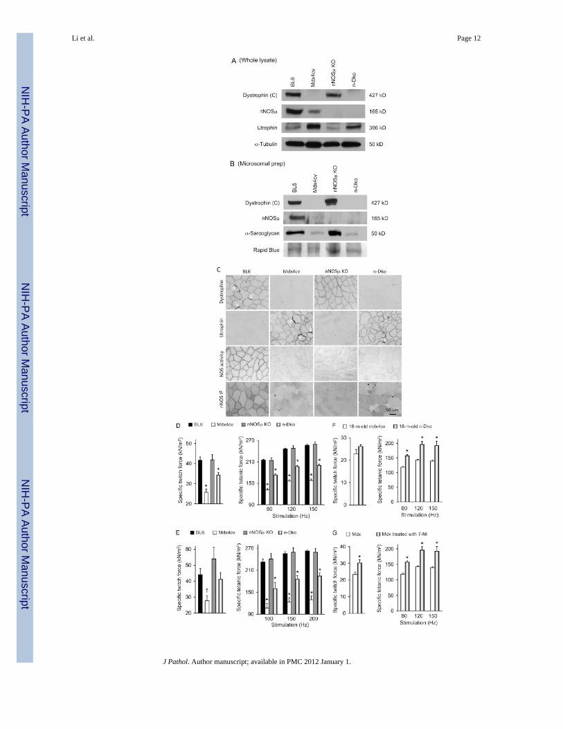
In dystrophin-deficient mice, nNOSμ was lost from the sarcolemma. However, it was notcompletely eliminated from muscle cells (Figures 1A–1C) [5–7]. To determine whethermislocalized nNOSμ affected muscle contractility, we generated n-dko mice by crossingnNOSμ-null mice with mdx4cv mice [16,17] (Figures 1A–1C). Both parental strains wereon the BL6 background.
We first examined force in young adult male mice (Figures 1D, 1E, and Table 1). The EDLmuscle of n-dko mice yielded significantly greater specific twitch and tetanic forces thanthat of mdx4cv mice (Figure 1D). Similar results were obtained from the TA muscle,although the improvement in specific twitch force appeared marginal in the TA muscle(Figure 1E). At 18 months of age, the EDL muscle of n-dko mice also showed significantlyhigher specific tetanic forces than that of mdx4cv mice (Figure 1F). However, geneticelimination of nNOSμ did not change the eccentric contraction profile (Supportinginformation, Supplementary Figures 1A–1C).
To validate the results of n-dko mice, we treated adult mdx mice with daily administrationof 7-NI. Mdx mice are on the BL10 background and they carry a different mutation. Onemonth after treatment, specific forces were significantly enhanced (Figure 1G).Interestingly, 7-NI-treated mdx mice appeared to be more resistant to eccentric contractioninjury during the first two rounds of lengthening contraction (Supporting information,Supplementary Methods, Supplementary Figure 1D).
Genetic elimination of nNOSμ does not increase specific muscle force in δ-sarcoglycanknockout mice
To further decipher the relationship between nNOSμ delocalization and force loss indystrophic mice, we created δ/n-dko mice by crossing nNOSμ-null mice with BL6background δ-sarcoglycan-null mice (Figure 2). We have previously shown that age- andgender-matched δ-sarcoglycan-null mice yielded significantly higher specific twitch andtetanic forces than those of mdx4cv mice [19]. Interestingly, nNOSμ was minimallydelocalized from the sarcolemma in δ-sarcoglycan-null mice (Figures 2A and 2B). Geneticelimination of nNOSμ did not increase specific force of δ-sarcoglycan-null mice either(Figure 2C).
nNOSμ delocalization alone is not sufficient to inhibit force in non-dystrophic muscleTo determine whether nNOSμ mislocalization per se impeded force production, wegenerated two strains of micro-dystrophin transgenic mdx mice. One strain expressed theR16–17/ΔC microgene which contains the nNOS-binding domain. The other strainexpressed the ΔR4–23/ΔC microgene that does not carry the nNOS-binding domain (Figure3A). Similar levels of micro-dystrophin expression were detected (Figures 3B–3D). Musclehistopathology was also effectively ameliorated in both strains (data not shown) [4].However, sarcolemmal nNOSμ expression was restored only in the R16–17/ΔC strain
Li et al. Page 4
J Pathol. Author manuscript; available in PMC 2012 January 1.
NIH
-PA Author Manuscript
NIH
-PA Author Manuscript
NIH
-PA Author Manuscript

(Figure 3C; data not shown for nNOS immunostaining). NOS activity was detected at thesarcolemma in the R16–17/ΔC, but not the ΔR4–23/ΔC, strain (Figure 3D). Interestingly,despite a considerably high cytosolic nNOSμ level, the ΔR4–23/ΔC transgenic mice yieldeda contractile profile identical to that of the R16–17/ΔC strain (Figure 3E and Supportinginformation, Supplementary Methods, Supplementary Figure 1E).
Delocalized nNOSμ induces nitrosative stress in dystrophin-deficient miceOur results so far suggest that nNOSμ delocalization selectively inhibits force production indystrophin-deficient muscle. To understand why nNOSμ delocalization reduced force indystrophic muscle, we compared muscle pathology in mdx4cv and n-dko mice. Consistentwith previous reports [9,18], we observed similar levels of myofiber size variation, centralnucleation, inflammation, regeneration, and fibrosis (Figure 4A and Supporting information,Supplementary Methods, Supplementary Figure 2). We also measured the severity ofsarcolemmal leakage by immunoglobulin infiltration and the serum CK level. There wasalso no significant difference between mdx4cv and n-dko mice (Figure 4A and Supportinginformation, Supplementary Methods, Supplementary Figure 2). Utrophin is a knowncompensatory protein for dystrophin. Its up-regulation may explain force gain in n-dkomice. To our surprise, utrophin appeared slightly reduced in n-dko mice compared with thatof mdx4cv mice (Figure 1A).
NO is one of the primary free radicals in muscle [14,15]. It is also a well-establishedregulator of muscle contractility [13,30]. To determine whether disruption of NO redoxhomeostasis contributed to our observation, we examined several nitrosative stress markersin mdx4cv and n-dko mice. 3-Nitrotyrosine is generated when the tyrosine residues aremodified by reactive nitrogen species (RNS) [13,14]. On western blot, we observedprominent 3-nitrotyrosine bands in mdx4cv mice (Figure 4B) [31,32]. Total cellular RyR1levels were quite comparable among the different strains. However, S-nitrosylated RyR1was greatly increased in mdx4cv mice (Figure 4C) [24,33]. Importantly, proteinnitrotyrosination and RyR1 S-nitrosylation were minimized in n-dko mice (Figures 4B and4C).
In the ΔR4–23/ΔC strain of micro-dystrophin transgenic mdx mice, nNOSμ delocalizationdid not reduce force (Figure 3E). One likely explanation was that delocalized nNOSμ didnot induce nitrosative stress in this context. We evaluated protein nitrotyrosination (Figure4D). Indeed, minimal 3-nitrotyrosine bands were detected in ΔR4–23/ΔC microgenetransgenic mdx mice (Figure 4D). Collectively, these results suggest that nNOSμdelocalization inhibits force via elevated RNS and subsequent nitrosative modification(Figure 5). In the absence of nitrosative stress, muscle force is not compromised.
DiscussionnNOS is a well-established signalling molecule [13,34]. It plays an important role in manyaspects of skeletal muscle activities [13]. Several different nNOS isoforms have beenidentified in skeletal muscle. These include 135 kD nNOSβ and 165 kD nNOSμ. nNOSβdoes not contain the PDZ domain and it resides within the Golgi complex [35]. nNOSμ isthe predominant nNOS isoform in muscle. It carries a unique N-terminal PDZ domain. Theβ-finger of the nNOSμ PDZ domain interacts with the PDZ domain of α-syntrophin [36,37].The groove of the nNOSμ PDZ domain interacts with dystrophin spectrin-like repeats 16/17[4]. Through these interactions, nNOSμ is anchored to the sarcolemma. Victor and co-workers examined the haemodynamic response during muscle contraction in mdx mice andDMD patients [10,38]. They demonstrated that NO generated from nNOSμ counteracts α-adrenergic vaso-constriction and hence prevents functional ischaemia in contracting muscle.The loss of dystrophin leads to nNOSμ delocalization from the sarcolemma. In the absence
Li et al. Page 5
J Pathol. Author manuscript; available in PMC 2012 January 1.
NIH
-PA Author Manuscript
NIH
-PA Author Manuscript
NIH
-PA Author Manuscript

of NO, muscle vessels cannot fully relax during contraction. Eventually, it leads to focalischaemic injury in myofibres that are already weakened by the loss of dystrophin. Recentstudies have further provided the physiological relevance of membrane-associated nNOSμ inmuscle fatigue and muscular dystrophy [4,39,40].
In addition to facilitating blood perfusion, NO also facilitates muscle repair by promotingsatellite cell activation and fusion, increasing trophic differentiating factors (follistatin andhepatocyte growth factor) and modifying chromatin [41–44]. As a matter of fact, severalstudies have revealed a beneficial effect of NO donors or NO-releasing agents (such asarginine) in mdx mice [45–48]. Wehling et al overexpressed nNOS in the cytoplasm and thesarcolemma via the transgenic approach. They found that supra-physiological levels ofnNOS mitigated histological lesions in mdx mice [49]. Collectively, these studies support aNO and/or nNOS-based therapy for DMD.
While increasing NO/nNOS levels appear beneficial, there is also evidence suggestingpossible disadvantages. Stamler and co-workers have provided convincing evidence that NOinhibits muscle force [50]. Furthermore, they demonstrated that NO-mediated forceinhibition occurs under high oxygen tension (a situation mimicking oxidative/nitrosativestress) [51]. Recent studies have begun to elucidate the underlying molecular mechanism[52,53]. Specifically, it was found that NO impairs skeletal muscle contractility vianitrosylative modification of RyR1 [33,52–54]. In addition to a negative effect on forceproduction, supra-physiological nNOS may directly contribute to muscular dystrophy. Inthis regard, Sunada et al have found that nNOS overactivation is involved in caveolin-3mutation-related limb-girdle muscular dystrophy [55]. A recent study by Wehling-Henrickset al also suggests that dietary supplementation of arginine, a NO donor, exacerbates fibrosisin mdx mice [56].
In dystrophin-deficient muscle, nNOSμ is lost from the sarcolemma but it is not eliminatedfrom muscle cells (Figures 1A–1C). Actually, it has been shown that the relative cytosolicNOS activity is elevated in dystrophin-null muscle [5–12]. While the loss of sarcolemmalnNOSμ causes ischaemic muscle injury, it remains controversial whether delocalizednNOSμ contributes to muscle disease in DMD [4,9,10,18].
The loss of muscle force is a major clinical manifestation of DMD. However, the molecularmechanisms underlying muscle force reduction are not completely understood. Stamler andco-workers have provided convincing evidence that NO inhibits muscle force under highoxygen tension [50,51]. Recent studies suggest that NO impairs skeletal muscle contractilityvia nitrosylative inhibition of RyR1 [33,53,54]. In DMD muscle, delocalized nNOSμ, highcytosolic free calcium, and elevated oxidative stress seem to have provided a great milieufor RNS-mediated RyR1 hyper-nitrosylation and force inhibition (Figure 5).
To determine whether delocalized nNOSμ has indeed contributed to force loss indystrophin-deficient mice, we used two independent approaches. First, we completelyremoved nNOSμ by crossing mdx4cv mice to the nNOSμ-null background. Second, weinactivated nNOS activity in mdx mice with 7-NI, a pharmacological nNOS inhibitor. Inboth cases, we observed a significant increase of specific muscle force (Figure 1).
Our hypothesis would predict nNOSμ delocalization as the initiating event that starts thechain reaction leading to nitrosative inhibition of muscle force (Figure 5). In the absence ofsignificant nNOSμ delocalization, muscle force should be better preserved. The results fromδ-sarcoglycan-null mice supported this argument. First, δ-sarcoglycan-deficient miceretained significant levels of sarcolemmal nNOSμ expression (Figures 2A and 2B). Second,δ-sarcoglycan-null mice yielded higher specific forces than mdx4cv mice despite the factthat both dystrophic models had similar muscle pathology such as elevated calcium,
Li et al. Page 6
J Pathol. Author manuscript; available in PMC 2012 January 1.
NIH
-PA Author Manuscript
NIH
-PA Author Manuscript
NIH
-PA Author Manuscript

myofibre necrosis, and inflammation [19,57–61]. Third, genetic elimination of nNOSμ fromδ-sarcoglycan-deficient mice had nominal impact on specific muscle force (Figure 2C).
To study the molecular mechanism(s) underlying nNOSμ delocalization-associated forceloss, we compared muscle pathology and utrophin expression between mdx4cv and n-dkomice. We did not find any evidence suggesting that improved muscle force in n-dko micewas due to histopathology amelioration or utrophin up-regulation (Figures 1A, 1C, and 4A,and Supporting information, Supplementary Methods, Supplementary Figure 2).Interestingly, several previous studies also revealed a disconnection between muscle forceand the histological lesion [19,22,54,62,63]. Hack et al first reported that muscledegeneration in γ-sarcoglycan-deficient mice did not lead to specific force reduction [63].Bogdanovich et al found that myostatin inhibition therapy increased muscle strength in γ-sarcoglycan-deficient mice but did not improve muscle pathology [62]. We also founddiscordance between the severity of muscle pathology and muscle force in mdx3cv,mdx4cv, and δ-sarcoglycan-null mice [19,22]. Bellinger et al further show that thedisconnection between force and histology may happen in normal mice. After 3 weeks ofchronic exercise, the authors observed a significant reduction of the EDL muscle specificforce but they did not detect any morphological change [54].
The primary cellular function of nNOS is to generate NO. Interaction of NO with reactiveoxygen species (ROS) produces highly reactive RNS such as peroxynitrite [13,15]. Hence,we investigated whether nNOSμ delocalization-associated force reduction is mediated bynitrosative stress. nNOS is activated by free calcium. While the cellular free calcium level isabnormally elevated in dystrophin-deficient muscle, recent studies suggest that theabbreviated synthetic dystrophin genes can rectify calcium defects [4,20,64–69]. Wegenerated a pair of micro-dystrophin transgenic mdx mice. One strain had normal nNOSμlocalization at the sarcolemma, while the other strain contained delocalized cytosolicnNOSμ (Figure 3). We have previously shown that these microgenes can effectivelyameliorate muscular dystrophy in mouse models of DMD [4,20]. We expected that acorrection of muscle disease would normalize calcium overload and oxidative stress.Consequently, it would prevent abnormal nNOS activation, RNS production, and nitrosativeforce inhibition even if there was delocalized nNOSμ. Indeed, we obtained the same muscleforces from both transgenic strains (Figure 3).
To further demonstrate the involvement of RNS hypernitrosylation, we measured nitrosativestress markers in mdx4cv and n-dko mice. Compared with mdx4cv mice, aberrantnitrotyrosination and RyR1 S-nitrosylation were corrected in n-dko mice (Figures 4B and4C). Evaluation of protein nitrotyrosination in micro-dystrophin transgenic mdx mice lentadditional support to our hypothesis. Irrespective of subcellular nNOSμ localization,minimal 3-nitrotyrosine bands were detected in these mice (Figure 4D).
In summary, we propose a model of RNS-mediated force inhibition in DMD (Figure 5). Indystrophin-deficient muscle, elevated free calcium activates delocalized nNOSμ andgenerates excessive NO. In the presence of ROS, NO is converted to RNS. Subsequentnitrosative modification of contractile proteins (in particular, S-nitrosylation of RyR1)further reduces the already compromised force (Figure 5). Genetic elimination of nNOSμ oradministration of the nNOS inhibitor 7-NI blocks the pathway and improves muscle force.In δ-sarcoglycan-deficient mice, the pathway is not initiated due to the absence of nNOSμdelocalization. The ΔR4–23/ΔC micro-dystrophin therapy corrects calcium mishandling andreduces ROS. As a result, nitrosative stress is prevented and muscle force is preserveddespite the failure of this microgene to anchor nNOSμ to the sarcolemma.
Li et al. Page 7
J Pathol. Author manuscript; available in PMC 2012 January 1.
NIH
-PA Author Manuscript
NIH
-PA Author Manuscript
NIH
-PA Author Manuscript

One potential confounding factor that is not studied here is inducible NOS (iNOS). As aconsequence of muscle inflammation, the iNOS level is elevated in dystrophin-deficientmuscle. This likely represents another important source of NO and RNS production [33].Assessing muscle force in iNOS/dystrophin double-deficient mice in the future may providea clue on the relative contribution of iNOS. Nevertheless, the results from our δ/n-dko micesuggest that participation from iNOS may be limited. Muscle inflammation is observed inboth δ-sarcoglycan-null mice and dystrophin-null mice. Genetic elimination of nNOSμshould not affect the iNOS level. If iNOS, rather than delocalized nNOSμ, plays adetermining role in NO/RNS-mediated force inhibition, then as in the case of δ/n-dko micewe would expect n-dko muscle to yield similar force to dystrophin-deficient muscle. This iscontradictory to our experimental results.
Supplementary MaterialRefer to Web version on PubMed Central for supplementary material.
AcknowledgmentsThis work was supported by grants from the National Institutes of Health (AR-49419; DD) and the MuscularDystrophy Association (DD). Chady Hakim was partially supported by a NIH training grant (T90DK70105). Wethank Dr Steve Yang for his help with the in situ TA muscle function assay. We thank Ms Chun Long and MsKeqing Zhang for excellent technical assistance. We thank Drs Elizabeth S Critser and Natalia Karasseva at theUniversity of Missouri Transgenic Animal Core for their help with generating the founder transgenic mice. Wethank Robert J McDonald, Jr, MD for his generous support to Duchenne muscular dystrophy research in the Duanlab.
ReferencesNote: References 1–9 are cited in the supporting information to this article.
1. Kunkel LM. 2004 William Allan Award address. Cloning of the DMD gene. Am J Hum Genet.2005; 76:205–214. [PubMed: 15714686]
2. Petrof BJ, Shrager JB, Stedman HH, et al. Dystrophin protects the sarcolemma from stressesdeveloped during muscle contraction. Proc Natl Acad Sci U S A. 1993; 90:3710–3714. [PubMed:8475120]
3. Rando TA. Role of nitric oxide in the pathogenesis of muscular dystrophies: a ‘two hit’ hypothesisof the cause of muscle necrosis. Microsc Res Tech. 2001; 55:223–235. [PubMed: 11748861]
4. Lai Y, Thomas GD, Yue Y, et al. Dystrophins carrying spectrin-like repeats 16 and 17 anchor nNOSto the sarcolemma and enhance exercise performance in a mouse model of muscular dystrophy. JClin Invest. 2009; 119:624–635. [PubMed: 19229108]
5. Brenman JE, Chao DS, Xia H, et al. Nitric oxide synthase complexed with dystrophin and absentfrom skeletal muscle sarcolemma in Duchenne muscular dystrophy. Cell. 1995; 82:743–752.[PubMed: 7545544]
6. Chao DS, Gorospe JR, Brenman JE, et al. Selective loss of sarcolemmal nitric oxide synthase inBecker muscular dystrophy. J Exp Med. 1996; 184:609–618. [PubMed: 8760814]
7. Chang WJ, Iannaccone ST, Lau KS, et al. Neuronal nitric oxide synthase and dystrophin-deficientmuscular dystrophy. Proc Natl Acad Sci U S A. 1996; 93:9142–9147. [PubMed: 8799168]
8. Kameya S, Miyagoe Y, Nonaka I, et al. Alpha1-syntrophin gene disruption results in the absence ofneuronal-type nitric-oxide synthase at the sarcolemma but does not induce muscle degeneration. JBiol Chem. 1999; 274:2193–2200. [PubMed: 9890982]
9. Crosbie RH, Straub V, Yun HY, et al. mdx muscle pathology is independent of nNOS perturbation.Hum Mol Genet. 1998; 7:823–829. [PubMed: 9536086]
10. Thomas GD, Sander M, Lau KS, et al. Impaired metabolic modulation of alpha-adrenergicvasoconstriction in dystrophin-deficient skeletal muscle. Proc Natl Acad Sci U S A. 1998;95:15090–15095. [PubMed: 9844020]
Li et al. Page 8
J Pathol. Author manuscript; available in PMC 2012 January 1.
NIH
-PA Author Manuscript
NIH
-PA Author Manuscript
NIH
-PA Author Manuscript

11. Torelli S, Brown SC, Jimenez-Mallebrera C, et al. Absence of neuronal nitric oxide synthase(nNOS) as a pathological marker for the diagnosis of Becker muscular dystrophy with rod domaindeletions. Neuropathol Appl Neurobiol. 2004; 30:540–545. [PubMed: 15488030]
12. Wells KE, Torelli S, Lu Q, et al. Relocalization of neuronal nitric oxide synthase (nNOS) as amarker for complete restoration of the dystrophin associated protein complex in skeletal muscle.Neuromuscul Disord. 2003; 13:21–31. [PubMed: 12467729]
13. Stamler JS, Meissner G. Physiology of nitric oxide in skeletal muscle. Physiol Rev. 2001; 81:209–237. [PubMed: 11152758]
14. Murrant CL, Reid MB. Detection of reactive oxygen and reactive nitrogen species in skeletalmuscle. Microsc Res Tech. 2001; 55:236–248. [PubMed: 11748862]
15. Jackson MJ, Pye D, Palomero J. The production of reactive oxygen and nitrogen species byskeletal muscle. J Appl Physiol. 2007; 102:1664–1670. [PubMed: 17082364]
16. Chapman VM, Miller DR, Armstrong D, et al. Recovery of induced mutations for X chromosome-linked muscular dystrophy in mice. Proc Natl Acad Sci U S A. 1989; 86:1292–1296. [PubMed:2919177]
17. Huang PL, Dawson TM, Bredt DS, et al. Targeted disruption of the neuronal nitric oxide synthasegene. Cell. 1993; 75:1273–1286. [PubMed: 7505721]
18. Chao DS, Silvagno F, Bredt DS. Muscular dystrophy in mdx mice despite lack of neuronal nitricoxide synthase. J Neurochem. 1998; 71:784–789. [PubMed: 9681470]
19. Li D, Long C, Yue Y, et al. Sub-physiological sarcoglycan expression contributes to compensatorymuscle protection in mdx mice. Hum Mol Genet. 2009; 18:1209–1220. [PubMed: 19131360]
20. Harper SQ, Hauser MA, DelloRusso C, et al. Modular flexibility of dystrophin: implications forgene therapy of Duchenne muscular dystrophy. Nature Med. 2002; 8:253–261. [PubMed:11875496]
21. Ervasti JM, Campbell KP. Membrane organization of the dystrophin–glycoprotein complex. Cell.1991; 66:1121–1131. [PubMed: 1913804]
22. Li D, Yue Y, Duan D. Preservation of muscle force in mdx3cv mice correlates with low-levelexpression of a near full-length dystrophin protein. Am J Pathol. 2008; 172:1332–1341. [PubMed:18385524]
23. Saito A, Seiler S, Chu A, et al. Preparation and morphology of sarcoplasmic reticulum terminalcisternae from rabbit skeletal muscle. J Cell Biol. 1984; 99:875–885. [PubMed: 6147356]
24. Forrester MT, Thompson JW, Foster MW, et al. Proteomic analysis of S-nitrosylation anddenitrosylation by resin-assisted capture. Nature Biotechnol. 2009; 27:557–559. [PubMed:19483679]
25. Liu M, Yue Y, Harper SQ, et al. Adeno-associated virus-mediated microdystrophin expressionprotects young mdx muscle from contraction-induced injury. Mol Ther. 2005; 11:245–256.[PubMed: 15668136]
26. Dellorusso C, Crawford RW, Chamberlain JS, et al. Tibialis anterior muscles in mdx mice arehighly susceptible to contraction-induced injury. J Muscle Res Cell Motil. 2001; 22:467–475.[PubMed: 11964072]
27. Mendez J, Keys A. Density and composition of mammalian muscle. Metabolism. 1960; 9:184–188.
28. Burkholder TJ, Fingado B, Baron S, et al. Relationship between muscle fiber types and sizes andmuscle architectural properties in the mouse hindlimb. J Morphol. 1994; 221:177–190. [PubMed:7932768]
29. Brooks SV, Faulkner JA. Contractile properties of skeletal muscles from young, adult and agedmice. J Physiol. 1988; 404:71–82. [PubMed: 3253447]
30. Powers SK, Jackson MJ. Exercise-induced oxidative stress: cellular mechanisms and impact onmuscle force production. Physiol Rev. 2008; 88:1243–1276. [PubMed: 18923182]
31. Dudley RW, Danialou G, Govindaraju K, et al. Sarcolemmal damage in dystrophin deficiency ismodulated by synergistic interactions between mechanical and oxidative/nitrosative stresses. Am JPathol. 2006; 168:1276–1287. [PubMed: 16565501]
Li et al. Page 9
J Pathol. Author manuscript; available in PMC 2012 January 1.
NIH
-PA Author Manuscript
NIH
-PA Author Manuscript
NIH
-PA Author Manuscript

32. Dudley RW, Khairallah M, Mohammed S, et al. Dynamic responses of the glutathione system toacute oxidative stress in dystrophic mouse (mdx) muscles. Am J Physiol Regul Integr CompPhysiol. 2006; 291:R704–R710. [PubMed: 16614063]
33. Bellinger AM, Reiken S, Carlson C, et al. Hypernitrosylated ryanodine receptor calcium releasechannels are leaky in dystrophic muscle. Nature Med. 2009; 15:325–330. [PubMed: 19198614]
34. Bredt DS. Endogenous nitric oxide synthesis: biological functions and pathophysiology. FreeRadic Res. 1999; 31:577–596. [PubMed: 10630682]
35. Percival JM, Anderson KN, Huang P, et al. Golgi and sarcolemmal neuronal NOS differentiallyregulate contraction-induced fatigue and vasoconstriction in exercising mouse skeletal muscle. JClin Invest. 2010; 120:816–826. [PubMed: 20124730]
36. Tochio H, Zhang Q, Mandal P, et al. Solution structure of the extended neuronal nitric oxidesynthase PDZ domain complexed with an associated peptide. Nature Struct Biol. 1999; 6:417–421.[PubMed: 10331866]
37. Hillier BJ, Christopherson KS, Prehoda KE, et al. Unexpected modes of PDZ domain scaffoldingrevealed by structure of nNOS-syntrophin complex. Science. 1999; 284:812–815. [PubMed:10221915]
38. Sander M, Chavoshan B, Harris SA, et al. Functional muscle ischemia in neuronal nitric oxidesynthase-deficient skeletal muscle of children with Duchenne muscular dystrophy. Proc Natl AcadSci U S A. 2000; 97:13818–13823. [PubMed: 11087833]
39. Percival JM, Anderson KN, Gregorevic P, et al. Functional deficits in nNOSmu-deficient skeletalmuscle: myopathy in nNOS knockout mice. PLoS ONE. 2008; 3:e3387. [PubMed: 18852886]
40. Kobayashi YM, Rader EP, Crawford RW, et al. Sarcolemma-localized nNOS is required tomaintain activity after mild exercise. Nature. 2008; 456:511–515. [PubMed: 18953332]
41. Colussi C, Gurtner A, Rosati J, et al. Nitric oxide deficiency determines global chromatin changesin Duchenne muscular dystrophy. FASEB J. 2009; 23:2131–2141. [PubMed: 19264835]
42. Colussi C, Mozzetta C, Gurtner A, et al. HDAC2 blockade by nitric oxide and histone deacetylaseinhibitors reveals a common target in Duchenne muscular dystrophy treatment. Proc Natl Acad SciU S A. 2008; 105:19183–19187. [PubMed: 19047631]
43. Filippin LI, Moreira AJ, Marroni NP, et al. Nitric oxide and repair of skeletal muscle injury. NitricOxide. 2009; 21:157–163. [PubMed: 19682596]
44. Anderson JE. A role for nitric oxide in muscle repair: nitric oxide-mediated activation of musclesatellite cells. Mol Biol Cell. 2000; 11:1859–1874. [PubMed: 10793157]
45. Archer JD, Vargas CC, Anderson JE. Persistent and improved functional gain in mdx dystrophicmice after treatment with L-arginine and deflazacort. FASEB J. 2006; 20:738–740. [PubMed:16464957]
46. Hnia K, Gayraud J, Hugon G, et al. L-arginine decreases inflammation and modulates the nuclearfactor-kappaB/matrix metalloproteinase cascade in mdx muscle fibers. Am J Pathol. 2008;172:1509–1519. [PubMed: 18458097]
47. Brunelli S, Sciorati C, D’Antona G, et al. Nitric oxide release combined with nonsteroidalantiinflammatory activity prevents muscular dystrophy pathology and enhances stem cell therapy.Proc Natl Acad Sci U S A. 2007; 104:264–269. [PubMed: 17182743]
48. Voisin V, Sebrie C, Matecki S, et al. L-arginine improves dystrophic phenotype in mdx mice.Neurobiol Dis. 2005; 20:123–130. [PubMed: 16137573]
49. Wehling M, Spencer MJ, Tidball JG. A nitric oxide synthase transgene ameliorates musculardystrophy in mdx mice. J Cell Biol. 2001; 155:123–132. [PubMed: 11581289]
50. Kobzik L, Reid MB, Bredt DS, et al. Nitric oxide in skeletal muscle. Nature. 1994; 372:546–548.[PubMed: 7527495]
51. Eu JP, Hare JM, Hess DT, et al. Concerted regulation of skeletal muscle contractility by oxygentension and endogenous nitric oxide. Proc Natl Acad Sci U S A. 2003; 100:15229–15234.[PubMed: 14645704]
52. Bellinger AM, Mongillo M, Marks AR. Stressed out: the skeletal muscle ryanodine receptor as atarget of stress. J Clin Invest. 2008; 118:445–453. [PubMed: 18246195]
Li et al. Page 10
J Pathol. Author manuscript; available in PMC 2012 January 1.
NIH
-PA Author Manuscript
NIH
-PA Author Manuscript
NIH
-PA Author Manuscript

53. Durham WJ, Aracena-Parks P, Long C, et al. RyR1 S-nitrosylation underlies environmental heatstroke and sudden death in Y522S RyR1 knockin mice. Cell. 2008; 133:53–65. [PubMed:18394989]
54. Bellinger AM, Reiken S, Dura M, et al. Remodeling of ryanodine receptor complex causes ‘leaky’channels: a molecular mechanism for decreased exercise capacity. Proc Natl Acad Sci U S A.2008; 105:2198–2202. [PubMed: 18268335]
55. Sunada Y, Ohi H, Hase A, et al. Transgenic mice expressing mutant caveolin-3 show severemyopathy associated with increased nNOS activity. Hum Mol Genet. 2001; 10:173–178.[PubMed: 11159934]
56. Wehling-Henricks M, Jordan MC, Gotoh T, et al. Arginine metabolism by macrophages promotescardiac and muscle fibrosis in mdx muscular dystrophy. PLoS ONE. 2010; 5:e10763. [PubMed:20505827]
57. Hack AA, Lam MY, Cordier L, et al. Differential requirement for individual sarcoglycans anddystrophin in the assembly and function of the dystrophin–glycoprotein complex. J Cell Sci. 2000;113:2535–2544. [PubMed: 10862711]
58. Nakamura TY, Iwata Y, Sampaolesi M, et al. Stretch-activated cation channels in skeletal musclemyotubes from sarcoglycan-deficient hamsters. Am J Physiol Cell Physiol. 2001; 281:C690–C699. [PubMed: 11443068]
59. Sampaolesi M, Yoshida T, Iwata Y, et al. Stretch-induced cell damage in sarcoglycan-deficientmyotubes. Pflugers Arch. 2001; 442:161–170. [PubMed: 11417209]
60. Iwata Y, Katanosaka Y, Shijun Z, et al. Protective effects of Ca2+ handling drugs against abnormalCa2+ homeostasis and cell damage in myopathic skeletal muscle cells. Biochem Pharmacol. 2005;70:740–751. [PubMed: 16009351]
61. Solares-Perez A, Sanchez JA, Zentella-Dehesa A, et al. Intracellular Ca2+ transients in delta-sarcoglycan knockout mouse skeletal muscle. Biochim Biophys Acta. 2010; 1800:373–379.[PubMed: 19931597]
62. Bogdanovich S, McNally EM, Khurana TS. Myostatin blockade improves function but nothistopathology in a murine model of limb-girdle muscular dystrophy 2C. Muscle Nerve. 2008;37:308–316. [PubMed: 18041051]
63. Hack AA, Cordier L, Shoturma DI, et al. Muscle degeneration without mechanical injury insarcoglycan deficiency. Proc Natl Acad Sci U S A. 1999; 96:10723–10728. [PubMed: 10485893]
64. Hopf FW, Turner PR, Steinhardt RA. Calcium misregulation and the pathogenesis of musculardystrophy. Subcell Biochem. 2007; 45:429–464. [PubMed: 18193647]
65. Turner PR, Westwood T, Regen CM, et al. Increased protein degradation results from elevated freecalcium levels found in muscle from mdx mice. Nature. 1988; 335:735–738. [PubMed: 3173492]
66. Friedrich O, Both M, Gillis JM, et al. Mini-dystrophin restores L-type calcium currents in skeletalmuscle of transgenic mdx mice. J Physiol. 2004; 555:251–265. [PubMed: 14594987]
67. Marchand E, Constantin B, Balghi H, et al. Improvement of calcium handling and changes incalcium-release properties after mini-or full-length dystrophin forced expression in culturedskeletal myotubes. Exp Cell Res. 2004; 297:363–379. [PubMed: 15212940]
68. Balghi H, Sebille S, Mondin L, et al. Mini-dystrophin expression down-regulates IP3-mediatedcalcium release events in resting dystrophin-deficient muscle cells. J Gen Physiol. 2006; 128:219–230. [PubMed: 16847098]
69. Teichmann MD, Wegner FV, Fink RH, et al. Inhibitory control over Ca(2+) sparks viamechanosensitive channels is disrupted in dystrophin deficient muscle but restored by mini-dystrophin expression. PLoS ONE. 2008; 3:e3644. [PubMed: 18982068]
Li et al. Page 11
J Pathol. Author manuscript; available in PMC 2012 January 1.
NIH
-PA Author Manuscript
NIH
-PA Author Manuscript
NIH
-PA Author Manuscript
Li et al. Page 12
J Pathol. Author manuscript; available in PMC 2012 January 1.
NIH
-PA Author Manuscript
NIH
-PA Author Manuscript
NIH
-PA Author Manuscript

Figure 1.Removing nNOSμ improves muscle force in dystrophin-deficient mice. (A) Representativewhole muscle lysate western blot for dystrophin, nNOSμ, and utrophin. α-Tubulin was usedas the loading control. n-Dko = nNOSμ/dystrophin double knockout mice. Dystrophin (C),an anti-dystrophin C-terminal domain specific antibody, was used in the study to reveal thefull-length dystrophin protein. (B) Representative microsomal preparation western blot fordystrophin, nNOSμ, and α-sarcoglycan. Rapid blue staining was used as the loading control.(C) Representative immunostaining photomicrographs for dystrophin (top row), utrophin(second row), and nNOS (bottom two rows). The third row shows representativephotomicrographs of in situ NOS activity staining. nNOS IF = nNOS immunofluorescencestaining. Arrow indicates neuromuscular junction. (D) Ex vivo analysis of the EDL muscleforce in young adult mice. N = 19 for BL6 mice; N = 12 for mdx4cv mice; N = 8 for nNOSμKO mice; N = 14 for n-dko mice. (E) In situ analysis of the TA muscle force in young adultmice. N = 6 for BL6 mice; N = 4 for mdx4cv mice; N = 3 for nNOSμ KO mice; N = 6 for n-dko mice. (F) Ex vivo analysis of the EDL muscle force in aged mdx4cv and n-dko mice. N= 5 for mdx4cv mice and N = 4 for n-dko mice. (G) Ex vivo analysis of the EDL muscleforce in adult mdx mice with or without 7-NI treatment. 7-NI = 7-nitroindazole. N = 10 foruntreated mdx mice and N = 8 for 7-NI-treated mdx mice. *Significantly different from theother strain(s) in the same group. † Significantly lower than that of nNOSμ KO.
Li et al. Page 13
J Pathol. Author manuscript; available in PMC 2012 January 1.
NIH
-PA Author Manuscript
NIH
-PA Author Manuscript
NIH
-PA Author Manuscript

Figure 2.nNOSμ expression and muscle forces in δ-sarcoglycan knockout mice and δ/n-dko mice. (A)Representative photomicrographs of nNOS immunofluorescence staining and NOS activitystaining in δSG KO and δ/n-dko muscles. (B) Representative whole muscle lysate andmicrosomal preparation western blots for nNOSμ. α-Tubulin and rapid blue staining wereused as the loading controls for whole lysate and microsomal western blots, respectively. (C)Specific twitch force and tetanic forces of the EDL muscle. N = 8 for δ-SG KO mice and N= 3 for δ/n-dko mice. δSG KO = δ-sarcoglycan knockout; δ/n-dko = δ-sarcoglycan/nNOSμdouble knockout.
Li et al. Page 14
J Pathol. Author manuscript; available in PMC 2012 January 1.
NIH
-PA Author Manuscript
NIH
-PA Author Manuscript
NIH
-PA Author Manuscript

Figure 3.Analysis of nNOSμ expression and EDL muscle force in micro-dystrophin transgenic mdxmice. (A) Schematic outline of the micro-dystrophin genes used in the study (not drawn toscale). Only the R16–17/ΔC microgene contains the dystrophin nNOSμ-binding domain. (B)Representative whole muscle lysate western blot for full-length dystrophin, micro-dystrophin, nNOSμ, and utrophin. α-Tubulin was used as the loading control. Micro-dystrophin (N), an anti-human dystrophin N-terminus specific antibody, was used to revealthe micro-dystrophin protein. Full-dystrophin (C), an anti-dystrophin C-terminal domainspecific antibody, was used to reveal the endogenous full-length murine dystrophin protein.(C) Representative microsomal preparation western blot for full-length dystrophin, micro-dystrophin, nNOSμ, and α-sarcoglycan. Rapid blue staining was used as the loading control.(D) Representative photomicrographs of micro-dystrophin immunofluorescence staining andNOS activity staining. (E) Specific twitch and tetanic forces of the EDL muscle. N = 7 forΔR4–23/ΔC transgenic mice and N = 6 for R16–17/ΔC transgenic mice.
Li et al. Page 15
J Pathol. Author manuscript; available in PMC 2012 January 1.
NIH
-PA Author Manuscript
NIH
-PA Author Manuscript
NIH
-PA Author Manuscript

Figure 4.Characterization of histopathology, serum creatine kinase, and nitrosative stress. (A) Leftpanel: central nucleation quantification. N = 6 for BL6 mice, N = 7 for mdx4cv mice, N = 6for nNOSμ KO mice, and N = 7 for n-dko mice. Right panel: serum creatine kinase levels. N= 9 for BL6 mice, N = 27 for mdx4cv mice, N = 3 for nNOSμ KO mice, and N = 6 for n-dkomice. (B) Representative western blot analysis of 3-nitrotyrosine in BL6, mdx4cv, nNOSμknockout, and n-dko mice. α-Tubulin was used as the loading control. (C) Representativewestern blots for total RyR1 (left panel) and S-nitrosylated RyR1 (middle and right panels)in BL6, mdx4cv, nNOSμ knockout, and n-dko mice. Rapid blue staining was used as theloading control. (D) Representative western blot analysis of 3-nitrotyrosine in BL10, mdx,and the ΔR4–23/ΔC and R16–17/ΔC strains of micro-dystrophin transgenic mice. α-Tubulinwas used as the loading control.
Li et al. Page 16
J Pathol. Author manuscript; available in PMC 2012 January 1.
NIH
-PA Author Manuscript
NIH
-PA Author Manuscript
NIH
-PA Author Manuscript

Figure 5.nNOSμ delocalization leads to nitrosative inhibition of muscle force in dystrophic mice.Dystrophin deficiency leads to the loss of sarcolemmal nNOSμ. In the presence of excessivecytosolic free calcium in dystrophic muscle, delocalized nNOSμ is abnormally activated andgenerates nitric oxide. Oxidative stress is another feature of muscular dystrophy. Elevatedreactive oxygen species (ROS) interacts with nitric oxide and produces pathogenic levels ofreactive nitrogen species (RNS). While the primary factor leading to specific muscle forcereduction may have come from sarcolemmal damage and other yet to be definedmechanisms, our results suggest that nNOSμ may partially contribute to specific forcereduction via inducing nitrosative stress. Complete elimination of nNOSμ (nNOSμ KO) orpharmacological inhibition of NOS activity (7-NI) improves specific force in dystrophin-null mice. The R16–17/ΔC micro-dystrophin gene restores sarcolemmal nNOS expression.Both R16–17/ΔC and ΔR4–23/ΔC microgenes correct sarcolemmal leakage, calcium mis-regulation, and oxidative/nitrosative stress. Consequently, they improve specific muscleforce. In δ-sarcoglycan knockout (δ-SG KO) mice, nNOSμ is not delocalized and specificmuscle force is better preserved.
Li et al. Page 17
J Pathol. Author manuscript; available in PMC 2012 January 1.
NIH
-PA Author Manuscript
NIH
-PA Author Manuscript
NIH
-PA Author Manuscript

NIH
-PA Author Manuscript
NIH
-PA Author Manuscript
NIH
-PA Author Manuscript
Li et al. Page 18
Tabl
e 1
Ana
tom
ic p
rope
rties
of t
he E
DL
and
TA m
uscl
es
Stra
inM
uscl
eA
ge (m
onth
s)N
Wei
ght (
mg)
CSA
(mm
2 )
BL6
EDL
519
10.8
4 ±
0.27
1.78
± 0
.40
Mdx
4cv
EDL
512
16.4
7 ±
0.70
*2.
67 ±
0.0
9*
nNO
Sμ K
OED
L5
88.
55 ±
0.7
0*1.
38 ±
0.1
1*
n-D
koED
L5
1412
.59
± 0.
261.
99 ±
0.0
4
Mdx
4cv
EDL
185
18.0
2 ±
0.77
*2.
65 ±
0.0
7*
n-D
koED
L18
415
.13
± 0.
202.
21 ±
0.0
2
Mdx
EDL
810
18.7
8 ±
0.59
*2.
88 ±
0.0
8*
7-N
I-tre
ated
mdx
EDL
88
13.2
3 ±
0.51
2.07
± 0
.06
ΔR4–
23/Δ
CED
L8
710
.74
± 0.
301.
68 ±
0.0
5
R16
–17/ΔC
EDL
86
10.7
5 ±
0.20
1.71
± 0
.04
δSG
KO
EDL
48
17.3
8 ±
0.65
*2.
60 ±
0.0
9*
δ/n-
Dko
EDL
43
11.2
7 ±
0.29
1.84
± 0
.05
BL6
TA4
648
.62
± 0.
935.
96 ±
0.1
3
Mdx
4cv
TA4
491
.20
± 3.
96*
10.6
1 ±
0.43
*
nNO
Sμ K
OTA
43
46.4
7 ±
1.64
5.57
± 0
.10
n-D
koTA
46
57.5
0 ±
5.70
6.90
± 0
.61
* Sign
ifica
ntly
diff
eren
t fro
m o
ther
stra
in(s
) of t
he sa
me
grou
p. n
-Dko
= n
NO
Sμ/d
ystro
phin
dou
ble
knoc
kout
; 7-N
I = 7
-nitr
oind
azol
e; δ
SG =
δ-s
arco
glyc
an; δ
/n-D
ko =
δ-s
arco
glyc
an/d
ystro
phin
dou
ble
knoc
kout
; AV
= a
deno
-ass
ocia
ted
viru
s.
J Pathol. Author manuscript; available in PMC 2012 January 1.


![Ligand Kedge X-ray Absorption Spectroscopy and DFT Calculations on [Fe 3 S 4 ] 0,+ Clusters: Delocalization, Redox, and Effect of the Protein Environment](https://static.fdokumen.com/doc/165x107/6314290dfc260b71020f6e78/ligand-kedge-x-ray-absorption-spectroscopy-and-dft-calculations-on-fe-3-s-4-0.jpg)