Genetic isolation and characterization of a splicing mutant of zebrafish dystrophin
10
Genetic isolation and characterization of a splicing mutant of zebrafish dystrophin Jeffrey R. Guyon 1,2, { , Julie Goswami 1,2 , Susan J. Jun 1,2 , Marielle Thorne 1,2 , Melanie Howell 1,2 , Timothy Pusack 1,2 , Genri Kawahara 1,2 , Leta S. Steffen 1,2 , Michal Galdzicki 1,2 and Louis M. Kunkel 1,2,3, 1 Division of Genetics, Children’s Hospital, Boston, MA, USA, 2 Department of Genetics, Harvard Medical School, Boston, MA, USA and 3 Howard Hughes Medical Institute, Children’s Hospital Boston, Boston, MA, USA Received June 16, 2008; Revised and Accepted October 8, 2008 Sapje-like (sap cl100 ) was one of eight potential zebrafish muscle mutants isolated as part of an early-pressure screen of 500 families. This mutant shows a muscle tearing phenotype similar to sapje (dys2/ 2) and both mutants fail to genetically complement suggesting they have a mutation in the same gene. Protein analysis confirms a lack of dystrophin in developing sapje-like embryos. Sequence analysis of the sapje-like dystro- phin mRNA shows that exon 62 is missing in the dystrophin transcript causing exon 63 to be translated out of frame terminating translation at a premature stop codon at the end of exon 63. Sequence analysis of sapje- like genomic DNA identified a mutation in the donor splice junction at the end of dystrophin exon 62. This mutation is similar to splicing mutations associated with human forms of Duchenne Muscular Dystrophy. Sapje-like is the first zebrafish dystrophin splicing mutant identified to date and represents a novel disease model which can be used in future studies to identify therapeutic compounds for treating diseases caused by splicing defects. INTRODUCTION Muscular dystrophy is the one of the most common X-linked human genetic disorders. Mutations in the dystrophin gene account for almost 90% of the human forms of the disease (1 – 3). Depending on disease severity, these mutations can cause Duchenne (more severe) or Becker (less severe) Muscular Dystrophy (4). While many different types of dystrophin mutations can cause muscular dystrophy, the most common are deletions (65%), mutations (30%) and duplications (5%) (5). Dystrophin is a large protein (427 kDa) that localizes in healthy muscle to the inside of the sarcolemmal membrane and is thought to function by connecting transmembrane pro- teins with the actin cytoskeleton inside muscle (6 – 8). Loss of dystrophin can destabilize the dystrophin-associated protein complex (DAPC) thereby increasing the fragility of the dis- eased muscle cell membrane. Mutations in many DAPC pro- teins have also been linked to different forms of muscular dystrophy (9–13). Once the membrane is compromised, calcium infiltrates the muscle and the fiber degenerates, eliciting a large immune response and the regeneration of additional diseased muscle fibers. While specific mutations in dystrophin were identified in 1986 as the cause of Duchenne Muscular Dystrophy (3), new treatments are only recently emerging based on either dystrophin replacement or gene cor- rection. These therapies seem promising and are currently being evaluated (reviewed in 14,15). Despite these advances, approaches to therapy would benefit from the establishment of additional new disease models adapted to high-throughput drug screening. Over the last eight years, muscular dystrophy has been investigated in zebrafish as a potential disease model (reviewed in 16). Morpholino experiments have shown that dystroglycan, dystrophin, d-sarcoglycan, titin and caveolin-3 are all required for normal muscle development in fish during the first seven days of development (17–21). In addition, four of the five available zebrafish muscle degener- ation mutants isolated from the 1996 Tuebingen screen (22) have now been characterized. Bassett et al. characterized the † Present address: Auke Bay Laboratories, National Marine Fisheries Service, Ted Stevens Marine Research Institute, Juneau, AK, USA. To whom correspondence should be addressed at: Program in Genomics Children’s Hospital Boston Enders Rm 570 300 Longwood Ave, Boston, MA 02115, USA. Tel: þ1 6173558200; Fax: þ1 6173557588; Email: [email protected] # The Author 2008. Published by Oxford University Press. All rights reserved. For Permissions, please email: [email protected] Human Molecular Genetics, 2009, Vol. 18, No. 1 202–211 doi:10.1093/hmg/ddn337 Advance Access published on October 28, 2008 by guest on May 18, 2015 http://hmg.oxfordjournals.org/ Downloaded from
-
Upload
independent -
Category
Documents
-
view
1 -
download
0
Transcript of Genetic isolation and characterization of a splicing mutant of zebrafish dystrophin

Genetic isolation and characterization of a splicingmutant of zebrafish dystrophin
Jeffrey R. Guyon1,2,{, Julie Goswami1,2, Susan J. Jun1,2, Marielle Thorne1,2, Melanie Howell1,2,
Timothy Pusack1,2, Genri Kawahara1,2, Leta S. Steffen1,2, Michal Galdzicki1,2
and Louis M. Kunkel1,2,3,�
1Division of Genetics, Children’s Hospital, Boston, MA, USA, 2Department of Genetics, Harvard Medical School,
Boston, MA, USA and 3Howard Hughes Medical Institute, Children’s Hospital Boston, Boston, MA, USA
Received June 16, 2008; Revised and Accepted October 8, 2008
Sapje-like (sapcl100) was one of eight potential zebrafish muscle mutants isolated as part of an early-pressurescreen of 500 families. This mutant shows a muscle tearing phenotype similar to sapje (dys2/2) and bothmutants fail to genetically complement suggesting they have a mutation in the same gene. Protein analysisconfirms a lack of dystrophin in developing sapje-like embryos. Sequence analysis of the sapje-like dystro-phin mRNA shows that exon 62 is missing in the dystrophin transcript causing exon 63 to be translated out offrame terminating translation at a premature stop codon at the end of exon 63. Sequence analysis of sapje-like genomic DNA identified a mutation in the donor splice junction at the end of dystrophin exon 62. Thismutation is similar to splicing mutations associated with human forms of Duchenne Muscular Dystrophy.Sapje-like is the first zebrafish dystrophin splicing mutant identified to date and represents a novel diseasemodel which can be used in future studies to identify therapeutic compounds for treating diseases caused bysplicing defects.
INTRODUCTION
Muscular dystrophy is the one of the most common X-linkedhuman genetic disorders. Mutations in the dystrophin geneaccount for almost 90% of the human forms of the disease(1–3). Depending on disease severity, these mutations cancause Duchenne (more severe) or Becker (less severe)Muscular Dystrophy (4). While many different types ofdystrophin mutations can cause muscular dystrophy, themost common are deletions (65%), mutations (30%) andduplications (5%) (5).
Dystrophin is a large protein (427 kDa) that localizes inhealthy muscle to the inside of the sarcolemmal membraneand is thought to function by connecting transmembrane pro-teins with the actin cytoskeleton inside muscle (6–8). Loss ofdystrophin can destabilize the dystrophin-associated proteincomplex (DAPC) thereby increasing the fragility of the dis-eased muscle cell membrane. Mutations in many DAPC pro-teins have also been linked to different forms of musculardystrophy (9–13). Once the membrane is compromised,
calcium infiltrates the muscle and the fiber degenerates,eliciting a large immune response and the regeneration ofadditional diseased muscle fibers. While specific mutationsin dystrophin were identified in 1986 as the cause of DuchenneMuscular Dystrophy (3), new treatments are only recentlyemerging based on either dystrophin replacement or gene cor-rection. These therapies seem promising and are currentlybeing evaluated (reviewed in 14,15). Despite these advances,approaches to therapy would benefit from the establishmentof additional new disease models adapted to high-throughputdrug screening.
Over the last eight years, muscular dystrophy has beeninvestigated in zebrafish as a potential disease model(reviewed in 16). Morpholino experiments have shown thatdystroglycan, dystrophin, d-sarcoglycan, titin and caveolin-3are all required for normal muscle development in fishduring the first seven days of development (17–21). Inaddition, four of the five available zebrafish muscle degener-ation mutants isolated from the 1996 Tuebingen screen (22)have now been characterized. Bassett et al. characterized the
†Present address: Auke Bay Laboratories, National Marine Fisheries Service, Ted Stevens Marine Research Institute, Juneau, AK, USA.
�To whom correspondence should be addressed at: Program in Genomics Children’s Hospital Boston Enders Rm 570 300 Longwood Ave, Boston, MA02115, USA. Tel: þ1 6173558200; Fax: þ1 6173557588; Email: [email protected]
# The Author 2008. Published by Oxford University Press. All rights reserved.For Permissions, please email: [email protected]
Human Molecular Genetics, 2009, Vol. 18, No. 1 202–211doi:10.1093/hmg/ddn337Advance Access published on October 28, 2008
by guest on May 18, 2015
http://hmg.oxfordjournals.org/
Dow
nloaded from
first of these fish in 2003 and showed that sapje carries a non-sense mutation in exon 4 of the dystrophin gene (23). Thesecond and third mutants (both allelic and designated candy-floss) were shown to carry mutations in laminin a2 (24),whereas the mutation in runzel has been linked to the titinlocus (25). In humans, mutations in laminin a2 have beenassociated with a congenital muscular dystrophy (26), andmutations in titin have been linked with various muscle dis-orders including Tibial muscular dystrophy, Familial DilatedCardiomyopathy and limb-girdle muscular dystrophy type 2J(27–31).
All four of the currently characterized zebrafish dystrophymutants were originally isolated as part of the 1996 Tuebingenscreen (22). These dystrophic mutants show a phenotype ofmuscle degeneration between two and five days post-fertilization (dpf). Since zebrafish embryos are transparent atearly stages, Granato et al. used muscle birefringence toassay muscle integrity. Birefringence is a physical propertyin which light is rotated as it passes through ordered matterand was used in the initial characterizations of muscle in the1950s (32).
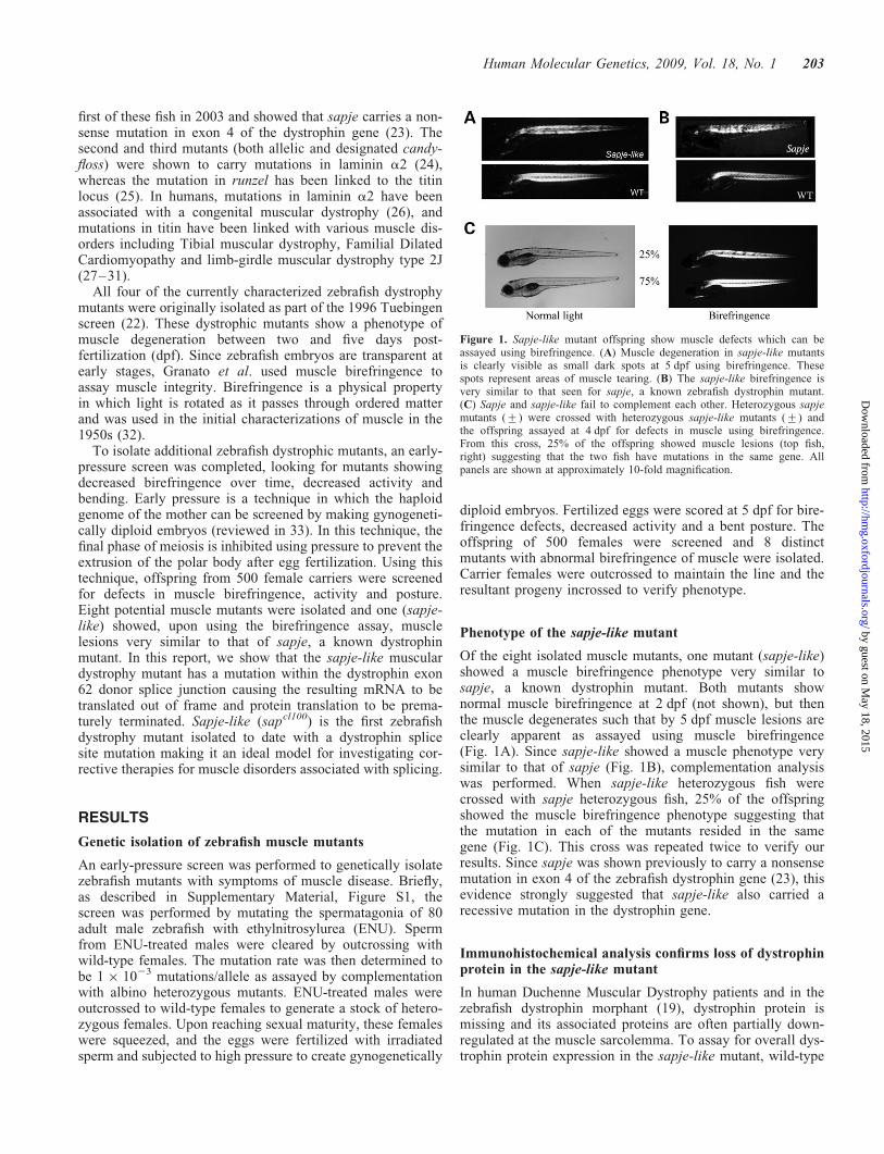
To isolate additional zebrafish dystrophic mutants, an early-pressure screen was completed, looking for mutants showingdecreased birefringence over time, decreased activity andbending. Early pressure is a technique in which the haploidgenome of the mother can be screened by making gynogeneti-cally diploid embryos (reviewed in 33). In this technique, thefinal phase of meiosis is inhibited using pressure to prevent theextrusion of the polar body after egg fertilization. Using thistechnique, offspring from 500 female carriers were screenedfor defects in muscle birefringence, activity and posture.Eight potential muscle mutants were isolated and one (sapje-like) showed, upon using the birefringence assay, musclelesions very similar to that of sapje, a known dystrophinmutant. In this report, we show that the sapje-like musculardystrophy mutant has a mutation within the dystrophin exon62 donor splice junction causing the resulting mRNA to betranslated out of frame and protein translation to be prema-turely terminated. Sapje-like (sapcl100) is the first zebrafishdystrophy mutant isolated to date with a dystrophin splicesite mutation making it an ideal model for investigating cor-rective therapies for muscle disorders associated with splicing.
RESULTS
Genetic isolation of zebrafish muscle mutants
An early-pressure screen was performed to genetically isolatezebrafish mutants with symptoms of muscle disease. Briefly,as described in Supplementary Material, Figure S1, thescreen was performed by mutating the spermatagonia of 80adult male zebrafish with ethylnitrosylurea (ENU). Spermfrom ENU-treated males were cleared by outcrossing withwild-type females. The mutation rate was then determined tobe 1 � 1023 mutations/allele as assayed by complementationwith albino heterozygous mutants. ENU-treated males wereoutcrossed to wild-type females to generate a stock of hetero-zygous females. Upon reaching sexual maturity, these femaleswere squeezed, and the eggs were fertilized with irradiatedsperm and subjected to high pressure to create gynogenetically
diploid embryos. Fertilized eggs were scored at 5 dpf for bire-fringence defects, decreased activity and a bent posture. Theoffspring of 500 females were screened and 8 distinctmutants with abnormal birefringence of muscle were isolated.Carrier females were outcrossed to maintain the line and theresultant progeny incrossed to verify phenotype.
Phenotype of the sapje-like mutant
Of the eight isolated muscle mutants, one mutant (sapje-like)showed a muscle birefringence phenotype very similar tosapje, a known dystrophin mutant. Both mutants shownormal muscle birefringence at 2 dpf (not shown), but thenthe muscle degenerates such that by 5 dpf muscle lesions areclearly apparent as assayed using muscle birefringence(Fig. 1A). Since sapje-like showed a muscle phenotype verysimilar to that of sapje (Fig. 1B), complementation analysiswas performed. When sapje-like heterozygous fish werecrossed with sapje heterozygous fish, 25% of the offspringshowed the muscle birefringence phenotype suggesting thatthe mutation in each of the mutants resided in the samegene (Fig. 1C). This cross was repeated twice to verify ourresults. Since sapje was shown previously to carry a nonsensemutation in exon 4 of the zebrafish dystrophin gene (23), thisevidence strongly suggested that sapje-like also carried arecessive mutation in the dystrophin gene.
Immunohistochemical analysis confirms loss of dystrophinprotein in the sapje-like mutant
In human Duchenne Muscular Dystrophy patients and in thezebrafish dystrophin morphant (19), dystrophin protein ismissing and its associated proteins are often partially down-regulated at the muscle sarcolemma. To assay for overall dys-trophin protein expression in the sapje-like mutant, wild-type
Figure 1. Sapje-like mutant offspring show muscle defects which can beassayed using birefringence. (A) Muscle degeneration in sapje-like mutantsis clearly visible as small dark spots at 5 dpf using birefringence. Thesespots represent areas of muscle tearing. (B) The sapje-like birefringence isvery similar to that seen for sapje, a known zebrafish dystrophin mutant.(C) Sapje and sapje-like fail to complement each other. Heterozygous sapjemutants (+) were crossed with heterozygous sapje-like mutants (+) andthe offspring assayed at 4 dpf for defects in muscle using birefringence.From this cross, 25% of the offspring showed muscle lesions (top fish,right) suggesting that the two fish have mutations in the same gene. Allpanels are shown at approximately 10-fold magnification.
Human Molecular Genetics, 2009, Vol. 18, No. 1 203
by guest on May 18, 2015
http://hmg.oxfordjournals.org/
Dow
nloaded from
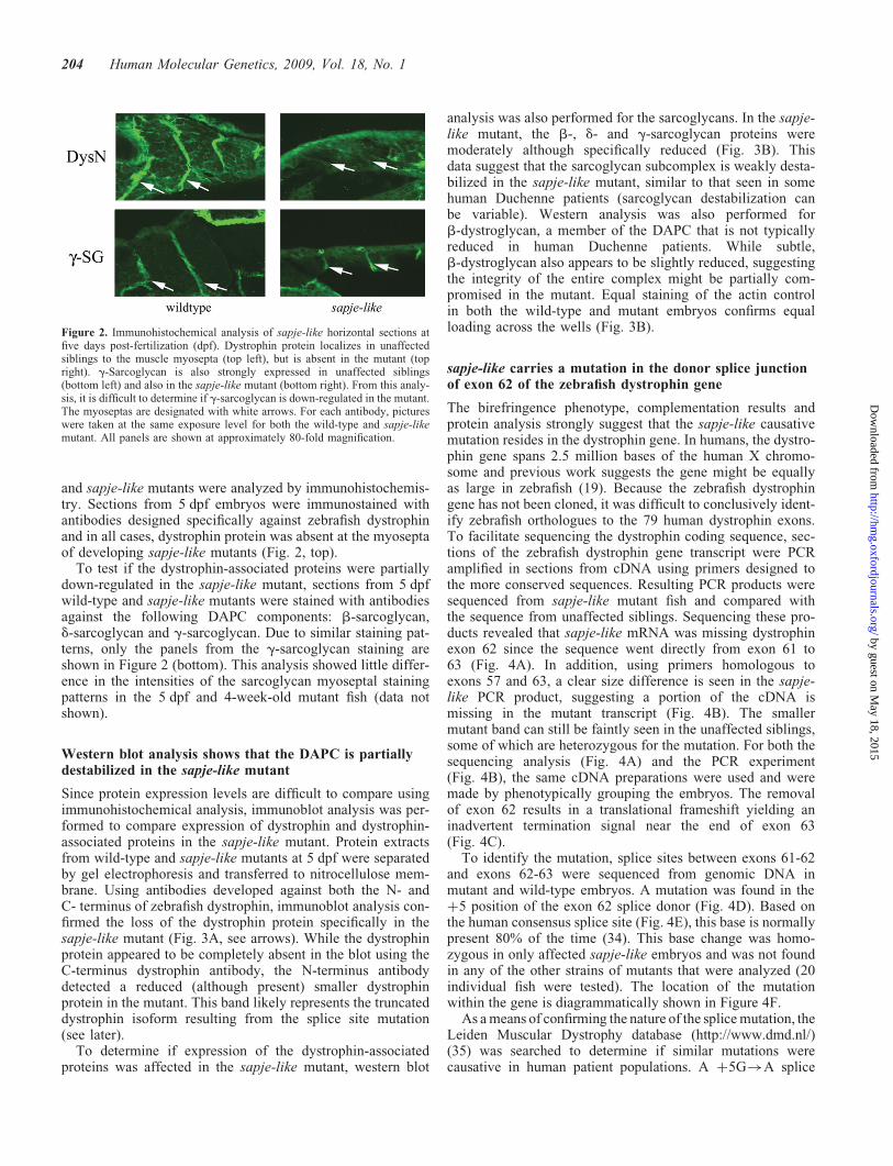
and sapje-like mutants were analyzed by immunohistochemis-try. Sections from 5 dpf embryos were immunostained withantibodies designed specifically against zebrafish dystrophinand in all cases, dystrophin protein was absent at the myoseptaof developing sapje-like mutants (Fig. 2, top).
To test if the dystrophin-associated proteins were partiallydown-regulated in the sapje-like mutant, sections from 5 dpfwild-type and sapje-like mutants were stained with antibodiesagainst the following DAPC components: b-sarcoglycan,d-sarcoglycan and g-sarcoglycan. Due to similar staining pat-terns, only the panels from the g-sarcoglycan staining areshown in Figure 2 (bottom). This analysis showed little differ-ence in the intensities of the sarcoglycan myoseptal stainingpatterns in the 5 dpf and 4-week-old mutant fish (data notshown).
Western blot analysis shows that the DAPC is partiallydestabilized in the sapje-like mutant
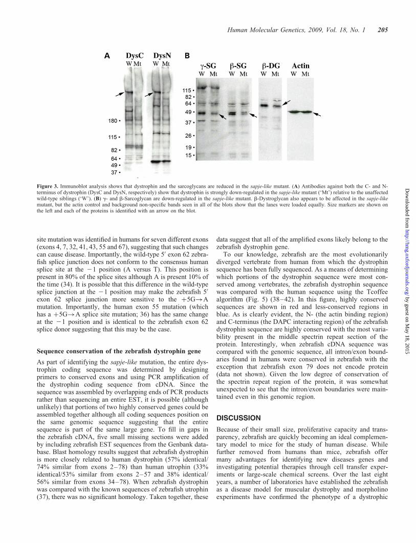
Since protein expression levels are difficult to compare usingimmunohistochemical analysis, immunoblot analysis was per-formed to compare expression of dystrophin and dystrophin-associated proteins in the sapje-like mutant. Protein extractsfrom wild-type and sapje-like mutants at 5 dpf were separatedby gel electrophoresis and transferred to nitrocellulose mem-brane. Using antibodies developed against both the N- andC- terminus of zebrafish dystrophin, immunoblot analysis con-firmed the loss of the dystrophin protein specifically in thesapje-like mutant (Fig. 3A, see arrows). While the dystrophinprotein appeared to be completely absent in the blot using theC-terminus dystrophin antibody, the N-terminus antibodydetected a reduced (although present) smaller dystrophinprotein in the mutant. This band likely represents the truncateddystrophin isoform resulting from the splice site mutation(see later).
To determine if expression of the dystrophin-associatedproteins was affected in the sapje-like mutant, western blot
analysis was also performed for the sarcoglycans. In the sapje-like mutant, the b-, d- and g-sarcoglycan proteins weremoderately although specifically reduced (Fig. 3B). Thisdata suggest that the sarcoglycan subcomplex is weakly desta-bilized in the sapje-like mutant, similar to that seen in somehuman Duchenne patients (sarcoglycan destabilization canbe variable). Western analysis was also performed forb-dystroglycan, a member of the DAPC that is not typicallyreduced in human Duchenne patients. While subtle,b-dystroglycan also appears to be slightly reduced, suggestingthe integrity of the entire complex might be partially com-promised in the mutant. Equal staining of the actin controlin both the wild-type and mutant embryos confirms equalloading across the wells (Fig. 3B).
sapje-like carries a mutation in the donor splice junctionof exon 62 of the zebrafish dystrophin gene
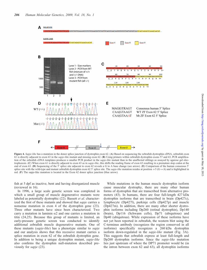
The birefringence phenotype, complementation results andprotein analysis strongly suggest that the sapje-like causativemutation resides in the dystrophin gene. In humans, the dystro-phin gene spans 2.5 million bases of the human X chromo-some and previous work suggests the gene might be equallyas large in zebrafish (19). Because the zebrafish dystrophingene has not been cloned, it was difficult to conclusively ident-ify zebrafish orthologues to the 79 human dystrophin exons.To facilitate sequencing the dystrophin coding sequence, sec-tions of the zebrafish dystrophin gene transcript were PCRamplified in sections from cDNA using primers designed tothe more conserved sequences. Resulting PCR products weresequenced from sapje-like mutant fish and compared withthe sequence from unaffected siblings. Sequencing these pro-ducts revealed that sapje-like mRNA was missing dystrophinexon 62 since the sequence went directly from exon 61 to63 (Fig. 4A). In addition, using primers homologous toexons 57 and 63, a clear size difference is seen in the sapje-like PCR product, suggesting a portion of the cDNA ismissing in the mutant transcript (Fig. 4B). The smallermutant band can still be faintly seen in the unaffected siblings,some of which are heterozygous for the mutation. For both thesequencing analysis (Fig. 4A) and the PCR experiment(Fig. 4B), the same cDNA preparations were used and weremade by phenotypically grouping the embryos. The removalof exon 62 results in a translational frameshift yielding aninadvertent termination signal near the end of exon 63(Fig. 4C).
To identify the mutation, splice sites between exons 61-62and exons 62-63 were sequenced from genomic DNA inmutant and wild-type embryos. A mutation was found in theþ5 position of the exon 62 splice donor (Fig. 4D). Based onthe human consensus splice site (Fig. 4E), this base is normallypresent 80% of the time (34). This base change was homo-zygous in only affected sapje-like embryos and was not foundin any of the other strains of mutants that were analyzed (20individual fish were tested). The location of the mutationwithin the gene is diagrammatically shown in Figure 4F.
As a means of confirming the nature of the splice mutation, theLeiden Muscular Dystrophy database (http://www.dmd.nl/)(35) was searched to determine if similar mutations werecausative in human patient populations. A þ5G!A splice
Figure 2. Immunohistochemical analysis of sapje-like horizontal sections atfive days post-fertilization (dpf). Dystrophin protein localizes in unaffectedsiblings to the muscle myosepta (top left), but is absent in the mutant (topright). g-Sarcoglycan is also strongly expressed in unaffected siblings(bottom left) and also in the sapje-like mutant (bottom right). From this analy-sis, it is difficult to determine if g-sarcoglycan is down-regulated in the mutant.The myoseptas are designated with white arrows. For each antibody, pictureswere taken at the same exposure level for both the wild-type and sapje-likemutant. All panels are shown at approximately 80-fold magnification.
204 Human Molecular Genetics, 2009, Vol. 18, No. 1
by guest on May 18, 2015
http://hmg.oxfordjournals.org/
Dow
nloaded from
site mutation was identified in humans for seven different exons(exons 4, 7, 32, 41, 43, 55 and 67), suggesting that such changescan cause disease. Importantly, the wild-type 50 exon 62 zebra-fish splice junction does not conform to the consensus humansplice site at the 21 position (A versus T). This position ispresent in 80% of the splice sites although A is present 10% ofthe time (34). It is possible that this difference in the wild-typesplice junction at the 21 position may make the zebrafish 50
exon 62 splice junction more sensitive to the þ5G!Amutation. Importantly, the human exon 55 mutation (whichhas a þ5G!A splice site mutation; 36) has the same changeat the 21 position and is identical to the zebrafish exon 62splice donor suggesting that this may be the case.
Sequence conservation of the zebrafish dystrophin gene
As part of identifying the sapje-like mutation, the entire dys-trophin coding sequence was determined by designingprimers to conserved exons and using PCR amplification ofthe dystrophin coding sequence from cDNA. Since thesequence was assembled by overlapping ends of PCR productsrather than sequencing an entire EST, it is possible (althoughunlikely) that portions of two highly conserved genes could beassembled together although all coding sequences position onthe same genomic sequence suggesting that the entiresequence is part of the same large gene. To fill in gaps inthe zebrafish cDNA, five small missing sections were addedby including zebrafish EST sequences from the Genbank data-base. Blast homology results suggest that zebrafish dystrophinis more closely related to human dystrophin (57% identical/74% similar from exons 2–78) than human utrophin (33%identical/53% similar from exons 2–57 and 38% identical/56% similar from exons 34–78). When zebrafish dystrophinwas compared with the known sequences of zebrafish utrophin(37), there was no significant homology. Taken together, these
data suggest that all of the amplified exons likely belong to thezebrafish dystrophin gene.
To our knowledge, zebrafish are the most evolutionarilydiverged vertebrate from human from which the dystrophinsequence has been fully sequenced. As a means of determiningwhich portions of the dystrophin sequence were most con-served among vertebrates, the zebrafish dystrophin sequencewas compared with the human sequence using the Tcoffeealgorithm (Fig. 5) (38–42). In this figure, highly conservedsequences are shown in red and less-conserved regions inblue. As is clearly evident, the N- (the actin binding region)and C-terminus (the DAPC interacting region) of the zebrafishdystrophin sequence are highly conserved with the most varia-bility present in the middle spectrin repeat section of theprotein. Interestingly, when zebrafish cDNA sequence wascompared with the genomic sequence, all intron/exon bound-aries found in humans were conserved in zebrafish with theexception that zebrafish exon 79 does not encode protein(data not shown). Given the low degree of conservation ofthe spectrin repeat region of the protein, it was somewhatunexpected to see that the intron/exon boundaries were main-tained even in this genomic region.
DISCUSSION
Because of their small size, proliferative capacity and trans-parency, zebrafish are quickly becoming an ideal complemen-tary model to mice for the study of human disease. Whilefurther removed from humans than mice, zebrafish offermany advantages for identifying new diseases genes andinvestigating potential therapies through cell transfer exper-iments or large-scale chemical screens. Over the last eightyears, a number of laboratories have established the zebrafishas a disease model for muscular dystrophy and morpholinoexperiments have confirmed the phenotype of a dystrophic
Figure 3. Immunoblot analysis shows that dystrophin and the sarcoglycans are reduced in the sapje-like mutant. (A) Antibodies against both the C- and N-terminus of dystrophin (DysC and DysN, respectively) show that dystrophin is strongly down-regulated in the sapje-like mutant (‘Mt’) relative to the unaffectedwild-type siblings (‘W’). (B) g- and b-Sarcoglycan are down-regulated in the sapje-like mutant. b-Dystroglycan also appears to be affected in the sapje-likemutant, but the actin control and background non-specific bands seen in all of the blots show that the lanes were loaded equally. Size markers are shown onthe left and each of the proteins is identified with an arrow on the blot.
Human Molecular Genetics, 2009, Vol. 18, No. 1 205
by guest on May 18, 2015
http://hmg.oxfordjournals.org/
Dow
nloaded from
fish at 5 dpf as inactive, bent and having disorganized muscle(reviewed in 16).
In 1996, a large scale genetic screen was completed inwhich a small group of muscle degenerative mutants werelabeled as potentially dystrophic (22). Bassett et al. character-ized the first of these mutants and showed that sapje carries anonsense mutation in exon 4 of the dystrophin gene (23).Three other mutants have since been characterized. Twocarry a mutation in laminin a2 and one carries a mutation intitin (24,25). Because this group of mutants is limited, anearly-pressure genetic screen was conducted to identifyadditional zebrafish muscle degenerative mutants. One ofthese mutants (sapje-like) has a phenotype similar to sapjeand our analysis shows that this recessive mutant carries asplice mutation in exon 62 of the zebrafish dystrophin gene.In addition to being a unique dystrophin mutant, sapje-likealso confirms the dystrophin null-mutation described pre-viously for sapje (23).
While mutations in the human muscle dystrophin isoformcause muscular dystrophy, there are many other humanforms of dystrophin that are transcribed from alternative pro-moters (43). In humans, there are four full-length 427 kDadystrophin isoforms that are transcribed in brain (Dp427c),lymphocyte (Dp427l), purkinje cells (Dp427p) and muscle(Dp427m). In addition, there are many other shorter dystro-phin isoforms including Dp260 (retinal dystrophin), Dp140(brain), Dp116 (Schwann cells), Dp71 (ubiquitous) andDp40 (ubiquitous). While expression of these isoforms havenot yet been reported in zebrafish, the western blot using theC-terminus antibody (recognizes the region conserved in allisoforms) specifically recognizes a 260 kDa dystrophinisoform down-regulated in the sapje-like mutant (Fig. 3A).This suggests that zebrafish express at least full-length andDp260 dystrophin isoforms. Since the sapje-like mutationlies just upstream of where the DP71 promoter would be (inthe intron between exon 62 and 63), all dystrophin isoforms
Figure 4. Sapje-like has a mutation in the donor splice junction of dystrophin exon 62. (A) Based on sequencing the zebrafish dystrophin cDNA, zebrafish exon61 is directly adjacent to exon 63 in the sapje-like mutant and missing exon 62. (B) Using primers within zebrafish dystrophin exons 57 and 63, PCR amplifica-tion of the zebrafish cDNA templates produces a smaller PCR product in the sapje-like mutant than in the unaffected siblings as assayed by agarose gel elec-trophoresis. (C) When exon 61 is directly adjacent to exon 63 as in sapje-like, this shifts the reading frame of exon 63 resulting in a premature stop codon at theend of exon 63. (D) Sequencing of the 50 splice site adjacent to exon 62 reveals a G to A base change (see arrow). (E) Comparison of the human consensus 50
splice site with the wild-type and mutant zebrafish dystrophin exon 62 50 splice site. The sapje-like mutation resides at position þ5 (G!A) and is highlighted inred. (F) The sapje-like mutation is located in the Exon 62 donor splice junction (blue arrow).
206 Human Molecular Genetics, 2009, Vol. 18, No. 1
by guest on May 18, 2015
http://hmg.oxfordjournals.org/
Dow
nloaded from
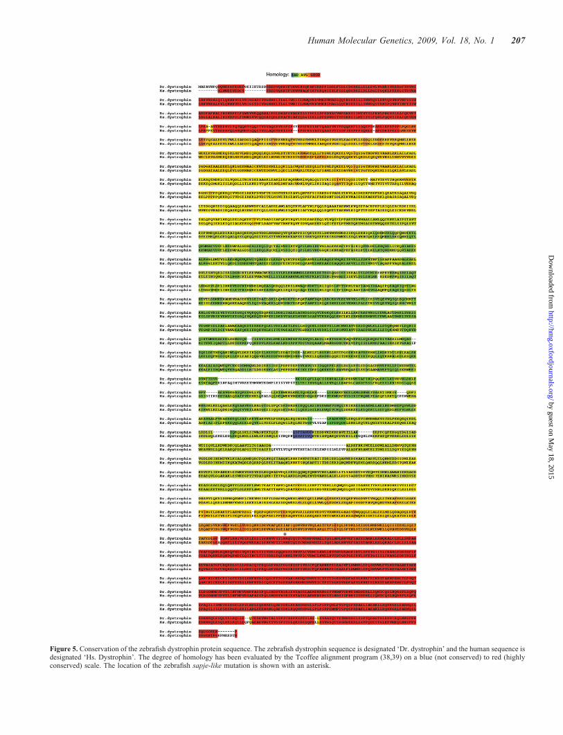
Figure 5. Conservation of the zebrafish dystrophin protein sequence. The zebrafish dystrophin sequence is designated ‘Dr. dystrophin’ and the human sequence isdesignated ‘Hs. Dystrophin’. The degree of homology has been evaluated by the Tcoffee alignment program (38,39) on a blue (not conserved) to red (highlyconserved) scale. The location of the zebrafish sapje-like mutation is shown with an asterisk.
Human Molecular Genetics, 2009, Vol. 18, No. 1 207
by guest on May 18, 2015
http://hmg.oxfordjournals.org/
Dow
nloaded from
larger than DP71 should be affected. As in human, defects inmuscle Dp427m cause the most overt phenotype (musculardystrophy) although there are additional defects in dystrophicpatients including cognitive defects which may be caused bychanges in other dystrophin isoforms. It is unclear whetherthe sapje-like mutation causes cognitive impairment orvisual defects although direct physical manifestations resultingfrom the mutation are almost identical to sapje in which onlythe largest dystrophin isoform is affected.
While genomic deletions cause the vast majority of thecases of Duchenne Muscular Dystrophy, splicing defects cancause muscular dystrophy and many other diseases. Sapje-likerepresents the first zebrafish dystrophic mutant characterizedto date that has dystrophy due to a splicing defect. As such,future drug screens using sapje-like are likely to uncovertherapeutics that work in a completely different manner thandrugs which would be able to correct sapje which carries anonsense mutation in exon 4 of the zebrafish dystrophingene. Since the muscular dystrophy phenotype in sapje-likecan be easily and specifically assayed using birefringence,this makes sapje-like valuable not just for finding drugscapable of correcting muscular dystrophy, but potentiallyany other disease caused by splicing defects.
The location of the sapje-like splicing defect was identifiedin the donor splice site of exon 62. The loss of exon 62 wasfirst noted experimentally by sequencing the complete zebra-fish dystrophin cDNA in affected and unaffected siblings.The fragment that was initially sequenced was amplifiedusing primers in exons flanking exon 62 (primers located inexon 57 and 63). In this scenario, the only product thatcould amplify in the splicing mutant was a transcript thatskipped exon 62 (since that exon was incapable of splicingand would yield a PCR product too big for amplification andsequencing). While spliced (exon 61 to exon 63) and unspliced(exon 62 to intron to exon 63) transcript may exist in the sapje-like mutant, no full-length dystrophin protein is produced andthe muscle degenerates due to the dystrophic phenotype.
Sapje-like is a novel zebrafish dystrophin mutant isolated aspart of a large genetic screen. Its characterization has shownthat it has symptoms of muscle degeneration similar to thatseen in human patients with Duchenne Muscular Dystrophy.We have identified the molecular lesion as a G!A mutationin the donor splice junction of zebrafish dystrophin exon 62.Sapje-like is the first characterized dystrophic mutant whichwas not isolated as part of the 1996 Tuebingen screen. Sincemany of the Tuebingen dystrophic mutants have already beencharacterized and there are at least 30 different forms of muscu-lar dystrophy, this suggests that additional dystrophic mutantsremain to be identified and validates our approach to geneticallyisolate additional mutants as part of our own genetic screen. Theidentification of sapje-like suggests that our screen was success-ful and that the characterization of our remaining musclemutants will help elucidate additional disease mechanisms.
MATERIALS AND METHODS
Early-pressure screen
As outlined in Supplementary Material, Figure S1 anddescribed in (44), 80 male AB strain adult zebrafish were
mutagenized with three treatments of 3 mM ENU (one treat-ment per week). Following treatment, 27 males were foundto be fertile. After clearing the males by random outcrossesfor a month, the mutation rate was assessed by complementa-tion analysis with heterozygous albino mutants. Of 2400scored embryos, 2 albinos were identified suggesting amutation rate of approximately 1.2 � 1023 mutations peralbino locus. This mutation rate is similar to that from pre-viously published reports. Mutagenized males were crossedwith wild-type AB females to create a stock of heterozygouscarriers which were grown to adulthood.
To perform the early-pressure screen, heterozygous femaleswere separated from the males. The night before squeezing,the females were primed by placing in a tank with malesalthough each sex was separated by a screen barrier. In themorning, the males were sacrificed and the testes wereground in Hanks Buffer to make a sperm stock. The spermwas subjected to UV irradiation for 3 min to crosslink theDNA. The females were anesthetized and systematicallysqueezed to extract the eggs and the eggs were fertilizedwith the UV-irradiated sperm. After squeezing, the femaleswere placed in numbered tanks to be later correlated withthe clutch of scored eggs. The sperm was activated byadding 1 ml of fish water and after 1 min 24 s, the fertilizedeggs were subjected to high pressure (8 000 pounds/inch) foran additional 4 min 36 s to inhibit the second phase ofmeiosis (polar body extrusion). Pressure was gently releasedand the eggs were placed in a plate with fish water. The fol-lowing day, the dead eggs were removed and the clutch wascounted if more than five eggs were viable (norm estimatedat 10–30 eggs). Gross phenotype was noticed at 1 dpf andmuscle structure, activity and bending confirmation assayedat 5 dpf. If the clutch showed any mutants of interest (recom-bination can make telomeric mutations hard to score by EP),the squeezed mother was outcrossed to wild-type AB malesand the resultant progeny incrossed to genetically verify phe-notype. The screen was complete when 500 viable clutcheswere scored. Of these, eight showed a muscle-specific pheno-type (not gross abnormalities) including three which show thehallmarks of muscular dystrophy (muscle degeneration overtime).
Birefringence
Muscle birefringence was analyzed by placing anesthesizedembryos on a glass polarizing filter and covering theembryos with second polarizing filter elevated to prevent theembryos from being harmed. The filters were then placed onan underlit dissecting scope (Leica, model MZ75) and thetop polarizing filter twisted until only the light refractingthrough the striated muscle was visible. Since the degree ofbirefringence is affected by the horizontal orientation of thefish, the fish were gently oscillated back and forth toaccount for differences in positioning.
Complementation analysis
Complementation analysis was performed twice. First to assaythe mutation rate by crossing mutagenized males with hetero-zygous albino females. Second, complementation was used to
208 Human Molecular Genetics, 2009, Vol. 18, No. 1
by guest on May 18, 2015
http://hmg.oxfordjournals.org/
Dow
nloaded from
determine if sapje and sapje-like were allelic by crossingheterozygous sapje-like mutants with heterozygous sapjemutants. Resultant progeny were scored using birefringence.
Immunoblot analysis
For separation of large proteins like dystrophin, 3–8% Tris–Acetate gradient gels were used and electrophoresis was at150 V for �2 h. Following separation, proteins were trans-ferred to 0.45 mm nitrocellulose membranes in a submergedtransfer apparatus according to the instructions provided bythe manufacturer (Invitrogen). For separation of smaller pro-teins (,100 kDa), 12% Tris–Glycine gradient gels (4% stack-ing) were used and electrophoresis was at 130 V for �2 h.Following separation, the proteins were transferred to0.45 mm nitrocellulose using a Semi-Dry Transfer Apparatusaccording to the instructions provided by the manufacturer(Bio-Rad). All antibodies used have either been described pre-viously or are commercially available.
Membranes were incubated with the following antibodies ina cocktail of 1� PBS (phosphate-buffered saline), 0.1%Tween-20 and 5% non-fat dry milk (NFDM): ‘DysC’ (1:333dilution) (19), ‘DysN’ (1:333 dilution) (18), d-sarcoglycan(1:300 dilution) (12,19), g-sarcoglycan ‘g-SG’ (1:333dilution) (18), b-sarcoglycan ‘b-SG’ (Novocastra) (1:20dilution) (19), b-dystroglycan ‘b-DG’ (Novocastra) (1:10dilution) (19) and actin ‘A2066 ‘(Sigma) (1:300 dilution)(19). Membranes were incubated with primary antibodies for1 h at 258C then washed two times quickly and three timesfor 10 min in a solution of 1� PBS with 0.1% Tween-20.
An anti-rabbit-conjugated goat HRP secondary antibody(1:5000 dilution) (Jackson ImmunoResearch Lab) was usedto detect the following primary antibodies: ‘DysC’, ‘DysN’,‘d-SG’, ‘g-SG’ and actin ‘A2066’. An anti-mouse-conjugatedgoat HRP secondary antibody (1:5000 dilution) (JacksonImmunoResearch Lab) was used to detect the ‘b-SG’ and‘b-DG’ primary antibodies. The membranes were incubatedwith 1:5000 dilution of secondary antibody in a solution of1� PBS with 0.1% Tween-20 and 5% NFDM for 1 h at258C before washing two times quickly and three times for10 min in a solution of 1� PBS with 0.1% Tween-20. Theblots were incubated with Western Lighting Chemilumines-cence Reagent Plus (Perkin Elmer) as described by the distri-butor and exposed to film for up to 5 min.
Immunohistochemistry
Juvenile (5-day-old) zebrafish were anesthetized in 0.3% tri-caine solution for 2–10 min, coated in Optimal Cutting Temp-erature Compound (Sakura) and frozen in liquidnitrogen-cooled 4-methyl butane. 5–10 mm sections (as indi-cated) were cut at 2208C using a Microm HM 505 E cryostat,transferred to silane-coated microscope slides and fixed byimmersing the slides in methanol for 3 min before equilibrat-ing in 1� PBS at 48C overnight.
To block non-specific binding of the antibodies, sectionswere incubated for 30 min at room temperature inPBSþ10% horse serum. Following blocking, the slides wereincubated overnight at 48C using the same antibodiesdescribed for immunoblot analysis. Slides were washed three
times in 1� PBS and sections incubated with FITC-conjugated secondary antibodies (Jackson ImmunoResearchLab) at a 1:100 dilution for 1 h at room temperature. Slideswere washed three times in 1� PBS before mounting in25 ml vectashield. Slides were analyzed using a Zeiss Axio-plan II microscope with a triple filter and the images storedusing either IP Lab or OpenLab Software.
Sequencing the zebrafish dystrophin gene
Zebrafish cDNA was synthesized for both wild-type and sapje-like 5 dpf embryos by first isolating total RNA with aQIAGEN RNA isolation kit. The poly A containing mRNAwas used as a template for cDNA synthesis using a One-CyclecDNA Synthesis Kit (Affymetrix). To specifically amplifysegments of the zebrafish dystrophin gene, PCR primerswere designed by finding regions of zebrafish chromosome 1exon sequences which could encode peptide fragmentshaving high homology to the analogous section of thehuman dystrophin protein sequence. Using this approach, 20PCR reactions were designed to amplify the complete dystro-phin cDNA in overlapping fragments. Overall, 12 000 bases ofthe zebrafish dystrophin cDNA were sequenced with a fewremaining bases filled in using pre-existing EST sequencesavailable in Genbank. For identification of the splice sitemutation, primers were designed from genomic sequenceoutside the splice regions and the region of interest amplifiedfrom wild-type and mutant genomic DNA.
PCR reactions were performed at 35 cycles of 1 min at948C, 2 min at 528C, and 1 min at 728C using a GC 2 PCRkit (Clontech). Finished PCR reactions were purified with aQIAGEN PCR Cleanup Kit and prepared for sequencing.
All DNA sequencing was performed in The Mental Retar-dation Research Center Sequencing Core Facility locatedwithin Children’s Hospital Boston. Sequencing reactionswere prepared with �50 ng of PCR product and 10 pm ofprimer in a 12 ml reaction volume. DNA was fluorescentlylabeled and analyzed on an Applied Biosystems 3730 DNAAnalyzer. The sequences were imported into Sequencher andcomparisons made between wild-type and sapje-like DNAand cDNA sequence to identify the exon missing in themutant fish and the causative mutation. In addition, at least10 linked SNPs were also identified as part of this analysis.
SUPPLEMENTARY MATERIAL
Supplementary Material is available at HMG Online.
ACKNOWLEDGEMENTS
We would like to acknowledge members of the Kunkel andZon laboratory for their experimental advice, help in perform-ing these experiments and the drafting of this manuscript.Specifically, we would like to thank Dr Yi Zhou and Dr LenZon (both of Children’s Hospital Boston) for their helpfuladvice and providing necessary fishroom space. In addition,we are grateful to Chris Lawrence and Tim Jones whomanaged our fish facility. We would also like to thankmembers of the Children’s Hospital Sequencing Facility
Human Molecular Genetics, 2009, Vol. 18, No. 1 209
by guest on May 18, 2015
http://hmg.oxfordjournals.org/
Dow
nloaded from
including Christine Briggs and Hal Schneider for sequencingthe zebrafish dystrophin PCR products. L.M.K. is an investi-gator of the Howard Hughes Medical Institute.
Conflict of Interest statement. None declared.
FUNDING
This work was supported by a National Institutes of HealthProgram Project Grant (grant number 5P50NS040828-07 toL.M.K.); the Bernard F. and Alva B. Gimbel Foundation ( toL.M.K.) and a Developmental Grant awarded by the MuscularDystrophy Association (to J.R.G). Funding for the Children’sHospital Sequencing Facility was provided by a NIH grant tothe Mental Retardation and Developmental DisabilitiesResearch Center (MRDDRC) Molecular Genetics Core Facil-ity at Children’s Hospital Boston (grant number5P30HD018655-26).
REFERENCES
1. Burghes, A.H., Logan, C., Hu, X., Belfall, B., Worton, R.G. and Ray, P.N.(1987) A cDNA clone from the Duchenne/Becker muscular dystrophygene. Nature, 328, 434–437.
2. Hoffman, E.P., Brown, R.H. Jr and Kunkel, L.M. (1987) Dystrophin: theprotein product of the Duchenne muscular dystrophy locus. Cell, 51,919–928.
3. Monaco, A.P., Neve, R.L., Colletti-Feener, C., Bertelson, C.J., Kurnit,D.M. and Kunkel, L.M. (1986) Isolation of candidate cDNAs for portionsof the Duchenne muscular dystrophy gene. Nature, 323, 646–650.
4. Hoffman, E.P., Fischbeck, K.H., Brown, R.H., Johnson, M., Medori, R.,Loike, J.D., Harris, J.B., Waterston, R., Brooke, M., Specht, L. et al.(1988) Characterization of dystrophin in muscle-biopsy specimens frompatients with Duchenne’s or Becker’s muscular dystrophy.N. Engl. J. Med., 318, 1363–1368.
5. Flanigan, K.M., von Niederhausern, A., Dunn, D.M., Alder, J., Mendell,J.R. and Weiss, R.B. (2003) Rapid direct sequence analysis of thedystrophin gene. Am. J. Hum. Genet., 72, 931–939.
6. Arahata, K., Ishiura, S., Ishiguro, T., Tsukahara, T., Suhara, Y., Eguchi,C., Ishihara, T., Nonaka, I., Ozawa, E. and Sugita, H. (1988)Immunostaining of skeletal and cardiac muscle surface membrane withantibody against Duchenne muscular dystrophy peptide. Nature, 333,861–863.
7. Bonilla, E., Samitt, C.E., Miranda, A.F., Hays, A.P., Salviati, G.,DiMauro, S., Kunkel, L.M., Hoffman, E.P. and Rowland, L.P. (1988)Duchenne muscular dystrophy: deficiency of dystrophin at the muscle cellsurface. Cell, 54, 447–452.
8. Zubrzycka-Gaarn, E.E., Bulman, D.E., Karpati, G., Burghes, A.H.,Belfall, B., Klamut, H.J., Talbot, J., Hodges, R.S., Ray, P.N. and Worton,R.G. (1988) The Duchenne muscular dystrophy gene product is localizedin sarcolemma of human skeletal muscle. Nature, 333, 466–469.
9. Araishi, K., Sasaoka, T., Imamura, M., Noguchi, S., Hama, H.,Wakabayashi, E., Yoshida, M., Hori, T. and Ozawa, E. (1999) Loss of thesarcoglycan complex and sarcospan leads to muscular dystrophy inbeta-sarcoglycan-deficient mice. Hum. Mol. Genet., 8, 1589–1598.
10. Bonnemann, C.G., Modi, R., Noguchi, S., Mizuno, Y., Yoshida, M.,Gussoni, E., McNally, E.M., Duggan, D.J., Angelini, C. and Hoffman,E.P. (1995) Beta-sarcoglycan mutations cause autosomal recessivemuscular dystrophy with loss of the sarcoglycan complex. Nat. Genet., 11,266–273.
11. Lim, L.E., Duclos, F., Broux, O., Bourg, N., Sunada, Y., Allamand, V.,Meyer, J., Richard, I., Moomaw, C., Slaughter, C. et al. (1995)Beta-sarcoglycan: characterization and role in limb-girdle musculardystrophy linked to 4q12. Nat. Genet., 11, 257–265.
12. Nigro, V., Moreira, E.d.S., Piluso, G., Vainzof, M., Belsito, A., Politano,L., Puca, A.A., Passos-Bueno, M.R. and Zatz, M. (1996) Autosomalrecessive limb-girdle muscular dystrophy (LGMD2F) is caused by amutation in the delta-sarcoglycan gene. Nat. Genet., 14, 195–198.
13. Noguchi, S., McNally, E.M., Othmane, K.B., Hagiwara, Y., Mizuno, Y.,Yoshida, M., Yamamoto, H., Bonnemann, C.G., Gussoni, E., Denton, P.H.et al. (1995) Mutations in the dystrophin-associated proteingamma-sarcoglycan in chromosome 13 muscular dystrophy. Science, 270,819–822.
14. Odom, G.L., Gregorevic, P. and Chamberlain, J.S. (2007) Viral-mediatedgene therapy for the muscular dystrophies: successes, limitations andrecent advances. Biochim. Biophys. Acta, 1772, 243–262.
15. Kunkel, L.M., Bachrach, E., Bennett, R.R., Guyon, J. and Steffen, L.(2006) Diagnosis and cell-based therapy for Duchenne musculardystrophy in humans, mice, and zebrafish. J. Hum. Genet., 51, 397–406.
16. Guyon, J.R., Steffen, L.S., Howell, M.H., Pusack, T.J., Lawrence, C. andKunkel, L.M. (2007) Modeling human muscle disease in zebrafish.Biochim. Biophys. Acta, 1772, 205–215.
17. Cheng, L., Guo, X.F., Yang, X.Y., Chong, M., Cheng, J., Li, G., Gui, Y.H.and Lu, D.R. (2006) Delta-sarcoglycan is necessary for early heart andmuscle development in zebrafish. Biochem. Biophys. Res. Commun., 344,1290–1299.
18. Guyon, J.R., Mosley, A.N., Jun, S.J., Montanaro, F., Steffen, L.S., Zhou,Y., Nigro, V., Zon, L.I. and Kunkel, L.M. (2005) Delta-sarcoglycan isrequired for early zebrafish muscle organization. Exp. Cell Res., 304,105–115.
19. Guyon, J.R., Mosley, A.N., Zhou, Y., Davidson, A.J., Sheng, X., Chiang,K., Brien, K.F.O., Volinski, J.M., Zon, L.I. and Kunkel, L.M. (2003) Thedystrophin associated protein complex in zebrafish. Hum. Mol. Genet., 12,601–615.
20. Nixon, S.J., Wegner, J., Ferguson, C., Mery, P.F., Hancock, J.F., Currie,P.D., Key, B., Westerfield, M. and Parton, R.G. (2005) Zebrafish as amodel for caveolin-associated muscle disease; caveolin-3 is required formyofibril organization and muscle cell patterning. Hum. Mol. Genet., 14,1727–1743.
21. Parsons, M.J., Campos, I., Hirst, E.M. and Stemple, D.L. (2002) Removalof dystroglycan causes severe muscular dystrophy in zebrafish embryos.Development, 129, 3505–3512.
22. Granato, M., Eeden, F.J.v., Schach, U., Trowe, T., Brand, M.,Furutani-Seiki, M., Haffter, P., Hammerschmidt, M., Heisenberg, C.P.,Jiang, Y.J. et al. (1996) Genes controlling and mediating locomotionbehavior of the zebrafish embryo and larva. Development, 123, 399–413.
23. Bassett, D.I., Bryson-Richardson, R.J., Daggett, D.F., Gautier, P., Keenan,D.G. and Currie, P.D. (2003) Dystrophin is required for the formation ofstable muscle attachments in the zebrafish embryo. Development, 130,5851–5860.
24. Hall, T.E., Bryson-Richardson, R.J., Berger, S., Jacoby, A.S., Cole, N.J.,Hollway, G.E., Berger, J. and Currie, P.D. (2007) The zebrafish candyflossmutant implicates extracellular matrix adhesion failure in lamininalpha2-deficient congenital muscular dystrophy. Proc. Natl. Acad. Sci.USA, 104, 7092–7097.
25. Steffen, L.S., Guyon, J.R., Vogel, E.D., Howell, M.H., Zhou, Y., Weber,G.J., Zon, L.I. and Kunkel, L.M. (2007) The zebrafish runzel musculardystrophy is linked to the titin gene. Dev. Biol., 309, 180–192.
26. Helbling-Leclerc, A., Zhang, X., Topaloglu, H., Cruaud, C., Tesson, F.,Weissenbach, J., Tome, F.M., Schwartz, K., Fardeau, M., Tryggvason, K.et al. (1995) Mutations in the laminin alpha 2-chain gene (LAMA2) causemerosin-deficient congenital muscular dystrophy. Nat. Genet., 11,216–218.
27. Knoll, R., Hoshijima, M., Hoffman, H.M., Person, V., Lorenzen-Schmidt,I., Bang, M.L., Hayashi, T., Shiga, N., Yasukawa, H., Schaper, W. et al.(2002) The cardiac mechanical stretch sensor machinery involves a Z disccomplex that is defective in a subset of human dilated cardiomyopathy.Cell, 111, 943–955.
28. Itoh-Satoh, M., Hayashi, T., Nishi, H., Koga, Y., Arimura, T., Koyanagi,T., Takahashi, M., Hohda, S., Ueda, K., Nouchi, T. et al. (2002) Titinmutations as the molecular basis for dilated cardiomyopathy. Biochem.Biophys. Res. Commun., 291, 385–393.
29. Gerull, B., Atherton, J., Geupel, A., Sasse-Klaassen, S., Heuser, A.,Frenneaux, M., McNabb, M., Granzier, H., Labeit, S. and Thierfelder, L.(2006) Identification of a novel frameshift mutation in the giant musclefilament titin in a large Australian family with dilated cardiomyopathy.J. Mol. Med., 84, 478–483.
30. Haravuori, H., Vihola, A., Straub, V., Auranen, M., Richard, I., Marchand,S., Voit, T., Labeit, S., Somer, H., Peltonen, L. et al. (2001) Secondarycalpain3 deficiency in 2q-linked muscular dystrophy: titin is the candidategene. Neurology, 56, 869–877.
210 Human Molecular Genetics, 2009, Vol. 18, No. 1
by guest on May 18, 2015
http://hmg.oxfordjournals.org/
Dow
nloaded from
31. Hackman, P., Vihola, A., Haravuori, H., Marchand, S., Sarparanta, J., DeSeze, J., Labeit, S., Witt, C., Peltonen, L., Richard, I. et al. (2002) Tibialmuscular dystrophy is a titinopathy caused by mutations in TTN, the geneencoding the giant skeletal-muscle protein titin. Am. J. Hum. Genet., 71,
492–500.
32. Schapira, G., Dreyfus, J.C. and Joly, M. (1952) Changes in the flowbirefringence of myosin as a result of muscular atrophy. Nature, 170,494–495.
33. Patton, E.E. and Zon, L.I. (2001) The art and design of genetic screens:zebrafish. Nat. Rev. Genet., 2, 956–966.
34. Zhang, M.Q. (1998) Statistical features of human exons and their flankingregions. Hum. Mol. Genet., 7, 919–932.
35. Aartsma-Rus, A., Van Deutekom, J.C., Fokkema, I.F., Van Ommen, G.J.
and Den Dunnen, J.T. (2006) Entries in the Leiden Duchenne musculardystrophy mutation database: an overview of mutation types andparadoxical cases that confirm the reading-frame rule. Muscle Nerve, 34,135–144.
36. Taylor, P.J., Maroulis, S., Mullan, G.L., Pedersen, R.L., Baumli, A.,Elakis, G., Piras, S., Walsh, C., Prosper-Gutierrez, B., De LaPuente-Alonso, F. et al. (2007) Measurement of the clinical utility of acombined mutation detection protocol in carriers of Duchenne and Beckermuscular dystrophy. J. Med. Genet., 44, 368–372.
37. Bohm, S., Jin, H., Hughes, S.M., Roberts, R.G. and Hinits, Y. (2008)Dystrobrevin and dystrophin family gene expression in zebrafish. GeneExpr. Patt., 8, 71–78.
38. Notredame, C., Higgins, D.G. and Heringa, J. (2000) T-Coffee: a novelmethod for fast and accurate multiple sequence alignment. J. Mol. Biol.,302, 205–217.
39. Poirot, O., O’Toole, E. and Notredame, C. (2003) Tcoffee@igs: a webserver for computing, evaluating and combining multiple sequencealignments. Nucleic Acids Res., 31, 3503–3506.
40. Notredame, C. and Abergel, C. (2003) Using multiple alignment methodsto assess the quality of genomic data analysis. In Andrade, M. (Ed.),Bioinformatics and Genomes: Current Perspectives. Horizon ScientificPress, Norfolk, pp. 30–50.
41. Notredame, C., Holme, L. and Higgins, D.G. (1998) COFFEE: a newobjective function for multiple sequence alignmnent. Bioinformatics, 14,407–422.
42. O’Sullivan, O., Suhre, K., Abergel, C., Higgins, D.G. and Notredame,C.3DCoffee: (2004) Combining protein sequences and structures withinmultiple sequence alignments. J. Mol. Biol., 340, 385–395.
43. Sadoulet-Puccio, H.M. and Kunkel, L.M. (1996) Dystrophin and itsisoforms. Brain Pathol., 6, 25–35.
44. Westerfield, M. (2000) The Zebrafish Book. A Guide for the Laboratory Useof Zebrafish (Danio rerio), 4th edn. University of Oregon Press, Eugene.
Human Molecular Genetics, 2009, Vol. 18, No. 1 211
by guest on May 18, 2015
http://hmg.oxfordjournals.org/
Dow
nloaded from