Bed height and material density effects on fluidized bed hydrodynamics
Upload
polytechniqueCategory
view
0download
0
C H A P T E R T W O
C
IS
*
{
urrent
SN 0
Labo(CNPalaiInstitRechand U
From Cilia Hydrodynamics to
Zebrafish Embryonic Development
Willy Supatto* and Julien Vermot†
Contents
1. In
Top
070
ratoRS)seauut derchniv
troduction
ics in Developmental Biology, Volume 95 # 2011
-2153, DOI: 10.1016/B978-0-12-385065-2.00002-5 All rig
ry for Optics and Biosciences, Ecole Polytechnique, Centre National de RechercheUMR 7645, and Institut National de Sante et de Recherche Medicale (INSER, Francee Genetique et de Biologie Moleculaire et Cellulaire (IGBMC), Institut National dee Medicale (INSERM) U964, Centre National de Recherche Scientifique (CNRS)ersite de Strasbourg, Illkirch, France
Else
hts
SciM
SaUM
34
2. M
otile Cilia and Zebrafish Development 353. S
tructure of Motile Cilia in the Developing Zebrafish 384. T
he Physics Side of Cilia-Mediated Flow: From Modeling toExperiment
414
.1. T he basics of hydrodynamics at the cilium scale: Stokes flowor fluid dynamics at low Reynolds number
414
.2. D irectional flow 424
.3. M ixing with chaotic advection 464
.4. T he left–right organizer: Interplay between models andexperiments
485. E
xperimental Investigation of Cilia-Driven Fluid Flowin Developing Embryos
515
.1. C hallenges and experimental models 515
.2. M apping microscopic flow field in living embryos 525
.3. M easuring cilia features in vivo 535
.4. N ext experimental challenges: Flow/cilia manipulation andfunctional imaging
576. R
ole of Cilia-Driven Flow in the Kupffer’s Vesicle in Zebrafish 577. R
ole of Cilia-Driven Flow in the Developing Inner Ear in Zebrafish 588. C
onclusion 61Ackn
owledgments 62Refe
rences 62vier Inc.
reserved.
entifique) U696,
nte et deR 1704,
33

34 Willy Supatto and Julien Vermot
Abstract
Embryonic development involves the cellular integration of chemical and physi-
cal stimuli. A key physical input is the mechanical stress generated during
embryonic morphogenesis. This process necessitates tensile forces at the tissue
scale such as during axis elongation and budding, as well as at the cellular scale
when cells migrate and contract. Furthermore, cells can generate forces using
motile cilia to produce flow. Cilia-driven flows are critical throughout embryonic
development but little is known about the diversity of the forces they exert and
the role of the mechanical stresses they generate. In this chapter, through an
examination of zebrafish development, we highlight what is known about the
role of hydrodynamics mediated by beating cilia and examine the physical
features of flow fields from the modeling and experimental perspectives.
We review imaging strategies to visualize and quantify beating cilia and the
flow they generate in vivo. Finally, we describe the function of hydrodynamics
during left–right embryonic patterning and inner ear development. Ideally,
continued progress in these areas will help to address a key conceptual problem
in developmental biology, which is to understand the interplay between
environmental constraints and genetic control during morphogenesis.
1. Introduction
Cilia are tail-like organelles that protrude out of nearly all vertebratecells. Their roles in moving fluid along epithelium, such as the respiratorytract or the fallopian tubes, are well known in humans (Baker and Beales,2009). Yet, this past decade identified many more functions for this organ-elle. Overall, one can define two types of cilia: motile cilia involved inmoving fluids and primary cilia (most often immotile) that can sensechemicals and/or mechanical inputs. This dual activity seems conserved inmost vertebrates (Shah et al., 2009), and it is thought that primary cilia andmotile cilia were once the same structure that specialized toward motility,sensitivity, or both ( Jekely and Arendt, 2006).
Cilia structure is variable and usually defines their function. In eukaryoticcells, cilia are made of microtubules and classified according to their internalmolecular arrangement and their ability to move. Usually, cells assembleonly one cilium but a number of specialized cells can assemble up to 300cilia. While motile cilia are commonly involved in cell motility, such assperm, their prominent role in the embryo is to generate fluid flow. Movingfluids can participate in numerous processes and cilia activities turn out to beinvolved in diverse developmental functions: from generating frictionalforces in the left–right organizer (McGrath et al., 2003) to act as a mixerin the olfactory pit (Castleman et al., 2009) or to attract particles in the innerear (Colantonio et al., 2009). Interestingly, the rules dictating these features

Cilia Hydrodynamics and Embryonic Development 35
strictly depend on fundamental fluid dynamics that were articulated 160years ago by Stokes (1851). This work, initially applied to aerodynamics andhydrodynamics, became slowly incorporated into microfluidics (Stone et al.,2004), bioengineering (Vilfan et al., 2010; White and Grosh, 2005;Yoganathan et al., 2004), microbiology (Purcell, 1997; Short et al., 2006;Solari et al., 2006), physiology (Purcell, 1977), and, more recently, devel-opmental biology (Cartwright et al., 2009).
The diversity of cilia motility and fluid mechanics in the embryo isstarting to be studied in vivo. The number of flows that can be generatedby such simple structure is limited, but the combination of cilia beatingpattern and particular topology of the environment can participate to buildup complex flows. Importantly, specific flow shapes are critical for embryo-nic development. The understanding of fluid mechanics principles and theuse of modeling are essential to address the emergence of complexitythrough simple hydrodynamic interactions. In addition, the constant prog-ress of live imaging has greatly changed the views of cilia flows. Severalexamples are paving the way for describing the complexity of cilia-drivenadvections and their roles in organizing flow in zebrafish embryos.
In this chapter, we focus on what is known about cilia-driven flowsduring zebrafish embryogenesis, with an emphasis on fluid dynamics and itsrelevance in the emergence of higher order aspects of morphogenesis. Wefirst review the roles of cilia motility during zebrafish embryonic develop-ment. Next, we consider basic rules explaining the fluid dynamic at work atthe cilium scale. We start with the simplest possible hydrodynamic rules,working up for basic geometry. We next address how modeling andexperimental approaches are driving the investigation of flow in vivo andconclude with the examination of fluid dynamics into organs whose devel-opment relies on fluid dynamics, the otolithic morphogenesis and left–rightaxis specification in zebrafish.
2. Motile Cilia and Zebrafish Development
Zebrafish embryo contains numerous cavities and tubes. As in manychordates, most of them are extensively ciliated. For example, the nervoussystem contains a ciliated epithelial cell layer that delimits the ventral canalof the spinal cord in mouse, xenopus, amphioxus, and zebrafish (Casparyet al., 2007; Dale et al., 1987; Kramer-Zucker et al., 2005; Nakao andIshizawa, 1984; Fig. 2.1). Other cavities such as the left–right organizer,also called the Kupffer’s vesicle (KV) in zebrafish, the developing kidneys,and the inner ear also contain cilia (Kramer-Zucker et al., 2005; Fig. 2.1).Cilia are usually visualized in situ using antibodies directed against theacetylated tubulin (Essner et al., 2002; Fig. 2.2), but motile cilia can also

A B
C
Kupffer’s vesicle
Inner earSpinal canal
Pronephros
Pronephros
Spinal canalOlfactory pit
9 hpf 24 hpf
72 hpf
Figure 2.1 Ciliated organs in the zebrafish embryo. Side views of 10 hpf (A), 24 hpf(B), and 72 hpf (C) showing reported areas containing motile cilia (black arrows). Scalebar: 150 mm.
36 Willy Supatto and Julien Vermot
be located in zebrafish embryo by looking at the expression of cilia specificgenes. A good example is foxj1, a transcription factor that controls theexpression of genes involved in the formation of motile cilia and which isimportant for embryonic development (Stubbs et al., 2008; Yu et al., 2008).Its expression starts at gastrulation in the forerunner cells, which constitutethe precursors of the KV early on during embryogenesis, and remains strongin the vesicle itself. Its expression is also detected in the ventral floor plate ofthe spinal cord, the inner ear, the pronephros, the developing kidneys(Aamar and Dawid, 2008), and in the olfactory pit, where motile cilia areactively mixing the environment (Castleman et al., 2009). In many verte-brates, cilia motility has been shown to be critical for the development andfunction of these organs. In zebrafish, the consequences of cilia immobilityhave been studied in detail using an array of cilia mutants (Drummond,2009). The most common phenotypes are the presence of kidney cysts, left–right defects, inner ear defects, a curly tail, and hydrocephaly. It is to notethat other transcription factors have been shown to control cilia assemblythrough the activation of foxj1 expression, in particular, the rfx family whichare differentially expressed in developing tissues. rfx genes are expressed indiverse developing organs, such as in the islet cell lineage of the pancreas invertebrates (Smith et al., 2010b; Soyer et al., 2010) or sensory neurons inCaenorhabditis elegans (Perkins et al., 1986) and Drosophila (Vandaele et al.,

gsc:egfp
Fura2Bodipy FL
A B C
E F
G
Ac. tubulin
D
Hd 90
80
70
60
50
40
30
20
1010
0 0 10 20 30 40 50 60 70 80 90
pkv
Directionalflow a
r
n
l
Figure 2.2 Anatomy of the Kupffer’s vesicle. (A and B) Visualization and localizationof the Kupffer’s vesicle with the gsc:GFP line. Confocal sections at increasing depthshowing the localization of the Kupffer’s vesicle just below the midline. (D) Labelingusing antibodies directed against acetylated tubulin reveals cilia localization in theKupffer’s vesicle. (E) Cell membrane labeling using Bodipy FL staining underlinesthe spherical shape of the vesicle. (F) Calcium indicator (Fura2) shows asymmetricactivation. This asymmetric activation is essential for the left–right embryonic pattern-ing. (G) Scheme representing the Kupffer’s vesicle in 3D and the direction of the cilia-driven flow. (H) Particle tracking in the Kupffer’s vesicle demonstrates the circularity ofthe flow in vivo. TheTg(gsc:eGFP) line is a kind gift from the Houart lab (King’s CollegeLondon, UK). Acetylated tubulin labeling was performed as in Colantonio et al. (2009),Bodipy FL labeling as in Hove et al. (2003). Panel (H) was obtained as described inSupatto et al. (2008).
Cilia Hydrodynamics and Embryonic Development 37
2001). Whether, these cells display motile cilia remains to be properlydemonstrated
The developmental origins of these phenotypes are unclear. The pres-ence of kidney cyst is not always due to cilia motility defects and has been

38 Willy Supatto and Julien Vermot
correlated with abnormal cellular rearrangement (Sullivan-Brown et al.,2008). Explanations for the curly tail phenotype are lacking, but it istempting to speculate that this phenotype is related with the abnormalextracellular matrix formation seen in polycystin mutants, as cilia functionand the polycystins are strongly interdependent (Mangos et al., 2010).In contrast, hydrocephaly seems specific to cilia motility defects. It correlateswith the absence of the spinal cord flow but experimental evidence islacking to explain how flow can contribute to brain cavity morphogenesis.Left–right defects and otolithic defects in the inner ear are better understoodand were clearly linked with fluid flow defects (Colantonio et al., 2009;Essner et al., 2005; Kramer-Zucker et al., 2005). The KV is a transient cavityvisible from 8 to 11 h after fertilization underneath the notochord whoserole is to break the intrinsic embryonic axis of symmetry (Essner et al., 2005;Kramer-Zucker et al., 2005; Fig. 2.2). Experimental evidence clearly showsthe presence of a directional flow (Fig. 2.2) which is critical to establish theleft-sided expression of genes involved in providing left identity, such assouthpaw and pitx2 (Kramer-Zucker et al., 2005).
Overall, a key issue in addressing the roles of motile cilia duringembryogenesis has been to deal with the difficulties in bridging theoreticalhydrodynamics with in vivo responses. In the following sections, we try toreconcile the theory of flow with what is known in vivo and to integratethis information into more complex structures such as the inner ear and theleft–right organizer of zebrafish.
3. Structure of Motile Cilia in the Developing
Zebrafish
A key parameter dictating cilia-mediated hydrodynamics is the type ofbeat they generate (Fig. 2.3). The beat pattern seems related with theinternal organization of the cilia but many unresolved questions remainconcerning the correlation of structure and cilia beat in different developingorgans. Motile cilia are composed of microtubules and are classified accord-ing to their microtubule organization into two groups: 9 þ 0 and 9 þ 2(Fig. 2.4). The axoneme of 9 þ 2 motile cilia is composed of nine periph-eral microtubule doublets and two central microtubules (the central pair).The motility depends on the presence of dynein arms that are attached tothe microtubules, the dynein regulatory complex (DRC) and by radialspokes (Lindemann and Lesich, 2010). Interestingly, vertebrates 9 þ 2cilia can significantly bend during its motion with effective and recoverystrokes, while 9 þ 0 cilia have an almost perfect circular motion (Nonakaet al., 1998, 2005). The structural basis of this difference is still open forinterpretation and the relationship between protein arrangement within

Cell surface
Fluid
Directionalflow
Directionalflow
Tiltangle
q
Tilt direction
Directionalflow
Cilium
Semi-coneangle
y
C Tilted conical motion
Fluid Fluid
B Asymmetric bending
Cilium
Recoverystroke
RecoverystrokeCilium
Effectivestroke
Effectivestroke
A Corkscrew-like motion
131
2345
6789
1011
12
No-slipboundary
Cell surface Cell surface
Figure 2.3 Pumping flow with motile cilia at low Reynolds numbers: three types of spatially asymmetric beating patterns observedexperimentally: (A) helical motion or corkscrew-like motion which pump fluids along the cilium, (B) asymmetric motion which is relatedto an asymmetric bending of the cilium during its movement, (C) cylindrical rotation with a tilted cilium. In (B) and (C), the effective strokecorresponds to the cilium momentum where fluid is moved efficiently in the direction of motion, whereas poor transport occurs during thecilia recovery phase.

9 + 2 9 + 0
A B C
Centralfiber
Outer fiber(doublet)
Membrane
Figure 2.4 Ultrastructure of cilia: schematic drawing depicting a transverse sectionthrough a 9 þ 2 cilium (for more information about the internal structure of the ciliumsee Lindeman in this book issue Lindemann, 2011) (A) and 9 þ 2 and 9 þ 0 cilia at 48hpf (B and C) observed in the zebrafish inner ear using TEM (black arrowhead points tothe membrane, white arrowhead to the outer fiber and the white arrow to the centralpair). The intrinsic organization of the cilia can be seen: 9 doublets of microtubulessurround the central pair in 9 þ 2 cilia. No central pair is seen in 9 þ 0 cilia. Scalebar: 100 nm. Electron microscopy imaging was performed as described in Pisamet al. (2002).
40 Willy Supatto and Julien Vermot
cilia and its three-dimensional (3D) motion remains to be better establishedin vertebrates. In protists, the presence of the central pair of microtubules iscritical in shaping the overall 3D motion of the beating cilium. Theso-called central pair hypothesis constitutes an attractive view of the struc-tural basis of cilia beat: the primary role of the associated dynein motors ofthe nine outer doublet microtubules power the microtubule sliding thatultimately results in flagellar bending whereas the central pair acts as acoordinator of the doublet sliding through the radial spokes and the DRC(Lindemann and Lesich, 2010). Several lines of evidence also suggest thatthe presence of the central pair helps in establishing more powerful planareffective strokes and that the central spokes act as a stress transducer (Smithand Yang, 2004). In humans and zebrafish, mutants of the radial spokesheads affect cilia motion (Castleman et al., 2009) and the DRC is critical forproper motility in zebrafish (Colantonio et al., 2009). Importantly, abnor-mal cilia motion found in human patients affected with primary ciliadyskinesia can be predicted from ultrastructural defects observed by trans-mission electron microscopy (Chilvers et al., 2003). Nevertheless, whilemost of the motile cilia are of 9 þ 2 type in zebrafish embryo (Sarmah et al.,2007), they are not all displaying a typical waveform motion. For example,the spinal canal cilia have been shown to display circular motion (Essneret al., 2005), even though 9 þ 2 cilia populate this structure. Importantly,the chirality of the ciliary structure originates from microtubule and dyneinorganization (Afzelius, 1999) and is thought to determine the rotationdirection of beating cilia (Hilfinger and Julicher, 2008).
Zebrafish constitutes a model of choice to address the functions of motorprotein because of its amenability to genetics and morpholino knockdown.

Cilia Hydrodynamics and Embryonic Development 41
It helped for the identification of the FGF signaling pathway as one of thefew signaling cascade involved in the control of cilia size and biogenesis(Neugebauer et al., 2009). Interestingly, many FGF roles during morpho-genesis correlate with phenotypes observed in mutants of motile cilia.Considering the cellular organization of cilia, the Notch signaling pathwayhas been shown to affect the multiciliated versus monociliated fate inzebrafish pronephros (Liu et al., 2007). It is to note that notch also controlscilia length in the KV (Lopes et al., 2010). In terms of motility, little isknown about potential factors that could control the direction and fre-quency of beating cilia in the zebrafish embryo. It has been proposed thatthe inositol kinase (Ipk1 or ippk) is controlling cilia length and frequency(Sarmah et al., 2007). Surprisingly, Ipk1 is localized in the centrosomes andbasal bodies, suggesting that its role in controlling cilia activity is not actingthrough the central pair, DRC, or the radial spokes.
Overall, most of these structural features will influence the fluid flowgenerated by cilia. Importantly, understanding the links between ciliumbeating pattern and the flow pattern generated by cilia requires understand-ing the specificity of fluid dynamics at this scale. In Section 3, we will discusshydrodynamics from the fluid mechanics point of view.
4. The Physics Side of Cilia-Mediated Flow: From
Modeling to Experiment
Paralleling the studies of cilia structure and function during embryo-genesis is a body of work that aims at understanding the basis of thehydrodynamics generated by cilia. In this part, we review what is knownabout the theory of cilia-mediated flow and its relevance when comparedwith in vivo observations.
4.1. The basics of hydrodynamics at the cilium scale: Stokesflow or fluid dynamics at low Reynolds number
Essentially, the main function of motile cilia is to generate fluid flow at themicrometer scale. However, fluid dynamics are governed by laws that arenot trivial at this scale and the resulting fluid flow presents features that arenot intuitive when used to human scale. In order to understand the funda-mental principles governing cilia-driven flow and behind its modeling andsimulation, it helps to go back to the basics of fluid mechanics theory andintroduce the Reynolds number (Re). This dimensionless number charac-terizes the nature of a fluid flow and the relative contribution of inertia andviscous dissipation. In practice, flows with the same Re will display the sameproperties. For an object of typical length Lmoving at typical velocity U, in

42 Willy Supatto and Julien Vermot
a fluid of dynamic viscosity � and density r, the Re is defined as Re ¼ULr/m. It also reads Re ¼ UL/u using the kinematic viscosity u ¼ m/r.The cilia-driven flow involved in zebrafish development exhibits charac-teristic scales, L < 100 mm and U < 100 mm s�1 (Table 2.1). The resultingRe using the kinetic viscosity of water (u � 106 mm2 s�1) is Re < 10�2. Asa consequence, due to the small length and velocity scales, the flow gener-ated by beating cilia is characterized by a low Re (Re � 1): it is governed byStokes equations and is referred to as creeping flow or Stokes flow. Moregenerally, fluid dynamics involved in most microscopic biological systems(Purcell, 1977) and in microfluidic devices (Stone et al., 2004) works at lowRe. At the human scale, a swimmer (L � 1 m and U � 1 m s�1) shouldswim in a fluid that is 108 times more viscous than water to experience suchfluid behavior.
From a modeling perspective, the low value of the Re enables thesimplification of the Navier–Stokes flow equation, the general model gov-erning fluid dynamics. As described in Fig. 2.5, each term of the Navier–Stokes flow equation scales as a force per unit volume and Re estimates therelative contribution of the inertial and viscous forces in this equation (finertiaand fviscosity terms in Fig. 2.5, respectively). IfRe � 1, the inertial forces canbe neglected compared to the viscous forces and the Navier–Stokes flowequation can be approximated by the linear Stokes flow equation (seeFig. 2.5 for details). This equation is the starting point of any modeling ofcilia-driven flows (see Cartwright et al., 2004; Smith et al., 2007; Vilfan andJulicher, 2006 for instance). The linear nature of this equation simplifies itssolving and allows applying the superposition theorem: for instance, theflow generated by an ensemble of cilia can be approximated as the sum ofthe flow induced by each single cilium. In addition, the linearity and thetime reversibility of Stokes flow equation provides fundamental propertiesof the flow that can be generated by motile cilia.
4.2. Directional flow
Once cilia beat in their fluidic environment, how can they generate adirectional flow? Whereas the main role of motile cilia is thought to beefficient fluid pumping, the first challenge resulting from the Stokes flowequation is the difficulty to obtain a net flow. The absence of inertia in sucha flow results in a velocity that is simply proportional to the force applied tothe fluid: in other words, it means that if a cilium stops beating and applyinga force on the surrounding fluid, the flow velocity drops instantaneously.More importantly, the time reversibility of the Stokes flow equation meansthat time asymmetry is not sufficient to create a net flow (reversing timedoes not affect the equation). This property has been described by Purcell asthe scallop theorem (Purcell, 1977): in the absence of inertia at low Re, thetime reversible movement of a scallop would not be sufficient to achieve

Table 2.1 Cilia properties in various embryonic models
Species Organ Structure
Mono- or
multiciliated
cells Frequency (Hz) Length (mm)
Rotation direction
(view from the
cilium tip) y (deg) c (deg)
Directional flow
velocity
(mm s�1)
Zebrafish Left–right organizer
(Kupffer’s vesicle)
9 þ 2a Monoa 29.7 � 0.3b
26.2 � 1.6a3.3 � 1.1a
3.7 � 0.8cClockwiseb,d,e
Counterclockwisea30 f 10–50f
Inner ear 9 þ 2
9 þ 0
(this study)
Monog 34 � 6g 5.9 � 0.2g �90h �10–50h
Central canal spinal
cord
9 þ 0a Monoa 12.3 � 3.4a 2.1 � 0.7a 0.45 � 0.03a
Pronephric ducts 9 þ 2a Mono and
multia20.0 � 3.2a 8.8 � 2a Clockwisea
Mouse Left–right organizer
(node)
9 þ 0d Monod 10.7 � 2.8i 5.5i Clockwisei 35–40i 40–50i 4 (leftward)i
�2 (rightward)i
50 j
Brain ventricles Multik 12–17k 8–10k Wave patternk
Medaka Left–right organizer
(Kupffer’s vesicle)
9 þ 2l Monol 42.7 � 2.6i 5i Clockwisei 35–40i 40–50i 7.4 � 3.6i
Xenopous Gastrocoel roof plate 20–25m 3–5m Clockwisem 3.5 (leftward)m
Cilia are characterized by their internal microtubule structure, by their number at the cell surface (mono- or multiciliated), their beating frequency, length, direction, and rotation direction. c andy give a good indication about the ability of cilia to generate a directional flow. When divergent informations are reported in the literature, several indications are present in the same box. For
example, both clockwise and counterclockwise rotation directions have been reported in zebrafish.a Kramer-Zucker et al. (2005).b Okabe et al. (2008).c Lopes et al. (2010).d Nonaka et al. (1998).e Okada et al. (1999).f Supatto et al. (2008).g Colantonio et al. (2009).h Wu et al. (2011).i Okada et al. (2005).j Nonaka et al. (2002).k Hirota et al. (2010).l Kobayashi et al. (2010).m Schweickert et al. (2007).

Characteristiclength L
Characteristicvelocity U
U
L
Fluid
finertia
finertia
finertia
finertia
fviscosity
fviscosity
=
=
Time t
1
fpressure fviscosity fext
fext
++
++
=
fpressure fviscosity fext
fext
++
+ +
=
=
=+∂u
r(u.—)u
rU Lm n
U L
m—2 u
m—2 u
m—2 u m
–—p
–—p
—
r∂t
Navier–Stokes flow equation:
scales with...
Stokes flow equation:
+
= =
=
∂ur(u.—)ur r∂t
∂∂x
∂∂y
U
U
L
∂∂z
L
L2
U2
r
mLU
U2
Re
If Re
0 =
1,��
Reynoldsnumber
L2
L
Figure 2.5 Fluid dynamics at low Reynolds number. The velocity field u of a fluid canbe generally described by the Navier–Stokes flow equation. Each term of this equationscales as a force per unit volume: finertia includes the time-dependent (r@u/@t) and thenonlinear (ru � r u) intertial components (blue), fpressure is the force generated by apressure gradient (green), fviscosity is the viscous dissipation term (orange), and fextcorresponds to external forces applied to the liquid (such as force generated by a motilecilium). The Reynolds number (Re) compares finertia with fviscosity to check if one ofthese terms can be neglected in the Navier–Stokes equation. Re ¼ ULr/m is obtainedby scaling each term using characteristic velocity U and length L of the system. Re � 1means that viscous forces dominate inertia. In this case, the inertial term can beneglected to obtain the Stokes flow equation.
Cilia Hydrodynamics and Embryonic Development 45
propulsion, the scallop would simply move back and forth, and the motionpattern would remain the same whether slow or fast, whether forward orbackward in time. Similarly, the proposition that a beating cilium couldgenerate a net flow simply by changing its angular velocity while rotating isincorrect (Raya and Belmonte, 2006). In order to produce a directionalflow at low Re, a beating cilium needs an asymmetry is space or shape.
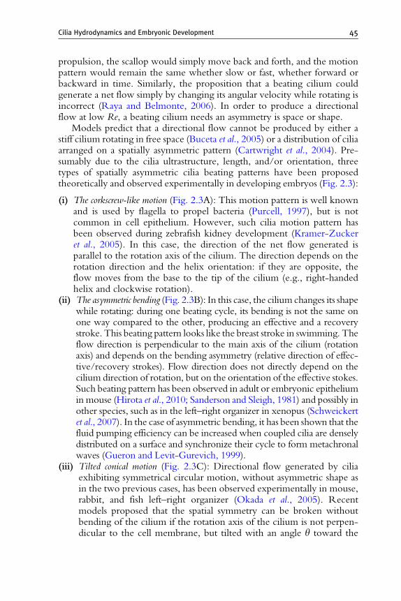
Models predict that a directional flow cannot be produced by either astiff cilium rotating in free space (Buceta et al., 2005) or a distribution of ciliaarranged on a spatially asymmetric pattern (Cartwright et al., 2004). Pre-sumably due to the cilia ultrastructure, length, and/or orientation, threetypes of spatially asymmetric cilia beating patterns have been proposedtheoretically and observed experimentally in developing embryos (Fig. 2.3):
(i) The corkscrew-like motion (Fig. 2.3A): This motion pattern is well knownand is used by flagella to propel bacteria (Purcell, 1997), but is notcommon in cell epithelium. However, such cilia motion pattern hasbeen observed during zebrafish kidney development (Kramer-Zuckeret al., 2005). In this case, the direction of the net flow generated isparallel to the rotation axis of the cilium. The direction depends on therotation direction and the helix orientation: if they are opposite, theflow moves from the base to the tip of the cilium (e.g., right-handedhelix and clockwise rotation).
(ii) The asymmetric bending (Fig. 2.3B): In this case, the cilium changes its shapewhile rotating: during one beating cycle, its bending is not the same onone way compared to the other, producing an effective and a recoverystroke. This beating pattern looks like the breast stroke in swimming. Theflow direction is perpendicular to the main axis of the cilium (rotationaxis) and depends on the bending asymmetry (relative direction of effec-tive/recovery strokes). Flow direction does not directly depend on thecilium direction of rotation, but on the orientation of the effective stokes.Such beating pattern has been observed in adult or embryonic epitheliumin mouse (Hirota et al., 2010; Sanderson and Sleigh, 1981) and possibly inother species, such as in the left–right organizer in xenopus (Schweickertet al., 2007). In the case of asymmetric bending, it has been shown that thefluid pumping efficiency can be increased when coupled cilia are denselydistributed on a surface and synchronize their cycle to form metachronalwaves (Gueron and Levit-Gurevich, 1999).
(iii) Tilted conical motion (Fig. 2.3C): Directional flow generated by ciliaexhibiting symmetrical circular motion, without asymmetric shape asin the two previous cases, has been observed experimentally in mouse,rabbit, and fish left–right organizer (Okada et al., 2005). Recentmodels proposed that the spatial symmetry can be broken withoutbending of the cilium if the rotation axis of the cilium is not perpen-dicular to the cell membrane, but tilted with an angle y toward the

46 Willy Supatto and Julien Vermot
normal of this surface (Fig. 2.3C; Cartwright et al., 2004; Smith et al.,2007; Vilfan and Julicher, 2006). In this case, the cilium interactionwith the cell surface plays a critical role: applying the no-slip boundarycondition (the velocity of the fluid at the cell surface must match thevelocity of the surface itself), the rotation forms an effective strokewhen the cilium is far from the surface and a recovery stroke close tothe surface (Fig. 2.3C). As the effective and recovery strokes aredefined by the interaction between cilium and cell surface, the direc-tion of the net flow generated depends on the cilium tilt and on the cellsurface. The directional flow is perpendicular to the direction of thecilium tilt and parallel to the surface. The orientation of the flowdepends on both the angular tilt orientation and the cilia rotationsense. The model predictions of a cilium tilt have been experimentallyconfirmed in vivo in mouse (Okada et al., 2005) and in zebrafish(Supatto et al., 2008; Fig. 2.6). Interestingly, among the three asym-metric beating patterns generated by biological cilia, the tilted conicalmotion is the easiest to reproduce artificially as it does not requirecontrolling the bending of the cilium. For this reason, the generationof directional flow at low Re in microfluidic devices can be obtainedusing stiff artificial cilia beating with tilted conical motion (Nonakaet al., 2005; Shields et al., 2010; Vilfan et al., 2010).
4.3. Mixing with chaotic advection
The second important consequence of Stokes flow properties is that mixingis hard at low Re. The absence of inertial and nonlinear terms in the Stokesflow equation prevents the occurrence of turbulent behaviors. Withoutturbulence, flow mixing relies on molecular diffusion and is usually slow.In this case, it is known that the only efficient way to create mixing is byusing chaotic advection (Stone et al., 2004). Chaotic advection enablesmolecular diffusion to take place on efficient time scales.
In general, the flow generated by motile cilia can help increasing therelative contribution of advection compared to diffusion in the transport ofparticles in a fluid. In fluid mechanics, this relative contribution is quantifiedby the Peclet number: Pe ¼ UL/D, with U and L the characteristic lengthand velocity scales (as used in Re) and D the particle diffusion coefficient.Pe � 1 means that the advection dominates the particle transport. Themixing efficiency thus increases with Pe (Stone et al., 2004).
In the case of chaotic advection, although the flow is laminar anddominated by viscous forces, fluid particle trajectories are chaotic, in thesense of being sensitive to initial conditions. In practice, it can be demon-strated experimentally or in simulations by following the trajectories ofseveral particles starting at close positions in space: if trapped into a chaotic
A Directional/vortical flow
C No-slip condition D Chaotic advection
30 mm.s–1
y (mm) y (mm)z (
mm)
x (m
m)
z (mm)
y (mm)
z (m
m)
Directionalflow
Vortical flow
Effective stroke
Chaoticadvection
Directional flow
No-slip condition
Cilium
Cell surface
Vortical flow Directional flow
10 0
5
10
15
20
25
20
15
10
5
010
5
0 510
15
15
10
5
Ave
rage
par
ticle
vel
ocity
in m
m/s
00 5 10
Distance from cell surface in mm15 20
5
10
15
20
10 5 5
5
10Vortical flow
Directional flow
Recovery stroke
Cilium
Cilium
Cell surface
Cell surface
15
10 15
50
0 mm.s–1
x (m
m)
x (m
m)
B Effective/recovery stroke
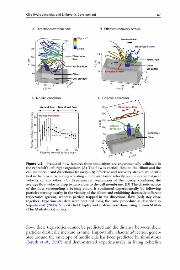
Figure 2.6 Predicted flow features from simulations are experimentally validated inthe zebrafish’s left–right organizer. (A) The flow is vortical close to the cilium and thecell membrane and directional far away. (B) Effective and recovery strokes are identi-fied in the flow surrounding a beating cilium with faster velocity on one side and slowervelocity on the other. (C) Experimental verification of the no-slip condition: theaverage flow velocity drop to zero close to the cell membrane. (D) The chaotic natureof the flow surrounding a beating cilium is confirmed experimentally by followingparticles starting nearby in the vicinity of the cilium and exhibiting drastically differenttrajectories (green), whereas particle trapped in the directional flow (red) stay closetogether. Experimental data were obtained using the same procedure as described inSupatto et al. (2008). Velocity field display and analysis were done using custom Matlab(The MathWorks) scripts.
Cilia Hydrodynamics and Embryonic Development 47
flow, these trajectories cannot be predicted and the distance between theseparticles drastically increase in time. Importantly, chaotic advection gener-ated around the envelope of motile cilia has been predicted by simulations(Smith et al., 2007) and demonstrated experimentally in living zebrafish

48 Willy Supatto and Julien Vermot
embryos (Supatto et al., 2008; Fig. 2.6D). Whether this chaotic behavior isactively used or avoided during embryonic development is interesting andremains unclear. Recent studies suggest that chaotic advection near beatingcilia could be an active mechanism involved in the otolith formation duringzebrafish inner ear development (Wu et al., 2011) and (Riley et al., 1997).Efficient fluid mixing using chaotic advection has been recently appliedin microfluidics with artificial motile cilia (Fahrni et al., 2009; Shieldset al., 2010).
4.4. The left–right organizer: Interplay between models andexperiments
Efforts have been made to address the hydrodynamics involved in the left–right symmetry breaking during early vertebrate development. Several keypredictions have been made through modeling of the leftward directionalflow generated in left–right organizers. This system illustrates well theinterplay between fluid mechanics, modeling, and experimental investiga-tion of cilia-driven flow in embryos. Whereas most of the experimental dataand models have been obtained in mouse, this system allows interestingcomparisons with the zebrafish left–right organizer (KV).
In recent years, several models and simulations have been developedbased on mouse node experimental data. In these theoretical studies, thecilium is modeled either as an infinitesimal sphere rotating in its place(Cartwright et al., 2004), a small sphere moving on a fixed trajectory inthe vicinity of a planar surface (Vilfan and Julicher, 2006) or a slender body(Smith et al., 2007). Simple time averaged modeling allows explaining thegeneration of a directional flow with tilted cilia (Cartwright et al., 2004),whereas more complex modeling considering the time-dependent dynam-ics of cilia allows capturing detailed features, such as the chaotic advectionoccurring in the vortical flow surrounding cilia (Smith et al., 2007). What-ever the complexity of the model used, it helps our understanding of thebiological process by explaining new observations, raising specific predic-tions, and suggesting the next set of experiments. In this section, we willreview the main flow features predicted by modeling and simulations basedon the mouse node system, describe the validation of these features in thezebrafish KV compared to the mouse node, and discuss the open questionsraised by each point.
The origin of the spatial asymmetry permitting the generation of adirectional flow within the left–right organizer was the first questionassessed by theorists. The proposition that cilia were beating with a tiltedconical motion (Fig. 2.3C) was first suggested by Cartwright et al. (2004)and further investigated by demonstrating the importance of the cell surfaceand the no-slip boundary condition to obtain efficient and recovery strokes(Smith et al., 2007; Vilfan and Julicher, 2006). The posterior tilt of cilia has

Cilia Hydrodynamics and Embryonic Development 49
been first observed in mouse (Okada et al., 2005). This topology promotes adirectional flow toward the left direction with cilia rotating clockwise.Among the asymmetric cilia beating patterns, it is interesting to mentionthat only the tilted conical motion allows breaking the left–right symmetrywithout preexisting left–right asymmetry at the scale of the cilium (Fig. 2.6)as the directional flow in this particular case is perpendicular to the tiltdirection, this pattern allows translating an anterior–posterior or dorsal–ventral asymmetry into a left–right asymmetry. This is not true for cork-screw-like motion or asymmetric bending: a net flow in the left–rightdirection would require having a preexisting left–right asymmetry at thelevel of the cilium. In zebrafish, the flow circulating around the dorsal–ventral axis within the KV (Fig. 2.2) cannot be due to a posterior tilt, butdue to a dorsal tilt. Importantly, this dorsal tilt has been observed in vivo(Supatto et al., 2008). Nevertheless, cilia with posterior tilt have beenobserved in the dorsal roof of the KV (Kramer-Zucker et al., 2005; Okabeet al., 2008). Such topology cannot produce the observed flow around thedorsal–ventral axis. It is noted that the beating pattern of KV’s cilia isdifferent from the mouse nodal cilia since they do not share the sameultrastructure (9 þ 2 instead of 9 þ 0 in mouse; Kramer-Zucker et al.,2005; Nonaka et al., 1998). Overall, a statistical analysis of the distributionof cilia orientations within the entire KV would greatly help to solve thisissue.
A key aspect of cilia-driven flow is the balance between effective/recovery strokes and the role of the no-slip conditions. In viscous fluids,the fluid will have zero velocity relative to a solid boundary. In case of ciliatilt, a fraction of the circular motion occurs while being closer to themembrane where moving fluid is more difficult. As a consequence, theinduced flow magnitude is smaller than when circular motion occurs farfrom the cell membrane, producing a net flow (Smith et al., 2007; Vilfan andJulicher, 2006). The no-slip condition has been demonstrated experimen-tally in zebrafish KV where the average flow velocity drops to 0 close to thecell surface (Fig. 2.6C) as well as in engineered cilia arrays (Shields et al.,2010). As a result, the flow velocity is stronger on the side of the effectivestroke, compared to the recovery stroke as shown experimentally(Fig. 2.6B). The optimal directional flow obtained for a tilt angle y � 35and a semi-cone angle c � 55 (Fig. 2.6), with y þ c � 90 (Smith et al.,2008, 2010a). Such angles have been reported in mouse, rabbit, and medakaembryos (Okada et al., 2005). The far field directional flow velocity scalewith 1/r2, with r the distance from the cilium (Cartwright et al., 2004; Smithet al., 2010a; Vilfan and Julicher, 2006). In fish, y � 35 (Supatto et al.,2008) or y � 45 (Kramer-Zucker et al., 2005) have been observed. Weshow in Fig. 2.7C that this angle can be even higher suggesting that ypresents strong fluctuations in vivo. The crucial role of this angle raises thequestion of its origin in vivo. In mouse, cilia tilt seems dynamic and

A B
Dorsal direction
y = 20°
q = 70°
Ciliumscanning artifact
Raster scanningat ~300 Hz (3.3 ms/line)
FluidCilium
Cell surface
Cell surface
C
2 mm
Figure 2.7 Characterizing 3D cilia features using standard laser scanning microscopyand scanning artifact. Cilia labeled with Bodipy TR are imaged in the zebrafishKupffer’s vesicle with two-photon laser scanning microscopy (A). The raster scanningis adjusted to be slow enough (3.3 ms per horizontal line) to capture several beatsduring the 2D image acquisition: in this case, stripped pattern is observed (arrows in A)corresponding to a slow scanning artifact. The distance between each strip correspondsto one period of cilium rotation: here, 10 lines correspond to �33 ms period and�30 Hz of beating frequency. Recording this pattern at different z-positions allows tomanually trace the contour of each stripped pattern and reconstruct the cilium envelopin 3D (B). The strong tilt direction toward the dorsal direction appears clearly. The tiltangle y and the semi-cone angle c can be measured (C): here, the tilt angle is specifi-cally strong with y � 70 and c � 20. Scale in (B) is 2 mm per tick. Experimental datawere obtained using zebrafish embryo preparation and labeling as described in Supattoet al. (2008). Two-photon excited fluorescence imaging was done at 820 nm wave-length using a Chameleon Ultra laser (Coherent), a Zeiss LSM510 microscope, anda 40/NA 1.1 objective lens (Zeiss) on six somite-stage embryo. Image displayand analysis were performed using ImageJ (http://rsb.info.nih.gov/ij/) and Imaris(Bitplane).
50 Willy Supatto and Julien Vermot
dependent on the planar cell polarity pathway (Marshall and Kintner, 2008).Most importantly, a clear coupling between cilia and directional flow hasbeen shown in ependymal cilia (Guirao et al., 2010). In mouse, it seems thatthe shape of the cell membrane and the planar polarity also plays a role incontrolling cilia tilt (Hashimoto et al., 2010).
Due to the low Re of the system, the influence of cilia vanishes quicklyin space. However, cilia motion causes unsteady behavior in its vicinity:mathematical modeling clearly shows that a vortical flow (unsteady) isgenerated close to the cilium and a directional flow (steady) far from thecilium (Smith et al., 2007). This chaotic nature of the vortical flow is seen inartificial setups (Shields et al., 2010) and in zebrafish KV (Supatto et al., 2008;Fig. 2.6D). So far, the relative contribution of directional and chaoticadvection in cilia-driven flow is not known. A simple back-of-the-envelopecalculation in the KV indicates that the chaotic advection occurs in morethan 50% of the total volume of the KV assuming the chaotic behavior at the

Cilia Hydrodynamics and Embryonic Development 51
periphery of the KV reaches up to 10 mm from the cell membrane in vesicleof 80 mm diameter (as shown experimentally in Supatto et al., 2008). Mixingis thus prominent in the KV, meaning that most of the fluid and factorssecreted in the vesicle would not travel through a linear path but will ratherhave a complex, erratic motion when close to the cilium.
More sophisticated models are now taking into account the geometry ofthe left–right organizer and the spatial distribution and the density of cilia inmice. Such models show interesting features, such as backward flow due toback pressure gradient in the enclosed nodal cavity (Cartwright et al., 2007;Smith et al., 2010a). A circular flow is thus generated in themouse node, verymuch like in fish. Yet, the backwardmotion observed inmouse spins aroundthe anterior–posterior axis contrary to the dorsoventral axis in fish. Further-more, there are cilia distributed all around the vesicle in fish as opposed tomice. Thus, while many features are common between mice and fish, eachspecies remain distinct and might have developed different ways to break theembryonic symmetry using the same basic ciliary machinery.
5. Experimental Investigation of Cilia-Driven
Fluid Flow in Developing Embryos
5.1. Challenges and experimental models
The experimental investigation of cilia-driven flow and its function duringembryonic development necessitates measuring parameters characterizingmotile cilia and fluid flow in vivo. As seen earlier, key information is held inthe fluid flow velocity field as well as in the cilia positioning, beating patternand orientation. Collecting this information in embryos is difficult becauseof the following challenges:
(i) Multiple time scales are required: cilia and flow dynamics are amongst thefastest processes occurring during embryonic morphogenesis andrequire high temporal resolution (up to �10 ms time resolution). Atthe same time, the long-term roles of cilia-mediated flow, such asactivating signal transduction pathways and the resulting cellular pro-cesses, can be slow (hours or days).
(ii) Multiple spatial scales are required: cilia act at the subcellular scale (mm) andthe flow-generated acts at the entire organ scale (mm).
(iii) Specific experimental imaging strategies that do not compromise nor-mal biology have to be developed. Ciliated structures are usuallylocated in cavities that are difficult to access experimentally. Moreover,they experience strong and large scale morphogenetic movements andare often located deep inside light scattering tissues.
The zebrafish embryo offers several advantages for visualizing fluid flowsin vivo. First, the embryo is transparent and has very little autofluorescence

52 Willy Supatto and Julien Vermot
and scattering, allowing an optimal use of optical imaging. Second, embryosare resistant to phototoxicity and are easy to culture permitting the use oflong-term light imaging and the collection of in vivo data at high resolution.Of note, recent zebrafish transgenic lines expressing fluorescent proteinsspecifically in cilia or in ciliated embryonic areas have been generated(Borovina et al., 2010; Tian et al., 2009). This facilitates the localization ofthe region of interest greatly and will certainly accelerate cilia imagingdevelopments. Nevertheless, imaging cilia-driven flows have been success-fully implemented in several vertebrate species. In particular, severalapproaches have been developed in mice, fish, frog, and rabbits to look atthe left–right organizer flow through the tracking of fluorescent beads in theenvironment of the cilia (Okada and Hirokawa, 2009; Okada et al., 2005;Schweickert et al., 2007). Tissue explants can also be an excellent approachwhen one can extract out and cultivate the ciliated cells without affectingtheir activity. Brain slices, and the primary culture of ependymal cells of theforebrain cavities or from the trachea, allow to image both flow and ciliaactivity (Guirao et al., 2010; Hirota et al., 2010; Zahm et al., 1990).However, it is difficult to know whether the boundary conditions in theexplants are intact, especially when looking at the far field flows.
5.2. Mapping microscopic flow field in living embryos
In the field of fluid mechanics, the experimental investigation of fluiddynamics usually relies on seeding the flow with tracer particles, imagingthe seeded flow and measuring the velocity field using image processing(Raffel et al., 1998). Particle image velocimetry (PIV) and particle trackingvelocimetry (PTV) are the most typical techniques to quantify flow veloc-ity. PIV is based on image cross-correlation to estimate the displacement ofpatterns between successive images in a statistical manner (Raffel et al.,1998). PIV is routinely used in fluid mechanics labs and has found recentapplications in developmental biology to map blood flow dynamics duringheart development (Hove et al., 2003) or to quantify the morphogeneticmovements shaping embryos (Supatto et al., 2005; Zamir et al., 2006). PTVis an alternative approach and relies on particle segmentation in each imagefollowed by the tracking of these objects between successive images. In fluidmechanics, these two techniques are related to the Eulerian and theLagrangian descriptions of the fluid flow: using PIV, the motion of thefluid is obtained at fixed positions in space distributed on a regular grid(Eulerian description such as in Hirota et al., 2010), whereas using PTV, thevelocity is measured along trajectories of specific particles trapped into theflow (Lagrangian description such as in Fig. 2.6). The choice between thesetwo descriptions usually depends on practical considerations: for instance,by following the unpredictable trajectories of particles, the Lagrangian

Cilia Hydrodynamics and Embryonic Development 53
description is well suited for demonstrating chaotic advection in the zebra-fish KV (Fig. 2.6D).
From an experimental perspective, PTV presents several advantages com-pared to PIV: it allows higher spatial resolution and manual or automatedparticle tracking that can be performed in 2D or 3D, whereas PIV usuallyworks only for 2Danalyses and cannot be performedmanually.Tobe accurate,PIV requires a high density of tracer particles, whereas PTV works at lowdensity. Commercial and free open source packages exist to perform PIV orPTV (see MatPIV http://folk.uio.no/jks/matpiv/, or the ImageJ pluginhttp://www.mosaic.ethz.ch/Downloads/ParticleTracker, for instance).
Both PIV and PTV have been used to investigate cilia-driven flow duringembryonic development, such as in the left–right organizer (PIV in mouse(Hirota et al., 2010), PTV in xenopus (Schweickert et al., 2007), or zebrafish(Supatto et al., 2008)). Seeding the flow with tracer particles remains a criticalstep for applying such strategy in developmental biology. In mice, flow in theleft–right organizer has been imaged in cultured embryos and the velocity ofthe steady flow has been measured by tracking the motion of beads (seeTable 2.1 and Okada and Hirokawa, 2009). In zebrafish, such an experimentnecessitates an injection of beads within the KV cavity using a needle. Inpractice, this step remains invasive when one wants to look at fine hydro-dynamics. An alternative approach has been recently developed to address theflow field with much milder side effects: the flow is seeded with microscopicfluorescently labeled cell debris generated by targeting a single cell withfemtosecond subcellular ablation as described in Fig. 2.8 (Supatto et al., 2008).
The experimental mapping of the flow field using PIV or PTV allowsquantifying basic features of the flow such as the average speed of thedirectional flow generated by beating cilia (Table 2.1). It also permits furthercharacterization of the flow and the experimental validation of predictionsfrom models and simulations, as well as feeding further modeling with preciseexperimental data. The investigation of the velocity field generated within thezebrafish KV using PTV is presented in Fig. 2.6. This study illustrates howexperimental investigation of the fluid flow can validate the flow featurespredicted by simulations (see Section 3). The flow features are measuredwithin the whole vesicle (Fig. 2.8), as well as in the vicinity of single cilia. Aspredicted by simulations, two types of flows are observed: a laminar flow,which is directional and breaks to left–right symmetry, and a vortical flowsurrounding the cilium. The chaotic nature predicted by simulations can bedemonstrated experimentally using such a quantitative approach (Fig. 2.6D).
5.3. Measuring cilia features in vivo
Investigating the role of cilia-driven flow in embryonic developmentrequires the experimental characterization of cilia in vivo: the key featuresincludes cilia spatial position and density, beating pattern, rotation speed and

A B
D E F
I J
C
Figure 2.8 Mapping the cilia-driven flow within the zebrafish Kupffer’s vesicle (KV)using femtosecond laser ablation, fast imaging and 3D-particle tracking. The KV cellsare labeled with Bodipy TR and a single cell lining the cavity is targeted with tightlyfocused femtosecond laser pulses at 820 nmwavelength (A). A subcellular femtosecondlaser ablation is performed to generate fluorescent microdebris seeding the flow (B).The nonlinear effect used in this technique allows a high spatial confinement and a lowinvasiveness, thus permitting the targeting of subcellular regions deep inside theembryo. Fast confocal 3D imaging at 4 z-stack per second capture the movement ofthe tracer particles: in (C), time-lapse images are superimposed to show the circulartrajectories of the flow around the dorsal–ventral axis of the embryo. Cilia close to theKV surface (D) can be identified by following particles trapped in a vortical flow(arrows in E and F) and mapping the flow using 3D-particle tracking (D). Experimentaldata were obtained using the same procedure as described in Supatto et al. (2008).Image display and analysis were performed using ImageJ (http://rsb.info.nih.gov/ij/)and Imaris (Bitplane).
54 Willy Supatto and Julien Vermot
direction, amplitude, tilt, or bending. However, these parameters cannot bemeasured experimentally using direct imaging of cilia using conventionaltechnologies, such as confocal microscopy. Since cilia are moving extremelyfast (Table 2.1) and in 3D, capturing cilia shape and motion requires both3D resolution and time resolution that are at the limit of the state-of-the-artimaging technology. We can easily estimate the ideal time resolution

Cilia Hydrodynamics and Embryonic Development 55
required to acquire 3D-time lapses capturing the full rotation movement ofa cilium beating at 30 Hz: with 10 images per z-stack and 10 z-stacks percilium rotation, it would require imaging at 3000 frame per second (fps) andacquiring z-stacks at 300 Hz with sufficient signal, which can be challengingwhen cilia are located deep in light scattering tissues. High-speed imaging ofcilia using bright field microscopy without fluorescent labeling has beenreported (acquisitions at 500 fps in Okada and Hirokawa, 2009). However,this technique lacks 3D resolution and depth penetration: it is only adaptedfor 2D arrays of cilia close to the embryo surface such as in the mouse nodeand does not provide measurements of every cilia features that wouldrequire 3D spatial reconstruction. Recent developments in the fast fluores-cence microscopy field allow high-speed fluorescence imaging (Vermotet al., 2008). 2D imaging of cilia with commercial fast confocal microscopyup to 900 fps has been recently reported (Hirota et al., 2010), correspondingin this case to typically 50 frames per cilium beat, which is more thanenough to capture the movement. However, the ideal z-stack acquisitionfrequency required to capture the full 3D movements of cilia is currentlyunreachable through commercial imaging setups. To circumvent this limi-tation, experimentalists have developed tricks to obtain the required infor-mation without fully capturing the 3D movements of beating cilia in vivo.We report in the following section several of these techniques used to studycilia-driven flow in the left–right organizer.
To demonstrate cilia beat with a tilted conical motion in the mouse left–right organizer (Fig. 2.3C), high-speed 2D bright field imaging has beenused to estimate cilia position, tilt angle y, semi-cone angle c, and tiltorientation (Nonaka et al., 2005; Okada et al., 1999, 2005). To circumventthe lack of 3D resolution, the 2D traces of cilia tips were analyzed andcompared with the 2D projection of an expected tilted cone. It allowedestimating the tilt angle and orientation, as described in Fig. 2.9. This ruseworks only if we assume the cilia are positioned on a 2D flat epitheliumperpendicular to the optical axis of the microscope. In the case of a curvedepithelium, such as in the zebrafish KV, it cannot give accurate measure-ment of cilia orientation.
An alternative approach to characterize cilia features without actual 3Dimaging of them has been recently reported in zebrafish (Supatto et al.,2008). From 3D particle trajectories trapped into the vortical flow sur-rounding a cilium (Figs. 2.6A and 2.8F), several features can be indirectlycharacterized: cilium position in space (Fig. 2.8F), tilt direction and angle y,rotation direction (clockwise/anticlockwise), and effective and recoverystrokes (Fig. 2.6B). Importantly, the particles trapped in the vicinity of acilium are moving much slower than the cilium itself in Stokes flow (Smithet al., 2010a). For this reason, mapping the flow surrounding a cilium allowsobtaining 3D data with lower time resolution than required for the direct3D imaging of cilia (Supatto et al., 2008). However, this indirect approach

A
R
R5 mm
A
A
L
L
P
P
B TiltNo tilt
Projected pattern
Cocentriccircle
EccentricElipse
EccentricD-shape
Small tilt
Extensive tilt
Figure 2.9 In vivo experimental characterization of cilia beating pattern in the mousenode without labeling or 3D resolution using DIC microscopy (figure reproduced fromNonaka et al., 2005). (A) Trace of node cilia in enhanced DIC images after backgroundsubtraction. The black spots represent the positions of cilia roots, and tip trajectories arein blue, green, and orange. (B) Relationship between observed trip trajectories asprojected pattern (circle, ellipse, or D-shape, corresponding to green, blue, and orangetrajectories in (A), respectively) and expected beating patterns in 3D. The arrows showthe clockwise rotation of cilia. A, P, L, and R refer to anterior, posterior, left, and rightsides of the node, respectively.
56 Willy Supatto and Julien Vermot
fails at characterizing properties such as cilium length, shape, rotation speed,or semi-cone angle c and requires an extensive analysis for characterizingeach investigated cilium.
Interestingly, experimental artifacts due to a lack of time resolution canbe used to extract useful information and measure cilia features in vivo.Hadjantonakis et al. (2008) reported a scanning artifact using slow acquisi-tion speed with standard laser scanning microscopy allows biologists todetect the presence of cilia in the mouse node. In this case, the laser scanningis slow enough to capture several traces of cilium signal during the 2Dacquisition of an image and produces a stripped pattern as shown in thezebrafish KV in Fig. 2.7A. Recording this pattern at different z-positionsallows reconstructing the cilium envelope in 3D (Fig. 2.7B) and to measurethe tilt orientation and angle y or the semi-cone angle c (Fig. 2.7C).In addition, the distance between the 2D stripes provide a measure of ciliumbeating frequency (Fig. 2.7A). This approach does not permit measuring thedirection of rotation (clockwise/anticlockwise).

Cilia Hydrodynamics and Embryonic Development 57
Bringing together these imaging approaches allow characterizing ciliafeatures in living embryos without requiring the full 3D capture of ciliabeats. Even if these technical ruses work better at low cilia density, theyopen access to simultaneous measurement of several cilia and permit tofollow processes at multiple spatial scales. For instance, flow mapping allowssimultaneously following the flow within an entire organ and close toindividual cilia (Supatto et al., 2008).
5.4. Next experimental challenges: Flow/cilia manipulationand functional imaging
While flow and cilia features can be measured experimentally in livingembryos using imaging, the full investigation of the role of cilia duringembryonic development brings other experimental challenges. Forinstance, to fully understand the biophysics of cilia and the interplaybetween mechanical properties and biological function, it can be criticalto manipulate cilia properties (density, clockwise/anticlockwise rotation,speed, tilt angle and direction, amplitude, direction, etc.) or the externalflow. Pioneering experimental studies reported the effect of manipulatingexternal flow on a ciliated epithelium (Guirao et al., 2010) and on the mouseembryo node (Nonaka et al., 2005) or the application of external forces oncilia using magnetic manipulations (Hill et al., 2010). However, suchmanipulation is extremely difficult to perform in live embryos, especiallywhen ciliated structures are internal. Working on explants or culturedepithelia allows direct access to cilia and facilitates measurements that arechallenging to perform in vivo, such as through magnetic manipulation (Hillet al., 2010). Alternative strategies using optical tweezers have been success-fully used in the zebrafish inner ear (Riley et al., 1997; Wu et al., 2011) andshould open access to other organs as well. Coupling structural imaging ofcilia, flow mapping and in vivo functional imaging of the biological responsesuch as calcium signaling, gene expression dynamics, or cellular processes isclearly the next challenge to meet. The constant progress in optics, molec-ular imaging and engineering will certainly help to reach this milestone.
6. Role of Cilia-Driven Flow in the Kupffer’s
Vesicle in Zebrafish
The KV is a transient cavity visible from 8 to 11 h after fertilizationunderneath the notochord (Fig. 2.2). The epithelium of the KV is made of9 þ 2 monociliated cells that generate a counterclockwise fluid flow(Fig. 2.2G–H). This flow triggers asymmetric calcium response on the leftside of the cavity (Fig. 2.2F; Francescatto et al., 2010; Sarmah et al., 2005)

58 Willy Supatto and Julien Vermot
and is involved in establishing and maintaining the left–right asymmetry ofthe body axis (Essner et al., 2005; Kramer-Zucker et al., 2005). Twohypotheses coexist to explain the role of flow in this process: either it biasesthe transport of biochemical signals toward the left side of the KV or itgenerates a physical stimulus which is read differently according to thedirection of the flow. So far, none of the two hypotheses has been ruledout. The asymmetric calcium response is dependent on two channel recep-tors, the ryanodine receptor ryr3 and pkd2 (TRPP2) that are expressed bythe KV cells (Francescatto et al., 2010). pkd2 is a mechanosensory receptorwhose function is tightly associated with primary cilia, thus it has beenproposed that flow is sensed by cilia through pkd2 which in turn triggerscalcium flux. However, a direct link between these three events is difficultto establish and theoretical work based on measures performed in the mousesuggests that the shear forces are too low for cilia to sense flow directionality(Cartwright et al., 2008). Even though the presence of sensory, immotilecilia is still a matter of debate in the fish (Borovina et al., 2010; Okabe et al.,2008), it is essential to quantify the flow forces generated within the left–right organizer to address this question. Typically, the flow map within theKV displays different velocities according to theA–P axis. It is not surprisingas cilia are enriched in the anterior pole of the vesicle (Okabe et al., 2008).This reflects the linear relationship postulated by the Stokes flow where theactivity of cilia adds to each other showing that directional flow is obtainedbecause of a dorsal cilia tilt. Overall, questions still remain whether flow,morphogen, and physical influences coexist in the process of left–rightspecification in vertebrates. It is certain that the theory of hydrodynamicscan lead us toward novel hypotheses, which will greatly benefit the devel-opmental biology field. Nevertheless, the biological mechanisms at workduring this process remain poorly understood in the zebrafish, especiallywhen considering that many observations in the mouse node such as thepresence of vesicular release in the vesicle and the presence of sensory ciliaare yet lacking in fish. The anatomy and the genetics have greatly divergedbetween fish and mice, and it is possible that slightly different mechanismsoperate in these two species.
7. Role of Cilia-Driven Flow in the Developing
Inner Ear in Zebrafish
Besides the well-known left–right flow, cilia-driven flows are neces-sary in multiple developing organs (Cartwright et al., 2009). Recent char-acterization of inner ear flow uncovered alternative roles to fluid motionduring embryogenesis. In zebrafish, as in humans and other vertebrates,balance is mediated by mechanical sensors in the inner ear. These sensors

24 hpf 48 hpf
A B C
Figure 2.10 Otolithic biomineralization in zebrafish. (A) Side view of a zebrafish at 20hpf showing the inner ear and the two otolithic aggregates. Otoliths first appear after18 h postfertilization in the otic cavity. (B and C) Transverse section through the otolithat 22 hpf (B) and 48 hpf (C) showing an electron dense inner part (nucleus) and radialbiomineralization starting around the nucleus viewed by transmission electron micros-copy (TEM). Radial biomineralization is prominent at that stage. Sensory cilia bundlesof the hair cells attached to the otolith at 48 hpf (arrow heads). Scale bar 100 mm in (A)and 5 mm in (B and C). Electron microscopy imaging was performed as described inPisam et al., 2002).
Cilia Hydrodynamics and Embryonic Development 59
consist of biomineralized composite crystals, called otoliths (ear-stones),situated atop cilia bundles on the surface of epithelial cells (Fig. 2.10).They provide an inertial mass that facilitates deflection of cilia bundles inresponse to vibration, gravity, and linear acceleration essential for hearingand balance through specialized cells called hair cells (Fig. 2.10; Yu et al.,2011). In zebrafish, the growth phase of otolith development constitutes akey step during the biomineralization process. Otoliths form at the top oftether cilia, located at the anterior and posterior poles of the inner ear fromsmaller dense clusters referred to as “spherules,” which are self-aggregatingparticles secreted from the apical portions of the epithelial cells lining theinner ear cavity (Fig. 2.10). At the site of cilium–otolith attachment areclusters of motile cilia inducing local flow. Electron microscopy analysisshows that otolith growth starts as a nucleus of spherules aggregating at thetop of a tether cilium (Fig. 2.10; Pisam et al., 2002). At 30 hpf, a mineralizedovoid otolith is visible (Pisam et al., 2002; Sollner et al., 2003) and concen-tric arrays of spherule deposition are seen at the periphery of the nascentotolith (Fig. 2.10; Pisam et al., 2002). Cilia-driven flow is required forproper otolith formation, but the nature of the flow dynamics at work inthe inner ear and the precise contribution of local flow at sites of biominer-alization are now the focus of recent investigations. On the basis of high-speed video microscopy of cilia motility and quantitative analysis of precur-sor particle movements in wild-type and immotile cilia mutant embryos, acilium-dependent hydrodynamic mechanism for otolith biogenesis hasbeen proposed (Fig. 2.11). In this model, motility of tether cilia at thepoles of the otic vesicle establishes a vortex that attracts otolith precursors

NT NT
NT
IEIE
IE
A B
C D
E
F
Figure 2.11 Cilia-mediated flow and otolith biogenesis. (A) Dorsal view of embryolabeled with anti-acetylated tubulin antibody showing the neural tube (NT) and axonalprojections as well as primary cilia throughout the embryos. (B) Side view of the innerear visualized through bright field microscopy showing otolith (arrowhead) within theinner ear (IE). (C) Cilia labeling in the inner ear using anti-acetylated tubulin antibodyreveals two types of cilia: long tether and short cilia. Immunohistochemistry wasperformed as described in Colantonio et al. (2009). (D) Otolith view with bright fieldmicroscopy as described in Colantonio et al. (2009), showing spherules next to it. (E andF) Model for motile cilia function during otolith biogenesis.
60 Willy Supatto and Julien Vermot

Cilia Hydrodynamics and Embryonic Development 61
(Fig. 2.11E and F), thereby biasing the distribution of precursor particlestowards the two patches of tether cilia. Such a mechanism would favorpreferential otolith seeding at the poles of the otic vesicle. At the otic vesiclepoles, tether cilia motility further serves to disperse precursor particleslocally. This model, while attractive, needs some clarifications in terms ofprecise hydrodynamics, most importantly in terms of the hydrodynamicbasis of spherule attraction. The hydrodynamic features in the inner ear areparticularly interesting because, as opposed to other known cilia-mediatedflows, there is no directional flow. It is the activity of isolated clusters ofbeating cilia that dictates a local flow, which is not able to take over thewhole cavity (Wu et al., 2011). It clearly shows that the topology, such asthe localization of cilia motility in key areas of the inner ear, is dictating thehydrodynamics in the organ. Importantly, the role of mixing in this systemis critical for dictating the shape of the otolith demonstrating that cilia cancombine different function through generation of different flow regime(Wu et al., 2011).
8. Conclusion
Throughout the years of cilia research conducted in the past, fluidmechanics and modeling greatly helped experimenters in identifying properquestions and in developing appropriate methods to address them. Today,many questions remain unanswered, which will undoubtedly drive ciliainvestigations in the coming years. For example, the crucial role of the tiltingangle in the genesis of directional flow raises important unknown questions:what are the structural origins of the tilt? How is it regulated and throughwhich dynamics? Last, how does the coupling between cilia and flowinfluence each other so that the system can reach a steady state? Furthermore,a lot remains to be said about the roles of chaotic advections in cilia-drivenflows. We showed that this is a prominent effect in the KV and in the innerear, but it is certainly true in other ciliated organs and species. The continu-ous effort to combine experimental approaches through live imaging, mod-eling, and forward and reverse genetics in the zebrafish are expected toprovide us answers to some of these interesting questions in the near future.
The study of the role of cilia-driven flow in embryonic development is anexciting example of interdisciplinary investigation. As reviewed here, thefruitful interplay between fluidmechanics, mechanical modeling, microscopy,image processing, experimental micromanipulation, and developmentalgenetics permits gaining important knowledge and promises future excitingdiscoveries in the field. A central driving force for the understanding of thebiology of cilia-flow during embryonic development is the continuous neces-sity for integrating knowledge and experimental data originating from various

62 Willy Supatto and Julien Vermot
disciplines (fluid mechanics, modeling, imaging and image processing, genet-ics, etc.). Another very recent trend is the fact that the biology of cilia structureis feeding biomimetic research and finds of applications inmicrofluidic devicesinspired by biology (Shields et al., 2010; Vilfan et al., 2010). There is a lot to betthat many other biological systems will benefit from such an integrated way ofapproaching science.
ACKNOWLEDGMENTS
We thank D. Wu and L. Thornton for thoughtful comments on the chapter. We are gratefulto N. Messaddeq and J. L. Weickert for help with the electron microscopy and the Fraserlaboratory at Caltech for sharing reagents. We also thank the IGBMC, Institut de Genetiqueet de Biologie Moleculaire et Cellulaire for assistance, the Caltech Biological Imaging Centerand the ImagoSeine imaging facility at Institut Jacques Monod for sharing equipment. J. V. issupported by the Human Frontier Science Program (HFSP), INSERM, and the Fondationpour la Recherche Medical (FRM). J. V. and W. S. are supported by Marie Curie Interna-tional Reintegration Grants within the 7th European Community Framework Programme.
REFERENCES
Aamar, E., and Dawid, I. B. (2008). Isolation and expression analysis of foxj1 and foxj1.2 inzebrafish embryos. Int. J. Dev. Biol. 52, 985–991.
Afzelius, B. A. (1999). Asymmetry of cilia and of mice and men. Int. J. Dev. Biol. 43,283–286.
Baker, K., and Beales, P. L. (2009). Making sense of cilia in disease: The human ciliopathies.Am. J. Med. Genet. C Semin. Med. Genet. 151C, 281–295.
Borovina, A., Superina, S., Voskas, D., and Ciruna, B. (2010). Vangl2 directs the posteriortilting and asymmetric localization of motile primary cilia. Nat. Cell Biol. 12, 407–412.
Buceta, J., Ibanes, M., Rasskin-Gutman, D., Okada, Y., Hirokawa, N., and Izpisua-Belmonte, J. C. (2005). Nodal cilia dynamics and the specification of the left/right axisin early vertebrate embryo development. Biophys. J. 89, 2199–2209.
Cartwright, J. H. E., Piro, O., and Tuval, I. (2004). Fluid-dynamical basis of the embryonicdevelopment of left-right asymmetry in vertebrates. Proc. Natl. Acad. Sci. USA 101,7234–7239.
Cartwright, J. H. E., Piro, N., Piro, O., and Tuval, I. (2007). Embryonic nodal flow and thedynamics of nodal vesicular parcels. J. R. Soc. Interface 4, 49–55.
Cartwright, J. H., Piro, N., Piro, O., and Tuval, I. (2008). Fluid dynamics of nodal flow andleft-right patterning in development. Dev. Dyn. 237, 3477–3490.
Cartwright, J. H., Piro, O., and Tuval, I. (2009). Fluid dynamics in developmental biology:Moving fluids that shape ontogeny. HFSP J. 3, 77–93.
Caspary, T., Larkins, C. E., and Anderson, K. V. (2007). The graded response to SonicHedgehog depends on cilia architecture. Dev. Cell 12, 767–778.
Castleman, V. H., Romio, L., Chodhari, R., Hirst, R. A., de Castro, S. C., Parker, K. A.,Ybot-Gonzalez, P., Emes, R. D., Wilson, S. W., Wallis, C., Johnson, C. A.,Herrera, R. J., et al. (2009). Mutations in radial spoke head protein genes RSPH9 andRSPH4A cause primary ciliary dyskinesia with central-microtubular-pair abnormalities.Am. J. Hum. Genet. 84, 197–209.

Cilia Hydrodynamics and Embryonic Development 63
Chilvers, M. A., Rutman, A., and O’Callaghan, C. (2003). Ciliary beat pattern is associatedwith specific ultrastructural defects in primary ciliary dyskinesia. J. Allergy Clin. Immunol.112, 518–524.
Colantonio, J. R., Vermot, J., Wu, D., Langenbacher, A. D., Fraser, S., Chen, J. N., andHill, K. L. (2009). The dynein regulatory complex is required for ciliary motility andotolith biogenesis in the inner ear. Nature 457, 205–209.
Dale, N., Roberts, A., Ottersen, O. P., and Storm-Mathisen, J. (1987). The morphology anddistribution of ‘Kolmer-Agduhr cells’, a class of cerebrospinal-fluid-contacting neuronsrevealed in the frog embryo spinal cord by GABA immunocytochemistry. Proc. R. Soc.Lond. B Biol. Sci. 232, 193–203.
Drummond, I. (2009). Studying cilia in zebrafish. Methods Cell Biol. 93, 197–217.Essner, J. J., Vogan, K. J., Wagner, M. K., Tabin, C. J., Yost, H. J., and Brueckner, M.
(2002). Conserved function for embryonic nodal cilia. Nature 418, 37–38.Essner, J. J., Amack, J. D., Nyholm, M. K., Harris, E. B., and Yost, H. J. (2005). Kupffer’s
vesicle is a ciliated organ of asymmetry in the zebrafish embryo that initiates left-rightdevelopment of the brain, heart and gut. Development 132, 1247–1260.
Fahrni, F., Prins, M. W. J., and van Ijzendoorn, L. J. (2009). Micro-fluidic actuation usingmagnetic artificial cilia. Lab Chip 9, 3413–3421.
Francescatto, L., Rothschild, S. C., Myers, A. L., and Tombes, R. M. (2010). The activationof membrane targeted CaMK-II in the zebrafish Kupffer’s vesicle is required for left-rightasymmetry. Development 137, 2753–2762.
Gueron, S., and Levit-Gurevich, K. (1999). Energetic considerations of ciliary beating andthe advantage of metachronal coordination. Proc. Natl. Acad. Sci. USA 96, 12240–12245.
Guirao, B., Meunier, A., Mortaud, S., Aguilar, A., Corsi, J. M., Strehl, L., Hirota, Y.,Desoeuvre, A., Boutin, C., Han, Y. G., Mirzadeh, Z., Cremer, H., et al. (2010).Coupling between hydrodynamic forces and planar cell polarity orients mammalianmotile cilia. Nat. Cell Biol. 12, 341–350.
Hadjantonakis, A. K., Pisano, E., and Papaioannou, V. E. (2008). Tbx6 regulates left/rightpatterning in mouse embryos through effects on nodal cilia and perinodal signaling. PLoSONE 3, e2511.
Hashimoto, M., Shinohara, K., Wang, J., Ikeuchi, S., Yoshiba, S., Meno, C., Nonaka, S.,Takada, S., Hatta, K., Wynshaw-Boris, A., and Hamada, H. (2010). Planar polarizationof node cells determines the rotational axis of node cilia. Nat. Cell Biol. 12, 170–176.
Hilfinger, A., and Julicher, F. (2008). The chirality of ciliary beats. Phys. Biol. 5. Articlenumber is 016003.
Hill, D. B., Swaminathan, V., Estes, A., Cribb, J., O’Brien, E. T., Davis, C. W., andSuperfine, R. (2010). Force generation and dynamics of individual cilia under externalloading. Biophys. J. 98, 57–66.
Hirota, Y., Meunier, A., Huang, S., Shimozawa, T., Yamada, O., Kida, Y. S., Inoue, M.,Ito, T., Kato, H., Sakaguchi, M., Sunabori, T., Nakaya, M. A., et al. (2010). Planarpolarity of multiciliated ependymal cells involves the anterior migration of basal bodiesregulated by non-muscle myosin II. Development 137, 3037–3046.
Hove, J. R., Koster, R. W., Forouhar, A. S., Acevedo-Bolton, G., Fraser, S. E., andGharib, M. (2003). Intracardiac fluid forces are an essential epigenetic factor for embry-onic cardiogenesis. Nature 421, 172–177.
Jekely, G., and Arendt, D. (2006). Evolution of intraflagellar transport from coated vesiclesand autogenous origin of the eukaryotic cilium. Bioessays 28, 191–198.
Kobayashi, D., Iijima, N., Hagiwara, H., Kamura, K., Takeda, H., and Yokoyama, T.(2010). Characterization of the medaka (Oryzias latipes) primary ciliary dyskinesia mutant,jaodori: Redundant and distinct roles of dynein axonemal intermediate chain 2 (dnai2) inmotile cilia. Dev. Biol. 347, 62–70.

64 Willy Supatto and Julien Vermot
Kramer-Zucker, A. G., Olale, F., Haycraft, C. J., Yoder, B. K., Schier, A. F., andDrummond, I. A. (2005). Cilia-driven fluid flow in the zebrafish pronephros, brainand Kupffer’s vesicle is required for normal organogenesis. Development 132, 1907–1921.
Lindemann, C. B. (2011). Experimental evidence for the geometric clutch hypothesis. Curr.Top. Dev. Biol. 95, 1–31.
Lindemann, C. B., and Lesich, K. A. (2010). Flagellar and ciliary beating: The proven andthe possible. J. Cell Sci. 123, 519–528.
Liu, Y., Pathak, N., Kramer-Zucker, A., and Drummond, I. A. (2007). Notch signalingcontrols the differentiation of transporting epithelia and multiciliated cells in the zebrafishpronephros. Development 134, 1111–1122.
Lopes, S. S., Lourenco, R., Pacheco, L., Moreno, N., Kreiling, J., and Saude, L. (2010).Notch signalling regulates left-right asymmetry through ciliary length control. Develop-ment 137, 3625–3632.
Mangos, S., Lam, P. Y., Zhao, A., Liu, Y., Mudumana, S., Vasilyev, A., Liu, A., andDrummond, I. A. (2010). The ADPKD genes pkd1a/b and pkd2 regulate extracellularmatrix formation. Dis. Model. Mech. 3, 354–365.
Marshall, W. F., and Kintner, C. (2008). Cilia orientation and the fluid mechanics ofdevelopment. Curr. Opin. Cell Biol. 20, 48–52.
McGrath, J., Somlo, S., Makova, S., Tian, X., and Brueckner, M. (2003). Two populationsof node monocilia initiate left-right asymmetry in the mouse. Cell 114, 61–73.
Nakao, T., and Ishizawa, A. (1984). Light- and electron-microscopic observations of the tailbud of the larval lamprey (Lampetra japonica), with special reference to neural tubeformation. Am. J. Anat. 170, 55–71.
Neugebauer, J. M., Amack, J. D., Peterson, A. G., Bisgrove, B. W., and Yost, H. J. (2009).FGF signalling during embryo development regulates cilia length in diverse epithelia.Nature 458, 651–654.
Nonaka, S., Tanaka, Y., Okada, Y., Takeda, S., Harada, A., Kanai, Y., Kido, M., andHirokawa, N. (1998). Randomization of left-right asymmetry due to loss of nodal ciliagenerating leftward flow of extraembryonic fluid in mice lacking KIF3B motor protein.Cell 95, 829–837.
Nonaka, S., Shiratori, H., Saijoh, Y., and Hamada, H. (2002). Determination of left-rightpatterning of the mouse embryo by artificial nodal flow. Nature 418, 96–99.
Nonaka, S., Yoshiba, S., Watanabe, D., Ikeuchi, S., Goto, T., Marshall, W. F., andHamada, H. (2005). De novo formation of left-right asymmetry by posterior tilt ofnodal cilia. PLoS Biol. 3, e268.
Okabe, N., Xu, B., and Burdine, R. D. (2008). Fluid dynamics in zebrafish Kupffer’s vesicle.Dev. Dyn. 237, 3602–3612.
Okada, Y., and Hirokawa, N. (2009). Observation of nodal cilia movement and measure-ment of nodal flow. Methods Cell Biol. 91, 265–285.
Okada, Y., Nonaka, S., Tanaka, Y., Saijoh, Y., Hamada, H., and Hirokawa, N. (1999).Abnormal nodal flow precedes situs inversus in iv and inv mice. Mol. Cell 4, 459–468.
Okada, Y., Takeda, S., Tanaka, Y., Belmonte, J. C., and Hirokawa, N. (2005). Mechanismof nodal flow: A conserved symmetry breaking event in left-right axis determination.Cell121, 633–644.
Perkins, L. A., Hedgecock, E. M., Thomson, J. N., and Culotti, J. G. (1986). Mutant sensorycilia in the nematode Caenorhabditis elegans. Dev. Biol. 117, 456–487.
Pisam, M., Jammet, C., and Laurent, D. (2002). First steps of otolith formation of thezebrafish: Role of glycogen? Cell Tissue Res. 310, 163–168.
Purcell, E. M. (1977). Life at low Reynolds number. Am. J. Phys. 45, 3–11.Purcell, E. M. (1997). The efficiency of propulsion by a rotating flagellum. Proc. Natl. Acad.
Sci. USA 94, 11307–11311.

Cilia Hydrodynamics and Embryonic Development 65
Raffel, M., Willert, C., and Kompenhans, J. (1998). Particle Image Velocimetry: A PracticalGuide. Springer-Verlag, Berlin.
Raya, A., and Belmonte, J. C. I. (2006). Left-right asymmetry in the vertebrate embryo:From early information to higher-level integration. Nat. Rev. Genet. 7, 283–293.
Riley, B. B., Zhu, C., Janetopoulos, C., and Aufderheide, K. J. (1997). A critical period ofear development controlled by distinct populations of ciliated cells in the zebrafish. Dev.Biol. 191, 191–201.
Sanderson, M. J., and Sleigh, M. A. (1981). Ciliary activity of cultured rabbit trachealepithelium—Beat pattern and metachrony. J. Cell Sci. 47, 331–347.
Sarmah, B., Latimer, A. J., Appel, B., and Wente, S. R. (2005). Inositol polyphosphatesregulate zebrafish left-right asymmetry. Dev. Cell 9, 133–145.
Sarmah, B., Winfrey, V. P., Olson, G. E., Appel, B., andWente, S. R. (2007). A role for theinositol kinase Ipk1 in ciliary beating and length maintenance. Proc. Natl. Acad. Sci. USA104, 19843–19848.
Schweickert, A., Weber, T., Beyer, T., Vick, P., Bogusch, S., Feistel, K., and Blum, M.(2007). Cilia-driven leftward flow determines laterality in Xenopus. Curr. Biol. 17,60–66.
Shah, A. S., Ben-Shahar, Y., Moninger, T. O., Kline, J. N., and Welsh, M. J. (2009). Motilecilia of human airway epithelia are chemosensory. Science 325, 1131–1134.
Shields, A. R., Fiser, B. L., Evans, B. A., Falvo, M. R., Washburn, S., and Superfine, R.(2010). From the Cover: Biomimetic cilia arrays generate simultaneous pumping andmixing regimes. Proc. Natl. Acad. Sci. USA 107, 15670–15675.
Short, M. B., Solari, C. A., Ganguly, S., Powers, T. R., Kessler, J. O., and Goldstein, R. E.(2006). Flows driven by flagella of multicellular organisms enhance long-range moleculartransport. Proc. Natl. Acad. Sci. USA 103, 8315–8319.
Smith, E. F., and Yang, P. (2004). The radial spokes and central apparatus: Mechano-chemical transducers that regulate flagellar motility. Cell Motil. Cytoskeleton 57, 8–17.
Smith, D. J., Gaffney, E. A., and Blake, J. R. (2007). Discrete cilia modeling with singularitydistributions: Application to the embryonic node and the airway surface liquid. Bull.Math. Biol. 69, 1477–1510.
Smith, D. J., Blake, J. R., and Gaffney, E. A. (2008). Fluid mechanics of nodal flow due toembryonic primary cilia. J. R. Soc. Interface 5, 567–573.
Smith, D., Smith, A., and Blake, J. (2010a). Mathematical embryology: The fluid mechanicsof nodal cilia. J. Eng. Math. 1–25, doi:10.1007/s10665-010-9383-y.
Smith, S. B., Qu, H. Q., Taleb, N., Kishimoto, N. Y., Scheel, D. W., Lu, Y., Patch, A. M.,Grabs, R., Wang, J. H., Lynn, F. C., Miyatsuka, T., Mitchell, J., et al. (2010b). Rfx6directs islet formation and insulin production in mice and humans. Nature 463, 775–780.
Solari, C. A., Ganguly, S., Kessler, J. O., Michod, R. E., and Goldstein, R. E. (2006).Multicellularity and the functional interdependence of motility and molecular transport.Proc. Natl. Acad. Sci. USA 103, 1353–1358.
Sollner, C., Burghammer, M., Busch-Nentwich, E., Berger, J., Schwarz, H., Riekel, C.,and Nicolson, T. (2003). Control of crystal size and lattice formation by starmaker inotolith biomineralization. Science 302, 282–286.
Soyer, J., Flasse, L., Raffelsberger, W., Beucher, A., Orvain, C., Peers, B., Ravassard, P.,Vermot, J., Voz, M. L., Mellitzer, G., and Gradwohl, G. (2010). Rfx6 is anNgn3-dependent winged helix transcription factor required for pancreatic islet celldevelopment. Development 137, 203–212.
Stokes, G. G. (1851). On the effect of the internal friction of fluids on the motion ofpendulums. Trans. Camb. Philos. Soc. 9, 8–106.
Stone, H. A., Stroock, A. D., and Ajdari, A. (2004). Engineering flows in small devices:Microfluidics toward a lab-on-a-chip. Annu. Rev. Fluid Mech. 36, 381–411.

66 Willy Supatto and Julien Vermot
Stubbs, J. L., Oishi, I., Belmonte, J. C. I., and Kintner, C. (2008). The forkhead proteinFoxj1 specifies node-like cilia in Xenopus and zebrafish embryos. Nat. Genet. 40,1454–1460.
Sullivan-Brown, J., Schottenfeld, J., Okabe, N., Hostetter, C. L., Serluca, F. C.,Thiberge, S. Y., and Burdine, R. D. (2008). Zebrafish mutations affecting cilia motilityshare similar cystic phenotypes and suggest a mechanism of cyst formation that differsfrom pkd2 morphants. Dev. Biol. 314, 261–275.
Supatto, W., Debarre, D., Moulia, B., Brouzes, E., Martin, J. L., Farge, E., andBeaurepaire, E. (2005). In vivo modulation of morphogenetic movements in Drosophilaembryos with femtosecond laser pulses. Proc. Natl. Acad. Sci. USA 102, 1047–1052.
Supatto, W., Fraser, S. E., and Vermot, J. (2008). An all-optical approach for probingmicroscopic flows in living embryos. Biophys. J. 95, L29–L31.
Tian, T., Zhao, L., Zhao, X. Y., Zhang, M., and Meng, A. M. (2009). A zebrafish gene trapline expresses GFP recapturing expression pattern of foxj1b. J. Genet. Genomics 36,581–589.
Vandaele, C., Coulon-Bublex, M., Couble, P., and Durand, B. (2001).Drosophila regulatoryfactor X is an embryonic type I sensory neuron marker also expressed in spermatids and inthe brain of Drosophila. Mech. Dev. 103, 159–162.
Vermot, J., Fraser, S., and Liebling, M. (2008). Fast fluorescence microscopy for imaging thedynamics of embryonic development. HFSP J. 2, 143–155.
Vilfan, A., and Julicher, F. (2006). Hydrodynamic flow patterns and synchronization ofbeating cilia. Phys. Rev. Lett. 96, 058102.
Vilfan, M., Potocnik, A., Kavcic, B., Osterman, N., Poberaj, I., Vilfan, A., and Babic, D.(2010). Self-assembled artificial cilia. Proc. Natl. Acad. Sci. USA 107, 1844–1847.
White, R. D., and Grosh, K. (2005). Microengineered hydromechanical cochlear model.Proc. Natl. Acad. Sci. USA 102, 1296–1301.
Wu, D., Freund, J. B., Fraser, S. E., Vermot, J. (2011). Mechanistic basis of otolith formationduring teleost inner ear development.DevelopmentalCell. doi:10.1016/j.devcel. 2010.12.006.
Yoganathan, A. P., He, Z., and Casey Jones, S. (2004). Fluid mechanics of heart valves.Annu. Rev. Biomed. Eng. 6, 331–362.
Yu, X. W., Ng, C. P., Habacher, H., and Roy, S. (2008). Foxj1 transcription factors aremaster regulators of the motile ciliogenic program. Nat. Genet. 40, 1445–1453.
Yu, X., Lau, D., Ng, C. P., and Roy, S. (2011). Cilia-driven fluid flow as an epigenetic cuefor otolith biomineralization on sensory hair cells of the inner ear. Development 138,487–494.
Zahm, J. M., Pierrot, D., Hinnrasky, J., Fuchey, C., Chevillard, M., Gaillard, D., andPuchelle, E. (1990). Functional activity of ciliated outgrowths from cultured human nasaland tracheal epithelia. Biorheology 27, 559–565.
Zamir, E. A., Czirok, A., Cui, C., Little, C. D., and Rongish, B. J. (2006). Mesodermal celldisplacements during avian gastrulation are due to both individual cell-autonomous andconvective tissue movements. Proc. Natl. Acad. Sci. USA 103, 19806–19811.
Copyright © 2022 FDOKUMEN