Zebrafish embryo development in a microfluidic flow-through system
Upload
independentCategory
view
0download
0
Bioelectric Signaling Regulates Size in Zebrafish FinsSimon Perathoner1*, Jacob M. Daane2, Ulrike Henrion3, Guiscard Seebohm3, Charles W. Higdon4,
Stephen L. Johnson4, Christiane Nusslein-Volhard1*, Matthew P. Harris2
1 Max Planck Institute for Developmental Biology, Tubingen, Germany, 2 Orthopedic Research Laboratories, Boston Children’s Hospital; Department of Genetics, Harvard
Medical School, Enders, Massachusetts, United States of America, 3 Abteilung Myozellulare Elektrophysiologie, Institut fur Genetik von Herzerkrankungen,
Universitatsklinikum Munster, Albert-Schweizer-Campus 1, Munster, Germany, 4 Department of Genetics, Washington University Medical School, St. Louis, Missouri,
United States of America
Abstract
The scaling relationship between the size of an appendage or organ and that of the body as a whole is tightly regulatedduring animal development. If a structure grows at a different rate than the rest of the body, this process is termedallometric growth. The zebrafish another longfin (alf) mutant shows allometric growth resulting in proportionally enlargedfins and barbels. We took advantage of this mutant to study the regulation of size in vertebrates. Here, we show that alfmutants carry gain-of-function mutations in kcnk5b, a gene encoding a two-pore domain potassium (K+) channel.Electrophysiological analysis in Xenopus oocytes reveals that these mutations cause an increase in K+ conductance of thechannel and lead to hyperpolarization of the cell. Further, somatic transgenesis experiments indicate that kcnk5b acts locallywithin the mesenchyme of fins and barbels to specify appendage size. Finally, we show that the channel requires the abilityto conduct K+ ions to increase the size of these structures. Our results provide evidence for a role of bioelectric signalingthrough K+ channels in the regulation of allometric scaling and coordination of growth in the zebrafish.
Citation: Perathoner S, Daane JM, Henrion U, Seebohm G, Higdon CW, et al. (2014) Bioelectric Signaling Regulates Size in Zebrafish Fins. PLoS Genet 10(1):e1004080. doi:10.1371/journal.pgen.1004080
Editor: Mary C. Mullins, University of Pennsylvania School of Medicine, United States of America
Received July 26, 2013; Accepted November 19, 2013; Published January 16, 2014
Copyright: � 2014 Perathoner et al. This is an open-access article distributed under the terms of the Creative Commons Attribution License, which permitsunrestricted use, distribution, and reproduction in any medium, provided the original author and source are credited.
Funding: This research was supported by the Max Planck Society (ZF-models EC Contract LSHG-CT-2003-503496), by the Interdisciplinary Centre of Clinicalresearch, University of Munster (See1/012/13 grant to GS) and NIH RO1-GM56988 grant. The funders had no role in study design, data collection and analysis,decision to publish, or preparation of the manuscript.
Competing Interests: The authors have declared that no competing interests exist.
* E-mail: [email protected] (SP); [email protected] (CNV)
Introduction
Organ growth is a complex process that requires attaining not
only a certain shape but also an appropriate size. The
maintenance of proper proportions between organs is tightly
regulated [1]. The growth of a structure at a different rate with
respect to the rest of the body results in changes in proportions
during development. Such allometric growth accounts for the
morphological differences between juvenile and adult stages in
numerous organisms. This process also contributes to changes in
shape and morphology during evolution [2,3].Growth is regulated by both organ-intrinsic signals as well as
growth factors and hormones that originate outside the target organ.
Their relative contribution can vary depending on the species or even
between different structures within the same organism [4,5]. Analysis
of chimeras, obtained from transplantation experiments during
embryonic stages, has shown that in many cases the final size of an
organ is independent of extrinsic factors, such as nutrients or
hormones, suggesting that determination of size and shape are organ-
autonomous properties [6]. For instance, reciprocal xenografts of
limb buds between salamander species of different sizes lead to limbs
that attain the final size of the donor species [7]. Further, grafting
experiments in avian models have shown that the mesenchyme
harbors the instructive information that specifies the final size and
shape of structures such as the limb and the beak [8–11].
The final size of an organ or appendage results from a
combination of cell number and cell size. Perturbation of the
Hippo pathway causes massive proliferation of Drosophila tissues
and tumorigenesis in mouse [12], while hyperactivation of the
TOR pathway stimulates cell growth and can trigger entry into the
cell cycle [13]. Locally acting molecules such as insulin-like growth
factors (IGFs) and fibroblast growth factors (FGFs) are essential
regulators of growth [6]. Yet, how these components are
integrated to establish proper patterning and size during
development as well as during regeneration is still unclear.
Two-pore domain potassium (K2P) channels are a family of
potassium (K+) channels that play an important role in determin-
ing membrane potential and cell excitability [14]. These leak K+
channels conduct instantaneous currents that are independent of
voltage and show open rectification, i.e. they mediate primarily
outward currents under physiological conditions. K2P channel
function is modulated by neurotransmitters and pharmacological
compounds as well as physiological parameters such as temper-
ature, oxygen, osmolarity and pH [15]. Due to their ability to
respond to multiple biological stimuli and their wide expression
across tissues, they are thought to control many physiological
processes besides determining the membrane potential. Although
these ion channels have not been implicated in organ size control
so far, evidence has been accumulating that endogenous bioelec-
trical signals orchestrate patterning and growth [16]. Endogenous
electrical currents are associated with limb development and
regeneration in vertebrates [17,18] and changes in voltage
accompany cessation of regenerative growth in earthworms [19].
In Xenopus laevis, a species with limited regenerative capacity,
PLOS Genetics | www.plosgenetics.org 1 January 2014 | Volume 10 | Issue 1 | e1004080
artificial induction of currents can enhance the regeneration
process [20,21], while chemical, pharmacological or molecular
inhibition of ionic currents can abrogate regeneration in this
species [22–24].
Fins are structures that show an enormous diversity in shape
and size in different fish species. They also possess a remarkable
regenerative capacity [25]; they can easily be manipulated and
unlike internal organs, fins do not have obvious limitations on
growth. The skeleton of zebrafish fins consists of a proximal
endochondral and a distal dermal skeletal component. The latter is
formed by segmented, concave fin rays, the lepidotrichia, which
originate from mesenchymal condensations [26]. Fins grow
through sequential addition of lepidotrichial segments at their
distal tip via migration of mesenchymal cells along the actino-
trichia, clusters of collagenous fibers that emerge from the tip of
each lepidotrichium [27,28]. Segment length slightly decreases
along the proximo-distal axis [26], but does not change once joints
are formed and segment boundaries are established [29]. In
zebrafish numerous fin mutants have been isolated over the years
[30–33]. Most of these mutants have reduced fins [34]. For
example, impairment of the ectodysplasin signaling causes loss of
fin rays in finless and nackt mutants [35], while in short fin (sof)
mutants defects in connexin 43 (cx43) lead to decreased fin size
with shorter segments [36]. A few mutants exhibit increased
allometric growth of the fin. Among these, longfin (lof) and rapunzel
(rpz) mutants have an increased number of ray segments [32,37],
whereas another longfin (alf) mutants tend to have elongated
segments [36]. So far, the genetic lesion has only been identified
for rpz, which is mutated in a novel teleost-specific gene with
unknown function [38].
Here, we report that the allometric fin overgrowth displayed by
alf mutants is due to the altered function of Kcnk5b, a K2P
channel. Our analysis indicates that mutant Kcnk5b acts locally
within the mesenchyme of fins and barbels to increase appendage
size. Furthermore, we demonstrate that K+ conductance is
required to cause allometric growth during development. Genetic
experiments suggest that kcnk5b may act independently of, or in
parallel to, cx43. Taken together our results provide in vivo
evidence for a role of K+ channels in the determination of
appendage size and proportion in the zebrafish.
Results
alf mutants display increased growth and proportion ofappendages
another longfin (alf dty86d) was identified in a large-scale mutagenesis
screen as a mutant with elongated fins and irregular segmentation
of the fin rays [30,34]. In a subsequent mutagenesis screen we
isolated a second mutation (alf d30mh) showing an identical
phenotype and mapping to the same chromosomal region as the
original alf allele (see below). Besides the longer fins, alf mutants
show overgrowth of the barbels, (Figure 1A, arrows). Homozygous
mutants have a stronger phenotype (Figure S1) and their fins tend
to be particularly susceptible to breakage leading to accretion of
bone around the lesions. Overgrown fins and barbels in alf
mutants retain their general organization; however, the fins have
an altered segmentation pattern, as joint formation is variable in
the mutants. On average, the length of lepidotrichial segments is
increased [36] (Figure 1B and 1C); however, structures appearing
as very short segments are occasionally observed (arrows in
Figure 1B). In contrast to other fin overgrowth mutants such as lof
or rpz [32,37], the number of segments is not increased in alf
mutants (Figure 1C).
Analysis of the caudal fins during development showed that the
increase in size seen in the mutants is due to an increased growth
rate (Figure 1D). Wild type (wt) fins exhibit only a slight increase in
relative growth during development (k = 1.29) as growth is
essentially isometric [32]. alf heterozygotes showed positive
allometric growth during development of the fin with an allometric
coefficient k near 2 (Figure 1D). Histological analysis of fins from
heterozygous fish does not reveal appreciable differences in the
size of scleroblasts and epidermal cells over those seen in wild type
sections (Figure 2A). However, increased staining of the prolifer-
ating cell nuclear antigen (PCNA) during fin regeneration suggests
that proliferation is increased in the mutants (Figure 2B).
In sof mutants defects in cx43 are known to cause a reduction in
both fin size and segment length [36]. We therefore tested whether
the alf overgrowth phenotype requires the function of cx43.
Crosses between alf dt30mh and a dominant sof allele, sof dj7e2, showed
no epistatic interaction between the two genes (Figure 1E),
suggesting that the two mutations most likely affect independent
processes that both contribute to the determination of final
appendage size during fin development.
The alf phenotype is due to missense mutations inkcnk5b
We mapped the alf mutations to overlapping regions on
chromosome 20 (Figure 3A). We further refined alf dty86d to a
genomic interval of 125 kb coding for 4 genes (bpnt1, ylpm1, kcnk5b,
and syt14). In both alf alleles, distinct missense mutations (W169L
and F241Y) were identified in the coding sequence for kcnk5b
(Figure 3B). This gene encodes a K2P channel. The affected
residues are highly conserved in kcnk5b homologs of other
vertebrate species (Figure 3C). Thus, the alf phenotype is due to
allelic mutations in kcnk5b.
To assess the nature of these alleles we generated a phenotypic
revertant (j131x8) of the dominant alf dty86d mutant (Figure 3D).
PCR analysis of genomic DNA showed the presence of a 384 bp
deletion leading to a frameshift and a premature termination codon.
The resulting protein is predicted to lack 3 of the 4 transmembrane
(TM) domains (Figure 3E). This suggests that the revertant is a null
mutation for kcnk5b. Homozygotes harboring the deletion are viable
and fertile; thus, kcnk5b is not essential for zebrafish development. As
kcnk5b has a close paralog in zebrafish, kcnk5a (Figure 4A), which is
expressed in similar tissues (Figure 4B), the lack of a loss-of-function
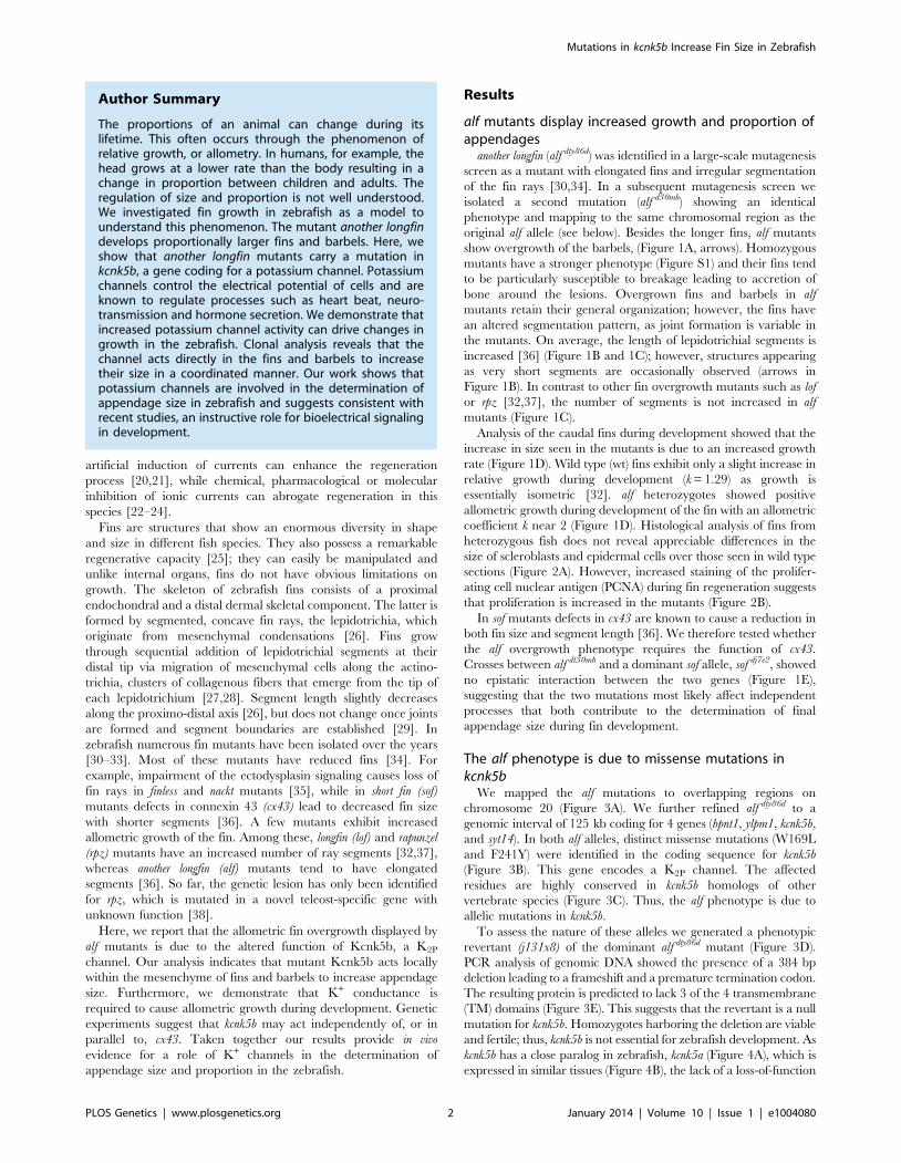
Author Summary
The proportions of an animal can change during itslifetime. This often occurs through the phenomenon ofrelative growth, or allometry. In humans, for example, thehead grows at a lower rate than the body resulting in achange in proportion between children and adults. Theregulation of size and proportion is not well understood.We investigated fin growth in zebrafish as a model tounderstand this phenomenon. The mutant another longfindevelops proportionally larger fins and barbels. Here, weshow that another longfin mutants carry a mutation inkcnk5b, a gene coding for a potassium channel. Potassiumchannels control the electrical potential of cells and areknown to regulate processes such as heart beat, neuro-transmission and hormone secretion. We demonstrate thatincreased potassium channel activity can drive changes ingrowth in the zebrafish. Clonal analysis reveals that thechannel acts directly in the fins and barbels to increasetheir size in a coordinated manner. Our work shows thatpotassium channels are involved in the determination ofappendage size in zebrafish and suggests consistent withrecent studies, an instructive role for bioelectrical signalingin development.
Mutations in kcnk5b Increase Fin Size in Zebrafish
PLOS Genetics | www.plosgenetics.org 2 January 2014 | Volume 10 | Issue 1 | e1004080

phenotype in normal development may be due to functional
redundancy between the paralogs. Together, these data endorse our
finding that kcnk5b is the gene responsible for the alf overgrowth
phenotype and demonstrate that these mutations are due to a gain
of function rather than haploinsufficiency.
alf mutations in kcnk5b lead to increased K+ conductanceand hyperpolarization
We used the known structure of human KCNK4 (K2p4.1) [39]
as a template for modeling Kcnk5b and assessing the mutations.
These models revealed that the affected amino acids are positioned
in two distinct TM domains towards the cytoplasmic side of the
protein (Figure 5A).
To assess how the identified amino acid substitutions might affect
Kcnk5b function, the channel properties were tested in a two-
electrode voltage clamp experiment in Xenopus oocytes. This
technique permits measurement of currents across the cell
membrane when the membrane potential is clamped to a given
value. Oocytes injected with kcnk5b(wt) cRNA react steadily to a
change in voltage and do not exhibit a delay in current flow, as is
expected for a K2P channel. A similar situation is also seen with
kcnk5b(W169L) or kcnk5b(F241Y) cRNAs. However, oocytes injected
with either of both mutant cRNAs show an almost two-fold increase
in K+ conductance over that of oocytes injected with wild type
cRNA (Figure 5B). The current-voltage relationship of the wild type
channel shows the typical outward rectification of a K2P channel,
i.e. current flows preferentially out of the cell, from the side of high
K+ concentration to the side of low K+ concentration [40]. In
contrast, the increase in K+ currents in the Kcnk5b mutant variants
is accompanied by reduced outward rectification (Figure 5C)
suggesting that the change in K+ conductance results from altered
biophysical features of Kcnk5b rather than a simple increase in the
number of channels at the plasma membrane.
K2P channels are often referred to as leak channels since they
account for the constant leaking current that sets the resting
membrane potential observed in neurons. They are known to
control both cell excitability and membrane potential [41], and the
human homolog of kcnk5b, KCNK5 (TASK2), was shown to
contribute significantly to the stabilization of the membrane
potential in articular chondrocytes [42]. Therefore, we hypothe-
sized that zebrafish Kcnk5b might also play a role in setting the
membrane potential. Indeed, the membrane potential values of
oocytes injected with wild type and mutant kcnk5b cRNAs are
correlated with the amplitude of the ion current measured at a
constant voltage of 50 mV (Figure 5D): the higher the conduc-
tance for K+ measured at 50 mV, the more negative the
membrane potential of the oocyte. Consistently, the mutant
channels lead to stronger hyperpolarization causing a shift in the
membrane potential towards 290 to 2100 mV, the equilibrium
potential for K+ in Xenopus oocytes.
kcnk5b acts locally to increase appendage sizeTo show where kcnk5b is expressed we performed in situ
hybridization experiments on adult fins, however no specific signal
Figure 1. alf mutants lead to an increase in size of the appendages of adult fish. (A) alf mutations are dominant and lead to overgrown finsand barbels in the adult. Arrows indicate maxillary barbels; the mutants shown are heterozygous. (B) Segment patterning in the dorsal fin of wild typeand heterozygous mutants. Brackets indicate one segment. Although the majority of segments show increased length, several short segments can beseen in the mutants (arrows). (C) Variation in segment length (top) and segment number (bottom) in the longest ray of the dorsal fin of mutants andwild type siblings (wt sib). Fish of similar standard length (SL) (i.e. distance between snout and caudal peduncle) were compared; all cases n = 4; errorbars: standard deviation; n.s.: not significant; *: p,0.02, ***: p,0.001. (D) Increased allometric scaling of heterozygous alf fins in development.k = allometric coefficient, Linear regression lines, wt R2 = 0.92; alf/+, R2 = 0.95; ***: p,0.001. (E) Crosses of sof with alf indicate that there is not epistaticinteraction between the two genes. Fin length was normalized with SL.doi:10.1371/journal.pgen.1004080.g001
Mutations in kcnk5b Increase Fin Size in Zebrafish
PLOS Genetics | www.plosgenetics.org 3 January 2014 | Volume 10 | Issue 1 | e1004080

Figure 2. Cell proliferation is increased in alf mutants. (A) Sections of wild type and heterozygous alf fins. No significant difference in cell size isseen in the two groups. (B) Antibody staining against PCNA on paraffin sections of regenerating fins 4 days post amputation (dpa). Chart showspercentage of proliferating nuclei (PCNA) over total nuclei (Hoechst). N = 3–4 sections of 4 individual fish **: p-value,0.01.doi:10.1371/journal.pgen.1004080.g002
Figure 3. The alf phenotype is due to gain-of-function mutations within the K+ channel kcnk5b. (A) alf mutations map to chromosome 20between z11841 and z21067. Gray: north markers; blue: south markers. (B) Electropherogram of kcnk5b at position 169 and 241 in mutants and wildtype siblings. (C) The amino acids affected in the mutants are well conserved among vertebrates. (D) A revertant of alfdty86d (j131x8) shows wild type-sized fins. (E) kcnk5bj131x8 fish harbor an intragenic deletion in kcnk5b that is predicted to cause a truncated protein lacking three transmembrane(TM) domains.doi:10.1371/journal.pgen.1004080.g003
Mutations in kcnk5b Increase Fin Size in Zebrafish
PLOS Genetics | www.plosgenetics.org 4 January 2014 | Volume 10 | Issue 1 | e1004080

above background was observed, indicating that expression levels
might be below detection with this technique. Nevertheless, RT-
PCR analysis showed that kcnk5b is expressed in fins of adult fish
(Figure 4B). To assess whether kcnk5b acts locally within fins and
barbels to control growth, we transplanted kcnk5bdt30mh/+ mutant
cells into wild type hosts (Figure S2A). Local overgrowth of these
structures was detected in 29 out of 120 chimeras raised to
adulthood (Figure S2B–D), whereas global overgrowth of all fins
and barbels was never observed. This suggests that the mutations
act locally within the appendages to increase their size. We further
attempted to induce the mutant phenotype by local overexpression
of the channel within fins and barbels of wild type fish. Whereas the
electrophysiological analysis indicated that the dominant kcnk5b
mutations lead to an increase of channel conductance, the current of
K+ ions through the plasma membrane depends not only on
individual channel conductance, but also on the number of channels
present in the membrane. Therefore, we argued that increasing the
number of channels should also promote fin overgrowth. We
generated a construct in which either kcnk5b(wt) or kcnk5b(W169L)
expression is driven by the elongation factor 1 alpha (ef1a) promoter
from Xenopus laevis; this promoter was recently shown to be active in
all major fin tissues [43]. To mark the cells that express the
transgene, DsRed expression was driven under a second ef1a
promoter positioned in cis within the same plasmid (Figure 6A). This
plasmid was injected into wild type one-cell stage zebrafish zygotes
along with Tol2 transposase mRNA as described before [43].
Injected fish were raised to adulthood, screened for DsRed positive
cells in the fins and the effects on growth were recorded. No
overgrowth was observed in fish injected with a control plasmid
expressing only DsRed under the ef1a promoter (0/240), despite the
presence of DsRed-positive cells in various tissues within the fin
(Figure S3). In about 40% of the fish injected with plasmids
encoding wild type or mutant kcnk5b and showing DsRed positive
cells in the fins we found a local overgrowth phenotype (Figure 6B
and H). Analysis showed a strong correlation of overgrowth with
DsRed positive mesenchymal tissue (89.2%, N = 37, Figure 6C, D
and I), whereas DsRed positive cells in other tissues were not
associated with increases in size. The fin ray segments were enlarged
in the overgrown fins similar to alf mutants (Figure 6D). The marked
fibroblast-like cells typically occupied the intra-ray space and were
excluded from the arteries (Figure 6E). These vessel-surrounding
clones extended along the actinotrichia to the distal ends of the
overgrown fins (Figure 6F). In the case of barbel overgrowth, DsRed
positive cells were found in the mesenchymal tissue surrounding the
central rod (Figure 6G), an acellular, non-cartilaginous, non-
mineralized structure that supports this organ [44]. In a few cases no
DsRed fluorescence signal could be detected within or next to
overgrown fin tissue (kcnk5b(W169L), 2/26; kcnk5b(wt), 2/11),
probably due to variegation of promoter activity [43]. In conclusion,
these findings indicate that kcnk5b overexpression within fibroblasts
of the mesenchyme is sufficient to induce fin outgrowth.
To test whether kcnk5b-induced overgrowth requires conductance
of K+ ions by the channel, we generated an overexpression construct
encoding a non-conductive version by mutating the GFG motif of
the selectivity filter to AAA, kcnk5b(GFGAAA). This modification
has previously been shown to block ion conductance in K+ channels
[45]. Electrophysiological measurements in Xenopus oocytes showed
that this channel is unable to conduct K+ (Figure 6J). The plasmid
was injected into wild type embryos along with Tol2 transposase
mRNA and injected fish were reared to adulthood and assessed for
overgrowth. No overgrowth was detected in these fish (Figure 6H),
although fins containing DsRed positive tissue (n = 32), including
fibroblasts (Figure 6J, inset), were found. These data indicate that
the increase in conductance of the Kcnk5b channel is essential for
the coordinated overgrowth of the fins and barbels in the mutants.
Discussion
K+ channels have long been associated with neurological
function, hormone secretion, and cardiomyocyte polarization
[46]. They are a diverse class of ion channels, which can be
grouped into four major families: inwardly rectifying (KIR),
voltage-dependent (KV), calcium-dependent (KCa) and two-pore
domain (K2P) potassium channels. KIR channels have recently
been shown to be involved in patterning in vertebrates and
invertebrates. In Drosophila loss-of-function mutations in Irk2 lead
to wing patterning defects [47]. Mutations in the human homolog,
Kir2.1, are associated with craniofacial and digital defects [48]. In
zebrafish establishment of the adult pigmentation pattern requires
the function of Kcnj13 (Kir7.1) [49]. Here, we report that gain-of-
function mutations in kcnk5b, a gene encoding a K2P channel, lead
to allometric overgrowth of the fins. This is the first time that a
member of this class of channels is shown to be involved in
regulation of growth and patterning in a vertebrate.
Implications of K+ channels in growth and proliferationThe size of an organ depends on cell size and cell number. The
mammalian homolog of kcnk5b has been implicated in both,
Figure 4. Vertebrate kcnk5 homologs and expression inzebrafish development. (A) Due to a whole genome duplicationevent, teleost fish have two kcnk5 paralogs that show early divergence.Numbers indicate bootstrap values in percentage (100 bootstrapreplications). Nodes with a bootstrap value lower than 95 werecollapsed. Dre, Danio rerio; Ola, Oryzias latipes; Gac, Gasterosteusaculeatus, Tru, Takifugu rubripes; Tni Tetraodon nigridoviridis, Gmo,Gadus morhua; Mmu, mus musculus; Gga, Gallus gallus; Xtr Xenopustropicalis. (B) RT-PCR of kcnk5a and kcnk5b shows comparableexpression between the two paralogs in multiple adult tissues,including fins.doi:10.1371/journal.pgen.1004080.g004
Mutations in kcnk5b Increase Fin Size in Zebrafish
PLOS Genetics | www.plosgenetics.org 5 January 2014 | Volume 10 | Issue 1 | e1004080

regulation of cell volume [50,51] and cell proliferation [52,53]. In
alf mutants we could detect an increase in cell proliferation but not
in cell size (Figure 2). Importantly, the mutant phenotype does not
arise simply by dysregulation of cell proliferation, which would
cause tumorous overgrowth; rather the overgrown structures in
the mutants preserve tissue organization and patterning.
It is unclear how K+ channels regulate proliferation. Studies have
proposed that this might occur through regulation of the membrane
potential [54]. In apparent contrast to some studies [55–58] but in
agreement with others [59,60], we found that hyperpolarization
caused by mutations in a K+ channel can lead to tissue overgrowth.
Although we observed a hyperpolarizing effect of the alf mutation in
Xenopus oocytes, we cannot exclude that this, in turn, triggers a
depolarization, either at cellular level or in the surrounding tissues
during development of the fin. In fact, experiments employing
depolarization-sensitive dyes, suggest that this might indeed be the
case (Figure S4). The importance of hyperpolarization during
growth is supported by regeneration studies in Xenopus [22,24].
Regenerating tadpole tails are initially depolarized, but, unlike tails
in the refractory state, subsequently undergo hyperpolarization.
Notably, impairing hyperpolarization through inhibition of V-
ATPase activity leads to a reduction of cell proliferation and failure
to regenerate [22]. Transient hyperpolarization of the cell might
lead to a cytosolic increase of the second messenger Ca2+, activate
integrin-dependent or PTEN phosphatase-dependent cascades, or
favor the uptake of mitogens such as serotonin through voltage-
dependent transporters [61]. Recent reports suggest that in some
cases K+ channels can induce cell proliferation independently of
their effect on membrane potential [62,63]. We show that, in the
case of Kcnk5b, conductance is essential for the regulation of fin
growth. Overexpression of a non-conducting version of the channel
does not cause a phenotype, whereas wild type and alf variants
induce local overgrowth.
Role of kcnk5b in size specificationOur analysis of transgenic mosaics indicates that cells of the
mesenchyme are sufficient to provide cues that alter the size of the
fins. This is consistent with results of classic xenograft studies
between chicken and quail where cells of the mesenchyme impart
donor-specific characteristics to the limbs [8,64]. During develop-
ment tetrapod limbs are patterned by signaling interactions
between mesenchymal cells and the overlying ectoderm. A
prominent signaling center, the apical ectodermal ridge (AER),
is active at the distal tip of the limb bud during this process. The
AER and the mesenchyme of the progress zone continuously
communicate with each other to direct limb outgrowth and
development. Similar epithelial-mesenchymal interactions from
the apical fin fold are likely to be required for the patterned
overgrowth of fins in alf mutants. In support of this mechanism, we
consistently find labeled mesenchymal cells in the distal-most
regions of overgrown tissue in mosaic animals.
AER signaling in amniotes requires connexin-mediated electri-
cal connectivity between cells to coordinate pattern and growth of
the vertebrate limb [65–67]. An analogous mechanism may be
functioning in fish. We show here that altering ionic communi-
cation in the developing fin of the zebrafish is sufficient to induce
Figure 5. Gain-of-function mutations in kcnk5b affect ionic conduction and lead to hyperpolarization of the cell. (A) Location of theamino acids altered in kcnk5b gain-of-function mutants. Kcnk5b protein was modeled on human KCNK4 (K2p4.1). GFG and GYG domains representthe selectivity pore of the channel. (B) Voltage clamp recordings from Xenopus oocytes injected with cRNA of wild type and mutant kcnk5b. Themembrane potential was clamped at a reference potential of 280 mV and then stepped to a test potential from +60 mV to 2100 mV for 500 ms. Thecurrent that is applied in order to clamp the voltage to a certain value corresponds to the current passing through the plasma membrane.Representative electrophysiological traces are shown. (C) The mutant channels display increased conductance over wild type channels expressed atcomparable levels. Error bars represent standard deviation. (D) Kcnk5b influences membrane potential (Vm) in oocytes. The mutant variants tend tohyperpolarize the cell (each point represents one oocyte).doi:10.1371/journal.pgen.1004080.g005
Mutations in kcnk5b Increase Fin Size in Zebrafish
PLOS Genetics | www.plosgenetics.org 6 January 2014 | Volume 10 | Issue 1 | e1004080

Mutations in kcnk5b Increase Fin Size in Zebrafish
PLOS Genetics | www.plosgenetics.org 7 January 2014 | Volume 10 | Issue 1 | e1004080

growth. Our analysis of the genetic interactions between alf and sof
indicate that Kcnk5b and Cx43 may act in parallel pathways to
modulate final fin size. In both mutants segment length and fin size
are correlated, however the role of segment patterning in size
regulation of the fin is unclear. In contrast to alf [36,68] (Figure 1)
and sof [33], the overgrowth mutants lof and rpz have wild type
sized lepidotrichial segments [32]. Moreover, the evx1 mutation,
which leads to fins rays devoid of joints, does not affect final fin size
in a wild type nor lof background [69].
Ionic currents and positional informationSeveral experiments suggest that bioelectrical signaling is a
shared common mechanism used across bilaterians to control
organ growth and patterning [21,70,71] and indicate that ion flow
may have an instructive role during development [72,73], as well
as regeneration [18,74]. Here, we provide genetic evidence
showing that changes in K+ channel activity result in allometric
scaling of an organ, rather than causing uncontrolled proliferation.
We favor a model for size regulation in which modulation of ionic
current by K+ channels within the organ shifts positional
information, thereby setting a different register of size during
development. In fact, there is evidence for a rostro-caudal and
medio-lateral gradient of voltage within the developing embryo
suggesting that electric fields are a component of the positional
information [75,76]. External electrical currents have been shown
to alter positional information in axial regenerates of planaria [77].
However, the underlying mechanism of signaling from electrical
fields is largely unknown, and possibly depends on electrical
coupling between cells [78]. This hypothesis is supported by
studies in pigment pattern formation, where both K+ channels and
connexins have been implicated in proper formation of the
zebrafish stripes [49,79–81]. Further studies will be needed to
uncover the signaling mechanism from K+ channels to regulate
size and pattern. However, our work, in concert with that of
others, clearly shows that ion flow is not just an epiphenomenal
event accompanying growth but one of the major factors
specifying pattern and form during development and regeneration.
Materials and Methods
Fish maintenanceZebrafish were bred and maintained as previously described
[82]. alf dty86d was isolated in the 1996 Tubingen screen [30,34] as
a mutant affecting adult fin formation. The alf dt30mh(pfau) mutant
was identified in F1 fish of a standard F3 screen (ZF Models) and
isolated based on its fin and barbel phenotype.
MeasurementsFish were anesthetized in tricaine solution for measurements; fin
length and standard length was measured using handheld calipers.
Fish were imaged under a stereo microscope (Zeiss, SteREO
Discovery) and measurements were performed using AxioVision
software (Zeiss). p-values from unpaired Student’s t-test were
obtained with Microsoft Excel.
Sections and PCNA stainingFin regenerates were fixed at 4 dpa in 4% PFA overnight and
decalcified with 0.5 M EDTA for 24 h. Sample were embedded in
paraffin and sectioned at 5 mm. Immunohistochemistry with anti-
PCNA antibody (Sigma) was performed as described [83].
Percentage of PCNA positive nuclei over Hoechst positive nuclei
was determined on three to four sections of four independent
samples for each genotype.
RT-PCR analysisAdult zebrafish organs were dissected on ice and stored in
RNALater (Invitrogen) at 4uC. Total RNA was isolated using
RNeasy Mini kit (Qiagen). cDNA was synthesized from 200 ng
RNA from each sample with SuperScript III and oligo(dT) primers
(Invitrogen). PCR analysis was performed using Taq polymerase S
(Genaxxon) with intron spanning primers (b-actin forward OSP-31,
TGC GGA ATA TCA TCT GCT TG, b-actin reverse OSP-32:
AGC ATC ATC TCC AGC GAA TC, kcnk5b forward OSP-390:
CAT TCC TCT GTG CCT CAC CT; kcnk5b reverse OSP-324
AGG CCA TCC ACA GAC TCA TC, Tm = 61uC, 30 cycles).
MappingMapping was performed as described [82]. The alf dty86d
mutation mapped between z11841 (5 recombinants/96 meioses)
and z21067 (2/96) and fine mapped using SNPs. alf dt30mh mapped
between z7803 (1/48) and z21067 (1/48). Full length kcnk5b was
cloned into pGEM-T Easy from cDNA of fin blastema amplified
with LA Taq polymerase (TaKaRa) (forward primer OSP-379:
TGG GAG TGT GGA GTG TGT GT, reverse OSP-382: TTT
TTG GTC CAG CTT TGG TC, Tm = 60uC, 45 cycles).
X-ray irradiation and screening for revertants of alfSperm from alf dty86d homozygotes was irradiated with X-rays
(1125 rads, Faxitron 43855D) and used to fertilize wild type eggs
(AB strain). F1 progeny was reared to approximately three weeks of
age (9433 fish) and screened for the alf phenotype. 11 fish showed
wild type fins. 10 of these survived to adult stages. SSLP analysis
revealed that 9 of these were deletions of some or all of the upper
arm of chromosome 20. q-RT-PCR of candidate genes in the
remaining revertant (j131x8) showed no change in transcript levels
for bpnt, ylpm1 and syt14, but little or no transcript for kcnk5b. PCR
analysis of genomic DNA showed that this revertant has a 384 bp
deletion of the 39 end of intron 2 and the 59 end of exon 3. This
deletion results in a frameshift and early truncation of the protein.
Modeling of Kcnk5bThe amino acid sequence of zebrafish Kcnk5b was retrieved
from Ensembl (http://www.ensembl.org) and used to search the
PDB database with HHpred (http://toolkit.tuebingen.mpg.de/)
Figure 6. Overexpression of kcnk5b is sufficient to cause fin overgrowth. (A) Construct used to create kcnk5b-expressing clones via Tol2transgenesis. (B) Individual fish expressing kcnk5b (W169L) (left) or kcnk5b (wt) (right) in mosaic clones display localized fin and barbel overgrowth.(C–F) Overgrowth is associated with DsRed expression (in red) within mesenchymal cells. (C) Calcein staining labels bone tissue (in green) of anovergrown fin (DsRed; kcnk5(W169L) expressing clone). (D) Mesenchymal clones are associated with increased segment length in the fin compared tonon-overgrown DsRed negative regions. (E) Fibroblast-like cells appear as DsRed positive cells within the fin rays (dotted line) that surround DsRednegative vasculature (arrows in E and F) which extend along the actinotrichia (fibrils within dotted lines in F) towards the distal end of the fin. (G)Overgrown barbels show DsRed signal within the mesenchyme (area within dotted line) but not in the vasculature (arrow). (H) Number of clonesassociated with overgrowth in different kcnk5b variants. (I) Proportion of different cell types labeled in overgrown tissues. (J) Electrophysiologicalrecordings of the non-conductive kcnk5b (GFGAAA) mutant in oocytes. Squares: kcnk5b (wt), purple stars: kcnk5b (F241Y)+kcnk5b (wt), blue circles:kcnk5b (W169L)+kcnk5b (wt), green triangles: + kcnk5b (GFGAAA)+kcnk5b (wt). Current was normalized to the measurement of wt current at 60 mV.Inset: DsRed+ fibroblasts in fish injected with the non-conductive construct do not lead to fin overgrowth.doi:10.1371/journal.pgen.1004080.g006
Mutations in kcnk5b Increase Fin Size in Zebrafish
PLOS Genetics | www.plosgenetics.org 8 January 2014 | Volume 10 | Issue 1 | e1004080

[84]. The first hit in the search (human KCNK4, PDB ID: 3um7
[39] identity 36%, similarity 0.646; 22nd March 2012) was used to
build the 3D model. The model was processed with MacPyMol
(http://pymol.org).
Electrophysiological measurementskcnk5b was subcloned from pGEM-T Easy to pSGEM
expression vector via SacII and SpeI sites. After linearization with
NheI, cRNA was synthesized with Ambion mMessage mMachine
(Invitrogen) and cleaned up with mRNeasy Mini Kit (Qiagen). X.
laevis oocytes were injected as described previously [85] (kcnk5b
single alleles: 4 ng wild type or mutant kcnk5b cRNA; co-injections
of two kcnk5b alleles: 2 ng cRNA each, for a total of 4 ng per
oocyte). Measurements were done from a holding potential of
280 mV with 0.5 s long pulses from 2100 to +60 mV with
increments of 20 mV. Recorded currents (n = 5–26) were aver-
aged and normalized to the mean value recorded for oocytes
injected with the wild type channel at +60 mV.
Generation of the kcnk5b(GFGAAA) non-conductivemutant
PCR mutagenesis was performed as described [86] using Pfu
polymerase (Fermentas) (OSP-15 CCC TGA CGA CTG TCG
CTG CAG CTG ACT ATG TGG CAG GGG C; OSP-16 CCT
GCC ACA TAG TCA GCT GCA GCG ACA GTC GTC AGG
GTG G, Tm = 70uC, 30 cycles) on pSGEM:kcnk5b(wt).
Cloning of overexpression vectorsef1a:DsRed vector. A ef1a:DsRed cassette generated with
KOD Hot Start DNA Polymerase (Toyobo) (primers: TAA TTT
AAA TAG ATC TTC GAG CAG GGG GAT CAT CTA ATC
A; CTA GAT GGC CAG ATC TGC CCG GGA CTT GAT
TAG GGT GAT GGT TCA CGT AGT G, Tm = 59uC, 30
cycles) from plasmid Ale237 (kind gift of Alessandro Mongera) was
inserted in plasmid 587jk (kind gift of Dr. Jana Krauß) using BglII
restriction sites through In-Fusion Advantage (Clontech) cloning
according to manufacturer’s protocol.
ef1a:DsRed; ef1a:kcnk5b wild type and mutant vectors.
The ef1a promoter was amplified from plasmid Ale237 (primers:
ATT AAT TCG AGC TCG GTA CCC CTC GAG CAG GGG
GAT CAT CT; GAA CAA GCA AGC TGG GTA CCC CGG
CCG TCG AGG AAT TCT TTG, Tm = 59uC, 30 cycles) and
inserted into the pSGEM vector at the KpnI restriction site using In-
Fusion Advantage (Clontech) cloning. The ef1a:kcnk5b cassette was
amplified from the resulting plasmid as above (primer: AAA CCT
AGG TCG AGC AGG GGG ATC ATC T; AAA CCT AGG
ATG ACC ATG ATT ACG CCA AGC TAT), digested with AvrII
and inserted into ef1a:DsRed vector using the SpeI restriction site.
InjectionsPlasmids (5–20 ng/ml), Tol2 mRNA (25 ng/ml) and 20% (v/v)
phenol red solution (Sigma- Aldrich, P0290-100ML) were injected
into the zygote of 1-cell stage embryos under a dissecting
microscope (Zeiss, Stemi 2000) using 275 Pa (40 psi) injecting
pressure for 100 ms (World Precision Instruments, Pneumatic
PicoPump PV820). Adults were analyzed with Zeiss, SteREO
Discovery and Zeiss LSM 5 Live.
TransplantationsTransplantations were performed as previously described [82].
At mid blastula stage (1000 cell stage), about 20–40 cells were
transplanted from the pfaudt30mh/+ donors into the recipient close to
the yolk cell and chimeras were raised to adulthood.
In vivo analysis of membrane potentialFluorescent dye experiments were performed by adapting
described protocols [22,87]. Briefly, wild type and mutant juvenile
fish (STL = 16–18 mm) were incubated in fluorescent dye diluted
1:2000 in fish water (stock solutions: DiSBAC2(3) (Bis-(1,3-
Diethylthiobarbituric Acid)Trimethine Oxonol, Life Technologies):
1 mg/ml in DMSO) for 30 min in the dark, anesthetized with
tricaine solution and placed on a custom-made chamber for confocal
imaging. The chamber was obtained by removing the bottom of a
55 mm plastic dish and by replacing it through a round cover slip
fastened with silicone. Fish were held in place with a tissue soaked in
dye and imaged upon excitation at 561 nm. Unstained animals
were imaged as a negative control. p-values from unpaired Student’s
t-test were obtained with Microsoft Excel.
Supporting Information
Figure S1 Phenotype of homozygous alf mutants. (A) wt, (B)
alf dty86d homozygous, (C) alf dt30mh homozygous. Scale bar: 10 mm
(TIF)
Figure S2 kcnk5b gain-of-function mutations affect local growth
of appendages. (A) Transplantation of kcnk5bdt30mh/+ cells into wt
albino hosts. If the mutation acts on a systemic level, mutant clones
should promote overgrowth of all appendages. If the mutation has
a local effect, overgrowth will be observed in patches. Chimeras
resulting from the transplantation experiments show overgrowth of
(B) single fins, (C) fin parts or (D) individual barbels.
(TIF)
Figure S3 The control plasmid ef1a:DsRed drives DsRed
expression in a wide range of cell types and tissues within the
fin. (A) lateral line, (B) vasculature, (C) osteoblasts, (D) fibroblasts,
(E) pigment cells (arrows), showing the typical stellated shape, and
(F) epidermis. Scale bar: 200 mm
(TIF)
Figure S4 Polarization of fins during growth. Voltage sensitive
dyes were used to assess changes in overall polarization of growing
caudal fins of wild type and alf juvenile fish. (A) DiSBAC2(3) staining
in wild type fins exhibited hyperpolarization localized to discrete
regions of the fin with variable detection of distal regions of altered
depolarization. (B) alf fins in contrast show high levels of
depolarization across the fin with variable patterns in different
tissues. (C) Quantification of average DiSBAC2(3) fluorescence
signal in wild type and mutant fins (average pixel intensity (12-bits) of
the fin in maximum intensity projections). ***: p,0.001, N = 21–23.
(D) Positive control of depolarization by treatment of the fins with
150 mM KCl (D9). DiBAC4(3), another dye sensitive to depolariza-
tion, showed similar effects, while DiSC3(5), a dye sensitive to
hyperpolarized states, was uninformative (data not shown).
(TIF)
Acknowledgments
The authors would like to thank Dr. Jana Krauß, Dr. Uwe Irion, and Dr.
Jennifer Lanni for valuable comments on earlier versions of the
manuscript, Andrey Fadeev, Ines Gehring, Hans-Martin Maischein,
Ursula Schach, and Iris Koch for technical assistance as well as Alessandro
Mongera for stimulating discussions.
Author Contributions
Conceived and designed the experiments: SP MPH SLJ. Performed the
experiments: SP JMD UH CWH. Analyzed the data: SP JMD UH CWH.
Contributed reagents/materials/analysis tools: SP GS SLJ CNV. Wrote
the paper: SP MPH CNV.
Mutations in kcnk5b Increase Fin Size in Zebrafish
PLOS Genetics | www.plosgenetics.org 9 January 2014 | Volume 10 | Issue 1 | e1004080

References
1. Lui JC, Baron J (2011) Mechanisms limiting body growth in mammals. Endocr
Rev 32: 422–440.
2. Huxley JS, Teissier G (1936) Terminology of relative growth. . Nature 137: 780–
781.
3. Gould SJ (1966) Allometry and size in ontogeny and phylogeny. Biol Rev CambPhilos Soc 41: 587–640.
4. Metcalf D (1963) The autonomous behaviour of normal thymus grafts. Aust J Exp
Biol Med Sci 41: SUPPL437–447.
5. Metcalf D (1964) Restricted growth capacity of multiple spleen grafts.Transplantation 2: 387–392.
6. Conlon I, Raff M (1999) Size Control in Animal Development. Cell 96: 235–
244.
7. Twitty VC, Schwind JL (1931) The growth of eyes and limbs transplanted
heteroplastically between two species of Amblystoma. Journal of ExperimentalZoology 59: 61–86.
8. Ohki-Hamazaki H, Katsumata T, Tsukamoto Y, Wada N, Kimura I (1997)
Control of the limb bud outgrowth in quail-chick chimera. Dev Dyn 208: 85–91.
9. Schneider RA, Helms JA (2003) The cellular and molecular origins of beakmorphology. Science 299: 565–568.
10. Sengel P (1971) The organogenesis and arrangement of cutaneous appendages
in birds. Adv Morphog 9: 181–230.
11. Zwilling E (1955) Ectoderm — mesoderm relationship in the development of the
chick embryo limb bud. Journal of Experimental Zoology 128: 423–441.
12. Harvey KF, Zhang X, Thomas DM (2013) The Hippo pathway and humancancer. Nat Rev Cancer 13: 246–257.
13. Tumaneng K, Russell RC, Guan K-L (2012) Organ size control by Hippo and
TOR pathways. Curr Biol 22: R368–379.
14. Goldstein SA, Bockenhauer D, O’Kelly I, Zilberberg N (2001) Potassium leak
channels and the KCNK family of two-P-domain subunits. Nat Rev Neurosci 2:175–184.
15. Talley EM, Sirois JE, Lei Q, Bayliss DA (2003) Two-pore-Domain (KCNK)
potassium channels: dynamic roles in neuronal function. Neuroscientist 9: 46–56.
16. Levin M (2007) Large-scale biophysics: ion flows and regeneration. Trends Cell
Biol 17: 261–270.
17. Altizer AM, Moriarty LJ, Bell SM, Schreiner CM, Scott WJ, et al. (2001)
Endogenous electric current is associated with normal development of thevertebrate limb. Developmental Dynamics 221: 391–401.
18. Borgens RB, Vanable JW, Jr., Jaffe LF (1977) Bioelectricity and regeneration:
large currents leave the stumps of regenerating newt limbs. Proc Natl AcadSci U S A 74: 4528–4532.
19. Kurtz I, Schrank AR (1955) Bioelectrical Properties of Intact and Regenerating
Earthworms, Eisenia foetida. Physiological Zoology 28: 322–330.
20. Borgens RB, Vanable JW, Jaffe LF (1979) Small artificial currents enhance
Xenopus limb regeneration. Journal of Experimental Zoology 207: 217–226.
21. Tseng AS, Beane WS, Lemire JM, Masi A, Levin M (2010) Induction ofvertebrate regeneration by a transient sodium current. J Neurosci 30: 13192–
13200.
22. Adams DS, Masi A, Levin M (2007) H+ pump-dependent changes in membranevoltage are an early mechanism necessary and sufficient to induce Xenopus tail
regeneration. Development 134: 1323–1335.
23. Reid B, Song B, Zhao M (2009) Electric currents in Xenopus tadpole tail
regeneration. Dev Biol 335: 198–207.
24. Adams DS, Tseng AS, Levin M (2013) Light-activation of the ArchaerhodopsinH(+)-pump reverses age-dependent loss of vertebrate regeneration: sparking
system-level controls in vivo. Biol Open 2: 306–313.
25. Morgan TH (1900) Regeneration in teleosts. Arch Entwickslungsmech 10: 120–131.
26. Grandel H, Schulte-Merker S (1998) The development of the paired fins in the
zebrafish (Danio rerio). Mech Dev 79: 99–120.
27. Goss RJ, Stagg MW (1957) The regeneration of fins and fin rays in Fundulus
heteroclitus. J Exp Zool 136: 487–507.
28. Haas HJ (1962) Studies on mechanisms of joint and bone formation in theskeleton rays of fish fins. Dev Biol 5: 1–34.
29. Iovine MK (2007) Conserved mechanisms regulate outgrowth in zebrafish fins.
Nat Chem Biol 3: 613–618.
30. van Eeden FJ, Granato M, Schach U, Brand M, Furutani-Seiki M, et al. (1996)
Genetic analysis of fin formation in the zebrafish, Danio rerio. Development123: 255–262.
31. Fisher S, Jagadeeswaran P, Halpern ME (2003) Radiographic analysis of
zebrafish skeletal defects. Dev Biol 264: 64–76.
32. Goldsmith MI, Fisher S, Waterman R, Johnson SL (2003) Saltatory control ofisometric growth in the zebrafish caudal fin is disrupted in long fin and rapunzel
mutants. Dev Biol 259: 303–317.
33. Iovine MK, Higgins EP, Hindes A, Coblitz B, Johnson SL (2005) Mutations in
connexin43 (GJA1) perturb bone growth in zebrafish fins. Dev Biol 278: 208–219.
34. Haffter P, Odenthal Jr, Mullins MC, Lin S, Farrell MJ, et al. (1996) Mutations
affecting pigmentation and shape of the adult zebrafish. Development Genesand Evolution 206: 260–276.
35. Harris MP, Rohner N, Schwarz H, Perathoner S, Konstantinidis P, et al. (2008)
Zebrafish eda and edar mutants reveal conserved and ancestral roles ofectodysplasin signaling in vertebrates. PLoS Genet 4: e1000206.
36. Sims K, Jr, Eble DM, Iovine MK (2009) Connexin43 regulates joint location in
zebrafish fins. Dev Biol 327: 410–418.
37. Iovine MK, Johnson SL (2000) Genetic analysis of isometric growth controlmechanisms in the zebrafish caudal Fin. Genetics 155: 1321–1329.
38. Green J, Taylor JJ, Hindes A, Johnson SL, Goldsmith MI (2009) A gain of
function mutation causing skeletal overgrowth in the rapunzel mutant. Dev Biol334: 224–234.
39. Brohawn SG, del Marmol J, MacKinnon R (2012) Crystal structure of the
human K2P TRAAK, a lipid- and mechano-sensitive K+ ion channel. Science335: 436–441.
40. Enyedi P, Czirjak G (2010) Molecular background of leak K+ currents: two-pore
domain potassium channels. Physiol Rev 90: 559–605.
41. Lesage F, Lazdunski M (2000) Molecular and functional properties of two-pore-domain potassium channels. Am J Physiol Renal Physiol 279: F793–801.
42. Clark RB, Kondo C, Belke DD, Giles WR (2011) Two-pore domain K+channels regulate membrane potential of isolated human articular chondrocytes.J Physiol 589: 5071–5089.
43. Tu S, Johnson SL (2011) Fate restriction in the growing and regenerating
zebrafish fin. Dev Cell 20: 725–732.
44. LeClair EE, Topczewski J (2010) Development and regeneration of the zebrafishmaxillary barbel: a novel study system for vertebrate tissue growth and repair.
PLoS One 5: e8737.
45. Kuzhikandathil EV, Oxford GS (2000) Dominant-negative mutants identify arole for GIRK channels in D3 dopamine receptor-mediated regulation of
spontaneous secretory activity. J Gen Physiol 115: 697–706.
46. Shieh CC, Coghlan M, Sullivan JP, Gopalakrishnan M (2000) Potassiumchannels: molecular defects, diseases, and therapeutic opportunities. Pharmacol
Rev 52: 557–594.
47. Dahal GR, Rawson J, Gassaway B, Kwok B, Tong Y, et al. (2012) An inwardlyrectifying K+ channel is required for patterning. Development 139: 3653–3664.
48. Tristani-Firouzi M, Etheridge SP (2010) Kir 2.1 channelopathies: the Andersen-
Tawil syndrome. Pflugers Arch 460: 289–294.
49. Iwashita M, Watanabe M, Ishii M, Chen T, Johnson SL, et al. (2006) Pigmentpattern in jaguar/obelix zebrafish is caused by a Kir7.1 mutation: implications
for the regulation of melanosome movement. PLoS Genet 2: e197.
50. Niemeyer MI, Cid LP, Barros LF, Sepulveda FV (2001) Modulation of the two-pore domain acid-sensitive K+ channel TASK-2 (KCNK5) by changes in cell
volume. J Biol Chem 276: 43166–43174.
51. Kirkegaard SS, Lambert IH, Gammeltoft S, Hoffmann EK (2010) Activation ofthe TASK-2 channel after cell swelling is dependent on tyrosine phosphoryla-
tion. Am J Physiol Cell Physiol 299: C844–853.
52. Santarius T, Bignell GR, Greenman CD, Widaa S, Chen L, et al. (2010) GLO1-A novel amplified gene in human cancer. Genes Chromosomes Cancer 49: 711–
725.
53. Alvarez-Baron CP, Jonsson P, Thomas C, Dryer SE, Williams C (2011) Thetwo-pore domain potassium channel KCNK5: induction by estrogen receptor
alpha and role in proliferation of breast cancer cells. Mol Endocrinol 25: 1326–
1336.54. Wang Z (2004) Roles of K+ channels in regulating tumour cell proliferation and
apoptosis. Pflugers Arch 448: 274–286.
55. Binggeli R, Weinstein RC (1986) Membrane potentials and sodium channels:hypotheses for growth regulation and cancer formation based on changes in
sodium channels and gap junctions. J Theor Biol 123: 377–401.
56. Chernet BT, Levin M (2013) Transmembrane voltage potential is an essentialcellular parameter for the detection and control of tumor development in a
Xenopus model. Dis Model Mech 6: 595–607.
57. Cone CD, Jr. (1971) Unified theory on the basic mechanism of normal mitoticcontrol and oncogenesis. J Theor Biol 30: 151–181.
58. Yang M, Brackenbury WJ (2013) Membrane potential and cancer progression.
Front Physiol 4: 185.
59. Patel SK, Jackson L, Warren AY, Arya P, Shaw RW, et al. (2013) A role for two-pore potassium (K2P) channels in endometrial epithelial function. J Cell Mol
Med 17: 134–146.
60. Wonderlin WF, Strobl JS (1996) Potassium channels, proliferation and G1progression. J Membr Biol 154: 91–107.
61. Blackiston DJ, McLaughlin KA, Levin M (2009) Bioelectric controls of cell
proliferation: ion channels, membrane voltage and the cell cycle. Cell Cycle 8:3519–3528.
62. Hegle AP, Marble DD, Wilson GF (2006) A voltage-driven switch for ion-
independent signaling by ether-a-go-go K+ channels. Proc Natl Acad Sci U S A103: 2886–2891.
63. Millership JE, Devor DC, Hamilton KL, Balut CM, Bruce JIE, et al. (2011)
Calcium-activated K+ channels increase cell proliferation independent of K+conductance. Am J Physiol Cell Physiol 300: C792–802.
64. Zwilling E (1959) Interaction between Ectoderm and Mesoderm in Duck-
Chicken Limb Bud Chimaeras. Journal of Experimental Zoology 142: 521–532.
65. Fallon JF, Kelley RO (1977) Ultrastruct analysis of the apical ectodermal ridgeduri;g vertebrate limb morphogenesis. II. Gap junctions as distinctive ridge
Mutations in kcnk5b Increase Fin Size in Zebrafish
PLOS Genetics | www.plosgenetics.org 10 January 2014 | Volume 10 | Issue 1 | e1004080

structures common to birds and mammals. J Embryol Exp Morphol 41: 223–
232.66. Flenniken AM, Osborne LR, Anderson N, Ciliberti N, Fleming C, et al. (2005) A
Gja1 missense mutation in a mouse model of oculodentodigital dysplasia.
Development 132: 4375–4386.67. Makarenkova H, Patel K (1999) Gap junction signalling mediated through
connexin-43 is required for chick limb development. Dev Biol 207: 380–392.68. Murciano C, Perez-Claros J, Smith A, Avaron F, Fernandez TD, et al. (2007)
Position dependence of hemiray morphogenesis during tail fin regeneration in
Danio rerio. Dev Biol 312: 272–283.69. Schulte CJ, Allen C, England SJ, Juarez-Morales JL, Lewis KE (2011) Evx1 is
required for joint formation in zebrafish fin dermoskeleton. Dev Dyn 240: 1240–1248.
70. Beane WS, Morokuma J, Lemire JM, Levin M (2013) Bioelectric signalingregulates head and organ size during planarian regeneration. Development 140:
313–322.
71. Hermle T, Saltukoglu D, Grunewald J, Walz G, Simons M (2010) Regulation ofFrizzled-dependent planar polarity signaling by a V-ATPase subunit. Curr Biol
20: 1269–1276.72. Hotary KB, Robinson KR (1992) Evidence of a role for endogenous electrical
fields in chick embryo development. Development 114: 985–996.
73. Pai VP, Aw S, Shomrat T, Lemire JM, Levin M (2012) Transmembrane voltagepotential controls embryonic eye patterning in Xenopus laevis. Development
139: 313–323.74. Jenkins LS, Duerstock BS, Borgens RB (1996) Reduction of the current of injury
leaving the amputation inhibits limb regeneration in the red spotted newt. DevBiol 178: 251–262.
75. Shi R, Borgens RB (1995) Three-dimensional gradients of voltage during
development of the nervous system as invisible coordinates for the establishmentof embryonic pattern. Dev Dyn 202: 101–114.
76. Metcalf MEM, Shi R, Borgens RB (1994) Endogenous ionic currents and
voltages in amphibian embryos. Journal of Experimental Zoology 268: 307–322.
77. Marsh G, Beams HW (1952) Electrical control of morphogenesis in regenerating
dugesia tigrina. I. Relation of axial polarity to field strength. Journal of Cellular
and Comparative Physiology 39: 191–213.
78. Oviedo NJ, Morokuma J, Walentek P, Kema IP, Gu MB, et al. (2010) Long-
range neural and gap junction protein-mediated cues control polarity during
planarian regeneration. Dev Biol 339: 188–199.
79. Watanabe M, Iwashita M, Ishii M, Kurachi Y, Kawakami A, et al. (2006) Spot
pattern of leopard Danio is caused by mutation in the zebrafish connexin41.8
gene. EMBO Rep 7: 893–897.
80. Watanabe M, Watanabe D, Kondo S (2012) Polyamine sensitivity of gap
junctions is required for skin pattern formation in zebrafish. Sci Rep 2: 473.
81. Inaba M, Yamanaka H, Kondo S (2012) Pigment pattern formation by contact-
dependent depolarization. Science 335: 677.
82. Nusslein-Volhard C, Dahm R (2002) Zebrafish: a practical approach. Oxford:
Oxford University Press.
83. Vandenplas S, Willems M, Huysseune A (2012) Dual BrdU-PCNA immuno-
detection of proliferative cells in dental and orofacial tissues of teleosts. Journal of
Applied Ichthyology 28: 336–340.
84. Hildebrand A, Remmert M, Biegert A, Soding J (2009) Fast and accurate
automatic structure prediction with HHpred. Proteins 77 Suppl 9: 128–132.
85. Strutz-Seebohm N, Gutcher I, Decher N, Steinmeyer K, Lang F, et al. (2007)
Comparison of potent Kv1.5 potassium channel inhibitors reveals the molecular
basis for blocking kinetics and binding mode. Cell Physiol Biochem 20: 791–800.
86. Zheng L, Baumann U, Reymond J-L (2004) An efficient one-step site-directed
and site-saturation mutagenesis protocol. Nucleic Acids Res 32: e115.
87. Oviedo NJ, Nicolas CL, Adams DS, Levin M (2008) Live Imaging of Planarian
Membrane Potential Using DiBAC4(3). CSH Protoc 2008: pdb prot5055.
Mutations in kcnk5b Increase Fin Size in Zebrafish
PLOS Genetics | www.plosgenetics.org 11 January 2014 | Volume 10 | Issue 1 | e1004080
Copyright © 2022 FDOKUMEN