Zebrafish models for ectopic mineralization disorders
17
REVIEW ARTICLE published: 08 May 2013 doi: 10.3389/fgene.2013.00074 Zebrafish models for ectopic mineralization disorders: practical issues from morpholino design to post-injection observations Mohammad Jakir Hosen 1,2 , Olivier M.Vanakker 1 *, Andy Willaert 1 , Ann Huysseune 3 , Paul Coucke 1 and Anne De Paepe 1 1 Center for Medical Genetics, Ghent University Hospital, Ghent, Belgium 2 Department of Genetic Engineering and Biotechnology, Shahjalal University of Science andTechnology, Sylhet, Bangladesh 3 Department of Biology, Ghent University, Ghent, Belgium Edited by: Theodore Perkins, Ottawa Hospital Research Institute, Canada Reviewed by: Zhiqun Zhang, Banyan Biomarkers Inc, USA David McMillen, University ofToronto Mississauga, Canada Marika Kapsimali, INSERM, France *Correspondence: Olivier M. Vanakker, Center for Medical Genetics, Ghent University Hospital, De Pintelaan 185, B-9000 Ghent, Belgium. e-mail: [email protected] Zebrafish (ZF, Danio rerio) has emerged as an important and popular model species to study different human diseases. Key regulators of skeletal development and calcium metabolism are highly conserved between mammals and ZF.The corresponding orthologs share sig- nificant sequence similarities and an overlap in expression patterns when compared to mammals, making ZF a potential model for the study of mineralization-related disorders and soft tissue mineralization.To characterize the function of early mineralization-related genes in ZF, these genes can be knocked down by injecting morpholinos into early stage embryos. Validation of the morpholino needs to be performed and the concern of aspecific effects can be addressed by applying one or more independent techniques to knock down the gene of interest. Post-injection assessment of early mineralization defects can be done using general light microscopy, calcein staining, Alizarin red staining, Alizarin red-Alcian blue double staining, and by the use of transgenic lines. Examination of general molec- ular defects can be done by performing protein and gene expression analysis, and more specific processes can be explored by investigating ectopic mineralization-related mecha- nisms such as apoptosis and mitochondrial dysfunction. In this paper, we will discuss all details about the aforementioned techniques; shared knowledge will be very useful for the future investigation of ZF models for ectopic mineralization disorders and to understand the underlying pathways involved in soft tissue calcification. Keywords: zebrafish, embryos, morpholino, mineralization, osteogenic pathways INTRODUCTION Mineralization is an essential step in the process of skeletal tissue formation, which needs to be rigorously controlled and restricted to specific regions. Though incompletely understood, multiple fac- tors – both agonists and inhibitors – have been shown to work synergistically in achieving physiological mineralization of bone and dental tissue (Hosen et al., 2012). In many cases, ectopic mineralization results from a disturbance of the complex inter- play between mineralization propagators and antagonists set out to regulate this process (Shanahan et al., 2011). Although most soft tissues can undergo calcification, certain ones – including skin, kidneys, cartilage and tendons, eyes, and vasculature – are considerably more prone. Pathologic mineralization in any of these tissues may result in disease with significant morbidity and mortality (Viegas et al., 2009). The mechanisms contributing to physiological calcification not only play a pivotal role in the patho- physiology and clinical manifestations of these diseases, but also in the prognosis and possible therapeutic options for rare as well as more common calcification disorders. Traditionally, mice have been the preferred models of human diseases, often through the development of Knockout (KO) ani- mals by targeted ablation of the corresponding genes.While the KO mice often show remarkable similarity to the human phenotype both at the genetic, gross morphologic, histopathologic, and ultra- structural level, this model system knows considerable limitations (Lieschke and Currie, 2007). It may take several years to develop a KO mouse and in some cases, development of a KO mouse of the corresponding human disease is not feasible due to the absence of the corresponding gene in the mouse genome such as the human SMAD9 gene (Li et al., 2007). These considerations, together with cost containment, the large space needed to keep the animals and low number of progeny, has prompted the search for alternative model systems to study mineralization-related disorders. The zebrafish (ZF, Danio rerio), a freshwater vertebrate belong- ing to the teleosts, has become an important model for the study of basic pathogenetic mechanisms in different human diseases. The popularity of the ZF as a model organism is due to some important properties including their easy maintenance in the laboratory at low cost, the production of large numbers of syn- chronous developing embryos per mating, the short reproductive cycle with external fertilization and rapid rate of development, the optical transparency of the embryos, and the possibility to con- duct high resolution in vivo imaging, availability of a wide range of molecular techniques such as large-scale genome mutagenesis www.frontiersin.org May 2013 |Volume 4 | Article 74 | 1
-
Upload
khangminh22 -
Category
Documents
-
view
0 -
download
0
Transcript of Zebrafish models for ectopic mineralization disorders
REVIEW ARTICLEpublished: 08 May 2013
doi: 10.3389/fgene.2013.00074
Zebrafish models for ectopic mineralization disorders:practical issues from morpholino design to post-injectionobservationsMohammad Jakir Hosen1,2, Olivier M. Vanakker 1*, Andy Willaert 1, Ann Huysseune3, Paul Coucke1 andAnne De Paepe1
1 Center for Medical Genetics, Ghent University Hospital, Ghent, Belgium2 Department of Genetic Engineering and Biotechnology, Shahjalal University of Science and Technology, Sylhet, Bangladesh3 Department of Biology, Ghent University, Ghent, Belgium
Edited by:Theodore Perkins, Ottawa HospitalResearch Institute, Canada
Reviewed by:Zhiqun Zhang, Banyan BiomarkersInc, USADavid McMillen, University of TorontoMississauga, CanadaMarika Kapsimali, INSERM, France
*Correspondence:Olivier M. Vanakker , Center forMedical Genetics, Ghent UniversityHospital, De Pintelaan 185, B-9000Ghent, Belgium.e-mail: [email protected]
Zebrafish (ZF, Danio rerio) has emerged as an important and popular model species to studydifferent human diseases. Key regulators of skeletal development and calcium metabolismare highly conserved between mammals and ZF. The corresponding orthologs share sig-nificant sequence similarities and an overlap in expression patterns when compared tomammals, making ZF a potential model for the study of mineralization-related disordersand soft tissue mineralization. To characterize the function of early mineralization-relatedgenes in ZF, these genes can be knocked down by injecting morpholinos into early stageembryos. Validation of the morpholino needs to be performed and the concern of aspecificeffects can be addressed by applying one or more independent techniques to knock downthe gene of interest. Post-injection assessment of early mineralization defects can be doneusing general light microscopy, calcein staining, Alizarin red staining, Alizarin red-Alcianblue double staining, and by the use of transgenic lines. Examination of general molec-ular defects can be done by performing protein and gene expression analysis, and morespecific processes can be explored by investigating ectopic mineralization-related mecha-nisms such as apoptosis and mitochondrial dysfunction. In this paper, we will discuss alldetails about the aforementioned techniques; shared knowledge will be very useful for thefuture investigation of ZF models for ectopic mineralization disorders and to understandthe underlying pathways involved in soft tissue calcification.
Keywords: zebrafish, embryos, morpholino, mineralization, osteogenic pathways
INTRODUCTIONMineralization is an essential step in the process of skeletal tissueformation, which needs to be rigorously controlled and restrictedto specific regions. Though incompletely understood,multiple fac-tors – both agonists and inhibitors – have been shown to worksynergistically in achieving physiological mineralization of boneand dental tissue (Hosen et al., 2012). In many cases, ectopicmineralization results from a disturbance of the complex inter-play between mineralization propagators and antagonists set outto regulate this process (Shanahan et al., 2011). Although mostsoft tissues can undergo calcification, certain ones – includingskin, kidneys, cartilage and tendons, eyes, and vasculature – areconsiderably more prone. Pathologic mineralization in any ofthese tissues may result in disease with significant morbidity andmortality (Viegas et al., 2009). The mechanisms contributing tophysiological calcification not only play a pivotal role in the patho-physiology and clinical manifestations of these diseases, but alsoin the prognosis and possible therapeutic options for rare as wellas more common calcification disorders.
Traditionally, mice have been the preferred models of humandiseases, often through the development of Knockout (KO) ani-mals by targeted ablation of the corresponding genes. While the KO
mice often show remarkable similarity to the human phenotypeboth at the genetic, gross morphologic, histopathologic, and ultra-structural level, this model system knows considerable limitations(Lieschke and Currie, 2007). It may take several years to develop aKO mouse and in some cases, development of a KO mouse of thecorresponding human disease is not feasible due to the absence ofthe corresponding gene in the mouse genome such as the humanSMAD9 gene (Li et al., 2007). These considerations, together withcost containment, the large space needed to keep the animals andlow number of progeny, has prompted the search for alternativemodel systems to study mineralization-related disorders.
The zebrafish (ZF, Danio rerio), a freshwater vertebrate belong-ing to the teleosts, has become an important model for the studyof basic pathogenetic mechanisms in different human diseases.The popularity of the ZF as a model organism is due to someimportant properties including their easy maintenance in thelaboratory at low cost, the production of large numbers of syn-chronous developing embryos per mating, the short reproductivecycle with external fertilization and rapid rate of development, theoptical transparency of the embryos, and the possibility to con-duct high resolution in vivo imaging, availability of a wide rangeof molecular techniques such as large-scale genome mutagenesis
www.frontiersin.org May 2013 | Volume 4 | Article 74 | 1

Hosen et al. Zebrafish models for ectopic mineralization disorders
and over-expression/knock-down approaches, and freely availableweb based resources, i.e., ZFIN, Zebrafish International ResourceCenter (ZIRC), Trans-NIH ZF initiative, ZF-Health, etc.
The ZF genomic sequence, which was completely mapped in2009 (Bhartiya et al., 2010), demonstrates about 70% homologywith the human genome, suggesting the evolutionary conserva-tion of a large number of genes and genetic pathways. Due to anancient genome duplication event that occurred after divergenceof actinopterygian and sarcopterygian ancestors, a substantialnumber of ZF genes are present in two or more copies (Woodset al., 2000; Lu et al., 2012). Although the genome duplicationcan complicate gene function analysis, in some cases, this resultedin the partitioning of ancestral gene functions between dupli-cated descendants. This role-separation by paralogs can providea unique opportunity to study the sub-functions of an individualhuman gene (Postlethwait et al., 2004).
To explore physiological and aberrant (soft tissue) mineraliza-tion in ZF, till now the bone, skin, scales, and vasculature have beenintensively studied. ZF have been proposed as a model to studyosteogenesis, bone metabolism, and remodeling based on study oftheir scales, although these structures are strictly speaking tooth-and not bone-related (Sire and Akimenko, 2004; Pasqualetti et al.,2012). Bone mineralization is an essential and well-orchestratedprocess in vertebrates in which crystals of calcium phosphate areorderly arranged into hydroxyapatite crystals in very close asso-ciation with collagen fibers to build the bone mineralized matrix(Kawasaki et al., 2009). The ZF skeleton shows high similaritywith human bones in terms of cells, matrix proteins, and molecu-lar signaling pathways involved. The latter include Notch, Wnt,and TGF beta/bone morphogenetic proteins (BMP) signalingpathways (Rusanescu et al., 2008).
The skin of ZF consists of two layers: the epidermis and thedermis. At 1 day post fertilization (dpf), two different layers repre-senting the epidermis and the dermis can be recognized. At 6 dpf,a two cell layer epidermis can be observed, clearly separated fromthe underlying connective tissue stroma (Li et al., 2011). Despiteseveral structural differences (lack of keratinization, presence ofseveral unicellular glands, etc.), the epidermis of the ZF showshigh homology with the human epidermis, and several similarmolecular genetic factors and mechanisms – including the retinol-binding protein 4 (RBP4) and apolipoprotein Eb (APOEB) – werereported in both species in the process of (epi)dermal develop-ment and homeostasis (Tingaud-Sequeira et al., 2006). However,the underlying molecular mechanisms of scale calcification whichoccur during ZF development are still incompletely understood.The scales of ZF, represent a significant reservoir of calcium and aresubjected to a specific mineralization pattern. In elasmoid scales,deposition of mineralization-related proteins occurs from the epi-dermis into the dermis (Sire et al., 1997a,b; Hong et al., 2011).Besides detailed morphological studies, characterization of themolecular events involved in scale development has been initiated,revealing the role of Sonic Hedgehog (Shh), BMP2, 4, osteocalcin,or osteopontin (Sire and Akimenko, 2004; Hong et al., 2011).
Because of the transparency of the embryonic stages, in vivoobservation of the heart rhythm as well as the vasculature andcirculation in ZF is possible and does not require physical inter-vention. The heart of ZF embryo starts beating within 26 hour
post fertilization (hpf; Baker et al., 1997) and undergoes loopingby 2 dpf (Stainier et al., 1996). A fully functioning vascular tree ispresent by 3 dpf (Sehnert and Stainier, 2002). At 4 dpf, cardiomy-ocyte proliferation thickens the ventricular wall (Antkiewicz et al.,2005) and by 5 dpf the heart has developed valves (Forouhar et al.,2004). To characterize the large number of cardiovascular mutantsin ZF, a comprehensive array of cellular, molecular, physiological,and genetic techniques has been developed (Warren and Fishman,1998; Xu et al., 2002). The vital nature of the heart poses a challengefor studying its function at a molecular level. Although targetedinactivation of mouse genes can provide a wealth of information,inactivation of heart-specific genes is frequently hampered by theearly embryonic lethality it creates. By contrast, the ability of ZFembryos to survive on diffused oxygen for several days without afunctioning cardiovascular system is an important distinguishingfeature. Using ZF, forward genetic screens have been conducted,identifying more than 100 genes required for heart formation andfunction (Chen et al., 1996; Stainier et al., 1996). Studies in ZF,mice, and humans indicate that Notch works in conjunction withother angiogenic pathways to pattern and stabilize the vasculature(Shawber and Kitajewski, 2004). Recent progress in cardiovascu-lar research has suggested that arterial and valve calcification is theresult of an active process of osteogenic differentiation, inducedby pro-atherogenic inflammatory response. At the molecular level,the calcification process is regulated by a network of signal-ing pathways, including Notch, Wnt, and TGFβ/BMP pathways,which control the master regulator of osteogenesis CBFA1/Runx2(Rusanescu et al., 2008).
One of the most substantial features of ZF in their use as amodel system to study ectopic mineralization is that the expres-sion of specific genes can be easily knocked down by injectionof 1–4 cell embryos with morpholino (MO)-based anti-senseoligonucleotides (Eisen and Smith, 2008). After MO injectionand analysis of its efficacy, a larger number of embryos andlarvae can be comprehensively screened for phenotypic mani-festations using light microscopy. Once the phenotype has beencharacterized, the embryos are readily amenable to further inves-tigation (Table 1) at the molecular and cellular level, includingcalcification-specific stains [i.e., calcein staining, Alizarin Red(AR-S) Staining, Alcian blue-Alizarin red double staining], theuse of mineralization specific transgenic lines, and analysis ofectopic calcification-related marker gene expression by quanti-tative real-time polymerase chain reaction (qPCR) or westernblot analysis, respectively. Also specific pathophysiological mech-anisms involved in aberrant mineralization, such as apoptosis andmitochondrial dysfunction can be assessed [e.g., using transferasedUTP nick end labeling (TUNEL) and MitoTracker staining].Hitherto, the number of reports applying MO injection to study(ectopic) mineralization processes and diseases is relatively scarce.Li et al. (2010) were able to KO about 84% of the abcc6a gene[a homolog gene of human ABCC6, causing pseudoxanthomaelasticum (PXE)] and suggested that abcc6a may also have a devel-opmental role. Hughes et al. (2004) knocked down the otop1(otopetrin) gene required for the formation of otoliths – largeextracellular biomineral particles involved in transducing soundinto neuronal signals – in the ZF ear, and with more than 96%of otop1 morphants failing to develop otoliths, demonstrated that
Frontiers in Genetics | Systems Biology May 2013 | Volume 4 | Article 74 | 2

Hosen et al. Zebrafish models for ectopic mineralization disorders
Table 1 | Overview of different methods that can be applied in zebrafish models for ectopic mineralization.
Methods Application Stages of application in MO approach
MO injection To evaluate the gene function by injecting synthetic anti-sense nucleotide oligomers 1–4 Cell stage embryos
Light microscopic
observation
Phenotypic screening after injection (seeTable 2). Post-injection to morphant death
RNA rescue
experiment
Validation of gene specificity by co-injection of MO and mRNA (encoding protein
from the targeted locus of other species
1–4 Cell stages of embryos
Western blotting Validation of the efficiency of TB MOs After phenotypic confirmation, 1–4 dpf,
until when effect of MO can be observed
PCR Expression profiling of targeted gene From 0- different time points
Validation of the efficiency of SJ MOs After phenotypic confirmation, 1–4 dpf,
until when effect of MO can be observed
Quantitative
real-time PCR
Expression profiling of targeted gene From 0- different time pointsValidation of the efficiency of SJ MOs After phenotypic confirmation, 1–4 dpf,
until when effect of MO can be observed
Calcein staining Fluorescent chromophores specifically bind to the calcified skeleton of live ZF
embryos
5 dpf to morphant death
Alizarin red S To identify calcium in tissue sections or whole mount embryos 4 dpf to morphant death
Alcian
blue-Alizarin red
double staining
Alcian blue stains cartilage blue and is used as a counterstaining to AR-S to
distinguish cartilage and bone
4 dpf to morphant death
Alizarin red stains as red in calcified matrix (calcified cartilage, bone)
IHC To detect presence and localization of (mineralization-related) protein in tissue
sections or whole mount embryos
0 hpf to morphant death
µCT imaging Useful for skeletal analysis, used to understand developmental processes of
three-dimensional embryos, embryo phenotyping, and quantitative modeling of
development
5 dpf
ISH To assess gene expression profiling in wild-type embryo and differential gene
expression in morphant
0–4 dpf of embryos, as until 4 dpf effect of
MO can be observed
MS To analyze differential protein expression by measuring the mass-to-charge ratio 0–4 dpf, until MO effect can be observed
2D gel
electrophoresis
To assess differential protein expression, where proteins are separated in the gel
according to their isoelectric point
0–4 dpf, until MO effect can be observed
Microarray Used to identify genome-wide expression of genes. In morphant differential
expression of different gene can be identified
0–4 dpf, until MO effect can be observed
Transcriptome
analysis
More sensitive compared to microarray, used to identify differential expression of
transcripts. By this method closely homologous genes can be distinguished,
alternatively spliced transcripts and non-coding RNAs can be characterized, and rare
transcripts which are undetectable in microarray analysis can be detected
0–4 dpf, until MO effect can be observed
TUNEL staining To assess in situ cell death in the whole mount embryo. TUNEL labels degraded
DNA products enzymatically or by a fluorescent probe and stains apoptotic bodies
30 hpf–4 dpf, until MO effect can be
observed
CMH2DCF
staining
Used to determine oxidative stress or level of ROS in live embryos 0–4 dpf, until MO effect can be observed
MitoTracker Red
CM-H2XRos
Used to determine mitochondrial membrane potentiality in live embryo 0–4 dpf, until MO effect can be observed
Chemical
screening
Used to identify small chemicals which can rescue the morphant phenotype and can
be predicted as a potential drug
0–4 dpf, until MO effect can be observed
MO, morpholino; PCR, polymer chain reaction; IHC, immunohistochemistry; ISH, in situ hybridization; MS, mass spectrophotometry; 2D, 2 dimensional.
www.frontiersin.org May 2013 | Volume 4 | Article 74 | 3
Hosen et al. Zebrafish models for ectopic mineralization disorders
otopetrin 1 has a conserved role in the timing and shaping ofotolith formation. In this paper, we highlight several experimentalprocedures which can be used to assess ectopic calcification andits related processes in ZF, illustrated with current knowledge onZF mineralization.
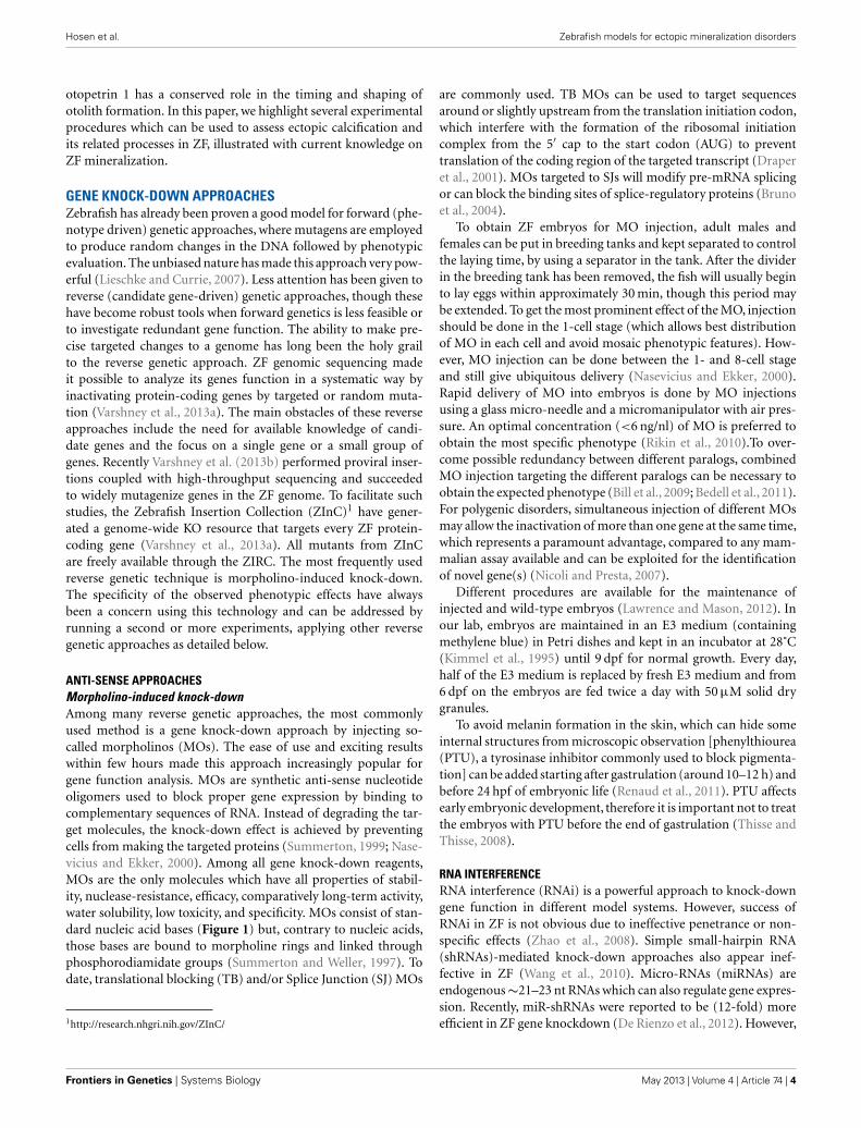
GENE KNOCK-DOWN APPROACHESZebrafish has already been proven a good model for forward (phe-notype driven) genetic approaches, where mutagens are employedto produce random changes in the DNA followed by phenotypicevaluation. The unbiased nature has made this approach very pow-erful (Lieschke and Currie, 2007). Less attention has been given toreverse (candidate gene-driven) genetic approaches, though thesehave become robust tools when forward genetics is less feasible orto investigate redundant gene function. The ability to make pre-cise targeted changes to a genome has long been the holy grailto the reverse genetic approach. ZF genomic sequencing madeit possible to analyze its genes function in a systematic way byinactivating protein-coding genes by targeted or random muta-tion (Varshney et al., 2013a). The main obstacles of these reverseapproaches include the need for available knowledge of candi-date genes and the focus on a single gene or a small group ofgenes. Recently Varshney et al. (2013b) performed proviral inser-tions coupled with high-throughput sequencing and succeededto widely mutagenize genes in the ZF genome. To facilitate suchstudies, the Zebrafish Insertion Collection (ZInC)1 have gener-ated a genome-wide KO resource that targets every ZF protein-coding gene (Varshney et al., 2013a). All mutants from ZInCare freely available through the ZIRC. The most frequently usedreverse genetic technique is morpholino-induced knock-down.The specificity of the observed phenotypic effects have alwaysbeen a concern using this technology and can be addressed byrunning a second or more experiments, applying other reversegenetic approaches as detailed below.
ANTI-SENSE APPROACHESMorpholino-induced knock-downAmong many reverse genetic approaches, the most commonlyused method is a gene knock-down approach by injecting so-called morpholinos (MOs). The ease of use and exciting resultswithin few hours made this approach increasingly popular forgene function analysis. MOs are synthetic anti-sense nucleotideoligomers used to block proper gene expression by binding tocomplementary sequences of RNA. Instead of degrading the tar-get molecules, the knock-down effect is achieved by preventingcells from making the targeted proteins (Summerton, 1999; Nase-vicius and Ekker, 2000). Among all gene knock-down reagents,MOs are the only molecules which have all properties of stabil-ity, nuclease-resistance, efficacy, comparatively long-term activity,water solubility, low toxicity, and specificity. MOs consist of stan-dard nucleic acid bases (Figure 1) but, contrary to nucleic acids,those bases are bound to morpholine rings and linked throughphosphorodiamidate groups (Summerton and Weller, 1997). Todate, translational blocking (TB) and/or Splice Junction (SJ) MOs
1http://research.nhgri.nih.gov/ZInC/
are commonly used. TB MOs can be used to target sequencesaround or slightly upstream from the translation initiation codon,which interfere with the formation of the ribosomal initiationcomplex from the 5′ cap to the start codon (AUG) to preventtranslation of the coding region of the targeted transcript (Draperet al., 2001). MOs targeted to SJs will modify pre-mRNA splicingor can block the binding sites of splice-regulatory proteins (Brunoet al., 2004).
To obtain ZF embryos for MO injection, adult males andfemales can be put in breeding tanks and kept separated to controlthe laying time, by using a separator in the tank. After the dividerin the breeding tank has been removed, the fish will usually beginto lay eggs within approximately 30 min, though this period maybe extended. To get the most prominent effect of the MO, injectionshould be done in the 1-cell stage (which allows best distributionof MO in each cell and avoid mosaic phenotypic features). How-ever, MO injection can be done between the 1- and 8-cell stageand still give ubiquitous delivery (Nasevicius and Ekker, 2000).Rapid delivery of MO into embryos is done by MO injectionsusing a glass micro-needle and a micromanipulator with air pres-sure. An optimal concentration (<6 ng/nl) of MO is preferred toobtain the most specific phenotype (Rikin et al., 2010).To over-come possible redundancy between different paralogs, combinedMO injection targeting the different paralogs can be necessary toobtain the expected phenotype (Bill et al., 2009; Bedell et al., 2011).For polygenic disorders, simultaneous injection of different MOsmay allow the inactivation of more than one gene at the same time,which represents a paramount advantage, compared to any mam-malian assay available and can be exploited for the identificationof novel gene(s) (Nicoli and Presta, 2007).
Different procedures are available for the maintenance ofinjected and wild-type embryos (Lawrence and Mason, 2012). Inour lab, embryos are maintained in an E3 medium (containingmethylene blue) in Petri dishes and kept in an incubator at 28˚C(Kimmel et al., 1995) until 9 dpf for normal growth. Every day,half of the E3 medium is replaced by fresh E3 medium and from6 dpf on the embryos are fed twice a day with 50 µM solid drygranules.
To avoid melanin formation in the skin, which can hide someinternal structures from microscopic observation [phenylthiourea(PTU), a tyrosinase inhibitor commonly used to block pigmenta-tion] can be added starting after gastrulation (around 10–12 h) andbefore 24 hpf of embryonic life (Renaud et al., 2011). PTU affectsearly embryonic development, therefore it is important not to treatthe embryos with PTU before the end of gastrulation (Thisse andThisse, 2008).
RNA INTERFERENCERNA interference (RNAi) is a powerful approach to knock-downgene function in different model systems. However, success ofRNAi in ZF is not obvious due to ineffective penetrance or non-specific effects (Zhao et al., 2008). Simple small-hairpin RNA(shRNAs)-mediated knock-down approaches also appear inef-fective in ZF (Wang et al., 2010). Micro-RNAs (miRNAs) areendogenous∼21–23 nt RNAs which can also regulate gene expres-sion. Recently, miR-shRNAs were reported to be (12-fold) moreefficient in ZF gene knockdown (De Rienzo et al., 2012). However,
Frontiers in Genetics | Systems Biology May 2013 | Volume 4 | Article 74 | 4

Hosen et al. Zebrafish models for ectopic mineralization disorders
FIGURE 1 | Binding of the anti-sense morpholino chain to the RNA chain. MOs have standard nucleic acid bases bound to morpholino ring, which arelinked through phosphorodiamidate groups, while RNA has ribose rings which are linked with phosphates.
the capability of germ-line transmission is very poor. Therefore,the potential of RNAi for stable and conditional gene knock-downin ZF remains uncertain.
ENGINEERED NUCLEASESTranscription activator-like effector nucleasesTranscription activator-like effector nucleases (TALENs) are apowerful and robust approach for efficient inactivation of a tar-geted gene. TALENs are artificial restriction enzymes generatedby fusing a TAL effector (derived from plant pathogenic bacteriaXanthomonas) DNA-binding domain to a DNA-cleavage domainor FokI nuclease. These restriction enzymes are introduced intocells to edit the genome in situ. When fused to the FokI nucleasedomain, TAL effectors recognize specific DNA sequences usinga straightforward DNA base recognition code. FokI cleaves onlywhen present as a dimer and the binding of two TAL effectors toDNA, which allows FokI to dimerize, will thus create a break in thenucleic acid strand. Because of the specificity of this DNA-binding,TAL effectors can bind virtually any DNA sequence (Cermak et al.,2011). TAL effectors possess several advantages, including ease ofdesign and easier optimization, shows few off-target effects and arethus potentially more reliable compared to other reverse geneticapproaches (Clark et al., 2011).
Zinc finger nucleasesAnother nuclease-based technology for efficient inactivation of atargeted gene in ZF (Sander et al., 2010, 2011) is the use of zincfinger nucleases (ZFNs). These are artificial restriction enzymesgenerated by fusing engineered zinc finger DNA-binding domainto a DNA-cleavage domain, and function as dimmers to introducetargeted DNA double-strand breaks. ZFN treated ZF can transmitmutant alleles (as if they were heterozygous carriers) and causeminimal collateral damage to the genomes of treated ZF. More-over, it is possible to also generate targeted knock-ins in ZF using
ZFNs. However, injected nucleases can be toxic to the embryo(Doyon et al., 2008; Meng et al., 2008) and together with the gapsin our understanding of sequence-specific DNA recognition byzinc fingers can still restrict the ability to construct ZFNs targetingany desired site in a genome (Lawson and Wolfe, 2011).
TARGETING INDUCED LOCAL LESIONS IN GENOMESTargeting induced local lesions in genomes (TILLING) is a quick,reliable, and increasingly popular method to identify chemicallyinduced point mutations in ZF (Wienholds et al., 2003). The TILL-ING method combines mutagenesis (by a chemical mutagen) witha sensitive DNA screening-technique that identifies single basemutations in a target gene. The TILLING method relies on the for-mation of DNA heteroduplexes between the wild type and mutantPCR fragment, formed when multiple alleles are amplified by PCRand are then heated and cooled. These heteroduplexes are thenincubated with celery derived CelI endonuclease, which recog-nizes and cleaves mismatches in the heteroduplex DNA generatedby small nuclear polymorphisms (SNPs) and point mutations(Oleykowski et al., 1998). Labeled digested fragments are sepa-rated and visualized on slab gel sequencers. Fragments generateddue to the presence of SNPs will be present in all animals. Thisapproach holds great promise for the rapid identification of largenumbers of mutant alleles from mutagenized libraries. However,the need for screening large number of F1 fish to find a lesion in agene of interest (Wienholds et al., 2002), the requirement of spe-cialized equipment, and a significant investment in computationalresources and personnel represent the initial bottleneck of thisapproach. To date, the “Welcome Trust Sanger Institute” has suc-cessfully provided 8402 genes (including abcc6) with mutationsunder the “Zebrafish Mutation Project (ZMP) KOs for diseasemodel” by using the TILLING approach2.
2http://www.sanger.ac.uk/Projects/D_rerio/zfm_DAS_conf.shtml
www.frontiersin.org May 2013 | Volume 4 | Article 74 | 5

Hosen et al. Zebrafish models for ectopic mineralization disorders
RETROVIRAL AND TRANSPOSON MEDIATED MUTAGENESISInsertional mutagens retrovirus or transposon can be utilized toidentify modified alleles of a target gene. The main advantage ofthese systems is readily identifiable tag that simplifies screeningfor carriers of a particular disrupted allele within the library (Jaoet al., 2008). It is also possible to generate conditional alleles inZF by including recombination sites flanking these gene-breakingelements (Petzold et al., 2009). A limitation of these insertionmutagenesis techniques is the inability to generate full null allelesin most instances.
DOMINANT NEGATIVE APPROACHHere, a mutant gene product is used to adversely affect the wild-type gene product within the same cell to get a reduced level ofgene activation. Two main Dominant negative (DN) approachesare: (1) mutation in a transcription factor that removes the activa-tion domain, which can block the wild-type transcription factor tobind with the DNA-binding site, resulting in a reduction of geneactivation, and (2) overexpression of a constitutively active protein(CAP) with mutation in/manipulation of the regulatory domain,leading to diminished opportunity for the regulatory subunit ofthe wild-type protein to bind with its receptor, and binding of amalfunctioning protein domain to the receptor will decrease thewild-type protein expression (Concordet et al., 1996). For exam-ple, in a protein which is functional as a dimer, a mutation leadingto removal of the functional domain while retaining the dimer-ized domain would cause a DN phenotype. Lanham et al. (2011)successfully used aryl hydrocarbon receptor 2 (Ahr2) as a DNapproach in ZF to protect developing ZF from dioxin toxicityby removing the C-terminal transactivation domain or replac-ing it with an inhibitory domain. CAP also been used to explorethe function of various components of signal transduction path-ways. The main advantage of DN approach over anti-sense RNAstrategies is the possibility of producing null mutations. The disad-vantages of DN approaches are that the technique is not applicableto all genes, has a relatively low throughput, and that it is usuallyimpossible to fully understand the true endogenous function ofthe molecule in vivo (Niwa and Slack, 2007).
PHARMACOLOGICAL APPROACHESPharmacological approaches (PA) are not true reverse geneticapproaches, because they do not depend on specific knowledgeof an individual gene of an organism. Once the treatment witha reagent reveals phenotypic consequences, this can be utilizedto understand interacting molecular pathways. Many pharmaco-logical reagents are used in the ZF model system including, e.g.,cyclopamine – an inhibitor of Shh signaling pathway – (Cooperet al., 1998) or SU5614 and SU1498-an inhibitor of VEGF/Flk-1tyrosine kinase signaling (Liang et al., 2001). Biochemical charac-terization of the pharmacological reagents is highly recommendedbefore applying them in the model system. Exert pleiotropic effectsis the bottleneck of using pharmacological reagents which canresult an unspecific phenotype (Skromne and Prince, 2008).
INDUCIBLE SYSTEMSMost of the aforementioned techniques used in the ZF gene-knock-down process are only suitable to evaluate phenotype and
gene function in early embryonic stages, as severe malfunction ofembryo leads to early death. So, gene function(s) at late stagesare missed, which can be overcome by inducible systems (IS)including heat shock promoters orgal4-UAS system. Applicationof IS can be done by regulating the activity of the protein orregulating the expression of the corresponding gene. In ZF, down-stream gene expression can be induced throughout the bodyby raising the temperature from 28.5 to 38˚C (Shoji and Sato-Maeda, 2008). Heat shock protein (hsp) promoters have beenused to regulate exogenous gene expression in a variety of studies.The hsp promoter can be used to search cis-acting transcrip-tional elements during ZF embryogenesis. Hsp:egfp (enhancedgreen fluorescence protein) can be injected into fertilized eggswith distal DNA fragments for tissue/cell type specific activa-tion (Islam et al., 2006). One such way for inducing heat shockresponse in targeted cell is by using a laser microbeam under themicroscope. The main advantage of this technique is visualiza-tion of a whole cell due to stable and longtime expression ofthe GFP protein, however laser ablation can be sensitive to thecells.
POST-INJECTION FOLLOW-UPEVALUATION OF THE PHENOTYPEPhenotypic screening is mainly done based on classic criteria(Haffter et al., 1996), which can provide important informationon gene function as well as the molecular events that underlie abiological process. Such screening focuses primarily on morpho-logical landmarks by dissecting microscopy. A significant numberof phenotypic traits can be screened for, as summarized in Table 2.The phenotypic screening is conducted at different time pointscorresponding to the embryonic developmental stages (Figure 2).Due to transient character of MO-induced gene knockdown, withefficient knockdown up to 4–5 dpf, phenotypic screening is prefer-ably done during this time frame (Nasevicius and Ekker, 2000; Billet al., 2009).
Morpholino mediated knockdown to study gene function inZF offers many advantages, including ease of delivery, a high effi-cacy, and rapid phenotypic screening. However, because of theirdilution in rapidly growing embryos, the effect of MOs is tempo-rary, preventing gene function analysis during the entire life cycleof ZF. Moreover, many genes – e.g., the macrophage stimulatingprotein Msp (recently found to have a role in the regulation ofcalcium homeostasis of adult ZF) (Huitema et al., 2012) – whichare expressed during the adult period and some organs such asthe skeleton which have only fully matured after 2–3 weeks can-not be studied sufficiently after MO knockdown. Altogether, thisemphasizes the usefulness of permanent mutants in addition toMO-induced models.
MORPHANT VALIDATIONEvery MO-induced phenotype must be validated to confirm thatit is due to gene-specific effects. Besides the application of anindependent knock-down approach as described above, severalMO-specific validation steps need to be addressed. First, to assessthe phenotypic variation and effect of the injection procedure,commercially available standard control MO needs to be injectedin parallel with the active MO. Secondly, for each targeted gene, the
Frontiers in Genetics | Systems Biology May 2013 | Volume 4 | Article 74 | 6

Hosen et al. Zebrafish models for ectopic mineralization disorders
Table 2 | Phenotypic traits which can be screened in ZF embryos
beyond early development.
System Phenotypic trait
Body axis Dorsilisation (48 hpf)
Ventrilisation (48 hpf)
Prechordal plate and hatching (24 hpf)
Tail (24–48 hpf)
Mesoderm Notochord formation, differentiation, and degeneration
(24 hpf)
Somite formation and patterning (24 hpf)
Central
nervous
system
Forebrain (24 hpf)Midbrain (24 hpf)
Hindbrain (24 hpf)
Neural tube – spinal cord (24 hpf)
Organs Vasculature (e.g., aortic arches, dorsal aorta,
common/posterior cardinal vein, blood island; 24–48 hpf)
Heart: morphology, beating (48 hpf)
Liver, kidney, gut (larva stadium)
Eye (24 hpf)
Ear (48 hpf)
Otoliths (48 hpf)
Pigmentation Cell number and pattern (48 hpf)
Melanin pigmentation (48 hpf)
Motility Muscles (48 hpf)
Pectoral and caudal fin (48 hpf – larva stadium)
Reduced motility (48 hpf)
For each trait, the timing when evaluation can start is mentioned. From this timing
on, serial evaluations in time are usually performed.
injection of both TB and SJ MO is recommended, to reveal the roleof either the entire protein-coding region of the gene, or of certainexons. MOs appear to have non-target-related phenotypes includ-ing overall developmental delay and cell death due to activationof p53-mediated apoptosis (Ekker and Larson, 2001), which canbe overcome by using stage matched (rather than age matched)control embryos and co-injection of an anti-p53 MO along withthe experimental MO. Injection of SJ MO generally causes skip-ping of the targeted exon or retention of the adjacent intron whileTB MO blocks gene transcription. These molecular effects can bevalidated by conducting western blotting in case of SJ MO’s andRT-PCR for TB MO’s. Further evidence of the specificity of thegene knockdown can be obtained by rescuing the phenotype bymRNA injection.
RNA rescue experimentsmRNA rescue is the most reliable approach to examine the speci-ficity of the effects of MO knockdown. To rescue the gene-specificphenotype, co-injection of the MO of interest and syntheticmRNA encoding the targeted protein can be done at the 1–4 cellstage of the embryo (Hyatt and Ekker, 1999; Bedell et al., 2011).many genes, knockdown by MOs followed by ubiquitous mRNAdelivery rarely results in truly rescued phenotypes (Bedell et al.,
FIGURE 2 | Developmental stages of zebrafish, from zygote to adult.Zygote: the newly formed fertilized egg after completion of the first zygoticcell cycle. Cleavage: zygotic cell cycles 2–7 occur rapidly and synchronously.Blastula: rapid and metasynchronous cell cycles (8, 9) occur, which give wayto lengthened, asynchronous ones at the midblastula transition, thenepiboly begins. Epiboly is the first coordinated cell movement in zebrafishembryos and begins before gastrulation. Gastrula: morphogeneticmovements of involution, convergence, and extension from the epiblast,hypoblast, and embryonic axis through the end of epiboly occur. Bud-100%epiboly is the stage where epiboly completely covers the yolk plug.Segmentation: Somites (after completion of epiboly and initial appearanceof the tail bud, first the somatic furrow forms and makes a boundary,between what will become the first and second somites), pharyngeal archprimordia, and neuromeres develop, primary organogenesis and earliestmovements take place, and the tail appears. Pharyngula: phylotypic stageof embryo, body axis straightens from its early curvature around the yolksac; circulation, pigmentation, and fins begin development. Hatching:completion of rapid morphogenesis of primary organ systems, cartilagedevelopment in head and pectoral fin, hatching occurs asynchronouslyacross individuals. Larval: swim bladder inflates; food-seeking and activeavoidance behaviors.
2011).The most important issues to consider in RNA rescue exper-iments are: (1) achievement of appropriate levels of injected MOand mRNA by co-injecting different concentrations of both com-ponents (Little and Mullins, 2004), (2) making sure that injectedsynthetic mRNA does not include the MO target sequence (Eisenand Smith, 2008). For TB MOs against the 5′ UTR sequence,the open reading frame can be cloned by PCR into a standardtranscription vector (Hyatt and Ekker, 1999). For MOs that tar-get part of the open reading frame, the rescue constructs canbe engineered to change the nucleotide sequence without alter-ing the encoded protein through degradation of the genetic code(Bill et al., 2009). Another frequently used approach for rescuingTB morphant is co-injection of mRNA from a different species.In their ZF model for PXE, Li et al. (2010) injected full-lengthmouse mRNA together with the MO; this co-injection com-pletely reversed the phenotypic effects of the MO and the rescuedembryos showed essentially the same morphology as controls. Co-injection of SJ MO and full-length mRNA (whose sequence isnon-homologous to the SJ MO) can rescue a SJ morphant (Clineet al., 2012).
www.frontiersin.org May 2013 | Volume 4 | Article 74 | 7

Hosen et al. Zebrafish models for ectopic mineralization disorders
Qualitative and quantitative validation methodsThe effect of TB MOs can be validated by western blotting, whichcorrelates reduced protein levels of the targeted gene with anobserved phenotype (Hutchinson and Eisen, 2006). The mainobstacle for performing western blot in ZF is the currentlylimited availability of antibodies that are specifically generatedto recognize ZF proteins, although a number of already avail-able antibodies from other origins show cross-reactivity with ZFproteins.
When using SJ MO, the phenotype can be validated with PCR.Qualitative evaluation of exon skipping or intron retention canbe done by performing RT-PCR with primers located in exonsupstream and downstream of the MO-targeted sequence showingsmaller and larger band respectively in addition to the originalband (Figure 3). Intron retention or exon skipping can resultin a frameshift and consequently nonsense-mediated decay andreduced transcript levels can be assessed quantitatively by usingquantitative real-time PCR (qRT-PCR).
CHARACTERIZATION OF (ECTOPIC) MINERALIZATIONPHENOTYPESCALCEIN STAININGCalcein (C30H26N2O13) is a fluorescent chromophore that specif-ically binds to calcium. As the skeletal system contains calcifiedstructures, calcein has been used to label bone structures and tostudy bone growth (Ducy et al., 2000). During calcein staining,fluorescent chromophores of calcein rapidly penetrate into ZFembryos and specifically bind to the calcified skeleton (Figure 4).Calcein staining can be used to follow the development of skeletalstructures in ZF embryos. Calcified skeletal structures appear in aprogressive fashion from head to tail. First appearance of calceinsignals, observed at ∼5 dpf are restricted to the head, followed bythe axial skeleton in the trunk (Du et al., 2001). Du et al. (2001)observed that the axial skeleton calcified in two domains. This waslater confirmed by AR-S staining (Bensimon-Brito et al., 2012).
The first domain consists of three anterior vertebral centra (cen-tra 3–5), whereas the second domain consists of the remainingabdominal and caudal centra which develop in an anterior-to-posterior direction. This confirms the sensitivity of calcein stainingfor visualizing mineralized structures in developing ZF embryosand its effectiveness for detecting defective bone structures andmineralization.
ALIZARIN RED S STAININGAlizarin Red S is an anthraquinone derivative used to identify cal-cium in tissue sections or whole mount embryos, where tissuecalcium forms an AR-S-calcium complex in a chelation process,producing a birefringent end product (Nejati-Yazdinejad, 2006).The reaction is not strictly specific for calcium, since magnesium,manganese, barium, strontium, and iron may interfere, but theseelements usually do not occur in sufficient concentration to inter-fere with the staining (Lievremont et al., 1982). AR-S stainingcan be performed on both live embryos and adult ZF as well ason fixed tissues. In live embryos, AR-S is used as a vital stainwhich labels mineralized matrix and is a very sensitive methodfor detecting bones (Kimmel et al., 1995; Walker and Kimmel,2007). On the other hand, in fixed embryos or adult fish, the fix-ation itself is a very critical step for good AR-S staining. As PFAnegatively affects bone staining, fixation with 4% PFA should berestricted to 2 h (Figure 5; Huitema et al., 2012) demonstrated anectopic mineralization phenotype in dragon fish (dgf) mutantsby Alizarin red staining, which exhibit ectopic mineralization inthe craniofacial and axial skeleton and encode a loss-of func-tion allele of ectonucleotide pyrophosphatase phosphodiesterase1 (enpp1).
Besides AR-S staining, an Alcian Blue-AR-S double stainingcan be used to distinguish cartilage and bone. Walker and Kim-mel (2007) developed an acid-free alcian blue-AR-S stainingmethod which is now widely used to stain cartilage and bonesimultaneously in ZF larvae.
FIGURE 3 | Evaluation of the effect of SJ MO in the PCR product.Hypothetical representation of intron retention and exon skipping as aconsequence of SJ MO injection. (A) MO is placed on exon2/intron2
border on targated genes pre-mRNA. The result can show (B) retention ofintron 2, or (C) skipping of exon 2. FW, forward primer; REV, reverseprimer.
Frontiers in Genetics | Systems Biology May 2013 | Volume 4 | Article 74 | 8

Hosen et al. Zebrafish models for ectopic mineralization disorders
FIGURE 4 | Calcein staining of 6 dpf embryos showing staining of theceratobranchials 5 (cb5), cleithrum (cl), dentary (d), entopterygoid (en),opercular bone (op), and parasphenoid (ps), in the skull, and anteriortip of the notochord (no).
µCT IMAGINGMicro computerized tomography (µCT) is a powerful and prac-tical tool for the skeletal analysis of diverse model organismsincluding ZF. It is used to understand developmental processesof three-dimensional embryos, embryo phenotyping, and quanti-tative modeling of development (Figure 6) (Metscher, 2009). Themethod is analogous to that used for the 3D imaging of humanstructures, on a smaller scale. It is dependent upon the interactionof large atoms with X-ray beams and requires the use of con-trast agents, still in development, for imaging anything other thanbone. But the usefulness of µCT imaging for developmental biol-ogy has been limited by the low inherent contrast of embryonictissues. Though it can be envisaged that µCT may also be usefulto demonstrate ectopic calcification in, e.g., soft tissues, its lim-ited sensitivity in embryos makes it a more useful technique inadult fish and hence the characterization of mutants instead ofmorphants. The ZF is the only well-developed vertebrate geneticmodel that is small enough to image the whole animal at cellresolutions using µCT (Cheng et al., 2011).
MOLECULAR CHARACTERIZATIONIMMUNOHISTOCHEMISTRYImmunohistochemistry (IHC) is a powerful and commonly usedtechnique in ZF for determining both the presence and localizationof antigens (e.g., endogenous protein) in cells of a tissue section,or whole mount embryos and larvae. Sample preparation, espe-cially sectioning and fixation of tissue, is very critical to maintaincell morphology, tissue architecture and the antigenicity of tar-get epitopes. Non-specific binding can cause high backgroundstaining which may mask the detection of the target antigen. Itis also possible to label both whole mount specimens and sectionswith two different antibodies, known as the double-labeling tech-nique. Double-labeling works particularly well with fluorescentconjugated secondary antibodies and these can be applied at thesame time.
FIGURE 5 | Alizarin Red staining of a 5 dpf embryo showing cleithrum(cl), opercular bone (op), parasphenoid (ps), and ceratobranchials 5(cb) with a set of three teeth.
FIGURE 6 | MicroCT scanning of whole 4 dpf embryos. The white spot inthe head region represents a signal for the mineralized otoliths.
Immunohistochemical approaches to reveal the localization ofsome calcification-related proteins demonstrated tissue distribu-tion and accumulation of Bgp and Matrix γ-carboxyglutamic acid(Gla) protein (Mgp) during the larval stage and in adult tissues ofZF by IHC shows that Bgp and Mgp proteins were located in allmineralized tissues during and after calcification including boneand calcified cartilage of branchial arches (Gavaia et al., 2006).Being a powerful method to identify specific proteins by using anantibody, the main obstacle is the availability of antibodies and thelimited cross-reactivity of ZF protein with antibodies originatingfrom other sources.
WHOLE MOUNT RNA IN SITU HYBRIDIZATIONWhole mount RNA in situ hybridization (ISH) is one of the oldestand most frequently used techniques used in the ZF research toinvestigate the site of expression of a particular gene in an intact
www.frontiersin.org May 2013 | Volume 4 | Article 74 | 9

Hosen et al. Zebrafish models for ectopic mineralization disorders
embryo (Jowett and Lettice, 1994). ISH allows specific nucleicacid sequences to be detected in morphologically preserved cellsor embryos and is also used to identify novel genes involved inthe same signaling pathways (Thisse and Thisse, 2008). Wholemount mRNA ISH can also be done to observe MO mediatedgene expression changes. In this case, cDNA of a gene of interest isused as a template for the synthesis of an anti-sense mRNA probe,which is used to recognize and bind to the endogenous transcriptthrough a color or fluorescence-based assay (Thisse and Thisse,2008). Finally, the expression of the gene can be observed under alight or fluorescent microscope.
The major advantage for using the ISH technique in ZF com-pared to other animal models is that, because of its transparencyand small size, the expression of a particular gene can be observedin the entire embryo. Though whole mount ISH is a quick andefficient method, the most significant caveat in ZF is the poorpenetration of the RNA probes after 2 days of development; onlysuperficial tissues (i.e., epithelial cells) are accessible to the probeat these later developmental stages (Thisse and Thisse, 2008). Toovercome this, embryos can be treated with proteinase K withoptimal concentration, incubation time and temperature for theembryonic stage to facilitate infiltration of the probes into thetissue.
ISH was performed to study the expression of the abcc6 gene(respectively the abcc6a and abcc6b isoform) during ZF devel-opment. While abcc6a was found to be expressed in Kupffer’svesicle, abcc6b expression was evident in the proximal tubulesof the embryonic kidney (Li et al., 2010).
In general, ISH is a laborious technique, taking about 3 days tocomplete the whole protocol. Additionally, it is difficult to handlesmall embryos because of their fragility throughout the protocol.So, semi-high-throughput procedures can alternatively be usedto facilitate these experiments by using multiwell plates (insteadof using single tube) and robotics (e.g., IntavisS AG) (Thisse andThisse,2008; Bouzaffour et al., 2009). To detect the differential pro-tein expression in morphant compared to control, ISH followedby image mapping software can be a useful tool. Alternatively,post-ISH embedding in plastic, followed by sectioning, yields highresolution images that allow detailed cellular localization of thetranscripts (Verstraeten et al., 2012).
ASSESSMENT OF GENE EXPRESSION USING QUANTITATIVEREAL-TIME PCR AND MICROARRAYSReal-time polymerase chain reaction, also called qPCR, is a labora-tory technique based on PCR, which is used to detect and measureminute amounts of nucleic acids in a wide range of samples. Thequantity can be either an absolute number of copies or a relativeamount. The expression pattern of mineralization-related genesin morphants (i.e., osteocalcin, alkaline phosphatase, bone sialo-protein) can be assessed by performing quantitative RT-PCR. Toreliably conduct qRT-PCR experiments it is highly recommendedto follow the MIQE guidelines (Bustin et al., 2009). MIQE is a setof guidelines that describe the minimum information necessaryfor evaluating qPCR experiments. Among the requirements is theneed to include at least three biological replicates (e.g., three inde-pendently injected clutches of embryos) to address the statisticalsignificance of differences in qPCR results between morphants
and controls. Furthermore, at least two reference genes should beincluded as internal controls for normalizing cellular mRNA data.Reference genes should be stably expressed, and their abundancesshould show strong correlation with the total amount of mRNApresent in the samples. In ZF embryos tuba1, bactin1, and elfα arethree stable reference genes which can be used for qPCR experi-ments comparing morphants and controls (McCurley and Callard,2008).
Microarray technology can be used to monitor transcriptomewide expression of genes in ZF embryos. Microarray analysis hasbeen employed to study the temporal activity of developmentallyregulated genes during ZF embryogenesis (Mathavan et al., 2005)but also in numerous studies where gene expression was com-pared between morphants and controls (Jenny et al., 2012; Weiet al., 2012).
The principle of Next Generation Sequencing (NGS) hasrecently been applied to transcriptome profiling (Wang et al.,2009), which offers several advantages compared to microarraysor quantitative RT-PCR. Most importantly, RNA-Seq transcrip-tome profiling can be used to identify rare transcripts which areundetectable with microarrays. Moreover, RNA-Seq allows moreprecise quantification of different transcripts (Wetterbom et al.,2010).
PROTEOMICSPhysiological mineralization is governed by highly coordinatedchanges in the expression of a large number of proteins. Under-standing these changes at the molecular level can provide impor-tant insights into physiological and disease mechanisms (Lucittet al., 2008). To elucidate these regulatory genetic networks ina ZF disease model, quantitation of protein expression duringgrowth and development is essential. In morphants, proteomicscan be used to identify proteins that are differentially expressedcompared to controls. It has been proposed that proteomic stud-ies should complement genome-wide expression profiling (Loveet al., 2004).
Many approaches can be used for quantitative protein stud-ies including two dimensional poly acrylamide gel electrophoresis(2D PAGE), mass spectrometry (MS), liquid chromatography (LC)and western blotting. Proteomic approaches are however incre-mentally challenging in ZF because of unavailability of specificantibodies and the high abundance of yolk proteins in embryos(Akhtar et al., 2009; Lobner et al., 2012). Further, many proteinidentifications have low reproducibility if the sensitivity of detec-tion is not carefully balanced against rates of false identificationerror (Lucitt et al., 2008). Therefore, rigorous statistical analysis isneeded to obtain high quality profiles of proteins.
In the early embryo, the cells forming the embryo consti-tute only a minor volume compared to the large yolk sac. Themajor yolk protein is vitellogenin, a phospholipo-glycoprotein,which functions as a nutritional source for the development ofthe embryo (Denslow et al., 1999). Link et al. (2006) developedan effective protocol for protein analysis from deyolked embryos.By pipetting with a narrow tip, the yolk sac can be disrupted. Abuffer of low osmolarity facilitates dissolving of the yolk. The dey-olking efficiency can be further increased by two additional washsteps. By removing the yolk, recovery of cellular proteins remained
Frontiers in Genetics | Systems Biology May 2013 | Volume 4 | Article 74 | 10
Hosen et al. Zebrafish models for ectopic mineralization disorders
high with only a minor reduction of housekeeping gene (mek andtubulin) observed.
Mass spectrometryMass spectrometry is an important method used to character-ize proteins by measuring the mass-to-charge ratio. The firststep in MS is the ionization of proteins, for which two commonmethods can be used: electrospray ionization (ESI) and matrix-assisted laser desorption/ionization (MALDI). Mass analysis ofproteins can be conducted using either time-of-flight (TOF) MSor Fourier transform ion cyclotron resonance (FT-ICR). Gener-ally, a protein sample is a complex mixture of different proteins.The concentration of a protein can vary between samples andan overwhelming number of peptides can make it difficult tointerpret the results. To overcome this problem, two methods,2D gel electrophoresis and high performance liquid chromatog-raphy, can be used to fractionate the proteins or their peptideproducts after enzymatic digestion. In MS, two ways are mainlyused to identify proteins, i.e., Peptide mass fingerprinting and Tan-dem MS. Observed fragment masses are matched with a databaseof predicted masses for given peptide sequences. Several meth-ods also allow quantitation of proteins by MS, i.e., stable isotopelabeling by amino acids in cell culture (SILAC), isotope codedaffinity tagging (ICAT), iRRAC (isobaric tags for relative andabsolute quantitation), or semi-quantitative MALDI analysis (inliner mode).
2D gel electrophoresisAfter removal of the predominant yolk proteins, high resolution2D gels in the acidic (pH 4–7) as well as in the basic range (pH 6–9)can be run, and proteins will be separated according to isoelectricpoint. Two biological replicates of each sample have to labeled withfluorophores (Cy3/Cy5), and an internal control from each samplelabeled with different fluorophores (Cy2) can be used to normal-ize label differences. DIGE gels are then stained and imaged usingemission wavelengths of the fluorophores. Link et al. (2006) estab-lished a protocol that is compatible with three color fluorescentlabeling using the Ettan DIGE system, which significantly reducesinter-gel variability compared to one color stains with a detectionlimit less than 1 ng protein. Analysis of 2D gels also allowed toresolve protein isoforms. Finally, validation of protein expressionchanges can be confirmed by western blotting and real-time PCRstudies.
Western blottingWestern blot is a widely accepted analytical technique used todetect specific proteins in a given sample of tissue homogenateor extract. The success of western blotting depends on the affin-ity and specificity of the antibodies and on the abundance of thetarget protein. If the yolk sac is not removed only 1 or 2 embryos(50–100 µg) can be loaded per lane on a gel to avoid overloadingeffects (Link et al., 2006). As previously stated, antibodies vali-dated for ZF proteins are not always available which can hamperthe use of this technique. Along with confirmation of TB MOefficiency, western blotting can also be used to detect differen-tial expression of a targeted protein in morphant compared tocontrol.
DISEASE SPECIFIC MECHANISMSIDENTIFICATION OF PROGRAMED CELL DEATH (APOPTOSIS)Apoptosis is a form of cell death in which a programed sequenceof events leads to the elimination of cells without inflammation.Apoptosis plays a pathophysiological role in many mineralization-related disorders including calcific aortic valve disease (Côté et al.,2012), osteoarthritis (Sun et al., 2012), and PXE (Mungrue et al.,2011).
Several methods have been developed for visualizing apop-totic cells in vitro or in fixed tissues, but few tools are availablefor visualizing apoptotic cells in live animals. Methods exist forlabeling apoptotic cells with fluorescent nucleic acid binding dyes,such as acridine orange, ethidium bromide, and propidium iodide(Lecoeur et al., 2002). A standardized technique to detect apop-totic cells in fixed tissue or fixed cells is terminal deoxynucleotidylTUNEL, which is based on end labeling of DNA degradation prod-ucts enzymatically or by a fluorescent probe (Figure 7) (Gavrieliet al., 1992). Another well-established method to detect apoptoticcells in vitro is based on loss-of membrane asymmetry duringapoptosis (Fadok et al., 1992). During apoptosis, the normal asym-metric distribution of phospholipids in the cell membrane is lost,and phosphatidylserine (PS) is exposed on the outer leaflet ofthe lipid bilayer. The calcium-dependent protein Annexin V (A5)binds PS with high affinity and fluorescently labeled A5 probeshave been widely used to detect apoptotic cells in vitro (VanGenderen et al., 2006).
Van Ham et al. (2010) recently introduced a new transgenicfluorescent marker allowing in vivo imaging of apoptotic cells tounderstand their dynamics. They fused secreted A5 (secA5) pro-tein to yellow fluorescent protein (YFP) (secA5-YFP) and showedthat this fusion product specifically labels apoptotic cells in livingZF; the fluorescent probe can characterize patterns of apoptosisin living ZF larvae and visualize cell death at single-cell resolu-tion in vivo. Labeled cells exhibit several other characteristics ofapoptotic cells, and the pattern of apoptotic cells observed by liveimaging was similar to previous findings using TUNEL.
DETECTION OF OXIDATIVE STRESSOxidative stress is an imbalance between the systemic manifes-tations of reactive oxygen species (ROS) and a biological system’sability to readily detoxify the reactive intermediates or to repair theresulting damage. Disturbances in the normal redox state of cellscan cause toxic effects through the production of peroxides andfree radicals that damage all components of the cell, including pro-teins, lipids, and DNA. Oxidative stress is increasingly implicatedas a possible pathogenic mechanism underlying a wide range ofdiseases including mineralization-related disorders such as athero-sclerosis, cardiovascular disease, or diabetes (Stephens et al., 2009).Oxidative stress in fibroblasts (Pasquali-Ronchetti et al., 2006),and elevated oxidative stress markers in the circulation (Garcia-Fernandez et al., 2008) of patients with PXE reveal its possible rolein the pathogenesis of this ectopic calcification disease.
To determine the level of ROS, fluorescent reporter moleculesCMH2DCF and Dihydrorhodamine can be administered, bothof which have been successfully applied to live embryos (Her-mann et al., 2004; Craven et al., 2005). Dihydrorhodamine isan uncharged fluorescent ROS indicator which passively diffuses
www.frontiersin.org May 2013 | Volume 4 | Article 74 | 11

Hosen et al. Zebrafish models for ectopic mineralization disorders
FIGURE 7 |Transferase dUTP nick end labeling staining of 5 dpf embryos. More fluorescent dots are observed in the tail region of Abcc6a-MO injected fish(right) demonstrating more apoptosis compared to the tail region of the wild-type fish (left).
across membranes where it is oxidized to cationic rhodamine123 which localizes in the mitochondria and exhibits greenfluorescence (Song et al., 2009).
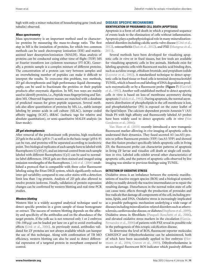
ANALYSIS OF MITOCHONDRIAL MEMBRANE POTENTIALITYRecent findings show that besides the production of ATP, mito-chondria also contribute to several other cellular functions, includ-ing redox homeostasis, calcium homeostasis, and cell death (Schef-fler, 2001). Mitochondrial mutations (inherited or somatic) areresponsible for many developmental abnormalities (Zhang et al.,2012). Mitochondrial dysfunction also plays an important rolein many mineralization diseases; i.e., in PXE and vascular calci-fication where decreased mitochondrial membrane potentiality(MMP) and reduced ion gradient has been reported. MeasuringMMP allows assessment of mitochondrial function and integrity(Nicholls and Budd, 2000).
Different methods exist to measure MMP in ZF, including stain-ing with membrane-potential-dependent dyes such as Rhodamine123, tetramethylrhodamine ethyl ester (TMRE), fluorescent car-bocyanine dye JC-1, or MitoTracker (Chazotte, 2010; Mitra andLippincott-Schwartz, 2010). MitoTracker is a commercially avail-able fluorescent dye, which labels mitochondria within live cells.Among available probes, MitoTracker Red is a red-fluorescent dyewidely used for labeling mitochondria in live ZF embryos/cells andits accumulation depends upon membrane potential (Figure 8).Live ZF embryos are incubated in the dark with 25–500 nM Mito-Tracker Red CM-H2XRos working solution for 2 h at 28.5˚C, andobserved under a fluorescent microscope. MitoTracker can alsostain the endoplasmic reticulum if embryos/cells are exposed to ahigher MitoTracker solution for a prolonged period of time. How-ever, high concentration and prolonged exposure can also blockmitochondrial activity, so a low concentration with short timeexposure is recommended to obtain specific staining (Ryu et al.,2011).
CHEMICAL SCREENING AND DRUG DISCOVERYDue to easy diffusion of chemicals through the skin, ZF allowsdisease-driven drug target identification and in vivo validation,thus representing an interesting bioassay tool for small moleculetesting and dissecting biological pathways (Pichler et al., 2003). Ina MO injection based reverse genetic approach, chemical screeningis very useful to find a chemical/drug which rescues morphant phe-notypes (Taylor et al., 2010). Peterson et al. (2000) screened 1100synthetic small molecules against ZF embryos arrayed in 96 wellplates to identify molecules that specifically modulated develop-mental processes, and found that one tetrazole derivative affectedotolith development. By adding and removing the chemical, theydetermined a critical stage for otolith development that occursbetween 14 and 26 h after fertilization. This demonstrates thatdrug screening in ZF can provide more insights in physiologicaland developmental processes.
Pichler et al. (2003) proposed two broad strategies for drugdevelopment in ZF. First, large-scale random chemical screen-ing against diseased or control ZF can be performed to observebiologically interesting phenotypic changes; secondly, functionalunderstanding of disease pathways can allow to determine specifictarget genes directly associated with the disease followed by chem-ical screening against that gene. In combination with microarray,ZF promise to be a cost and efficient bioassay that can simulta-neously uncover drug candidates, estimate toxicity, primary andsecondary drug targets, and phenotypic outcomes (Pichler et al.,2004). As many ectopic mineralization disorders are currentlystill intractable, this model may prove efficient to further exploretherapeutic options.
SYSTEMS BIOLOGY APPROACHESThe multidisciplinary approaches of systems biology allow usto quantitatively study of the fundamental principles of a bio-logical system, aiming at better understanding the connections
Frontiers in Genetics | Systems Biology May 2013 | Volume 4 | Article 74 | 12
Hosen et al. Zebrafish models for ectopic mineralization disorders
FIGURE 8 | Detection of mitochondrial membrane potentiality on2 dpf embryos by MitoTracker Red CM-H2XRos staining. (A)Control fish showing no fluorescent staining, and (B) staining with
500 nM MitoTracker Red CM-H2XRos for 2 h showing fluorescentstaining of mitochondria in the head region of a mitochondrial diseasemodel.
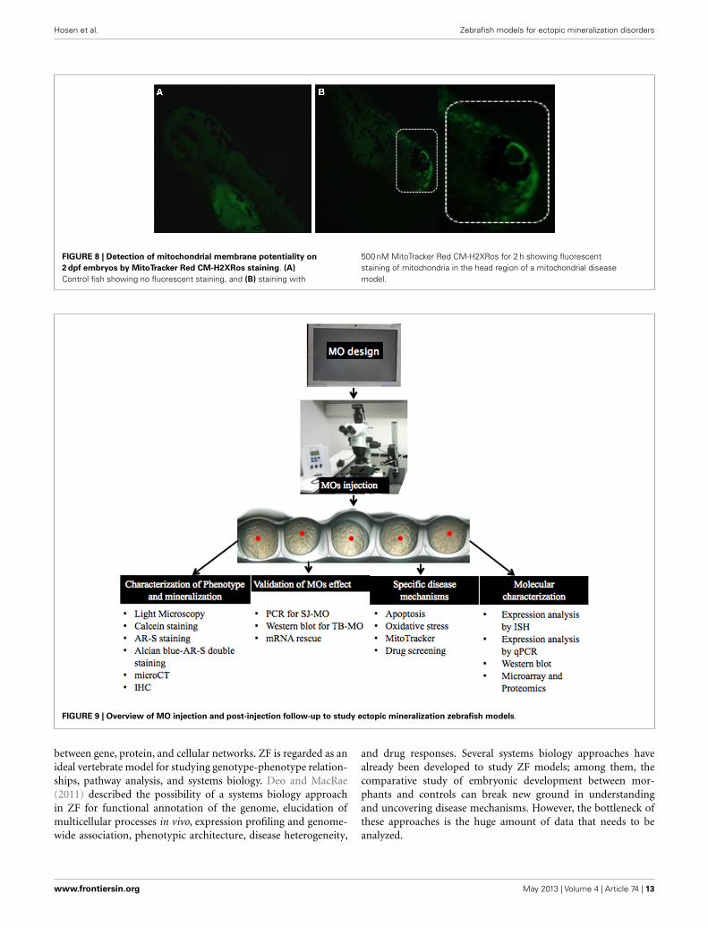
FIGURE 9 | Overview of MO injection and post-injection follow-up to study ectopic mineralization zebrafish models.
between gene, protein, and cellular networks. ZF is regarded as anideal vertebrate model for studying genotype-phenotype relation-ships, pathway analysis, and systems biology. Deo and MacRae(2011) described the possibility of a systems biology approachin ZF for functional annotation of the genome, elucidation ofmulticellular processes in vivo, expression profiling and genome-wide association, phenotypic architecture, disease heterogeneity,
and drug responses. Several systems biology approaches havealready been developed to study ZF models; among them, thecomparative study of embryonic development between mor-phants and controls can break new ground in understandingand uncovering disease mechanisms. However, the bottleneck ofthese approaches is the huge amount of data that needs to beanalyzed.
www.frontiersin.org May 2013 | Volume 4 | Article 74 | 13

Hosen et al. Zebrafish models for ectopic mineralization disorders
IN TOTO IMAGINGMegason and Fraser (2007) recently described “Digital Fishproject” to upload ZF development into computer for betterunderstanding how genome computes the formation of an embryofrom an egg. In their project they combine confocal/2-photonimaging with genetics, genomics, synthetic biology, and computa-tional analysis to watch biological circuits function in vivo and usethese data in cell-based modeling. They developed “in toto imag-ing” to track all cells in a developing tissue and record cell-basedquantitative data by using fluorescent reporters. In this imagingprocess they used “segmentation marker” to track cells, captured3D time-lapse movies on confocal and custom built 2-photonmicroscope, and finally used a software package called GoFigureto determine complete lineages and cell-based frameworks for usein modeling.
FLIP TRAPSFlip traps is a novel gene trap technique to generate endogenouslyexpressed functional fluorescent fusion protein, and they gener-ate Cre conditional alleles. Fluorescent fusion protein is used tonon-invasively quantify expression and localization of proteinsin vivo. Flip Traps in combination with “in toto imaging” can dig-itize expression and phenotype with single-cell resolution for usein molecular and cellular modeling of developmental processes(Megason and Fraser, 2007).
COMPUTATIONAL APPROACHMorelli et al. (2012) recently described the use of computationalmodeling (algorithm and simulation) to understand embryonicdevelopment. They investigated four developmental patterningstrategies of the embryo: (1) gradients of signaling moleculesreleased from localized source cell population, (2) balance betweenactivator-inhibitor mechanism leading to formation of spatialpatterns, such as stripes and spots in a two dimensional space,(3) synchronization of cellular oscillations controlling rhyth-mic and sequential subdivision of body axis into segments, and(4) mechanical deformation changing the pattern of a cellu-lar population. It is shown that theoretical along with experi-mental/computational data can play an important role to dis-close mechanisms of development. Following the same approachbetween control and morphant embryos will give an opportunityto understand underlying differential mechanisms. The Prereq-uisite for using this approach is that the level of description andmodel type are matched to quantitative, precise, and accurate data.This will beyond any doubt require a multidisciplinary team ofspecialists.
IN VIVO MODELINGGenetic and genomic features, conservation of intermolecular net-work, as well as physiologic and phenotypic features have made ZF
an important model for systems biology studies. Deo and MacRae(2011) described homology and high-throughput phenotypingstrategies which can be used for genetic or chemical screeningon a scale compatible with in vivo validation for systems biology.
Advent and validation of MO and increasing efficiency oftransgenesis have made it possible to study hundreds of geneswith specific phenotypes. Generally, phenotypic investigation ofthese approaches has been limited by phenotyping throughput.Now, the possibility of automated, quantitative phenotypes canlead to more comprehensive screenings. Moreover, applying aknown causal mutation background in ZF models can aid touncover disease mechanism. This type of analysis is also very usefulto understand genotype-phenotype correlations including pene-trance, pleiotropy, and pathogenicity. ZF disease models combinedwith known/approved libraries of drugs may enable collectionof datasets which can be highly informative not only for diseasenetwork architecture, but also for pharmacogenetics.
CONCLUSIONEctopic mineralization disorders feature some important med-ical issues to be resolved because of their complex pathogenesis,uncertainties on the mechanisms that deposit mineral in tissuesor how to remove it from the tissues, and the unavailability ofspecific drugs and treatments. Until now, many experiments havebeen done both on patients and mammalian model organismsbut the pathophysiological mechanisms of many ectopic miner-alization disorders are still incompletely understood. Because ofthe limitations to perform studies on human tissues – often dueto the invasive procedures needed to obtain them – and the highcost, long breeding time and complexity to achieve knockdownof genes in mouse models, the ZF has come to attention as analternative model organism. Several studies have shown that thereare many similarities in the molecular pathways and mechanismsinvolved in (pathological) mineralization between ZF and mam-malians, even if the phenotypic consequences are not identicalfor obvious reasons. Using MO injection based knockdown, ZFcan be an important disease model to study mineralization dis-orders. This is because phenotypic and molecular genetic resultscan be obtained within hours, because of the possibility of closeobservation in the developing transparent embryos and becauseof the easy application of techniques in the post-injection period.The concern of off-target or aspecific findings can be addressedby applying one or more alternative knock-down approaches.By considering aforementioned advantages, ZF can be used asa novel model organism for ectopic mineralization disorders.After MO-based knockdown, the described (Figure 9) valida-tion and post-injection follow-up can be applied to gain insightsin the mechanisms and future therapeutics of mineralizationdisorders.
REFERENCESAkhtar, T., Li, J., Olden, T., and Wallace,
K. N. (2009). Use of phospholipaseA2 for antigen retrieval in zebrafishwhole-mount immunohistochem-istry. Zebrafish 6, 223–227.
Antkiewicz, D. S., Burns, C. G.,Carney, S. A., Peterson, R. E.,
and Heideman, W. (2005).Heart malformation is an earlyresponse to TCDD in embry-onic zebrafish. Toxicol. Sci. 84,368–377.
Baker, K., Warren, K. S., Yellen, G., andFishman, M. C. (1997). Defective“pacemaker”current in a zebrafish
mutant with a slow heart rate.Proc. Natl. Acad. Sci. U.S.A. 94,4554–4559.
Bedell, V. M., Westcot, S. E., andEkker, S. C. (2011). Lessons frommorpholino-based screening inzebrafish. Brief. Funct. Genomics 10,181–188.
Bensimon-Brito, A., Cardeira, J., Can-cela, M. L., Huysseune, A., andWitten, P. E. (2012). Distinct pat-terns of notochord mineraliza-tion in zebrafish coincide withthe localization of osteocalcin iso-form 1 during early vertebral cen-tra formation. BMC Dev. Biol.
Frontiers in Genetics | Systems Biology May 2013 | Volume 4 | Article 74 | 14

Hosen et al. Zebrafish models for ectopic mineralization disorders
12:28. doi:10.1186/1471-213X-12-28
Bhartiya, D., Maini, J., Sharma, M.,Joshi, P., Laddha, S. V., Jalali,S., et al. (2010). FishMap Zvupdate-a genomic regulatory map ofzebrafish. Zebrafish 7, 179–180.
Bill, B. R., Petzold, A. M., Clark, K. J.,Schimmenti, L. A., and Ekker, S. C.(2009). A primer for morpholino usein zebrafish. Zebrafish 6, 69–77.
Bouzaffour, M., Dufourcq, P., Lecaudey,V., Hass, P., and Vriz, S. (2009). Fgfand Sdf-1 pathways interactduring zebrafish fin regen-eration. PLoS ONE 4:e5824.doi:10.1371/journal.pone.0005824
Bruno, I. G., Jin, W., and Cote, G.J. (2004). Correction of aberrantFGFR1 alternative RNA splicingthrough targeting of intronic regula-tory elements. Hum. Mol. Genet. 13,2409–2420.
Bustin, S. A., Benes, V., Garson, J. A.,Hellemans, J., Kubista, M., Mueller,R., et al. (2009). Clin. Chem. 55,511–522.
Cermak, T., Doyle, E. L., Christian,M., Wang, L., Zhang, Y., Schmidt,C., et al. (2011). Efficient designand assembly of custom TALEN andother TAL effector-based constructsfor DNA targeting. Nucleic Acids Res.39, 7879.
Chazotte, B. (2010). Labeling mito-chondria with Mito Tracker dyes.Cold Spring Harb. Protoc. 8, 990–992.
Chen, J. N., Haffter, P., Odenthal, J.,Vogelsang, E., Brand, M., van Eeden,F. J., et al. (1996). Mutations affect-ing the cardiovascular system andother internal organs in zebrafish.Development 123, 293–302.
Cheng, K. C., Xin, X., Clark, D. P.,and Riviere, P. L. (2011). Whole-animal imaging, gene function, andthe zebrafish phenome project. Curr.Opin. Gene. Dev. 21, 620–629.
Clark, K. J., Voytas, D. F., and Ekker,S. C. (2011). A TALE of two nucle-ases: gene targeting for the masses?Zebrafish 8, 147–149.
Cline, A., Gao, N., Flanagan-Steet,H., Sharma, V., Rosa, S., Sonon,R., et al. (2012). A zebrafish modelof PMM2-CDG reveals alteredneurogenesis and a substrate-accumulation mechanism forN-linked glycosylation deficiency.Mol. Biol. Cell 23, 4175–4187.
Concordet, J. P., Lewis, K. E., Moore,J. W., Goodrich, L. V., Johnson, R.L., Scott, M. P., et al. (1996). Spa-tial regulation of a zebrafish patchedhomologue reflects the role of sonichedgehog and protein kinase A inneural tube and somite patterning.Development 122, 2835–2846.
Cooper, M. K., Porter, J. A., Young, K. E.,and Beachy, P. A. (1998). Teratogen-mediated inhibition of target tissueresponse to Shh signaling. Science280, 1603–1607.
Côté, N., El Husseini, D., Pépin, A.,Guauque-Olarte, S., Ducharme, V.,Bouchard-Cannon, P., et al. (2012).ATO acts as a survival signal andprevents the mineralization of aor-tic valve. J. Mol. Cell. Cardiol. 52,1191–1202.
Craven, S. E., French, D., Ye, W.,de Sauvage, F., and Rosenthal,A. (2005). Loss of Hspa9b inzebrafish recapitulates the ineffectivehematopoiesis of the myelodysplas-tic syndrome. Blood 105, 3528–3534.
De Rienzo, G., Gutzman, J. H., andSive, H. (2012). Efficient shRNA-mediated inhibition of gene expres-sion in zebrafish. Zebrafish 9,97–107.
Denslow, N. D., Chow, M. C., Kroll, K. J.,and Green, L. (1999). Vitellogenin asa biomarker of exposure for estrogenor estrogen mimics. Ecotoxicology 8,485–488.
Deo, R. C., and MacRae, C. A. (2011).The zebrafish: scalable in vivo mod-eling for systems biology. WillyInterdiscip. Rev. Syst. Biol. Med. 3,335–346.
Doyon, Y., McCammon, J. M., Miller,J. C., Faraji, F., Ngo, C., Katibah, G.E., et al. (2008). Heritable targetedgene disruption in zebrafish usingdesigned zinc-finger nucleases. Nat.Biotechnol. 26, 702–708.
Draper, B. W., Morcos, P. A., and Kim-mel, C. B. (2001). Inhibition ofzebrafish fgf8 Pre-mRNA splicingwith morpholino oligos: a quantifi-able method for gene knockdown.Genesis 30, 154–156.
Du, S. J., Frenkel, V., Kindschi, G., andZohar, Y. (2001). Visualizing normaland defective bone development inzebrafish embryos using the fluores-cent chromophore calcein. Dev. Biol.238, 239–246.
Ducy, P., Amling, M., Takeda, S.,Priemel, M., Schilling, A. F., Beil, F.T., et al. (2000). Leptin inhibits boneformation through a hypothalamicrelay: a central control of bone mass.Cell 100, 197–207.
Eisen, J. S., and Smith, J. C. (2008). Con-trolling morpholino experiments:don’t stop making antisense. Devel-opment 135, 1735–1743.
Ekker, S. C., and Larson, J. D. (2001).Morphant technology in modeldevelopmental systems. Genesis 30,89–93.
Fadok,V. A., Savill, J. S., Haslett, C., Brat-ton, D. L., Doherty, D. E., Campbell,P. A., et al. (1992). Different
populations of macrophages useeither the vitronectin receptor or thephosphatidylserine receptor to rec-ognize and remove apoptotic cells. J.Immunol. 149, 29–35.
Forouhar, A. S., Hove, J. R., Calvert, C.,Flores, J., Jadvar, H., and Gharib, M.(2004). Electrocardiographic char-acterization of embryonic zebrafish.Conf. Proc. IEEE Eng. Med. Biol. Soc.5, 3615–3617.
Garcia-Fernandez, M. I., Gheduzzi, D.,Boraldi, F., Paolinelli, C. D., Sanchez,P., Valdivielso, P., et al. (2008).Parameters of oxidative stress arepresent in the circulation of PXEpatient. Biochem. Biophys. Acta 1782,474–481.
Gavaia, P. J., Simes, D. C., Ortiz-Delgado, J. B., Viegas, C. S., Pinto, J.P., Kelsh, R. N., et al. (2006). Osteo-calcin and matrix Gla protein inzebrafish and sengal sole: compar-ative gene and protein expressionduring larval development throughadulthood. Gene Expr. Patterns 6,637–652.
Gavrieli, Y., Sherman, Y., and Ben-Sasson, S. A. (1992). Identificationof programmed cell death in situvia specific labeling of nuclear DNAfragmentation. J. Cell Biol. 119,493–501.
Haffter, P., Granato, M., Brand, M.,Mllins, M. C., Maischein, H. M.,Brand, M., et al. (1996). The iden-tification of genes with unique andessential functions in the develop-ment of the zebrafish. Development123, 1–36.
Hermann, A. C., Millard, P. J., Blake, S.L., and Kim, C. H. (2004). Develop-ment of respiratory burst assay usingzebrafish kidneys and embryos. J.Immunol. Methods 292, 119–129.
Hong, Y. J., Choi, Y. W., Myung, K.B., and Choi, H. Y. (2011). Theimmunohistochemical patternsof calcicication-related moleculesin epidermis and dermis of thezebrafish. Ann. Dermatol. 23,299–303.
Hosen, M. J., Lamoen, A., De Paepe,A., and Vanakker, O. (2012).Histopathology of pseudoxanthomaelasticum and related disorders: his-tological hallmarks and diagnosticclues. Scientifica 2012, 1–15.
Hughes, I., Blasiole, B., Huss, D., War-chol, M. E., Rath, N. P., Hurle, B.,et al. (2004). Otopetrin1 is requiredfor otolith formation in the zebrafishDanio rerio. Dev. Biol. 276, 391–402.
Huitema, L. F., Renn, J., Logister, I.,Gray, J. K., Waltz, S. E., Flik, G., etal. (2012). Macrophage-stimulatingprotein and calcium homeostasis inzebrafish. FASEB J. 26, 4092–4101.
Hutchinson,S. A., and Eisen, J. S. (2006).Islet1 and Islet2 have equivalent abil-ities to promote motoneuron for-mation and to specify motoneuronsubtype identity. Development 133,2137–2147.
Hyatt, T. M., and Ekker, S. C. (1999).Vectors and techniques for ectopicgene expression in zebrafish. Meth-ods Cell Biol. 59, 117–126.
Islam, M. E., Kikuta, H., Inoue, F.,Kanai, M., Kawakami, A., Parvin,M. S., et al. (2006). Three enhancerregions regulate gbx2 gene expres-sion in the isthmic region duringzebrafish development. Mech. Dev.123, 907–924.
Jao, L. E., Maddison, L., Chen, W.,and Burgess, S. M. (2008). Usingretroviruses as a mutagenesis toolto explore the zebrafish genome.Brief. Funct. Genomics Proteomics 7,427–443.
Jenny, M. J., Aluru, N., and Hahn, M. E.(2012). Effects of short-term expo-sure to 2,3,7,8-tetrachlorodibenzo-p-dioxin on microRNA expresson inzebrafish. Toxicol. Appl. Pharmacol.264, 262–273.
Jowett, T., and Lettice, L. (1994). Whole-mount in-situ hybridizations onzebrafish embryos using a mix-ture of digoxigenin-labeled andfluorescein-labeled probes. TrendsGenet. 10, 73–74.
Kawasaki, K., Buchanan, A. V., andWeiss, K. M. (2009). Biomineraliza-tion in humans: making the hardchoices in life. Annu. Rev. Genet. 43,119–142.
Kimmel, C. B., Ballard, W. W., Kimmel,S. R., Ullmann, B., and Schilling, T.F. (1995). Stages of embryonic devel-opment of the zebrafish. Dev. Dyn.203, 253–310.
Lanham, K. A., Prasch, A. L., Weina,K. M., Peterson, R. E., and Hei-deman, W. (2011). A dominantnegative zebrafish Ahr2 partiallyprotects developing zebrafish fromdioxin toxicity. PLoS ONE 6:e28020.doi:10.1371/journal.pone.0028020
Lawrence, C., and Mason, T. (2012).Zebrafish housing systems: a reviewof basic operating principles andconsiderations for design and func-tionality. ILAR J. 53, 179–191.
Lawson, N. D., and Wolfe, S. A.(2011). Forward and reverse geneticapproaches for the analysis of verte-brate development in the zebrafish.Dev. Cell 21, 48–64.
Lecoeur, H., de Oliveira-Pinto, L. M.,and Gougeon, M. L. (2002). Multi-parametric flow cytometric analysisof biochemical and functional eventsassociated with apoptosis and onco-sis using the 7-aminoactinomycin
www.frontiersin.org May 2013 | Volume 4 | Article 74 | 15

Hosen et al. Zebrafish models for ectopic mineralization disorders
D assay. J. Immunol. Methods 265,81–96.
Li, C. F., MacDonald, J. R., Wie, R. Y.,Ray, J., Lau, K., Kandel, C., et al.(2007). Human sterile alpha motifdomain 9, a novel gene identified asdown-regulated in aggressive fibro-matosis, is absent in the mouse. BMCGenomics 8:92. doi:10.1186/1471-2164-8-92
Li, Q., Frank, M., Thisse, C. L., Thisse,B. V., and Uitto, J. (2011). Zebrafish:a model system to study heritableskin diseases. J. Invest. Dermatol. 131,565–571.
Li, Q., Sadowski, S., Frank, M., Chai, C.,Varadi, A., Ho, S. Y., et al. (2010). Theabcc6a gene expression is requiredfor normal zebrafish development. J.Invest. Dermatol. 11, 1–8.
Liang, D., Chang, J. R., Chin, A. J.,Smith, A., Kelly, C., Weinberg, E. S.,et al. (2001). The role of vascularendothelial growth factor (VEGF)in vasculogenesis, angiogenesis, andhematopoiesis in zebrafish develop-ment. Mech. Dev. 108, 29–43.
Lieschke, G., and Currie, P. D. (2007).Animal models of human disease:zebrafish swim into view. Nat. Rev.Genet. 8, 353–367.
Link, V., Shevchenko, A., and Heisen-berg, C. (2006). Proteomics of earlyzebrafish embryos. BMC Dev. Biol.6:1. doi:10.1186/1471-213X-6-1
Little, S. C., and Mullins, M. C. (2004).Twisted gastrulation promotes BMPsignaling in zebrafish dorsal-ventralaxial patterning. Development13123, 5825–5835.
Lievremont, M., Potus, J., and Guillou,B. (1982). Use of alizarin red S forhistochemical staining of Ca2+ inthe mouse; some parameters of thechemical reaction in vitro. Acta Anat.(Basel) 3, 3268–3280.
Lobner, C., Wee, S., Ler, S. G., Li, R.H., Carnery, T., Blackstock, W., etal. (2012). Expanding the zebrafishembryo proteome using multiplefractionation approaches and tan-dem mass spectrometry. Proteomics11, 1879–1882.
Love, D. R., Pichler, F. B., Dodd, A.,Copp, B. R., and Greenwood, D.R. (2004). Technology for high-throughput screens: the present andfuture. Curr. Opin. Biotechnol. 6,564–571.
Lu, J. G., Peatman, E., Tang, H. B.,Lewis, J., and Liu, Z. J. (2012). Pro-filing of gene duplication patterns ofsequenced teleost genomes: evidencefor rapid lineage-specific genomeexpansion mediated by recent tan-dem duplications. BMC Genomics13:246. doi:10.1186/1471-2164-13-246
Lucitt, M. B., Price, T. S., Pizarro, A.,Wu, W., Yocum, A. K., Seiler, C., etal. (2008). Analysis of the zebrafishproteome during embryonic devel-opment. Mol. Cell. Proteomics 7,981–994.
Mathavan, S., Lee, S. G., Mak, A.,Miller, L. D., Murthy, K. R.,Govindarajan, K. R., et al.(2005). Transcriptome analysisof zebrafish embryogenesis usingmicroarrays. PLoS Genet. 1:e29.doi:10.1371/journal.pgen.0010029
McCurley, A. T., and Callard, G. V.(2008). Characterization of house-keeping genes in zebrafish: male-female differences and effects of tis-sue type, developmental stage andchemical treatment. BMC Mol. Biol.9:102. doi:10.1186/1471-2199-9-102
Megason, S. G., and Fraser, S. E. (2007).Imaging in systems biology. Cell 130,784–795.
Meng, X., Noyes, M. B., Zhu, L. J.,Lawson, N. D., and Wolfe, S. A.(2008). Targeted gene inactivationin zebrafish using engineered zinc-finger nucleases. Nat. Biotechnol. 26,695–701.
Metscher, B. D. (2009). MicroCT fordevelopmental biology: a versatiletool for high-contrast 3D imagingat histological resolutions. Dev. Dyn.38, 632–640.
Mitra, K., and Lippincott-Schwartz, J.(2010). Analysis of mitochondrialdynamics and functions using imag-ing approaches. Curr. Protoc. CellBiol. 4, 1–21.
Morelli, L. G., Uriu, K., Ares, S.,and Oates, A. C. (2012). Com-putational approaches to devel-opmental patterning. Science 336,187–191.
Mungrue, I. N., Zhao, P., Yao, Y., Meng,H., Rau, C., Havel, J. V., et al. (2011).Abcc6 deficiency causes increasedinfarct size and apoptosis in amouse cardiac ischemia-reperfusionmodel. Arterioscler. Throm. Vasc.Biol. 31, 2806–2812.
Nasevicius, A., and Ekker, S. C. (2000).Effective targeted gene “knock-down” in zebrafish. Nat. Genet. 26,216–220.
Nejati-Yazdinejad, M. (2006). Spec-trophotometric determination oftrace amount of copper (ii) ironbased on complexation with ananthraquinone derivative. Anal. Sci.22, 617–619.
Nicholls, D. G., and Budd, S. L. (2000).Mitochondria and neuronal sur-vival. Physiol. Rev. 80, 315–360.
Nicoli, S., and Presta, M. (2007).The zebrafish/tumor xenograftangiogenesis assay. Nat. Protoc. 2,2918–2923.
Niwa, R., and Slack, F. J. (2007). Ins andouts of RNA interference analysis.Cell 2008, 34–36.
Oleykowski, C. A., Bronson Mullins, C.R., Godwin, A. K., and Yeung, A.T. (1998). Mutation detection usinga novel plant endonuclease. NucleicAcids Res. 26, 4597–4602.
Pasqualetti, S., Banfi, G., and Mari-otti, M. (2012). The zebrafish scaleas model to study the bone min-eralization process. J. Mol. Hist. 43,589–595.
Pasquali-Ronchetti, I., Garcia-Fernandez, M. I., Boraldi, F.,Quaglino, D., Gheduzzi, D., DeVincenzi Paolinelli, C., et al. (2006).Oxidative stress in fibroblastsfrom patients with pseudoxan-thoma elasticum: possible relein the pathogenesis of clinicalmanifestations. J. Pathol. 208,54–61.
Peterson, R. T., Link, B. A., Dowl-ing, J. E., and Schreiber, S. L.(2000). Small molecule develop-mental screens reveal the logic andtiming of vertebrate development.Proc. Natl. Acad. Sci. U.S.A. 97,12965–12969.
Petzold, A. M., Balciunas, D., Sivasubbu,S., Clark, K. J., Bedell, V. M., Westcot,S. E., et al. (2009). Nicotine responsegenetics in the zebrafish. Proc. Natl.Acad. Sci. U.S.A. 106, 18662–18667.
Pichler, F. B., Dodd, A., and Love, D.R. (2004). Global gene expressionanalysis in the zebrafish: the chal-lenge and the promise. Drug Discov.Today Technol. 1, 79–84.
Pichler, F. B., Laurenson, S., Williams,L. C., Dodd, A., Copp, B. R., andLove, D. R. (2003). Chemical dis-covery and global gene expressionanalysis in zebrafish. Nat. Biotechnol.21, 879–883.
Postlethwait, J., Amores, A., Cresko, W.,Singer, A., and Yan, Y. L. (2004).Subfunction partitioning, the tel-wost radiation and the annotationof the human genome. Trends Genet.20, 481–490.
Renaud, O., Herbomel, P., and Kissa,K. (2011). Studying cell behaviorin whole zebrafish embryos by con-focal live imaging: application tohematopoietic stem cells. Nat. Pro-toc. 6, 1897–1904.
Rikin, A., Rosenfeld, G. E., McCartin,K., and Evans, T. (2010). A reversergenetic approach to test functionalredundancy during embryogenesis.J. Vis. Exp. 11, 2020.
Rusanescu, G., Weissleder, R., andAikawa, E. (2008). Notch signal-ing in cardiovascular disease andcalcification. Curr. Cardiol. Rev. 4,148–156.
Ryu, S., Beutner, G., Kinnally, K. W.,Dirksen, R. T., and Sheu, S. (2011).Single channel characterization ofthe mitochondrial ryanodine recep-tor in heart mitoplasts. J. Biol. Chem.24, 21324–21339.
Sander, J. D., Dahlborg, E. J., Good-win, M. J., Cade, L., Zhang, F.,Cifuentes,D., et al. (2011). Selection-free zinc-finger-nuclease engineer-ing by context-dependent assem-bly (CoDA). Nat. Methods 8,67–69.
Sander, J. D., Maeder, M. L., Reyon, D.,Voytas, D. F., Joung, J. K., and Dobbs,D. (2010). ZiFiT (Zinc Finger Tar-geter): an updated zinc finger engi-neering tool. Nucleic Acids Res. 38,W462–W468.
Scheffler, I. E. (2001). Mitochondriamake a comeback. Adv. Drug Deliv.Rev. 49, 3–26.
Sehnert, A. J., and Stainier, D. Y. (2002).A window to the heart: can zebrafishmutants help us understand heartdisease in humans? Trends Genet. 18,491–494.
Shanahan, C. M., Crouthamel, M.H., Kapustin, A., and Giachelli, C.M. (2011). Arterial calcification inchronic kidney disease: kye roles forcalcium and phosphate. Circ. Res. 6,697–711.
Shawber, C. J., and Kitajewski, J. (2004).Notch function in the vasculature:insights from zebrafish, mouse andman. Bioessays 26, 225–234.
Shoji, W., and Sato-Maeda, M. (2008).Application of heat shock promoterin transgenic zebrafish. Dev. GrowthDiffer. 50, 401–406.
Sire, J. Y., and Akimenko, M. A. (2004).Scale development in fish: a review,with description of sonic hedgehog(shh) expression in the zebrafish. Int.J. Dev. Biol. 48, 233–247.
Sire, J. Y.,Allizard, F., Babiar, O., Bourgu-ingnon, J., and Quilhac, A. (1997a).Scale development in zebrafish. J.Anat. 190, 545–561.
Sire, J. Y., Quilhac, A., Bourguignon, J.,and Allizard, F. (1997b). Evidencefor participation of the epidermis inthe deposition of superficial layer ofscales in zebrafish (Danio rerio). J.Morphol. 231, 161–174.
Skromne, I., and Prince, V. E. (2008).Current perspectives in zebrafishreverse genetics: moving forward.Dev. Dyn. 137, 861–882.
Song, Y. Q., Selak, M. A., Watson, C. T.,Coutts, C., Scherer, P. C., Panzer, J.A., et al. (2009). Mechanisms under-lying metabolic and neural defectsin zebrafish and human multipleAcyl-CoA dehydrogenase deficiency(MADD). PLoS ONE 4:e8329.doi:10.1371/journal.pone.0008329
Frontiers in Genetics | Systems Biology May 2013 | Volume 4 | Article 74 | 16
Hosen et al. Zebrafish models for ectopic mineralization disorders
Stainier, D. Y., Fouquet, B., Chen, J.N., Warren, K. S., Weinstein, B. M.,and Meiler, S. E. (1996). Mutationsaffecting the formation and functionof the cardiovascular system in thezebrafish embryo. Development 123,285–292.
Stephens, J. W., Khanolkar, M. P.,and Bain, S. C. (2009). The bio-logical relevance and measurementof plasma markers of oxidativestress in diabetes and cardiovas-cular disease. Atherosclerosis 202,321–329.
Summerton, J. (1999). Morpholinoantisense oligomers: the case foran RNase H-independent structuraltype. Biochim. Biophys. Acta 1489,141–158.
Summerton, J., and Weller, D. (1997).Morpholino antisense oligomers:design, preparation and properties.Antisense Nucleic Acid Drug Dev. 7,187–195.
Sun, Y., Mauerhan, D. R., Chaturvedi,D., Hanley, E. N. Jr., and Gruber,H. E. (2012). Biological effects ofphosphate on fibroblast-like syn-oviocytes. ISRN Cell Biol. 2012,1–6.
Taylor, K. L., Grant, N. J., Temperley, N.D., and Patton, E. E. (2010). Smallmolecule screening in zebrafish: anin vivo approach to identifying newchemical tools and drug leads. CellCommun. Signal. 8, 1–14.
Thisse, C., and Thisse, B. (2008). High-resolution in situ hybridization towhole-mount zebrafish embryos.Nat. Protoc. 3, 59–69.
Tingaud-Sequeira, A., Forgue, J.,Andre, M., and Babin, P. J.(2006). Epidermal transientdown-regulation of retinol-bindingprotein 4 and mirror expressionof apolipoprotein Eb and estrogenreceptor 2a during zebrafish fin and
scale development. Dev. Dyn. 235,3071–3079.
Van Genderen, H., Kenis, H., Lux, P.,Ungeth, L., Maassen, C., Deckers,N., et al. (2006). In vitro measure-ment of cell death with the annexinA5 affinity assay. Nat. Protoc. 1,363–367.
Van Ham, T. J., Mapes, J., Kokel, D., andPeterson, R. T. (2010). Live imagingof apoptotic cells in zebrafish. FASEBJ. 24, 4336–4342.
Varshney, G. K., Huang, H., Zhang, S.,Lu, J., Gildea, D. E., Yang, Z., et al.(2013a). The zebrafish insertion col-lection (ZInC): a web based, search-able collection of zebrafish muta-tions generated by DNA insertion.Nucleic Acids Res. 41, 861–864.
Varshney, G. K., Lu, J., Gildea, D. E.,Huang, H., Pei, W., Yang, Z., et al.(2013b). A large-scale zebrafish geneknockout resource for the genome-wide study of gene function. GenomeRes. 23, 1–9.
Verstraeten, B., Sanders, E., and Huysse-une, A. (2012). “Whole mountimmunohistochemistry and in situhybridization of larval and adultzebrafish dental tissues,” in Meth-ods in Molecular Biology, Meth-ods in Odontogenesis, Vol. 887, ed.C. Kioussi (Totowa, NJ: HumanaPress), 179–191.
Viegas, C. S. B., Cavaco, S., Neves, P. L.,Ferreira, A., Joao, A., Williamson, M.K., et al. (2009). Gla-rich protein is anovel vitamin K-dependent proteinpresent in serum that accumulatesat sites of pathological calcifications.Am. J. Pathol. 175, 2288–2298.
Walker, M. B., and Kimmel, C. B. (2007).A two-color acid-free cartilage andbone stain for zebrafish larvae.Biotech. Histochem. 82, 23–28.
Wang, L., Zhou, J. Y., Yaho, J. H., Lu,D. R., and Qiao, X. J. (2010). U6
promoter-driven siRNA injectionhas nonspecific effects in zebrafish.Biochem. Biophys. Res. Commun. 39,1363–1368.
Wang, Z., Gerstein, M., and Snyder,M. (2009). RNA-seq: a revolutionarytool for transcriptomics. Nat. Rev.Genet. 10, 57–63.
Warren, K. S., and Fishman, M.C. (1998). Physiological genomics:mutant screens in zebrafish. Am. J.Physiol. 275, H1–H7.
Wei, L., Hoole, D., and Sun, B. (2012).Identification of apoptosis relatedgenes and transcription variationsin response to microcystin-LR inzebrafish liver. Toxicol. Ind. Healthdoi:10.1177/0748233712462443
Wetterbom, A., Ameur, A., Feuk, L., Gyl-lensten, U., and Cavelier, L. (2010).Identification of novel exons andtranscribed regions by chimpanzeetranscriptome sequencing. GenomeBiol. 11, R78.
Wienholds, E., Koudijs, M. J., vanEeden, F. J., Cuppen, E., and Plas-terk, R. H. (2003). The microRNA-producing enzyme Dicer1 is essen-tial for zebrafish development. Nat.Genet. 35, 217–218.
Wienholds, E., Schulte-Merker, S.,Walderich, B., and Plasterk, R. H.(2002). Target-selected inactivationof the zebrafish rag1 gene. Science297, 99–102.
Woods, I. G., Kelly, P. D., Chu, F., Ngo-Hazelett, P., Yan, Y., Huang, H., et al.(2000). A comparative map of thezebrafish genome. Genome Res. 10,1903–1914.
Xu, X., Meiler, S. E., Zhong, T. P.,Mohideen, M., Crossley, D. A.,Burggren, W. W., et al. (2002).Cardiomyopathy in zebrafish dueto mutation in an alternativelyspliced exon of titin. Nat. Genet. 30,205–209.
Zhang, C., Huang, V. H., Simon, M.,Sharma, L. K., Fan, W., Hass, R.,et al. (2012). Heteroplasmic muta-tions of the mitochondrial genomecause paradoxical effects on mito-chondrial functions. FASEB J. 26,4914–4924.
Zhao, X. F., Fjose, A., Larsen, N.,Helvik, J. V., and Drivenes, O. (2008).Treatment with small interferingRNA affects the microRNA path-way and causes unspecific defectsin zebrafish embryos. FEBS J. 275,2177–2184.
Conflict of Interest Statement: Theauthors declare that the research wasconducted in the absence of any com-mercial or financial relationships thatcould be construed as a potential con-flict of interest.
Received: 23 October 2012; accepted: 15April 2013; published online: 08 May2013.Citation: Hosen MJ, Vanakker OM,Willaert A, Huysseune A, Coucke P andDe Paepe A (2013) Zebrafish models forectopic mineralization disorders: prac-tical issues from morpholino design topost-injection observations. Front. Genet.4:74. doi: 10.3389/fgene.2013.00074This article was submitted to Frontiers inSystems Biology, a specialty of Frontiersin Genetics.Copyright © 2013 Hosen, Vanakker,Willaert , Huysseune, Coucke andDe Paepe. This is an open-access articledistributed under the terms of theCreative Commons Attribution License,which permits use, distribution andreproduction in other forums, providedthe original authors and source are cred-ited and subject to any copyright noticesconcerning any third-party graphicsetc.
www.frontiersin.org May 2013 | Volume 4 | Article 74 | 17