Emotional arousal of beginning physics teachers during extended experimental investigations

CHAPTER 11
Monitoring Sleep and Arousal in Zebrafish
Jason Rihel*, David A. Prober†, and Alexander F. Schier*,‡,§,{*Department of Molecular and Cellular Biology, Harvard University, Cambridge, Massachusetts
†Division of Biology, California Institute of Technology, Pasadena, California
‡Division of Sleep Medicine, Harvard University, Cambridge, Massachusetts
§Center for Brain Science, Harvard University, Cambridge, Massachusetts
{Harvard Stem Cell Institute, Harvard University, Cambridge, Massachusetts
I. IntroductionII. Behavior, Genetics, and Pharmacology of Zebrafish Sleep
A. BehaviorB. Genetics and Pharmacology
III. Methods for Monitoring Sleep/Wake Behavior in ZebrafishA. Methodological Considerations—from Drosophila to DanioB. Experimental Design and SetupC. Monitoring and Analysis of Sleep/Wake Behaviors
IV. ConclusionReferences
Abstract
Zebrafish has emerged in the past 5 years as a model for the study of sleep and wakebehaviors. Experimental evidence has shown that periods of behavioral quiescence inzebrafish larvae and adults are sleep-like states, as these rest bouts are regulated by thecircadian cycle, are associated with decreases in arousal, and are increased followingrest deprivation. Furthermore, zebrafish share with mammals a hypocretin/orexinsystem that promotes wakefulness, and drugs that alter mammalian sleep have similareffects on zebrafish rest. In this chapter, we review the zebrafish sleep literature anddescribe a long-term, high-throughput monitoring system for observing sleep and wakebehaviors in larval zebrafish.
METHODS IN CELL BIOLOGY, VOL. 100 978-0-12-384892-5Copyright � 2010 Elsevier Inc. All rights reserved. 281 DOI: 10.1016/B978-0-12-384892-5.00011-6

I. Introduction
Sleep is essential, time consuming, and conserved across the animal kingdom, yet itremains one of the major mysteries of biology. What is the function of sleep, and how is itregulated by genes and neurons? Since the discovery of characteristic electroencephalo-graphic (EEG) signatures for states of sleep and waking in the late 1930s (Davis et al.,1937), mammalian model systems have dominated sleep research. However, behavioralobservations over the past decade have demonstrated that non-mammalian systems,includingDrosophila (Hendricks et al., 2000; Shaw et al., 2000), Caenorhabditis elegans(Raizen et al., 2008; Van Buskirk and Sternberg, 2007), and zebrafish (Prober et al.,2006; Yokogawa et al., 2007; Zhdanova et al., 2001), have sleep-like states. These“simple” model systems allow researchers to bring large-scale genetics and in vivoimaging to bear on fundamental questions of sleep biology. Zebrafish is an attractivemodel because it combines the facile genetics of invertebrates with brains that aremorphologically and molecularly analogous to mammals. In this chapter, we reviewthe progress of sleep studies in zebrafish and discuss the high-throughput methods(Fig. 1) we have developed to study larval zebrafish sleep/wake behaviors.
II. Behavior, Genetics, and Pharmacology of Zebrafish Sleep
A. Behavior
Sleep is a period of reversible, inattentive behavioral quiescence that can be dis-tinguished from quiet wakefulness using several behavioral criteria (Borbely and
96 hpf
H2O in
28 °C
H2O
out
White + IR LEDs
Videocamera
Translucentchamber
Trackingcomputer
Fig. 1 High-throughput tracking of zebrafish locomotor behavior. Zebrafish larvae older than 96 hpf are placedinto a 96-well plate filled with blue water. The plate is then placed into a translucent chamber that is illuminatedfrom the bottom continuously by infrared (IR) light emitting diodes (LEDs) and from 9:00 AM to 11:00 PMwithwhite LEDs. Water heated to 28°C is pumped continuously through (water goes in and out of the chamber) theplate chamber to maintain temperature and humidity. The plate is observed by a video camera connected to acomputer with video tracking software. The chamber, lights, and camera are housed inside a box (not shown) toprevent extraneous light from interfering with the experiment. Modified from Fig. 4 in Prober et al., 2006.
282 Jason Rihel et al.

Tobler, 1996; Campbell and Tobler, 1984; Cirelli, 2009). Sleep-like rest periods aretypically associated with a species-specific posture and location, and are regulated in acircadian (about 24 h) manner (Borbely and Tobler, 1996). Importantly, sleep isassociated with an increased arousal threshold in response to stimuli, although strongstimuli can still wake the animal (thereby distinguishing sleep from coma or stupor).Finally, sleep is thought to be under homeostatic control, such that depriving an animalof sleep results in a subsequent increase in the duration or intensity of sleep, known assleep rebound (Borbely and Tobler, 1996). Using automated tracking systems toobserve individual animals for several days (see Section III), a variety of non-mammalian model systems, including fruit flies (Hendricks et al., 2000; Shaw et al.,2000), C. elegans (Raizen et al., 2008; Van Buskirk and Sternberg, 2007), and bothlarval (Prober et al., 2006; Zhdanova et al., 2001) and adult zebrafish (Yokogawa et al.,2007), have been shown to exhibit sleep-like states that meet these behavioral criteria.
1. Criterion 1—Quiescent Behavior Regulated by an Endogenous Circadian Rhythm
Zebrafish larvae first exhibit active spontaneous locomotor activity around 96h postfertilization (hpf), shortly after inflation of the swim bladder (Hurd and Cahill, 2002; Proberet al., 2006). When maintained on a 14 h:10 h light:dark cycle, these swim bouts occurmaximally during the lights-on phase and are tightly synchronized with the light stimulus(Fig. 2A). If raised on a light:dark cycle and then transferred to constant dark conditions, thespontaneous locomotor activity of >96hpf larvae and adults continues to cycle with acircadian rhythm of ~25–25.5 h, with the phase set by the prior entraining light:dark cycle(Hurd and Cahill, 2002; Hurd et al., 1998). These observations demonstrate that zebrafishhave an endogenously controlled circadian rhythm behavior that can be entrained by light.Consistent with the behavioral observations, rhythmic components of the molecularmachinery that controls circadian rhythms in Drosophila and mammals also exhibit rhyth-mic expressionwith a light-entrainable circadian period in zebrafish. Such genes include thezebrafish orthologs ofPeriod1 (Dekens andWhitmore, 2008),Period3 (Kaneko andCahill,2005; Pando et al., 2001), Clock (Whitmore et al., 1998), Cryptochrome (Kobayashi et al.,2000), and the clock-binding partner Bmal (Cermakian et al., 2000).
2. Criterion 2—Increased Arousal Threshold/Decreased Responsiveness during Quiescence
Throughout the 24 h light:dark cycle, zebrafish larvae exhibit bouts of quiescence thatcan last for several minutes or longer (Fig. 2B and C) and that occur maximally at night.During these quiescent periods, both zebrafish larvae and adults have an accompanyingincrease in arousal threshold, as measured by a decreased responsiveness of larvaeto taps at night versus the day (Zhdanova et al., 2001), an increase in response time tolarge changes in light intensity (Prober et al., 2006), or a decreased responsivenessto electrical stimuli in adults (Yokogawa et al., 2007). By correlating the length ofa quiescent bout with the concomitant change in arousal state, sleep is defined as aquiescent bout lasting at least 1 min in larvae and at least 6 s in adults. This definitionof a sleep-like state has served well in the analysis of molecules that regulate sleep andhas been effective in uncovering conserved pathways (see Section II-B). The definitioncan now be refined by testing changes in arousal across multiple sensory modalities,
11. Monitoring Sleep and Arousal in Zebrafish 283

experimental conditions, and behavioral situations, including a detailed analysisof changes in arousal states during different times of the day and night. For example,a recent report notes that larval respiration rate is reduced and arousal thresholdelevated in nighttime rest compared to daytime rest (Zhdanova, 2006), indicating thatthese two quiescent states may not be equivalent. We also observe changes in averagesleep bout length and in waking activity (see section III.C for details) during the dayand night that may reflect underlying differences between these states (Table I).
3. Criterion 3—Sleep Rebound Following Sleep Deprivation (Homeostatic Regulation of Sleep)
Two studies have demonstrated an increase in total sleep amount in larval and adultzebrafish following nighttime sleep deprivation. Zhdanova and colleagues used a
50
40
30
20
10
0
Sec
on
ds
of
acti
vity
/10
min
(C)
(B)
(A)
14 0 14 0 14
Zeitgeber time
876543210
Min
0 10 20 30 40 50
Sec
on
ds
of
acti
vity
/min
Lights off
Sleep latency(6 min)
Sleep bout(6 min)
Active bout(5 min)
14 0 14 0 14
Zeitgeber time
10
8
6
4
2
0Min
ute
s o
f sl
eep
/10
min
(D) Behavioral fingerprint
Sle
epla
tenc
y
Act
ivity
Sle
epto
tal
Sle
ep B
out
leng
th
Sle
ep B
out
num
ber
SD –3 –2 –1 0 1 2 3
Wak
ing
activ
ity
Drug 1
Drug 2
Drug 3
Fig. 2 Zebrafish sleep/wake data. (A) The average activity of a single wild type larva is plotted per 10 minfor two light:dark cycles starting at ~110 hpf. Activity occurs maximally during each day period. The grayarea is expanded in (B). Zeitgeber time 0 = lights on; 14 = lights off. (B) The average activity of the samefish in (A) (gray area), expanded and replotted per 1 min. Examples of sleep latency, sleep bout, sleep boutlength, active bout, and active bout length are indicated. (C) The behavior of the same fish shown in (A) and(B), plotted as minutes of sleep per 10 min. Sleep occurs maximally during each night period. (D) Bynormalizing each behavioral parameter to wild type controls, the data can be transformed into a behavioralfingerprint. Each square of the fingerprint represents the average relative value in standard deviations (black,higher than controls; white, lower than controls) for a single behavioral measurement. The black and whitebars across the top represent night and day measurements, respectively. In this example, fingerprints areshown for three different drugs that increase sleep bout lengths, leading to increased total sleep.
284 Jason Rihel et al.

mechanical shaker to deprive larvae of rest during the day and the night and found thatnight time deprivation decreased subsequent larval locomotor activity more than daytime deprivation (Zhdanova et al., 2001). Furthermore, following nighttime sleepdeprivation, the larvae had an increased arousal threshold response to a tap stimulus.Together, these data suggest that nighttime deprivation causes a sleep rebound in boththe amount and the depth of sleep. However, the decrease in general locomotion mayindicate that the mechanical deprivation had caused some harm or stress. A long-termbehavioral assessment demonstrating a return to baseline activity levels would help
TABLE ITypical Sleep/Wake Measures Obtained from Wild Type Larvae (TLAB�TLAB Cross) in a 14 h:10 hLight:Dark Cycle
Mean (± SD) Median
TOTAL SLEEP (minutes)Day 6 189 138 135Day 7 110 127 60Night 6 252 125 240Night 7 219 118 202
# SLEEP BOUTSDay 6 29 21 24Day 7 37 34 26Night 6 58 23 57Night 7 64 20 63
SLEEP LENGTH (minutes)Day 6 7.7 5.4 6.6Day 7 2.8 1.9 2.3Night 6 4.5 2.8 3.9Night 7 3.5 2.4 3.1
SLEEP LATENCY (minutes)Day 6 24.8 70.6 4Day 7 59.2 147.0 7Night 6 21.2 28.0 13Night 7 19.6 27.0 14
AVERAGE ACTIVITY (seconds/minute)Day 6 4.04 1.88 3.90Day 7 4.18 1.95 4.00Night 6 0.85 0.46 0.78Night 7 0.85 0.42 0.79
WAKING ACTIVITY (seconds/minute)Day 6 5.01 1.67 4.80Day 7 4.58 1.88 4.30Night 6 1.29 0.50 1.21Night 7 1.21 0.41 1.19
n= 321; 14 h Day, 10 h NightEach parameter (±SD) was calculated from the behavioral data of 321 wild type larvae. Day 6 starts at 120 hpf, and night 6 starts at134 hpf. Day 7 starts at 144 hpf, and night 7 starts at 158 hpf. Sleep latency at night is calculated as the time from lights out to thefirst sleep bout; sleep latency during the day is calculated as the time from lights on to the first sleep bout.
11. Monitoring Sleep and Arousal in Zebrafish 285

clarify this point. Furthermore, it is unclear whether the arousal threshold changesoccurred specifically during times of sleep-like states (see Section II-A, criterion 2).Large changes in arousal states during waking activity may also be an indicator of poorhealth. In another study, adult zebrafish were deprived of rest for 6 h with either electricshocks or light (Yokogawa et al., 2007). Shock-induced deprivation at night resultedonly in a modest rebound in total sleep relative to yoked controls; changes in arousalthreshold following deprivation were not examined. Curiously, light-induced depriva-tion resulted in no observed sleep rebound, which may reflect a strong masking effectof light or the effect of light on the circadian rhythm in zebrafish.Although these results are encouraging, much more work needs to be done to firmly
establish that rest is under homeostatic control in zebrafish. In particular, it remains tobe demonstrated that either the depth or duration of sleep rebound increases withincreasing amounts of sleep deprivation. Also underexplored are the behavioral con-sequences of short- and long-term sleep deprivation, including the time course of thereturn to normal sleep patterns and altered performance in behavioral tasks (seeZhdanova, 2006). Finally, rebound sleep following short-term exposure to arousingdrugs or genetic manipulations that dramatically reduce total sleep (see Section II-B)should also be investigated, as these treatments may represent less invasive and morereproducible methods for sleep deprivation.
B. Genetics and Pharmacology
Genetic and pharmacological experiments in zebrafish indicate that mechanisms thatregulate mammalian sleep are conserved in zebrafish. Of these, the most studied is thehypocretin/orexin (Hcrt) system, which has been shown to increase wakefulness inmammals (Sakurai, 2007). Hcrt peptide is produced by a population of hypothalamicneurons that project throughout the brain, particularly to other known wake-promotingcenters (Peyron et al., 1998). Deficiency in Hcrt signaling can be caused by loss-of-function mutations in the peptide (Chemelli et al., 1999) or its receptors (Lin et al.,1999; Willie et al., 2003) or by a selective loss of Hcrt-producing neurons (Hara et al.,2001; Peyron et al., 2000; Thannickal et al., 2000). In mammals, loss of Hcrt signalingleads to narcolepsy, a disease characterized by excessive daytime sleepiness, unstablesleep/wake states, and sudden loss of muscle tone during waking. Larval and adultzebrafish express their single hcrt ortholog in a small number of hypothalamic neuronsthat project to putative wake-promoting centers of the brain and down the spinal cord,regions that also express the single hcrt receptor (Appelbaum et al., 2009; Faraco et al.,2006; Kaslin et al., 2004; Prober et al., 2006). As expected for a wake-promotingpeptide, overexpression of hcrt in larval zebrafish leads to increased wakefulness at theexpense of rest (Prober et al., 2006). Furthermore, in vivo observation of Hcrt neuralactivity using the bioluminescent reporter GFP-Aequorin reveals that they are maxi-mally active during episodes of spontaneous locomotor activity and inactive duringrest (Naumann et al., 2010), consistent with results obtained in mammals (Lee et al.,2005; Mileykovskiy et al., 2005). Finally, adult zebrafish with mutations in the Hcrtreceptor exhibit sleep fragmentation (Yokogawa et al., 2007), another hallmark of
286 Jason Rihel et al.

narcolepsy (Mochizuki et al., 2004; Overeem et al., 2001). Taken together, these dataconfirm that at least some aspects of Hcrt’s regulatory role in sleep/wake behavior arepreserved in zebrafish.
There are some discrepancies between the Hcrt data obtained in larvae and adultsthat should be noted. Paradoxically, Hcrt receptor mutant adults were reported tohave a small decrease in sleep at night and to be slightly hyperactive at night, althoughthe latter effect was only significant compared to unrelated non-mutagenized wild typeanimals (Yokogawa et al., 2007). These effects were not seen in Hcrt receptor mutantlarvae under constant dim light conditions (Appelbaum et al., 2009). In addition, somebehavioral effects (e.g., hypoactivity and increased sleep in constant dark) appearonly in Hcrt receptor heterozygotes (Yokogawa et al., 2007). The reasons for thesediscrepancies are unclear but might include background mutations or the smallnumber of adults that were analyzed. In addition, injection of a very high dose(280–2800 pmol/g body weight) of Hcrt peptide into adult zebrafish brains led to aslight decrease in activity (Yokogawa et al., 2007). This observation contrasts with thelong-term and strong increase in wakefulness following Hcrt overexpression inzebrafish larvae (Prober et al., 2006) and the arousing effects of moderate levels(2.8–28 pmol/g body weight) of Hcrt peptide injected into adult goldfish (Nakamachiet al., 2006). Additional genetic studies and more extensive behavioral analyses willbe needed to resolve these discrepancies. Currently, the preponderance of evidenceindicates that Hcrt has arousing effects in teleosts, and that loss of Hcrt receptor inadults leads to fragmented sleep/wake states, as in mammals.
Other evidence for the conservation of sleep-regulatory mechanisms in zebrafishcomes from pharmacological studies. Tests of known sedative and hypnotic compounds,including the hormone melatonin (Zhdanova et al., 2001), the GABA receptor agonistsbaclofen, phenobarbital, and diazepam (Renier et al., 2007; Zhdanova et al., 2001), andthe alpha-2 adrenergic receptor agonist clonidine (Ruuskanen et al., 2005), demonstratedthat these compounds dose-dependently decrease locomotor activity and increase rest inzebrafish larvae 2–3 h following drug treatment. We have confirmed and extended theseresults through an unbiased screen of the long-term (3 days) effects of nearly 4000 smallmolecules on larval zebrafish sleep/wake behaviors. We found that many modulators ofneurotransmitter systems, including the noradrenaline, serotonin, dopamine, GABA,glutamate, histamine, adenosine, and melatonin systems, induce similar sleep/wakephenotypes in zebrafish as observed in mammals (Rihel et al., 2010).
Overall, the behavioral, genetic, and pharmacological evidence indicates that asleep-like state exists in both larval and adult zebrafish and that this state is regulatedby mechanisms that are conserved among vertebrates. Now that the conceptualgroundwork for studying sleep in zebrafish has been established, future screens andexperiments can begin to dissect novel mechanisms of sleep/wake regulation in earn-est. Indeed, the pharmacological screen has already identified several pathways,including the ether-a-go-go related gene (ERG) potassium channel, verapamil-sensitive L-type calcium channels, and the immunomodulatory nuclear factor ofactivated T cells (NFAT) and nuclear factor kappa B (NF-�B) pathways, as targetsfor future zebrafish sleep/wake studies (Rihel et al., 2010).
11. Monitoring Sleep and Arousal in Zebrafish 287

III. Methods for Monitoring Sleep/Wake Behavior in Zebrafish
A. Methodological Considerations—from Drosophila to Danio
Two major methods have been used to track locomotor behavior of animals, either bycounting the number of times an animal breaks an infrared beam or by direct analysis ofmovements captured by video. Pioneering work on circadian rhythms (Konopka andBenzer, 1971) and sleep (Hendricks et al., 2000; Shaw et al., 2000) in Drosophilapredominantly used the infrared beam break method, which measures when the flycrosses an infrared beam in the center of a tube. Some work on zebrafish (Hurd et al.,1998) and goldfish (Azpeleta et al., 2010; Iigo and Tabata, 1996; Vera et al., 2007) alsoused this method to assess locomotion. Although useful for low time-resolution analysisof circadian rhythms, the beam break method suffers from blind spots, where an animalmay move without crossing the beam. Indeed, a direct comparison of results frominfrared beam breaks to direct video recording suggests that the beam break methodcan overestimate total sleep in flies by 10–90% (Zimmerman et al., 2008). More fine-scale measures of sleep structure, including sleep latency, sleep bout number, and sleepbout length (see Section III-C for descriptions of these measures), can be even moredramatically overestimated (Zimmerman et al., 2008). In addition, to obtain an accurateassessment of these important sleep parameters, individual animals must be trackedunambiguously throughout the experiment. Because sleep bouts occur non-simultaneously among individuals, methods that average activity across a populationof animals lack details of sleep architecture. While methods that allow for the simulta-neous and unambiguous tracking of animals within the same arena are under develop-ment (Branson et al., 2009; Grover et al., 2009; Kato et al., 2004; Straw et al., 2010),most available methods require animals to be individually housed.There are a growing number of methods for automated detection of zebrafish locomo-
tion using video-based analysis. These include commercially available zebrafish trackingsystems from Noldus Information Technology (http://www.noldus.com) and ViewpointLife Sciences, Inc. (http://www.vplsi.com) as well as custom algorithms designed for theanalysis of short-term responses to stimuli captured by a high-speed camera (e.g.,Burgess and Granato, 2007; Fontaine et al., 2008). To simultaneously observe hundredsof animals over several days with minimal user input, we use a relatively simple frame-by-frame background subtraction method within an analysis suite from Viewpoint LifeSciences. We find that counting pixel changes per frame at a low frame rate (15 framesper second) gives a reliable readout of the timing and duration of each larva’s locomotoractivity and rest for days or weeks of continuous behavioral recording.
B. Experimental Design and Setup
Because sleep behavior can be strongly influenced by prior environmental condi-tions and experiences (Ganguly-Fitzgerald et al., 2006 and personal observation), theconditions for raising larvae prior to behavioral testing must be rigorously maintained.Following fertilization, embryos and larvae are raised from a single cell on a 14 h:10 h
288 Jason Rihel et al.

light:dark (LD) cycle at 28°C in petri dishes with conventional embryo water at adensity of no more than 50 larvae per 100 mm dish. If the effect of a mutation or atransgene (e.g., heat shock driving Hcrt overexpression; Prober et al., 2006) onbehavior is being tested, all comparisons are ideally done within the same clutch orbatch, raised in the same petri dish, and not pre-sorted by genotype. Each day, thedishes are cleared of any sick larvae, water levels are readjusted as necessary, and thechorions are removed post hatching.
Between 96 and 110 hpf, single larvae are placed into each well of a flat-bottom,square-well 96-well plate (650 µl well volume, Whatman) filled with embryo bluewater. For the best optical properties, each well is filled so that the meniscus is flat andnearly flush with the top of the well. To test the long-term behavioral effects of smallmolecules, drugs may be added directly to the wells at this time by pipetting compounddissolved in DMSO. Usually the desired final concentration of drug in each well isbetween 100 nM and 1mM, and the final DMSO concentration should not exceed 1%(above this level, DMSO can have behavioral consequences).
The 96-well plate is then placed into the zebrafish tracking setup (custom modifiedfrom Viewpoint Life Sciences; see Fig. 1). Inside a box, the plate chamber is illumi-nated continuously with an infrared LED panel and from 9:00 AM to 11:00 PM withwhite LEDs. The plate is monitored by a video camera (Dinion one-third inch Mono-chrome Bosch camera) fitted with a fixed-angle megapixel lens (50mm focal length;Computar) and a filter that transmits infrared light. To slightly humidify the box andmaintain a constant temperature, distilled water heated to 28°C is continuouslypumped through the plate chamber. Embryo water is added daily to each well tomaintain high water levels. Although the larvae need not be fed until the 7th day ofdevelopment, paramecia can also be added to each well daily (they will not be detectedby the software). By adding paramecia to the wells, we have monitored larvae in thesame 96-well plate continuously from day 4 to day 14 of development without anoticeable decline in health (JR, unpublished data). Older animals can be monitored inplates with larger well sizes (e.g., 6-, 12-, and 24-well plates).
C. Monitoring and Analysis of Sleep/Wake Behaviors
To collect the movement data from each larva in the 96-well plate, we use ViewpointLife Sciences Videotrack software running in the quantization mode. In this softwarepackage, a detection threshold is set to distinguish the dark fish from the whitebackground (in our setup, the threshold is 40, although this value depends on theinfrared lighting used). For each camera frame, any pixels darker than this thresholdthat change are detected as a movement and stored in a raw data file as pixels changedper frame for each larva. These data are further processed by setting a threshold valuefor the number of pixels that must change to constitute larval movement instead ofrandom pixel noise (the “freeze” threshold; for our experimental setups, a cutoff of 4pixels). The data are then converted into total seconds spent moving per minute foreach larva by summing the total time of pixel changes that exceed the threshold.A sample 56 h activity trace from a single fish is plotted in Fig. 2A.
11. Monitoring Sleep and Arousal in Zebrafish 289
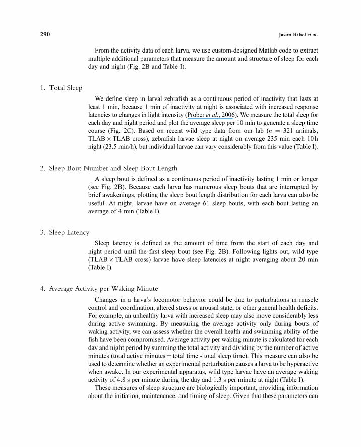
From the activity data of each larva, we use custom-designed Matlab code to extractmultiple additional parameters that measure the amount and structure of sleep for eachday and night (Fig. 2B and Table I).
1. Total Sleep
We define sleep in larval zebrafish as a continuous period of inactivity that lasts atleast 1 min, because 1 min of inactivity at night is associated with increased responselatencies to changes in light intensity (Prober et al., 2006). We measure the total sleep foreach day and night period and plot the average sleep per 10 min to generate a sleep timecourse (Fig. 2C). Based on recent wild type data from our lab (n = 321 animals,TLAB�TLAB cross), zebrafish larvae sleep at night on average 235 min each 10 hnight (23.5 min/h), but individual larvae can vary considerably from this value (Table I).
2. Sleep Bout Number and Sleep Bout Length
A sleep bout is defined as a continuous period of inactivity lasting 1 min or longer(see Fig. 2B). Because each larva has numerous sleep bouts that are interrupted bybrief awakenings, plotting the sleep bout length distribution for each larva can also beuseful. At night, larvae have on average 61 sleep bouts, with each bout lasting anaverage of 4 min (Table I).
3. Sleep Latency
Sleep latency is defined as the amount of time from the start of each day andnight period until the first sleep bout (see Fig. 2B). Following lights out, wild type(TLAB�TLAB cross) larvae have sleep latencies at night averaging about 20 min(Table I).
4. Average Activity per Waking Minute
Changes in a larva’s locomotor behavior could be due to perturbations in musclecontrol and coordination, altered stress or arousal state, or other general health deficits.For example, an unhealthy larva with increased sleep may also move considerably lessduring active swimming. By measuring the average activity only during bouts ofwaking activity, we can assess whether the overall health and swimming ability of thefish have been compromised. Average activity per waking minute is calculated for eachday and night period by summing the total activity and dividing by the number of activeminutes (total active minutes= total time - total sleep time). This measure can also beused to determine whether an experimental perturbation causes a larva to be hyperactivewhen awake. In our experimental apparatus, wild type larvae have an average wakingactivity of 4.8 s per minute during the day and 1.3 s per minute at night (Table I).These measures of sleep structure are biologically important, providing information
about the initiation, maintenance, and timing of sleep. Given that these parameters can
290 Jason Rihel et al.

be selectively perturbed by pharmacological agents (Rihel et al., 2010), they are likelycontrolled by at least partially independent regulatory mechanisms. These parameterscan be presented as a multi-dimensional “behavioral fingerprint” to facilitate compar-isons among multiple genotypes, experimental manipulations, or small molecules.Each measurement is normalized to matched controls and then combined to create avector that accounts for all of the parameters (see Fig. 2D; Rihel et al., 2010).Clustering algorithms and principal component analyses can be used to organizelarge datasets by phenotype and to uncover small molecules or genotypes that havesimilar effects across multiple zebrafish sleep/wake behavioral parameters.
IV. Conclusion
Only a decade old, the study of sleep in non-mammalian systems is still in its infancy.While early zebrafish sleep studies have focused on establishing the existence of beha-vioral sleep regulated by conserved mechanisms, the challenge ahead is to use thezebrafish sleep model to uncover heretofore unsuspected aspects of the neuronal andgenetic control of sleep/wake regulation. Recent studies that potentially link Hcrt signalingto pineal gland regulation (Appelbaum et al., 2009) and that uncover novel small moleculeregulators of sleep/wake states (Rihel et al., 2010) may represent two such discoveries.
A major advantage of the zebrafish system is the ability to efficiently perform geneticand pharmacological screens in a cost-, space-, and labor-effective manner. With this inmind, we have described an automated high-throughput method for observing long-termsleep/wake behavior in larval zebrafish. This methodology is highly flexible and caneasily be adapted to other behaviors, for example to observe larval responses totemperature, vibration, noxious chemicals, and changes in light intensity (Emran et al.,2007, 2008, 2010; Prober et al., 2008). In principle, the behavioral space of futurescreens could be expanded by testing these and other behavioral modalities in conjunc-tion with long-term sleep/wake behavioral monitoring, incorporating all the data into asingle multi-dimensional behavioral fingerprint. Such screens could not only uncovernovel mutants that affect specific behaviors but also identify correlated sets of behaviorsthat are regulated by similar underlying mechanisms. With new techniques to directlyobserve neural activity in behaving zebrafish, including neuroluminescence, whole-braincalcium imaging, and optogenetic techniques to manipulate the activity of neurons withlight, future studies will also begin to directly elucidate the activities and functions ofneural circuits that underlie sleep/wake behaviors.
References
Appelbaum, L., Wang, G. X., Maro, G. S., Mori, R., Tovin, A., Marin, W., Yokogawa, T., Kawakami, K.,Smith, S. J., Gothilf, Y., Mignot, E., and Mourrain, P. (2009). Sleep–wake regulation and hypocretin–melatonin interaction in zebrafish. Proc. Natl. Acad. Sci. U. S. A. 106, 21942–21947.
Azpeleta, C., Martinez-Alvarez, R. M., Delgado, M. J., Isorna, E., and De Pedro, N. (2010). Melatoninreduces locomotor activity and circulating cortisol in goldfish. Horm. Behav. 57, 323–329.
11. Monitoring Sleep and Arousal in Zebrafish 291

Borbely, A. A., and Tobler, I. (1996). Sleep regulation: Relation to photoperiod, sleep duration, wakingactivity, and torpor. Prog. Brain Res. 111, 343–348.
Branson, K., Robie, A. A., Bender, J., Perona, P., and Dickinson, M. H. (2009). High-throughput ethomics inlarge groups of Drosophila. Nat. Methods 6, 451–457.
Burgess, H. A., and Granato, M. (2007). Modulation of locomotor activity in larval zebrafish during lightadaptation. J. Exp. Biol. 210, 2526–2539.
Campbell, S. S., and Tobler, I. (1984). Animal sleep: A review of sleep duration across phylogeny. Neurosci.Biobehav. Rev. 8, 269–300.
Cermakian, N., Whitmore, D., Foulkes, N. S., and Sassone-Corsi, P. (2000). Asynchronous oscillations oftwo zebrafish CLOCK partners reveal differential clock control and function. Proc. Natl. Acad. Sci.U. S. A. 97, 4339–4344.
Chemelli, R. M., Willie, J. T., Sinton, C. M., Elmquist, J. K., Scammell, T., Lee, C., Richardson, J. A.,Williams, S. C., Xiong, Y., Kisanuki, Y., et al. (1999). Narcolepsy in orexin knockout mice: Moleculargenetics of sleep regulation. Cell 98, 437–451.
Cirelli, C. (2009). The genetic and molecular regulation of sleep: From fruit flies to humans. Nat. Rev.Neurosci. 10, 549–560.
Davis, H., Davis, P. A., Loomis, A. L., Harvey, E. N., and Hobart, G. (1937). Changes in human brainpotentials during the onset of sleep. Science 86, 448–450.
Dekens, M. P., and Whitmore, D. (2008). Autonomous onset of the circadian clock in the zebrafish embryo.EMBO J. 27, 2757–2765.
Emran, F., Rihel, J., Adolph, A. R., and Dowling, J. E. (2010). Zebrafish larvae lose vision at night. Proc.Natl. Acad. Sci. U. S. A. 107, 6034–6039.
Emran, F., Rihel, J., Adolph, A. R., Wong, K. Y., Kraves, S., and Dowling, J. E. (2007). OFF ganglion cellscannot drive the optokinetic reflex in zebrafish. Proc. Natl. Acad. Sci. U. S. A. 104, 19126–19131.
Emran, F., Rihel, J., and Dowling, J. E. (2008). A behavioral assay to measure responsiveness of zebrafish tochanges in light intensities. J. Vis. Exp 20, http://www.jove.com/index/details.stp?id=923, doi: 10.3791/923.
Faraco, J. H., Appelbaum, L., Marin, W., Gaus, S. E., Mourrain, P., and Mignot, E. (2006). Regulation ofhypocretin (orexin) expression in embryonic zebrafish. J. Biol. Chem. 281, 29753–29761.
Fontaine, E., Lentink, D., Kranenbarg, S.,Muller, U. K., van Leeuwen, J. L., Barr, A. H., andBurdick, J.W. (2008).Automated visual tracking for studying the ontogeny of zebrafish swimming. J. Exp. Biol. 211, 1305–1316.
Ganguly-Fitzgerald, I., Donlea, J., and Shaw, P. J. (2006). Waking experience affects sleep need inDrosophila. Science 313, 1775–1781.
Grover, D., Yang, J., Ford, D., Tavare, S., and Tower, J. (2009). Simultaneous tracking of movement andgene expression in multiple Drosophila melanogaster flies using GFP and DsRED fluorescent reportertransgenes. BMC Res. Notes 2, 58.
Hara, J., Beuckmann, C. T., Nambu, T., Willie, J. T., Chemelli, R. M., Sinton, C. M., Sugiyama, F., Yagami,K., Goto, K., Yanagisawa, M., and Sakurai, T. (2001). Genetic ablation of orexin neurons in mice results innarcolepsy, hypophagia, and obesity. Neuron 30, 345–354.
Hendricks, J. C., Finn, S. M., Panckeri, K. A., Chavkin, J., Williams, J. A., Sehgal, A., and Pack, A. I. (2000).Rest in Drosophila is a sleep-like state. Neuron 25, 129–138.
Hurd, M. W., and Cahill, G. M. (2002). Entraining signals initiate behavioral circadian rhythmicity in larvalzebrafish. J. Biol. Rhythms 17, 307–314.
Hurd, M. W., Debruyne, J., Straume, M., and Cahill, G. M. (1998). Circadian rhythms of locomotor activityin zebrafish. Physiol. Behav. 65, 465–472.
Iigo, M., and Tabata, M. (1996). Circadian rhythms of locomotor activity in the goldfish Carassius auratus.Physiol. Behav. 60, 775–781.
Kaneko, M., and Cahill, G. M. (2005). Light-dependent development of circadian gene expression intransgenic zebrafish. PLoS Biol. 3, e34.
Kaslin, J., Nystedt, J. M., Ostergard, M., Peitsaro, N., and Panula, P. (2004). The orexin/hypocretin system inzebrafish is connected to the aminergic and cholinergic systems. J. Neurosci. 24, 2678–2689.
Kato, S., Nakagawa, T., Ohkawa, M., Muramoto, K., Oyama, O., Watanabe, A., Nakashima, H., Nemoto, T.,and Sugitani, K. (2004). A computer image processing system for quantification of zebrafish behavior.J. Neurosci. Methods 134, 1–7.
292 Jason Rihel et al.
Kobayashi, Y., Ishikawa, T., Hirayama, J., Daiyasu, H., Kanai, S., Toh, H., Fukuda, I., Tsujimura, T., Terada,N., Kamei, Y., Yuba, S., Iwai, S., et al. (2000). Molecular analysis of zebrafish photolyase/cryptochromefamily: Two types of cryptochromes present in zebrafish. Genes Cells 5, 725–738.
Konopka, R. J., and Benzer, S. (1971). Clock mutants of Drosophila melanogaster. Proc. Natl. Acad. Sci.U. S. A. 68, 2112–2116.
Lee, M. G., Hassani, O. K., and Jones, B. E. (2005). Discharge of identified orexin/hypocretin neurons acrossthe sleep–waking cycle. J. Neurosci. 25, 6716–6720.
Lin, L., Faraco, J., Li, R., Kadotani, H., Rogers, W., Lin, X., Qiu, X., de Jong, P. J., Nishino, S., andMignot, E. (1999). The sleep disorder canine narcolepsy is caused by a mutation in the hypocretin (orexin)receptor 2 gene. Cell 98, 365–376.
Mileykovskiy, B. Y., Kiyashchenko, L. I., and Siegel, J. M. (2005). Behavioral correlates of activity inidentified hypocretin/orexin neurons. Neuron 46, 787–798.
Mochizuki, T., Crocker, A., McCormack, S., Yanagisawa, M., Sakurai, T., and Scammell, T. E. (2004).Behavioral state instability in orexin knock-out mice. J. Neurosci. 24, 6291–6300.
Nakamachi, T., Matsuda, K., Maruyama, K., Miura, T., Uchiyama, M., Funahashi, H., Sakurai, T., andShioda, S. (2006). Regulation by orexin of feeding behaviour and locomotor activity in the goldfish.J. Neuroendocrinol. 18, 290–297.
Naumann, E. A., Kampff, A. R., Prober, D. A., Schier, A. F., and Engert, F. (2010). Monitoring neuralactivity with bioluminescence during natural behavior. Nat. Neurosci. 13, 513–520.
Overeem, S., Mignot, E., van Dijk, J. G., and Lammers, G. J. (2001). Narcolepsy: Clinical features, newpathophysiologic insights, and future perspectives. J. Clin. Neurophysiol. 18, 78–105.
Pando, M. P., Pinchak, A. B., Cermakian, N., and Sassone-Corsi, P. (2001). A cell-based system thatrecapitulates the dynamic light-dependent regulation of the vertebrate clock. Proc. Natl. Acad. Sci.U. S. A. 98, 10178–10183.
Peyron, C., Faraco, J., Rogers, W., Ripley, B., Overeem, S., Charnay, Y., Nevsimalova, S., Aldrich, M.,Reynolds, D., Albin, R., Li, R., Hungs, M., et al. (2000). A mutation in a case of early onset narcolepsyand a generalized absence of hypocretin peptides in human narcoleptic brains. Nat. Med. 6, 991–997.
Peyron, C., Tighe, D. K., van den Pol, A. N., de Lecea, L., Heller, H. C., Sutcliffe, J. G., and Kilduff, T. S.(1998). Neurons containing hypocretin (orexin) project to multiple neuronal systems. J. Neurosci.18, 9996–10015.
Prober, D. A., Rihel, J., Onah, A. A., Sung, R. J., and Schier, A. F. (2006). Hypocretin/orexin overexpressioninduces an insomnia-like phenotype in zebrafish. J. Neurosci. 26, 13400–13410.
Prober, D. A., Zimmerman, S., Myers, B. R., McDermott, B. M. Jr., Kim, S. H., Caron, S., Rihel, J., Solnica-Krezel, L., Julius, D., Hudspeth, A. J., and Schier. A. F. (2008). Zebrafish TRPA1 channels are required forchemosensation but not for thermosensation or mechanosensory hair cell function. J. Neurosci.28, 10102–10110.
Raizen, D. M., Zimmerman, J. E., Maycock, M. H., Ta, U. D., You, Y. J., Sundaram, M. V., and Pack, A. I.(2008). Lethargus is a Caenorhabditis elegans sleep-like state. Nature 451, 569–572.
Renier, C., Faraco, J. H., Bourgin, P., Motley, T., Bonaventure, P., Rosa, F., and Mignot, E. (2007). Genomicand functional conservation of sedative-hypnotic targets in the zebrafish. Pharmacogenet. Genomics17, 237–253.
Rihel, J., Prober, D. A., Arvanites, A., Lam, K., Zimmerman, S., Jang, S., Haggarty, S. J., Kokel, D., Rubin,L. L., Peterson, R. T., and Schier., A. F. (2010). Zebrafish behavioral profiling links drugs to biologicaltargets and rest/wake regulation. Science 327, 348–351.
Ruuskanen, J. O., Peitsaro, N., Kaslin, J. V., Panula, P., and Scheinin, M. (2005). Expression and function of alpha-adrenoceptors in zebrafish: Drug effects, mRNA and receptor distributions. J. Neurochem. 94, 1559–1569.
Sakurai, T. (2007). The neural circuit of orexin (hypocretin): Maintaining sleep and wakefulness. Nat. Rev.Neurosci. 8, 171–181.
Shaw, P. J., Cirelli, C., Greenspan, R. J., and Tononi, G. (2000). Correlates of sleep and waking in Drosophilamelanogaster. Science 287, 1834–1837.
Straw, A. D., Branson, K., Neumann, T. R., and Dickinson, M. H. (2010). Multi-camera real-time three-dimensional tracking of multiple flying animals. J. R. Soc. Interface, doi:10.1098/rsif.2010.0230.
11. Monitoring Sleep and Arousal in Zebrafish 293

Thannickal, T. C., Moore, R. Y., Nienhuis, R., Ramanathan, L., Gulyani, S., Aldrich, M., Cornford, M., andSiegel, J. M. (2000). Reduced number of hypocretin neurons in human narcolepsy. Neuron 27, 469–474.
Van Buskirk, C., and Sternberg, P. W. (2007). Epidermal growth factor signaling induces behavioralquiescence in Caenorhabditis elegans. Nat. Neurosci. 10, 1300–1307.
Vera, L. M., De Pedro, N., Gomez-Milan, E., Delgado, M. J., Sanchez-Muros, M. J., Madrid, J. A., andSanchez-Vazquez, F. J. (2007). Feeding entrainment of locomotor activity rhythms, digestive enzymes andneuroendocrine factors in goldfish. Physiol. Behav. 90, 518–524.
Whitmore, D., Foulkes, N. S., Strahle, U., and Sassone-Corsi, P. (1998). Zebrafish Clock rhythmic expres-sion reveals independent peripheral circadian oscillators. Nat. Neurosci. 1, 701–707.
Willie, J. T., Chemelli, R. M., Sinton, C. M., Tokita, S., Williams, S. C., Kisanuki, Y. Y., Marcus, J. N.,Lee, C., Elmquist, J. K., Kohlmeier, K. A., Leonard, C. S., Richardson, J. A., et al. (2003). Distinctnarcolepsy syndromes in Orexin receptor-2 and Orexin null mice: Molecular genetic dissection of non-REM and REM sleep regulatory processes. Neuron 38, 715–730.
Yokogawa, T., Marin, W., Faraco, J., Pezeron, G., Appelbaum, L., Zhang, J., Rosa, F., Mourrain, P., andMignot, E. (2007). Characterization of sleep in zebrafish and insomnia in hypocretin receptor mutants.PLoS Biol. 5, e277.
Zhdanova, I. V. (2006). Sleep in zebrafish. Zebrafish 3, 215–226.Zhdanova, I. V., Wang, S. Y., Leclair, O. U., and Danilova, N. P. (2001). Melatonin promotes sleep-like statein zebrafish. Brain Res. 903, 263–268.
Zimmerman, J. E., Raizen, D. M., Maycock, M. H., Maislin, G., and Pack, A. I. (2008). A video method tostudy Drosophila sleep. Sleep 31, 1587–1598.
294 Jason Rihel et al.
Copyright © 2022 FDOKUMEN