Computerized craniofacial reconstruction using CT-derived implicit surface representations
Upload
independentCategory
view
1download
0
357Development 123, 357-367 Printed in Great Britain © The Company of Biologists Limited 1996DEV3324
Mutations affecting craniofacial development in zebrafish
Stephan C. F. Neuhauss, Lilianna Solnica-Krezel*, Alexander F. Schier, Fried Zwartkruis†, Derek L. Stemple,Jarema Malicki, Salim Abdelilah, Didier Y. R. Stainier‡ and Wolfgang Driever§
Cardiovascular Research Center, Massachusetts General Hospital and Harvard Medical School, 149 13th Street, Charlestown,MA 02129, USA
*Present address: Department of Molecular Biology, Vanderbilt University, Box 1820, Station B, Nashville, TN 37235†Present address: Laboratory for Physiological Chemistry, Utrecht University, Universiteitsweg 100, 3854 CG Utrecht, The Netherlands‡Present address: School of Medicine, Department of Biochemistry and Biophysics, UCSF, San Francisco, CA 94143-0554, USA§Author for correspondence (e-mail: [email protected])
In a large-scale screen for mutations affecting embryogen-esis in zebrafish, we identified 48 mutations in 34 geneticloci specifically affecting craniofacial development.Mutants were analyzed for abnormalities in the cartilagi-nous head skeleton. Further, the expression of markergenes was studied to investigate potential abnormalities inmutant rhombencephalon, neural crest, and pharyngealendoderm. The results suggest that the identified mutationsaffect three distinct aspects of craniofacial development. Inone group, mutations affect the overall pattern of the cran-iofacial skeleton, suggesting that the genes are involved in
SUMMARY
the specification of these elements. Another large group ofmutations affects differentiation and morphogenesis ofcartilage, and may provide insight into the genetic controlof chondrogenesis. The last group of mutations leads to theabnormal arrangement of skeletal elements and mayuncover important tissue-tissue interactions underlyingjaw development.
Key words: Danio rerio, craniofacial mutants, cartilage, pharyngealarches
INTRODUCTION
The head skeleton in vertebrates mainly comprises derivativesof the cranial neural crest, a transient migratory population ofcells unique to vertebrates (Noden, 1983, 1975; Le Lievre,1978; Couly et al., 1993). During closure of the neural tube,the cranial neural crest cells migrate from the dorsal aspect ofthe neural tube into the frontonasal process and the pharyngealarches. Cranial neural crest derivatives include a large part ofthe head skeleton, dermis of the face, most of the skull, someneurons and glia of several cranial ganglia, and Schwann cellsof the cranial nerves (Le Douarin, 1982). Cranial neural crestcells appear to migrate according to their rhombomeric origin(Lumsden et al., 1991). For example, cells from rhombomere2 migrate to the first pharyngeal (mandibular) arch, whereascells from rhombomere 4 migrate into the second pharyngeal(hyoid) arch. Transplantation experiments indicate, that cranialcrest cells may be determined before the onset of migration(Noden, 1983).
After the pharyngeal arches become populated with neuralcrest-derived cells, chondrogenesis can be first observed asmesenchymal, neural crest-derived cells undergo condensa-tion. The pharyngeal endoderm has been identified asnecessary for the induction of cartilage formation (Gravesonand Armstrong, 1987; Seufert and Hall, 1990). Most of thecartilage formed by neural crest-derived cells is subsequentlyreplaced by osteoblasts during bone formation.
While more recent studies of craniofacial development have
focused on chick and mouse embryos, similar conclusionsregarding craniofacial development have been obtained inlower vertebrates, such as teleosts (Langille and Hall, 1987,1988; Schilling, 1993; Miyake and Hall, 1994). In thezebrafish, Danio rerio, anterior cranial neural crest cells startto emigrate around intermediate segmentation stages, followedshortly by cells of progressively more caudal levels. Thosecells migrate ventrally, subsequently forming a reiterated seriesof seven pharyngeal arch primordia separated by endodermalepithelial pouches. Labeling studies suggest that arch precur-sors are fated near the onset of migration and produce clonesconfined to single arches and probably single cell types. Fur-thermore, the fate of these cells can be predicted by theirposition within the precursor populations. The chondrogenicneural crest cells are the most medial and the last to emigrate.By 72 hours post fertilization (hpf), many of the neural crest-derived mesenchymal cells have differentiated into long rowsof cartilage within each arch making up the pharyngealskeleton (Schilling, 1993; Schilling and Kimmel, 1994).
Most of our understanding of craniofacial developmentstems from embryological experiments. Mutational analyses inmice have provided new insights. Skeletal development can bedisrupted by mutations at several steps in the pathway. Defectsin the patterning of the head skeleton have been described formice carrying mutations in several of the Hox genes (Chisakaand Capecchi, 1991; Gendron-Maguire et al., 1993; Rijli et al.,1993). Several mutations, such as short ear and brachypodism,both of which encode growth factors of the TGFβ superfam-

358 S. C. F. Neuhauss and others
ily affect the formation of the mesenchymal condensations(Kingsley et al., 1992; Storm et al., 1994).
Recently, conditions for efficient recovery of mutations inzebrafish have been described (Mullins et al., 1994; Solnica-Krezel et al., 1994). We have used a genetic approach to studycraniofacial development in zebrafish. In a genome-wide largescale screen for mutations with abnormal embryogenesis(Driever et al., 1996), we have identified mutations in 34genetic loci affecting specific aspects of craniofacial develop-ment. We analyzed the pharyngeal skeleton of the mutants ata morphological level using a stain for proteoglycans. Forseveral mutations we also investigated the organization of therhombencephalon and the pharyngeal arch primordia for alter-ation of the expression pattern of marker genes.
Our results suggest that the identified mutations can be cat-egorized into three groups. (1) Mutations affecting the layoutof the pharyngeal skeleton. These mutations are characterizedby absence or displacement of individual elements of the pha-ryngeal skeleton. (2) Mutations affecting the differentiationand morphogenesis of cartilaginous skeletal elements and (3)mutations affecting the spatial arrangement of normallyformed pharyngeal skeletal elements.
MATERIALS AND METHODS
Mutations were induced by the alkylating mutagen N-ethyl-N-nitro-surea (ENU) and recovered in an F2 screen as previously described(Solnica-Krezel et al., 1994; Driever et al., 1996) All embryos weremaintained at 28.5°C and staged according to development in hours(hpf) or days (dpf) postfertilization.
Phenotypic analysisLarvae were anesthetized in 0.02% buffered 3-aminobenzoic acidmethyl ester (Sigma) and observed under a dissecting microscope.Embryos were embedded in 2% methylcellulose and photographedeither under a dissecting scope or using differential interferencecontrast (DIC) optics on a Zeiss Axiophot microscope (Westerfield,1994).
Cartilage was stained with Alcian blue, using a modified protocolof Kelly and Bryden (1983). Larvae at 5 dpf or older were fixedovernight in 4% phosphate-buffered paraformaldehyde and washedseveral times in phosphate-buffered saline with 0.1% Tween-20(PBT). In order to enhance their optical clarity, the specimens werebleached in 30% hydrogen peroxide for 2 hours or until the eyes weresufficiently translucent. The embryos were rinsed in PBT and trans-ferred into a filtered, Alcian blue solution (1% concentratedhydrochloric acid, 70% ethanol, 0.1% Alcian blue) and stainedovernight. Specimens were cleared in acidic ethanol (5% concentratedhydrochloric acid, 70% ethanol) for 4 hours, dehydrated in an ethanolseries and stored in glycerol. Specimens were analyzed and docu-mented in glycerol under epillumination on a dissecting microscope.
Whole-mount in situ hybridization was performed with digoxi-genin-labeled RNA probes essentially as described by Oxtoby andJowett (1993). Embryos were scored in PBT and documented afterclearing in benzylbenzoate/benzyl alcohol (2:1). The following probeswere used: krx-20 (Oxtoby and Jowett, 1993), pax 2 (Krauss et al.,1991), hlx-1 (Fjose et al., 1994), dlx-2, dlx-3 and dlx-4 (Ekker et al.,1992; Akimenko et al., 1994).
Immunocytochemistry with the monoclonal antibody zn-5(Trevarrow et al., 1990) was performed on early pharyngula embryos(24 -28 hpf) after fixation in 4% paraformaldehyde (4 hours at roomtemperature). Subsequent steps were performed essentially as previ-ously described (Solnica-Krezel and Driever, 1994). Hybridomasupernatant was used at a 1:5 dilution.
For metacrylate sections, larvae were fixed overnight in 4%paraformaldehyde (4°C, overnight) and dehydrated in ethanol, thenembedded in JB-4 resin, according to the manufacturer’s instructions(Polyscience Inc.). Sections (5 µm) were cut on a Leica 2065microtome.
Genetic analysisGenetic segregation of individual mutations was determined byassessing on average 1000 embryos from crosses of heterozygousparents for any given mutation. In order to test potential allelism ofisolated mutations, complementation testing among members of thephenotypically defined groups of mutations was performed. Comple-mentation between two mutations was tested by crossing identifiedheterozygous parents of each mutation and screening their offspringfor the mutant phenotype. A minimum of 30 embryos per comple-mentation cross was analyzed. If all embryos developed as wild-type,mutations were considered to be in distinct loci. If about 25% of theembryos develop the mutant phenotype, they were considered to beallelic. All mutations segregate as Mendelian recessive traits. A totalof 278 successful complementation crosses were performed with14,886 embryos scored.
Heterozygous parents of mutant embryos were outcrossed and theheterozygotes were reidentified in sibling incrosses.
RESULTS
The pharyngeal skeleton of 5-day old zebrafishlarvaeThe pharyngeal skeleton of the zebrafish consists of 7 pharyn-geal (visceral) arches with the anterior two forming jaw andthe posterior five forming gill structures.
After 3 dpf the cartilage of the pharyngeal skeleton of all 5gill arches can be stained with Alcian blue. In a 5 dpf larva,individual components of the pharyngeal skeleton can bereliably identified (Fig. 1). By analogy to other vertebrates, thepharyngeal arch from which individual components derive canbe assigned (De Beer, 1937; Langille and Hall, 1987; Miyakeand Hall, 1994). The first pharyngeal (mandibular) arch deriv-atives are: Meckel’s cartilage and the quadrate with thepterygoid process. Second pharyngeal (hyoid) arch derivativesinclude the ceratohyal, the hyosymplectic, the interhyal and thebasihyal. The third to seventh pharyngeal arch (first to fifth gillarch derivatives) include the basibranchial, the cerato-branchials and hypobranchials. Also visible are the trabeculaeof the chondrocranium, which are fused to form the ethmoidplate. The zebrafish pharyngeal skeleton develops in a waysimilar to that of other teleosts, e.g. Salmo salar (De Beer,1937) and Oryzias latipes (Langille and Hall, 1987).
Identification of mutations affecting craniofacialdevelopmentMutations affecting craniofacial development were identifiedin an F2 screen as described by Driever et al. (1996). Zebrafishlarvae were screened by visual inspection under the dissectingmicroscope. Abnormalities in the jaw and gill structures canbe detected best at 5 dpf. Two major categories of abnormalcraniofacial phenotypes were formed.
The first category comprise mutations that lead to reducedcraniofacial differentiation accompanied by overall delay ofdevelopment. This syndrome manifests itself by a generaldelay in growth made apparent by small eye size, reduced bodylength and smaller pectoral fins and was observed in 24%

359Craniofacial mutants in zebrafish
Fig. 1. Pharyngeal skeleton of a day-5 zebrafish larva stained for cartilagewith Alcian blue. (A) Ventral view.Individual cartilaginous elements ofthe pharyngeal skeleton areidentifiable. The two trabeculae fusemedially to form the ethmoid plate(eth). First arch derivatives areMeckel’s cartilage (mk) and quadrate(qu). Second arch derivatives arebasihyal (bh), ceratohyal (ch),hyosymplectic (hys). Gill arch
derivatives are basibranchials (bb) and ceratobranchials I-V (cb i-v). Also labeled are cartilages of the pectoral fins (pec fin). (B) Lateral viewreveals the pterygoid process of the quadrate (pty) and the auditory capsule (aud). Scale bar is 100 µm.
Fig. 2. Mutations affecting the layout of the pharyngeal skeleton. (A-H) Lateral view of a living day-5 larvae. (I-P) Ventral view of Alcian bluestained larvae. (A,I) Wild-type embryos. (B,J) little richardm433. (C,K) mother superiorm188. (D,L) thinnerm214. (E,M) quadrom271.(F,N) nocyranom579. (G,O) bielakm429. (H,P) mont blancm610. For details, see text. wt, wild-type; bh, basihyal; cb, ceratobranchial;ch, ceratohyal; and eth, ethmoid plate. Scale bar, 100 µm.
(587/2383) of all embryonic and early larval lethal mutationsfound in the screen. These pleiotropic mutations appear tocause a non-specific general delay in development, possiblydue to some metabolic defect, and may not be involved specif-ically in craniofacial development. We excluded thesemutations from further analysis. A sample of 16 independent
mutations leading to such phenotypes were subjected toskeletal analysis. All but one had a craniofacial phenotype con-sistent with a general delay in development. The entire pha-ryngeal skeleton was found to be properly formed and differ-entiated but was reduced in size. In only one isolate did theceratohyal not reach the midline (Fig. 7F).

360 S. C. F. Neuhauss and others
The second category includes mutations that affect specificaspects of craniofacial development. We recovered 48 inde-pendent mutations and each was subjected to further pheno-typic and genetic characterization.
The nature of the defects observed allowed us to classifythese mutants into three groups: (1) mutations affecting thelayout of the pharyngeal skeleton; (2) mutations affecting thedifferentiation and morphogenesis of cartilaginous elements ofthe pharyngeal skeleton; and (3) mutations affecting the spatialarrangement of normally formed skeletal elements. Severalother mutations presented in this manuscript do not fit intothese categories, but appear also to affect specific aspects ofcraniofacial development.
Complementation analysis was performed within eachgroup. The 48 isolated mutations fall into 34 complementationgroups. Table 1 presents a brief summary, including pheno-typic aspects not related to craniofacial development.
Group I: Mutations affecting the layout of thepharyngeal skeletonAlcian blue staining produced evidence of the absence or dis-placement of individual elements of the pharyngeal skeletonfor 9 mutations in 7 loci. All of the mutations are early larvallethal, meaning that they hatch as free swimming larvae but donot inflate their swim bladder and are unable to feed.
Mutant little richard (lit) alleles (m181 and m433) cause asevere reduction of the jaw and gill apparatus (Fig. 2B,J).Meckel’s cartilage and quadrate are reduced both in size androstral extent. The ceratohyal is bent caudally and is smaller.The basihyal and hyosymplectic cartilage are present but aresmaller and positioned more caudally. In the majority ofhomozygous mutant embryos only the first two and the fifthceratobranchials are detectable. In a few cases the third cera-tobranchial is visible. The two alleles produce similar jaw phe-notypes but differ in other aspects. In litm433 mutants at 2 dpfthe auditory vesicle is much smaller and does not form semi-circular canals. Most homozygous mutant embryos in thisgroup have cardiac edema and reduced circulation due to mal-formations of the atrioventricular valve. In contrast, homozy-gous litm181 mutants have normal circulation and only a slightlysmaller auditory capsule. The pharyngeal arches are populatedslightly later by dlx-positive cells (see below).
The mother superior (mosm188) mutation leads to a severereduction of the pharyngeal skeleton sometimes accompaniedby a ventral displacement of Meckel’s cartilage (Fig 2C,K).Skeletal analysis reveals the absence of the basihyal and nodetectable ceratobranchials with the occasional exception ofceratobranchial V. The hyosymplectic and the ceratohyal aremissing or reduced in the majority of embryos. In about halfof the mutant embryos the trabeculae are not fused, so that theethmoid plate is not formed.
Most thinner (thim214) mutant embryos develop only the firstand the fifth ceratobranchial, while some have none or up tothree visible ceratobranchials (Fig. 2D,L). All lower jaw struc-tures are reduced in their rostral extent, whereas the ethmoidplate is only slightly smaller.
Skeletal analysis of quadro (quom271) mutant embryosreveals that the rostral extent of the basihyal and the gill archesis reduced (Fig. 2E,M). The ceratohyal is shorter and deflectedcaudally. In a few embryos one of the ceratohyals extends overthe midline. The hyosymplectic is much smaller and
sometimes not detectable. The ceratobranchials are reduced innumber with the majority of embryos developing only three.The entire gill arch complex is shifted caudally and the cera-tobranchials are bent caudally rather than dorsally. Malforma-tions of the auditory capsule are not necessarily bilaterallysymmetric and range from very small empty auditory capsulesto duplications of well-formed ears.
For nocyrano (nocm579) the majority of mutant embryoshave trabeculae that are not fused to form the ethmoid plate,and are not connected to the basal plate (Fig. 2F,N). In someembryos the trabeculae are only weakly stained or not visibleat all. The lower jaw is slightly delayed in its formation.Meckel’s cartilage is dorsally bent in relation to the quadrate,reminiscent of the haim114 and rochenm193 phenotype (Fig.6C,D).
In homozygous bielak (bilm429) embryos, the ethmoid plate,as well as Meckel’s cartilage, quadrate, ceratohyal andhyosymplectic are smaller (Fig. 2G,O). The ceratohyal pointscaudally. While the first and the fifth ceratobranchials arevisible in all mutant embryos, the second ceratobranchial israrely present and the other ones are not detectable.
In mutant embryos of two indistinguishable mont blanc(mob) alleles (m610 and m780), all skeletal elements of the headare smaller (Fig. 2H,P). Meckel’s cartilage, quadrate andbasihyal are reduced both in size and rostral extent. In abouthalf of the mutant embryos the basihyal is laterally displacedfrom its medial position. Ceratohyal and hyosymplectic are verysmall and do not extend toward the midline or are notdetectable. The trabeculae are thinner, reduced in their rostralextent and do not fuse. The ceratobranchials are not visible withthe rare exception of very small first and fifth ceratobranchials.
To determine whether the observed phenotypes correlatewith patterning defects in the rhombencephalon, the expressionof krx-20, pax b and hlx-1 was analyzed in mutant embryos foreach locus of this group. The expression patterns of krx-20(Oxtoby and Jowett, 1993) and pax b (Krauss et al., 1991) wereanalyzed at early segmentation stages (12-15 hpf), to assess theanterior-posterior patterning of the rhombencephalon. Thesegenes are expressed in rhombomere 3 and 5, and in themidbrain, respectively. At early pharyngula stages (24-28 hpf)hlx-1 (Fjose et al., 1994) is expressed in paired stripes in allrhombomeres. The expression pattern of these three genesindicates no abnormalities in the rhombencephalon organiz-ation for any of the mutants in this group. Antibody stainingusing zn-5 (Trevarrow et al., 1990), a monoclonal antibodywhich labels the endodermal pouches of the pharyngeal arches,revealed no structural abnormalities in mutant embryos (datanot shown).
To detect abnormalities in the formation of the pharyngealarches we analyzed the expression of dlx-2, dlx-3 and dlx-4(Ekker et al., 1992; Akimenko et al., 1994) at early pharyngulastages (24-28 hpf). These markers are expressed in a combi-natorial fashion in the pharyngeal arch primordia (Akimenkoet al., 1994). For mutants affected in litm433 and mosm188 theexpression of dlx genes in the pharyngeal arches is slightlydelayed. whereas no difference between mutant and wild-typeembryos could be detected for the other loci.
Group II: Mutations affecting cartilage differentiationand morphogenesisCartilage differentiation and morphogenesis appear to be

361Craniofacial mutants in zebrafish
Table 1. Summary of the phenotypesGenetic loci Alleles Craniofacial defect Other phenotypes Refs
Group Ilittle richard (lit) m181, m433 Reduced number of CB Reduction of otic capsule, reduced circulation c
(in m433)mother superior (mos) m188 Absence or reduction of CH, absence of BH Smaller eyes, slightly reduced pigmentation,
and fewer or none CB supernumerary , neuromast organs in trunkthinner (thi) m214 Reduced number of CB Smaller eyesquadro (quo) m271 Reduced and deflected CH, HYS, reduced Variable ear malformations (reductions and c
number of CB duplications)bielak (bil) m429 Reduction of mandibular and hyoid structures, Lack of xanthophores, fewer and smaller
absence or fewer CB iridiophores, smaller eyesmont blanc (mob) m610, m780 CH, HYS reduced or not detectable, fewer CB Slightly reduced pigmentation and eye size
or not detectablenocyrano (noc) m579 ETH not formed, trabeculae not fused with
basal plate or absentGroup II
bulldog (bul) m137, m421, m494, Cartilage growth and differentiation, Kinked and shorter pectoral fin, fin degeneration m606, m757 semicircular canals of inner ear fail to form
jekyll (jek) m151, m310 Cartilage differentiation affected, no staining Kinked and shorter pectoral fin, atrio-ventricular dwith Alcian blue, semicircular canals of valve formationinner ear fail to form
mr hyde (hyd) m205 Cartilage growth and differentiation, Kinked and shorter pectoral fin, fin degenerationsemicircular canals of inner ear fail to form
round (rnd) m211, m641, m713,m715 Cartilage growth and differentiation, Kinked and shorter pectoral fin, fin degeneration
semicircular canals of inner ear fail to formcrusher (cru) m299 Cartilage growth and differentiation, Shorter pectoral fin
semicircular canals of inner ear fail to formzhivago (zhi) m315 Cartilage growth and differentiation, Shorter pectoral fin
semicircular canals of inner ear fail to formstumpf (stp) m365 Cartilage growth and differentiation Shorter pectoral finstrangelove (stn) m617 Cartilage growth and differentiation, Shorter pectoral fin
semicircular canals of inner ear fail to formfeelgood (fel) m662 Cartilage growth and differentiation, Shorter pectoral fin
semicircular canals of inner ear fail to formapparatchik (apa) m364 Pharyngeal skeleton reduced, CH and QU Pectoral fins slightly shorter
thicker and shorterrieux (rix) m526 Pharyngeal skeleton reduced, CH and QU Pectoral fins slightly shorter
thicker and shorterkimble (kim) m533 Pharyngeal skeleton reduced, CH and QU Pectoral fins slightly shorter
thicker and shorter, semicircular canals ofinner ear fail to form
postdoc (pos) m485 Pharyngeal skeleton reduced, CH bend caudally Pectoral fins kinked and very reduced in size
Group IIIlong jaw (ljw) m82 Ventral displacement of MKspitzmaul (sil) m636 Ventral displacement of MKdegenerator (deg) m412 Ventral displacement of MK Tectal degenerationthe boss (tbo) m182 Ventral displacement of MKdoolittle (doo) m239 Ventral displacement of MK, CH collapsedrochen (roh) m193 Dorsal displacement of MK Head appears flat, smaller eyeshai (hai) m114 Dorsal displacement of MK Head appears flat, smaller eyesflycatcher (flc) m370 BH and BB displaced ventrally Smaller eyespelican (pel) m202, m207 BH and BB displaced ventrallygrossmaul (gro) m643, m752 BH and BB displaced ventrally
Othersbrak (brk) m452 Bending of individual skeletal elements Severe reduction of melanocytes and lack of eye
pigmentationmaggot (mgt) m350, m503, m635 Cartilage growth and differentiation Notochord bcaptain hook (cpt) m169 ETH absent, compressed lower jaw Gastrulation awhite bread (wib) m384 Reduced lower jaw, no stained CB Lack of xantho- and iridiophores, smaller eyes
BB, basibranchial; BH, basihyal; CB; ceratobranchial(s); CH; ceratohyal; ETH; ethmoid plate; MK, Meckel’s cartilage and QU, quadrate. See also Fig. 1.Other phenotypic aspects of these mutants are described by: a, Solnica-Krezel et al., 1996; b, Stemple et al., 1996; c, Malicki et al., 1996; d, Stainier et al.,1996.
defective for all elements of the craniofacial skeleton for 21mutations in 13 loci (Table 1). These mutations were initiallyrecovered in the visual screen by virtue of a common set ofphenotypes characterized by a reduction in body length and a
shortened head with craniofacial elements not projectingbeyond the eyes (Fig. 3A,C,D).
All mutations in this group cause reduction in the size of thepharyngeal skeleton and most fail to form proper projections

362 S. C. F. Neuhauss and others
in the inner ear after initial outpocketing. Therefore the semi-circular canals of the inner ear are not formed. The intensityof Alcian blue staining in the mutants is generally lower thanin their wild-type siblings. The skeletal elements, pectoral finsand the auditory capsule are similarly affected.
Since the pharyngeal endoderm is implicated in tissue inter-actions necessary for chondrogenesis (Graveson andArmstrong, 1987; Seufert and Hall, 1990), we have analyzedthe endodermal pouches of the pharyngeal endoderm in thesemutants. Staining with the monoclonal antibody zn-5 revealedno abnormalities in the formation of the endodermal pouchesin any of the mutants in this group (data no shown).
All the alleles of bulldog (bul) – m137,m421, m494, m606, m757 – and round(rnd) – m211, m641, m731, m715 – aswell as mr hyde(hydm205) andcrusher(crum299) result in the outline ofthe cartilage rods being poorly defined at5 dpf (Fig. 3E-H). The auditory capsuleis reduced in size and the semi-circularcanals do not form (Fig. 4). The chon-drocytes do not reach their full size andthe amount and density of the extracel-lular matrix seems to be reduced asjudged from sectioned material. We havealso analyzed older embryos to detect apotential delay in differentiation. Thephenotype remains the same at 8 dpf,which argues against a general delay.The cartilage cells in the pectoral fins aresimilarly affected.
Fig. 3. Mutations affecting chondrogenesis.(A) Mutants are shorter and have reducedjaw structures. Wild-type (bottom) andmutant mr hyde205 larva (top) at day 5 ofdevelopment. The mutant is shorter and hasa reduced jaw. Wild-type embryos have aninflated swim bladder. (C) Dorsal view ofwild-type day-5 larva. (D) Dorsal view of aday-5 mr hyde205 larva. Notice the rostralposition of the eyes and the smaller and bentpectoral fins. (B,E-Q). Ventral views ofAlcian blue stained larvae at day 5 ofdevelopment. (B) Wild-type larva.(E-M) Mutant larvae defective in cartilageformation. Skeletal elements appear smallerand are weakly stained. (E) bulldog494, theceratobranchials are present but smaller;(F) mr hydem205; (G) roundm713; (H)crusherm299; (I) jekyllm151; (J) zhivagom315;(K) stumpfm365; (L) strangelovem617 and (M)feelgoodm662. N-Q depict mutant larvae thatdisplay stunted growth of skeletal elements.(N) apparatchik m364; (O) rieuxm526 (noticethe thicker and shorter ceratohyal) and (P)kimblem533. (Q) In postdocm485 thepharyngeal skeleton is reduced and theceratohyal is bent ventrally. The pectoral finis reduced. wt, wild-type; ch, ceratohyal; cb,ceratobranchials; mk, Meckel’s cartilage;pec fin; pectoral fin and qu, quadrate. Scalebars, 100 µm.
Embryos mutant at the jekyll locus (m151, m310) are char-acterized by very weak or absent staining with Alcian blue(Fig. 3I). Use of DIC optics and histological sections revealthat the cartilage in the head is present but smaller and doesnot stain with Alcian blue (Fig 5). The pharyngeal skeleton isreduced to an extent similar to that of bul, rnd, hyd and crumutants.
Mutants in zhivagom315, stumpfm365, strangelovem617 andfeelgoodm662 are less severely affected as judged by Alcianblue stain intensity (Fig. 3J-M). Pectoral fins are only slightlyreduced in size and not kinked. The phenotype remains thesame from 5 to 10 dpf for strangelovem617 and feelgoodm662

363Craniofacial mutants in zebrafish
Fig. 4. Lateral view of the ear at day6 of development. (A) In the wild-type the semicircular canals can beseen. (B) Ear of a jekyllm151 larva.The epithelial projections are formedbut do not elongate and fail to formthe semicircular canals. Arrowpoints to the cranial projection.Scale bar 50 µm.
larvae. With the exception of stumpfm365 embryos the semicir-cular canals fail to form.
In apparatchikm364, rieuxm526 and kimblem533 cartilageelements are smaller but stain normally (Fig. 3N-P). Thequadrate and ceratohyal appear to be thicker and slightlymisshapen. In kimblem533 the semicircular canals of the innerear do not form.
In postdocm485 the cartilage is quite normal, but the individ-ual cartilage bars are reduced in size (Fig. 3Q). The ceratohyalis bent caudally and the ceratobranchials are smaller. In somemutant embryos the ceratobranchials are fused into a looselypacked mass of cartilaginous cells. As for the other members
Fig. 6. Mutations affecting the spatial relationship between elements of and their corresponding Alcian blue staining (B,D,F,H). (A,B) Wild typedetails see text. Scale bar, 100 µm.
of this group, the pectoral fins are very reduced in size and theembryos are shorter.
Group III: Mutations affecting the spatialarrangement of pharyngeal skeletal elementsFor 12 mutations defining 10 genetic loci cartilaginouselements form normally but are perturbed in their spatialarrangement (Table 1). A ventral view of a mutant headskeleton reveals no difference from the pattern of wild-typesiblings. Only observation from a lateral perspective allowsidentification of the mutant phenotypes (Fig. 6). Some mutantscause a ventral bend of the cartilage rod formed by the basihyal
Fig. 5. Sagittal sections of the gill archregion of day-5 larvae. Sections werestained with methylene blue. (A) Wild-type. (B) jekyllm151. Notice that thewhole gill region is more compressedand that there is a lack of purplestaining surrounding the cartilage cells.h, hyoid arch; g1-5, gill arches. Scalebar, 50 µm.
the pharyngeal skeleton. Lateral view of live day-5 larvae (A,C,E,G) (wt). (C,D) haim114. (E,F) grossmaul m643. (G,H) spitzmaulm636. For
364 S. C. F. Neuhauss and others
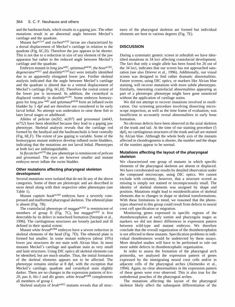
and the basibranchials, which results in a gaping jaw. The othermutations result in an abnormal angle between Meckel’scartilage and the quadrate.
Mutant haim114 and rochenm193 larvae are characterized bya dorsal displacement of Meckel’s cartilage in relation to thequadrate (Fig. 6C,D). Therefore the jaw appears to be shorter.This is not due to a reduction in size of any element of the jawapparatus but rather to the reduced angle between Meckel’scartilage and the quadrate.
Embryos mutant in long jawm82, spitzmaulm636, the bossm182,degeneratorm412 and doolittlem239 loci were initially identifieddue to an apparently elongated lower jaw. Further skeletalanalysis indicated that the angle between Meckel’s cartilageand the quadrate is altered due to a ventral displacement ofMeckel’s cartilage (Fig. 6G,H). Therefore the rostral extent ofthe lower jaw is increased. In addition, the ceratohyal isdisplaced ventrally in doolittlem239. Some embryos homozy-gous for long jaw m82 and spitzmaulm636 form an inflated swimbladder by 5 dpf and are therefore not considered to be earlylarval lethal. No attempt was undertaken to raise these fish tolater larval stages or adulthood.
Alleles of pelican (m202, m207) and grossmaul (m643,m752) have been identified because they lead to a gaping jawphenotype. Skeletal analysis reveals that the cartilage rodformed by the basihyal and the basibranchials is bent ventrally(Fig, 6E,F). The extent of jaw gaping is variable. Some of thehomozygous mutant embryos develop inflated swim bladders,indicating that the mutations are not larval lethal. Phenotypesat both loci are indistinguishable.
In flycatcherm370 the jaw phenotype is reminiscent of pelicanand grossmaul. The eyes are however smaller and mutantembryos never inflate the swim bladder.
Other mutations affecting pharyngeal skeletondevelopmentSeveral mutations were isolated that do not fit any of the abovecategories. Some of them are pleiotropic and are described inmore detail along with their respective other phenotypes (seeTable 1).
Mutant captain hookm169 embryos have a severely com-pressed and malformed pharyngeal skeleton. The ethmoid plateis absent (Fig. 7B).
The craniofacial phenotype of maggotm350 is reminiscent ofmembers of group II (Fig. 7C), but maggotm350 is firstdetectable by its defect in notochord formation (Stemple et al.,1996). The cartilaginous structures are loosely packed and illdefined in their spatial extent.
Mutant white breadm384 embryos have a severe reduction inskeletal elements of the head (Fig. 7D). The ethmoid plate isformed but smaller. In some mutant embryos (about 10%)lower jaw structures do not stain with Alcian blue. In mostmutants Meckel’s cartilage and quadrate stain as very smalland faint structures. Using DIC optics all skeletal elements canbe identified, but are much smaller. Thus, the initial formationof the skeletal elements appears not to be affected. Thephenotype remains similar at 8 dpf, with the exception thatMeckel’s cartilage, quadrate and ceratohyal stain slightlydarker. There are no changes in the expression patterns of krx-20, pax b, hlx-1 and dlx genes. white breadm384 complementsall members of group I.
Skeletal analysis of brakm452 mutants reveals that all struc-
tures of the pharyngeal skeleton are formed but individualelements are bent to various degrees (Fig. 7E).
DISCUSSION
During a systematic genetic screen in zebrafish we have iden-tified mutations in 34 loci affecting craniofacial development.The fact that only a single allele has been found for 26 out ofthe 34 loci, indicates that our screen has not approached satu-ration (see also Driever et al., 1996). Additionally, our visualscreen was designed to find rather dramatic abnormalities.Future screens, using DIC optics, or markers like Alcian bluestaining, will recover mutations with more subtle phenotypes.Similarly, interesting craniofacial abnormalities appearing aspart of a pleiotropic phenotype might have gone unnoticedwithout the application of cartilage stains.
We did not attempt to recover mutations involved in ossifi-cation. Our screening procedure involving dissecting micro-scope inspection, as well as the time frame of evaluation, wasinsufficient to accurately reveal abnormalities in early boneformation.
No obvious defects have been observed in the axial skeletonof the trunk and tail. At the time of our staining procedure (5dpf), no cartilaginous structures of the trunk and tail are stainedby Alcian blue. Although the whole body axis of the mutantsaffected in chondrogenesis is shorter, the number and the shapeof the somites appear to be normal.
Mutations affecting the layout of the pharyngealskeletonWe characterized one group of mutants in which specificelements of the pharyngeal skeleton are absent or displaced.We have corroborated our results by detailed observation underthe compound microscope, using DIC optics. We cannotexclude with certainty, however, that a structure scored asmissing is simply not stained or inconspicuously small. Theidentity of skeletal elements was assigned by shape andposition. Mutations might lead to misidentification of skeletalelements due to changes in shape or skeletal transformations.With these limitations in mind, we reasoned that the pheno-types observed in this group could result from defects in neuralcrest cell specification or migration.
Monitoring genes expressed in specific regions of therhombencephalon at early somite and pharyngula stages asmarkers we did not detect differences in expression patternbetween wild-type and mutant embryos. We thereforeconclude that the overall organization of the rhombencephalonis not affected in these mutants. Specification problems in indi-vidual rhombomeres would be undetected by these assays.More detailed studies will have to be performed to rule outmore subtle defects in rhombencephalic organization.
In order to assess the formation of the pharyngeal archprimordia, we analyzed the expression pattern of genesexpressed by the immigrating neural crest cells and/or inadjacent cells of the pharyngeal arches (Akimenko et al.,1994). Again, no clear abnormalities in the expression patternof these genes were ever observed. This is also true for theendodermal pouches of the pharyngeal arches.
The mutations affecting the layout of the pharyngealskeleton likely affect the subsequent differentiation of the

365Craniofacial mutants in zebrafish
Fig. 7. Ventral view ofAlcian blue stained day-5mutant embryos, that havenot been assigned to any ofthe three groups. (A) Wild-type. (B) captain hookm169.(C) maggotm350. (D) whitebreadm384. (E) brakm452.(F) An example of a mutantthat was initiallycharacterized as having anon-specific delay indevelopment. Alcian bluestaining reveals that theceratohyal is malformed anddoes not reach the midline.ch, ceratohyal; eth, ethmoidplate; mk, Meckel’scartilage and qu, quadrate.Scale bar, 100 µm.
chondrogenic neural crest cells in a region-specific way but notthe initial formation of the pharyngeal arches.
Expression and mutational studies have implicated Hoxgenes in patterning of the branchial region (Hunt andKrumlauf, 1992). These genes have sharp anterior borders ofexpression which coincide with rhombomere boundaries(reviewed by Hunt et al., 1991). In general, cranial neural crestcells emigrating from the rhombencephalon retain the Hoxexpression pattern of their original rhombencephalic origin.This pattern is unaltered, at least in rostrally transposed rhom-bomeres, even after transplantation to ectopic sites within thehindbrain (Guthrie et al., 1992; Kuratani and Eichele, 1993;Prince and Lumsden, 1994; Grapin-Botton et al., 1995) Micewith a disruption of the hoxa-2 gene exhibit multiple cranialskeletal defects. Second pharyngeal arch-derived skeletalelements are absent in these mutants due to their transforma-tion into more anterior structures (Gendron-Maguire et al.,1993; Rijli et al., 1993). Mice carrying a deletion of the hoxa-3 gene are deficient in neural crest-derived structures. Cranio-facial defects include altered shape and size of the mandible,neck cartilage and the absence of the lesser horn of the hyoid.In addition, they have severely deficient, or are missing, athymus and thyroid, and parathyroid glands. Their heart andmajor blood vessels are also malformed (Chisaka andCapecchi, 1991). The migration of neural crest cells appears tobe unaffected in mice deficient in hoxa-3 (Manley andCapecchi, 1995). Double mutants defective in the paralogousgenes hoxa-3 and hoxd-3 have more extensive skeletal defectsthan the combination of the phenotypes. This has been inter-preted in a model in which Hox genes differentially regulatethe proliferation rate of chondrogenic cells (Condie andCapecchi, 1993).
The phenotypes that we observe in our group I layoutmutations are consistent with the involvement of these genesin regulating the expression or downstream effects of Hoxgenes. Similar to the mouse mutations, specific elements of thepharyngeal arches are malformed without a deficit in neuralcrest migration.
An alternative explanation is given by a time-boundarymodel. In the wild-type embryos, the pharyngeal skeletondevelops roughly in an anterior-posterior sequence, with the
exception of ceratobranchial V, which develops at about thesame time as ceratobranchial I (Kimmel et al., 1995). Acommon aspect of the pharyngeal skeletal phenotypes in thisgroup is that rostral elements (mandibular and hyoid elements)are less likely to be affected than more caudal elements (gillarch elements), with the exception of the ceratobranchial V.According to this model the given mutation causes a time-dependent block; that is cartilage forming before the block willdifferentiate whereas cartilage that would normally form afterthe block does not.
Mutations affecting cartilage formation andmorphogenesisWe isolated a group of mutations that are likely to be affectedin chondrogenesis. Mutations in this group are characterizedby abnormal head morphology, smaller jaw structures, eardefects and a general reduction in body size. Skeletal analysisrevealed that all elements of the pharyngeal skeleton areinitially laid down but then fail to differentiate into propercartilage bars. We postulate that mutations in this group are notaffected in neural crest cell migration or the initial formationof the pharyngeal arches, but rather in chondrogenesis. In orderto distinguish between a general delay in cartilage formationand a specific interruption in the chain of events leading tocartilage formation, we analyzed the pharyngeal skeleton inmost mutations at stages of development later than 5 dpf. Wedid not observe a further progression in cartilage formation,arguing against a mere delay in chondrogenesis.
Following the establishment of the pharyngeal arches, chon-drogenesis is initiated in chondrogenic neural crest cells. Thefirst morphological sign is aggregation of mesenchymal cells.These condensations form the rough outline of the prospectiveskeletal elements. Subsequently the condensed cells willelaborate an extracellular matrix and differentiate intocartilage. Strong candidates for signaling molecules incartilage formation are the bone morphogenetic proteins(BMP). They were originally isolated by virtue of their abilityto induce bone formation at ectopic sites (Rosen and Thies,1992). In two characterized mouse mutations cartilage con-densation is affected. Mice homozygous for the short ear (se)and the brachypodism (pb) mutations have multiple skeletal

366 S. C. F. Neuhauss and others
abnormalities (Lynch, 1921; Green and Green, 1942;Landauer, 1952). The mutations are in the genes encodingBMP-5 and GDF-5 (growth and differentiation factor 5)respectively; both molecules are members of the TGFβ super-family (Kingsley et al., 1992; Storm et al., 1994).
Apart from skeletal defects, most members of this group failto form semicircular canals of the inner ear. In wild-typeembryos, the canals are formed by three paired axial projec-tions into the lumen of the otic vesicle that eventually fuse ataround 64 hpf (Waterman and Bell, 1984). In homozygousmutant embryos the projections are initially formed but fail toextend and fuse (Figure 4). This phenotype is similar to alter-ations seen after the injection into the otic vesicle of agents thatinterfere with the extracellular matrix (Haddon and Lewis,1991; Gerchman et al., 1995). Therefore, abnormalities insemicircular canal formation may be due to a deficit in theextracellular matrix. These findings are consistent with thereduction of Alcian blue stain intensity. Alcian blue stains car-bohydrate moieties on proteoglycans of the extracellularmatrix. A failure of chondrogenic cells to differentiate ordefects in genes encoding extracellular matrix componentscould result in the formation of a deficient or incomplete extra-cellular matrix.
A deficiency in aggrecan, a major cartilage specific proteo-glycan core protein, has been shown to be causal in nanomelia,a recessive embryonic lethal mutation in chicken (Li et al.,1993). The phenotype of nanomelic chick embryos is similarto some of the mutations described here, with regards to theirskeletal defects and the hypoplasia of the limbs. In mice thecartilage matrix deficiency (cmd) mutations produces adeletion in the aggrecan gene, leading to a similar phenotype(Watanabe et al., 1994).
Mutations affecting other aspects of craniofacialdevelopmentAnother aspect of craniofacial development concerns themechanical properties of the formed cartilaginous skeleton.This process might be affected in homozygous brakm452
embryos, where the pharyngeal skeleton is properly laid downand appears to be differentiated into cartilage bars. Individualelements however are bent to various degrees. This could beexplained by a deficit in mechanical stability of the cartilagebars with the observed malformations resulting from mechan-ical stress.
An additional aspect of the formation of the pharyngealskeleton is its proper connections to other tissues in the head.In order to function, the jaw apparatus needs to be connectedto various muscles via tendons. We identified a group ofmutations that affect the spatial arrangement of individualelements of the pharyngeal skeleton. It is conceivable thatthese mutations do not act on the skeletal elements themselves,but rather on their accessory structures.
ProspectsOur work initiates a genetic analysis of craniofacial develop-ment in zebrafish. The amenable genetics of zebrafish willmake it possible to study genetic interactions among thesegenes and to find additional mutations. The optical propertiesof the zebrafish embryos will enable us to follow the assemblyof the head skeleton and to study mutant phenotypes at thecellular level in vivo. The combination of both a genetic and
embryological approaches will be fruitful for the study oftissue interactions during formation of the head skeleton. Theadvances in zebrafish genome resources (Postlethwait et al.,1994; Knapik et al., 1996) will hopefully soon link genetic dataobtained in zebrafish with molecular data obtained in othersystems and help to elucidate the molecular nature of themutations described in this study.
We thank Colleen Boggs, Jane Belak, Heather Goldsboro and LisaAnderson for excellent technical assistance. We like to thank ElizaMountcastle-Shah and Kendrick A. Goss for critical reading of themanuscript. We are especially indebted to Dr Chuck Kimmel forhelpful comments on the manuscript. The following colleagues kindlyprovided us with cDNA clones: T. Jowett, S. Krauss, A. Fjose, andM.-A. Akimenko. This work was supported by NIH RO1-HD29761and a sponsored research agreement with Bristol Myers-Squibb (toW. D.). Further support in form of fellowships came from EMBO andSwiss National Fund (to A. S.), Helen Hay Whitney Foundation (toD. L. S. and D. Y. S.) and the Damon Runyon - Walter WinchellCancer Research Fund (to J. M.).
REFERENCES
Akimenko, M. A., Ekker, M., Wegner, J., Lin, W. and Westerfield, M.(1994). Combinatorial expression of three zebrafish genes related to distal-less: part of a homeobox gene code for the head. J. Neurosci. 14, 3475-86.
Chisaka, O. and Capecchi, M. R. (1991). Regionally restricted developmentaldefects resulting from targeted disruption of the mouse homeobox gene hox-1.5. Nature 350, 473-479.
Condie, B. G. and Capecchi, M. R. (1993). Mice with targeted disruption inthe paralogous genes hoxa-3 and hoxd-3 reveal synergistic interactions.Nature 370, 304-307.
Couly, G. F., Coltey, P. M. and Le Douarin, N. M. (1993). The triple origin ofskull in higher vertebrates: a study in quail-chick chimeras. Development117, 409-429.
De Beer, G. R. (1937). The Development of the Vertebrate Skull. London:Oxford University Press.
Driever, W., Solnica-Krezel, L., Schier, A. F., Neuhauss, S. C. F., Malicki,J., Stemple, D. L., Stainier, D. Y. R., Zwartkruis, F., Abdelilah, S.,Rangini, Z., Belak, J. and Boggs, C. (1996). A genetic screen for mutationsaffecting embryogenesis in zebrafish. Development 123, 37-46.
Ekker, M., Akimenko, M. A., Bremiller, R. and Westerfield, M. (1992).Regional expression of three homeobox transcripts in the inner ear ofzebrafish embryos. Neuron 9, 27-35.
Fjose, A., Izpisua-Belmonte, J. C., Fromental-Ramain, C. and Duboule, D.(1994). Expression of the zebrafish gene hlx-1 in the prechordal plate andduring CNS development. Development 120, 71-81.
Gendron-Maguire, M., Mallo, M., Zhang, M. and Gridley, T. (1993). Hoxa-2 mutant mice exhibit homeotic transformation of skeletal elements derivedfrom cranial neural crest cells. Cell 75, 1317-1331.
Gerchman, E., Hilfer, S. R. and Brown, J. W. (1995). Involvement ofextracellular matrix in the formation of the inner ear. Dev. Dyn. 202, 421-432.
Grapin-Botton, A., Bonnie, M.-A., McNaughton, L. A., Krumlauf, R. andLeDouarin, N. M. (1995). Plasticity of transposed rhombomeres: Hox geneinduction is correlated with phenotypic modifications. Development 121,2707-2721.
Graveson, A. C. and Armstrong, J. B. (1987). Differentiation of cartilagefrom cranial neural crest in the axolotl (Ambystoma mexicanum).Differentiation 35, 16-20.
Green, E. L. and Green, M. C. (1942). The development of threemanifestations of the short ear gene in the mouse. J. Morph. 70, 1-19.
Guthrie, S., Muchamore, I., Kuroiwa, A., Marshall, H., Krumlauf, R. andLumsden, A. (1992). Neuroectodermal autonomy of Hox-2.9 expressionrevealed by rhombomere transposition. Nature 356, 157-159.
Haddon, C. M. and Lewis, J. H. (1991). Hyaluronan as a propellant forepithelial movement: the development of semicircular canals in the inner earof Xenopus. Development 112, 541-550.

367Craniofacial mutants in zebrafish
Hunt, P. and Krumlauf, R. (1992). Hox codes and positional specification invertebrate embryonic axes. Ann. Rev. Cell Biol. 8, 227-56.
Hunt, P., Whiting, J., Muchamore, I., Marshall, H. and Krumlauf, R.(1991). Homeobox genes and models for patterning the hindbrain andbranchial arches. Development Supplement 1, 187-196.
Kelly, W. L. and Bryden, M. M. (1983). A modified differential stain forcartilage and bone in whole mount preparations of mammalian fetuses andsmall vertebrates. Stain Technology 58, 131-134.
Kimmel, C. B., Ballard, W. W., Kimmel, S. R., Ullmann, B. and Schilling,T. F. (1995). Stages of embryonic development of the zebrafish. Dev. Dyn.203, 253-310.
Kingsley, D. M., E., B. A., Grubber, J. M., Marker, P. C., Russell, L. B.,N.G., C. and Jenkins, N. A. (1992). The mouse short ear skeletalmorphogenesis locus is associated with defects in a bone morphogeneticmember of the TGFβ superfamily. Cell 71, 399-410.
Knapik, E. W., Goodman, A., Atkinson, O. S., Roberts, C. T., Shiozawa,M., Sim, C. U., Weksler-Zangen, S., Trolliet, M. R., Futrell, C., Innes, B.A., Koike, G., McLaughlin, M. G., Pierre, L., Simon, J. S., Vilallonga, E.,Roy, M., Chiang, P.-W., Fishman, M. C., Driever, W. and Jacob, H. J.(1996). A reference cross DNA panel for zebrafish (Danio rerio) anchoredwith simple sequence length polymorphisms. Development 123, 451-460.
Krauss, S., Johansen, T., Korzh, V. and Fjose, A. (1991). Expression patternof zebrafish pax genes suggests a role in early brain regionalization. Nature353, 267-270.
Kuratani, S. C. and Eichele, G. (1993). Rhombomere transplantationrepatterns the segmental organization of cranial nerves and reveals cell-autonomous expression of homeodomain protein. Development 117, 105-117.
Landauer, W. (1952). Brachypodism, a recessive mutation of house-mice. J.Hered. 43, 293-298.
Langille, R. M. and Hall, B. K. (1987). Development of the head skeleton ofthe Japanese medaka Oryzias latipes. J. Morph. 193, 135-158.
Langille, R. M. and Hall, B. K. (1988). Role of the neural crest in developmentof the cartilaginous cranial and visceral skeleton of the medaka, Oryziaslatipes (Teleostei). Anat. Embryol. 177, 297-305.
Le Douarin, N. M. (1982). The Neural Crest. Cambridge: CambridgeUniversity Press.
Le Lievre, C. (1978). Participation of neural crest-derived cells in the genesisof the skull in birds. J. Embryol. Exp. Morphol. 47, 17-37.
Li, H., Schwartz, N. B. and Vertel, B. M. (1993). cDNA cloning of chickcartilage condroitin sulfate (aggrecan) core protein and identification of astop codon in the aggrecan gene associated with the chondrodystrophynanomelia. J. Biol. Chem. 268, 23504-11.
Lumsden, A., Sprawson, N. and Graham, A. (1991). Segmental origin andmigration of neural crest cells in the hindbrain region of the chick embryo.Development 113, 1281-1291.
Lynch, C. J. (1921). Short ears, an autosomal mutation in the house mouse.Am. Natur. 55, 421-426.
Malicki, J., Schier, A. F., Solnica-Krezel, L., Stemple, D. L., Neuhauss, S.C. F., Stainier, D. Y. R., Abdelilah, S., Rangini, Z., Zwartkruis, F. andDriever, W. (1996). Mutations affecting development of the zebrafish ear.Development 123, 275-283.
Manley, N. R. and Capecchi, M. R. (1995). The role of Hoxa-3 in mousethymus and thyroid development. Development 121, 1989-2003.
Miyake, T. and Hall, B. K. (1994). Development of in vitro organ culturetechniques for differentiation and growth of cartilages and bones from teleostfish and comparisons with in vivo skeletal development. J. Exp. Zool. 268,22-43.
Mullins, M. C., Hammerschmidt, M., Haffter, P. and Nüsslein-Volhard, C.(1994). Large-scale mutagenesis in the zebrafish: in search of genescontrolling development in a vertebrate. Curr. Biol. 4, 189-202.
Noden, D. M. (1975). An analysis of the migratory behavior of avian cephalicneural crest cells. Dev. Biol. 42, 106-130.
Noden, D. M. (1983). The role of the neural crest in patterning of avian cranialskeletal, connective, and muscle cells. Dev. Biol. 96, 144-165.
Oxtoby, E. and Jowett, T. (1993). Cloning of the zebrafish krox-20 gene (krx-20) and its expression during hindbrain development. Nucl. Acids Res. 21,1087-95.
Postlethwait, J. H., Johnson, S. L., Midson, C. N., Talbot, W. S., Gates, M.,Ballinger, E. W., Africa, D., Andrews, R., Carl, T., Eisen, J. S. et al.(1994). A genetic linkage map for the zebrafish. Science 264, 699-703.
Prince, V. and Lumsden, A. (1994). Hoxa-2 expression in normal andtransposed rhombomeres: independent regulation in the neural tube andneural crest. Development 120, 911-923.
Rijli, F. M., Mark, M., Lakkaraju, S., Dierich, A., Dolle, P. and Chambon,P. (1993). A homeotic transformation is generated in rostral branchial regionof the head by disruption of Hoxa-2 which acts as a selector gene. Cell 75,1333-1349.
Rosen, V. and Thies, R. S. (1992). The BMP proteins in bone formation andrepair. Trends Genet. 8, 97-102.
Schilling, T. F. (1993). Cell Lineage and Mutational Studies of Cranial NeuralCrest Development in the Zebrafish. PhD Thesis. University of Oregon,Eugene
Schilling, T. F. and Kimmel, C. B. (1994). Segment and cell type lineagerestrictions during pharyngeal arch development in the zebrafish embryo.Development 120, 483-494.
Seufert, D. W. and Hall, B. K. (1990). Tissue interactions involving cranialneural crest in cartilage formation in Xenopus laevis (Daudin). Cell Diff. Dev.32, 153-166.
Solnica-Krezel, L. and Driever, W. (1994). Microtubule arrays of thezebrafish yolk cell: organization and function during epiboly. Development120, 2443-2455.
Solnica-Krezel, L., Schier, A. F. and Driever, W. (1994). Efficient recoveryof ENU-induced mutations from the zebrafish germline. Genetics 136, 1401-1420.
Solnica-Krezel, L., Stemple, D. L., Mountcastle-Shah, E., Rangini, Z.,Neuhauss, S. C. F., Malicki, J., Schier, A. F., Stainier, D. Y. R.,Zwartkruis, F., Abdelilah, S. and Driever, W. (1996). Mutations affectingcell fates and cellular rearrangements during gastrulation in zebrafish.Development 123, 67-80.
Stainier, D. Y. R., Fouquet, B., Chen, J.-N., Warren, K. S., Weinstein, B.M., Meiler, S., Mohideen, M.-A. P. K., Neuhauss, S. C. F., Solnica-Krezel, L., Schier, A. F., Zwartkruis, F., Stemple, D. L., Malicki, J.,Driever, W. and Fishman, M. C. (1996). Mutations affecting the formationand function of the cardiovascular system in the zebrafish embryo.Development 123, 285-292.
Stemple, D. L., Solnica-Krezel, L., Zwartkruis, F., Neuhauss, S. C. F.,Schier, A. F., Malicki, J., Stainier, D. Y. R., Abdelilah, S., Rangini, Z.,Mountcastle-Shah, E. and Driever, W. (1996). Mutations affectingdevelopment of the notochord in zebrafish. Development 123, 117-128.
Storm, E. E., Huynh, T. V., Copeland, N. G., Jenkisn, N. A., Kingsley, D. M.and Lee, S. J. (1994). Limb alterations in brachypodism mice due tomutations in a new memebr of the TGF-β superfamily. Nature 368, 639-643.
Trevarrow, B., Marks, D. L. and Kimmel, C. B. (1990). Organization ofhindbrain segments in the zebrafish embryo. Neuron 4, 669-679.
Watanabe, H., Kimata, K., Line, S., Strong, D., Gao, L. Y., Kozak, C. A.and Yamada, Y. (1994). Mouse cartilage matrix deficiency (cmd) caused bya 7 bp deletion in the aggrecan gene. Nature Genet. 7, 154-157.
Waterman, R. E. and Bell, D. H. (1984). Epithelial fusion during earlysemicircular canal formation in the embryonic zebrafish, Brachydanio rerio.Anat. Rec. 210, 101-114.
Westerfield, M. (1994). The Zebrafish Book. Eugene: University of OregonPress.
(Accepted 8 January 1996)
Copyright © 2022 FDOKUMEN