Sublethal Antibiotic Treatment Leads to Multidrug Resistance via Radical-Induced Mutagenesis
Upload
khangminh22Category
view
0download
0
Insertional mutagenesis in zebrafish identifies two novel genes, pescadillo and dead eye, essential for embryonic development Miguel L. AUende, 1 Adam Amsterdam, 1 Thomas Becker, Koichi Kawakami, Nicholas Gaiano, and Nancy Hopkins 2
Center for Cancer Research, Department of Biology, Massachusetts Institute of Technology, Cambridge, Massachusetts 02139 USA
Recently our laboratory described an efficient method for generating retroviral provirus insertions in the zebrafish germ line, and we showed that provirus insertions induce embryonic mutations at a frequency of roughly one mutant per 70 insertions. To date we have isolated four insertional mutants and, using the proviruses as a molecular tag, have cloned the genes disrupted in three of them. The proviruses in all three mutants lie within or just 5' of the first coding exon, point in the opposite transcriptional orientation from the gene, and disrupt transcription. Here we present a molecular characterization of two genes identified by this method and describe the associated mutant phenotypes. The pescadillo (pes) gene is predicted to encode a protein of 582 amino acids with no recognizable functional motifs, which is highly conserved from yeast to humans, pes mRNA is expressed widely and dynamically during the first 3 days of embryogenesis. Prominent sites of expression are the eyes and optic rectum on day 1, the fin buds, liver primordium, and gut on day 2, and the branchial arches on day 3. Beginning at day 3 of embryogenesis, pes mutant embryos exhibit small eyes, a reduced brain and visceral skeleton, shortened fins, and a lack of expansion of the liver and gut, and then die on the sixth day of development. The dead eye (dye) gene encodes a protein of 820 amino acids that is homologous to genes of unknown function in human, mouse, and Xenopus, and that has weak homology with the yeast NIC96 (nucleoporin-interacting component) gene. dye mutants can be recognized on day 2 of embryogenesis by the presence of necrotic cells in the tectum and eyes. dye mutants die on day 5 of development. These results demonstrate the power of insertional mutagenesis in zebrafish for rapidly finding and characterizing novel genes essential for embryonic development. Using our current methodology, we estimate that our laboratory could screen -25,000 insertions in 2-3 years, identifying perhaps 250-350 embryonic lethal genes. Assuming that all genes are accessible to proviral insertion, the wider application of this approach could lead to the rapid identification of the majority of genes that are required for embryonic development of this vertebrate.
[Key Words: Danio rerio; mutant; teleost; organogenesis; branchial arches]
Received August 20, 1996; revised version accepted October 29, 1996.
The zebrafish is a superb model organism for identifying genes essential in vertebrate development (Streisinger et al. 1981; Kimmel 1989; Rossant and Hopkins 1992; Nfis- slein-Volhard 1994). The ability to breed and maintain large numbers of adult animals in the laboratory makes classical genetics feasible, and the optical transparency of the zebrafish embryo makes it ideal for visualizing early developmental processes. This year two laboratories completed systematic large-scale mutant screens for em- bryonic lethal and visible mutations in the zebrafish (Mullins et al. 1994; Solnica-Krezel et al. 1994; Driever
1These authors contributed equally to this work. 2Corresponding author.
et al. 1996; Haffter et al. 1996). Using ethyl nitrosourea (ENU) as the mutagen, the laboratories of Nfisslein-Vol- hard and Driever recovered several thousand chemically induced mutations that affect diverse aspects of early development in the zebrafish. About 70% of the mutants were considered to be nonspecific, and -30% are asso- ciated with more specific, usually lethal, defects in pat- terning and morphogenesis. The latter include muta- tions affecting gastrulation, pattern formation, organo- genesis, structural organization of the central nervous system, and basic behaviors. Altogether -350 genes with relatively specific developmental defects have been iden- tified by complementation tests based on phenotypes en- countered in these screens. It is estimated, although very
GENES & DEVELOPMENT 10:3141-3155 ~ 1996 by Cold Spring Harbor Laboratory Press ISSN 0890-9369/96 $5.00 3141
Cold Spring Harbor Laboratory Press on January 28, 2022 - Published by genesdev.cshlp.orgDownloaded from

Allende et al.
roughly, that there are -2400 genes with essential or visible functions in the fish embryo and that -50% were identified in the chemical mutagenesis screens (Haffter et al. 1996).
Despite the wealth of new genetic information emerg- ing from chemical mutagenesis screens in the zebrafish, knowledge about the molecular nature of the affected genes and their products will not be immediately forth- coming. Cloning the mutated genes will depend upon the development of reagents for positional cloning in the zebrafish. For now this technology remains laborious and expensive because of the large size (-1.6 x 10⁹ bp) of the zebrafish genome.
As an alternative to chemical mutagenesis, recently we developed a method for generating insertional mu- tants in zebrafish using integration of retroviral provi- ruses into the genome (Lin et al. 1994; Gaiano et al. 1996a, b). Although the frequency of mutagenesis is con- siderably lower than that of chemical mutagenesis, the molecular tag provided by the inserted retroviral provi- rus allows the immediate isolation of flanking genomic fragments, which are likely to include the disrupted gene. Of the four zebrafish insertional mutants isolated thus far, we quickly cloned genes disrupted in three of them. We believe these disrupted genes are likely to be responsible for the phenotypes of the respective mu- tants. Cloning was extremely rapid because in all three cases the provirus that is linked genetically to the mu- tant phenotype integrated close to coding sequences of the gene it disrupted, because so many gene sequences are now available in the data base, and because the dis- rupted genes are highly conserved evolutionarily (this study; Gaiano et al. 1996b).
Here we describe the molecular characterization of the genes mutated in two insertional mutants pescadillo (pes) and dead eye (dye) and present a preliminary char- acterization of the mutant phenotypes. The pes gene en- codes a novel protein of unknown function that is very highly conserved across species: Homologous sequences are present in human, mouse, Caenorhabditis elegans, and yeast. The dye gene encodes a protein homologous to predicted proteins identified in human, mouse, and Xenopus (Nagase et al. 1995; Hudson et al. 1996). Verte- brate dye proteins share limited similarity with the prod- uct of a yeast gene NIC96 (nucleoporin-interacting com- ponent of 96 kD), which has been shown to be essential for viability (Grandi et al. 1993).
These results provide strong support for the prediction that genetic analysis in zebrafish will identify many novel genes essential for vertebrate development. If the retroviral mutagen we have used integrates at random into the fish genome, given the efficiency of mutagenesis we have observed to date, it should be possible within a few years for a number of fish laboratories together to identify and clone the majority of the genes essential for the early development of this vertebrate species.
lethal phenotypes were sought among the progeny of crosses between pairs of Fz fish heterozygous for a single identical proviral insertion (Gaiano et al. 1996b). We screened by observing at least 25 live embryos under a dissecting microscope on days 1, 2, and 5 after fertiliza- tion, and scored for consistent abnormalities visible in 25% of the embryos as described by Haffter et al. (1996). Four recessive lethal mutations tightly linked to proviral insertions have been identified. The no arches (nar) mu- tation and gene are described elsewhere (Gaiano et al. 1996b). The 80A mutation has not been studied further as a disrupted gene has not yet been identified for this mutant. Here we describe the genes and phenotypes as- sociated with proviral insertions 67D and 404, which have been named pescadillo (allele designation pes hi2, referred to hereafter as pes) and dead eye (allele designa- tion dye hi4, referred to hereafter as dye), respectively.
Evidence that dye is an insertional mutant and preliminary characterization of the mutant phenotype
The dye mutation was recovered from a cross between two F2 fish heterozygous for a single proviral insertion designated 404 (Fig. 1). Defects are first apparent in em- bryos at day 2 of development when mutant embryos display necrosis of the tectum and the eyes. By day 5, most of the anterior head structures are reduced, includ- ing the eyes and the forebrain. Most, if not all, of the pharyngeal skeleton is absent, and the tectum and cere- bellum are barely discernible. The embryos display edema and fail to develop a swim bladder, characteristics present in most embryonic lethal mutations in fish.
Because our zebrafish are not inbred and background mutations are present, we first sought evidence of whether the dye mutation was caused by proviral inser- tion. PCR analysis showed that in crosses of fish hetero- zygous for the 404 provirus insertion, all mutant em- bryos were transgenic, a result consistent with linkage of the insertion to the mutation. To determine whether the
R e s u l t s
In an ongoing insertional mutagenesis screen, visible or
Figure 1. Wild-type (top) and dead eye (bottom) zebrafish em- bryos at 72 hr after fertilization, dye mutants are recognized by the small head structures and protruding eyes. Bar, 100 ~m.
3142 GENES & DEVELOPMENT
Cold Spring Harbor Laboratory Press on January 28, 2022 - Published by genesdev.cshlp.orgDownloaded from

Zebrafish insertional mutants pcs and dye
provirus and mutan t phenotype are t ightly linked, we tested whether mutan t embryos are invariably homozy- gous for the insertion-bearing chromosome. Using in- verse PCR we cloned a fragment of fish DNA adjacent to the provirus and identified a single-copy probe from this fragment. This probe yielded bands on a Southern blot that were diagnostic for either the transgenic chromo- some or for its nontransgenic homolog. We used this probe on a Southern blot of DNA samples isolated from 57 mutan t embryos and 110 phenotypically wild-type siblings (data not shown). All the mutan t embryos were homozygous for the chromosome carrying the 404 inser- tion, whereas wild-type embryos were either heterozy- gous or were homozygous for the chromosome lacking the insertion. This result indicates that the insertion is
t ightly l inked to the muta t ion and suggests that the in- sertion caused the mutat ion.
To obtain a prel iminary characterization of the dye mutant phenotype, we prepared sections of mutan t and wild-type embryos at day 5 after ferti l ization (Fig. 2). By this stage, dye~dye embryos show severe defects in the structure of the brain and cranial skeleton (Fig. 2B,D). Although the major brain subdivisions (fore-, mid-, and hindbrain) can be recognized in the mutan t (Fig. 2B, D), they are substantial ly reduced in size compared wi th the same structures in the wild type (Fig. 2A). There is also a near complete absence of the pharyngeal skeleton, and only the posterior neurocranium is evident.
Low-power microscopic analysis revealed necrosis in the brain of dye~dye mutan t embryos relative to wild
Figure 2. Sagittal sections of 5-day-old wild-type (A), dead eye (B,D), and pescadillo (C) mutant embryos. Anterior is to the left and dorsal is up. The plane of section is medial in A,B, and C, and mediolateral (to include the eye) in D. dye mutant embryos lack most structures in the ventral head such as elements of the visceral skeleton (indicated by an asterisk, black arrowhead and arrows in A, cf. B). At this stage, the forebrain (f) and tectum of the midbrain (t) are grossly reduced; the hindbrain (hb), albeit smaller, appears less affected. Cartilage can be seen underlying the caudal brain in the dye mutant but anteriorly it is short and fragmented when compared to wild type (A vs. B, white arrowhead). The trabeculae, normally connecting the ethmoid plate and the posterior neurocranium, jut into the eyes (e, in D), which are recessed mediocaudally in dye mutants (D). In pes mutant animals, the brain, particularly the tectum (t), is smaller than in wild type, the neurocranium is shorter and thicker, and the posterior pharyngeal arches (arrows) lack differ- entiated cartilage (A vs. C). At the position of the five branchial arches, however (arrows in C), mesenchymal tissue is organized into segmental bundles. The liver (li) is very reduced and the yolk (y) has not been consumed. (e) Eye; (f) forebrain; (h) heart; (hb) hindbrain; (li) liver; (t) tectum (dorsal midbrain); (y)yolk; (asterisk)Meckel's cartilage (first arch, Pl); (black arrowhead)hyoid (second arch, P2); (black arrows) branchial arches (P3 through P7); (white arrowhead) anterior neurocranium. Bar, 100 ~m.
GENES & DEVELOPMENT 3143
Cold Spring Harbor Laboratory Press on January 28, 2022 - Published by genesdev.cshlp.orgDownloaded from

Allende et al.
type beginning at -48 hr (Fig. 3 cf. B with A). To deter- mine whether apoptotic cells are present in dye em- bryos, 2-day-old wild-type and mutant embryos were in- jected with acridine orange, a vital dye diagnostic for apoptotic cell death (Abrams et al. 1993; Furutani-Seiki et al. 1996). This analysis revealed extensive staining in the brain and neural tube of dye~dye mutant embryos relative to their wild-type siblings (Fig. 3 cf. D with C). Sections of 2-day-old embryos revealed the presence of dead cells by this stage in the midbrain, cerebellum, hindbrain, and eyes of dye mutant embryos relative to wild type (Fig. 3 cf. F with E).
Isolation of the dye gene and demonstration that its expression is disrupted in mutants
If insertion 404 is responsible for the dye mutation, a prediction is that it lies in or near, and disrupts the ex- pression of, a gene expressed during embryonic develop- ment. We cloned sequences adjacent to the 404 insertion and sequenced 1.4 kb on the 3' and 2.1 kb on the 5' side of the provirus. This 3.5-kb sequence was used to search the GenBank data base using the BLAST algorithm (Alts- chul et al. 1990). This search identified a region of 180 nucleotides whose predicted translation product was highly homologous to the first 60 amino acids of proteins encoded by genes from human, mouse, and Xenopus. This sequence begins 450 bp to the 5' side of the provi- ms, such that the transcription units of the provirus and the putative gene are divergent (Fig. 4A). The human gene was cloned from the myeloid cell line KG1 and designated KIAA0095 (Nagase et al. 1995; GenBank ac- cession no. D42085). The Xenopus gene, which is 84% identical to the human gene, was identified by differen- tial display PCR and designated An4a (Hudson et al. 1996; GenBank accession no. U63919). In addition, translations of expressed sequence tags (ESTs) from both human and mouse were highly homologous to these 60 amino acids. Both the human and Xenopus genes share large regions of homology with the Saccharomyces cer- evisiae NIC96 gene, whose product is part of the nuclear pore complex (Grandi et al. 1993, 1995); however, the BLAST search with the fish genomic sequence did not identify the NIC96 gene as there is no homology be- tween the yeast and vertebrate genes in the first 60 amino acids.
Two nested oligonucleotides corresponding to se- quences within the putative zebrafish protein coding re- gion were used to perform 3' rapid amplification of cDNA ends (RACE) from 48-hr embryonic RNA (see Ma- terials and Methods; Fig. 4A), resulting in the isolation of a 2.4-kb cDNA fragment, approximately the size ex- pected based on the human and Xenopus genes. This product was subcloned and both strands were sequenced. In addition, RT-PCR was performed between a down- stream primer matching sequences within the 3' RACE product and an upstream primer corresponding to the genomic sequence, which produced a fragment of the expected size (450 bp; Fig. 5). This fragment was also sequenced, confirming that the RNA that produced the
3' RACE product includes the genomic sequences iden- tified adjacent to the proviral insertion. Because both the 3' RACE and RT-PCR products were generated from 48- hr RNA, the dye gene must be transcribed by this time, when the phenotype is first apparent in the mutant.
The RT-PCR and 3' RACE sequences were compiled and translated. The predicted amino acid sequence is shown in Figure 4B, along with the predicted amino acid sequence of the human and Xenopus homologs. The ze- brafish gene is 83% identical to the human gene (with one gap) and 79% identical to the Xenopus gene over its entire length. In addition, when the full-length predicted protein sequence of dye was used in a data-base search, significant but limited homology was found to the yeast NIC96 gene product, as was found previously with the other vertebrate dye homologs (Nagase et al. 1995; data not shown).
To determine whether the 404 proviral insertion af- fects expression of the dye gene, we performed RT-PCR on RNA extracted from phenotypically wild-type or mu- tant embryos obtained from a cross of two fish hetero- zygous for this insertion. As shown in Figure 5, a dye- specific RT-PCR product is detected when RNA from phenotypically wild-type embryos is used as a template, whereas no band can be detected when RNA isolated from phenotypically mutant embryos was used. This in- dicates that expression of the dye gene is abolished or is decreased beyond the level of detection by proviral in- sertion 404.
The pes gene encodes a highly conserved novel protein
Previously we reported that the proviral insertion desig- nated 67D is linked genetically to, and presumably caused, the zebrafish pescadillo mutation (Gaiano et al. 1996b). This insertion lies within the first coding exon of a gene that was discovered because of high homology between genomic sequences flanking the 67D insertion and a human EST (GenBank accession no. R13806). The regions of homology were presumed to be exon se- quences and were used to design primers for RT-PCR. Analysis of the amplified RT-PCR products confirmed the predicted intron-exon structure in the region and showed that the provirus lies 80 bp upstream of the pu- tative methionine initiation codon (Gaiano et al. 1996b; data not shown).
To learn more about the pes gene and its encoded pro- tein, we screened a zebrafish cDNA library prepared from day 3 embryonic mRNA (kind gift of Dr. Kai Zinn, California Institute of Technology, Pasadena) using a 300-bp cDNA fragment amplified by RT-PCR as a probe. Two clones with cDNA inserts of apparently equal size were obtained and one was sequenced. This clone in- cludes a 2214-bp insert. A putative protein coding region was identified in this sequence based on homology with the human EST sequence. This coding region corre- sponds to the longest open reading frame (ORF) found and encodes a protein of 582 amino acids (Fig. 6). The region surrounding the presumed methionine ATG ini- tiation codon conforms to the consensus translation start site (Kozak 1984) and is preceded by an in-frame
3144 GENES & DEVELOPMENT
Cold Spring Harbor Laboratory Press on January 28, 2022 - Published by genesdev.cshlp.orgDownloaded from

Zebrafish insertional mutants pcs and dye
. , . ~
~ 2 c ~
o
z , ~ ~
~ z ~ ~ ~ .
~.~.~ ~
. , . .~
. , , - i
- ~ ' ~
GENES & DEVELOPMENT 3145
Cold Spring Harbor Laboratory Press on January 28, 2022 - Published by genesdev.cshlp.orgDownloaded from

AUende et al.
Figure 4. Proviral insertion 404 lies adja- cent to the dye gene. (A) A sequence of 3.5 kb flanking proviral insertion 404 was cloned by inverse PCR (see Materials and Methods). The region of homology to the human KIAA0095 and Xenopus An4a genes is shaded. The provirus has inserted 450 bp upstream of the putative ATG and its tran- scription runs in the opposite direction to that of the open reading frame. The position of primers 404-2 and 404-3, used in 3' RACE, is indicated. {B) The predicted amino acid sequence of the dye gene was compared with that of the human and frog genes using the Lasergene alignment tool. Amino acids identical between any two or all three of the genes are shaded. There is a one amino acid gap at position 215 in the human gene. Amino acid identity between the fish and human genes is 83%, between the fish and Xenopus 79%, and between the human and Xenopus 84%. Amino acids 1-60 of the dye gene were obtained from the genomic se- quence originally found to lie adjacent to the proviral insertion, amino acids 49-820 were deduced from the 3' RACE product. Four independent RT-PCR reactions were carried out between primers 404-2 and 404-8 or 404-9 (see Materials and Methods) and the product from each was subcloned and sequenced to confirm the continuity of the expressed sequence. All four RT-PCR isolates contained two base pair changes relative to the 3' RACE product. Amino acids 111 and 114 reflect the sequence contained in these RT-PCR products. The 5' end of the dye gene was not determined, thus the assignment of the translation start site is based on the homology to the human and Xenopus genes. The zebrafish dye cDNA sequence has been deposited in the GenBank data base (accession no. U77595).
stop codon located 21 nuc leo t ides upst ream. The 5' and 3' un t r ans l a t ed regions are 99 and 368 bp, respectively. The en t i re c D N A sequence ups t ream of the proviral in- ser t ion poin t is con t iguous w i t h the genomic DNA, sug- gest ing tha t the inse r t ion is w i t h i n the first exon. It can- not be ruled out, however , tha t the cDNAs obta ined are i ncomple t e and tha t there are addi t ional exons fur ther ups t ream. No poly(A) sequence was found at the 3' ter- m i n u s of th is cDNA.
Three h u m a n c D N A clones con ta in ing the EST iden- t if ied by compute r search were obta ined from A T C C and the longes t was sequenced in its ent i re ty . Compar i son of the pu ta t ive pro te ins encoded by the h u m a n and ze- brafish t ranscr ip ts show 74% iden t i ty {Fig. 6).
Fur ther data base searches us ing the predicted pes amino acid sequence were carried out against the trans- la ted GenBank data base (TBLASTN) (Altschul et al. 1990}. S igni f icant ly homologous sequences were identi- fied in cosmid clones of genomic D N A isolated in the C. elegans and yeas t genome projects (GenBank accession no. D75131 and yeas t ORF n a m e YGR103W, respec- t ivelyl, and in mouse EST sequences (GenBank accession no. AA003101). A m i n o acid iden t i t y be tween the ze- braf ish pes prote in and the S. cerevisiae homolog is - 5 4 % in the a m i n o - t e r m i n a l th i rd of the protein, 39% overal l (Fig. 6).
Ana lys i s of the predicted pes prote in sequence did not reveal any k n o w n mot i f s tha t would suggest a possible
Figure 5. Proviral insertion 404 disrupts transcription of the dye gene. Embryos from a cross between fish heterozygous for insertion 404 were sorted by phenotype on day 3, and RNA was prepared from pools of wild-type or mutant embryos. RT-PCR was performed either with primers to detect the dye transcript (lanes 2-5), or the pes transcript (lanes 6-9) as a positive control. Lanes 2,3,6, and 7 used wild-type RNA as template; lanes 4,5,8, and 9 used dye~dye RNA as template. Wild-type embryos con- tain both dye and pes transcripts (lanes 2 and 6), whereas dye/ dye embryos express pes but not dye (lanes 8 and 4). Lanes 3,5, 7, and 9 are the products of reactions lacking reverse tran- scriptase. Both products span introns, and in both cases the bands were isolated, subcloned, and sequenced to confirm that they truly represent their respective genes. (Lane 1) 123-bp ladder.
3146 GENES & DEVELOPMENT
Cold Spring Harbor Laboratory Press on January 28, 2022 - Published by genesdev.cshlp.orgDownloaded from

Zebrafish insertional mutants pcs and dye
Figure 6. Amino acid sequence alignment of the predicted zebrafish pes protein with the human and yeast homologs as compiled by the Lasergene alignment tool. Shaded regions correspond to identities among the sequences. Underlined amino acids correspond to a highly acidic region conserved in pes homologs. The zebrafish and human cDNA sequences have been deposited in the GenBank data base (accession nos. U77627 and U78310, respectively).
cellular function. A highly acidic region at the carboxyl terminus of the protein is conserved among the ze- brafish, human, and yeast genes (underlined in Fig. 6). Most of the pes protein is predicted to have an s-helix structure as determined by analysis with the Robson/ Garnier secondary structure algorithm (M. Robinson, pets. comm.).
Expression of the pes gene is developmental ly regulated
If muta t ion of the pes gene is responsible for the mutant phenotype in pes/pes embryos, gene expression would be expected to occur at or before the mutan t phenotype be- comes visible at day 3 of embryogenesis. Thus, we ana- lyzed the t iming and tissue distribution of expression of the pes gene during embryogenesis by Northern blot and in situ hybridization.
A 300-bp pes cDNA fragment was labeled radioac- tively and hybridized to RNA prepared from several em- bryonic stages and adult fish in a Northern blot (Fig. 7). Two transcripts, 2.2 and 1.9 kb, were detected, with the larger being more prevalent during embryogenesis. Un- fertilized eggs and gastrulating embryos (6 hr after fertil- ization) have low levels of the transcripts, whereas strong zygotic expression is seen by 12 hr. Transcript levels decrease after - 2 4 hr. In adult fish, only females have detectable pes RNA and dissection of the ovaries shows that it is restricted to this organ (Fig. 7).
A 2-kb cDNA fragment of the pes gene was used to synthesize digoxigenin-labeled RNA probes for whole mount in situ hybridization. A sense-strand-specific probe did not produce signal at the stages examined. Us- ing an antisense-strand probe, pes transcripts could not be detected in 3- or 6-hr embryos, but were detected be- ginning at 12 hr in the eye and brain primordia (not shown). Between 18 hr and 3 days of development, the expression of pes message is highly dynamic. At 18-24 hr, strong expression is detected in the eye, forebrain,
tectum, and somites, whereas lower levels of transcript are seen in the hindbrain and in cells f lanking the hind- brain (Fig. 8A; data not shown). During the second day of development (28-36 hr) expression levels begin to de- crease and by 48 hr the distr ibution of transcripts is re- stricted to the ganglion cell layer of the eye, the ventral forebrain, cells in the posterior tec tum at the midbra in - hindbrain boundary, rows of ce l l s in the pharyngeal arches, the pectoral fin buds, the liver and pancreatic primordia, and the presumptive gut (Fig. 8B; data not shown). At 72 hr, the tectal row of cells continues to express pes transcripts. RNA levels in the liver are lower than at 48 hr, whereas the developing gut continues to express high levels of pes mRNA. Striking expression is seen in a series of stripes coincident wi th the expanding branchial arches (Fig. 8C). The distr ibution of pes tran- scripts in embryos older than 72 hr was not analyzed in
Figure 7. Northern analysis. A pes cDNA fragment was hy- bridized to total RNA isolated from the indicated embryonic stages and adult zebrafish by Northern blot analysis. The num- bers shown correspond to hours after fertilization; (ue) unfertil- ized egg RNA; (ov) ovary. The last four lanes on the right cor- respond to RNA from adult female, adult male, adult female without ovary, and ovary. A 2.2-kb product is prevalent during embryogenesis, whereas a 1.9-kb band is expressed at low lev- els in the embryo but is the more abundant product in adult ovary. RNA from adult male and from females whose ovaries have been removed have no detectable pes product.
GENES & DEVELOPMENT 3147
Cold Spring Harbor Laboratory Press on January 28, 2022 - Published by genesdev.cshlp.orgDownloaded from

Allende et al.
Figure 8. Restricted expression of pes between days 1 and 3 after fertilization. (A) Twenty-eight-hour-old embryo whose yolk has been removed, observed dorsally; anterior is to the left. Note heavy expression in the eye primordia, in the forebrain, the tectum (arrowhead), and in the asymetrically localized liver primordium (arrow). (B) Thirty-hour-old embryo was dissected as in A and shows heavy hybridization in the developing liver (arrow) and in the fin buds (arrowheads). Anterior is to the left. (C) Seventy-two-hour-old embryo. Expression of pes in the tec- tum is reduced to a row of cells (arrowhead), compare with stain at 28 hr (A). Expression in the pharyngeal arches can be seen at this stage (arrow). The pancreas and gut express heavily (dark stain to the right of arch staining). Anterior is to the left and dorsal is up. (A,C) Bar, 100 txm; (B) bar, 50 ~m.
whole mount material because of-the difficulty in ob- taining efficient penetration of the probe to all tissues.
In situ hybridized embryos were sectioned to confirm and further specify the sites of expression inferred from the whole mount preparations (data not shown). We as- certained that high levels of pes message are found in all pharyngeal arch primordia and in the nascent anterior neurocranium (the e thmoid plate) at day 2 after fertiliza- tion. We also observed heavy expression in the gut epi- the l ium and in the pancreas at day 3.
Previous Northern blot analysis had shown that pes transcripts could not be detected in 5-day-old homozy- gous mutan t embryos (Gaiano et al. 1996b). To rule out the possibility that the absence of pes RNA at day 5 is attributable to loss of expressing tissues, in situ hybrid- ization was carried out on progeny obtained from crosses of heterozygous pes/+ parent fish before the appearance of the mutant phenotype. When 1- or 2-day-old embryos were tested, - 2 5 % of the animals showed no detectable staining (38 of 163 in six experiments), whereas 100% of embryos obtained from wild-type crosses (177 of 177 in seven experiments) were stained (data not shown). This furthers the notion that in homozygous mutan t embryos pes transcripts either are not synthesized at all or are present in undetectable amounts. A similar analysis done on 3-day-old embryos, when the pes phenotype be- comes apparent, confirmed that it is the mutan t animals in which hybridization signal is not observed.
The pes mutation affects the development of a subset of embryonic primordia that correlate with sites of strong pes gene expression
The pes mutant phenotype is first evident under a dis- secting microscope on the third day of development as a reduction in the size of the head and eyes and incomplete extension of the jaw when compared to wild type. To further analyze the deficiency wi th in the cranial skele- ton, we stained differentiated cartilage in mutan t and wild-type embryos wi th alcian blue (Dingerkus and Uhler 1977). The early pharyngeal skeleton normal ly consists of a series of seven dist inct arches: the mandib- ular (P1) and hyoid (P2), both of which will form the jaw, and five branchial arches (P3-P7), which eventual ly will support the gills (Schilling et al. 1996) (Fig. 9). In addi- tion, alcian blue labels the developing neurocranium, which underlies the brain, and cartilage in the fin pri- mordia.
A striking aspect of the pes phenotype is the absence of stained cartilage in the five branchial arches (P3-P7) and a severe reduction of the jaw arches (P1 and P2) relative to wild type. This is apparent by 80 hr after fertilization, shortly after cartilage begins to develop in the arches (Fig. 9A, top). In day 5 wild-type embryos, the skeletal architecture is more complex than at day 3, whereas in mutants, cartilage in the anterior jaw arches appears un- changed (Fig. 9A, center, B). Although cartilage fails to dif- ferentiate in the branchial arches of mutan t embryos, mesenchymal tissue is organized segmental ly in this re- gion as can be seen in longitudinal sections through the
3148 GENES & DEVELOPMENT
Cold Spring Harbor Laboratory Press on January 28, 2022 - Published by genesdev.cshlp.orgDownloaded from

Zebrafish insertional mutants pcs and dye
Figure 9. Pharyngeal cartilage formation is defective in pes mutants. (A) Ventral views of 3 (top) and 5 (center) day-old wild-type (wt) and mutant (pes) embryos stained with alcian blue. Note the differing head sizes between wild type and mutant at day 3 and the lack of growth of cartilage in the mutant by day 5. The principal cartilaginous elements of the wild-type and mutant pharyngeal skeleton are shown schematically below the stained embryos: the first arch (P1), including Meckel's cartilage (m) and palatoquadrate (pq); the second arch (P2), including the hyomandibular (hm), the paired ceratohyals (ch) and the medial basihyal (bh), and branchial arches, P3-P7. In the mutant, both elements of Meckel's cartilage have fused at the midline, the hyomandibular is reduced, the ceratohyals are pointing ventrocaudally, and branchial arch cartilages are absent. (B) Lateral views of day 5 wild-type (wt) and mutant (pes) embryos stained with alcian blue. The short, ventrally protruding Meckel's cartilage (m) and ceratohyal (ch) are the only stained structures seen in the pharyngeal region of the mutant. Bar, 100 ~m.
GENES & DEVELOPMENT 3149
Cold Spring Harbor Laboratory Press on January 28, 2022 - Published by genesdev.cshlp.orgDownloaded from
Allende et al.
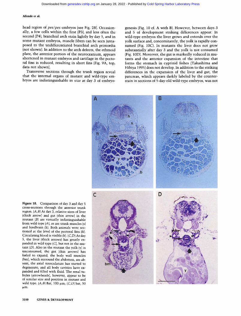
head region of pes/pes embryos (see Fig. 2B). Occasion- ally, a few cells wi th in the first (P3), and less often the second (P4), branchial arch stain l ightly by day 5, and in some mutan t embryos, muscle fibers can be seen juxta- posed to the undifferentiated branchial arch primordia (not shown). In addition to the arch defects, the ethmoid plate, the anterior portion of the neurocranium, appears shortened in mutan t embryos and cartilage in the pecto- ral fins is reduced, resulting in short fins (Fig. 9A, top; data not shown).
Transverse sections through the trunk region reveal that the internal organs of mutan t and wild-type em- bryos are indist inguishable in size at day 3 of embryo-
genesis (Fig. 10 cf. A wi th B). However, between days 3 and 5 of development striking differences appear. In wild-type embryos the liver grows and extends over the yolk surface and, concomitantly, the yolk is rapidly con- sumed (Fig. 10C). In mutants the liver does not grow substantial ly after day 3 and the yolk is not consumed (Fig. 10D). Moreover, the gut is markedly reduced in mu- tants and the anterior expansion of the intest ine that forms the stomach in cyprinid fishes (Takashima and Hibiya 1995) does not develop. In addition to the striking differences in the expansion of the liver and gut, the pancreas, which appears darkly labeled by the counter- stain in sections of 5-day-old wild-type embryos, was not
Figure 10. Comparison of day 3 and day 5 cross-sections through the anterior trunk region. (A,B) At day 3, relative sizes of liver (thick arrowl and gut (thin arrow) in the mutant {B) are virtually indistinguishable from wild type (A), as are trunk muscles (s) and hindbrain (h). Both animals were sec- tioned at the level of the pectoral fins {fi). Circulating blood is visible (b). (C,D) At day 5, the liver (thick arrows) has greatly ex- panded in wild type (C), but not in the mu- tant (D). Also in the mutant the yolk (Yl is unconsumed, the gut (thin arrows) has failed to expand, the body wall muscles ibm), which surround the abdomen, are ab- sent, the axial musculature has started to degenerate, and all body cavities have ex- panded and filled with fluid. The renal tu- bules (arrowheadsl, however, appear to be of similar size and position in mutant and wild type. (A,B) Bar, 100 ~m; (C,D) bar, 50 b~m.
3150 GENES & DEVELOPMENT
Cold Spring Harbor Laboratory Press on January 28, 2022 - Published by genesdev.cshlp.orgDownloaded from

Zebrafish insertional mutants pcs and dye
discernible in mutants . Furthermore, in mutants the ax- ial muscula ture shows signs of degeneration and the body wall muscle, which surrounds the abdominal cav- ity in wild-type embryos, is absent (Fig. 10 cf. C with D).
At the level of resolution presented here, there appears to be a striking correlation between defects in pes mu- tant embryos and regions where pes is normally ex- pressed at high levels earlier in development: the bran- chial arches, liver and gut, as well as brain, eyes, fin buds, and e thmoid plate. In most cases the primordia develop, but between days 3 and 5 they fail to expand. Importantly, other structures appear normal in mutants at day 5, although many other regions of the animal are severely affected. Tissues that appear normal include the notochord, the pronephros, and renal tubules (Fig. 10 cf. C wi th D; data not shown). Expression of pes message was not detected in these tissues at any stage in wild- type animals. To establish f i rmly that the pes phenotype is restricted entirely to cells that normal ly express pes product will require further experiments. Likewise, we cannot yet rule out that pes m R N A expression occurs at some point during embryogenesis in tissues that appear normal in mutan t animals.
D i s c u s s i o n
We have presented evidence that dye and pes are inser- tional mutan ts and we have described the genes whose disruption is l ikely to be responsible for the mutan t phe- notypes. In the case of dye, we showed that (1) a single proviral insert ion is l inked genetically to the mutan t phenotype; (2) the provirus lies just 5' of the putative ATG codon of a gene whose transcript is expressed in embryos; and (3) the insertion abolishes detectable ex- pression of this gene. In the case of pes, previously we had shown genetic linkage of a single provirus to the mutant , shown that the provirus lies in the 5' exon of a gene that is expressed in embryos, and shown that the provirus abolishes detectable gene expression. Here we
have provided additional strong support that pes is an insertional mutan t in the gene we identified by showing that at least many sites of pes expression in wild-type embryos correspond to regions of the embryo that fail to reach normal size in pes/pes mutants . The evidence pre- sented suggests that it is highly l ikely that mutat ions in the pes and dye genes are responsible for the mutan t phenotypes observed. Defini t ive proof wil l require e i ther
rescue of the mutan t phenotype by introducing the g e n e
or its product into mutan t animals, or possibly targeted mutat ion of these genes. Nei ther technology has yet been reported in zebrafish. Targeted disruption could be performed in mice (Mansour et al. 1988; Joyner et al. 1989; Schwartzberg et al. 1989; Zijlstra et al. 1989) as the pes and dye genes are so highly conserved among verte-
brates. Only a positive result would be informative in a knockout experiment, however, as mice and fish may differ in their genetic redundancy and as homologous genes could serve different functions even among verte- brate species.
Ease of molecular cloning of genes disrupted by proviral insertion
The virtue of insertional mutagenesis is the ease of clon- ing the mutated genes. In mice, this virtue has not al- ways been realized. DNA insertions in the mouse ge- nome frequently cause complex rearrangements at the site of insertion, which can make it difficult to identify the mutated gene responsible for the observed phenotype (Jaenisch 1988). Retrovirus proviral insertions in mice can disrupt genes by integrating into coding or noncod- ing exons, introns, or regulatory regions. The location of the provirus relative to recognizable coding regions or transcripts determines whether it is easy to locate the gene whose disruption causes the mutan t phenotype.
In our case, we identified rapidly genes disrupted in three of the four insertional mutants (Table 1). The most recent case, dye, required only 3 weeks of work by a
Table 1. Retrovirally induced insertional mutants in zebrafish
Insertion Mutant no. name Allele Disrupted gene
Onset of phenotype Reference
38M no arches nar hi~
67D pescadillo pes hi2
80A - 8 0 A h i 3
404 dead eye dye hi4
nar, homologous to Drosophila clipper, a zinc finger ribonuclease a
pes, homology to unknown genes in human, mouse and yeast
unidentified
dye, homology to genes in human b and frog, c similarity to yeast NIC96 protein a
Early day 3 Gaiano et al. (1996b)
Late day 3 This work and Gaiano et al. (1996b)
Late day 2 Gaiano et al {1996b)
Early day 2 This work
aBai and Tolias (1996). bNagase et al. (1993). CHudson et al. (1993). dGrandi et al. (1993).
GENES & DEVELOPMENT 3151
Cold Spring Harbor Laboratory Press on January 28, 2022 - Published by genesdev.cshlp.orgDownloaded from

Allende et al.
single individual. The reasons for this speed were (1) the proximity of the mutagenic proviral insertions to coding sequences, and (2) the fact that the coding sequence was homologous to sequences present in the data base. We have not yet located a gene near the 80A insertion. Pos- sibly we do not yet have enough sequence data (1 kb on one side, 4 kb on the other). Alternatively, this inser- tional mutant may involve a gene sequence that is not in the data base or one that is not evolutionarily conserved, or the 80A insertion may lie in a large intron or in a regulatory sequence distant from the coding region of the putative disrupted gene.
It is interesting that the proviruses in three of the four mutants we isolated integrated just upstream of the ATG initiation codon of a gene and that all three point in the opposite transcriptional orientation from the gene they disrupt. These numbers are too small to allow con- clusions, but the following points are relevant. Despite much effort, whether mouse and chicken C-type retro- viruses integrate randomly into the host cell genome re- mains controversial (Vijaya et al. 1986; Rohdewohld et al. 1987; Shih et al. 1988; Scherdin et al. 1990; Withers- Ward et al. 1994). Some studies suggest preferred inte- gration into 5' ends of genes and into actively transcribed regions, whereas others suggest that integration is essen- tially random across the genome. In many studies bio- logical selection is operating. For example, in our case only insertions that disrupt genes will be detected as mutants. The position of the integrations we have ob- served in mutants might reflect the most probable way for the virus to disrupt gene expression. Provirus inser- tions into introns, for example, might not affect tran- scription of fish genes as they sometimes do in mice. We have sequenced 1-2 kb of DNA adjacent to 42 randomly selected proviruses present in transgenic lines of fish that did not harbor embryonic mutations (K. Kawakami, N. Gaiano, D. Grosshans, M. Allende, A. Amsterdam, T. Becker, and N. Hopkins, unpubl.). The data do not reveal preferred sequences and are consistent with random in- tegration, although such data are limited.
It has been reported that in mice approximately one in 20 provirus integrations causes an embryonic phenotype (Jaenisch 1988). The frequency we have obtained for fish insertional mutants (approximately one in 70) is consid- erably lower. However, our numbers are still very small, and the genetic requirements for embryonic develop- ment may not be comparable between mice and fish. In addition, it will be important to learn whether muta- genic insertions are limited to the 5' ends of genes in the fish. If so, reengineering the viral vector might increase its mutagenicity at other locations, increasing signifi- cantly the target size for gene disruption and thus the frequency of mutants.
Relationship of insertional mutants to previously identified zebrafish mutants
Even more important than the location of the provirus relative to the gene it disrupts is the related question of whether all genes can be mutated by proviral insertion.
Most insertional systems show bias. In Drosophila, P elements readily target only about one-third to one-half of the genes that can be mutated to an embryonic phe- notype by chemical mutagenesis (Spradling et al. 1995). Even if this is the case with retroviruses, we might ex- pect that the collection of insertional mutants will ulti- mately contain a distribution of phenotypic classes sim- ilar to that encountered in the chemical mutagenesis screens. So far, the four insertional mutants we identi- fied show distinct phenotypes that become manifest rel- atively late in embryogenesis (although the dye pheno- type begins to be visible by day 2). It is probably not surprising that these insertional mutants present post- hatching defects, as most of the chemical mutants did.
Although most late-appearing mutants were discarded in the chemical screens (Driever et al. 1996; Haffter et al. 1996}, some were kept that affected specifically the jaw, liver, and gut, all of which are relatively late-developing structures. Among the collection of chemically induced mutants classified as having specific defects in pharyn- geal arch development, one designated babyface (bab) has deficiencies in the cranial skeleton that are strik- ingly similar to those seen in pes mutants (Schilling et al. 1996). Complementation tests will be needed to de- termine whether bab or other chemically induced mu- tations are allelic to pes.
The chemical mutagenesis screens isolated a large number of mutants whose primary phenotype was ne- crosis restricted to the central nervous system (Furutani- Seiki et al. 1996). However, because of the large number of mutants that fell into this class, complementation tests were not performed and therefore, the number of genes involved remains unknown. Based on the pheno- type observed here, it appears that dye would belong to this class of mutants.
Understanding the dye and pes mutant phenotypes
The dye and pes mutant phenotypes described here are both distinctive, dye embryos exhibit extensive necrosis of the central nervous system, whereas pes embryos fail to expand normally at least many of the primordia in which the pes gene is expressed: the branchial arches, liver, gut, brain, eyes, fin buds, and ethmoid plate. To understand the molecular basis for the mutant pheno- types we observed, it will be necessary to understand the normal cellular function of the products of the dye and pes genes, pes is a novel gene and does not contain any motifs that might suggest a clearly identifiable function, making the task demanding. In the case of dye, a possible function is suggested by its relation to the yeast NIC96 gene, whose product is part of the nuclear pore com- plex. The NIC96 protein interacts physically with sev- eral nucleoporins, including nucleoskeletal-like protein 1 (NSP1), the yeast homolog of the vertebrate p62 nucle- oporin that is required for the assembly of transport- competent nuclear pore complexes (Finlay et al. 1991; Grandi et al. 1993, 1995; Zabel et al. 1996). NIC96 is an essential gene for cell growth in S. cerevisiae and appears to be required for the formation of nuclear pore corn-
3152 GENES & DEVELOPMENT
Cold Spring Harbor Laboratory Press on January 28, 2022 - Published by genesdev.cshlp.orgDownloaded from
Zebrafish insertional mutants pcs and dye
plexes (Grandi et al. 1993; Zabel et al. 1996). Although the degree of homology between dye and NIC96 is sta- tistically significant, to determine whether they perform a similar biological function will require further experi- ments.
Transcripts for the pes gene are contributed mater- nally to the egg, but then decrease during the first hours after fertilization. Although this could reflect a "house- keeping" function for pes, some patterning genes in other organisms also display maternal and zygotic ex- pression. During embryogenesis, pes expression in- creases rapidly during the first day of development, and later decreases gradually. No expression can be detected in adult animals suggesting a specific embryonic require- ment for this gene. The results described here suggest that the pes gene product may be required for the growth of a subset of embryonic organs. Regions of the embryo that express the gene heavily subsequently fail to reach normal size in mutants . Particularly striking in this re- spect are the gill arches, liver, and gut. In contrast, re- gions of wild-type embryos in which expression was not detected by in situ hybridization appear normal in mu- tants, including the notochord, pronephros and renal tu- bules. The defects observed in pes/pes embryos suggest that cell types of diverse embryonic origins are affected and, moreover, that the expression pattern of the gene is not restricted to specific cell types.
The fact that the dye and pes mutat ions exhibit dis- tinctive embryonic phenotypes, including defects in the growth or maintenance of specific tissues, suggests that they may be important in cell biological processes, al- though possibly not in pattern formation and morpho- genesis. Such genes could also have medical relevance. Current ly we are mapping their homologs on human chromosomes to determine whether these genes corre- spond to disease loci.
Feasibility of genetic screens
Forward genetic approaches are usually powerful only when large-scale screens are possible, because the num- ber of genes that affect any one developmental process is small. Although this will also be true for insertional mu- tagenesis in zebrafish, the situation is somewhat differ- ent because the mutan ts that we isolate can be viewed wi th in the broad picture provided by the chemical mu- tagenesis screens. Nonetheless, we have at tempted to develop a technology that will make large scale mu- tagenesis screens possible, and we are continuing to try to increase the number of insertions that can be screened.
Although our current mutagenesis frequency is - 7 0 - fold lower than that in the chemical screens, in chemical mutagenesis, as the muta t ions are unmarked, it was nec- essary to obtain about five successful pair matings for every mu tan t identified (Mullins et al. 1994; Solnica- Krezel et al. 1994; Driever et al. 1996; Haffter et al. 1996). Because we track insertions by PCR and Southern blot and only mate fish heterozygous for the same lesion, we require only one successful cross to identify a mu-
tant. Thus, the amount of work, al though clearly greater in our case, is not 70 t imes greater per mutan t than in chemical mutagenesis screens.
Given our experience to date, and at the muta t ion fre- quency we have observed, we est imate that eight scien- tists will be able to screen -25 ,000 insertions in 2-3 years, obtaining - 2 5 0 - 3 5 0 mutants . If the distribution of phenotypes among these mutan t s proves to be similar to that in the chemical screens, - 3 0 % of these should have specific early developmental defects and at least several should affect the patterning and morphogenesis of almost every embryonic structure.
The zebrafish has long been admired for the ease with which early development can be visualized and it has been pursued as a model system because of the possibil- ity that the organism might serve as a tool for rapidly identifying genes essential for vertebrate development. The results presented here suggest that insertional mu- tagenesis, in conjunction with chemical mutagenesis screens, may help the fish to realize this potential.
Mater ia l s and m e t h o d s
Animals
Zebrafish (Danio rerio} were kept and raised essentially accord- ing to standard conditions (Westerfield 1995) and using prac- tices established in our laboratory (Culp et al. 1991). The aquar- ium systems used were designed specifically for housing large numbers of animals in small containers (Mullins et al. 1994) and were purchased from K.-J. Schwarz Glas Aquarienbau (G6ttin- gen, Germany). Fertilization was achieved by natural spawning and embryos were raised at 28~ and staged according to Kim- mel et al. (1995). The insertional mutant pilot screen was car- ried out by inbreeding fish harboring identical proviral inser- tions (Gaiano et al. 1996b) and scoring their progeny for several morphological criteria under low magnification as described (Haffter et al. 1996). Identified mutants have been named ac- cording to the conventions established for zebrafish (Wester- field 1995; M. Mullins, pets. comm.); the superscript letters indicate the laboratory designation (h) and the insertional na- ture of the mutation (i).
Isolation of genomic sequence flanking proviral insertion 404
Inverse PCR was used to clone genomic DNA fragments on each side of proviral insertion 404. Genomic DNA from fish heterozygous for the 404 insertion was digested with either NcoI and BspHI (for the 3' flanking sequence) or BglII (for the 5' flanking sequence), extracted with phenol/chloroform, and eth- anol precipitated. T4 DNA ligase was then added to the DNA diluted to 2 ~g/ml to circularize the fragments. PCR was then carried out with expand high fidelity PCR system (Boehringer Mannheim) and pairs of primers from the provirus such that one primer was in the long terminal repeat (LTR) oriented 5' to 3' toward the genomic DNA and the other was in the middle of the virus oriented 5' to 3' toward either the NcoI or BglII site: NU5 (GTAAGATCTCGAGTGATTGACTACCCGTCAG) and NV1 (GTACTCTATAGGCTTCAGCTGG) were used for the 3' flanking sequence and NU3 (GTAAGATCTCGAGCCAA- ACCTACAGGTGGGGTCT) and NV2 (GCGGTACCAGC- CCTCACTCCTTCTCTAGG) were used for the 5' flanking se- quence. The PCR program was 30 cycles of 94~ for 15 sec, 55~ for 30 sec, 68~ for 4 min (plus 20 sec per cycle after
GENES & DEVELOPMENT 3153
Cold Spring Harbor Laboratory Press on January 28, 2022 - Published by genesdev.cshlp.orgDownloaded from

Allende et ai.
cycle 10), preceded by a 2-min denaturation step at 94~ and followed by a 10-min extension step at 72~
3' RACE and RT-PCR
RNA was isolated from day 2 or 3 embryos by the guanidinium hydrochloride method (Westerfield 1995). 3' RACE for the iso- lation of the dye gene and RT-PCR for the detection of both dye and pes transcripts was carried out with a commercially avail- able kit (GIBCO-BRL Life Sciences) according to the manufac- turer's instructions, with the use of expand high fidelity PCR system (Boehringer Mannheim) for the PCR. For 3' RACE, first strand synthesis from 1 ~g of day 2 RNA was performed using the supplied AP primer. One round of PCR was performed on 10% of this sample using the supplied AUAP downstream primer and an upstream primer (404-2; CATGGATACT- GAGGGTTTTGGGGAGC) that overlaps the first 8 amino ac- ids of the ORF found in the genomic sequence. This reaction was fractionated on a low melt agarose gel; DNA from a 2.6-kb band unique to the presence of both primers was purified and 2% of this sample was used as a template for a second round of PCR in which the upstream primer (404-3; CACCA- GAACCTCTCAAGACACAGC) overlapped amino acids 49-56 of the same ORF. The 2.4-kb product was subcloned into pB- luescript II (Stratagene) for sequencing. Both rounds of PCR used the same PCR program as the inverse PCR except that the an- nealing temperature was 60~
For RT-PCR, 0.5 g.g of RNA from either wild-type or dye day 3 embryos was used for first strand synthesis with a primer specific to either the dye gene (404-9; TGCTGGCACCAG- CAGGACG) or the pes gene (P20; TACTCTCTGAATTTGC- CAACG). PCR was performed on 10% of this sample using either 404-2 and 404-8 (TCCCAGCAGGGTGTGCAAC) that lies upstream of 404-9, or P20 and P15 (TGCAAGCTTCTG- GAGACGCACGTTAG) that lies in the 5' UTR of pes. The PCR program was 30 cycles of 94~ for 30 sec, 60~ for 1 min, 72~ for 2 min, preceded by a 2-min denaturation step at 94~ and followed by a 5-min extension step at 72~
Northern blot and cDNA isolation
For the Northern blot analysis, total RNA {15 ~g) from each sample was fractionated on a 2 M formaldehyde agarose gel and transferred to a nylon filter (Hybond N +, Amersham). The blot was probed with a radioactively labeled 292-bp RT-PCR cDNA fragment (corresponding to nucleotides 61-353 of the pes cDNA). Exposure was performed on Kodak BioMax MS film for 6 days. The same cDNA fragment was used as a probe to screen a 3-day embryonic eDNA library (gift of Dr. Kai Zinn). Plaques (5 x l0 s) were screened and two positive clones were identified and isolated. Both clones contained inserts of identical length by PCR and one of them was sequenced in its entirety on both strands. The human cDNA clones were purchased from Amer- ican Type Culture Collection {Rockville, MD), and were se- quenced on both strands. Sequence alignment was accom- plished by using the Lasergene software (DNAStar, Inc.}.
Histology and acridine orange labeling
Alcian blue staining was done essentially as described (Dingerkus and Uhler 1977; Gaiano et al. 1996b). The nomen- clature for skeletal elements is that described by Schilling et al. {199@
For tissue sectioning, embryos were fixed in 4% paraformal- dehyde/PBS, dehydrated and embedded in Polybed 812 epoxy resin (Polysciences). Specimens were cut into 1- to 2-~tm sec-
tions that were counterstained subsequently with a solution of 0.05% crystal violet, 0.01% methylene blue, and 0.05% borax at 95~
For detection of apoptotic cells, anesthetized embryos were injected with a solution of 1 jzg/ml of acridine orange (Sigma) into the yolk sac (Furutani-Seiki et al. 1996). Embryos were allowed to recover for 30-60 min and were visualized under a Nikon Microphot SA microscope with an EPI-FL3 fluorescence attachment using a 450- to 490-nm excitation filter and a 520- nm long pass emission filter.
In situ hybridization
UTP-11 digoxigenin-labeled RNA probes were prepared as sug- gested by the manufacturer (Boehringer Mannheim Biochemi- cals). The probe used was an in vitro transcription product of a 2-kb fragment of the pes cDNA (corresponding to nucleotides 214--2214 of the pes cDNA). Anesthetized embryos were fixed in 4% paraformaldehyde in PBS at 4~ for 12-16 hr and were dehydrated in methanol at -20~ for at least 1 hr. In situ hy- bridization was carried out essentially after Jowett and Lettice (1994). Proteinase K treatment was for 10 min at 10 ~g/ml for embryos up to 24 hr old and at 25 g.g/ml for older embryos. Prehybridization and hybridization temperature was 65~ with a probe concentration of 1 ~g/ml. Hybridized embryos were cleared in glycerol or in methyl salicylate and were photo- graphed under a Leica Wild M3Z dissecting scope or a Nikon Microphot SA microscope.
A c k n o w l e d g m e n t s
We thank Dean Thompson, Elizabeth Abrams, and Brett Hay- ward for fish care, Kai Zinn for the cDNA library, Catherine Willet and Agustin Zapata for advice on histology and com- ments on the manuscript, Patricia Reilly for instruction on tis- sue sections, David Grosshans for sequencing help, and Murray Robinson for protein sequence analysis. The participation of Matthew Voas and Shawn Burgess in the isolation of the dye mutant is deeply appreciated. We are indebted to Charles B. Kimmel and Rachel Warga for comments on the localization of in situ hybridization stain, to Christiane Nfisslein-Volhard for many helpful discussions and to Bob Bosselman for his interest. M.A. was supported by the National Institutes of Health (NIH) (postdoctoral fellowship 5F32-HD07818), T.B. by the Cancer Research Fund of the Damon Runyon-Walter Winchell Founda- tion, Fellowship DRG-1274, K.K. by the Yamada Science Foun- dation and the Toyobo Biotechnology Foundation, and A.A. by a Howard Hughes Medical Institute predoctoral fellowship. N.H. is supported by grants from the Human Frontier Science Program, the NIH, and Amgen, Inc. Support was also obtained through a Core Grant (CA14051) from the National Cancer In- stitute to the Center for Cancer Research. Gene sequences have been deposited in the GenBank database under accession nos. U77595 (zebrafish dye), U77627 {zebrafish pes), and U78310 (human pes).
The publication costs of this article were defrayed in part by payment of page charges. This article must therefore be hereby marked "advertisement" in accordance with 18 USC section 1734 solely to indicate this fact.
R e [ e r e n c e s
Abrams, J.M., K. White, L.I. Fessler, and H. Steller. 1993. Pro- grammed cell death during Drosophila embryogenesis. De- velopment 117: 29-43.
Altschul, S., W. Gish, W. Miller, E. Myers, and D. Lipman. 1990. Basic local alignment search tool. J. Mol. Biol. 215: 403-410.
Bai, C. and P.P. Tolias. 1996. Cleavage of RNA hairpins medi-
3154 GENES & DEVELOPMENT
Cold Spring Harbor Laboratory Press on January 28, 2022 - Published by genesdev.cshlp.orgDownloaded from

Zebrafish insertional mutants pcs and dye
ated by a developmentally regulated CCCH zinc-finger pro- tein. Mol. Cell. Biol. (in press).
Culp, P., C. Nhsslein-Volhard, and N. Hopkins. 1991. High- frequency germ-line transmission of plasmid DNA se- quences injected into fertilized zebrafish eggs. Proc. Natl. Acad. Sci. 88: 7953-7957.
Dingerkus, G. and D. Uhler. 1977. Enzyme clearing of alcian blue stained whole small vertebrates for demonstration of cartilage. Stain Tech. 32: 229-231.
Driever, W., L. Solnica-Krezel, A. Schier, S. Neuhauss, J. Maliki, D. Stemple, D. Stainier, F. Zwartkruis, S. Abdelilah, Z. Rangini, J. Belak, and C. Boggs. 1996. A genetic screen for mutations affecting embryogenesis in zebrafish. Develop- men t 123: 37--46.
Finlay, D.R., E. Weier, P. Bradley, J. Horecka, and D.J. Forbes. 1991. A complex of nuclear pore proteins required for pore function. J. Cell Biol. 114: 169-183.
Furutani-Seiki, M., Y-J. Jiang, M. Brand, C-P. Heisenberg, C. Houart, D. Beuchle, F.J.M. van Eeden, M. Granato, P. Haffter, M. Hammerschmidt, D. Kane, R. Kelsh, M. Mullins, J. Odenthal, and C. Nfisslein-Volhard. 1996. Neural degen- eration mutants in the zebrafish, Danio rerio. Development 123: 229-239.
Gaiano, N., M. Allende, A. Amsterdam, K. Kawakami, and N. Hopkins. 1996a. Highly efficient germ-line transmission of proviral insertions in zebrafish. Proc. Natl. Acad. Sci. 93: 7777-7782.
Gaiano, N., A. Amsterdam, K. Kawakami, M. Allende, T. Becker, and N. Hopkins. 1996b. Insertional mutagenesis and rapid cloning of essential genes in zebrafish. Nature 383: 829-832.
Grandi, P., V. Doye, and E.C. Hurt. 1993. Purification of NSP1 reveals complex formation with "GLFG" nucleoporins and a novel nuclear pore protein NIC96. EMBO J. 12: 3061-3071.
Grandi, P., N. Schlaich, H. Tekotte, and E.C. Hurt. 1995. Func- tional interaction of Nic96p with a core complex consisting of Nspl, Nup49p and a novel protein Nup57p. EMBO 1. 14: 76-87.
Haffter, P., M. Granato, M. Brand, M. Mullins, M. Hammer- schmidt, D. Kane, J. Odenthal, F. van Eeden, Y. Jiang, C-P. Heisenberg, R. Kelsh, M. Furutani-Seiki, L. Vogelsang, D. Beuchle, U. Schach, C. Fabian, and C. Nhsslein-Volhard. 1996. The identification of genes with unique and essential functions in the development of the zebrafish, Danio rerio. Development 123: 1-36.
Hudson, J.W., V.B. Alarcon, and R.P. Elinson. 1996. Identifica- tion of new localized RNAs in the Xenopus oocyte by dif- ferential display PCR. Dev. Genet. 19: 190--198.
Jaenisch, R. 1988. Transgenic animals. Science 240: 1468-1474. Joyner, A.L., W.L. Skames, and J. Rossant. 1989. Production of a
mutation in mouse En-2 gene by homologous recombination in embryonic stem cells. Nature 338: 153-156.
Jowett, T. and L. Lettice. 1994. Whole mount in situ hybridiza- tion on zebrafish embryos using a mixture of digoxigenin- and fluorescein-labelled probes. Trends Genet. 10: 73-74.
Kimmel, C.B. 1989. Genetics and early development of ze- brafish. Trends Genet. 5: 283-288.
Kimmel, C.B., W. Ballard, S. Kimmel, B. Ullmann, and T. Schill- ing. 1995. Stages of embryonic development of the zebrafish. Dev. Dyn. 203: 253-310.
Kozak, M. 1984. Compilation and analysis of sequences up- stream from the translational start site in eukaryotic mR- NAs. Nucleic Acids Res. 12: 857-872.
Lin, S., N. Gaiano, P. Culp, J. Burns, T. Friedman, J.-K. Yee, and N. Hopkins. 1994. Integration and germ-line transmission of a pseudotyped retroviral vector in zebrafish. Science
265: 666-669. Mansuor, S.L., K.R. Thomas, and M.R. Capecchi. 1988. Disrup-
tion of the proto-oncogene int-2 in mouse embryo-derived stem cells: A general strategy for targeting mutations to non- selectable genes. Nature 336: 348-352.
Mullins, M., M. Hammerschmidt, P. Haffter, and C. Nhsslein- Volhard. 1994. Large-scale mutagenesis in the zebrafish: In search of genes controlling development in a vertebrate. Curr. Biol. 4: 189-202.
Nagase, T., N. Miyajima, A. Tanaka, T. Sazuka, N. Seki, S. Sato, S. Tabata, K. Ishikawa, Y. Kawarabayasi, H. Kotani, and N. Nomura. 1995. Prediction of the coding sequences of un- identified human genes III: The coding sequences of 40 new genes (KIAA0081-KIAA0120) deduced by analysis of cDNA clones from human cell line KG-1. D N A Res. 2: 37-43.
Nhsslein-Volhard, C. 1994. Of flies and fishes. Science 266: 572-574.
Rohdewohld, H., H. Weiher, W. Reik, R. Jaenisch, and M. Breindl. 1987. Retrovirus integration and chromatin struc- ture: Moloney Murine Leukemia proviral integration sites map near DNase I-hypersensitive sites. J. Virol. 61: 336-343.
Rossant, J. and N. Hopkins. 1992. Of fin and fur: Mutational analysis of vertebrate embryonic development. Genes & Dev. 6: 1-13.
Scherdin, U., K. Rhodes, and M. Breindl. 1990. Transcriptionally active genome regions are preferred targets for retrovirus in- tegration. J. Virol. 64: 907-912.
Schilling, T., T. Piotrowski, H. Grandel, M. Brand, C-P. Heisen- berg, Y-J. Jiang, D. Beuchle, M. Hammerschmidt, D. Kane, M. Mullins, F. van Eeden, R. Kelsh, M. Furutani-Seiki, M. Granato, P. Haffter, J. Odenthal, R. Warga, T. Trowe, and C. Nhsslein-Volhard. 1996. Jaw and branchial arch mutants in zebrafish I: Branchial arches. Development 123: 329-344.
Schwartzberg, P.O., S.P. Goff, and E.J. Robertson. 1989. Germ- line transmission of a c-abl mutation produced by targeted gene disruption of ES cells. Science 246: 799-803.
Shih, C.-C., J.P. Stoye, and J.M. Coffin. 1988. Highly preferred targets for retrovirus integration. Cell 53: 531-537.
Solnica-Krezel, L., A. Schier, and W. Driever. 1994. Efficient recovery of ENU-induced mutations from the zebrafish ger- mline. Genetics 136: 1401-1420.
Spradling, A.C., D. Stern, I. Kiss, J. Roote, T. Laverty, and G.M. Rubin. 1995. Gene disruptions using P transposable ele- ments: An integral component of the Drosophila genome project. Proc. Natl. Acad. Sci. 92: 10924-10830.
Streisinger, G., C. Walker, N. Dower, D. Knauber, and F. Singer. 1981. Production of clones of homozygous diploid zebrafish (Brachydanio rerio). Nature 291: 293-296.
Takashima, F. and T. Hibiya. 1995. An atlas of fish histology, 2nd edition. Kodansha Ltd, Tokyo, Japan.
Vijaya, S., D. Steffen, and H. Robinson. 1986. Acceptor sites for retroviral integrations map near DNase I-hypersensitive sites in chromatin. J. Virol. 60: 683-692.
Westerfield, M. 1995. The zebrafish book. University of Oregon Press, Eugene, OR.
Withers-Ward, E.S., Y. Kitamura, J.P. Barnes, and J.M. Coffin. 1994. Distribution of targets for avian retrovirus DNA inte- gration in vivo. Genes & Dev. 8: 1473-1487.
Zabel, U., V. Doye, H. Tekotte, R. Wepf, P. Grandi, and E.C. Hurt. 1996. Nic96p is required for nuclear pore formation and functionally interacts with a novel nucleoporin, Nup188p. J. Cell Biol. 133: 1141-1152.
Zijlstra, M., E. Li, F. Sajjadi, S. Subramani, and R. Jaenisch. 1989. Germ-line transmission of a disrupted ~2 microglobulin gene produced by homologous recombination in embryonic stem cells. Nature 342: 435-438.
GENES & DEVELOPMENT 3155
Cold Spring Harbor Laboratory Press on January 28, 2022 - Published by genesdev.cshlp.orgDownloaded from

10.1101/gad.10.24.3141Access the most recent version at doi: 10:1996, Genes Dev.
M L Allende, A Amsterdam, T Becker, et al. pescadillo and dead eye, essential for embryonic development.Insertional mutagenesis in zebrafish identifies two novel genes,
References
http://genesdev.cshlp.org/content/10/24/3141.full.html#ref-list-1
This article cites 36 articles, 18 of which can be accessed free at:
License
ServiceEmail Alerting
click here.right corner of the article or
Receive free email alerts when new articles cite this article - sign up in the box at the top
Copyright © Cold Spring Harbor Laboratory Press
Cold Spring Harbor Laboratory Press on January 28, 2022 - Published by genesdev.cshlp.orgDownloaded from
Copyright © 2022 FDOKUMEN