Mural cell-derived laminin-α5 plays a detrimental role in ...
Upload
independentCategory
view
0download
0
Characterization of Vascular Mural Cells During ZebrafishDevelopment
Massimo M. Santoro1,2,3, Gabriella Pesce1,2, and Didier Y. Stainier21Molecular Biotechnology Center, University of Torino, Via Nizza, 52, 10126, Torino, Italy2Department of Biochemistry and Biophysics, Programs in Developmental Biology, Genetics andHuman Genetics, and Cardiovascular Research Institute, University of California San Francisco,San Francisco, CA 94158, USA3Department of Environmental and Life Sciences, University of Piemonte Orientale “A. Avogadro”,Alessandria, 15100, Italy
AbstractDevelopment and maturation of the nascent cardiovascular system requires the recruitment of muralcells (MCs) around the vascular tree in a process called vascular myogenesis. Understanding theorigin and development of vascular MCs has been hampered by difficulties in observing these cellsin vivo and performing defined genetic and experimental manipulations in available modelorganisms. Here, we investigate the origin of vascular MCs using molecular and genetic tools inzebrafish. We show that vascular MCs are present around the lateral dorsal aortae (LDA) and anteriormesenteric arteries (AMA) of developing animals, and that they also contribute to the outflow tractof the developing heart and ventral aorta (VA). Genetic data indicate that the vascular MCs of theLDA and AMA do not arise from blood or endothelial progenitors but from other derivatives of thelateral plate mesoderm. We further show that zebrafish vascular MCs share many of themorphological, molecular and functional characteristics of vascular smooth muscle cells andpericytes found in higher vertebrates. These data establish the zebrafish as a useful cellular andgenetic model to study vascular myogenesis as well as tumor angiogenesis and other MC-associateddiseases.
IntroductionIn vertebrates, the development and maturation of the cardiovascular system require therecruitment of smooth muscle cells in a process called vascular myogenesis (Carmeliet,2000; Jain, 2003). During this process, smooth muscle cell precursors, called mural cells(MCs), differentiate and surround vascular endothelial vessels promoting the maturation andspecialization of the cardiovascular system (Carmeliet, 2003). During development, thesevascular MCs undergo proliferation and/or differentiation depending on the size of the vessels.On large vessels, they become multi-layered and referred to as vascular smooth muscle cells(SMCs). On smaller vessels, mural cells remain as single sparse cells and usually referred toas pericytes (PCs). Large arterial vessels have strong, elastic vessel walls with densepopulations of concentrically-arranged SMCs to withstand the higher blood pressures. SMCs
corresponding authors: [email protected]; [email protected]'s Disclaimer: This is a PDF file of an unedited manuscript that has been accepted for publication. As a service to our customerswe are providing this early version of the manuscript. The manuscript will undergo copyediting, typesetting, and review of the resultingproof before it is published in its final citable form. Please note that during the production process errors may be discovered which couldaffect the content, and all legal disclaimers that apply to the journal pertain.
NIH Public AccessAuthor ManuscriptMech Dev. Author manuscript; available in PMC 2010 August 1.
Published in final edited form as:Mech Dev. 2009 ; 126(8-9): 638–649. doi:10.1016/j.mod.2009.06.1080.
NIH
-PA Author Manuscript
NIH
-PA Author Manuscript
NIH
-PA Author Manuscript

in these large vessels support and regulate blood flow. Veins are generally irregularly coveredby SMCs and have valves to prevent backflow of blood (Scultetus et al., 2001). Small bloodvessels, which are composed of endothelial cells (ECs) surrounded by a basal lamina, areloosely covered by single PCs, which serve in both scaffolding and signaling functions, andcan also regulate vasoconstriction and vasodilation within the vascular bed. It is not knownwhether PCs and SMCs have distinct progenitors or whether they represent phenotypic variantsof the same lineage (Bergers and Song, 2005; Majesky, 2007).
In mammals SMCs are characterized by a set of contractile proteins that are distinct from thoseexpressed in skeletal and cardiac muscles (Owens, 1995). Several dynamic molecular markerspresent in mammalian vascular SMCs are used for their detection, including smooth muscle-myosin heavy chain (MYH11) (Babij et al., 1991; Frid et al., 1993; Madsen et al., 1998; Mianoet al., 1994), smooth muscle alpha-actin (ACTA2) (Gabbiani et al., 1981; Hungerford et al.,1996; Mack and Owens, 1999), Sm22alpha (aka TRANSGELIN) (Li et al., 1996; Kim et al.,1997), Smoothelin (van der Loop et al., 1996; Kramer et al., 1999), Desmin and Calponin(Duband et al., 1993; Owens et al., 2004). The expression patterns of these markers can varydepending on the developmental stage of the vascular system. In particular, Sm22alpha andsmooth muscle alpha-actin are the first markers expressed in SMCs and for this reason areconsidered embryonic SMC markers in mammals (Majesky, 2007).
Intriguingly, unlike skeletal or cardiac muscle cells which permanently exit the cell cycle andare terminally (and essentially irreversibly) differentiated, SMCs retain remarkable plasticityand can undergo rather profound and reversible phenotypic changes in response to changes inlocal environmental cues. The high degree of mammalian smooth muscle plasticity, especiallyduring postnatal development, predisposes it to phenotypic switching, and thus may play amajor role in human disease (Halayko and Solway, 2001). Unregulated growth, differentiationand plasticity of mammalian vascular SMCs is a key feature of vascular diseases such asatherosclerosis, restenosis and hypertension, as well as monogenic diseases such astelangiectasia and retinopathies (Ross, 1999; Conway et al., 2001; Sata et al., 2002; Hao et al.,2003). Several genes identified to play a key role in vascular myogenesis are also associatedwith human disease (Carmeliet, 2003; Rensen et al., 2007). Furthermore, in response to specificvascular injuries, vascular SMCs dramatically increase their rate of proliferation, migrationand synthetic capacity and play a critical role in vascular repair and tumor angiogenesis(Gerhardt and Semb, 2008).
A limitation in studying mammalian vascular myogenesis is the lack of an easily manipulatablein vivo system to analyse smooth muscle development and differentiation. The zebrafish hasalready been established as a powerful model system to study cardiovascular development(Stainier, 2001; Lawson and Weinstein, 2002; Beis et al., 2005; Santoro et al., 2007). Thezebrafish is also a genetically accessible vertebrate with an optically clear embryo permittinghigh-resolution in vivo cell imaging. Surprisingly, the existence of vascular SMCs or PCs hasnot yet been fully reported in this model organism. We carried out a series of morphological,molecular and functional studies to visualize and characterize vascular myogenesis in thedeveloping zebrafish. Since we were able to detect only a few cells surrounding earlyembryonic vessels, we decided to name these perivascular cells, mural cells (MCs). Our studiesshow that zebrafish embryos develop vascular MCs that share many of the characteristicfeatures of embryonic SMCs found in higher vertebrates, and we provide evidence to supporta functional role for zebrafish MCs during cardiovascular development. These studies identifythe zebrafish as an embryological and genetic model system to further study the development,differentiation and plasticity of vascular MCs in vivo.
Santoro et al. Page 2
Mech Dev. Author manuscript; available in PMC 2010 August 1.
NIH
-PA Author Manuscript
NIH
-PA Author Manuscript
NIH
-PA Author Manuscript

ResultsVascular smooth muscle cells are present in zebrafish larvae
Understanding the molecular mechanisms regulating vascular myogenesis remains a majorissue, in part because of the challenges of studying this process in amniotes (Majesky, 2007).We therefore decided to investigate the origin of vascular MCs in zebrafish, particularlyfocusing on their contribution to the trunk vasculature (mainly dorsal aorta and cardinal vein)during development. We started by comparing the ultrastructure of the dorsal aorta (DA) andposterior cardinal vein (PCV) at various stages. Light microscopy images of 3-months oldzebrafish revealed striking differences in vessel wall caliber between the DA and PCV, withthe former clearly characterized by thick layers of vascular SMCs around the ECs (Figure 1)(Miano et al., 2006). Interestingly, the PCV did not show the same extent of vascular SMCcoverage as in mammals. To better characterize these vascular SMCs we analysed samples ofDA and PCV by transmission electron microscopy (TEM) at earlier time points. Analyses of20 day-old DA revealed that at least two to three layers of synthetic vascular SMCs are presentin the DA, while single and dispersed SMCs were found in the PCV (Supplementary Figure1). Interestingly, a dense layer of extracellular matrix, the internal elastic lamina (IEL), liesbetween the EC and the SMC layers, constituting the basement membrane. The synthetic SMCsin the DA at 20 dpf did not yet exhibit the ultrastructural features of fully differentiated adultvascular SMCs. Differentiated, contractile vascular SMCs are elongated, whereasundifferentiated, synthetic SMCs have a cobblestone morphology and contain a high numberof organelles involved in protein synthesis. Moreover, synthetic SMCs show higher growthrates and higher migratory activity than contractile SMCs (Hao et al., 2003). Collectively, theseresults reveal a basic ultrastructural organization of the DA in zebrafish that is similar to thatin mammals with the notable exception of a delay in zebrafish SMC maturation.
Markers of embryonic smooth muscle cells identify specific perivascular tissue duringzebrafish development
The transcriptional program controlling smooth muscle differentiation is poorly understood.Unlike skeletal and cardiac muscle, SMCs do not terminally differentiate but retain remarkableplasticity and exhibit a diverse range of phenotypes under various physiological andpathological conditions (Halayko and Solway, 2001). Due to their heterogeneous origin andtranscriptional profiles, no single marker can be used to identify all SMCs. However, whenthese markers are assayed in combination, and are considered in the morphological context ofdeveloping organs, they can be used to identify cells of the SMC lineage. A variety of genesand gene products have been identified in mammals and other vertebrates as useful markersof the relative state of induction and differentiation of SMCs (Owens, 1995; Owens et al.,2004). These markers include mainly contractile, cytoskeletal and cytoskeleton-associatedproteins such as smooth muscle alpha-actin, Sm22alpha, smooth muscle myosin heavy chain,and Smoothelin. Similarly, in zebrafish, the heterogeneity of MCs precludes their identificationby single markers (Georgijevic et al., 2007; Davis et al., 2008). Therefore, we identified andcloned a panel of zebrafish MC markers including the genes encoding Transgelin (Sm22alpha-b), Transgelin2 (Sm22alpha-a), Acta2, Myh11, Smoothelin, Calponin, and Desmin. Amongthose we found that acta2 and transgelin were expressed specifically in the perivascular regionof zebrafish larvae (Figure 2). Starting at 72 hpf these genes were not only expressed in visceralSMCs (Georgijevic et al., 2007) and the fin bud, but also in a specific region of the vasculaturecorresponding to the anterior portion of the DA, the lateral DA (LDA) as well as the primitiveinternal carotid arteries (PICA). Cross-section analyses revealed specific staining around thedorsal aorta and in the mesenchyme surrounding the gut epithelium. At this time, expressionwas relatively weak and discontinuous throughout the length of the dorsal aorta. However, thisexpression became more evident at 96 hpf especially around the DA and LDA. At this laterstage, acta2 and transgelin expression also became evident in the heart, specifically the outflow
Santoro et al. Page 3
Mech Dev. Author manuscript; available in PMC 2010 August 1.
NIH
-PA Author Manuscript
NIH
-PA Author Manuscript
NIH
-PA Author Manuscript
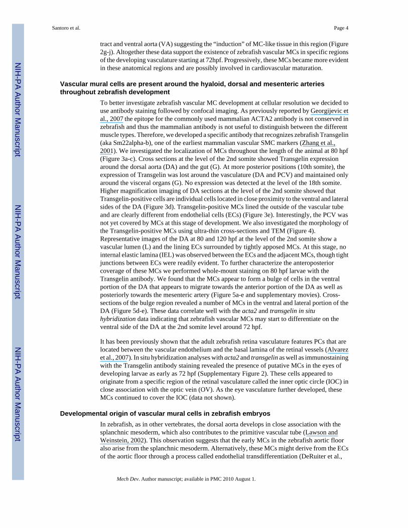
tract and ventral aorta (VA) suggesting the “induction” of MC-like tissue in this region (Figure2g-j). Altogether these data support the existence of zebrafish vascular MCs in specific regionsof the developing vasculature starting at 72hpf. Progressively, these MCs became more evidentin these anatomical regions and are possibly involved in cardiovascular maturation.
Vascular mural cells are present around the hyaloid, dorsal and mesenteric arteriesthroughout zebrafish development
To better investigate zebrafish vascular MC development at cellular resolution we decided touse antibody staining followed by confocal imaging. As previously reported by Georgijevic etal., 2007 the epitope for the commonly used mammalian ACTA2 antibody is not conserved inzebrafish and thus the mammalian antibody is not useful to distinguish between the differentmuscle types. Therefore, we developed a specific antibody that recognizes zebrafish Transgelin(aka Sm22alpha-b), one of the earliest mammalian vascular SMC markers (Zhang et al.,2001). We investigated the localization of MCs throughout the length of the animal at 80 hpf(Figure 3a-c). Cross sections at the level of the 2nd somite showed Transgelin expressionaround the dorsal aorta (DA) and the gut (G). At more posterior positions (10th somite), theexpression of Transgelin was lost around the vasculature (DA and PCV) and maintained onlyaround the visceral organs (G). No expression was detected at the level of the 18th somite.Higher magnification imaging of DA sections at the level of the 2nd somite showed thatTransgelin-positive cells are individual cells located in close proximity to the ventral and lateralsides of the DA (Figure 3d). Transgelin-positive MCs lined the outside of the vascular tubeand are clearly different from endothelial cells (ECs) (Figure 3e). Interestingly, the PCV wasnot yet covered by MCs at this stage of development. We also investigated the morphology ofthe Transgelin-positive MCs using ultra-thin cross-sections and TEM (Figure 4).Representative images of the DA at 80 and 120 hpf at the level of the 2nd somite show avascular lumen (L) and the lining ECs surrounded by tightly apposed MCs. At this stage, nointernal elastic lamina (IEL) was observed between the ECs and the adjacent MCs, though tightjunctions between ECs were readily evident. To further characterize the anteroposteriorcoverage of these MCs we performed whole-mount staining on 80 hpf larvae with theTransgelin antibody. We found that the MCs appear to form a bulge of cells in the ventralportion of the DA that appears to migrate towards the anterior portion of the DA as well asposteriorly towards the mesenteric artery (Figure 5a-e and supplementary movies). Cross-sections of the bulge region revealed a number of MCs in the ventral and lateral portion of theDA (Figure 5d-e). These data correlate well with the acta2 and transgelin in situhybridization data indicating that zebrafish vascular MCs may start to differentiate on theventral side of the DA at the 2nd somite level around 72 hpf.
It has been previously shown that the adult zebrafish retina vasculature features PCs that arelocated between the vascular endothelium and the basal lamina of the retinal vessels (Alvarezet al., 2007). In situ hybridization analyses with acta2 and transgelin as well as immunostainingwith the Transgelin antibody staining revealed the presence of putative MCs in the eyes ofdeveloping larvae as early as 72 hpf (Supplementary Figure 2). These cells appeared tooriginate from a specific region of the retinal vasculature called the inner optic circle (IOC) inclose association with the optic vein (OV). As the eye vasculature further developed, theseMCs continued to cover the IOC (data not shown).
Developmental origin of vascular mural cells in zebrafish embryosIn zebrafish, as in other vertebrates, the dorsal aorta develops in close association with thesplanchnic mesoderm, which also contributes to the primitive vascular tube (Lawson andWeinstein, 2002). This observation suggests that the early MCs in the zebrafish aortic flooralso arise from the splanchnic mesoderm. Alternatively, these MCs might derive from the ECsof the aortic floor through a process called endothelial transdifferentiation (DeRuiter et al.,
Santoro et al. Page 4
Mech Dev. Author manuscript; available in PMC 2010 August 1.
NIH
-PA Author Manuscript
NIH
-PA Author Manuscript
NIH
-PA Author Manuscript

1997). To address this point, we took advantage of two zebrafish mutants, cloche (clos5) andhands off (hans6). cloche mutant embryos lack all endothelial precursors in the head and trunkregions and do not form functional blood vessels (Stainier et al., 1995). The hands off geneencodes the bHLH transcription factor Hand2 and is expressed in the lateral plate mesodermduring gastrulation and somitogenesis stages, and hans6 mutants have defects in lateral platemesoderm-derived tissues (Yelon et al., 2000). We tested for the presence of vascular MCs inthese mutants using the Transgelin antibody (Figure 6). Interestingly, while clos5 mutantsshowed Transgelin-positive cells in the region corresponding to the aortic floor, hans6 mutantscompletely lacked these cells. These experiments suggest that in zebrafish, MCs derive fromthe lateral plate mesoderm and not from the transdifferentiation of ECs.
Vascular mural cells are present in the developing outflow tract in zebrafishThe zebrafish is a widely accepted model for the study of early cardiogenesis (Stainier,2001; Beis et al., 2005). Much of this research has been concentrated on the development ofthe atrium and ventricle, at stages long before the outflow tract is formed. As a result, thisstructure has been poorly characterized. Several reports have shown that the adult zebrafishoutflow tract is invested with a considerable number of smooth muscle layers (Hu et al.,2000; Grimes et al., 2006). We therefore decided to investigate the onset of zebrafish MCinduction in the bulbus arteriosus (BA) and outflow tract. Whole mount in situ hybridizationanalyses showed that acta2 and transgelin are expressed in the heart region at 96 hpf (Figure2). To gain a better resolution of these MC-positive cardiac regions, we analyzed thin crosssections of whole mount stained larvae at 96hpf (Figure 7a-b). These analyses confirmed thatthe region involved is the BA and the proximal part of the VA. We confirmed the presence ofthis MC coverage in the outflow tract by Transgelin staining (Fig. 7c-e). Immunofluorescenceon whole mount and cross sections with Transgelin antibody clearly showed a single layer ofvascular MCs around the VA at 96 hpf. Interestingly, at 72 hpf, while the BA was alreadyformed and several layers of cuboidal cells were evident, the outflow tract did not yet expressthese MC markers (data not shown). These data are in agreement with a previouscharacterization of the zebrafish outflow tract where a cellular structure committed to becomethe BA appears after 48 hpf (Grimes et al., 2006), a stage when the atrium and ventricle arealready formed and pumping blood through a rudimentary circulatory system. We speculatethat between 72 and 96 hpf these cells are induced to differentiate into vascular MCs andcontribute to the future outflow tract.
DiscussionMammalian vascular SMCs are a highly heterogeneous cell population with diverse embryonicorigins, properties, and gene expression profiles. A major difficulty in the smooth muscle fieldhas been the lack of an easily manipulatable model system for the study of SMC biology invivo. Surprisingly, the development of vascular SMCs, in particular vaSMCs and PCs, has notyet been characterized in zebrafish, even though the transparency and easy manipulation ofembryos has established it as an excellent model system to study the development of a varietyof organ systems. Here we analyzed the development and distribution of zebrafish vascularMCs and investigated the temporal and spatial expression patterns of several MC genes duringcardiovascular development. Additionally, we examined the morphology of zebrafish vascularMCs with different microscopy techniques and identified these cells in the larval and adultvasculature. The goal of this study is to begin to provide an understanding of the ontogeny ofzebrafish vascular SMCs and PCs and the morphological events that lead to their induction,differentiation and maturation. This understanding will help investigate the cellular andmolecular mechanisms that regulate their development and to evaluate the importance of SMCbiology as a susceptibility factor for vascular diseases like atherosclerosis, retinopathies andhereditary haemorrhagic telangiectasia (HHT). The importance of developing a model system
Santoro et al. Page 5
Mech Dev. Author manuscript; available in PMC 2010 August 1.
NIH
-PA Author Manuscript
NIH
-PA Author Manuscript
NIH
-PA Author Manuscript

to study SMCs resides on the extensive contribution of these cells to various pathologicalconditions. The zebrafish represents an interesting new approach to connect developmentalbiology and associated diseases (Dodd et al., 2000; Lieschke and Currie, 2007; Chico et al.,2008). Our data suggest an evolutionarily conserved developmental origin and function ofSMCs indicating that the zebrafish can be used to study the genetics of vascular SMC-associated human diseases. Generation of transgenic zebrafish lines with fluorescently labelledvascular MCs, as well as the identification and characterization of mutant lines showing defectsin zebrafish vascular MC development will be the next steps. In fact, some zebrafish mutantphenotypes identified in genetic screens for developmental regulators resemble human diseasestates and these mutations, once isolated molecularly, will provide candidate genes forevaluation in human disorders (Lieschke and Currie, 2007). The identification of mutantphenotypes resembling human vascular diseases will be very useful to i) dissect new geneticpathways involved in SMC-associated diseases, ii) study the genetic origin of pathologicalconditions associated with MCs and iii) facilitate the set-up of new therapeutic applicationsfor these pathologies.
The origin of vertebrate vascular SMCs is complex with contributions from severalindependent cell lineages (Gittenberger-de Groot et al., 1998; Majesky, 2007). Neural crestcells (NCs) give rise to vascular (Wilm et al., 2005) SMCs in the cephalic region and in thecardiac outflow tract region, and are required for patterning the great vessels (Hutson et al.,2003). The pro-epicardial organ contributes to SMCs in the coronary arteries and ventrallyemigrating neural tube cells may contribute to SMCs in the great vessels and the coronaryvessels in chicken (Mikawa and Gourdie, 1996; Gittenberger-de Groot et al., 1998). Themesothelium also gives rise to SMCs in mesenteric vessels in mouse (Wilm et al., 2005). Inposterior parts of the body, vascular MCs are assumed to derive primarily from the splanchniclateral plate mesoderm (Gittenberger-de Groot et al., 1999), but the precise source of thesecells has not been rigorously determined. Observations of the early pattern of SMC contractileprotein expression in mouse and quail aorta suggest that vascular MC differentiation is inducedin mesenchymal cells that line the endothelium (Hungerford et al., 1996; Takahashi et al.,1996). Vascular SMC contractile proteins are first observed on the ventral side of the vessel,and it has been hypothesized that MCs are only induced in the ventral area of the aorta andlater migrate to populate the lateral and dorsal areas. Our findings are in agreement with thesefindings and suggest that vascular MCs in the trunk originate from the lateral plate mesodermas in other vertebrate model organisms (Wiegreffe et al., 2007; Wasteson et al., 2008).However, cell lineage experiments need to be carried out to determine the precise origin ofMCs in different anatomical regions. Establishment of transgenic lines for zebrafish MCsshould also be a useful tool to study the perivascular origin of mesenchymal stem cells andtheir potency in myogenesis, adipogenesis, chondrogenesis and tissue regeneration (Cossu andBianco, 2003; Crisan et al., 2008).
We also noticed that from early developmental stages until adulthood, zebrafish arteries andveins are differently covered by SMCs. While the aorta is covered with vascular MCs from anearly time point, the cardinal vein is not. Lack of MCs in the posterior cardinal vein of zebrafishlarvae is not surprising given that only a few scattered SMCs were found in the walls of thecaudal vein and the posterior cardinal vein of 20 dpf animals (Figure 1 and SupplementaryFigure 1). This situation resembles that found in the carp (Cyprinus carpio)(Satchell, 1992).Gravity exerts significant effect on venous return in terrestrial animals, affecting venouspressure and ventricular filling pressure. Hence, venous compliance must be continuouslyadjusted in terrestrial animals by MC contractility to ensure proper cardiac filling. In contrast,gravitational stress in water is much less than in a terrestrial environment. The specific gravityof water is similar to that of blood, resulting in constant transmural pressure along the lengthof the posterior cardinal vein (Scultetus et al., 2001;Satchell, 1992). Therefore, modulation ofvenous blood pressure by smooth muscle contractility may not be crucial in juvenile zebrafish.
Santoro et al. Page 6
Mech Dev. Author manuscript; available in PMC 2010 August 1.
NIH
-PA Author Manuscript
NIH
-PA Author Manuscript
NIH
-PA Author Manuscript

On the other hand, we found an early vascular MC coverage in the anterior mesenteric arteryof zebrafish larvae suggesting that this is a crucial vascular region to control blood flow towardsthe gastro-intestinal tract region at early time points.
While the use of atrioventricular-specific molecular markers has allowed extensivecharacterization of the development of the atrium and ventricle, the lack of any bulbusarteriosus (BA)-specific markers has meant that this region of the zebrafish heart is poorlycharacterized and quite possibly misunderstood (Hu et al., 2000; Grimes et al., 2006). Inzebrafish, the BA forms by 48 hpf as an independent structure, at a stage when the atrium andventricle are already formed and pumping blood through a rudimentary circulatory system.Because it forms so late, it is usually excluded from studies of the true cardiac chambers, anddetailed analyses of its development need to be carried out. The function of the BA in fish isto reduce the high ventricular pressure during systole, and to maintain constant blood flow intothe ventral aorta and gills during diastole. The elastic walls of the BA maintain a strong andeven pressure on the blood flowing to the gills, maintaining a steady flow. The ability of thebulbous wall to expand and hold much of the stroke volume is critical to this process (Bushnellet al, 1992). Our finding of extensive smooth muscle coverage in the outflow tract and closelyassociated vessels is consistent with a role for the BA in maintaining an even blood pressure.It will be important to identify the progenitors of these outflow tract MCs as their biology getsfurther investigated (Sato and Yost, 2003).
Our results provide the first analysis of zebrafish vascular myogenesis. Collectively, these dataprovide new insights into the emergence of differentiated vascular MCs and PCs in zebrafish,thus providing a foundation for future studies on the gene expression profile and function ofthese cells in wild-type and mutant animals.
Materials and MethodsZebrafish husbandry
Zebrafish, Danio rerio, were maintained and staged based on developmental time andmorphological criteria as previously described (Santoro et al., 2007). Fish were kept under a14-h light and 10-h dark photoperiod at approximately 28°C. Following fertilization, eggs werecollected and embryos- wild type or mutant- were raised at 28°C under standard laboratoryconditions in the presence of 0.003% 1-phenyl-2-thiourea to prevent formation of melaninpigments, greatly facilitating visualization of the vascular system.
Whole mount in situ hybridizationWild-type and mutant larvae were collected at 72 or 96 hpf, fixed in 4% paraformaldehyde(PFA) in PBS overnight at 4°C, then transferred into 100% Methanol (MeOH) for at least 2hat -20°C. Whole-mount in situ hybridization was performed as previously described (Santoroet al., 2007), using Digoxigenin-labeled antisense RNA probes of acta2 and transgelin. Foracta2, a 3′-UTR–specific probe was generated using the following primers: 5′-gtcccgtaattctgctattg-3′ and 5′-aattattgctgagctttatt-3′. For transgelin2, a specific probe wasgenerated using the following primers: 5′-atggcaaataaaggtccgtc-3′ and 5′-gtcgtccgtaaccggtcatt-3′. For transgelin, a specific probe was generated using the followingprimers: 5′-actatggcaaacaaggggcc-3′ and 5′-acgcccctcttttggagccc-3′. To perform the in situhybridization, the larvae were rehydrated using successive dilutions of methanol (75%, 50%,25%) in PBS. Then the larvae were permeabilized by digestion with proteinase K (10 μg/ml)at room temperature for 30 minutes to allow the RNA probe to penetrate. Proteinase K digestionwas stopped by incubation for 30 minutes in 4% (w/v) paraformaldehyde (PFA) in PBS.Hybridization took place overnight at 65°C in the presence of approximately 150 ng antisenseDIG-labeled RNA probes. After washing, the hybridized larvae were incubated overnight in a
Santoro et al. Page 7
Mech Dev. Author manuscript; available in PMC 2010 August 1.
NIH
-PA Author Manuscript
NIH
-PA Author Manuscript
NIH
-PA Author Manuscript

1:10,000 dilution of alkaline phosphatase-conjugated anti-DIG antibody (Roche) at 4°C. Colorreactions (using nitroblue tetrazolium and 5-bromo-4-chloro-3-indolyl phosphate assubstrates) were used to detect the hybridized mRNA transcripts. The larvae were thenembedded in 17% Gelatin-PBS-Azide, fixed in 4% paraformaldehyde for at least 24 h at roomtemperature and cut with a VT1000S vibratome (Leica) into 50 μm sections. Processed sampleswere mounted in Vectashield (Vector Laboratories) and images were acquired using a ZeissAxiocam.
Histology and transmission electron microscopy (TEM)Wild type animals were fixed in 2% glutaraldehyde in 0.1M sodium cacodylate buffer (pH 7.4)for two hours at room temperature or overnight at 4 °C. Semi-thin sections were cut at 0.5 or1.0 micron and a metal loop was used to collect thick sections, and transfer them to a drop ofdistilled water on a glass slide. Sections were dried down on a glass slide by placing the slideon a slide warmer lamp. After the sections were completely dry, they were covered with a fewdrops of staining solution (1% Toluidine Blue and 2% borate in Distilled Water) for 1-2 minutesdepending on the darkness of staining desired. Excess stain was gently rinsed off with distilledwater. Slides were air dried and mounted. For TEM analysis, wild type animals were fixed in2% glutaraldehyde in 0.1M sodium cacodylate buffer (pH 7.4) for two hours at roomtemperature or overnight at 4 °C. Samples were dissected in cold PBS, and fixed in 2%formaldehyde (Polysciences) and 2% glutaraldehyde in 0.1 M sodium cacodylate buffer (pH7.4) for 2 hours at room temperature. Embryos were post-fixed with 1% osmium tetraoxide,followed by staining in 1% uranyl acetate in 70% ethanol, dehydrated through a graded seriesof ethanol washes, followed by propylene oxide, and embedded in resin (EMBED 812 KIT,Electron Microscopy Sciences) for 72 hours at 60 °C in the vacuum oven. 80 nm sections werestained with uranyl acetate, followed by lead citrate and viewed on a Hitachi 7000 electronmicroscope.
Antibody production and embryo immunostainingA polyclonal anti-Transgelin (aka Sm22alpha-b) antibody was generated using theTGYGRPRQIMNP peptide sequence. Antigen-affinity purification was performed accordingto CovanceTM protocols. The larvae were washed at room temperature with PT buffer (0.3%Triton X-100 in phosphate-buffered saline) for 5 min, rinsed three times with phosphate-buffered saline (PBS), and then fixed in 3% paraformaldehyde (PFA) overnight at 4 °C. Thefixed larvae were permeabilized with 1% BSA, 1% DMSO and 0.5% Triton X-100 in PBS for1 hour at room temperature and then incubated with primary antibody overnight at 4°C. Afterwashing in PBT-S (0.1% Triton X-100, 1% BSA in PBS) for 2 hours and incubated withsecondary antibodies and TOPRO (Molecular Probes) overnight at 4°C, the larvae were washedin PBT-S and then in PBS, and finally embedded in 1.5% low melting agarose. Images wereacquired using a Zeiss LSM5 Pascal confocal microscope and Zeiss confocal projectionsoftware.
Supplementary MaterialRefer to Web version on PubMed Central for supplementary material.
AcknowledgmentsWe would like to thank members of the Stainier and Santoro laboratories for invaluable support and discussion. Wethank S. Huling and the UCSF liver centre for help with electron microscopy. Support for this research came from theHuman Frontier Science Program and Regione Piemonte (M.M.S.), as well as the American Heart Association, NIHand Packard Foundation (D.Y.R.S.). M.M.S. is currently supported by a Human Frontiers Science Program CareerDevelopment Award.
Santoro et al. Page 8
Mech Dev. Author manuscript; available in PMC 2010 August 1.
NIH
-PA Author Manuscript
NIH
-PA Author Manuscript
NIH
-PA Author Manuscript

ReferencesAlvarez Y, Cederlund ML, Cottell DC, Bill BR, Ekker SC, Torres-Vazquez J, Weinstein BM, Hyde DR,
Vihtelic TS, Kennedy BN. Genetic determinants of hyaloid and retinal vasculature in zebrafish. BMCDev Biol 2007;7:114. [PubMed: 17937808]
Babij P, Kelly C, Periasamy M. Characterization of a mammalian smooth muscle myosin heavy-chaingene: complete nucleotide and protein coding sequence and analysis of the 5′ end of the gene. ProcNatl Acad Sci U S A 1991;88:10676–80. [PubMed: 1961735]
Beis D, Bartman T, Jin SW, Scott IC, D'Amico LA, Ober EA, Verkade H, Frantsve J, Field HA, WehmanA, Baier H, Tallafuss A, Bally-Cuif L, Chen JN, Stainier DY, Jungblut B. Genetic and cellular analysesof zebrafish atrioventricular cushion and valve development. Development 2005;132:4193–204.[PubMed: 16107477]
Bergers G, Song S. The role of pericytes in blood-vessel formation and maintenance. Neuro Oncol2005;7:452–64. [PubMed: 16212810]
Bushnell, PG.; Jones, DR.; Farrell, AP. The Arterial System. In: Hoar, WS.; Randall, DJ.; Farrell, AP.,editors. The Cardiovascular System. Vol. XII. Academic Press Inc.; San Diego, CA: 1992. p.89-139.Part A
Carmeliet P. Mechanisms of angiogenesis and arteriogenesis. Nat Med 2000;6:389–95. [PubMed:10742145]
Carmeliet P. Angiogenesis in health and disease. Nat Med 2003;9:653–60. [PubMed: 12778163]Chico TJ, Ingham PW, Crossman DC. Modeling cardiovascular disease in the zebrafish. Trends
Cardiovasc Med 2008;18:150–5. [PubMed: 18555188]Conway EM, Collen D, Carmeliet P. Molecular mechanisms of blood vessel growth. Cardiovasc Res
2001;49:507–21. [PubMed: 11166264]Cossu G, Bianco P. Mesoangioblasts--vascular progenitors for extravascular mesodermal tissues. Curr
Opin Genet Dev 2003;13:537–42. [PubMed: 14550421]Crisan M, Yap S, Casteilla L, Chen CW, Corselli M, Park TS, Andriolo G, Sun B, Zheng B, Zhang L,
Norotte C, Teng PN, Traas J, Schugar R, Deasy BM, Badylak S, Buhring HJ, Giacobino JP, LazzariL, Huard J, Peault B. A perivascular origin for mesenchymal stem cells in multiple human organs.Cell Stem Cell 2008;3:301–13. [PubMed: 18786417]
Davis JL, Long X, Georger MA, Scott IC, Rich A, Miano JM. Expression and comparative genomics oftwo serum response factor genes in zebrafish. Int J Dev Biol 2008;52:389–96. [PubMed: 18415940]
DeRuiter MC, Poelmann RE, VanMunsteren JC, Mironov V, Markwald RR, Gittenberger-de Groot AC.Embryonic endothelial cells transdifferentiate into mesenchymal cells expressing smooth muscleactins in vivo and in vitro. Circ Res 1997;80:444–51. [PubMed: 9118474]
Dodd A, Curtis PM, Williams LC, Love DR. Zebrafish: bridging the gap between development anddisease. Hum Mol Genet 2000;9:2443–9. [PubMed: 11005800]
Duband JL, Gimona M, Scatena M, Sartore S, Small JV. Calponin and SM 22 as differentiation markersof smooth muscle: spatiotemporal distribution during avian embryonic development. Differentiation1993;55:1–11. [PubMed: 8299876]
Frid MG, Printesva OY, Chiavegato A, Faggin E, Scatena M, Koteliansky VE, Pauletto P, GlukhovaMA, Sartore S. Myosin heavy-chain isoform composition and distribution in developing and adulthuman aortic smooth muscle. J Vasc Res 1993;30:279–92. [PubMed: 8399989]
Gabbiani G, Schmid E, Winter S, Chaponnier C, de Ckhastonay C, Vandekerckhove J, Weber K, FrankeWW. Vascular smooth muscle cells differ from other smooth muscle cells: predominance of vimentinfilaments and a specific alpha-type actin. Proc Natl Acad Sci U S A 1981;78:298–302. [PubMed:7017714]
Georgijevic S, Subramanian Y, Rollins EL, Starovic-Subota O, Tang AC, Childs SJ. Spatiotemporalexpression of smooth muscle markers in developing zebrafish gut. Dev Dyn 2007;236:1623–32.[PubMed: 17474123]
Gerhardt H, Semb H. Pericytes: gatekeepers in tumour cell metastasis? J Mol Med 2008;86:135–44.[PubMed: 17891366]
Santoro et al. Page 9
Mech Dev. Author manuscript; available in PMC 2010 August 1.
NIH
-PA Author Manuscript
NIH
-PA Author Manuscript
NIH
-PA Author Manuscript

Gittenberger-de Groot AC, DeRuiter MC, Bergwerff M, Poelmann RE. Smooth muscle cell origin andits relation to heterogeneity in development and disease. Arterioscler Thromb Vasc Biol1999;19:1589–94. [PubMed: 10397674]
Gittenberger-de Groot AC, Vrancken Peeters MP, Mentink MM, Gourdie RG, Poelmann RE.Epicardium-derived cells contribute a novel population to the myocardial wall and theatrioventricular cushions. Circ Res 1998;82:1043–52. [PubMed: 9622157]
Grimes AC, Stadt HA, Shepherd IT, Kirby ML. Solving an enigma: arterial pole development in thezebrafish heart. Dev Biol 2006;290:265–76. [PubMed: 16405941]
Halayko AJ, Solway J. Molecular mechanisms of phenotypic plasticity in smooth muscle cells. J ApplPhysiol 2001;90:358–68. [PubMed: 11133929]
Hao H, Gabbiani G, Bochaton-Piallat ML. Arterial smooth muscle cell heterogeneity: implications foratherosclerosis and restenosis development. Arterioscler Thromb Vasc Biol 2003;23:1510–20.[PubMed: 12907463]
Hu N, Sedmera D, Yost HJ, Clark EB. Structure and function of the developing zebrafish heart. AnatRec 2000;260:148–57. [PubMed: 10993952]
Hungerford JE, Owens GK, Argraves WS, Little CD. Development of the aortic vessel wall as definedby vascular smooth muscle and extracellular matrix markers. Dev Biol 1996;178:375–92. [PubMed:8812136]
Hutson LD, Jurynec MJ, Yeo SY, Okamoto H, Chien CB. Two divergent slit1 genes in zebrafish. DevDyn 2003;228:358–69. [PubMed: 14579375]
Jain RK. Molecular regulation of vessel maturation. Nat Med 2003;9:685–93. [PubMed: 12778167]Kim S, Ip HS, Lu MM, Clendenin C, Parmacek MS. A serum response factor-dependent transcriptional
regulatory program identifies distinct smooth muscle cell sublineages. Mol Cell Biol 1997;17:2266–78. [PubMed: 9121477]
Kramer J, Aguirre-Arteta AM, Thiel C, Gross CM, Dietz R, Cardoso MC, Leonhardt H. A novel isoformof the smooth muscle cell differentiation marker smoothelin. J Mol Med 1999;77:294–8. [PubMed:10023782]
Lawson ND, Weinstein BM. In vivo imaging of embryonic vascular development using transgeniczebrafish. Dev Biol 2002;248:307–18. [PubMed: 12167406]
Li L, Miano JM, Cserjesi P, Olson EN. SM22 alpha, a marker of adult smooth muscle, is expressed inmultiple myogenic lineages during embryogenesis. Circ Res 1996;78:188–95. [PubMed: 8575061]
Lieschke GJ, Currie PD. Animal models of human disease: zebrafish swim into view. Nat Rev Genet2007;8:353–67. [PubMed: 17440532]
Mack CP, Owens GK. Regulation of smooth muscle alpha-actin expression in vivo is dependent on CArGelements within the 5′ and first intron promoter regions. Circ Res 1999;84:852–61. [PubMed:10205154]
Madsen CS, Regan CP, Hungerford JE, White SL, Manabe I, Owens GK. Smooth muscle-specificexpression of the smooth muscle myosin heavy chain gene in transgenic mice requires 5′-flankingand first intronic DNA sequence. Circ Res 1998;82:908–17. [PubMed: 9576110]
Majesky MW. Developmental basis of vascular smooth muscle diversity. Arterioscler Thromb Vasc Biol2007;27:1248–58. [PubMed: 17379839]
Miano JM, Cserjesi P, Ligon KL, Periasamy M, Olson EN. Smooth muscle myosin heavy chainexclusively marks the smooth muscle lineage during mouse embryogenesis. Circ Res 1994;75:803–12. [PubMed: 7923625]
Miano JM, Georger MA, Rich A, De Mesy Bentley. Ultrastructure of zebrafish dorsal aortic cells.Zebrafish 2006;3:455–63. [PubMed: 18377225]
Mikawa T, Gourdie RG. Pericardial mesoderm generates a population of coronary smooth muscle cellsmigrating into the heart along with ingrowth of the epicardial organ. Dev Biol 1996;174:221–32.[PubMed: 8631495]
Owens GK. Regulation of differentiation of vascular smooth muscle cells. Physiol Rev 1995;75:487–517. [PubMed: 7624392]
Owens GK, Kumar MS, Wamhoff BR. Molecular regulation of vascular smooth muscle celldifferentiation in development and disease. Physiol Rev 2004;84:767–801. [PubMed: 15269336]
Santoro et al. Page 10
Mech Dev. Author manuscript; available in PMC 2010 August 1.
NIH
-PA Author Manuscript
NIH
-PA Author Manuscript
NIH
-PA Author Manuscript

Rensen SS, Doevendans PA, van Eys GJ. Regulation and characteristics of vascular smooth muscle cellphenotypic diversity. Neth Heart J 2007;15:100–8. [PubMed: 17612668]
Ross R. Atherosclerosis-an inflammatory disease. New England Journal of Medicine 1999;340:115–126.[PubMed: 9887164]
Santoro MM, Samuel T, Mitchell T, Reed JC, Stainier DY. Birc2 (cIap1) regulates endothelial cellintegrity and blood vessel homeostasis. Nat Genet 2007;39:1397–402. [PubMed: 17934460]
Sata M, Saiura A, Kunisato A, Tojo A, Okada S, Tokuhisa T, Hirai H, Makuuchi M, Hirata Y, Nagai R.Hematopoietic stem cells differentiate into vascular cells that participate in the pathogenesis ofatherosclerosis. Nat Med 2002;8:403–9. [PubMed: 11927948]
Satchell, GH. The Venous System. In: Hoar, WS.; Randall, DJ.; Farrell, AP., editors. The CardiovascularSystem. Vol. XII. Academic Press Inc.; San Diego, CA: 1992. p. 141-183.Part A
Sato T, Yost HJ. Cardiac neural crest contributes to cardiomyogenesis in zebrafish. Dev Biol2003;257:127–139. [PubMed: 12710962]
Scultetus AH, Villavicencio JL, Rich NM. Facts and fiction surrounding the discovery of the venousvalves. J Vasc Surg 2001;33:435–41. [PubMed: 11174802]
Stainier DY, Weinstein BM, Detrich HW 3rd, Zon LI, Fishman MC. Cloche, an early acting zebrafishgene, is required by both the endothelial and hematopoietic lineages. Development 1995;121:3141–50. [PubMed: 7588049]
Stainier DY. Zebrafish genetics and vertebrate heart formation. Nat Rev Genet 2001;2:39–48. [PubMed:11253067]
Takahashi K, Tazunoki T, Okada T, Ohgami K, Miwa T, Miki A, Shibata N. The 5′-flanking region ofthe human smooth muscle cell calponin gene contains a cis-acting domain for interaction with amethylated DNA-binding transcription repressor. J Biochem 1996;120:18–21. [PubMed: 8864837]
van der Loop FT, Schaart G, Timmer ED, Ramaekers FC, van Eys GJ. Smoothelin, a novel cytoskeletalprotein specific for smooth muscle cells. J Cell Biol 1996;134:401–11. [PubMed: 8707825]
Wasteson P, Johansson BR, Jukkola T, Breuer S, Akyurek LM, Partanen J, Lindahl P. Developmentalorigin of smooth muscle cells in the descending aorta in mice. Development 2008;135:1823–32.[PubMed: 18417617]
Wiegreffe C, Christ B, Huang R, Scaal M. Sclerotomal origin of smooth muscle cells in the wall of theavian dorsal aorta. Dev Dyn 2007;236:2578–85. [PubMed: 17685486]
Wilm B, Ipenberg A, Hastie ND, Burch JB, Bader DM. The serosal mesothelium is a major source ofsmooth muscle cells of the gut vasculature. Development 2005;132:5317–28. [PubMed: 16284122]
Yelon D, Ticho B, Halpern ME, Ruvinsky I, Ho RK, Silver LM, Stainier DY. The bHLH transcriptionfactor hand2 plays parallel roles in zebrafish heart and pectoral fin development. Development2000;127:2573–82. [PubMed: 10821756]
Zhang JC, Kim S, Helmke BP, Yu WW, Du KL, Lu MM, Strobeck M, Yu Q, Parmacek MS. Analysisof SM22alpha-deficient mice reveals unanticipated insights into smooth muscle cell differentiationand function. Mol Cell Biol 2001;21:1336–44. [PubMed: 11158319]
Santoro et al. Page 11
Mech Dev. Author manuscript; available in PMC 2010 August 1.
NIH
-PA Author Manuscript
NIH
-PA Author Manuscript
NIH
-PA Author Manuscript

Figure 1. Histological analyses of the dorsal aorta and posterior cardinal vein of 3 months-oldzebrafisha-d) Toluidine blue-stained transverse sections of the trunk vasculature showing the dorsalaorta (DA) and posterior cardinal vein (PCV). A thick layer (arrows in b,c) of vascular SMCs(vaSMCs) is present around the DA but not the PCV, where single vaSMCs (arrow in d) areinterspersed along the circumference of the PCV. Scale bars, 100 μm. * peri-aorticmelanocytes. Sections are at the level of the 10th somite.
Santoro et al. Page 12
Mech Dev. Author manuscript; available in PMC 2010 August 1.
NIH
-PA Author Manuscript
NIH
-PA Author Manuscript
NIH
-PA Author Manuscript
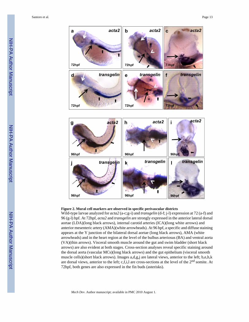
Figure 2. Mural cell markers are observed in specific perivascular districtsWild-type larvae analyzed for acta2 (a-c;g-i) and transgelin (d-f; j-l) expression at 72 (a-f) and96 (g-l) hpf. At 72hpf, acta2 and transgelin are strongly expressed in the anterior lateral dorsalaortae (LDA)(long black arrows), internal carotid arteries (ICA)(long white arrows) andanterior mesenteric artery (AMA)(white arrowheads). At 96 hpf, a specific and diffuse stainingappears at the Y junction of the bilateral dorsal aortae (long black arrows), AMA (whitearrowheads) and in the heart region at the level of the bulbus arteriosus (BA) and ventral aorta(VA)(thin arrows). Visceral smooth muscle around the gut and swim bladder (short blackarrows) are also evident at both stages. Cross-section analyses reveal specific staining aroundthe dorsal aorta (vascular MCs)(long black arrows) and the gut epithelium (visceral smoothmuscle cells)(short black arrows). Images a,d,g,j are lateral views, anterior to the left; b,e,h,kare dorsal views, anterior to the left; c,f,i,l are cross-sections at the level of the 2nd somite. At72hpf, both genes are also expressed in the fin buds (asterisks).
Santoro et al. Page 13
Mech Dev. Author manuscript; available in PMC 2010 August 1.
NIH
-PA Author Manuscript
NIH
-PA Author Manuscript
NIH
-PA Author Manuscript
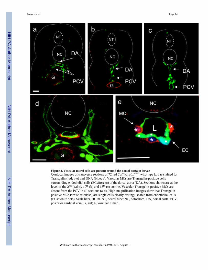
Figure 3. Vascular mural cells are present around the dorsal aorta in larvaeConfocal images of transverse sections of 72 hpf Tg(flk1:gfp)s843 wild-type larvae stained forTransgelin (red; a-e) and DNA (blue; e). Vascular MCs are Transgelin-positive cellssurrounding endothelial cells (ECs)(green) of the dorsal aorta (DA). Sections shown are at thelevel of the 2nd (a,d,e), 10th (b) and 18th (c) somite. Vascular Transgelin-positive MCs areabsent from the PCV in all sections (a-d). High-magnification images show that Transgelin-positive MCs (white asterisks) are single cells clearly distinguishable from endothelial cells(ECs: white dots). Scale bars, 20 μm. NT, neural tube; NC, notochord; DA, dorsal aorta; PCV,posterior cardinal vein; G, gut; L, vascular lumen.
Santoro et al. Page 14
Mech Dev. Author manuscript; available in PMC 2010 August 1.
NIH
-PA Author Manuscript
NIH
-PA Author Manuscript
NIH
-PA Author Manuscript

Figure 4. TEM of single mural cells surrounding endothelial vessels in larvaeTransmission electron microscopy (TEM) images of transverse sections of 72 (a,c,e) and 120(b,d,f) hpf larvae. c and e) higher magnification of MCs and ECs boxed in a). MCs and ECsexhibit different morphologies, and tight junctions are evident between ECs (black arrows). d-f) higher magnification images of MCs and ECs boxed in b. Both ECs and MCs exhibit a moreelongated morphology at 96 than 72 hpf. L: lumen. Scale bars, 5 μm.
Santoro et al. Page 15
Mech Dev. Author manuscript; available in PMC 2010 August 1.
NIH
-PA Author Manuscript
NIH
-PA Author Manuscript
NIH
-PA Author Manuscript
Figure 5. Vascular mural cells first appear in the anterior region of the dorsal aorta(a-c) Confocal images of horizontal sections of a 72 hpf Tg(flk1:GFP)s843 wild-type larvastained for Transgelin (red) show that MCs develop as a “bulge” of cells at the Y junctionbetween the lateral dorsal aortae (LDA) and the dorsal aorta (DA). Images (a-d) are ventralviews, anterior to the top. d) Schematic representation of vascular MCs (red) and ECs (green)forming the anterior trunk vasculature at 72 hpf. e) Confocal image of a transverse section ofa 72 hpf Tg(flk1:GFP)s843 wild-type larva stained for Transgelin (red). Vascular MCs arepresent and appear to originate at the ventral region of the DA. Scale bars, 20 μm. NT, neuraltube; NC, notochord; DA, dorsal aorta; PCV, posterior cardinal vein; G, gut; L, vascular lumen.
Santoro et al. Page 16
Mech Dev. Author manuscript; available in PMC 2010 August 1.
NIH
-PA Author Manuscript
NIH
-PA Author Manuscript
NIH
-PA Author Manuscript

Figure 6. Vascular mural cells derive from the lateral plate mesoderm but not from the blood orendothelial lineagesConfocal images of transverse sections, at different magnifications, of 72 hpf Tg(flk1:GFP)s843 wild-type (a,d) or cloche (clos5) (b,e) or hands-off (hans6) (c,f) mutant larvaeanalyzed for Transgelin expression (red). While wild-type (wt) and clos5 mutant larvae showTransgelin-positive cells around both the dorsal aorta and visceral organs, hans6 mutant larvaecompletely lack both vascular and visceral MCs. Sections are at the level of the1st-2nd somite.Scale bars, 20 μm. NT, neural tube; DA, dorsal aorta; SB, swim bladder; G, gut; MA, mesentericartery.
Santoro et al. Page 17
Mech Dev. Author manuscript; available in PMC 2010 August 1.
NIH
-PA Author Manuscript
NIH
-PA Author Manuscript
NIH
-PA Author Manuscript

Figure 7. Vascular mural cells contribute to the ventral aorta and the bulbus arteriosus96 hpf wild-type larvae analyzed for transgelin (a) and acta2 (b) expression. transgelin andacta2 are expressed in the bulbus arteriosus (BA) and ventral aorta (VA). (c) Confocal imageof a sagittal section of a 96 hpf Tg(flk1:EGFP)s843 wild-type larva stained for Transgelin (red)show that vascular MCs surround the VA and contribute to the BA. (d) Confocal image of atranverse section of the VA shows single MCs around the ECs (white dots). (e) Confocalprojection of the heart region of a 120 hpf Tg(flk1:EGFP)s843 wild-type larva stained forTransgelin (red). At this stage of development, vascular MCs are present in the BA as well asVA. Scale bar, 20 μm. V, ventricle; L, vascular lumen.
Santoro et al. Page 18
Mech Dev. Author manuscript; available in PMC 2010 August 1.
NIH
-PA Author Manuscript
NIH
-PA Author Manuscript
NIH
-PA Author Manuscript
Copyright © 2022 FDOKUMEN