Endogenous laminin is required for human airway smooth muscle cell maturation
Upload
khangminh22Category
view
2download
0
RESEARCH Open Access
Mural cell-derived laminin-α5 plays adetrimental role in ischemic strokeAbhijit Nirwane1, Jessica Johnson1, Benjamin Nguyen1, Jeffrey H. Miner2 and Yao Yao1*
Abstract
At the blood-brain barrier (BBB), laminin-α5 is predominantly synthesized by endothelial cells and mural cells. Endotheliallaminin-α5 is dispensable for BBB maintenance under homeostatic conditions but inhibits inflammatory cell extravasationin pathological conditions. Whether mural cell-derived laminin-α5 is involved in vascular integrity regulation, however,remains unknown. To answer this question, we generated transgenic mice with laminin-α5 deficiency in mural cells (α5-PKO). Under homeostatic conditions, no defects in BBB integrity and cerebral blood flow (CBF) were observed in α5-PKOmice, suggesting that mural cell-derived laminin-α5 is dispensable for BBB maintenance and CBF regulation underhomeostatic conditions. After ischemia-reperfusion (MCAO) injury, however, α5-PKO mice displayed less severe neuronalinjury, including reduced infarct volume, decreased neuronal death, and improved neurological function. In addition, α5-PKO mice also showed attenuated vascular damage (milder BBB disruption, reduced inflammatory cell infiltration,decreased brain edema, and diminished hemorrhagic transformation). Mechanistic studies revealed less severe tightjunction protein (TJP) loss and pericyte coverage reduction in α5-PKO mice after ischemia-reperfusion injury, indicatingthat the attenuated ischemic injury in α5-PKO mice is possibly due to less severe vascular damage. These findingssuggest that mural cell-derived laminin-α5 plays a detrimental role in ischemic stroke and that inhibiting its signalingmay have a neuroprotective effect.
Keywords: Blood-brain barrier, Ischemic stroke, MCAO, Mural cells, Laminin
IntroductionThe blood-brain barrier (BBB) is a dynamic structuremainly composed of brain microvascular endothelial cells(BMECs), pericytes, astrocytes, and a non-cellular compo-nent---the basement membrane (BM) [7, 57, 77]. Bytightly regulating substance exchange between the CNSand circulation system, the BBB functions to maintainCNS homeostasis. Accumulating evidence suggests thatBBB disruption contributes to the pathogenesis and pro-gression of various neurological disorders [48, 81, 82]. Forexample, BBB breakdown affects inflammatory cell infil-tration and is associated with the development/progres-sion of ischemia-reperfusion injury [15, 32, 75]. It shouldbe noted that the majority of BBB studies focus on its cel-lular constituents, and the role of the BM in BBB regula-tion remains largely unknown.
The BM consists of highly organized extracellularmatrix proteins synthesized by astrocytes, BMECs, andmural cells, which include both pericytes and vascularsmooth muscle cells (vSMCs) [29, 51, 67, 76]. Laminin,the only protein that is absolutely required for BM forma-tion, is a trimer composed of α, β, and γ subunits [20, 51,76, 77]. Among all five genetic variants of the α subunits,laminin-α4 and -α5 are highly expressed in blood vesselsthroughout the body [29, 67, 76]. Unlike laminin-α4,which is ubiquitously distributed in the vasculature,laminin-α5 expression shows a patchy pattern at smallervessels [73]. The major cell types that synthesizelaminin-α5 in the vasculature are BMECs and mural cells[26, 29, 46, 62, 65, 67, 80]. Recent studies demonstratedthat knockout of laminin-α5 in endothelial cells failed toaffect BBB permeability under homeostatic conditions[25, 63]. In TNFα-induced inflammation, however,these mutants showed significantly enhanced neutrophilextravasation in cremaster muscle [63]. In collagenase-in-duced intracerebral hemorrhage (ICH) model, these mu-tants displayed exacerbated inflammatory cell infiltration
* Correspondence: [email protected] of Pharmaceutical and Biomedical Sciences, University ofGeorgia, 240 W Green Street, Athens, GA 30602, USAFull list of author information is available at the end of the article
© The Author(s). 2019 Open Access This article is distributed under the terms of the Creative Commons Attribution 4.0International License (http://creativecommons.org/licenses/by/4.0/), which permits unrestricted use, distribution, andreproduction in any medium, provided you give appropriate credit to the original author(s) and the source, provide a link tothe Creative Commons license, and indicate if changes were made. The Creative Commons Public Domain Dedication waiver(http://creativecommons.org/publicdomain/zero/1.0/) applies to the data made available in this article, unless otherwise stated.
Nirwane et al. Acta Neuropathologica Communications (2019) 7:23 https://doi.org/10.1186/s40478-019-0676-8
[25]. In addition, in the experimental autoimmune en-cephalomyelitis (EAE) model, decreased T cell infiltrationinto the brain and reduced disease susceptibility & severitywere observed in laminin-α4 null mice [73], which exhib-ited compensatory and ubiquitous expression oflaminin-α5 along the vasculature [73]. These findings sug-gest that endothelial laminin-α5 plays an inhibitory role ininflammatory cell extravasation under pathological condi-tions, although it is dispensable for BBB maintenanceunder physiological conditions [25, 63]. Whether muralcell-derived laminin-α5 is involved in BBB regulationunder physiological and pathological conditions, however,remains unknown. Given that mural cell-derivedlaminin-α5 is an important component of the BM at theBBB [51, 76], we hypothesize that mural cell-derivedlaminin-α5 may also contribute to BBB integrity. In thisstudy, we investigated the functions of mural cell-derivedlaminin-α5 in BBB regulation under homeostatic condi-tions and in ischemic stroke.
Materials and methodsMiceThe experimental protocols were reviewed and approvedby the Institutional Animal Care and Use Committee atthe University of Georgia and were in accordance withthe National Institute of Health Guide for Care and Useof Laboratory Animals. The Animal Research: ReportingIn Vivo Experiments (ARRIVE) guidelines for reportingexperiments involving animals were strictly followed.Laminin-α5flox/flox mice were generated as describedpreviously [50]. Pdgfrβ-Cre+ mice were a generous giftfrom Dr. Volkhard Lindner. These two transgenic lineswere crossed to generate laminin-α5flox/flox: Pdgfrβ-Cre+
(α5-PKO) mice. Their wildtype littermates were used ascontrols. In this study, 194 mice (102 control and 92 α5-PKO) were used. All mice were housed in the animal fa-cility at the University of Georgia with free access towater and food.
Middle cerebral artery occlusion (MCAO)Eight-week-old control and α5-PKO mice were subjectedto 45min of focal cerebral ischemia produced by transi-ent intraluminal occlusion of the middle cerebral arteryusing a filament as described previously [49, 68]. Briefly,mice were anesthetized with 2,2,2-tribromoethyl alcohol(250 mg/kg, i.p.). A midline neck incision was made andthe common carotid artery (CCA), external carotid ar-tery (ECA), and internal carotid artery (ICA) on theright side were carefully isolated. The ECA and CCAwere ligated distal to the carotid bifurcation. The ICAwas clipped temporarily. A 6–0 silicone monofilamentsuture (Doccol) with a 0.21 mm diameter was introducedinto the CCA via an incision, advanced 9mm distal tothe carotid bifurcation and secured in place. Successful
occlusion of the middle cerebral artery was confirmedwith the PeriCam PSI HR system (Perimed) based onlaser speckle contrast analysis technology. Animalsshowing diminished blood flow of at least 80% duringocclusion with at least 75% recovery of blood flow afterreperfusion were used for experimentation. The bodytemperature was maintained at 37.0 ± 0.5 °C during thesurgery using a heating pad. Animals had free access tofood and water throughout the reperfusion period. Thisischemic model led to ~ 30% and ~ 20% mortality ratesfor control and α5-PKO mice, respectively.
Body weight loss and neurological functionThe body weight loss was evaluated daily from days 1 to7 after surgery. Neurological function was assessed usingthe modified neurological severity scores (mNSS) sys-tem, which evaluates motor, sensory, reflex and balancefunctions, as described previously [16, 38, 58]. Briefly,mice were scored based on their performances in a var-iety of tests as described in Additional file 1: Table S1.The sum of these scores (0–14) was used to reflect theirneurological function after MCAO. Higher scores indi-cate worse neurological function. Animals were habitu-ated to the testing environment prior to experimentsand the investigator who scored the animals was blindedto the genotypes.
Brain sectioningSerial sectioning was used in this study. Briefly,20 μm-thick serial sections were cut with Cryostat (Mi-cro HM 550, Thermo Scientific). Eight sections evenlydistributed along the rostral-to-caudal axis were col-lected from each brain.
Infarct volume and neuronal deathBrain infarct volume was quantified as infarct volume per-centage (%) as described previously [43, 53, 56]. Briefly,cresyl violet-stained brain sections were imaged using theNikon Eclipse Ti microscope. The areas of the contralat-eral hemisphere (Ci), ipsilateral hemisphere (Ii), and ipsi-lateral non-ischemic region (Ni) were determined usingthe Image J software (NIH), and the infarct volume (%)
was calculated as:Infarctvolumeð%Þ ¼ ð
X
i
ððIi−Ni
IiÞCiÞ
2P
iCi
Þx100.Neuronal death was assessed using Fluoro-Jade C (FJC)
staining as described previously [59, 79]. Specifically, thenumber of FJC+ cells was counted in each field. At least 3random fields from each section, 8 serial sections perbrain, and 4 animals were used for quantification.
Nirwane et al. Acta Neuropathologica Communications (2019) 7:23 Page 2 of 18
BBB permeabilityEvans blue (EB) and/or FITC-Dextran (4kD) were usedto assess BBB permeability as described previously [15].Briefly, control and α5-PKO mice were injectedretro-orbitally with 80 μl EB (2%, Sigma E2129) and/or50 μl FITC-Dextran (25 mg/ml, Sigma FD4). Fornon-ischemic study, FITC-Dextran was allowed to cir-culate for 12 h. After transcardial perfusion, the brainswere collected, homogenized in formamide, and centri-fuged at 20,000 rpm for 20 min. The fluorescenceintensity of the supernatant was measured using aSpectraMax M2 plate reader (Molecular Devices) at450/550 nm. Mice without FITC-Dextran injection wereused to determine baseline reading, which was sub-tracted from raw reading to obtain FITC-Dextran leak-age. Leakage in α5-PKO mice was normalized to that incontrols. For ischemic study, both tracers were injected4 h before mice were transcardially perfused at eachtime point after injury. Each brain hemisphere was ho-mogenized in formamide and centrifuged at 20,000 rpmfor 20 min. The absorbance and fluorescence intensityof the supernatant were measured using a SpectraMaxM2 plate reader at 620 nm and 450/550 nm, respect-ively. EB or FITC-Dextran leakage was defined as thedifference of absorbance or fluorescence intensity be-tween contralateral and ipsilateral hemispheres. Leak-age in α5-PKO mice was normalized to that in controls.
Brain edemaBrain edema was assessed using both brain water con-tent [79] and brain swelling [33] as described previously.Briefly, control and α5-PKO mice were transcardiallyperfused with PBS. Brains were collected and cut intoleft and right hemispheres. The weights of each hemi-sphere before and after drying at 85 °C for 4 h were mea-sured and defined as wet and dry weights, respectively.Brain water content (%) was calculated as (Wet Weight -Dry Weight) / Wet Weight × 100. Brain swelling (%) wascalculated as (Final wet weightipsi – Initial wet weightipsi)/ Initial wet weightipsi × 100. In this equation, final wetweightipsi is the wet weight of ipsilateral hemisphere. Ini-tial wet weightipsi is defined as (Wet weightcontra / Dryweightcontra) x Dry weightipsi.
Immunofluorescence analysesImmunofluorescence analyses were performed accordingto standard protocols. Briefly, brain sections and/or cellswere fixed in 4% PFA for 15 min at room temperatureand washed in PBS 3 times. Next, the sections and cellswere blocked in blocking buffer (5% normal donkeyserum in PBS + 1% BSA + 0.3% Triton X-100) for 2 h atroom temperature, followed by incubation withanti-Laminin-α2 (1:400, Sigma L0663), anti-Laminin-α5(1:800, generated as described in [47]), anti-Smooth
Muscle Actin-α (SMA)-FITC (1:1000, Sigma F3777),anti-Hemoglobin (1:200, Cloud-Clone PAB409Mu01),anti-Ly6G (1:200, Biolegend 108,402), anti-CD3 (1:200,eBioscience 14–0032-82), anti-CD68 (1:200, Biolegend137,002), anti-PDGFRβ (1:200, Cell Signaling 3169S),anti-ZO-1(1:400, ThermoFisher 61–7300), anti-Claudin-5 (1:200, Invitrogen 35–2500), anti-AQP4 (1:200, Milli-pore AB3594), and anti-CD31 (1:200, BD Bioscience553,370) antibodies overnight at 4 °C. After extensivewashes in PBS, the sections and/or cells were incubatedwith the following secondary antibodies: Alexa Fluor-488 conjugated donkey anti-rabbit (1:1000, InvitrogenA21206), Alexa Fluor-594 conjugated donkey anti-rabbit(1:1000, Invitrogen A21207), FITC conjugated goatanti-mouse (1:500, BD Pharmingen 554,001), AlexaFluor-594 conjugated donkey anti-mouse (1:1000, Invi-trogen A21203), FITC conjugated goat anti-rat (1:500,BD Pharmingen 554,016), Alexa Fluor-594 conjugateddonkey anti-rat (1:1000, Invitrogen A21209), and AlexaFluor-647 conjugated goat anti-rat (1:1000, InvitrogenA21247) for 2 h at room temperature. Then, the sectionsand/or cells were washed in PBS 3 times and mountedin Fluoromount-G with DAPI. Images were taken undera Nikon Eclipse Ti microscope or LSM710 confocalmicroscope. Image processing was performed using Ima-geJ and Adobe Photoshop.
Image analysesBrain angioarchitecture analyses were performed usingthe open source “Angiotool” software (National CancerInstitute, USA) as described previously [83]. Specifically,CD31-stained brain sections were used for analyses. Ves-sel length, defined as the sum of Euclidean distances be-tween the pixels of all vessels; vessel density, defined asthe percentage of area occupied by vessels inside the ex-plant area; and branching index, defined as the numberof vessel junctions per unit area, were computed in thecortex and striatum. Thresholding was applied to re-move small particles so that only actual vessels werequantified [24]. For quantification, 8 serial sections alongthe rostral-to-caudal axis were analyzed for each brainand 4 mice were used. Data in α5-PKO mice were nor-malized to that in controls.For pericyte coverage, PDGFRβ- and CD31-positive
fluorescent areas were determined using ImageJ areameasurement tool. Pericyte coverage was determined asthe percentage (%) of PDGFRβ-positive fluorescent areacovering CD31-positive capillary area, as previously de-scribed [8]. For TJP and AQP4 coverage, ZO-1/Clau-din-5/AQP4- and CD31-positive fluorescent areas weredetermined using ImageJ area measurement tool. ZO-1/Claudin-5/AQP4 coverage was determined as the per-centage (%) of ZO-1/Claudin-5/AQP4-positive fluores-cent area covering CD31-positive capillary area. For
Nirwane et al. Acta Neuropathologica Communications (2019) 7:23 Page 3 of 18

inflammatory cell infiltration, total numbers of Ly6G+/CD3+/CD68+ cells were counted. For hemoglobin stain-ing, mean fluorescence intensity was used. For quantifi-cation, at least three random fields from each section, 8serial sections along the rostral-to-caudal axis for eachbrain, and 4–5 animals were used. All data analyses wereperformed on z-projection (10-12 μm) images by a blindedinvestigator.For laminin-α5 immunocytochemistry, the percentage
of laminin-α5+ cells were calculated. For quantification,6 independent experiments were performed and at least50 cells were examined in each experiment.
Brain mural cell isolationPrimary mural cells were isolated from mouse brainsusing a well-established protocol. Briefly, brains werecollected under aseptic conditions. After removing men-inges, the brains were minced with a blade and tritu-rated. Brain tissue was then incubated with 0.1%collagenase at 37 °C for 1 h followed by centrifuge at700 g for 8 min. The pellet was resuspended in 17% ster-ile dextran solution and centrifuged at 6000 g for 20 minat 4 °C. Blood vessel-containing pellet was washed inDMEM for 3 times and further digested in 1 mg/ml col-lagenase/dispase (Roche, 11,097,113,001) for 2 h withconstant shaking at 37 °C. Next, red blood cells (RBCs)were removed by washing the pellet in RBC lysis buffer.The pellet was resuspended in sorting buffer and passedthrough a 40 μm cell strainer. The single-cell solutionwas then stained with anti-CD31-APC (1:100, Biolegend102,509), anti-CD45-FITC (1:100, Biolegend 103,108),and anti-Pdgfrβ-PE (1:100, eBioscience 12–1402) for 30min at 4 °C. After extensive wash, the cells were sub-jected to FACS. Sorted mural cells (Pdgfrβ+CD31−
CD45−) were grown in Pericyte Medium (ScienCell,1201) and used for immunocytochemistry.
Transmission electron microscopy (TEM)Eight-week-old control and α5-PKO mice were anesthe-tized and perfused with PBS followed by 0.1M sodiumcacodylate buffer containing 2% paraformaldehyde and2% glutaraldehyde. After perfusion, brain tissue was dis-sected out, fixed overnight, and post-fixed in 1% osmiumtetroxide and 1% K-ferrocyanide. Next, the tissue was enbloc stained with 2% uranyl acetate and embedded inresin. Ultra-thin sections were cut on an RMC MT-Xmicrotome (Boeckeler Instruments) and post-stainedwith 2% uranyl acetate and 1% lead citrate. Sectionswere examined and photographed using JEOL JEM1011(JEOL) at 80 kV.
Western blottingCortex and striatum were carefully dissected and imme-diately homogenized on ice. Total protein concentration
was determined using the BCA Protein Assay Kit (Pierce23,227). Equal amount of protein was loaded and sepa-rated in SDS-PAGE and transferred to PVDF mem-branes (Millipore). Next, the membranes were probedwith primary antibodies [anti-Laminin-α2 (1:500, SigmaL0663), anti-Laminin α5 (1:800, generated as describedin [47]), Claudin-5 (1:500, ThermoFisher 35–2500),ZO-1 (1:500, ThermoFisher 61–7300), and anti-GAPDH(1:1000, Abcam AB9484)] over night at 4 °C, followed byappropriate horseradish peroxidase-conjugated second-ary antibodies [donkey anti-mouse (1:2500, JacksonImmunoResearch Laboratory 715–035-151), donkeyanti-rabbit (1:2500, Jackson ImmunoResearch Labora-tory 711–035-152), and donkey anti-rat (1:2500, JacksonImmunoResearch Laboratory 712–035-153] at roomtemperature for 1 h. Then, target proteins were visual-ized using the ChemiDoc Imaging System (Bio-Rad). Forquantification, the density of target blots was normalizedto that of GAPDH, and the expression of target proteinsin α5-PKO brains was normalized to that in controlbrains. Four animals were used for quantification.
Statistical analysesAll statistical analyses were performed using the Graph-Pad Prism 6 software. For normally distributed measure-ments, unpaired Student’s t-test was used to determinestatistical significance between two groups, and one-wayanalysis of variance (ANOVA) followed by Tukeypost-hoc test was used for three or more groups. Formeasurements that are not normally distributed, thenon-parametric Mann-Whitney U test (two groups) andKruskal-Wallis test (three or more group) were used.Significance was set at p < 0.05. Data were presented asmean ± SD.
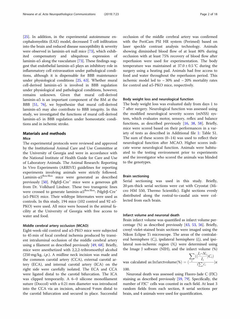
ResultsLaminin-α5 is indeed abrogated in mural cells in α5-PKOmiceThe α5-PKO mice are born at the expected Mendelian ra-tio, fail to show gross abnormalities, and have a normallifespan. Using lineage-tracing technique, we have demon-strated that Pdgfrβ-Cre specifically targets mural cells inthe brain [26]. Immunohistochemistry revealedlaminin-α2 and laminin-α5 expression in the cortex ofboth control and α5-PKO mice (Fig. 1a). To quantitativelydetermine the expression levels of laminin-α2 andlaminin-α5, western blot analysis was performed usingcortical tissue. As expected, comparable levels oflaminin-α2 were found in control and α5-PKO mice (Fig.1b). Laminin-α5, on the other hand, was slightly reducedin α5-PKO mice, although statistical significance was notreached (Fig. 1b). Similar results were observed in the stri-atum (not shown). The residual expression of laminin-α5in α5-PKO brains is probably from endothelial cells, which
Nirwane et al. Acta Neuropathologica Communications (2019) 7:23 Page 4 of 18
synthesize laminin-511 and -411 [29, 62, 65]. To furtherdetermine if laminin-α5 expression is abrogated in muralcells in α5-PKO mice, we isolated primary mural cellsfrom control and α5-PKO brains using a well-establishedprotocol [9, 26, 78] and examined laminin-α5 expressionin these cells. Isolated cells expressed mural cell markerSMA (Fig. 1c), suggesting they were indeed mural cells.Immunocytochemistry revealed laminin-α5 expression incontrol but not α5-PKO mural cells (Fig. 1c). Quantifica-tion showed that almost all control mural cells expressedlaminin-α5, whereas more than 95% of α5-PKO muralcells were negative for laminin-α5 (Fig. 1d). These resultsindicate that laminin-α5 is indeed abrogated in mural cellsin α5-PKO mice.
Brain angioarchitecture is unaffected in α5-PKO mice underhomeostatic conditionsTo determine if α5-PKO mice have abnormal brainangioarchitecture, we analyzed vessel length, vessel dens-ity, and branching index in both cortex and striatum usingthe “Angiotool” software. None of these parametersshowed significant differences in the cortex (Additionalfile 1: Figure S1) or striatum (not shown) in α5-PKO micecompared to the controls (Additional file 1: Figure S1),
strongly suggesting that loss of laminin-α5 in mural cellsdoes not affect brain angioarchitecture.
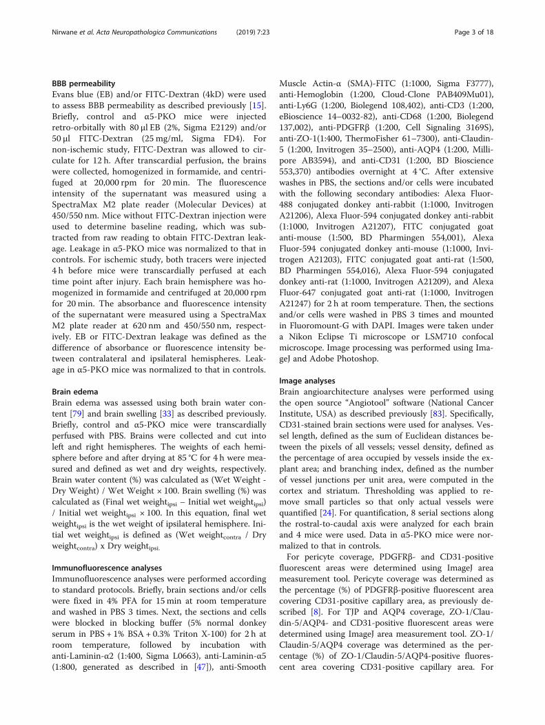
BBB integrity and cerebral blood flow (CBF) are unchangedin α5-PKO mice under homeostatic conditionsTo investigate if BBB integrity is disrupted in α5-PKOmice, IgG leakage was examined by immunohistochemis-try. No IgG signal was detected in the cortex (Fig. 2a) orstriatum (not shown) of control or α5-PKO mice, suggest-ing that the BBB is not leaky to molecules with a size of150kD or above. Next, FITC-Dextran (4kD), a smaller dye,was used to assess BBB integrity. Comparable levels ofFITC-Dextran were found in control and α5-PKO brains(Fig. 2b), suggesting intact BBB integrity in α5-PKO miceunder homeostatic conditions. To investigate if CBF is al-tered in α5-PKO mice, real-time CBF in middle cerebralartery regions was measured. Representative laser speckleimages of CBF in control and α5-PKO mice are shown inFig. 2c. Quantification revealed no significant difference inCBF between control and α5-PKO mice (Fig. 2d), suggest-ing unaffected CBF in α5-PKO mice under homeostaticconditions. Together, these findings suggest that loss ofmural cell-derived laminin-α5 does not affect BBB integ-rity or CBF under homeostatic conditions.
Fig. 1 Lama5 expression is abrogated in mural cells in α5-PKO mice. a Representative images of laminin-α2 (green) and laminin-α5 (red) stainingin the cortex of control and α5-PKO mice. Scale bar = 100 μm. b Representative western blotting and quantification of laminin-α2 and laminin-α5levels in the cortex of control and α5-PKO mice. n = 4. c Representative images of smooth muscle actin-α (SMA, green) and laminin-α5 (red)staining in primary mural cells isolated from control and α5-PKO brains. Scale bar = 50 μm. d Quantification showing the lack of laminin-α5expression in primary mural cells isolated from α5-PKO brains. n = 6 independent experiments with at least 50 cells examined in each experiment.Student’s t-test, ***p < 0.001, compared to controls
Nirwane et al. Acta Neuropathologica Communications (2019) 7:23 Page 5 of 18
TJP expression and tight junction structure are unalteredin α5-PKO mice under homeostatic conditionsBMECs express high levels of TJPs at the intercellularspace forming tight junctions, which contribute to BBBintegrity [3, 17, 78]. To determine if TJP expression isaltered, we examined the levels of two TJPs, ZO-1 andclaudin-5, in control and α5-PKO brains. Immunohis-tochemistry revealed similar distribution patterns ofZO-1 and claudin-5 in control and α5-PKO brains.Specifically, both proteins co-localized well with bloodvessel marker CD31, in both cortex (Fig. 3a, b) and stri-atum (not shown). Similarly, western blotting was per-formed to quantify TJP expression in control andα5-PKO brains. No significant differences in ZO-1 andclaudin-5 levels were observed between genotypes in ei-ther cortex (Fig. 3c) or striatum (not shown). Consist-ent with these biochemical findings, TEM studyrevealed no obvious defects in the structure of tightjunctions (Fig. 3d, arrowheads). These results suggestthat mural cell-derived laminin-α5 plays a dispensablerole in the regulation of TJP expression and tight junc-tion structure under homeostatic conditions.
Pericyte coverage and astrocyte polarity are unaltered inα5-PKO mice under homeostatic conditionsPericyte coverage on capillaries plays an important rolein maintaining BBB integrity [4, 5, 8, 18]. To determine
if pericyte coverage is altered in α5-PKO brains, we per-formed immunohistochemistry against PDGFRβ andCD31 (Fig. 4a). Quantification revealed comparable peri-cyte coverage in control and α5-PKO mice in both cor-tex (Fig. 4b) and striatum (not shown), suggesting thatpericyte coverage is unaffected in α5-PKO mice underhomeostatic conditions.Astrocytes express AQP4 exclusively in their end-
feet, contributing to BBB maintenance [5, 26, 78]. Todetermine if the polarized distribution of AQP4 isaltered in α5-PKO brains, we performed immunohis-tochemistry against AQP4 and CD31 (Fig. 4c). Quanti-fication revealed comparable AQP4 coverage incontrol and α5-PKO mice in both cortex (Fig. 4d) andstriatum (not shown), suggesting that astrocyte polar-ity is unchanged in α5-PKO mice under homeostaticconditions.
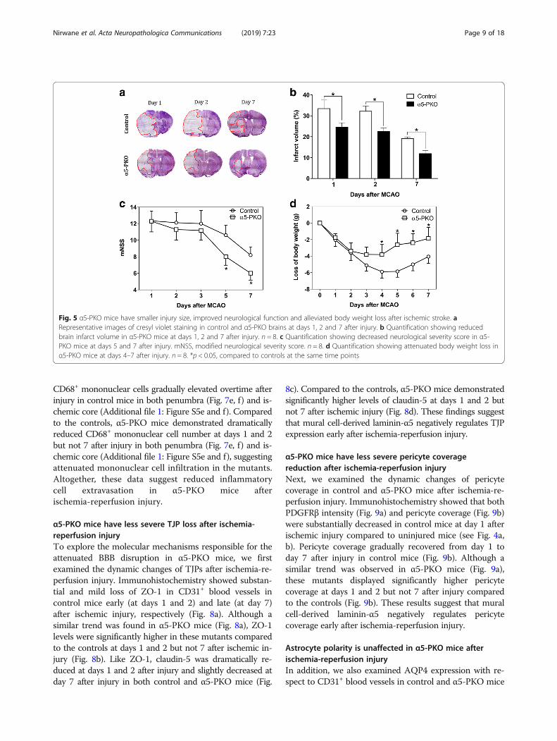
α5-PKO mice have smaller infarct volume and improvedneurological function after ischemia-reperfusion injuryTo investigate the function of mural cell-derivedlaminin-α5 in ischemia-reperfusion injury, we firstexamined brain infarct volume at various time pointsafter MCAO. Control mice demonstrated large infarctvolume at days 1 and 2 after injury, which was reduceddramatically by day 7 after injury (Fig. 5a). A similar trendwas observed in α5-PKO mice (Fig. 5a). Quantification
Fig. 2 BBB integrity and CBF are unaffected in α5-PKO mice under homeostatic conditions. a Representative images of IgG (red) and CD31(green) staining in the cortex of control and α5-PKO mice. Scale bar = 100 μm. b Quantification showing comparable levels of FITC-Dextran incontrol and α5-PKO brains. n = 5. c Representative laser speckle images of CBF/brain perfusion in control and α5-PKO brains. d Quantificationshowing similar levels of CBF/brain perfusion in control and α5-PKO brains. n = 5
Nirwane et al. Acta Neuropathologica Communications (2019) 7:23 Page 6 of 18

revealed significantly smaller infarct volume (Fig. 5b) inα5-PKO mice at all three time points compared to thecontrols, suggesting reduced ischemic injury. To visualizethe spatial distribution of infarct areas in control andα5-PKO brains at day 1 after injury, 5 brain sections alongthe rostral-to-caudal axis (with equal distance) were usedfor analyses. Similarly, α5-PKO mice demonstrated dimin-ished infarct volume compared to the controls (Additionalfile 1: Figure S2). Consistent with the reduced infarctvolume, significantly lower neurological severity scorewas detected in α5-PKO mice at days 5 and 7 after in-jury (Fig. 5c), indicating improved neurological func-tion. In addition, the α5-PKO mice also displayedsubstantially less body weight loss at days 4–7 after in-jury (Fig. 5d). Together, these results suggest betterpathological and functional outcomes in α5-PKO miceafter ischemia-reperfusion injury. To determine any gen-der differences, these parameters were also analyzed in agender-specific manner. Compared to male mice, femalemice showed smaller infarct volume, lower neurologicalscore, and less body weight loss independent of genotype,
although these changes did not reach statistical signifi-cance (Additional file 1: Figure S3).
α5-PKO mice have reduced neuronal death afterischemia-reperfusion injuryTo investigate if loss of mural cell-derived laminin-α5 af-fects neuronal death after ischemic injury, we performedFJC staining, which labels degenerating neurons [11, 72].FJC+ cells were identified in both penumbra (Additionalfile 1: Figure S4a) and ischemic core (Additional file 1:Figure S4c) in control and α5-PKO brains. Consistentwith previous finding that FJC+ cells peak at 24 h afterischemic injury [41], quantitative data showed a continu-ous decline of FJC+ cell number in both penumbra(Additional file 1: Figure S4b) and ischemic core(Additional file 1: Figure S4d) in control mice from days1 to 7 after ischemia-reperfusion injury. Although a simi-lar trend was found in α5-PKO mice, these mutantsshowed significantly fewer FJC+ cells in both regions(Additional file 1: Figure S4) at all three time points
Fig. 3 Tight junctions are unaffected in α5-PKO mice under homeostatic conditions. a,b Representative images of ZO-1 (green)/CD31 (red) (a)and Claudin-5 (green)/CD31 (red) (b) staining in the cortex of control and α5-PKO mice. Scale bar = 50 μm. c Representative western blotting andquantification of ZO-1 and Claudin-5 levels in the cortex of control and α5-PKO mice. n = 4. d TEM images showing normal tight junctionstructure in control and α5-PKO brains. Black arrowheads indicate tight junctions. TEM, transmission electron microscopy. Scale bar = 800 nm
Nirwane et al. Acta Neuropathologica Communications (2019) 7:23 Page 7 of 18

compared to the controls. These results suggest reducedneuronal death in α5-PKO mice after ischemic injury.
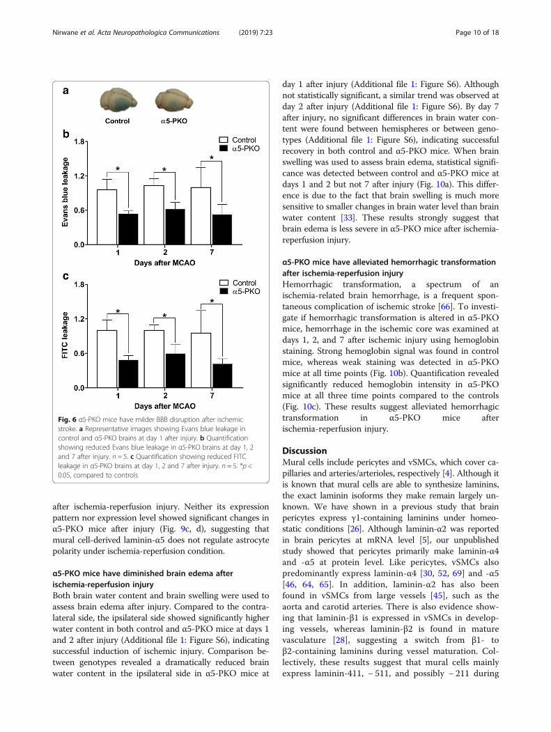
α5-PKO mice have milder BBB disruption after ischemia-reperfusion injuryBBB permeability was assessed by measuring EB andFITC-Dextran leakage at days 1, 2 and 7 after injury.Representative whole-brain images showing EB leakagein control and α5-PKO mice at day 1 after injury areshown in Fig. 6a. Compared to the controls, significantlyreduced EB leakage was detected in α5-PKO mice atdays 1, 2 and 7 after injury (Fig. 6b). Consistent with EBdata, dramatically diminished FITC-Dextran leakage wasfound in α5-PKO mice at all three time points (Fig. 6c).These results suggest milder BBB damage in α5-PKOmice after ischemia-reperfusion injury.
α5-PKO mice have decreased inflammatory cell infiltrationafter ischemia-reperfusion injuryAccumulating evidence demonstrates that immune cellsinfiltrate into the brain and modulate disease progres-sion after ischemic stroke [27, 31]. To investigate if
inflammatory cell extravasation is affected in α5-PKOmice, we examined the infiltration of Ly6G+ neutrophils,CD3+ lymphocytes, and CD68+ mononuclear cells inboth penumbra and ischemic core at days 1, 2, and 7after injury. In control mice, the number of Ly6G+ neu-trophils peaked at early time points (days 1 and 2) afterinjury and gradually declined over time in both penumbra(Fig. 7a, b) and ischemic core (Additional file 1: Figures S5aand b). Compared to the controls, α5-PKO mice showedsignificantly decreased Ly6G+ neutrophil number at days 1and 2 but not 7 after injury in both penumbra (Fig. 7a, b)and ischemic core (Additional file 1: Figures S5a and b),suggesting diminished neutrophil infiltration in the mu-tants. Unlike Ly6G+ neutrophils, the number of CD3+ lym-phocytes gradually increased over time after injury incontrol mice in both penumbra (Fig. 7c, d) and ischemiccore (Additional file 1: Figure S5c and d). Compared to thecontrols, α5-PKO mice displayed substantially less CD3+
lymphocytes in both penumbra (Fig. 7c, d) and ischemiccore (Additional file 1: Figure S5c and d) at all three timepoints, suggesting decreased lymphocyte infiltration in themutants. Similar to CD3+ lymphocytes, the number of
Fig. 4 Pericyte coverage and AQP4 coverage are unaffected in α5-PKO mice under homeostatic conditions. a Representative images of PDGFRβ(green) and CD31 (red) staining in the cortex of control and α5-PKO mice. Scale bar = 50 μm. b Quantification showing comparable pericytecoverage in the cortex of control and α5-PKO mice. n = 4. c Representative images of AQP4 (green) and CD31 (red) staining in the cortex ofcontrol and α5-PKO mice. Scale bar = 50 μm. d Quantification showing comparable AQP4 coverage in the cortex of control and α5-PKO mice.n = 4. AQP4, aquaporin-4
Nirwane et al. Acta Neuropathologica Communications (2019) 7:23 Page 8 of 18
CD68+ mononuclear cells gradually elevated overtime afterinjury in control mice in both penumbra (Fig. 7e, f) and is-chemic core (Additional file 1: Figure S5e and f). Comparedto the controls, α5-PKO mice demonstrated dramaticallyreduced CD68+ mononuclear cell number at days 1 and 2but not 7 after injury in both penumbra (Fig. 7e, f) and is-chemic core (Additional file 1: Figure S5e and f), suggestingattenuated mononuclear cell infiltration in the mutants.Altogether, these data suggest reduced inflammatorycell extravasation in α5-PKO mice afterischemia-reperfusion injury.
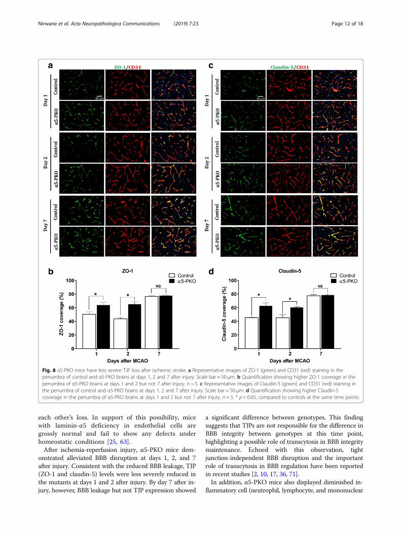
α5-PKO mice have less severe TJP loss after ischemia-reperfusion injuryTo explore the molecular mechanisms responsible for theattenuated BBB disruption in α5-PKO mice, we firstexamined the dynamic changes of TJPs after ischemia-re-perfusion injury. Immunohistochemistry showed substan-tial and mild loss of ZO-1 in CD31+ blood vessels incontrol mice early (at days 1 and 2) and late (at day 7)after ischemic injury, respectively (Fig. 8a). Although asimilar trend was found in α5-PKO mice (Fig. 8a), ZO-1levels were significantly higher in these mutants comparedto the controls at days 1 and 2 but not 7 after ischemic in-jury (Fig. 8b). Like ZO-1, claudin-5 was dramatically re-duced at days 1 and 2 after injury and slightly decreased atday 7 after injury in both control and α5-PKO mice (Fig.
8c). Compared to the controls, α5-PKO mice demonstratedsignificantly higher levels of claudin-5 at days 1 and 2 butnot 7 after ischemic injury (Fig. 8d). These findings suggestthat mural cell-derived laminin-α5 negatively regulates TJPexpression early after ischemia-reperfusion injury.
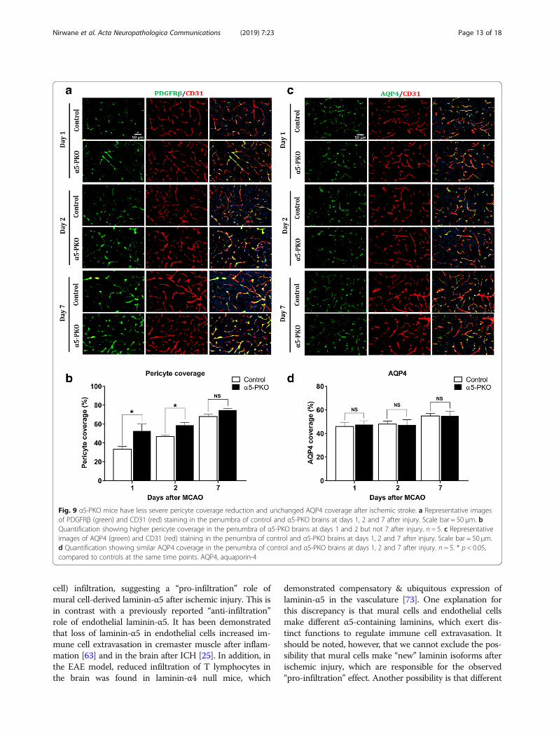
α5-PKO mice have less severe pericyte coveragereduction after ischemia-reperfusion injuryNext, we examined the dynamic changes of pericytecoverage in control and α5-PKO mice after ischemia-re-perfusion injury. Immunohistochemistry showed that bothPDGFRβ intensity (Fig. 9a) and pericyte coverage (Fig. 9b)were substantially decreased in control mice at day 1 afterischemic injury compared to uninjured mice (see Fig. 4a,b). Pericyte coverage gradually recovered from day 1 today 7 after injury in control mice (Fig. 9b). Although asimilar trend was observed in α5-PKO mice (Fig. 9a),these mutants displayed significantly higher pericytecoverage at days 1 and 2 but not 7 after injury comparedto the controls (Fig. 9b). These results suggest that muralcell-derived laminin-α5 negatively regulates pericytecoverage early after ischemia-reperfusion injury.
Astrocyte polarity is unaffected in α5-PKO mice afterischemia-reperfusion injuryIn addition, we also examined AQP4 expression with re-spect to CD31+ blood vessels in control and α5-PKO mice
Fig. 5 α5-PKO mice have smaller injury size, improved neurological function and alleviated body weight loss after ischemic stroke. aRepresentative images of cresyl violet staining in control and α5-PKO brains at days 1, 2 and 7 after injury. b Quantification showing reducedbrain infarct volume in α5-PKO mice at days 1, 2 and 7 after injury. n = 8. c Quantification showing decreased neurological severity score in α5-PKO mice at days 5 and 7 after injury. mNSS, modified neurological severity score. n = 8. d Quantification showing attenuated body weight loss inα5-PKO mice at days 4–7 after injury. n = 8. *p < 0.05, compared to controls at the same time points
Nirwane et al. Acta Neuropathologica Communications (2019) 7:23 Page 9 of 18
after ischemia-reperfusion injury. Neither its expressionpattern nor expression level showed significant changes inα5-PKO mice after injury (Fig. 9c, d), suggesting thatmural cell-derived laminin-α5 does not regulate astrocytepolarity under ischemia-reperfusion condition.
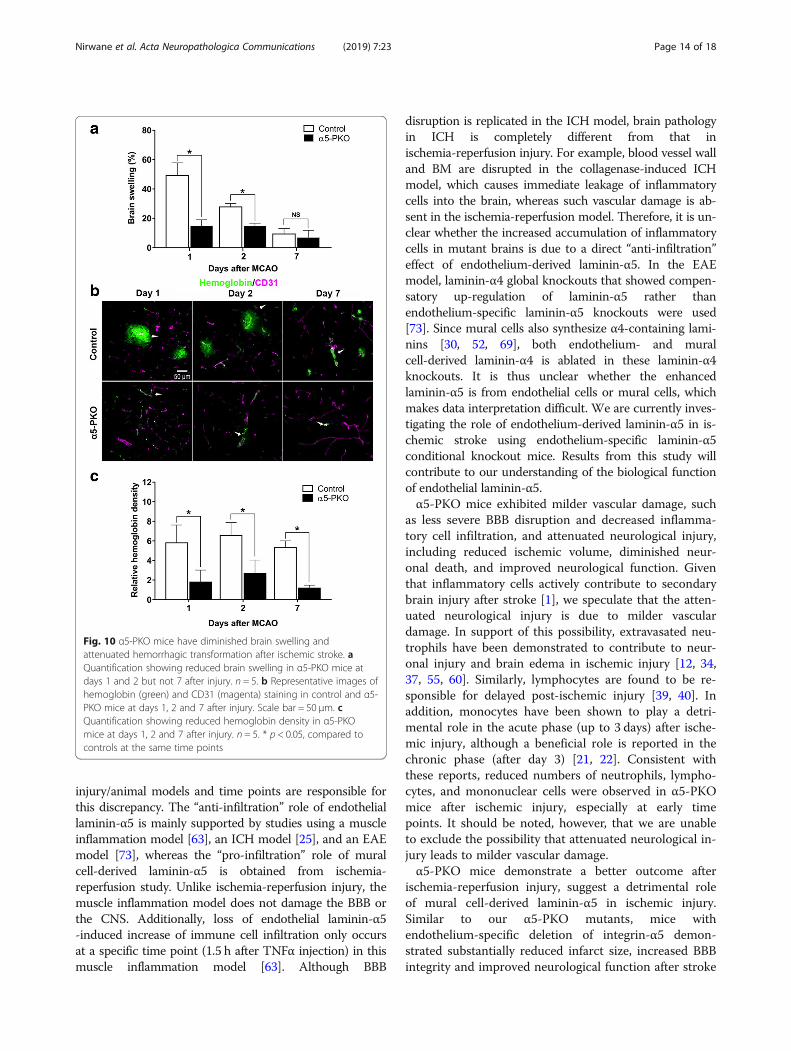
α5-PKO mice have diminished brain edema afterischemia-reperfusion injuryBoth brain water content and brain swelling were used toassess brain edema after injury. Compared to the contra-lateral side, the ipsilateral side showed significantly higherwater content in both control and α5-PKO mice at days 1and 2 after injury (Additional file 1: Figure S6), indicatingsuccessful induction of ischemic injury. Comparison be-tween genotypes revealed a dramatically reduced brainwater content in the ipsilateral side in α5-PKO mice at
day 1 after injury (Additional file 1: Figure S6). Althoughnot statistically significant, a similar trend was observed atday 2 after injury (Additional file 1: Figure S6). By day 7after injury, no significant differences in brain water con-tent were found between hemispheres or between geno-types (Additional file 1: Figure S6), indicating successfulrecovery in both control and α5-PKO mice. When brainswelling was used to assess brain edema, statistical signifi-cance was detected between control and α5-PKO mice atdays 1 and 2 but not 7 after injury (Fig. 10a). This differ-ence is due to the fact that brain swelling is much moresensitive to smaller changes in brain water level than brainwater content [33]. These results strongly suggest thatbrain edema is less severe in α5-PKO mice after ischemia-reperfusion injury.
α5-PKO mice have alleviated hemorrhagic transformationafter ischemia-reperfusion injuryHemorrhagic transformation, a spectrum of anischemia-related brain hemorrhage, is a frequent spon-taneous complication of ischemic stroke [66]. To investi-gate if hemorrhagic transformation is altered in α5-PKOmice, hemorrhage in the ischemic core was examined atdays 1, 2, and 7 after ischemic injury using hemoglobinstaining. Strong hemoglobin signal was found in controlmice, whereas weak staining was detected in α5-PKOmice at all time points (Fig. 10b). Quantification revealedsignificantly reduced hemoglobin intensity in α5-PKOmice at all three time points compared to the controls(Fig. 10c). These results suggest alleviated hemorrhagictransformation in α5-PKO mice afterischemia-reperfusion injury.
DiscussionMural cells include pericytes and vSMCs, which cover ca-pillaries and arteries/arterioles, respectively [4]. Although itis known that mural cells are able to synthesize laminins,the exact laminin isoforms they make remain largely un-known. We have shown in a previous study that brainpericytes express γ1-containing laminins under homeo-static conditions [26]. Although laminin-α2 was reportedin brain pericytes at mRNA level [5], our unpublishedstudy showed that pericytes primarily make laminin-α4and -α5 at protein level. Like pericytes, vSMCs alsopredominantly express laminin-α4 [30, 52, 69] and -α5[46, 64, 65]. In addition, laminin-α2 has also beenfound in vSMCs from large vessels [45], such as theaorta and carotid arteries. There is also evidence show-ing that laminin-β1 is expressed in vSMCs in develop-ing vessels, whereas laminin-β2 is found in maturevasculature [28], suggesting a switch from β1- toβ2-containing laminins during vessel maturation. Col-lectively, these results suggest that mural cells mainlyexpress laminin-411, − 511, and possibly − 211 during
Fig. 6 α5-PKO mice have milder BBB disruption after ischemicstroke. a Representative images showing Evans blue leakage incontrol and α5-PKO brains at day 1 after injury. b Quantificationshowing reduced Evans blue leakage in α5-PKO brains at day 1, 2and 7 after injury. n = 5. c Quantification showing reduced FITCleakage in α5-PKO brains at day 1, 2 and 7 after injury. n = 5. *p <0.05, compared to controls
Nirwane et al. Acta Neuropathologica Communications (2019) 7:23 Page 10 of 18

development; and laminin-421, − 521, and possibly −221 in adulthood under homeostatic conditions.In this study, we failed to detect any changes in BBB
permeability and CBF between control and α5-PKO miceunder physiological conditions, suggesting that muralcell-derived α5-containing laminins are dispensable forBBB maintenance and CBF regulation under homeostaticconditions. Unlike these α5-PKO mice, mutants withlaminin-γ1 deficiency (all γ1-containing laminins) inmural cells showed BBB breakdown and hydrocephalus inC57Bl6-FVB mixed background [26], suggesting an im-portant role of mural cell-derived γ1-containing laminins
in BBB maintenance and hydrocephalus pathogenesis,although we cannot exclude the possibility that BBB dis-ruption is secondary to hydrocephalus. Together, thesefindings suggest the existence of compensation betweenmural cell-derived α5-containing laminins and α4/α2-con-taining laminins. In addition, it is also possible that thelack of phenotype in α5-PKO mice under homeostaticconditions is due to compensation by laminin isoformsfrom endothelial cells and/or astrocytes, which are in closeproximity of mural cells [67]. For example, mural cell-derived α5-containing laminins and endothelial cell-derived laminin-511 may be able to compensate for
Fig. 7 α5-PKO mice show decreased inflammatory cell infiltration after ischemic stroke. a Representative images of Ly6G (red) staining in thepenumbra of control and α5-PKO brains at days 1, 2 and 7 after injury. Scale bar = 50 μm. b Quantification showing reduced extravasation ofLy6G+ neutrophils in the penumbra of α5-PKO brains at days 1 and 2 but not 7 after injury. n = 5. c Representative images of CD3 (red) stainingin the penumbra of control and α5-PKO brains at days 1, 2 and 7 after injury. Scale bar = 50 μm. d Quantification showing reduced extravasationof CD3+ lymphocytes in the penumbra of α5-PKO brains at days 1, 2 and 7 after injury. n = 5. e Representative images of CD68 (red) staining inthe penumbra of control and α5-PKO brains at days 1, 2 and 7 after injury. Scale bar = 50 μm. f Quantification showing reduced extravasation ofCD68+ mononuclear cells in the penumbra of α5-PKO brains at days 1 and 2 but not 7 after injury. n = 5. * p < 0.05, compared to controls at thesame time points
Nirwane et al. Acta Neuropathologica Communications (2019) 7:23 Page 11 of 18
each other’s loss. In support of this possibility, micewith laminin-α5 deficiency in endothelial cells aregrossly normal and fail to show any defects underhomeostatic conditions [25, 63].After ischemia-reperfusion injury, α5-PKO mice dem-
onstrated alleviated BBB disruption at days 1, 2, and 7after injury. Consistent with the reduced BBB leakage, TJP(ZO-1 and claudin-5) levels were less severely reduced inthe mutants at days 1 and 2 after injury. By day 7 after in-jury, however, BBB leakage but not TJP expression showed
a significant difference between genotypes. This findingsuggests that TJPs are not responsible for the difference inBBB integrity between genotypes at this time point,highlighting a possible role of transcytosis in BBB integritymaintenance. Echoed with this observation, tightjunction-independent BBB disruption and the importantrole of transcytosis in BBB regulation have been reportedin recent studies [2, 10, 17, 36, 71].In addition, α5-PKO mice also displayed diminished in-
flammatory cell (neutrophil, lymphocyte, and mononuclear
Fig. 8 α5-PKO mice have less severe TJP loss after ischemic stroke. a Representative images of ZO-1 (green) and CD31 (red) staining in thepenumbra of control and α5-PKO brains at days 1, 2 and 7 after injury. Scale bar = 50 μm. b Quantification showing higher ZO-1 coverage in thepenumbra of α5-PKO brains at days 1 and 2 but not 7 after injury. n = 5. c Representative images of Claudin-5 (green) and CD31 (red) staining inthe penumbra of control and α5-PKO brains at days 1, 2 and 7 after injury. Scale bar = 50 μm. d Quantification showing higher Claudin-5coverage in the penumbra of α5-PKO brains at days 1 and 2 but not 7 after injury. n = 5. * p < 0.05, compared to controls at the same time points
Nirwane et al. Acta Neuropathologica Communications (2019) 7:23 Page 12 of 18
cell) infiltration, suggesting a “pro-infiltration” role ofmural cell-derived laminin-α5 after ischemic injury. This isin contrast with a previously reported “anti-infiltration”role of endothelial laminin-α5. It has been demonstratedthat loss of laminin-α5 in endothelial cells increased im-mune cell extravasation in cremaster muscle after inflam-mation [63] and in the brain after ICH [25]. In addition, inthe EAE model, reduced infiltration of T lymphocytes inthe brain was found in laminin-α4 null mice, which
demonstrated compensatory & ubiquitous expression oflaminin-α5 in the vasculature [73]. One explanation forthis discrepancy is that mural cells and endothelial cellsmake different α5-containing laminins, which exert dis-tinct functions to regulate immune cell extravasation. Itshould be noted, however, that we cannot exclude the pos-sibility that mural cells make “new” laminin isoforms afterischemic injury, which are responsible for the observed“pro-infiltration” effect. Another possibility is that different
Fig. 9 α5-PKO mice have less severe pericyte coverage reduction and unchanged AQP4 coverage after ischemic stroke. a Representative imagesof PDGFRβ (green) and CD31 (red) staining in the penumbra of control and α5-PKO brains at days 1, 2 and 7 after injury. Scale bar = 50 μm. bQuantification showing higher pericyte coverage in the penumbra of α5-PKO brains at days 1 and 2 but not 7 after injury. n = 5. c Representativeimages of AQP4 (green) and CD31 (red) staining in the penumbra of control and α5-PKO brains at days 1, 2 and 7 after injury. Scale bar = 50 μm.d Quantification showing similar AQP4 coverage in the penumbra of control and α5-PKO brains at days 1, 2 and 7 after injury. n = 5. * p < 0.05,compared to controls at the same time points. AQP4, aquaporin-4
Nirwane et al. Acta Neuropathologica Communications (2019) 7:23 Page 13 of 18
injury/animal models and time points are responsible forthis discrepancy. The “anti-infiltration” role of endotheliallaminin-α5 is mainly supported by studies using a muscleinflammation model [63], an ICH model [25], and an EAEmodel [73], whereas the “pro-infiltration” role of muralcell-derived laminin-α5 is obtained from ischemia-reperfusion study. Unlike ischemia-reperfusion injury, themuscle inflammation model does not damage the BBB orthe CNS. Additionally, loss of endothelial laminin-α5-induced increase of immune cell infiltration only occursat a specific time point (1.5 h after TNFα injection) in thismuscle inflammation model [63]. Although BBB
disruption is replicated in the ICH model, brain pathologyin ICH is completely different from that inischemia-reperfusion injury. For example, blood vessel walland BM are disrupted in the collagenase-induced ICHmodel, which causes immediate leakage of inflammatorycells into the brain, whereas such vascular damage is ab-sent in the ischemia-reperfusion model. Therefore, it is un-clear whether the increased accumulation of inflammatorycells in mutant brains is due to a direct “anti-infiltration”effect of endothelium-derived laminin-α5. In the EAEmodel, laminin-α4 global knockouts that showed compen-satory up-regulation of laminin-α5 rather thanendothelium-specific laminin-α5 knockouts were used[73]. Since mural cells also synthesize α4-containing lami-nins [30, 52, 69], both endothelium- and muralcell-derived laminin-α4 is ablated in these laminin-α4knockouts. It is thus unclear whether the enhancedlaminin-α5 is from endothelial cells or mural cells, whichmakes data interpretation difficult. We are currently inves-tigating the role of endothelium-derived laminin-α5 in is-chemic stroke using endothelium-specific laminin-α5conditional knockout mice. Results from this study willcontribute to our understanding of the biological functionof endothelial laminin-α5.α5-PKO mice exhibited milder vascular damage, such
as less severe BBB disruption and decreased inflamma-tory cell infiltration, and attenuated neurological injury,including reduced ischemic volume, diminished neur-onal death, and improved neurological function. Giventhat inflammatory cells actively contribute to secondarybrain injury after stroke [1], we speculate that the atten-uated neurological injury is due to milder vasculardamage. In support of this possibility, extravasated neu-trophils have been demonstrated to contribute to neur-onal injury and brain edema in ischemic injury [12, 34,37, 55, 60]. Similarly, lymphocytes are found to be re-sponsible for delayed post-ischemic injury [39, 40]. Inaddition, monocytes have been shown to play a detri-mental role in the acute phase (up to 3 days) after ische-mic injury, although a beneficial role is reported in thechronic phase (after day 3) [21, 22]. Consistent withthese reports, reduced numbers of neutrophils, lympho-cytes, and mononuclear cells were observed in α5-PKOmice after ischemic injury, especially at early timepoints. It should be noted, however, that we are unableto exclude the possibility that attenuated neurological in-jury leads to milder vascular damage.α5-PKO mice demonstrate a better outcome after
ischemia-reperfusion injury, suggest a detrimental roleof mural cell-derived laminin-α5 in ischemic injury.Similar to our α5-PKO mutants, mice withendothelium-specific deletion of integrin-α5 demon-strated substantially reduced infarct size, increased BBBintegrity and improved neurological function after stroke
Fig. 10 α5-PKO mice have diminished brain swelling andattenuated hemorrhagic transformation after ischemic stroke. aQuantification showing reduced brain swelling in α5-PKO mice atdays 1 and 2 but not 7 after injury. n = 5. b Representative images ofhemoglobin (green) and CD31 (magenta) staining in control and α5-PKO mice at days 1, 2 and 7 after injury. Scale bar = 50 μm. cQuantification showing reduced hemoglobin density in α5-PKOmice at days 1, 2 and 7 after injury. n = 5. * p < 0.05, compared tocontrols at the same time points
Nirwane et al. Acta Neuropathologica Communications (2019) 7:23 Page 14 of 18
[54], highlighting an adverse effect of endothelialintegrin-α5 in ischemic stroke. Together, these findingssuggest that mural cell-derived α5-containing lamininsand endothelial integrin-α5 may use a converging signal-ing pathway to modulate the development/progressionof ischemic stroke, although integrin-α5 is not a classicallaminin receptor [6, 74]. Identifying the receptors anddownstream signaling pathways may provide innovativemolecular targets with therapeutic potential in ischemicstroke.Due to the multiphasic nature of ischemic stroke, this
study has a few limitations. First, only the transient is-chemic model was used in this study. The transient is-chemic model involves both ischemia and reperfusion.However, most strokes found in human patients only in-volve ischemia without reperfusion [19, 23, 42, 61, 70].Thus, it is important to test the biological function ofmural cell-derived laminin-α5 in the permanent ische-mic model. Second, only one ischemic duration (45 min)was used in this study. It is known that longer occlusioncauses more severe injury [44, 49]. Currently, various is-chemic durations ranging from 30 to 120 min have beenused in rodent MCAO studies [13, 14, 35, 49]. We chose45-min ischemia for two reasons: (1) compared to otherdurations, 45-min ischemia consistently induced signifi-cant ischemic injury with less mortality in our hands,and (2) significant differences in stroke outcomes be-tween control and α5-PKO mice were observed with45-min ischemia. Other ischemic durations should betested in future studies. Third, only young mice wereused in this study. Aging is a risk factor for ischemicstroke and actively influences stroke outcomes [19, 23,42]. Therefore, it is important to examine the biologicalfunction of mural cell-derived laminin-α5 in ischemicstroke using aged mice in the future. Fourth, unlikeprevious studies reporting improved outcomes in youngfemale mice [19, 23, 42], we failed to observe genderdifferences in infarct volume, neurological severityscore, and body weight loss. It should be noted that,although not statistically significant, a trend toward at-tenuated injury was observed in female mice independ-ent of genotype. This discrepancy may be explained bythe relatively small animal number used in each groupand/or other factors, such as the severity of injury andsensitivity of assays. Future research is needed to ad-dress these limitations.
ConclusionsCollectively, our results suggest that mural cell-derivedlaminin-α5 is dispensable for BBB maintenance and CBFregulation under homeostatic condition. In ischemicstroke, however, loss of mural cell-derived laminin-α5 at-tenuates vascular damage and improves stroke outcomes,
indicating a detrimental role of mural cell-derivedlaminin-α5 in ischemic stroke. These findings identifymural cell-derived laminin-α5 as a molecular target withtherapeutic potential in ischemic stroke.
Additional file
Additional file 1: Table S1. Modified Neurologic Severity Scores(mNSS) system. Figure S1. Angioarchitecture is unaltered in α5-PKOmice under homeostatic conditions. Figure S2. Spatial distribution ofinfarct area in control and α5-PKO mice at day 1 after ischemic injury.Figure S3. Comparison of gender-specific effects in control and α5-PKOmice after ischemic stroke. Figure S4. α5-PKO mice show reduced neuronaldeath in both penumbra and ischemic core after ischemic stroke. Figure S5.α5-PKO mice have reduced inflammatory cell infiltration after ischemicstroke. Figure S6. α5-PKO mice have reduced brain water content afterischemic stroke. (DOCX 2830 kb)
AbbreviationsBBB: Blood-brain barrier; BM: Basement membrane; BMECs: Brainmicrovascular endothelial cells; CBF: Cerebral blood flow; CCA: Commoncarotid artery; CNS: Central nervous system; EAE: Experimental autoimmuneencephalomyelitis; ECA: External carotid artery; ICA: Internal carotid artery;ICH: Intracerebral hemorrhage; MCAO: Middle cerebral artery occlusion;SMA: Smooth muscle actin-α; TEM: Transmission electron microscopy;TJP: Tight junction protein; vSMCs: Vascular smooth muscle cells
AcknowledgmentsWe thank Dr. Volkhard Lindner for the Pdgfrβ-Cre mice.
FundingThis study was supported, in part, by the American Heart Association grant16SDG29320001 (to YY) and NIH R01DK078314 (to JHM).
Availability of data and materialsAll data generated or analyzed during this study are included in thispublished article and its supplementary information files.
Authors’ contributionsYY conceived & designed the experiments, performed data analyses, andwrote the manuscript; AN performed the experiments, collected & analyzedthe data, wrote the manuscript; JJ and BN contributed to data analyses; JHMgenerated the laminin-α5 floxed mice & laminin-α5 antibody and contrib-uted to data analyses. All authors have read and approved the finalsubmission.
Ethics approvalAll applicable international, national, and/or institutional guidelines for thecare and use of animals were followed. All procedures performed in studiesinvolving animals were in accordance with the ethical standards of theinstitution or practice at which the studies were conducted.
Consent for publicationNot applicable.
Competing interestsThe authors declare that they have no competing interests.
Publisher’s NoteSpringer Nature remains neutral with regard to jurisdictional claims inpublished maps and institutional affiliations.
Author details1Department of Pharmaceutical and Biomedical Sciences, University ofGeorgia, 240 W Green Street, Athens, GA 30602, USA. 2Division ofNephrology, Department of Medicine, Washington University School ofMedicine, St. Louis, MO, USA.
Nirwane et al. Acta Neuropathologica Communications (2019) 7:23 Page 15 of 18
Received: 31 December 2018 Accepted: 10 February 2019
References1. Ahmad M, Graham SH (2010) Inflammation after stroke: mechanisms and
therapeutic approaches. Transl Stroke Res 1:74–84. https://doi.org/10.1007/s12975-010-0023-7
2. Andreone BJ, Chow BW, Tata A, Lacoste B, Ben-Zvi A, Bullock K, Deik AA, GintyDD, Clish CB, Gu C (2017) Blood-brain barrier permeability is regulated by lipidtransport-dependent suppression of Caveolae-mediated transcytosis. Neuron94:581–594. https://doi.org/10.1016/j.neuron.2017.03.043
3. Andreone BJ, Lacoste B, Gu C (2015) Neuronal and vascular interactions.Annu Rev Neurosci 38:25–46. https://doi.org/10.1146/annurev-neuro-071714-033835
4. Armulik A, Genove G, Betsholtz C (2011) Pericytes: developmental,physiological, and pathological perspectives, problems, and promises. DevCell 21:193–215. https://doi.org/10.1016/j.devcel.2011.07.001
5. Armulik A, Genove G, Mae M, Nisancioglu MH, Wallgard E, Niaudet C, He L,Norlin J, Lindblom P, Strittmatter K et al (2010) Pericytes regulate the blood-brain barrier. Nature 468:557–561. https://doi.org/10.1038/nature09522
6. Aumailley M (2013) The laminin family. Cell Adhes Migr 7:48–55. https://doi.org/10.4161/cam.22826
7. Baeten KM, Akassoglou K (2011) Extracellular matrix and matrix receptors inblood-brain barrier formation and stroke. Dev Neurobiol 71:1018–1039.https://doi.org/10.1002/dneu.20954
8. Bell RD, Winkler EA, Sagare AP, Singh I, LaRue B, Deane R, Zlokovic BV (2010)Pericytes control key neurovascular functions and neuronal phenotype inthe adult brain andduring brain aging. Neuron 68:409–427. https://doi.org/10.1016/j.neuron.2010.09.043
9. Bell RD, Winkler EA, Singh I, Sagare AP, Deane R, Wu Z, Holtzman DM,Betsholtz C, Armulik A, Sallstrom J et al (2012) Apolipoprotein E controlscerebrovascular integrity via cyclophilin A. Nature 485:512–516. https://doi.org/10.1038/nature11087nature11087
10. Ben-Zvi A, Lacoste B, Kur E, Andreone BJ, Mayshar Y, Yan H, Gu C (2014)Mfsd2a is critical for the formation and function of the blood-brain barrier.Nature 509:507–511. https://doi.org/10.1038/nature13324
11. Bian GL, Wei LC, Shi M, Wang YQ, Cao R, Chen LW (2007) Fluoro-jade C canspecifically stain the degenerative neurons in the substantia nigra of the 1-methyl-4-phenyl-1,2,3,6-tetrahydro pyridine-treated C57BL/6 mice. Brain Res1150:55–61. https://doi.org/10.1016/j.brainres.2007.02.078
12. Buck BH, Liebeskind DS, Saver JL, Bang OY, Yun SW, Starkman S, Ali LK, KimD, Villablanca JP, Salamon N et al (2008) Early neutrophilia is associated withvolume of ischemic tissue in acute stroke. Stroke 39:355–360. https://doi.org/10.1161/STROKEAHA.107.490128
13. Cai M, Zhang W, Weng Z, Stetler RA, Jiang X, Shi Y, Gao Y, Chen J (2017)Promoting neurovascular recovery in aged mice after ischemic stroke-prophylactic effect of Omega-3 polyunsaturated fatty acids. Aging Dis 8:531–545. https://doi.org/10.14336/AD.2017.0520
14. Carmichael ST (2005) Rodent models of focal stroke: size, mechanism, andpurpose. NeuroRx 2:396–409. https://doi.org/10.1602/neurorx.2.3.396
15. Chen B, Friedman B, Cheng Q, Tsai P, Schim E, Kleinfeld D, Lyden PD (2009)Severe blood-brain barrier disruption and surrounding tissue injury. Stroke40:e666–e674. https://doi.org/10.1161/STROKEAHA.109.551341
16. Chen J, Zhang C, Jiang H, Li Y, Zhang L, Robin A, Katakowski M, Lu M,Chopp M (2005) Atorvastatin induction of VEGF and BDNF promotes brainplasticity after stroke in mice. J Cereb Blood Flow Metab 25:281–290.https://doi.org/10.1038/sj.jcbfm.9600034
17. Chow BW, Gu C (2015) The molecular constituents of the blood-brainbarrier. Trends Neurosci 38:598–608. https://doi.org/10.1016/j.tins.2015.08.003
18. Daneman R, Zhou L, Kebede AA, Barres BA (2010) Pericytes are required forblood-brain barrier integrity during embryogenesis. Nature 468:562–566.https://doi.org/10.1038/nature09513
19. Dirnagl U (2006) Bench to bedside: the quest for quality in experimentalstroke research. J Cereb Blood Flow Metab 26:1465–1478. https://doi.org/10.1038/sj.jcbfm.9600298
20. Durbeej M (2010) Laminins. Cell Tissue Res 339:259–268. https://doi.org/10.1007/s00441-009-0838-2
21. ElAli A, Jean LeBlanc N (2016) The role of monocytes in ischemic strokepathobiology: new avenues to explore. Front Aging Neurosci 8:29. https://doi.org/10.3389/fnagi.2016.00029
22. Fang W, Zhai X, Han D, Xiong X, Wang T, Zeng X, He S, Liu R, Miyata M, XuB et al (2018) CCR2-dependent monocytes/macrophages exacerbate acutebrain injury but promote functional recovery after ischemic stroke in mice.Theranostics 8:3530–3543. https://doi.org/10.7150/thno.24475
23. Fisher M, Feuerstein G, Howells DW, Hurn PD, Kent TA, Savitz SI, Lo EH,Group S (2009) Update of the stroke therapy academic industry roundtablepreclinical recommendations. Stroke 40:2244–2250. https://doi.org/10.1161/STROKEAHA.108.541128
24. Gambardella L, Zudaire E, Vermeren S (2012) Quantitative analysis ofangiogenesis in the allantois explant model. The textbook of angiogenesis andLymphangiogenesis: methods and applications. Springer, Dordrecht, pp 189–204
25. Gautam J, Miner JH, Yao Y (2019) Loss of endothelial laminin alpha5exacerbates hemorrhagic brain injury. Transl Stroke Res. https://doi.org/10.1007/s12975-019-0688-5
26. Gautam J, Zhang X, Yao Y (2016) The role of pericytic laminin in bloodbrain barrier integrity maintenance. Sci Rep 6(36450). https://doi.org/10.1038/srep36450
27. Gesuete R, Stevens SL, Stenzel-Poore MP (2016) Role of circulating immunecells in stroke and preconditioning-induced protection. Acta NeurochirSuppl 121:39–44. https://doi.org/10.1007/978-3-319-18497-5_7
28. Glukhova M, Koteliansky V, Fondacci C, Marotte F, Rappaport L (1993)Laminin variants and integrin laminin receptors in developing and adulthuman smooth muscle. Dev Biol 157:437–447. https://doi.org/10.1006/dbio.1993.1147
29. Hannocks MJ, Pizzo ME, Huppert J, Deshpande T, Abbott NJ, Thorne RG,Sorokin L (2018) Molecular characterization of perivascular drainagepathways in the murine brain. J Cereb Blood Flow Metab 38:669–686.https://doi.org/10.1177/0271678X17749689
30. Iivanainen A, Kortesmaa J, Sahlberg C, Morita T, Bergmann U, Thesleff I,Tryggvason K (1997) Primary structure, developmental expression, andimmunolocalization of the murine laminin alpha4 chain. J Biol Chem 272:27862–27868. https://doi.org/10.1074/jbc.272.44.27862
31. Jin R, Yang G, Li G (2010) Inflammatory mechanisms in ischemic stroke: roleof inflammatory cells. J Leukoc Biol 87:779–789. https://doi.org/10.1189/jlb.1109766
32. Jin X, Liu J, Yang Y, Liu KJ, Yang Y, Liu W (2012) Spatiotemporal evolution ofblood brain barrier damage and tissue infarction within the first 3h afterischemia onset. Neurobiol Dis 48:309–316. https://doi.org/10.1016/j.nbd.2012.07.007
33. Keep RF, Hua Y, Xi G (2012) Brain water content: a misunderstoodmeasurement? Transl Stroke Res 3:263–265. https://doi.org/10.1007/s12975-012-0152-2
34. Kim J, Song TJ, Park JH, Lee HS, Nam CM, Nam HS, Kim YD, Heo JH (2012)Different prognostic value of white blood cell subtypes in patients withacute cerebral infarction. Atherosclerosis 222:464–467. https://doi.org/10.1016/j.atherosclerosis.2012.02.042
35. Kraft P, Göb E, Schuhmann MK, Göbel K, Deppermann C, Thielmann I,Herrmann AM, Lorenz K, Brede M, Stoll G et al (2013) FTY720 amelioratesacute ischemic stroke in mice by reducing thrombo-inflammation but notby direct neuroprotection. Stroke 44:3202–3210. https://doi.org/10.1161/STROKEAHA.113.002880
36. Krueger M, Hartig W, Reichenbach A, Bechmann I, Michalski D (2013) Blood-brain barrier breakdown after embolic stroke in rats occurs withoutultrastructural evidence for disrupting tight junctions. PLoS One 8:e56419.https://doi.org/10.1371/journal.pone.0056419
37. Kumar AD, Boehme AK, Siegler JE, Gillette M, Albright KC, Martin-Schild S(2013) Leukocytosis in patients with neurologic deterioration after acuteischemic stroke is associated with poor outcomes. J Stroke Cerebrovasc Dis22:e111–e117. https://doi.org/10.1016/j.jstrokecerebrovasdis.2012.08.008
38. Li Y, Chen J, Wang L, Lu M, Chopp M (2001) Treatment of stroke in rat withintracarotid administration of marrow stromal cells. Neurology 56:1666–1672. https://doi.org/10.1212/WNL.56.12.1666
39. Liesz A, Hu X, Kleinschnitz C, Offner H (2015) Functional role of regulatorylymphocytes in stroke: facts and controversies. Stroke 46:1422–1430. https://doi.org/10.1161/STROKEAHA.114.008608
40. Liesz A, Zhou W, Mracsko E, Karcher S, Bauer H, Schwarting S, Sun L, BruderD, Stegemann S, Cerwenka A et al (2011) Inhibition of lymphocytetrafficking shields the brain against deleterious neuroinflammation afterstroke. Brain 134:704–720. https://doi.org/10.1093/brain/awr008
41. Liu F, Schafer DP, McCullough LD (2009) TTC, fluoro-Jade B and NeuNstaining confirm evolving phases of infarction induced by middle cerebral
Nirwane et al. Acta Neuropathologica Communications (2019) 7:23 Page 16 of 18
artery occlusion. J Neurosci Methods 179:1–8. https://doi.org/10.1016/j.jneumeth.2008.12.028
42. Macleod MR, Fisher M, O'Collins V, Sena ES, Dirnagl U, Bath PM, Buchan A,van der Worp HB, Traystman R, Minematsu K et al (2009) Good laboratorypractice: preventing introduction of bias at the bench. Stroke 40:e50–e52.https://doi.org/10.1161/STROKEAHA.108.525386
43. McBride DW, Klebe D, Tang J, Zhang JH (2015) Correcting for brainswelling’s effects on infarct volume calculation after middle cerebral arteryocclusion in rats. Transl Stroke Res 6:323–338. https://doi.org/10.1007/s12975-015-0400-3
44. McColl BW, Carswell HV, McCulloch J, Horsburgh K (2004) Extension ofcerebral hypoperfusion and ischaemic pathology beyond MCA territoryafter intraluminal filament occlusion in C57Bl/6J mice. Brain Res 997:15–23.https://doi.org/10.1016/j.brainres.2003.10.028
45. McLean SE, Mecham BH, Kelleher CM, Mariani TJ, Mecham RP (2005)Extracellular matrix gene expression in the developing mouse aorta. AdvDev Biol 15:81–128. https://doi.org/10.1016/S1574-3349(05)15003-0
46. Miner JH, Lewis RM, Sanes JR (1995) Molecular cloning of a novel lamininchain, alpha 5, and widespread expression in adult mouse tissues. J BiolChem 270:28523–28526. https://doi.org/10.1074/jbc.270.48.28523
47. Miner JH, Patton BL, Lentz SI, Gilbert DJ, Snider WD, Jenkins NA, CopelandNG, Sanes JR (1997) The laminin alpha chains: expression, developmentaltransitions, and chromosomal locations of alpha1-5, identification ofheterotrimeric laminins 8-11, and cloning of a novel alpha3 isoform. J CellBiol 137:685–701. https://doi.org/10.1083/jcb.137.3.685
48. Montagne A, Barnes SR, Sweeney MD, Halliday MR, Sagare AP, Zhao Z, TogaAW, Jacobs RE, Liu CY, Amezcua L et al (2015) Blood-brain barrierbreakdown in the aging human hippocampus. Neuron 85:296–302. https://doi.org/10.1016/j.neuron.2014.12.032
49. Morris GP, Wright AL, Tan RP, Gladbach A, Ittner LM, Vissel B (2016) Acomparative study of variables influencing ischemic injury in the longa andKoizumi methods of intraluminal filament middle cerebral artery occlusionin mice. PLoS One 11:e0148503. https://doi.org/10.1371/journal.pone.0148503
50. Nguyen NM, Kelley DG, Schlueter JA, Meyer MJ, Senior RM, Miner JH (2005)Epithelial laminin alpha5 is necessary for distal epithelial cell maturation,VEGF production, and alveolization in the developing murine lung. Dev Biol282:111–125. https://doi.org/10.1016/j.ydbio.2005.02.031
51. Nirwane A, Yao Y (2019) Laminins and their receptors in the CNS. Biol RevCamb Philos Soc 94:283–306. https://doi.org/10.1111/brv.12454
52. Petajaniemi N, Korhonen M, Kortesmaa J, Tryggvason K, Sekiguchi K,Fujiwara H, Sorokin L, Thornell LE, Wondimu Z, Assefa D et al (2002)Localization of laminin alpha4-chain in developing and adult human tissues.J Histochem Cytochem 50:1113–1130. https://doi.org/10.1177/002215540205000813
53. Renolleau S, Aggoun-Zouaoui D, Ben-Ari Y, Charriaut-Marlangue C (1998) Amodel of transient unilateral focal ischemia with reperfusion in the P7neonatal rat: morphological changes indicative of apoptosis. Stroke 29:1454–1460. https://doi.org/10.1161/01.STR.29.7.1454
54. Roberts J, de Hoog L, Bix GJ (2017) Mice deficient in endothelial α5 integrinare profoundly resistant to experimental ischemic stroke. J Cereb BloodFlow Metab 37:85–96. https://doi.org/10.1177/0271678X15616979
55. Ross AM, Hurn P, Perrin N, Wood L, Carlini W, Potempa K (2007) Evidence ofthe peripheral inflammatory response in patients with transient ischemicattack. J Stroke Cerebrovasc Dis 16:203–207. https://doi.org/10.1016/j.jstrokecerebrovasdis.2007.05.002
56. Rousselet E, Kriz J, Seidah NG (2012) Mouse model of intraluminal MCAO:cerebral infarct evaluation by cresyl violet staining. J Vis Exp:e4038. https://doi.org/10.3791/4038
57. Sá-Pereira I, Brites D, Brito MA (2012) Neurovascular unit: a focus onpericytes. Mol Neurobiol 45:327–347. https://doi.org/10.1007/s12035-012-8244-2
58. Schaar KL, Brenneman MM, Savitz SI (2010) Functional assessments in therodent stroke model. Exp Transl Stroke Med 2:13. https://doi.org/10.1186/2040-7378-2-13
59. Schmued LC, Stowers CC, Scallet AC, Xu L (2005) Fluoro-Jade C results inultra high resolution and contrast labeling of degenerating neurons. BrainRes 1035:24–31. https://doi.org/10.1016/j.brainres.2004.11.054
60. Segel GB, Halterman MW, Lichtman MA (2011) The paradox of theneutrophilˈs role in tissue injury. J Leukoc Biol 89:359–372. https://doi.org/10.1189/jlb.0910538
61. Sena ES, van der Worp HB, Bath PM, Howells DW, Macleod MR (2010)Publication bias in reports of animal stroke studies leads to majoroverstatement of efficacy. PLoS Biol 8:e1000344. https://doi.org/10.1371/journal.pbio.1000344
62. Sixt M, Engelhardt B, Pausch F, Hallmann R, Wendler O, Sorokin LM (2001)Endothelial cell laminin isoforms, laminins 8 and 10, play decisive roles in T cellrecruitment across the blood-brain barrier in experimental autoimmuneencephalomyelitis. J Cell Biol 153:933–946. https://doi.org/10.1083/jcb.153.5.933
63. Song J, Zhang X, Buscher K, Wang Y, Wang H, Di Russo J, Li L, Lütke-EnkingS, Zarbock A, Stadtmann A et al (2017) Endothelial basement membranelaminin 511 contributes to endothelial junctional tightness and therebyinhibits leukocyte transmigration. Cell Rep 18:1256–1269. https://doi.org/10.1016/j.celrep.2016.12.092
64. Sorokin LM, Pausch F, Durbeej M, Ekblom P (1997) Differential expression offive laminin alpha (1-5) chains in developing and adult mouse kidney. DevDyn 210:446–462. https://doi.org/10.1002/(SICI)1097-0177(199712)210:4<446::AID-AJA8>3.0.CO;2-G
65. Sorokin LM, Pausch F, Frieser M, Kroger S, Ohage E, Deutzmann R (1997)Developmental regulation of the laminin alpha5 chain suggests a role inepithelial and endothelial cell maturation. Dev Biol 189:285–300. https://doi.org/10.1006/dbio.1997.8668
66. Sussman ES, Connolly ES Jr (2013) Hemorrhagic transformation: a review ofthe rate of hemorrhage in the major clinical trials of acute ischemic stroke.Front Neurol 4:69. https://doi.org/10.3389/fneur.2013.00069
67. Thomsen MS, Routhe LJ, Moos T (2017) The vascular basement membranein the healthy and pathological brain. J Cereb Blood Flow Metab 37:3300–3317. https://doi.org/10.1177/0271678X17722436
68. Thored P, Wood J, Arvidsson A, Cammenga J, Kokaia Z, Lindvall O (2007)Long-term neuroblast migration along blood vessels in an area withtransient angiogenesis and increased vascularization after stroke. Stroke 38:3032–3039. https://doi.org/10.1161/STROKEAHA.107.488445
69. Thyboll J, Kortesmaa J, Cao R, Soininen R, Wang L, Iivanainen A, Sorokin L,Risling M, Cao Y, Tryggvason K (2002) Deletion of the laminin alpha4 chainleads to impaired microvessel maturation. Mol Cell Biol 22:1194–1202.https://doi.org/10.1128/MCB.22.4.1194-1202.2002
70. van der Worp HB, Howells DW, Sena ES, Porritt MJ, Rewell S, O'Collins V,Macleod MR (2010) Can animal models of disease reliably inform humanstudies? PLoS Med 7:e1000245. https://doi.org/10.1371/journal.pmed.1000245
71. Villasenor R, Lampe J, Schwaninger M, Collin L (2018) Intracellular transportand regulation of transcytosis across the blood-brain barrier. Cell Mol LifeSci. https://doi.org/10.1007/s00018-018-2982-x
72. Wang J, Tsirka SE (2005) Tuftsin fragment 1-3 is beneficial when deliveredafter the induction of intracerebral hemorrhage. Stroke 36:613–618. https://doi.org/10.1161/01.STR.0000155729.12931.8f
73. Wu C, Ivars F, Anderson P, Hallmann R, Vestweber D, Nilsson P, Robenek H,Tryggvason K, Song J, Korpos E et al (2009) Endothelial basementmembrane laminin α5 selectively inhibits T lymphocyte extravasation intothe brain. Nat Med 15:519–527. https://doi.org/10.1038/nm.1957
74. Wu X, Reddy DS (2012) Integrins as receptor targets for neurologicaldisorders. Pharmacol Ther 134:68–81. https://doi.org/10.1016/j.pharmthera.2011.12.008
75. Yang Y, Rosenberg GA (2011) Blood-brain barrier breakdown in acute andchronic cerebrovascular disease. Stroke 42:3323–3328. https://doi.org/10.1161/STROKEAHA.110.608257
76. Yao Y (2017) Laminin: loss-of-function studies. Cell Mol Life Sci 74:1095–1115. https://doi.org/10.1007/s00018-016-2381-0
77. Yao Y (2019) Basement membrane and stroke. J Cereb Blood Flow Metab39:3–19. https://doi.org/10.1177/0271678X18801467
78. Yao Y, Chen ZL, Norris EH, Strickland S (2014) Astrocytic laminin regulatespericyte differentiation and maintains blood brain barrier integrity. NatCommun 5(3413). https://doi.org/10.1038/ncomms4413
79. Yao Y, Tsirka SE (2012) The CCL2-CCR2 system affects the progression andclearance of intracerebral hemorrhage. Glia 60:908–918. https://doi.org/10.1002/glia.22323
80. Yousif LF, Di Russo J, Sorokin L (2013) Laminin isoforms in endothelial andperivascular basement membranes. Cell Adhes Migr 7:101–110. https://doi.org/10.4161/cam.22680
81. Zhao Z, Nelson AR, Betsholtz C, Zlokovic BV (2015) Establishment anddysfunction of the blood-brain barrier. Cell 163:1064–1078. https://doi.org/10.1016/j.cell.2015.10.067
Nirwane et al. Acta Neuropathologica Communications (2019) 7:23 Page 17 of 18
82. Zlokovic BV (2008) The blood-brain barrier in health and chronicneurodegenerative disorders. Neuron 57:178–201. https://doi.org/10.1016/j.neuron.2008.01.003
83. Zudaire E, Gambardella L, Kurcz C, Vermeren S (2011) A computational toolfor quantitative analysis of vascular networks. PLoS One 6:e27385. https://doi.org/10.1371/journal.pone.0027385
Nirwane et al. Acta Neuropathologica Communications (2019) 7:23 Page 18 of 18
Copyright © 2022 FDOKUMEN