Seaweed Extracts as Substitutes of Synthetic Hormones for ...
Upload
independentCategory
view
4download
0
J. Funct. Biomater. 2013, 4, 162-177; doi:10.3390/jfb4030162
Journal of
Functional
Biomaterials ISSN 2079-4983
www.mdpi.com/journal/jfb/
Article
Epoxy Cross-Linked Collagen and Collagen-Laminin Peptide
Hydrogels as Corneal Substitutes
Li Buay Koh 1,†
, Mohammad Mirazul Islam 2,3,†
, Debbie Mitra 4,†
, Christopher W. Noel 4,†
,
Kimberley Merrett 3,4
, Silvia Odorcic 4, Per Fagerholm
3, William. Bruce Jackson
4, Bo Liedberg
5,
Jaywant Phopase 1,†
and May Griffith 2,3,†,
*
1 Integrative Regenerative Medicine Center, Department of Physics, Chemistry and Biology,
Linköping University, SE 581 83 Linköping, Sweden; E-Mails: [email protected] (L.B.K.);
[email protected] (J.P.) 2
Swedish Nanoscience Center, Karolinska Institute, 171 77 Stockholm , Sweden;
E-Mail: [email protected] 3
Integrative Regenerative Medicine Center & Department of Clinical and Experimental Medicine,
Cell Biology Building, Linköping University, SE 581 85 Linköping, Sweden;
E-Mails: [email protected] (K.M.); [email protected] (P.F.) 4
Ottawa Hospital Research Institute, University of Ottawa Eye Institute, 501 Smyth Rd. Ottawa,
ON K1H 8L6, Canada; E-Mails: [email protected] (D.M.);
[email protected] (C.W.N.); [email protected] (S.O.);
[email protected] (W.B.J.) 5
Center for Biomimetic Sensor Science, Nanyang Technological University, Research Technoplaza,
Story 6, 50 Nanyang Drive, Singapore 637553; E-Mail: [email protected]
† These authors contributed equally to this work.
* Author to whom correspondence should be addressed; E-Mail: [email protected];
Tel.: +46-01-328-1756; Fax: +46-10-103-4273.
Received: 7 April 2013; in revised form: 3 July 2013 / Accepted: 6 August 2013 /
Published: 28 August 2013
Abstract: A bi-functional epoxy-based cross-linker, 1,4-Butanediol diglycidyl ether
(BDDGE), was investigated in the fabrication of collagen based corneal substitutes. Two
synthetic strategies were explored in the preparation of the cross-linked collagen scaffolds.
The lysine residues of Type 1 porcine collagen were directly cross-linked using
OPEN ACCESS

J. Funct. Biomater. 2013, 4 163
l,4-Butanediol diglycidyl ether (BDDGE) under basic conditions at pH 11. Alternatively,
under conventional methodology, using both BDDGE and 1-Ethyl-3-(3-dimethyl
aminopropyl) carbodiimide (EDC)/N-hydroxysuccinimide (NHS) as cross-linkers, hydrogels
were fabricated under acidic conditions. In this latter strategy, Cu(BF4)2·XH2O was used to
catalyze the formation of secondary amine bonds. To date, we have demonstrated that both
methods of chemical cross-linking improved the elasticity and tensile strength of the collagen
implants. Differential scanning calorimetry and biocompatibility studies indicate comparable,
and in some cases, enhanced properties compared to that of the EDC/NHS controls.
In vitro studies showed that human corneal epithelial cells and neuronal progenitor cell
lines proliferated on these hydrogels. In addition, improvement of cell proliferation on the
surfaces of the materials was observed when neurite promoting laminin epitope, IKVAV,
and adhesion peptide, YIGSR, were incorporated. However, the elasticity decreased with
peptide incorporation and will require further optimization. Nevertheless, we have shown
that epoxy cross-linkers should be further explored in the fabrication of collagen-based
hydrogels, as alternatives to or in conjunction with carbodiimide cross-linkers.
Keywords: biomimetic materials; cross-linking; collagen; cornea; tissue engineering
1. Introduction
Corneal disease leading to vision loss is a major cause of blindness worldwide and a World Health
Organization priority disease, particularly in the developing world [1]. While many causes of corneal
blindness are treatable by donor human corneal transplantation, there is a severe shortage of high
quality donor tissues despite innovations such as split grafts [2] and the use of gamma sterilization
techniques to allow for processing of otherwise suboptimal tissue implants. Hence, there has been a
range of technologies under development to augment and possibly replace the need for donor corneas,
from fully cell-based injections of stem cells to promote regeneration [3] to the fabrication of
biomaterials as prosthetic replacements that may be enhanced to enable corneal tissue regeneration [4].
We have now shown that biomimetic collagen-based materials that exhibit characteristics similar to
that of the native extracellular matrix (ECM) can be developed as scaffolds to enable regeneration of
the human cornea through recruitment of endogenous progenitor cells. Collagen is the most abundant
ECM constituent in the body and contains the molecular recognition elements needed to elicit cellular
responses and potentially carry out functions normally innate to that of the native tissue [5–7]. In a
Phase I clinical study, 1-Ethyl-3-(3-dimethyl aminopropyl) carbodiimide (EDC) cross-linked
recombinant human collagen (RHC) hydrogels were fabricated into corneal implants and grafted into
10 patients [8]. Two-year post-operative results showed that all 10 patients had regenerated their
epithelium, stroma, and nerves, leading to restoration of the tear film and touch sensitivity in all
corneas. At three years, post-operative, the corneas remained stable without immunosuppression.
However, these early implants were susceptible to microcrack formation and shearing [8]. Hence, in
our clinical study, overlying sutures were used to retain the implants. The tight sutures crossing at the
center of the cornea unfortunately impeded epithelial overgrowth on the implant toward the center,

J. Funct. Biomater. 2013, 4 164
creating an ulcer-like condition that resulted in astigmatism as well as haze and graft thinning.
Implants that were more elastic and amenable to the use of interrupted or continuous sutures that are
placed peripherally without crossing the central cornea would have circumvented the suture-induced
problem and given a better visual outcome. A new cross-linking strategy was therefore needed to
fabricate implants of a higher elasticity. The incorporation of epoxy-based cross-linkers into collagen
has been shown to enhance its mechanical properties. This was dependent upon coupling time, pH, and
concentration of the cross-linking agent [9–11]. The bi-functional epoxy-based chemical cross-linker,
l,4-Butanediol diglycidyl ether (BDDGE), in particular, has been found to produce materials with high
tensile strength and improved elasticity under various conditions [11]. There are several ways in which
BDDGE may be introduced into collagen materials, such as through direct cross-linking methods, by
post-treatment of extracted collagen tissue or treatment of pre-cross-linked collagen material [12,13].
Typically, BDDGE is successively incorporated into pre-treated collagen tissue, followed by addition
of other cross-linkers such as glutaradehyde or EDC [14]. Very recently, studies employing the
epoxy-based BDDGE cross-linking of hydrogels have been reported [15,16].
Cross-linking of bi-functional BDDGE is pH dependent and occurs through secondary amine bond
formation via epoxide ring opening by amine groups of collagen under basic pH conditions
(Scheme 1). We examined BDDGE both as a stand-alone cross-linker and together with the use of
EDC/ NHS sequential to BDDGE in the development of a potential corneal implant with high elasticity.
Scheme 1. Scheme illustrating the predicted collagen cross-linking using BDDGE and
EDC/NHS coupling.
Our earlier clinical trial results demonstrated that stroma in-growth to the central portions of the
EDC based cross-linked implants took between 12 and 18 months [8], and nerve touch sensitivity was
below that of healthy corneas. Thus, in addition to improving the mechanical properties, we also
explored methods for enhancing cell and nerve in-growth into our BDDGE cross-linked
collagen-based corneal implants.
To enhance both cell and nerve in-growth, we specifically examined the incorporation of two
different laminin peptides, YIGSR and IKVAV, into the collagen hydrogel via EDC/NHS and BDDGE
cross-linking. Laminin is a glycoprotein comprised of three polypeptide chains: A (440 KDa), B1 (230
J. Funct. Biomater. 2013, 4 165
KDa) and B2 (220 KDa). It is present in the basement membranes of cells and directly influences cell
growth, migration, and proliferation [17,18]. Various peptides on the laminin macromolecule have
distinctive biological effects [19]. We have previously demonstrated that a hydrogel matrix incorporating
YIGSR promoted rapid epithelial cell overgrowth and both stromal cell and neurite extension into the
implants through an acrylamide backbone [20]. In this project, we incorporated YIGSR and IKVAV
laminin peptides using EDC/NHS coupling, directly into cell-free collagen hydrogels cross-linked with
BDDGE and examined their effect on corneal epithelial cell attachment and neurite outgrowth.
2. Results and Discussion
2.1. pH and BDDGE Cross-Linking
Very few reports have established a direct chemical cross-linking method of BDDGE to
uncross-linked collagen materials [21]. We conducted a series of experiments to determine the
feasibility of directly cross-linking collagen using BDDGE. We varied the pH, collagen concentration,
and use of a follow-on cross-linker. At pH 5, 10% porcine type I collagen did not form hydrogels with
BDDGE alone. Hydrogels were not successfully fabricated under acidic conditions of esterification
(pH 5, MES buffer), with a 1:1 BDDGE solution to collagen. However cross-linking of porcine collagen
was observed, when Cu (II) tetrafluoroborate catalyst (Cu(BF4)2·xH2O) was used at pH 5. This catalyst
has been shown to catalyze the formation of secondary amine bonds when in the presence of an amine
and epoxide [22]. Amine bonds provide greater stability to collagen based materials and may
subsequently enhance the mechanical properties and resistance towards enzymatic degradation [11].
Additionally, doubly cross-linked hydrogels (referred to as hybrid hydrogels) were fabricated by
cross-linking with BDDGE followed by EDC/NHS at pH 5, showed gelation within 72 h at room
temperature. Optimal cross-linking was achieved at a BDDGE:coll-NH2 ratio of 1:1, with 30% Cu
catalyst loading, followed by immediate addition of EDC/NHS (0.5:1:NH2).
An alternative strategy for BDDGE cross-linking of collagen without a catalyst was also explored.
The iso-electric point of the lysine residues is roughly between pKa 10 and 11 [23]. Cross-linking
around this pKa should result in optimal coupling since a high population of the amino groups of the
lysine residues would no longer be positively charged [22]. Indeed, we showed that direct chemical
coupling of BDDGE to NH2-collagen (1:1) at pH 11 resulted in hydrogel formation within 12 h at
room temperature. By comparison, control collagen hydrogels coupled with a 0.7 EDC/NHS equimolar
ratio relative to the amine groups in the collagen fibers cross-linked within 10 minutes at room
temperature. BDDGE cross-linking is markedly slower than EDC/NHS cross-linking. This, however,
will allow for incorporation of a delivery system where careful, homogenous mixing is required.
2.2. Properties of BDDGE-Collagen-Hydrogels
The properties of 10% collagen hydrogels cross-linked under different pH conditions are summarized
in Table 1.
J. Funct. Biomater. 2013, 4 166
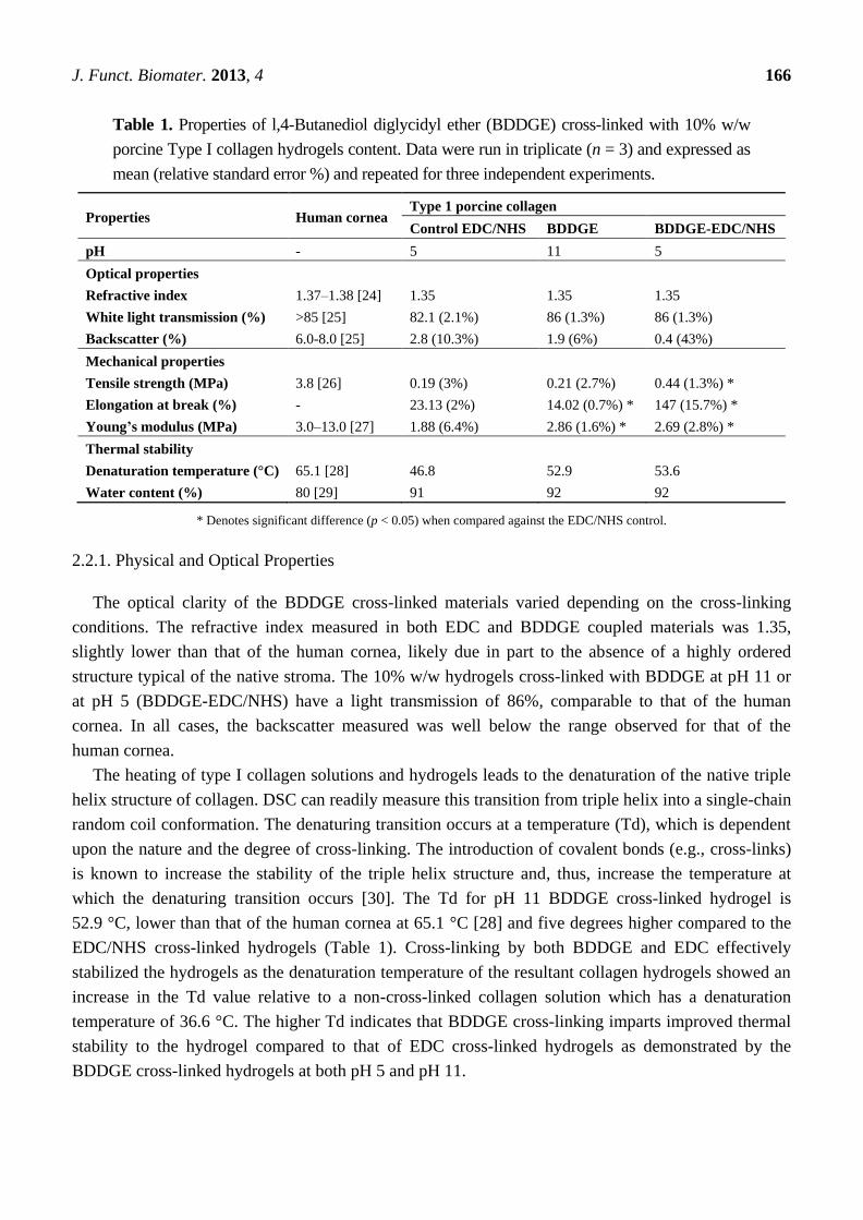
Table 1. Properties of l,4-Butanediol diglycidyl ether (BDDGE) cross-linked with 10% w/w
porcine Type I collagen hydrogels content. Data were run in triplicate (n = 3) and expressed as
mean (relative standard error %) and repeated for three independent experiments.
Properties Human cornea Type 1 porcine collagen
Control EDC/NHS BDDGE BDDGE-EDC/NHS
pH - 5 11 5
Optical properties
Refractive index 1.37–1.38 [24] 1.35 1.35 1.35
White light transmission (%) >85 [25] 82.1 (2.1%) 86 (1.3%) 86 (1.3%)
Backscatter (%) 6.0-8.0 [25] 2.8 (10.3%) 1.9 (6%) 0.4 (43%)
Mechanical properties
Tensile strength (MPa) 3.8 [26] 0.19 (3%) 0.21 (2.7%) 0.44 (1.3%) *
Elongation at break (%) - 23.13 (2%) 14.02 (0.7%) * 147 (15.7%) *
Young’s modulus (MPa) 3.0–13.0 [27] 1.88 (6.4%) 2.86 (1.6%) * 2.69 (2.8%) *
Thermal stability
Denaturation temperature (°C) 65.1 [28] 46.8 52.9 53.6
Water content (%) 80 [29] 91 92 92
* Denotes significant difference (p < 0.05) when compared against the EDC/NHS control.
2.2.1. Physical and Optical Properties
The optical clarity of the BDDGE cross-linked materials varied depending on the cross-linking
conditions. The refractive index measured in both EDC and BDDGE coupled materials was 1.35,
slightly lower than that of the human cornea, likely due in part to the absence of a highly ordered
structure typical of the native stroma. The 10% w/w hydrogels cross-linked with BDDGE at pH 11 or
at pH 5 (BDDGE-EDC/NHS) have a light transmission of 86%, comparable to that of the human
cornea. In all cases, the backscatter measured was well below the range observed for that of the
human cornea.
The heating of type I collagen solutions and hydrogels leads to the denaturation of the native triple
helix structure of collagen. DSC can readily measure this transition from triple helix into a single-chain
random coil conformation. The denaturing transition occurs at a temperature (Td), which is dependent
upon the nature and the degree of cross-linking. The introduction of covalent bonds (e.g., cross-links)
is known to increase the stability of the triple helix structure and, thus, increase the temperature at
which the denaturing transition occurs [30]. The Td for pH 11 BDDGE cross-linked hydrogel is
52.9 °C, lower than that of the human cornea at 65.1 °C [28] and five degrees higher compared to the
EDC/NHS cross-linked hydrogels (Table 1). Cross-linking by both BDDGE and EDC effectively
stabilized the hydrogels as the denaturation temperature of the resultant collagen hydrogels showed an
increase in the Td value relative to a non-cross-linked collagen solution which has a denaturation
temperature of 36.6 °C. The higher Td indicates that BDDGE cross-linking imparts improved thermal
stability to the hydrogel compared to that of EDC cross-linked hydrogels as demonstrated by the
BDDGE cross-linked hydrogels at both pH 5 and pH 11.

J. Funct. Biomater. 2013, 4 167
2.2.2. Mechanical Properties of BDDGE-Collagen Hydrogels
The tensile strength of collagen hydrogels cross-linked with BDDGE under basic conditions at
pH 11 was 0.21 MPa, similar to that of the EDC/NHS cross-linked gels (Table 1). The BDDGE
cross-linked hydrogels, however, were stiffer with Young’s modulus at 2.86 MPa compared to that of
the EDC hydrogels at 1.88 MPa. Accordingly, they were slightly less elastic than the EDC cross-linked
hydrogels. The stiffer BDDGE hydrogels were likely a result of the cross-linking being carried out
close to the pKa of lysine at 10.4 [23], allowing a higher degree of binding of lysine residues
to BDDGE.
The EDC/NHS and BDDGE cross-linked 10% collagen hydrogels showed a higher tensile strength
of 0.44 MPa, compared to the 0.21 MPa for the pH 11 BDDGE cross-linked hydrogels. In addition,
these hydrogels were highly elastic, allowing for a 147% elongation at break. In most cases, materials
with high elasticity have not been fabricated without compromising tensile strength values in methods
where direct cross-linking is used [9–11].
2.3. In Vitro Biological Stability
To assess biocompatibility, a degradation study was conducted to measure the relative resilience of
BDDGE cross-linked hydrogels to collagenase degradation, compared to EDC/NHS hydrogel controls.
The BDDGE cross-linked hydrogels (pH 11) took twice as long to be completely digested, i.e., 16 h
compared to 8 h for EDC/NHS cross-linked hydrogels (Figure 1). The sequentially cross-linked
collagen hydrogels demonstrated similar stability to the EDC/NHS controls. When interpreting these
results, it is important to consider that the EDC and sequentially cross-linked hydrogels were formed at
pH 5 whereas the BDDGE gels were synthesized at pH 11. Thus, the protein conformation likely
would have been altered during the cross linking process and this may have had downstream effects on
the collagenase degradation profiles. Additionally, the variability in collagenase resistance could, in
part, be due to the chemical bonding. BDDGE facilitates an addition reaction between an epoxide
group and lysine residue generating a secondary amine and hydroxyl group, whereas EDC activates the
carboxylic acids on the collagen fibers towards amide formation.
Figure 1. Degradation profile of 10% w/w BDDGE cross-linked hydrogels after exposure
to collagenase and compared to EDC/NHS cross-linked hydrogels.
0 2 4 6 8 10 12 14 16
0
20
40
60
80
100 A
Re
sid
ua
l m
as
s (
%)
Time (Hours)
pH 11
BDDGE-EDC/NHS
EDC/NHS
EDC/NHS w/o collagenase
J. Funct. Biomater. 2013, 4 168
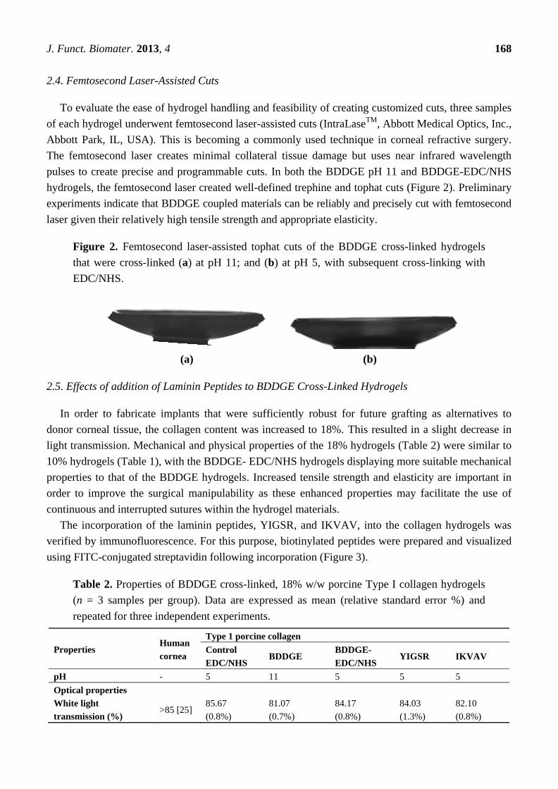
2.4. Femtosecond Laser-Assisted Cuts
To evaluate the ease of hydrogel handling and feasibility of creating customized cuts, three samples
of each hydrogel underwent femtosecond laser-assisted cuts (IntraLaseTM
, Abbott Medical Optics, Inc.,
Abbott Park, IL, USA). This is becoming a commonly used technique in corneal refractive surgery.
The femtosecond laser creates minimal collateral tissue damage but uses near infrared wavelength
pulses to create precise and programmable cuts. In both the BDDGE pH 11 and BDDGE-EDC/NHS
hydrogels, the femtosecond laser created well-defined trephine and tophat cuts (Figure 2). Preliminary
experiments indicate that BDDGE coupled materials can be reliably and precisely cut with femtosecond
laser given their relatively high tensile strength and appropriate elasticity.
Figure 2. Femtosecond laser-assisted tophat cuts of the BDDGE cross-linked hydrogels
that were cross-linked (a) at pH 11; and (b) at pH 5, with subsequent cross-linking with
EDC/NHS.
(a) (b)
2.5. Effects of addition of Laminin Peptides to BDDGE Cross-Linked Hydrogels
In order to fabricate implants that were sufficiently robust for future grafting as alternatives to
donor corneal tissue, the collagen content was increased to 18%. This resulted in a slight decrease in
light transmission. Mechanical and physical properties of the 18% hydrogels (Table 2) were similar to
10% hydrogels (Table 1), with the BDDGE- EDC/NHS hydrogels displaying more suitable mechanical
properties to that of the BDDGE hydrogels. Increased tensile strength and elasticity are important in
order to improve the surgical manipulability as these enhanced properties may facilitate the use of
continuous and interrupted sutures within the hydrogel materials.
The incorporation of the laminin peptides, YIGSR, and IKVAV, into the collagen hydrogels was
verified by immunofluorescence. For this purpose, biotinylated peptides were prepared and visualized
using FITC-conjugated streptavidin following incorporation (Figure 3).
Table 2. Properties of BDDGE cross-linked, 18% w/w porcine Type I collagen hydrogels
(n = 3 samples per group). Data are expressed as mean (relative standard error %) and
repeated for three independent experiments.
Properties Human
cornea
Type 1 porcine collagen
Control
EDC/NHS BDDGE
BDDGE-
EDC/NHS YIGSR IKVAV
pH - 5 11 5 5 5
Optical properties
White light
transmission (%) >85 [25]
85.67
(0.8%)
81.07
(0.7%)
84.17
(0.8%)
84.03
(1.3%)
82.10
(0.8%)

J. Funct. Biomater. 2013, 4 169
Table 2. Cont.
Properties Human
cornea
Type 1 Porcine Collagen
Control
EDC/NHS BDDGE
BDDGE-
EDC/NHS YIGSR IKVAV
Mechanical properties
Tensile strength (MPa) 3.8 [26] 0.12
(14.4%)
0.10
(17.3%)
0.16
(7.2%)
0.13
(4.4%)
0.17
(44%)
Elongation at break (%) - 44.52
(24.4%)
16.63
(6.2%) *
120.48
(9.3%) *
23.81
(5.9%)
44.76
(2.6%)
Young’s modulus
(MPa)
3.0–13.0
[27]
0.64
(39.6%)
1.21
(15.7%) *
0.21
(8.2%) *
0.98
(1.7%)
0.55
(46%)
Thermal properties
Denaturation
temperature (°C) 65.1 [28] 49.1 65.9 49.5 47.2 55.0 & 64.4
* Denotes significant difference (p < 0.05) when compared against the EDC/NHS benchmark.
Figure 3. Visualization of biotin-IKVAV peptides incorporated into BDDGE-EDC/NHS
collagen hydrogel, with FITC-streptavidin. The inset shows the EDC/NHS control
hydrogel without peptide. Scale bar = 100 μm.
In Vitro Biological Stability of Laminin Peptide-BDDGE-Collagen Hydrogels
In the case of 18% w/w collagen hydrogels, increased stability was observed against collagenase
due to incorporation of peptide, IKVAV and YIGSR (Figure 4). Residual mass was more than 50% in
case of both peptide-incorporated hydrogels, which was significantly (p < 0.001) higher than the
control hydrogel at 10 h. Although there was no significant difference in retention of residual mass in
case of both IKVAV and YIGSR hydrogels after 10 h, IKVAV exhibited greater resistance. We
reasoned that this might be due to the formation of additional cross-linking by IKVAV due to the
number of lysine residues within the peptide. The greater overall stability of IKVAV containing
hydrogels could be due to the formation of interpenetrating networks within the hydrogel, as shown by
the two discrete Td values (Table 2).

J. Funct. Biomater. 2013, 4 170
Figure 4. Degradation profile of 18% w/w BDDGE cross-linked hydrogels after exposure
to collagenase and compared to EDC/NHS cross-linked hydrogels.
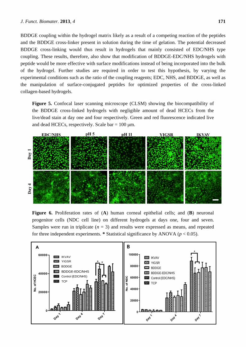
2.6. In Vitro Biocompatibility and Performance
It is important to evaluate the biocompatibility of the BDDGE coupled hydrogels as the clinically
tested EDC cross-linked biomaterials were found to promote corneal cell and nerve regeneration in all
our patients. Live/Dead staining of immortalized human corneal epithelial cells (HCECs) seeded on
BDDGE cross-linked hydrogels fabricated under different conditions showed very low/no dead cells
(Figure 5). This demonstrated that the hydrogels were non-cytotoxic. The cell adhesion and spread on
the hydrogels was also morphologically distinct by microscopic examination as the cells were flattened
and spread out on the hydrogels. The proliferation rate of HCECs and neuronal progenitors (derived
from rodent dorsal root ganglia fused with neuroblastoma cells (NDCs)), on BDDGE
cross-linked hydrogels were also examined. A MTS assay showed that all the hydrogels tested
successfully supported HCEC proliferation, albeit at different rates (Figure 6). The differences in
proliferation were most distinct by day seven of culture after pre-seeding on the 9 mm diameter
hydrogels of 500 μm thickness. The rate of cell proliferation on BDDGE cross-linked hydrogels was
slower than that on the EDC/NHS cross-linked hydrogels (Figure 6A). Addition of YIGSR
peptide, however, increased the proliferation rate to the level of cells grown on EDC/NHS
cross-linked hydrogels.
NDC proliferation was also supported by all the hydrogels. However, the highest proliferation rate
was observed in the IKVAV collagen-hydrogels (Figure 6B).
Hydrogels with incorporated IKVAV and YIGSR had statistical significant influence (p < 0.05) on
cell proliferation of NDCs and HCECs. By day seven of culture, the peptides were able to
differentially affect cell growth. YIGSR enhanced HCEC growth while IKVAV enhanced neuronal
cell growth. This was in keeping with previous reports in the literature.
Although incorporation of IKVAV and YIGSR peptides enhanced neuronal and epithelial cell
growth, the marked improvement in the mechanical properties of the hydrogels offered by the BDDGE
cross-linking following by EDC/NHS enhancement was lost. Hydrogels showed properties that were
similar to those of EDC/NHS cross-linked benchmarks only. This suggests a lack of incorporation of
0 2 4 6 8 10
0
20
40
60
80
100
Re
sid
ua
l m
as
s (
%)
Time (Hours)
EDC/NHS
YIGSR
IKVAV
B
J. Funct. Biomater. 2013, 4 171
BDDGE coupling within the hydrogel matrix likely as a result of a competing reaction of the peptides
and the BDDGE cross-linker present in solution during the time of gelation. The potential decreased
BDDGE cross-linking would thus result in hydrogels that mainly consisted of EDC/NHS type
coupling. These results, therefore, also show that modification of BDDGE-EDC/NHS hydrogels with
peptide would be more effective with surface modifications instead of being incorporated into the bulk
of the hydrogel. Further studies are required in order to test this hypothesis, by varying the
experimental conditions such as the ratio of the coupling reagents; EDC, NHS, and BDDGE, as well as
the manipulation of surface-conjugated peptides for optimized properties of the cross-linked
collagen-based hydrogels.
Figure 5. Confocal laser scanning microscope (CLSM) showing the biocompatibility of
the BDDGE cross-linked hydrogels with negligible amount of dead HCECs from the
live/dead stain at day one and four respectively. Green and red fluorescence indicated live
and dead HCECs, respectively. Scale bar = 100 μm.
Figure 6. Proliferation rates of (A) human corneal epithelial cells; and (B) neuronal
progenitor cells (NDC cell line) on different hydrogels at days one, four and seven.
Samples were run in triplicate (n = 3) and results were expressed as means, and repeated
for three independent experiments. * Statistical significance by ANOVA (p < 0.05).
Day
1
Day 4
Day 7
0
20000
40000
60000IKVAV
YIGSR
BDDGE
BDDGE-EDC/NHS
Control (EDC/NHS)
TCP
No
. o
f H
CE
C
*
A
*
Day
1
Day
4
Day
7
0
20000
40000
60000
80000
100000
No
. o
f N
DC
IKVAV
YIGSR
BDDGE
BDDGE-EDC/NHS
Control (EDC/NHS)
TCP
B*

J. Funct. Biomater. 2013, 4 172
3. Experimental Section
3.1. Materials
Porcine Type I collagen purchased from Sewon Cellontech (Seoul, South Korea) was lyophilized
and reconstituted to make either a 10 w/w% or 18% w/w% solution. All other reagents were of
analytical grade and used as received. 1-Ethyl-3-(3-dimethyl aminopropyl) carbodiimide (EDC) and
1,4-Butanediol diglycidyl ether (BDDGE) were both supplied by Sigma-Aldrich (Oakville, Ontario,
Canada), Copper (II) Tetrafluoroborate Cu(BF4)2 was supplied by Strem Chemicals, (Newburyport,
MA, USA) and N-hydroxysuccinimide (NHS) by Fluka (Buchs, Switzerland). Phosphate-buffered
saline (PBS, pH 7.2) was prepared via tablets and obtained from Calbiochem Corp., (Darmstadt,
Germany). Milli-Q deionized water was used throughout all necessary experiments.
The peptides IKVAV and YIGSR-NH2 were synthesized on symphony automated peptide
synthesizer (Protein Technologies Inc., Tucson, AZ, USA) using standard fluorenylmethoxycarbonyl
(Fmoc) chemistry with HCTU (ChemPep Inc., Wellington, FL, USA) as the activating reagent. The
synthesis was performed on a 0.1 mmol scale with Fmoc-Val-PEG-PS and Fmoc-PAL-PS resin
(Applied Biosystems, Sweden) using a four-fold excess of amino acid in each coupling. The peptides
were cleaved from the resin by treatment with a mixture of trifluoroacetic acid (TFA), and water
(95:5 v/v; 10 mL per gram of polymer) for 2 h at room temperature. After filtration, TFA was
evaporated and the peptides were precipitated by the addition of cold diethyl ether, centrifuged and
lyophilized. The crude products were purified by reversed-phase HPLC on a semi-preparative C-18
column (Grace Vydac) and identified from MALDI-TOF spectra (Applied Biosystems Voyager
DE-STR, Stockholm, Sweden).
3.2. Preparation of Collagen Cross-Linked Materials
0.4 mL aliquots of either 10 or 18 w/w% collagen were weighed out and mixed in a T-piece system,
using a methodology similar to one we previously described [31]. For gels performed in basic
conditions a buffer of 0.036 M Na2CO3 and 0.064 M NaHCO3 at pH 10 was used to fill the T-piece
whereas a 0.625 MES buffer was used in acidic conditions. The collagen solution was adjusted to
either pH 5 or 11 with microliter quantities of 2N aqueous NaOH, followed by thorough mixing.
Calculated volumes of aqueous solutions of BDDGE, Cu(BF4)2, EDC and its co-reagent NHS were
added to their respective solutions. For epoxide gels, the final ―cross-linker-doped‖ collagen solution
was mixed 150× at 4 °C whereas carbodiimide solutions were only capable of being mixed 50×.
Laminin-derived cell-adhesive peptides were incorporated within the epoxide gels using the pH 5
formulations, with 0.001 equimolar of peptide relative to mole of amine in collagen. Additionally,
biotinylated peptide and streptavidin was used to investigate the binding affinity between the peptides
within the BDDGE hydrogel matrix. For incorporation of YIGSR and IKVAV peptides, a starting
concentration of 18% w/w% collagen was used to allow for dilution of the collagen content. Confocal
fluorescence microscopy (using a LSM700 confocal microscope, Carl Zeiss AB, Stockholm, Sweden)
was used to examine the presence of biotinylated peptide inside the hydrogel. The final collagen
solution was dispensed as flat sheet into glass molds, cured at 100% humidity at room temperature for
J. Funct. Biomater. 2013, 4 173
24 h, post-cured at 37 °C for 1 day, then washed extensively in PBS to remove any non-cross-linked
substrate. Hydrogels cross-linked sequentially by both BDDGE and EDC/NHS were cured for 72 h.
3.3. Optical Property Measurements
Refractive indices of fully hydrated epoxide and carbodimide cross-linked hydrogels were recorded
using an Abbe refractometer (Model C10, VEE GEE, Scientific Inc., Kirkland, WA, USA). The
experiment was performed at 21 °C with bromonaphthalene as the calibration agent. Light
transmission and back-scattering measurements were carried out at room temperature for white light
(quartz-halogen lamp source) and for narrow spectral regions (centered at 450, 500, 550, 600, and
650 nm). Briefly, a custom-built instrument was used to measure the percent transmission of samples
as compared to open beam intensity [30]. The relative percent of light back scattered from the collimated
beam by the sample was measured with a circular array of 8 photodiodes, 30 degrees off axis.
3.4. Mechanical Property Measurements
The tensile strength, Young’s moduli and elongation at break of the hydrogels were determined on an
Instron electromechanical universal tester (Model 3342) equipped with Series IX/S software, using a
crosshead speed of 10 mm·min−1
and a gauge length for testing of 5 mm. Hydrogels with 0.55 mm
thickness were equilibrated in PBS and cut into 10 mm × 5 mm rectangular sheets. A minimum of three
specimens was measured for each hydrogel formulation and repeated for three independent experiments.
3.5. Equilibrium Water Content Measurement
After removal from the molds, hydrogels were immersed in a PBS solution for 5 days at 4 °C. After
removal of surface water through gentle blotting on filter paper, the samples were immediately
weighed on a microbalance to record the ―wet weight‖ (W0) of the sample. They were allowed to dry
at room temperature under vacuum to constant weight (W). The total equilibrated water content of the
hydrogels (Wt) was calculated according to the following equation:
Wt = (W − W0)/W × 100% (1)
3.6. Thermal Properties: Differential Scanning Calorimetry (DSC)
The thermal properties of the hydrogels were tested using a Q2000 differential scanning calorimeter
(TA Instruments, New Castle, DE). Heating scans were recorded within the range of 8 to 80 °C at a
scan rate of 5 °C·min−1
. Pre-weighed samples of the PBS-equilibrated hydrogels (weights ranging
from 5 to 10 mg) were surface-dried with filter paper and hermetically sealed in an aluminum pan to
prevent water evaporation. A resulting heat flux versus temperature curve was then used to calculate
the denaturing temperature (Td). Tmax of the endothermic peak gives the denaturing temperature.
3.7. In Vitro Degradation
Briefly, hydrated cross-linked hydrogels (approximately 50 mg) were placed in vials containing
5 mL of a 5 U/mL collagenase in a PBS solution (Type I Collagenase from Clostridium histolyticum,

J. Funct. Biomater. 2013, 4 174
318 U/mg solid, Sigma-Aldrich, Oakville, ON, Canada), refreshed every 8 h. The vials were incubated
in an oven at 37 °C. The gels were weighed at different time intervals after removal of surface water
through blotting. The residual mass of the hydrogels was tracked as a function of time, relative to their
initial hydrated weight.
3.8. Femtosecond Laser-Assisted Cuts of Epoxide and Carbodiimide Cross-linked Hydrogels: Feasibility
Both hydrogels and cadaveric porcine control corneas underwent femtosecond laser-assisted
trephine (cylindrical) and tophat cuts. Cylindrical cuts were programmed to span the entire depth of the
hydrogel at a cut angle of 90 degrees, while the tophat cut was programmed with 90 degree anterior
and posterior side cut angles. The dimensions, shape, and side cut angles were then captured for
analysis using an optical profilometry system developed by Rejean Munger and David Priest at the
Ottawa Hospital, Ottawa, Ontario, Canada. Optical profilometry captures silhouette images of cut
hydrogels using a high-resolution camera positioned against a backlight. These images are subsequently
analyzed using ImageJ Software (Java-based image processing, NIH freeware) to calculate their
precise dimensions and side cut angles.
3.9. In Vitro Biocompatibility and Performance
To observe the biological effect of the different hydrogels, immortalized human corneal epithelial
cells (HCEC) [32] and rodent hybrid dorsal root ganglia-neuroblastoma cells (NDC) [33] were seeded
directly on the hydrogels. For cell culture, hydrogels of 6 mm diameter size were cut and placed in
24-well plate followed by sterilization with 1% v/v chloroform and 3X antibiotic solution consisting of
300 unit/mL penicillin and 300 µg/mL streptomycin. HCEC and NDC were seeded at 5 × 10⁴
cells/well respectively with a small volume of medium (50 µL) on the top of the hydrogel and
incubated for 30 min. HCEC and NDC were maintained in EpiGRO™ medium (Millipore) and
DMEM supplemented with 10% FCS and 1% penicillin and spectinomycin, respectively. Medium was
replaced on alternative days and culture was maintained for 7 days.
For both HCEC and NDC, proliferation was measured by the colorimetric MTS assay
(CellTiter 96 Aqueous One solution, Promega, Madison, WI). Dehydrogenase enzymes from
mitochondria within live cells convert the yellow tetrazolium salt [3-(4,5-dimethylthiazol-2-yl)-5-
(3carboxymethoxyphenyl)-2(4-sulfophenyl)-2H-tetrazolium] to purple formazancrystals. Herein, the
amount of formazan crystal formation is directly proportional to the number of live cells. The MTS
assay was performed on days 1, 4 and 7 of cell culture. Hydrogels were rinsed three times in PBS and
subsequently transferred into new wells prior to MTS assay to avoid the absorbance from any cells that
may have been growing on the tissue culture plastic beneath the hydrogels. Subsequently, 20% v/v of
MTS reagent in cell culture medium was added and cultures were incubated further for 4 h at 37 °C.
Absorbance was measured at 490 nm using a spectrophotometric microplate reader (VERSA Max
Microplate Reader, Molecular Device, Sunnyvale, CA, USA).
The biocompatibility of the different hydrogel formulations with HCEC survival was examined by
using live/dead staining on day 1 and day 4 of culture. Calceinacetoxymethyl (Calcein AM) and
ethidium homodimer-1 (EthD-1) (Invitrogen) were used to determine the viability of cells on the
hydrogels. The concentration of Calcein AM and EthD-1staining solution were 2 µM and 4 µM
J. Funct. Biomater. 2013, 4 175
respectively. The fluorescence images were visualized under fluorescent confocal microscope
(LSM700, Carl Zeiss). Morphological inspection of the cells for flattening and spread (as opposed to
rounded cells) gave an indication of the ability of each hydrogel formulation to support cell adhesion
and spread.
Cell proliferation studies on hydrogels were repeated separately for three times and run in triplicate
(n = 3). Data obtained from the different experiments are presented as mean values. Statistical
significance of differences between two hydrogels cell proliferation was analyzed using single factor
analysis of variance (ANOVA).
4. Conclusions
The BDDGE hydrogels exhibit high tailorability, given that the mechanical properties could be
manipulated depending on the conditions in which the hydrogels were fabricated. Specifically, the
BDDGE-EDC/NHS hydrogels, fabricated by BDDGE coupling followed by carbodiimide chemistry
resulted in materials with high elasticity. Interestingly, the slow gelation time of the BDDGE
cross-linking may allow for drug encapsulation within the collagen matrix upon fabrication for
therapeutic use. In addition, the YIGSR and IKVAV peptide-BDDGE-based hydrogels may also serve
as ideal candidates for corneal substitutes given their ability to enhance epithelial and nerve cell growth.
Acknowledgments
This work was supported by an EU ERA-net nanomedicine grant for the I-CARE project (MG, PF,
BL), Konung Gustaf V and Drottning Margarets Frimurarestiftelse (PF) and some early work by a
CIHR Canada Regenerative Medicine team grant to MG. CN was supported by an NSERC Canada
studentship for his work in Sweden and Canada. We thank Kashif Baigand, Rejean Munger, University
of Ottawa Eye Institution, Ottawa Hospital for their assistance with the laser cut analysis, and Deryn
Fogg, Department of Chemistry, University of Ottawa for use of her laboratory facilities.
Conflicts of Interest
The authors declare no conflict of interest.
References
1. Shin, H.; Jo, S.; Mikos, A.G. Biomimetic materials for tissue engineering. Biomaterials 2003, 24,
4353–4364.
2. Heindl, L.M.; Riss, S.; Adler, W.; Bucher, F.; Hos, D.; Cursiefen, C. Split cornea transplantation:
Relationship between storage time of split donor tissue and outcome. Ophthalmology 2013,
in press.
3. Du, Y.Q.; Carlson, E.C.; Funderburgh, M.L.; Birk, D.E.; Pearlman, E.; Guo, N.X.; Kao, W.W.Y.;
Funderburgh, J.L. Stem cell therapy restores transparency to defective murine corneas. Stem Cells
2009, 27, 1635–1642.
4. Ruberti, J.W.; Sinha Roy, A.; Roberts, C.J. Corneal biomechanics and biomaterials. Annu. Rev.
Biomed. Eng. 2011, 13, 269–295.
J. Funct. Biomater. 2013, 4 176
5. Yang, C.L.; Hillas, P.J.; Baez, J.A.; Nokelainen, M.; Balan, J.; Tang, J.; Spiro, R.; Polarek, J.W.
The application of recombinant human collagen in tissue engineering. Biodrugs 2004, 18,
103–119.
6. Ma, P.X. Biomimetic materials for tissue engineering. Adv. Drug. Deliv. Rev. 2008, 60, 184–198.
7. Hollister, S.J. Porous scaffold design for tissue engineering. Nat. Mater. 2005, 4, 518–524.
8. Fagerholm, P.; Lagali, N.S.; Merrett, K.; Jackson, W.B.; Munger, R.; Liu, Y.W.; Polarek, J.W.;
Soderqvist, M.; Griffith, M. A biosynthetic alternative to human donor tissue for inducing corneal
regeneration: 24-month follow-up of a phase 1 clinical study. Sci. Transl. Med. 2010, 2,
doi: 10.1126/scitranslmed.3001022.
9. Tampieri, A.; Sandri, M.; Landi, E.; Pressato, D.; Francioli, S.; Quarto, R.; Martin, I. Design of
graded biomimetic osteochondral composite scaffolds. Biomaterials 2008, 29, 3539–3546.
10. Nam, K.; Murakoshi, A.; Kimura, T.; Fujisato, T.; Kitamura, S.; Kishida, A. Study on the physical
properties of tissue-engineered blood vessels made by chemical cross-linking and polymer-tissue
cross-linking. J. Artif. Organs. 2009, 12, 47–54.
11. Zeeman, R. Cross-linking of Collagen-Based Materials. Ph.D. Dissertation, Universiteit Twente,
Enschede, Netherlands, 1998.
12. Zeeman, R.; Dijkstra, P.J.; van Wachem, P.B.; van Luyn, M.J.A.; Hendriks, M.; Cahalan, P.T.;
Feijen, J. Successive epoxy and carbodiimide cross-linking of dermal sheep collagen. Biomaterials
1999, 20, 921–931.
13. Zeeman, R.; Dijkstra, P.J.; van Wachem, P.B.; van Luyn, M.J.A.; Hendriks, M.; Cahalan, P.T.;
Feijen, J. Crosslinking and modification of dermal sheep collagen using 1,4-butanediol diglycidyl
ether. J. Biomed. Mater. Res. 1999, 46, 424–433.
14. Zeeman, R.; Dijkstra, P.J.; van Wachem, P.B.; van Luyn, M.J.A.; Hendriks, M.; Cahalan, P.T.;
Feijen, J. The kinetics of 1,4-butanediol diglycidyl ether crosslinking of dermal sheep collagen.
J. Biomed. Mater. Res. 2000, 51, 541–548.
15. La Gatta, A.; Schiraldi, C.; Papa, A.; D’Agostino, A.; Cammarota, M.; de Rosa, A.; de Rosa, M.
Hyaluronan scaffolds via diglycidyl ether crosslinking: Toward improvements in composition and
performance. Carbohyd. Polym. 2013, 96, 536–544.
16. Nicoletti, A.; Fiorini, M.; Paolillo, J.; Dolcini, L.; Sandri, M.; Pressato, D. Effects of different
crosslinking conditions on the chemical-physical properties of a novel bio-inspired composite
scaffold stabilised with 1,4-butanediol diglycidyl ether (BDDGE). J. Mater. Sci.-Mater. M. 2013,
24, 17–35.
17. Kleinman, H.K.; Cannon, F.B.; Laurie, G.W.; Hassell, J.R.; Aumailley, M.; Terranova, V.P.;
Martin, G.R.; Duboisdalcq, M. Biological activities of laminin. J. Cell. Biochem. 1985, 27,
317–325.
18. Timpl, R.; Rohde, H.; Robey, P.G.; Rennard, S.I.; Foidart, J.M.; Martin, G.R. Laminin—A
glycoprotein from basement membranes. J. Biol. Chem. 1979, 254, 9933–9937.
19. Mecham, R.P. Laminin receptors. Annu. Rev. Cell. Biol. 1991, 7, 71–91.
20. Li, F.; Griffith, M.; Li, Z.; Tanodekaew, S.; Sheardown, H.; Hakim, M.; Carlsson, D.J.
Recruitment of multiple cell lines by collagen-synthetic copolymer matrices in corneal regeneration.
Biomaterials 2005, 26, 3093–3104.

J. Funct. Biomater. 2013, 4 177
21. Chen, Y.-H.; Yeh, M.-H.; Lai, H.-M. Preparation of a Biodegradable Thermal-Sensitive Gel
System. U.S. Patent 20040013733, 30 December 2002.
22. Kamal, A.; Ramu, R.; Azhar, M.A.; Khanna, G.B.R. Copper(II) tetrafluoroborate-catalyzed
ring-opening of epoxides by amines. Tetrahedron Lett. 2005, 46, 2675–2677.
23. André, I.; Linse, S.; Mulder, F.A.A. Residue-specific pKa determination of lysine and arginine
side chains by indirect 15N and 13C NMR spectroscopy: Application to apo calmodulin. J. Am.
Chem. Soc. 2007, 129, 15805–15813.
24. Patel, S.; Marshall, J.; Fitzke, F.W. Refractive index of the human corneal epithelium and stroma.
J. Refract. Surg. 1995, 11, 100–105.
25. Liu, Y.; Griffith, M.; Watsky, M.A.; Forrester, J.V.; Kuffova, L.; Grant, D.; Merrett, K.;
Carlsson, D.J. Properties of porcine and recombinant human collagen matrices for optically clear
tissue engineering applications. Biomacromolecules 2006, 7, 1819–1828.
26. Zeng, Y.J.; Yang, J.; Huang, K.; Lee, Z.H.; Lee, X.Y. A comparison of biomechanical properties
between human and porcine cornea. J. Biomech. 2001, 34, 533–537.
27. Crabb, R.A.B.; Chau, E.P.; Evans, M.C.; Barocas, V.H.; Hubel, A. Biomechanical and
microstructural characteristics of a collagen film-based corneal stroma equivalent. Tissue Eng.
2006, 12, 1565–1575.
28. Merrett, K.; Fagerholm, P.; McLaughlin, C.R.; Dravida, S.; Lagali, N.; Shinozaki, N.;
Watsky, M.A.; Munger, R.; Kato, Y.; Li, F.F.; et al. Tissue-engineered recombinant human
collagen-based corneal substitutes for implantation: Performance of type I versus type III
collagen. Invest. Ophth. Vis. Sci. 2008, 49, 3887–3894.
29. Tighe, B. Eye contact. Chem. Brit. 1992, 28, 241–244.
30. Angele, P.; Abke, J.; Kujat, R.; Faltermeier, H.; Schumann, D.; Nerlich, M.; Kinner, B.;
Englert, C.; Ruszczak, Z.; Mehrl, R.; et al. Influence of different collagen species on
physico-chemical properties of crosslinked collagen matrices. Biomaterials 2004, 25, 2831–2841.
31. Liu, Y.W.; Gan, L.H.; Carlsson, D.J.; Fagerholm, P.; Lagali, N.; Watsky, M.A.; Munger, R.;
Hodge, W.G.; Priest, D.; Griffith, M. A simple, cross-linked collagen tissue substitute for corneal
implantation. Invest. Ophth. Vis. Sci. 2006, 47, 1869–1875.
32. Griffith, M.; Osborne, R.; Munger, R.; Xiong, X.J.; Doillon, C.J.; Laycock, N.L.C.; Hakim, M.;
Song, Y.; Watsky, M.A. Functional human corneal equivalents constructed from cell lines.
Science 1999, 286, 2169–2172.
33. Wood, J.N.; Bevan, S.J.; Coote, P.R.; Dunn, P.M.; Harmar, A.; Hogan, P.; Latchman, D.S.;
Morrison, C.; Rougon, G.; Theveniau, M.; et al. Novel cell lines display properties of nociceptive
sensory neurons. P. R. Soc. B Biol. Sci. 1990, 241, 187–194.
© 2013 by the authors; licensee MDPI, Basel, Switzerland. This article is an open access article
distributed under the terms and conditions of the Creative Commons Attribution license
(http://creativecommons.org/licenses/by/3.0/).
Copyright © 2022 FDOKUMEN