In Vitro Biocompatibility of New PVA-Based Hydrogels as Vitreous Body Substitutes
21
Journal of Biomaterials Science 23 (2012) 555–575 brill.nl/jbs In Vitro Biocompatibility of New PVA-Based Hydrogels as Vitreous Body Substitutes Stefania Lamponi a,∗ , Gemma Leone a , Marco Consumi a , Giuseppe Greco b and Agnese Magnani a a Department of Pure and Applied Medicinal Chemistry, University of Siena, via Aldo Moro 2, 53110 Siena, Italy b Casa di Cura Rugani, loc. Montarioso, 53100 Siena, Italy Received 15 September 2010; accepted 1 January 2011 Abstract In order to synthesize injectable hydrogels suitable as vitreous body substitutes, a new method based on the use of trisodium trimetaphosphate (STMP) to cross-link PVA was recently proposed. Hydrogels with different molar ratios between STMP and PVA were realised. The aim of the present study was the evaluation of the biocompatibility of the different STMP/PVA hydrogels synthesised by analysing the effects of their in vitro interaction with cultures of mouse fibroblasts NIH3T3, primary human microvascular endothelial cells adult (HMVECad) and human lens cells. Cytotoxicity of hydrogels was first evaluated by analysing cell density and proliferation. Morphological and morphometric analysis of cell in contact with hydrogels was then performed using light microscopy and scanning electron microscopy, respectively. Moreover, cell adhesion and growth onto the hydrogels surface was evaluated and correlated to the amount of adsorbed proteins. At last, the biocompatibility of the sheared STMP/PVA 1:8 hydrogel was tested. The in vitro data of all the STMP/PVA hydrogels demonstrated their good biocompatibility, and indicated that the 1:8 sample was the most promising as vitreous body substitute. © Koninklijke Brill NV, Leiden, 2012 Keywords Hydrogels, cytotoxicity, biocompatibility, vitreous body, fibroblasts, endothelial cells, human lens cells 1. Introduction Hydrogels are three-dimensional cross-linked polymeric networks able to absorb large amounts of water without dissolving or losing their structure. They can be synthesized by natural polymers, such as hyaluronan [1], alginates [2], chitosan [3] and collagen [4], as well as by synthetic polymers, such as poly(ethylene gly- col) [5], poly(ethylene eoxide) [6], poly(methacrylic acid-co-acrylamide) [7] and * To whom correspondence should be addressed. Tel.: (39-57) 723-4386; Fax: (39-57) 723-4177; e-mail: [email protected] © Koninklijke Brill NV, Leiden, 2012 DOI:10.1163/092050611X554499
-
Upload
independent -
Category
Documents
-
view
5 -
download
0
Transcript of In Vitro Biocompatibility of New PVA-Based Hydrogels as Vitreous Body Substitutes

Journal of Biomaterials Science 23 (2012) 555–575brill.nl/jbs
In Vitro Biocompatibility of New PVA-Based Hydrogels asVitreous Body Substitutes
Stefania Lamponi a,∗, Gemma Leone a, Marco Consumi a, Giuseppe Greco b
and Agnese Magnani a
a Department of Pure and Applied Medicinal Chemistry, University of Siena, via Aldo Moro 2,53110 Siena, Italy
b Casa di Cura Rugani, loc. Montarioso, 53100 Siena, Italy
Received 15 September 2010; accepted 1 January 2011
AbstractIn order to synthesize injectable hydrogels suitable as vitreous body substitutes, a new method based onthe use of trisodium trimetaphosphate (STMP) to cross-link PVA was recently proposed. Hydrogels withdifferent molar ratios between STMP and PVA were realised. The aim of the present study was the evaluationof the biocompatibility of the different STMP/PVA hydrogels synthesised by analysing the effects of theirin vitro interaction with cultures of mouse fibroblasts NIH3T3, primary human microvascular endothelialcells adult (HMVECad) and human lens cells. Cytotoxicity of hydrogels was first evaluated by analysingcell density and proliferation. Morphological and morphometric analysis of cell in contact with hydrogelswas then performed using light microscopy and scanning electron microscopy, respectively. Moreover, celladhesion and growth onto the hydrogels surface was evaluated and correlated to the amount of adsorbedproteins. At last, the biocompatibility of the sheared STMP/PVA 1:8 hydrogel was tested. The in vitro dataof all the STMP/PVA hydrogels demonstrated their good biocompatibility, and indicated that the 1:8 samplewas the most promising as vitreous body substitute.© Koninklijke Brill NV, Leiden, 2012
KeywordsHydrogels, cytotoxicity, biocompatibility, vitreous body, fibroblasts, endothelial cells, human lens cells
1. Introduction
Hydrogels are three-dimensional cross-linked polymeric networks able to absorblarge amounts of water without dissolving or losing their structure. They can besynthesized by natural polymers, such as hyaluronan [1], alginates [2], chitosan[3] and collagen [4], as well as by synthetic polymers, such as poly(ethylene gly-col) [5], poly(ethylene eoxide) [6], poly(methacrylic acid-co-acrylamide) [7] and
* To whom correspondence should be addressed. Tel.: (39-57) 723-4386; Fax: (39-57) 723-4177; e-mail:[email protected]
© Koninklijke Brill NV, Leiden, 2012 DOI:10.1163/092050611X554499
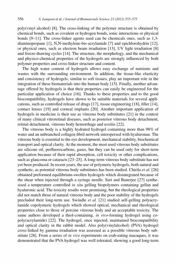
556 S. Lamponi et al. / Journal of Biomaterials Science 23 (2012) 555–575
poly(vinyl alcohol) [8]. The cross-linking of the polymer structure is obtained bychemical bonds, such as covalent or hydrogen bonds, ionic interactions or physicalbonds [9–11]. The cross-linker agents used can be chemicals ones, such as 1,3-diaminopropane [1], N,N-methylene-bis-acrylamide [7] and epichlorohydrin [12],or physical ones, such as electron beam irradiation [13], UV light irradiation [8]and freeze-thawing cycles [14]. The structure, the morphology, and the mechanicaland physico-chemical properties of the hydrogels are strongly influenced by bothpolymer properties and cross-linker structure and content.
The high water content of hydrogels allows easy exchange of nutrients andwastes with the surrounding environment. In addition, the tissue-like elasticityand consistency of hydrogels, similar to soft tissues, play an important role in theintegration of these biomaterials into the human body [15]. Finally, another advan-tage offered by hydrogels is that their properties can easily be engineered for theparticular application of choice [16]. Thanks to these properties and to the goodbiocompatibility, hydrogels have shown to be suitable materials for several appli-cations, such as controlled release of drugs [17], tissue engineering [18], filler [14],contact lenses [19] and corneal implants [20]. Another important application ofhydrogels in medicine is their use as vitreous body substitutes [21] in the contextof many clinical vitroretinal diseases, such as posterior vitreous body detachment,retinal detachment, vitreous body hemorrhage and uveitis [22].
The vitreous body is a highly hydrated hydrogel containing more than 98% ofwater and an unbranched collagen fibril network interspersed with hyaluronan. Thevitreous body is essential to the eye development, mechanical stability, biochemicaltransport and optical clarity. At the moment, the most used vitreous body substitutesare silicone oil, perfluorocarbons, gases, but they can be used only for short-termapplication because of their rapid absorption, cell toxicity or other complications,such as glaucoma or cataracts [23–25]. A long-term vitreous body substitute has notyet been produced. In recent years, the use of polymeric hydrogels, both natural andsynthetic, as potential vitreous body substitutes has been studied. Chirila et al. [26]obtained preformed equilibrium-swollen hydrogels which disintegrated because ofthe shear when injected through a syringe needle. Suri and Banerjee [27] synthe-sised a temperature controlled in situ gelling biopolymers containing gellan andhyaluronic acid. The toxicity results were promising, but the rheological propertiesdid not match those of natural vitreous body and the poor stability of the hydrogelsprecluded their long-term use. Swindle et al. [21] studied self-gelling polyacry-lamide copolymeric hydrogels which showed optical, mechanical and rheologicalproperties close to those of porcine vitreous body and an acceptable toxicity. Thesame authors developed a thiol-containing, in vivo-forming hydrogel using co-poly(acrylamide) [22]. The hydrogel, once injected, maintained biocompatibilityand optical clarity in the rabbit model. Also poly(vinylalcohol) (PVA) hydrogelcross-linked by gamma irradiation was assessed as a possible vitreous body sub-stitute [28]. From a series of in vivo experiments on crab-eating macaques, it wasdemonstrated that the PVA hydrogel was well tolerated, showing a good long-term

S. Lamponi et al. / Journal of Biomaterials Science 23 (2012) 555–575 557
biocompatibility. Histopathologic examination by light and electron microscopythree months after the hydrogel injection demonstrated no significant changes inophtalmoscopic findings.
Recently a new method to cross-link PVA was proposed [29], based on theuse of a non-toxic substance as cross-linker agent, trisodium trimetaphosphate(STMP), usually used to cross-link starch in the food industry. Hydrogels withSTMP/PVA molar ratios 1:4, 1:6 and 1:8 were realised. The hydrogels were char-acterised by infrared spectroscopy and rheological analysis. IR analysis providedthe chemical composition of the synthesised hydrogels, confirming the success ofthe cross-linking reaction. On the basis of rheological analysis, light transmittanceand water content measurements STMP/PVA 1:8 was identified as the most suit-able hydrogel for a vitreous body substitute. Consequently, the 1:8 sample wasfurther characterised in terms of thixothropic behaviour, injectability and perme-ability. STMP/PVA 1:8 showed good injectability with a negligible change of itsrheological properties. Moreover, the diffusion coefficient (DC) of a model mole-cule (phenylalanine) through the hydrogel was found to be very similar to its DC inwater.
As the synthetic injectable hydrogels must have a very high biocompatibility, afundamental aspect in their characterisation is the knowledge of the hydrogel cyto-toxicity and the effects of their interactions with cells. It is, therefore, important toevaluate the morphofunctional and/or cytotoxic effects of the hydrogels, in order todefine the most suitable formulation to be used without collateral effects on cell ac-tivity, beside the knowledge of the three-dimensional structure and the rheologicalproperties of the hydrogels.
The aim of the present study was the evaluation of the biocompatibility ofthe three hydrogels previously synthesised with different STMP/PVA ratios (1:4,1:6 and 1:8) in terms of effects of their in vitro interaction with cultures ofNIH3T3 mouse fibroblasts and primary human microvascular endothelial cellsadult (HMVECad). Cytotoxicity of hydrogels was evaluated by cell density andproliferation analysis, through the measurements of the rate of cell viability withWST-1 assay, and by morphological and morphometric analysis using light andscanning electron microscopy, respectively. Cell adhesion and growth directly onthe hydrogels’ surface was evaluated by electron microscopy and correlated to theamount of adsorbed proteins. Moreover, the adhesion and proliferation of humanlens cells to the most promising hydrogel in terms of cell compatibility, i.e., 1:8STMP/PVA, was studied. At last, the biocompatibility of sheared STMP/PVA 1:8was also tested.
2. Materials and Methods
2.1. Materials
Medium 131 was supplied by Invitrogen. Dulbecco’s Modified Eagle’s Medium(DMEM), trypsin solution, and all the solvents used for cell culture were pur-
558 S. Lamponi et al. / Journal of Biomaterials Science 23 (2012) 555–575
chased from Lonza. Glutaraldehyde (25% solution in water) and sodium cacodylatetrihydrate (98%) were supplied by Sigma-Aldrich. NIH3T3 mouse tumor fibrob-lasts were purchased from American Type Culture Collection and primary humanmicrovascular endothelial cells adult (HMVECad) from Invitrogen. High-densitypolyethylene (HDPE) was supplied by US Pharmacopeia, and organo-tin-stabilizedpoly(vinylchloride) by Gradko International.
2.2. Hydrogel Synthesis
The PVA-based hydrogels were synthesized as described previously [29]. Briefly,poly(vinyl alcohol) (PVA) was cross-linked with different amounts of trisodiumtrimetaphosphate (STMP). A 5% (w/v) solution of PVA was prepared in alkalifiedwater (pH 12 with 2 M NaOH). Samples with three different STMP/PVA molarratios (1:4; 1:6 and 1:8) were obtained. The reaction mixtures were stirred for 2additional h at pH 12 maintained by the addition of 2 M NaOH. The obtained hy-drogels (STMPS/PVA 1:4, STMP/PVA 1:6, STMP/PVA 1:8) were then poured intoPetri dishes and dried at room temperature for 18 h. The obtained soft films wererinsed with large volumes of distilled water and further dried until no weight losscould be detected.
2.3. Morphological Characterisation of Hydrogels
The morphological analysis of hydrogels was performed by scanning electron mi-croscopy (SEM) using the procedure already described [1]. Briefly, water-swollenhydrogels (5 mg) were placed in cryotubes and cooled with liquid nitrogen. Aftercooling, the hydrogels were lyophilised, mounted on SEM stubs, gold-sputtered byan automatic sputter coater (BAL-TEC MED 010, Balzers) and analysed by SEM(XL20, FEI) at 10 kV accelerating voltage.
2.4. Cell Cultures
NIH3T3 and HMVECad at passage 4 were used for citotoxicity and cytocompati-bility experiments, respectively.
HMVECad were propagated in Medium 131 and NIH3T3 in DMEM at 37◦Cin a humidified atmosphere containing 5% CO2. Both the culture media weresupplemented with 10% fetal calf serum, 1% L-glutamine–penicillin–streptomycinsolution and 1% non-essential amino-acid solution. Once at confluence, cells werewashed with 0.1 M PBS, taken up with trypsin-EDTA solution and then centrifugedat 1000 rpm for 5 min. The pellet was re-suspended in complete medium solution(dilution 1:15).
2.5. Sample Preparation
Of each swollen hydrogel (STMP/PVA 1:4, 1:6 and 1:8) 10 mg was sterilised with70% EtOH for 1 h at room temperature. Then the EtOH was removed, sampleswashed three times by culture medium and tested by cell cultures. HDPE was usedas negative control; organo-tin-stabilized poly(vinylchloride) (PVC) was used aspositive material. All samples were set up in triplicate.

S. Lamponi et al. / Journal of Biomaterials Science 23 (2012) 555–575 559
2.6. Cytotoxicity and Cytocompatibility Assay: NIH3T3 and HMVECad Density,Proliferation and Viability
To evaluate the in vitro cytotoxicity of hydrogels, the direct contact test, proposedby ISO 10993-5, Biological evaluation of medical devices — Part 5: Tests for cy-totoxicity: in vitro methods [30], was used. This test is suitable for samples withvarious shapes, sizes or physical states (i.e., liquid or solid). The same procedurewas utilised in order to test the cytocompatibility of the hydrogels.
NIH3T3 or HMVECad (of each type 1000 cells were suspended in 1 ml com-plete medium), for cytotoxicity or cytocompatibility, respectively, were seeded ineach well of a 24-well circular multidish and incubated at 37◦C in a 5% CO2 at-mosphere for 24 h. Then, the hydrogel samples were carefully placed on the celllayer, ensuring that each specimen covered approx. 1/10 of the cell-layer surface.In order to prevent unnecessary movement of the specimens, as this could causephysical trauma to the cells, each sample was covered and fixed by a co-cultureinsert with a PET membrane with pores of 0.4 µm diameter (Falcon).
2.7. Evaluation of Cell Proliferation and Viability
The number of cells in contact with hydrogels was evaluated by an inverted opticalmicroscopy (IX 50, Olympus) at the following intervals: 24, 48 and 72 h. Cellswere counted directly or by previous fixing in 2.5% glutaraldehyde in 100 mMsodium cacodylate and staining with toluidine blue. Images of cells in contact withhydrogels were captured by a digital camera applied to the microscope (CamediaC5050, Olympus).
Cell viability after 24, 48 and 72 h of contact with different hydrogels wasevaluated by the WST-1 assay (Roche).The cell proliferation reagent WST-1 isa ready-to-use substrate which measures the metabolic activity of viable cells.This colourimetric assay is based on the reduction of tetrazolium (WST-1) salts tocoloured formazan compounds by living (metabolically active) cells. Tetrazoliumsalt (WST-1) is cleaved to soluble formazan dye by mitochondrial dehydrogenase,which is only active in viable cells. The increase of enzyme activity leads to anincrease of the production of formazan dye, so the quantity of formazan dye, mea-sured by absorbance at 450 nm, is directly proportional to the living cells in culture.The procedure is rapid, accurate and can assay as low as 1000 cells/well (assayrange is 1000 to 100 000 cells/well).
At intervals of 24 h the medium was removed from each well, WST-1 was addedand incubated at 37◦C in 5% CO2 for 3 h. At the end of the incubation period, theWST-1 was removed and the amount of formazan salt produced by viable cells wasmeasured by reading the absorbance at 450 nm using an ELISA plate reader (BIO-TRAK II Microplate Reader and Washer, Biochrom). The absorbance was directlycorrelated to viable cell number using a standard curve previously obtained.

560 S. Lamponi et al. / Journal of Biomaterials Science 23 (2012) 555–575
2.8. Cell Morphology
After 3 days in culture, the morphology of NIH3T3 and HMVECad in contact withthe different hydrogels was evaluated by SEM using the procedure described pre-viously [31]. Briefly, the culture medium was removed, samples washed twice byPBS without Ca2+ and Mg2+ and fixed with 2.5% glutaraldehyde in 100 mM Na-cacodylate for 1 h at 4◦C. Then, the samples were washed overnight in 100 mMcacodylate buffer and postfixed in 1% osmium tetroxide in 100 mM cacodylatebuffer. After fixation, the samples were dehydrated with ethanol and dried by sub-limation with t-butanol by vacuum pump. Finally, the samples were coated with100% gold by a sputter-coater (BAL-TEC MED 010) and analysed by SEM (XL20)at 10 kV accelerating voltage.
The analysis of HMVECad morphometric parameters after 72 h of culture withthe different hydrogels was performed on a Toshiba Portègè M300 notebook usingthe Image J program (available on the internet at http://rsb.info.-nih.gov/nih-image).
2.9. NIH3T3 and HMVECad Adhesion and Proliferation on the Hydrogel Surface
The adhesion and proliferation of NIH3T3 fibroblasts and HMVECad on theSTMP-modified PVA hydrogel surfaces were also evaluated. Fifteen and ten mil-ligrams of each swollen and EtOH-sterilised hydrogel in the form of a thin layerof 2 mm × 15 mm and 2 mm × 10 mm (height × diameter), respectively, wereplaced on the bottom of each 24-well multidish. Cells were seeded onto the hydro-gel surfaces at 25 000 cells/cm2 in complete medium. Each sample was covered andfixed by a co-culture insert with a PET membrane with pores of 0.4 µm diameter(Falcon), in order to prevent unnecessary movement of the specimens and to forcethe cells to contact only the sample surface.
At intervals of 24, 48 and 72 h, co-culture inserts were removed from cultureconditions, the specimens analysed firstly by optical microscopy and then by SEM.Samples for SEM analysis were processed by the procedure already described (Sec-tion 2.8).
2.10. Adhesion and Proliferation of Human Lens Cells on STMP/PVA 1:8
The vitreous body is concave in the front part to accommodate the crystalline lensand is closely applied to the retina around the wall of the eyeball. Therefore, anartificial vitreous body will contact lens cells. For this reason the ability of humanlens cells to adhere and proliferate on the surface of the best STMP/PVA hydrogelin terms of cell compatibility was evaluated.
The human lens tissues for in vitro experiments were obtained from patients dur-ing cataract operation. Briefly, in order to remove a cataract, a circular hole in thelens capsule (capsulorexi) was created. Then the inner nucleus of the lens was re-moved by a phacoemulsification machine (phaco, lens; emulsification, to liquify).At this point also the softer cortex was removed and the back of the capsule pol-ished up.

S. Lamponi et al. / Journal of Biomaterials Science 23 (2012) 555–575 561
The tissues, once removed from the lens, were put on thin layers (2 mm × 15 mm(height × diameter) in order to completely cover the bottom of multidish wells) ofmedium-swollen STMP/PVA 1:8 hydrogel, previously sterilised by dipping in 70%EtOH and incubated at 37◦C in 5% CO2. After 30 days of culture, the presenceof cells on the hydrogels was evaluated by SEM. Samples for SEM analysis wereprocessed by the procedure already described (Section 2.8).
2.11. Bradford Assay
In order to evaluate the amount of adsorbed proteins to the hydrogels, the Brad-ford assay was performed. The Bradford assay is a dye-binding assay in which adifferential colour change of a dye occurs in response to various concentrations ofprotein. The absorbance maximum for an acidic solution of Coomassie BrilliantBlue G-250 dye shifts from 465 nm to 595 nm when binding to protein occurs.Comparison to a standard curve provides a relative measurement of protein con-centration.
Samples (5 mg) were first incubated with human serum (2 ml for each sample)for 24 h at 37◦C. Then, the samples were subjected to a two-step elution: (1) 2 ml10% PBS (pH 7.4) for 24 h and (2) 2 ml 0.1% SDS for 24 h.
Then the eluates were used for the Bradford assay.
2.12. Biocompatibility of Sheared STMP/PVA 1:8 Hydrogel
As the STMP/PVA 1:8 hydrogel was previously demonstrated to be able to passthrough a needle without significant damage to the cross-linking of the gel [29], thebiocompatibility towards NIH3T3 and HMVECad of the swollen STMP/PVA 1:8hydrogel after injection was also evaluated. Of the medium-swollen STMP/PVA 1:8hydrogel 10 mg was sterilised with 70% EtOH for 1 h at room temperature. Thenthe EtOH was removed and the hydrogel washed three times by culture medium.The hydrogel was placed inside a syringe and subjected to a mechanical stimulusconsisting of piston movement. The piston movement was repeated two to threetimes to obtain a fluid compound. The fluid passed through a 19 gauge needle andinstantaneously resumed the original hydrogel consistency. After injection throughthe 19 gauge needle, the sheared STMP/PVA was carefully placed on cell layer fol-lowing the procedure previously described (Section 2.6). After 24 h of incubation,morphology and viability of adhered NIH3T3 and HMVECad were evaluated byoptical microscopy and WST-1 assay, respectively, as reported in Section 2.7.
2.13. Statistical Analysis
Multiple comparisons were performed by one-way ANOVA and individual dif-ferences tested by Fisher’s test after the demonstration of significant intergroupdifferences by ANOVA. Differences with P < 0.05 were considered significant.
562 S. Lamponi et al. / Journal of Biomaterials Science 23 (2012) 555–575
3. Results
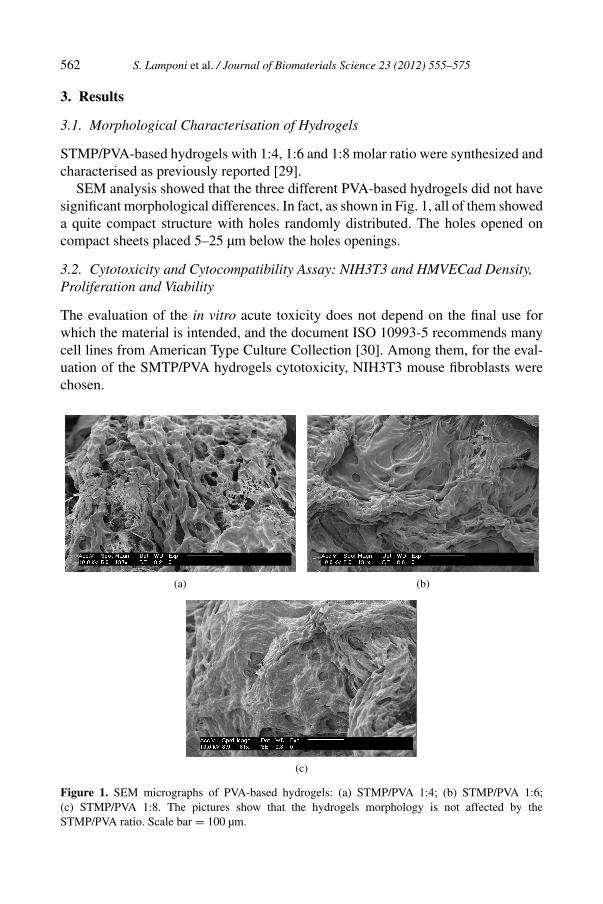
3.1. Morphological Characterisation of Hydrogels
STMP/PVA-based hydrogels with 1:4, 1:6 and 1:8 molar ratio were synthesized andcharacterised as previously reported [29].
SEM analysis showed that the three different PVA-based hydrogels did not havesignificant morphological differences. In fact, as shown in Fig. 1, all of them showeda quite compact structure with holes randomly distributed. The holes opened oncompact sheets placed 5–25 µm below the holes openings.
3.2. Cytotoxicity and Cytocompatibility Assay: NIH3T3 and HMVECad Density,Proliferation and Viability
The evaluation of the in vitro acute toxicity does not depend on the final use forwhich the material is intended, and the document ISO 10993-5 recommends manycell lines from American Type Culture Collection [30]. Among them, for the eval-uation of the SMTP/PVA hydrogels cytotoxicity, NIH3T3 mouse fibroblasts werechosen.
(a) (b)
(c)
Figure 1. SEM micrographs of PVA-based hydrogels: (a) STMP/PVA 1:4; (b) STMP/PVA 1:6;(c) STMP/PVA 1:8. The pictures show that the hydrogels morphology is not affected by theSTMP/PVA ratio. Scale bar = 100 µm.

S. Lamponi et al. / Journal of Biomaterials Science 23 (2012) 555–575 563
For the evaluation of cytocompatibility cells are usually chosen according to theend-use of the material under investigation. The vitreous body is contained in athin hyoid membrane and fills the cavity behind the crystalline lens of the eye. Thevitreous body is not penetrated by blood vessels but is nourished at its peripheryby vessels of the retina. As a consequence, a synthetic vitreous body substitutemust not alter the structure and functions of the endothelial cells. For this reasonHMVECad were chosen for the assessment of the SMTP/PVA hydrogels cytocom-patibility.
Non-confluent adhered cells were incubated with 10 mg of each swollen sampleuntil confluence was reached. Samples were analysed at intervals of 24 h and theresults are reported in Fig. 2. All the tested samples (STMP/PVA 1:4, STMP/PVA1:6, STMP/PVA 1:8) were not cytotoxic, neither for mouse 3T3 fibroblasts, norfor HMVECad, and they did not interfere with cell growth. In fact, the number ofadhered cells/mm2 in contact with the hydrogels was not statistically different incomparison to the negative control (HDPE). Looking at Fig. 2, a slight, not statis-tically significant, difference among the number of cells in contact with the threehydrogels was revealed for both the cell types. In fact, the number of cells in con-tact with the hydrogel reached the highest value for STMP/PVA 1:8 and it decreasedin the case of STMP/PVA 1:6 and STMP/PVA 1:4. This trend may be due to thedifferent cross-linking degree of the hydrogels. In fact, the STMP/PVA 1:8, withthe lowest cross-linking degree and, thus, a less rigid structure, allows an easierexchange of gases and nutrients promoting a higher cell growth [29].
The optical microscopy images (Figs 3 and 4), revealed an increasing cell densityfrom STMP/PVA 1:4 to SMTP/PVA 1:8. The sample with the highest percentageof PVA (Figs 3b and 4b) showed the same cell density as the negative control (Figs3a and 4a). The cell density decreased in SMTP/PVA 1:6 (Figs 3c and 4c) andreached the lowest value in the SMTP/PVA 1:4 (Figs 3d and 4d).
(a) (b)
Figure 2. Number of (a) adhered NIH3T3 and (b) HMVECad in contact with the three different hy-drogels as a function of incubation time evaluated by optical microscopy cell count. Data are means ±SE of five counts for each sample tested in triplicate. No value is statistically different versus control(HDPE). (A) STMP/PVA 1:4, (B) STMP/PVA 1:6 and (C) STMP/PVA 1:8.

564 S. Lamponi et al. / Journal of Biomaterials Science 23 (2012) 555–575
(a) (b)
(c) (d)
Figure 3. Optical microscopy images of NIH3T3 after 72 h of culture in contact with (a) HDPE,(b) STMP/PVA 1:8, (c) STMP/PVA 1:6 and (d) STMP/PVA 1:4. Magnification 20×, scale bar =50 µm. This figure is published in colour in the online edition of this journal, which can be accessedvia http://www.brill.nl/jbs
NIH3T3 mouse fibroblasts and HMVECad primary endothelial cells prolifera-tion and viability in contact with the hydrogels was evaluated by WST-1 assay overthree days at intervals of 24 h. The results, reported in Fig. 5, demonstrated thatthe mitochondrial activity of fibroblasts and endothelial cells after 24, 48 and 72 hof contact with the three different substrates, showed the same characteristics. Noimpairment of the mitochondrial function or sign of cytotoxicity was found in thecell lines.
3.3. Cell Morphology
The morphology of NIH3T3 and HMVECad in contact with the three different hy-drogels was evaluated by SEM analysis after 72 h of culture. SEM analysis demon-strated that the morphology of both NIH3T3 and HMVECad was maintained whenthe STMP/PVA hydrogels were present. In the presence of the specimens, as wellas of the negative control, NIH3T3 showed an excellent spreading and their densityincreased with increasing PVA content in the hydrogel (Fig. 6). In fact, as already

S. Lamponi et al. / Journal of Biomaterials Science 23 (2012) 555–575 565
(a) (b)
(c) (d)
Figure 4. Optical microscopy images of HMVECad after 72 h of culture in contact with (a) HDPE,(b) STMP/PVA 1:8, (c) STMP/PVA 1:6 and (d) STMP/PVA 1:4. Magnification 20×, scale bar =50 µm.
(a) (b)
Figure 5. Number of viable (a) NIH3T3 and (b) HMVECad cells in contact with the three differ-ent hydrogels as a function of incubation time evaluated by WST-1 assay. Data are means ± SEof three experiments run in triplicate. No value is statistically significant versus control (HDPE).(A) STMP/PVA 1:4, (B) STMP/PVA 1:6 and (C) STMP/PVA 1:8.
observed by both optical microscopy analysis and proliferation assay, the number ofadhered and viable cells increased from STMP/PVA 1:4 to STMP/PVA 1:8. A con-

566 S. Lamponi et al. / Journal of Biomaterials Science 23 (2012) 555–575
(a) (b)
(c) (d)
Figure 6. SEM micrographs of NIH3T3 after 72 h of culture in contact with (a) negative control(HDPE), (b) STMP/PVA 1:8, (c) STMP/PVA 1:6 and (d) STMP/PVA 1:4. Cell density increases withthe increasing STMP/PVA molar ratio. Cells in contact with the 1:8 sample (c), as well as those incontact with the negative control (a), reach confluence. In contrast, fibroblasts in contact with both the1:4 (d) and 1:6 (c) hydrogels are not confluent.
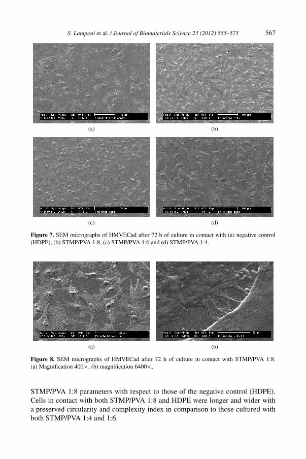
fluent monolayer of NIH3T3 covered the polystyrene plate on both STMP/PVA 1:8and HDPE after 72 h of culture and the cell ultrastructure was the same in contactwith each specimen. The characteristic morphology of spindle-shaped cells waspresent in all the samples. The adhesion of the cells to the surface was normal andidentical on each sample. No evidence of cytotoxicity, such as cell lysis and blebformation, was found. The same trend was observed for HMVECad. SEM analysisdemonstrated that also the endothelial cells morphology was maintained in pres-ence of the specimens (Fig. 7). The cells appeared flat, spread on the polystyreneand formed lamellae and philopodia between adjacent cells (Fig. 8). No alterationof both ultrastructure, and adhesion was observed for all the tested specimens. Nomorphological evidence of cytotoxicity was found.
Morphometric measurements of polystyrene-adhered HMVECad after 72 h ofculture in presence of the specimens were performed in order to demonstrate thehigh cell compatibility of the three hydrogels. By using a morphometric approachbased on five parameters (i.e., feret diameter, perimeter, area, circularity indexand complexity index; Table 1) no significant differences were detected in the
S. Lamponi et al. / Journal of Biomaterials Science 23 (2012) 555–575 567
(a) (b)
(c) (d)
Figure 7. SEM micrographs of HMVECad after 72 h of culture in contact with (a) negative control(HDPE), (b) STMP/PVA 1:8, (c) STMP/PVA 1:6 and (d) STMP/PVA 1:4.
(a) (b)
Figure 8. SEM micrographs of HMVECad after 72 h of culture in contact with STMP/PVA 1:8.(a) Magnification 400×, (b) magnification 6400×.
STMP/PVA 1:8 parameters with respect to those of the negative control (HDPE).Cells in contact with both STMP/PVA 1:8 and HDPE were longer and wider witha preserved circularity and complexity index in comparison to those cultured withboth STMP/PVA 1:4 and 1:6.

568 S. Lamponi et al. / Journal of Biomaterials Science 23 (2012) 555–575
Table 1.Morphometric characteristics of cultured endothelial cells
Parameter STMT/PVA 1:4 STMT/PVA 1:6 S/MT/PVA 1:8 Control (HDPE)
Feret diameter (µm) 54 ± 6a 45 ± 5a 76 ± 9 69 ± 10Perimeter (µm) 144 ± 15b 128 ± 16b 187 ± 16 187 ± 16Area (µm2) 1357 ± 365c 1257 ± 304c 2186 ± 308 2435 ± 426Circularity index 0.39 ± 0.07 0.45 ± 0.05 0.38 ± 0.07 0.42 ± 0.08Complexity index (µm−1) 0.106 ± 0.007d 0.102 ± 0.008d 0.085 ± 0.012 0.077 ± 0.012
Feret diameter, longest axis; circularity index, feret diameter/short axis length; complexity index,perimeter/area.a–d P < 0.05, significantly different in comparison to both STMP/PVA 1:8 and control.
3.4. NIH3T3 and HMVECad Adhesion and Proliferation on the Hydrogel Surface
Both fibroblasts and endothelial cells were seeded directly on STMP/PVA hydro-gels in order to investigate their bio-specific adhesiveness. For each specimen twodifferent dimensions (height × diameter) of hydrogels layers were tested: 2 mm ×15 mm, in order to completely cover the bottom of multidish wells, and 2 mm ×10 mm, so that a small part of the wells bottom were not covered by the samples.The behaviour of both NIH3T3 and HMVECad was evaluated at predetermined in-tervals of 24, 48 and 72 h by optical microscopy and after 72 h of culture by SEM.
Regarding the 2 mm × 15 mm samples, no cells, either fibroblasts or endothelialcells, attached and spread on the surface and along the thickness of any of the threePVA-based hydrogels (Fig. 9) demonstrating the low adhesion properties of thesespecimens, probably due to the low amount of adsorbed proteins, as demonstratedby the Bradford assay data reported in Table 2. As observed for the PVA hydrogels,the STMP/PVA networks showed an extremely hydrophilic nature, reflected in thehigh value of water content ranging from 90 to 96% [29] and resistance to proteinadsorption [8, 32], that negatively influence cell adhesion to the hydrogels. More-over, once removed the specimens from the bottom of the multidish wells after 72 hof culture, no cells were found adhered to the polystyrene, demonstrating that thestructure of the hydrogels prevents the cells to cross them.
With regard to the 2 mm × 10 mm hydrogel layers, both NIH3T3 and HMVECadonce seeded on the specimens surface moved to the bottom of the multidish wellsand adhered to the polystyrene plate surrounding the hydrogels (Fig. 10), empha-sising once again the high cell compatibility of the three hydrogels and their nonfouling properties (lacking of adhesiveness towards cells).
3.5. Human Lens Cell Adhesion and Proliferation on STMP/PVA 1:8
On the basis of the reported results, the STMP/PVA 1:8 hydrogel was selected fortesting with human lens cells. Human anterior chamber was put directly on hydrogelsurface in order to investigate its bio-specific adhesiveness. The dimension of hy-drogel layers tested was 2 mm × 15 mm (height × diameter) in order to completely

S. Lamponi et al. / Journal of Biomaterials Science 23 (2012) 555–575 569
(a) (d)
(b) (e)
(c) (f)
Figure 9. SEM micrographs of STMP/PVA hydrogels 72 h after cell seeding on the surface of thespecimens. (a) STMP/PVA 1:4 + HMVECad, (b) STMP/PVA 1:6 + HMVECad, (c) STMP/PVA1:8 + HMVECad, (d) STMP/PVA 1:4 + NIH3T3, (e) STMP/PVA 1:6 + NIH3T3 and (f) STMP/PVA1:8 + NIH3T3. No cells, both fibroblasts and endothelial, adhered on the surface of the samples.
cover the bottom of the multidish wells. The behaviour of cells was evaluated bySEM after 30 days of culture.
The results are shown in Fig. 11. No cells were found attached and spread on thesurface of the hydrogel, demonstrating the low adhesion properties of this specimenalso towards specific human lens cells. Moreover, as observed for fibroblasts andendothelial cells, once the specimens were removed from the bottom of the multi-

570 S. Lamponi et al. / Journal of Biomaterials Science 23 (2012) 555–575
Table 2.Amount of proteins released by 5 mg hydrogels
Sample PBS (mg/ml) SDS (mg/ml)
STMT/PVA 1:4 0.0150 ± 0.0003 0.5810 ± 0.0116STMT/PVA 1:6 0.0270 ± 0.0005 0.2090 ± 0.0042STMT/PVA 1:8 0.0120 ± 0.0002 0.0150 ± 0.0007
(a) (b)
Figure 10. Optical microscopy images of (a) HMVECad and (b) NIH3T3 adhering to the polystyrene(PS) surrounding the STMP/PVA 1:8 hydrogel. Magnification 20×.
(a) (b)
Figure 11. SEM images of hydrogel samples after 30 days of culture with human anterior chamber:(a) low magnification (400×) and (b) high magnification (1600×).
dish wells (after 30 days of culture), no cells were found adhered to the polystyreneplate due to the structure of the hydrogel which does not allow the cells to cross it.
3.6. Biocompatibility of the Sheared STMP/PVA 1:8 Hydrogel
Non-confluent adhered fibroblasts and endothelial cells were incubated with 10 mgof swollen, sheared STMP/PVA 1:8 hydrogel. Samples were analysed after 24 hand the results are reported in Fig. 12. The sheared hydrogel was not cytotoxic for

S. Lamponi et al. / Journal of Biomaterials Science 23 (2012) 555–575 571
Figure 12. Percentage of viable NIH3T3 and HMVECad after 24 h of contact with the shearedSTMP/PVA 1:8 hydrogel evaluated by WST-1 assay. Data are mean ± SE of three experiments run intriplicate. No value is statistically significant versus control (HDPE).
either mouse fibroblasts or human endothelial cells and it did not interfere with cellviability and growth. No impairment of the mitochondrial function was found inboth the cell lines. In fact, the percentage of viable cells in contact with the sam-ple was not statistically different in comparison to the negative control (HDPE),demonstrating that the injection of the hydrogel through a needle did not changeits biocompatibility characteristics. The optical microscopy images (Fig. 13) revealthat the morphology of both NIH3T3 and HMVECad was maintained when thesheared hydrogel was present. In the presence of the STMP/PVA 1:8, as well as ofthe HDPE, mouse fibroblast showed an excellent spreading and after 24 h of culturea confluent monolayer of cells covered the polystyrene plate (Fig. 13a and 13b). Thesame trend was observed for HMVECad (Fig. 13c and 13d), even if in this case cellsdid not reach confluence in 24 h. Endothelial cells morphology was maintained,cells appeared flat, spread on polystyrene and formed lamellae and philipodia be-tween adjacent cells.
4. Discussion
The success of an implant is based on the understanding of the cell–material interac-tions, that, first of all, must not alter cell viability and functions. The present findingson the in vitro assessment of biocompatibility of three STMP/PVA hydrogels (1:4,1:6 and 1:8) towards NIH3T3 fibroblast line and primary HMVECad demonstratedthat all the hydrogels are biocompatible and not cytotoxic for these cells. Anyway,the differences observed among the specimens in terms of (1) cell proliferation and(2) HMVECad morphology, indicated that the three hydrogels are biocompatiblebut to a different extent. The differences in biocompatibility degree may be due tothe structural diversity of the three specimens and not to their morphological char-acteristics as demonstrated by rheological properties, light transmittance [29] andSEM analysis. In fact, by a morphological point of view there are no differencesamong the three hydrogels. All of them show the same structure with holes ran-

572 S. Lamponi et al. / Journal of Biomaterials Science 23 (2012) 555–575
(a) (b)
(c) (d)
Figure 13. Optical microscopy image of NIH3T3 and HMVECad after 24 h of culture with (a, c)HDPE and (b, d) sheared STMP/PVA 1:8. Magnification 20×, scale bar 50 µm. This figure is pub-lished in colour in the online edition of this journal, which can be accessed via http://www.brill.nl/jbs
domly distributed and the same degree of micro-roughness. In contrast, from therheological point of view, the STMP/PVA 1:8 shows a less compact structure incomparison to the other two hydrogels allowing an easier exchange of gases andnutrients [29] which promoted a higher cell growth.
Cell shape and size strongly depend on the spatial arrangement of the micro-tubule cytoskeleton and are closely related to cell function [33]. Endothelial cells incontact with both STMP/PVA 1:8 and HDPE (negative control) resulted longer andwider with a preserved circularity and complexity index in comparison to those cul-tured with both STMP/PVA 1:4 and 1:6. It cannot be excluded that these differencesreflect, at least in part, a different functional state probably due to the different rhe-ological properties of STMP/PVA 1.8 in comparison to both 1:4 and 1:6 samples.The 1:8 hydrogel, for its characteristics, promotes not only a higher cell growth butalso a preserved cell structure. It is noteworthy that the reduced perimeter/area ratiosuggests for many types of cultured cells a lower shape complexity, a parameter
S. Lamponi et al. / Journal of Biomaterials Science 23 (2012) 555–575 573
that normally is under tight control to ensure a normal cell architecture and tissuepattern [33].
The degree of cell adhesion onto the hydrogels is influenced by the properties ofthe three-dimensional network, such as hydrophilicity [34], hydrophobicity [35],charge density [36] and incorporation of biologically active molecules [8, 15].Highly hydrophilic materials do not favour cell adhesion, and this phenomenonon hydrogels increases with increasing the water content [37] because of the lowamount of adsorbed proteins [38]. In fact, it is well described that implanted mate-rials are immediately coated with proteins from blood and interstitial fluids, and itis through this adsorbed layer that cells sense foreign surfaces. Cells do not adheredirectly to biomaterials but through the adsorbed proteins [39]. Hence, it is the ad-sorbed proteins layer, rather than the surface itself, to which cells initially respond.PVA hydrogels have been shown to resist protein adsorption and cell adhesion[32]. In order to promote cell adhesion many attempts have been done for mod-ifying PVA-based hydrogels: conjugation with fibronectin [15] and cell-adhesiveRGDS peptides [8] or by blending with chitosan [40]. The high hydrophilicity ofthe SMTP/PVA hydrogels presented here, whose water content value is in the rangeof 90–96% [29], inhibits proteins adsorption on the specimens, as demonstratedby Bradford assay, and this could explain why the cells were never observed ad-hered to the hydrogels. Moreover, as the seeded cells on SMTP/PVA surface werenot found to cross the three-dimensional network, the hydrogels were demonstratedto have the suitable mechanical properties for preventing cell migration throughthem. When the experiments are performed with hydrogel samples smaller thanthe diameter of the bottom well (10 mm), cells can slip along the highly hydratedhydrogel structure, falling down to the bottom of well dish, adhering and growingon polystyrene surface maintaining tight contact with the hydrogels. The integrityof the morphology of polystyrene-adhered cells in contact with the hydrogels andtheir tight contact with the specimens, points out the high biocompatibility of theSTMP/PVA hydrogels.
The hydrogels’ biocompatibility and their ability to support cell adhesion andgrowth conjugated with their rheological and physico-chemical properties [29], letus to indicate these gels for ophtalmologic applications (e.g., vitreous body sub-stitute). For most tissue engineering applications, it is a general requirement thatcells adhere to the supporting material or scaffold that is being used [41]; for vit-reous body replacement the most important requisite is that the materials, besidesnot being cytotoxic and highly biocompatible, are not colonised by cells in orderto avoid opacities which would compromise the function of the implant. For thisreason the adhesion and proliferation of human lens cells to the most promising hy-drogel in terms of cell compatibility, i.e., the 1:8 STMP/PVA, has been studied. Aswell as for fibroblasts and endothelial cells, human lens cells do not adhere and pro-liferate onto the hydrogels surface and do not cross the three-dimensional network.Moreover, after injection through a 19 gauge needle the STMP/PVA 1:8 hydrogeldoes not change its characteristics of biocompatibility. Thus, the properties of the

574 S. Lamponi et al. / Journal of Biomaterials Science 23 (2012) 555–575
SMTP/PVA 1:8 hydrogel makes it eligible for the use as vitreous body substitute,also in consideration of the good in vivo and in vitro indications for PVA-based hy-drogels long-term safety and stability. In fact, it has been demonstrated by Maroukaet al. [28] that PVA hydrogels, obtained by gamma irradiation, injected into vitre-ous body cavity after vitrectomy in crab-eating macaques, do not induce significantchanges in ophtalmoscopic findings. No abnormal rising of intraocular pressure isrecognized; the electroretinogram does not show a meaningful amplitude weakness;from the photon counting of flare cell meter, significant break of the blood–aqueousbarrier and blood–retinal barrier is not observed. Moreover, histopathological exam-ination reveals that all layers of the retina are intact and no loss of tissue is evidenteven if some vacuolations of the inner retina are found in some specimens. PVA-hydrogels have also demonstrated stability during swelling at 37◦C for a 6-monthperiod, indicating suitability for long-term biomedical applications [42, 43].
In conclusion, all the STMP/PVA hydrogels properties, i.e., high cell compati-bility and inhibition of cell adhesion on the surface, make them suitable as vitreousbody substitutes. Among them, the 1:8 sample, showing the best behaviour in termsof biocompatibily, is the most suitable candidate for such an application.
References
1. R. Barbucci, G. Leone and S. Lamponi, J. Biomed. Mater. Res. B 76, 33 (2006).2. J. A. Rowley, G. Madlambayan and D. J. Mooney, Biomaterials 20, 45 (1999).3. Z. M. Wu, X. G. Zhang, C. Zheng, C. X. Li, S. M. Zhang, R. N. Dong and D. M. Yu, Eur. J.
Pharm. Sci. 37, 198 (2009).4. M. B. Yaylaoglu, C. Yildiz, F. Korkusuz and V. Hasirci, Biomaterials 20, 1513 (1999).5. M. Diez, P. Mela, V. Seshan, M. Möller and M. C. Lensen, Small 5, 2756 (2009).6. P. Schexnailder, E. Loizou, L. Porcar, P. Butler and G. Schmidt, Phys. Chem. Phys. 11, 2760
(2009).7. N. V. Gupta and H. G. Shivakumar, Curr. Drug Deliv. 6, 505 (2009).8. R. H. Schmedlen, K. S. Masters and J. L. West, Biomaterials 23, 4325 (2002).9. H. A. Aliyar, W. J. Foster, P. D. Hamilton and N. Ravi, Polym. Prepr. 45, 469 (2004).
10. T. Betancourt, J. Prado, K. Soo and N. A. Peppas, J. Biomed. Mater. Res. 93, 175 (2010).11. N. A. Peppas and A. G. Mikos, in: Hydrogels in Medicine and Pharmacy: Fundamentals,
N. A. Peppas (Ed.), p. 1. CRC Press, Boca Raton, FL (1986).12. Y. Hu, X. Zhou, Y. Lu, C. Hu and X. Hu, Int. J. Pharm. 371, 89 (2009).13. M. F. Abou Taleb, H. L. Abd El-Mohdy and H. A. Abd El-Rehim, J. Hazard. Mater. 168, 68
(2009).14. L. Dini, E. Panzarini, M. A. Miccoli, V. Miceli, C. Protopapa and P. A. Ramires, Tissue Cells 37,
479 (2005).15. C. R. Nuttelman, D. J. Mortisen, S. M. Henry and K. S. Anseth, J. Biomed. Mater. Res. 57, 217
(2001).16. A. C. Jen, M. C. Wake and A. G. Mikos, Biotechnol. Bioeng. 50, 357 (1996).17. A. Chiumiento, A. Dominguez, S. Lamponi, R. Villalonga and R. Barbucci, J. Mater. Sci. Mater.
Med. 17, 427 (2006).18. H. Shin, K. Zygourakis, M. C. Farach-Carson, M. J. Yaszemski and A. G. Mikos, Biomaterials
25, 895 (2004).

S. Lamponi et al. / Journal of Biomaterials Science 23 (2012) 555–575 575
19. B. Muller, US Patent 5,508,317 (1996).20. D. Myung, N. Farooqui, L. L. Zheng, W. Koh, S. Gupta, A. Bakri, J. Noolandi, J. R. Cochran,
C. V. Frank and C. N. Ta, J. Biomed. Mater. Res. A 90, 70 (2009).21. K. E. Swindle, P. D. Hamilton and N. Ravi, J. Biomed. Mater. Res. A 87, 656 (2008).22. K. E. Swindle-Reilly, M. Shah, P. D. Hamilton, T. A. Eskin, S. Kaushal and N. Ravi, Invest.
Ophtalmol. Vis. Sci. 50, 48 (2009).23. P. K. Leaver, in: Basic and Advanced Vitreous Surgery, G. W. Blankenship, M. Stirpe, M. Gonvers
and S. Binder (Eds), p. 387. Liviana Press, Padova (1986).24. S. Saxena and L. Gopal, Indian J. Ophtalmol. 44, 191 (1996).25. N. Soman and R. Banerjee, Biomed. Mater. Eng. 13, 59 (2003).26. T. V. Chirila and Y. Hong, Polym. Int. 46, 183 (1998).27. S. Suri and R. Banerjee, J. Biomed. Mater. Res. A 79, 650 (2006).28. S. Marouka, T. Matsuura, K. Kawasaki, M. Okamoto, H. Yoshiaki, M. Kodama, M. Sugiyama and
M. Annaka, Curr. Eye Res. 31, 599 (2006).29. G. Leone, M. Consumi, M. Aggravi, A. Donati, S. Lamponi and A. Magnani, J. Mater. Sci. Mater.
Med. 21, 2491 (2010).30. International standard ISO 10993-5, 1st edn. ISO, Geneva (1992).31. S. Pezzatini, R. Solito, L. Morbidelli, S. Lamponi, E. Boanini, A. Bigi and M. Ziche, J. Biomed.
Mater. Res. A 76, 656 (2006).32. K. Smetana, Biomaterials 14, 1046 (1993).33. S. M. Rafelski and W. F. Marshall, Nature Rev. Cell Biol. 9, 593 (2008).34. T. Nam Kimura and A. Kishida, Biomaterials 28, 3153 (2007).35. Y. Ogushi, S. Sakai and K. Kawakami, Macromol. Biosci. 10, 262 (2009).36. Y. M. Chen, J. P. Gong, M. Tanaka, K. Yasuda, S. Yamamoto, M. Shimomura and Y. Osada,
J. Biomed. Mater. Res. A 88, 74 (2009).37. S. Nagaoka, H. Tanzawa and J. Suzuki, In Vitro Cell Dev. Biol. 26, 51 (1990).38. C. J. Wilson, R. E. Clegg, D. I. Leavesley and M. J. Pearcy, Tissue Eng. 11, 1 (2005).39. B. Ratner, J. Biomed. Mater. Res. A 27, 837 (1993).40. D. T. Mathews, Y. A. Birney, P. A. Cahill and G. B. McGuinness, J. Biomed. Mater. Res. B 84,
531 (2008).41. J. E. Leslie-Barbick, J. J. Moon and J. L. West, J. Biomater. Sci. Polymer Edn 20, 1753 (2009).42. C. M. Hassan, J. E. Stewart and N. A. Peppas, Eur. J. Pharm. Biopharm. 49, 161 (2000).43. C. M. Hassan and N. A. Peppas, Macromolecules 33, 2472 (2000).