Institutionalisms and Structuralisms: Opposites, Substitutes, or Relatives
Upload
khangminh22Category
view
1download
0
Healthcare 2014, 2, 356-400; doi:10.3390/healthcare2030356
healthcare ISSN 2227-9032
www.mdpi.com/journal/healthcare
Review
Wound Healing: Biologics, Skin Substitutes, Biomembranes and
Scaffolds
Krishna S. Vyas and Henry C. Vasconez *
Division of Plastic Surgery, Department of Surgery, University of Kentucky, Kentucky Clinic K454,
740 South Limestone, Lexington, KY 40536, USA; E-Mail: [email protected]
* Author to whom correspondence should be addressed; E-Mail: [email protected];
Tel.: +1-859-323-8082; Fax: +1-859-323-3823.
Received: 26 May 2014; in revised form: 8 July 2014 / Accepted: 19 August 2014 /
Published: 10 September 2014
Abstract: This review will explore the latest advancements spanning several facets of
wound healing, including biologics, skin substitutes, biomembranes and scaffolds.
Keywords: biologics; skin substitutes; biomembranes; scaffolds; wound healing
Abbreviations
RNA Ribonucleic Acid
IL-6 Interleukin 6
TNF-α Tumor Necrosis Factor Alpha
LTC4 Leukotriene C4
TXB2 Thromboxane B2
UVB Ultraviolet B
MIF Migration Inhibitory Factor
NO Nitric Oxide
RCT Randomized Controlled Trial
TBSA Total Body Surface Area
STSG Split-Thickness Skin Graft
COX-2 Cyclooxygenase-2
IL-1β Interleukin-1 beta
NF-κB Nuclear Factor kappa-light-chain-enhancer of activated B cells
OPEN ACCESS

Healthcare 2014, 2 357
IL-10 Interleukin 10
ATP Adenosine Triphosphate
KATP Potassium Channels
CEA Cultured Epithelial Autograft
HDE Humanitarian Device Exemption
DFU Diabetic Foot Ulcers
PMA Premarket Approval
LOS Length of Stay
TGF-β Transforming Growth Factor-beta
CACs Circulating Angiogenic Cells
OPN Osteopontin
CCPE Collagen Coated Porous Polyethylene
PMB Poly(2-methacryloyloxyethyl phosphorylcholine-co-n-butyl methacrylate)
UPPE Uncoated Porous Polyethylene
DNA Deoxyribonucleic Acid
P3HT Photosensitive Polymer Poly (3-hexylthiophene)
CGS Collagen/Gelatin Sponge
1. Introduction
The healing of wounds is a complex process that involves the activation and synchronization of
intracellular, intercellular and extracellular elements, including coagulatory and inflammatory events,
fibrous tissue accretion, deposition of collagen, epithelialization, wound contraction, tissue granulation and
remodeling [1]. This process occurs via activation of local and systemic cells to restore tissue integrity
through regeneration and scar formation, and often these cumulative processes result in satisfactory
repair of damaged sites. Disruptions caused by tissue loss, inadequate blood flow, and comorbid
disease states can lead to chronic wounds that are difficult to manage [2]. There are many strategies
that have been applied to the treatment of wounds in the past. Early on, these were based on empirical
deduction and unsubstantiated determinations. Although there was a general resistance to new concepts
and modalities that impeded progress, advancements in the treatment of wounds have, nevertheless,
evolved [3]. Over the past two decades, advancements in the clinical understanding of wounds and their
pathophysiology have commanded significant biomedical innovations in the treatment of acute, chronic,
and other types of wounds. This review will explore the latest advancements spanning several facets of
wound healing, including biologics, skin substitutes, biomembranes and scaffolds.
2. Biologics for Wound Healing
2.1. Description
Biologic wound healing therapies are those that are intended to facilitate the re-establishment of the
innate repair mechanisms, and may involve the application of active biological agents, such as
plant-derived active biomolecules which exhibit antioxidant, antimicrobial, or anti-inflammatory attributes.
Biologic dressings prevent evaporative water loss, heat loss, protein and electrolyte loss, and

Healthcare 2014, 2 358
contamination. They also permit autolytic debridement and develop a granular wound bed. Biological skin
equivalents, epidermal growth factors, stem cell therapies, and tissue engineering might also be utilized [2].
2.2. Mechanisms and Indications
Monoterpenes represent an extensive and varied family of naturally occurring terpene-based
chemical compounds that comprise the majority of essential oils. These compounds exhibit
anti-inflammatory, antibacterial, and antioxidant attributes [4,5]. The primary mechanisms proposed
for various monoterpenes encompass: antimicrobial activity (inhibition of microorganism ribonucleic
acid (RNA) and protein biosynthesis); anti-inflammation (lowers the generation of interleukin 6 (IL-6)
and tumor necrosis factor alpha (TNF-α) in mast cells, inhibition and alteration of leukotriene C4
(LTC4) release and thromboxane B2 (TXB2) release, respectively); antioxidation (inhibits the production
of ultraviolet B (UVB)-induced free radicals photoprotective effects and oxidative stress); fibroblast
growth and macrophage migration inhibitory factor (MIF) effects. The anti-inflammatory action of the
monoterpenes is often correlated to their wound-healing effects. Monoterpenes include compounds
such as borneol, thymol, α-terpineol, genipin, aucubin, d-Limonene and sericin that have either direct
or indirect activities in wound healing. Although monoterpenes are poorly studied in the context of
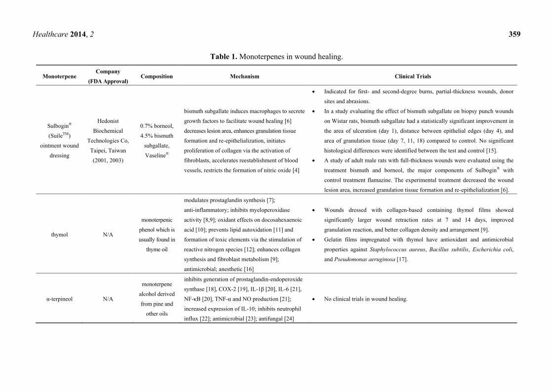
wound healing, studies suggest that they are promising for the treatment of chronic wounds (Table 1).
Mai et al. [6] investigated the ointment Sulbogin® (marketed as Suile
TM), comprised of borneol (a
bicyclic monoterpenoid alcohol), bismuth subgallate and Vaseline®
, and found it to hasten excision
wound closure in adult male Sprangue-Dawley rats. Although the specific mechanism remains elusive,
it is thought that bismuth subgallate may induce macrophages to secrete growth factors to facilitate
wound healing. It was found to decrease the lesion area, enhance granulation tissue formation and
re-epithelialization, initiate the proliferation of collagen via the activation of fibroblasts, accelerate the
reestablishment of blood vessels, and restrict the formation of nitric oxide (NO) [4,6].
The monoterpenoid phenol, thymol, demonstrates multiple beneficial bioactivities toward the
healing of wounds. These attributes encompass the modulation of prostaglandin synthesis [7],
imparting anti-inflammatory effects in neutrophils, the inhibition of myeloperoxidase activity and a
decreased influx of leukocytes [8,9], positive antioxidant effects on docosahexaenoic acid (an omega-3
fatty acid) concentrations [10], the prevention of lipid autoxidation [11] and formation of toxic
elements via the stimulation of reactive nitrogen species [12], and antimicrobial activity [13,14]. The
capacity of thymol for direct wound healing involves its being correlated with elevated concentrations,
in the central nervous system, of macrophage MIF, as well as enhanced anti-inflammatory related
tissue granulation. Furthermore, it influences collagen synthesis and fibroblast metabolism, leading to
augmented fibroblast growth in vitro [9].
Healthcare 2014, 2 359
Table 1. Monoterpenes in wound healing.
Monoterpene Company
(FDA Approval) Composition Mechanism Clinical Trials
Sulbogin®
(SuileTM)
ointment wound
dressing
Hedonist
Biochemical
Technologies Co,
Taipei, Taiwan
(2001, 2003)
0.7% borneol,
4.5% bismuth
subgallate,
Vaseline®
bismuth subgallate induces macrophages to secrete
growth factors to facilitate wound healing [6]
decreases lesion area, enhances granulation tissue
formation and re-epithelialization, initiates
proliferation of collagen via the activation of
fibroblasts, accelerates reestablishment of blood
vessels, restricts the formation of nitric oxide [4]
Indicated for first- and second-degree burns, partial-thickness wounds, donor
sites and abrasions.
In a study evaluating the effect of bismuth subgallate on biopsy punch wounds
on Wistar rats, bismuth subgallate had a statistically significant improvement in
the area of ulceration (day 1), distance between epithelial edges (day 4), and
area of granulation tissue (day 7, 11, 18) compared to control. No significant
histological differences were identified between the test and control [15].
A study of adult male rats with full-thickness wounds were evaluated using the
treatment bismuth and borneol, the major components of Sulbogin® with
control treatment flamazine. The experimental treatment decreased the wound
lesion area, increased granulation tissue formation and re-epithelialization [6].
thymol N/A
monoterpenic
phenol which is
usually found in
thyme oil
modulates prostaglandin synthesis [7];
anti-inflammatory; inhibits myeloperoxidase
activity [8,9]; oxidant effects on docosahexaenoic
acid [10]; prevents lipid autoxidation [11] and
formation of toxic elements via the stimulation of
reactive nitrogen species [12]; enhances collagen
synthesis and fibroblast metabolism [9];
antimicrobial; anesthetic [16]
Wounds dressed with collagen-based containing thymol films showed
significantly larger wound retraction rates at 7 and 14 days, improved
granulation reaction, and better collagen density and arrangement [9].
Gelatin films impregnated with thymol have antioxidant and antimicrobial
properties against Staphylococcus aureus, Bacillus subtilis, Escherichia coli,
and Pseudomonas aeruginosa [17].
α-terpineol N/A
monoterpene
alcohol derived
from pine and
other oils
inhibits generation of prostaglandin-endoperoxide
synthase [18], COX-2 [19], IL-1β [20], IL-6 [21],
NF-κB [20], TNF-α and NO production [21];
increased expression of IL-10; inhibits neutrophil
influx [22]; antimicrobial [23]; antifungal [24]
No clinical trials in wound healing.

Healthcare 2014, 2 360
Table 1. Cont.
Monoterpene Company
(FDA Approval) Composition Mechanism Clinical Trials
genipin N/A
fruit extract
aglycone
derived from
iridoid glycoside
crosslinking agent [25,26]; antioxidant [27];
anti-inflammatory [28]; stimulates NO production;
inhibits lipid peroxidation; elevates potential of
mitochondrial membranes; elevates secretion of
insulin; increases ATP levels; closes KATP
channels [29]
No clinical trials in wound healing.
Genipin hydrogels [30], nanogels [31], and genipin cross-linked scaffolds [32]
have potential application in skin tissue engineering [33] and wound dressings
[34–36] and demonstrate excellent biocompatibility and low cytotoxicity in
scaffolding models [37,38]. In biomaterials studies, genipin-crosslinked gels
enhance fibroblast attachment [39] and vascularization of engineered
tissues [38,40] and exhibit bacterial inhibition [41].
Genipin-crosslinked gelatin-silk fibroin hydrogels have been shown to induce
pluripotent cells to differentiate into epidermal lineages [42]. Genipin as a
crosslinking agent is also utilized in controlling drug delivery in multiple
systems [43].
aucubin N/A iridoid glycoside
found in plants
anti-inflammatory [44], antimicrobial, antioxidant,
chemopreventive agent
No clinical trials in wound healing.
In a study of male mice with full-thickness buccal mucosal oral wounds, 0.1%
aucubin-treated mice demonstrated earlier re-epithelization and matrix
formation and decreased numbers of inflammatory cells compared to
saline-treated controls at 1, 3, and 5 days, suggesting utility of topical aucubin
in oral wound healing [45].

Healthcare 2014, 2 361
Table 1. Cont.
Monoterpene Company
(FDA Approval) Composition Mechanism Clinical Trials
d-Limonene N/A
orange-peel
derived terpene
d-Limonene
anti-angiogenic, anti-inflammatory; decreases
systemic cytokines; inhibits expression of
endothelial P-selectin
No clinical trials in wound healing.
Topical d-Limonene and its metabolite perillyl alcohol were tested in murine
models of chemically-induced dermatitis and mechanical skin lesions. Both
significantly reduced the severity and extent of chemically-induced dermatitis.
Lower levels of the inflammatory cytokines IL-6 and TNF-α, reduced
neovascularization, and lower levels of P-selectin expression were observed in
both models. Both d-Limonene and perillyl alcohol demonstrated anti-
inflammatory effects in wound healing. Together, these effects contribute to the
wound healing effects of d-Limonene [46].
Nanophyto-modified wound dressings with limonene are resistant to
Staphylococcal and Pseudomonal colonization and biofilm formation compared to
uncoated controls [47].
Topical limonene and other terpenes can increase permeation of silver
sulphadiazine by increasing its partitioning into eschars. Burn wound
antimicrobial therapy may be improved through the use of terpenes [48].

Healthcare 2014, 2 362
Table 1. Cont.
Monoterpene Company
(FDA Approval) Composition Mechanism Clinical Trials
sericin N/A
protein created
by silkworms
(Bombyx mori)
stimulates migration of fibroblasts; generates
collagen in wounds, leading to activation of
epithelialization; anti-inflammatory; initiates
propagation and attachment of skin fibroblasts
and keratinocytes
Double blinded randomized controlled trial (RCT) of 65 burn wounds of
greater than 15% total body surface area (TBSA) were randomly assigned to
either control (silver zinc sulfadiazine cream) or treatment (silver zinc
sulfadiazine cream with sericin cream at a concentration of 100 μg/mL). Time
to complete healing was significantly shorter for the treatment group
(22.42 ± 6.33 days) compared to the control group (29.28 ± 9.27 days).
No infections or adverse reactions were found in any of the wounds [49].
A clinical study on silk sericin-releasing wound dressing was compared to the
wound dressing Bactigras® in a clinical trial in patients with split-thickness skin
graft (STSG) donor sites. The sericin dressing was less adhesive to the wound
and potentially less traumatic. Wounds treated with the silk sericin dressing
exhibited significantly faster rates to complete healing (12 ± 5.0 days compared
to 14 ± 5.2 days) and significantly reduced pain during the first four days post-
operatively [50]. In rat models, silk sericin dressing also demonstrated
accelerated wound healing and greater epithelialization and type III collagen
formation in full-thickness wounds [51–53].
Several animal studies conclude that sericin promotes the wound healing
process without causing inflammation [54]. Sericin treated full-thickness skin
wounds in rats demonstrated less inflammation, greater wound size reduction
and shorter mean time to healing compared to control (betadine treated full-
thickness skin wounds). Examination after 15 days of 8% sericine treatment
revealed complete healing, increased collagen formation, and no ulceration
compared to cream base-treated wounds which demonstrated inflammatory
exudates and ulceration [55].
3D hydrogels [56] and cultured fibroblasts and keratinocytes on three-
dimensional sericin matrices can potentially be used as skin equivalents in
wound repair [57].
Sericin/chitosan composite nanofibers demonstrate wide spectrum bactericidal
activity [58]. Sericin enriched wound dressings represent significant promise in
wound healing biologics [35,59,60].
Healthcare 2014, 2 363
The monoterpenoid alcohol, α-terpineol conveys its wound healing [61] and anti-inflammatory
activities via the inhibition of the generation of prostaglandin-endoperoxide synthase enzymes [18],
cyclooxygenase-2 (COX-2) [19], interleukin-1 beta (IL-1β) [20] and IL-6 cytokines [21], nuclear factor
kappa-light-chain-enhancer of activated B cells (NF-κB) [20], TNF-α and NO production [21]. Increased
expression of the anti-inflammatory cytokine interleukin 10 (IL-10) is also observed. Additionally, it
exhibits inhibitory effects on neutrophil influx [22], as well as robust antimicrobial [23] and antifungal
activities [24]. Significant activity in tissue/scar formation is also observed with α-terpineol [61].
Cross-linkers are one of the many factors that affect the mechanical and biological properties of
scaffolds used in tissue engineering. The iridoid (a secondary monoterpenoid metabolite) compound
genipin may serve as a biocompatible crosslinking agent that imparts minimal cytotoxicity [25,26].
Additionally, it is an antioxidant [27] and anti-inflammatory that stimulates the generation of NO while
inhibiting lipid peroxidation [28]. It also serves to elevate the potential of mitochondrial membranes, to
elevate the secretion of insulin, to increase adenosine triphosphate (ATP) levels and to close potassium
channels (KATP) [29], among other positive effects in wound healing [36,62]. Aucubin (an iridoid
glycoside) was found to have beneficial pharmacological activities on a number of fronts,
encompassing dermal wound healing [44,45,63], and capacities as an anti-inflammatory [44],
antimicrobial [64], and antioxidant [65]. In addition to various specific biochemical effects, it also
shows promise as a non-cytotoxic chemopreventive agent [66].
D’Alessio et al. [46] revealed that the prototype monoterpene d-Limonene in combination with its
metabolite perillyl alcohol, which is derived from orange-peel, exhibited considerable anti-angiogenic,
anti-inflammatory properties, epidermal repair and wound healing effects in murine models.
These compounds also lowered the generation of systemic cytokines and inhibited the expression of
endothelial P-selectin. Topical treatment resulted in more rapid and improved wound closure.
Aramwit et al. [49] revealed that a protein derived from the silkworm cocoon called silk sericin
acted to enhance the capacity for wound (second-degree burns) healing when incorporated into a
common silver zinc sulfadiazine antimicrobial cream. At a concentration of 100 μg/mL, sericin was
shown to stimulate the migration of fibroblasts. Siritientong et al. [35] discovered that silk sericin had
the capacity to generate collagen in wounds, which led to the activation of epithelialization. Further, it
served to reduce inflammation [67] and to initiate the propagation and attachment of human skin
fibroblasts and keratinocytes [55,68,69].
2.3. Contraindications
Contraindications for biologics such as the monoterpenes are low. Acute toxicity of the
monoterpenes is low via the oral and dermal routes of exposure in animal models [70].
3. Skin Substitutes for Wound Healing
3.1. Description
Skin substitutes are tissue-engineered products designed to replace, either temporarily or
permanently, the form and function of the skin. Skin substitutes are often used in chronic, non-healing
ulcers, such as pressure ulcers, diabetic neuropathic ulcers and vascular insufficiency ulcers.

Healthcare 2014, 2 364
These wounds contribute to substantial morbidity such as increased risk for infection, limb amputation,
and death. Skin substitutes have the potential to improve rates of healing and reduce complications in
a variety of other skin wounds including, but not limited to, wounds from burn injuries, ischemia,
pressure, trauma, surgery and skin disorders. Skin substitutes are also used in patients whose ability to
heal is compromised and in situations where skin coverage is inadequate. Goals for treating acute and
chronic wounds with skin substitutes are to provide temporary coverage or permanent wound closure,
to reduce healing time, to reduce post-operative contracture, to improve function, and to decrease
morbidity from more invasive treatments such as skin grafting.
Skin substitutes can be categorized according to whether they are acellular or cellular.
Acellular products, such as cadaveric human dermis with removed cellular components, contain
a scaffold or matrix of hyaluronic acid, collagen, or fibronectin. Cellular products contain living cells
such as keratinocytes and fibroblasts within a matrix. These cells can be autologous, allogeneic,
or from another species. Skin substitutes can be divided into three major categories: dermal
replacement, epidermal replacement and dermal/epidermal replacement. They can also be used as
either permanent or temporary wound coverings.
A large number of skin substitutes are commercially available or in development. Table 2 details
epidermal, dermal, and combined, full-thickness skin replacements that have clinical and experimental
evidence of efficacy in wound healing. Information regarding type of skin replacement, regulatory
status and year of United States Food and Drug Administration (U.S. FDA) approval, product
description, indications, clinical and experimental trials according to wound type, and advantages and
disadvantages for each product are detailed.
Epidermal skin replacements require a skin biopsy from which keratinocytes are isolated
and cultured on top of fibroblasts. Epicel®
(Genzyme Tissue Repair Corporation, Cambridge, MA,
USA) is an epidermal skin substitute composed of cultured autogeneous keratinocytes used for
permanent coverage in partial or full-thickness wounds. Laserskin® (Fidia Advanced Biopolymers,
Abano Terme, Italy) is composed of autologous keratinocytes and fibroblasts cultured on a
laser-microperforated biodegradable matrix of benzyl esterified hyaluronic acid.

Healthcare 2014, 2 365
Table 2. Skin substitutes for wound healing.
Epidermal Skin Replacement
Biologic Company
(FDA Approval)
Product Description
Product Description FDA Indications
(Other Indications) Clinical Trials
Advantages
Disadvantages
Epicel®
Genzyme Tissue Repair
Corporation
Cambridge, MA, USA
(2007)
Permanent skin substitute
Living Cell Therapy
Cultured Epithelial
Autograft (CEA)
autologous keratinocytes
with murine fibroblasts are
cultured to form epidermal
autografts which are then
processed into sheets and
placed onto petroleum
gauze [71]. It is used as an
adjuvant to STSG or alone
if STSG are not available
due to
the extent or severity of the
burns.
Humanitarian Device
Exemption (HDE) for
treatment of deep dermal
or full thickness burns
(greater than or equal to
30% TBSA); grafting
after congenital nevus
removal
(diabetic and venous
ulcers)
Burns
No RCT have been conducted to evaluate the effectiveness of this product in
improving health outcomes for deep dermal/full thickness burns.
In a large, single center trial, Epicel® CEA was applied to 30 burn patients
with a mean TBSA of 37% ± 17% TBSA. Epicel® achieved permanent
coverage of a mean of 26% TBSA compared to conventional autografts
(mean 25%). Final CEA take was a mean 69% ± 23%.
Ninety percent of these severely burned patients survived [72].
Advantages
Use of autologous cells
obviates rejection
Permanent large area wound
coverage, especially in
extensive burns [73]
Disadvantages
Long culture time (3 weeks)
Variable take rate
Poor long-term results
1 day shelf life [74]
Expensive
Risk of blistering,
contractures, and infection

Healthcare 2014, 2 366
Table 2. Cont.
Epidermal Skin Replacement Biologic Company
(FDA Approval)
Product Description
Product Description FDA Indications
(Other Indications) Clinical Trials
Advantages
Disadvantages
Laserskin®
Fidia Advanced
Biopolymers
Abano Terme, Italy
Permanent skin substitute
autologous keratinocytes
and fibroblasts derived
from a skin biopsy
cultured on
a laser-microperforated
biodegradable matrix of
benzyl esterified
hyaluronic acid [75,76].
Cells proliferate and
migrate through the
matrix. Microperforations
allow for drainage of
wound exudate.
(diabetic foot ulcers and
venous leg ulcers,
partial thickness burns,
vitiligo) [77,78]
Diabetic Foot Ulcers (DFUs)
A multicenter RCT with unhealed (≥1 month) DFUs randomized 180
patients to receive intervention (Hyalograft-3D® autograft and then
Laserskin® autograft after two weeks) or control (paraffin gauze). At 12
weeks, a 50% reduction in the intervention group was achieved significantly
faster compared to control (40 versus 50 days). Complete ulcer healing was
similar in both groups. The rate of ulcer reduction was greater in the
treatment group. There was a significantly (3.65-fold) better chance of
wound healing in a subgroup of hard-to-heal ulcers following autograft
treatment of dorsal ulcers [79].
In a study of chronic (>6 months) foot ulcers over 15 cm2 in type 2
diabetic patients older than 65 years treated with Hyalograft-3D® and
Laserskin® autograft, all ulcers healed at 12 months except for one, with a
median healing time of 21 weeks [80].
In a study of 14 patients with chronic (>6 months), non-healing foot ulcers
secondary to type 2 diabetes treated with Laserskin® autograft, 11/14 lesions
were completely healed between 7 and 64 days post-transplantation [81].
Wounds
In a retrospective observational study in 30 patients with chronic wounds not
responding to conventional therapy, keratinocytes on Laserskin® to treat
superficial wounds or fibroblasts on Hyalograft-3D® to treat deep leg ulcers
were applied; the wounds were then dressed with nanocrystalline silver
dressing. A reduction in wound dimension and exudates and an increase in
wound bed score was observed. The group treated with keratinocytes had a
significantly greater degree of healing compared to those treated with allogenic
fibroblasts [82].
Collagen matrices such as Integra® have been poor recipients of cultured
keratinocytes, although some studies report successes in the use of Laserskin®
on the neodermis of Integra® after the silicone membrane is removed 14–21
days post-grafting [83,84].
Advantages
Use of autologous cells
obviates rejection
Can be produced in shorter
period of time than confluent
epidermal sheets
Does not require the use of
the enzyme dispase to
remove the sheets from
culture flasks, in contrast to
CEA
Good graft take
Low rate of infection
Ease of handling during
application
Transparency allows wound
to be visualized during
dressing changes
Disadvantages
Only available in Europe
2 day shelf life
Expensive
Healthcare 2014, 2 367
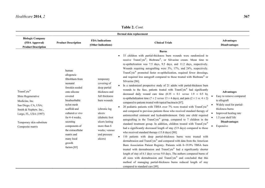
Table 2. Cont.
Dermal skin replacement Biologic Company
(FDA Approval)
Product Description
Product Description FDA Indications
(Other Indications) Clinical Trials
Advantages
Disadvantages
TransCyte®
Shire Regenerative
Medicine, Inc.
San Diego, CA, USA;
Smith & Nephew, Inc.,
Largo, FL, USA (1997)
Temporary skin substitute
Composite matrix
human
allogeneic
fibroblasts from
neonatal
foreskin seeded
onto silicone
covered
bioabsorbable
nylon mesh
scaffold and
cultured ex vivo
for 4–6 weeks,
secreting
components of
the extracellular
matrix and
many local
growth
factors [85]
temporary
covering of
deep partial
thickness and
full thickness
burn wounds
(chronic leg
ulcers
(diabetic foot
ulcers lasting
more than 6
weeks; venous
and pressure
ulcers)
Burns
33 children with partial-thickness burn wounds were randomized to
receive TransCyte®, Biobrane®, or Silvazine cream. Mean time to
re-epithelization was 7.5 days, 9.5 days, and 11.2 days, respectively.
Wounds requiring autografting were 5%, 17%, and 24%, respectively.
TransCyte® promoted faster re-epithelization, required fewer dressings,
and required less autograft compared to those treated with Biobrane® or
Silvazine [86].
In a randomized prospective study of 21 adults with partial-thickness burn
wounds to the face, patients treated with TransCyte® had significantly
decreased daily wound care time (0.35 ± 0.1 versus 1.9 ± 0.5 h),
re-epithelialization time (7 ± 2 versus 13 ± 4 days), and pain (2 ± 1 vs. 4 ± 2)
compared to patients treated with topical bacitracin [87].
20 pediatric patients with TBSA over 7% were treated with TransCyte®
and compared to previous patients those who received standard therapy of
antimicrobial ointment and hydrodebridement. Only one child required
autografting in the TransCyte® group, compared to 7 children in the
standard treatment group. In addition, children treated with TransCyte®
had a significantly decreased length of stay (5.9 days) compared to those
who received standard therapy (13.8 days) [88].
110 patients with deep partial-thickness burns were treated with
dermabrasion and TransCyte® and compared with data from the American
Burn Association Patient Registry. Patients with 0–19.9% TBSA burn
treated with dermabrasion and TransCyte® had a significantly shorter
length of stay of 6.1 days versus 9.0 days. The authors compared burns of
all sizes with dermabrasion and TransCyte® and concluded that this
method of managing partial-thickness burns reduced length of stay
compared to standard care [89].
Advantages
Easy to remove compared
to allograft
Widely used for partial-
thickness burns
Improved healing rate
1.5 year shelf life
Disadvantages
Expensive

Healthcare 2014, 2 368
Table 2. Cont.
Dermal skin replacement
Biologic Company
(FDA Approval)
Product Description
Product Description FDA Indications
(Other Indications) Clinical Trials
Advantages
Disadvantages
Wounds
A randomized prospective comparison study of TransCyte® and silver
sulfadiazine on 11 patients with paired wound sites was performed.
Wounds treated with TransCyte® had significantly quicker healing times
to re-epithelialization (mean 11.14 days vs. 18.14 days). Wound evaluations
at 3, 6, and 12 months revealed that wounds treated with TransCyte®
healed with significantly less hypertrophic scarring than those treated with
silver sulfadiazine [90].
Dermagraft®
Shire Regenerative
Medicine, Inc.
San Diego, CA, USA (2001)
Permanent or temporary skin
substitute
Living Cell Therapy
Allogenic matrix derived
from human neonatal
fibroblast
cryopreserved allogenic
neonatal fibroblasts
derived from neonatal
foreskin and cultured on
bioabsorbable collagen on
polyglactin (Dexon) or
polyglactin-910 (Vicryl)
mesh for several
weeks [91]. The
biodegradable mesh
disappears after 3–4 weeks
Premarket approval
(PMA) for full-thickness
diabetic lower extremity
ulcers present for longer
than 6 weeks extending
through the dermis but
not to the tendon, muscle,
or bone [92]
(Chronic wounds, and
noninfected wounds. It
can be used as a
temporary or permanent
covering to support take
of meshed STSG on
excised burn
wounds [93,94])
DFUs
A multicenter RCT with 314 patients with chronic DFUs to Dermagraft®
or conventional therapy was performed. At 12 weeks, 30% of the
Dermagraft® patients had complete wound closure compared to 18.3% of
control patients. Although the incidence of adverse events was similar for
both groups, the Dermagraft group (19%) experienced significantly fewer
ulcer-related adverse events (infection, osteomyelitis, cellulitis) compared
to the control group (32.5%) [95].
A prospective, multicenter RCT in 28 patients with chronic DFUs
(>6 weeks duration) comparing intervention (Dermagraft® + saline gauze)
to control (saline gauze) was performed. By week 12, significantly more
DFUs healed in the intervention (71.4%) compared to the control (14.3%).
Wounds closed significantly faster in patients treated with Dermagraft®
and the percentage of patients with wound infection was less in the
Dermagraft® group [96].
Advantages
Semitransparency allows
continuous observation of
underlying wound surface
Cell bank fibroblasts have
been tested for safety and
there have been no safety
issues thus far
Easier to remove and higher
patient satisfaction compared
to allograft [94]
Equivalent or better than
allograft for graft take [93],
wound healing time, wound
exudate and infection
No adverse reactions, such as
evidence of rejection [93]

Healthcare 2014, 2 369
Table 2. Cont.
Dermal skin replacement
Biologic Company
(FDA Approval)
Product Description
Product Description FDA Indications
(Other Indications) Clinical Trials
Advantages
Disadvantages
DFUs
The DOLCE trial (ID: NCT01450943) is a randomized, single-blind,
comparative trial to compare the differences among acellular matrices
(Oasis® (Healthpoint, Ltd Fort Worth, TX, USA), cellular matrices
(Dermagraft® (Shire Regenerative Medicine, Inc.), and standard of care in
the treatment of DFUs using the primary outcome of complete wound
closure by 12 weeks [97].
A multicenter clinical trial of Dermagraft® in the treatment of DFUs in 62
patients after sharp debridement was performed. Patients received dressing
changes with saline gauze or polyurethane foam dressings weekly. By
week 12, 27/62 (44%) patients had complete wound closure, and 32/62
(52%) healed by week 20. Median time to healing was 13 weeks.
Dermagraft® was safe and effective in the treatment of non-healing DFUs
[98].
A prospective multicenter randomized single-blinded study to evaluate
wound healing in 50 patients with DFUs was performed. Patients were
randomized into one of four groups (three separate dosages of
Dermagraft® and one control group). A dose response curve was observed
and ulcers treated with the highest dosage of Dermagraft® healed
significantly more than those treated with conventional wound closure
methods. 50% (6/12) of the Dermagraft® and 8% (1/13) of the control
ulcers healed completely. The percentage of ulcers to complete closure
was significantly greater in the Dermagraft® group (50% or 6/12)
compared to the control group (8% or 1/13) [99].
Disadvantages
Used for temporary coverage
6 month shelf life
Contraindications
Clinically infected ulcers
Ulcers with sinus tracts
Hypersensitivity to bovine
products

Healthcare 2014, 2 370
Table 2. Cont.
Dermal skin replacement
Biologic Company
(FDA Approval)
Product Description
Product Description FDA Indications
(Other Indications) Clinical Trials
Advantages
Disadvantages
Venous leg ulcers
A prospective multicenter RCT to evaluate Dermagraft® + compressive
therapy versus compressive therapy alone in the treatment of venous leg
ulcers was conducted. For ulcers ≤12 months duration, 49/94 (52%)
patients in the Dermagraft® group versus 36/97 (37%) patients in the
control group healed at 12 weeks and this was statistically significant. For
ulcers ≤10 cm2, complete healing at 12 weeks was observed in 55/117
(47%) patients in the Dermagraft® group compared with 47/120 (39%)
patients in the control group, and this was statistically significant. Both
groups experienced similar rates of adverse events [100].
A prospective RCT in 18 patients with venous leg ulcers treated with
Dermagraft® + compression therapy or compression therapy alone was
performed. Healing was assessed through ulcer tracing and planimetry.
The rate of healing was significantly improved in patients treated with
Dermagraft® [101].
AlloDerm®/
Strattice®
LifeCell Corporation
Branchburg, NJ, USA
(1992)
Permanent skin substitute
Living Cell Therapy
Human skin allograft derived
from donated human cadaver
lyophilized human
acellular cadaver dermal
matrix serves as a
scaffold for tissue
remodeling [85]
Burns, full thickness
wounds [102]
(breast surgery [103–105],
soft tissue
reconstruction [106])
Burns
Three patients with full-thickness burns of the extremities were treated with
AlloDerm® dermal grafts followed by thin autografts. Functional
performance and aesthetics were considered good to excellent [107].
The average graft take rate in 12 patients with full-thickness burn injuries in
joint areas was 91.5% at one year post AlloDerm® with ultrathin autograft.
All patients had near normal range of motion at one year and aesthetic
results were judged fair to good by both surgeons and patients [108].
Healthcare 2014, 2 371
Table 2. Cont.
Dermal skin replacement Biologic
Company
(FDA Approval)
Product
Description
Product
Description
FDA
Indications
(Other
Indications)
Clinical Trials Advantages
Disadvantages
Wounds
36 patients with oral mucosal defects reconstructed
with AlloDerm® grafts were evaluated. 34/36 cases
(94.4%) were successfully replaced with mucosa and
2 grafts failed. Graft contraction occurred in 7/34
(20.6%) of patients with lip or buccal defects [109].
Advantages
Immediate permanent wound coverage
Allows grafting of ultra-thin STSG as one-stage procedure
Template for dermal regeneration
Immunologically inert since the cells responsible for immune response and graft
rejection are removed during the processing
Reduced scarring
Can vascularize over exposed bone and tendon
2 year shelf life
Good aesthetic and functional outcomes (less hypertrophic scar rates, good
movement)
Injectable micronized form is also available (Cymetra®)
Disadvantages
Risk of transmission of infectious diseases, although no cases of viral
transmission have been reported
No viral or prion screening
Collection fluid risk (seroma, hematoma, infection)
Possibility of donor rejection
Expensive
Requires two procedures
Inability to replace both dermal and epidermal components simultaneously
Healthcare 2014, 2 372
Table 2. Cont.
Dermal skin replacement Biologic Company
(FDA Approval)
Product Description
Product Description FDA Indications
(Other Indications) Clinical Trials
Advantages
Disadvantages
Biobrane®
Smith & Nephew, St.
Petersburg, FL, USA
Temporary skin substitute
Acellular matrix
acellular dermal matrix
made of porcine type I
collagen that is
incorporated onto a
porous nylon mesh with
a silicone membrane.
The semipermeable
membrane allows for
penetration of antibiotics,
drainage of exudates, and
control of evaporative
water losses. The nylon
and silicone membrane
allow for adherence to
the wound surface [110].
Partial thickness burns
within 6 hours and donor
sites of split thickness
skin grafts [111] with
low bacterial counts and
without eschar or
debris [112]; treatment
of toxic epidermal
necrolysis [113] and
paraneoplastic
pemphigus
(dermabrasion, skin-graft
harvesting, and laser
resurfacing, chronic
wounds, venous
ulcers [110])
Burns
In a retrospective chart review of children aged 4 weeks to 18 years with
an average of 6% TBSA partial thickness burns, patients with Biobrane®
healed significantly faster compared than those treated with beta glucan
collagen (9 days vs. 13 days). Patients requiring inpatient treatment had
shorter length of hospital stay (2.6 vs. 4.1 days) [114]
In a prospective randomized study in pediatric patients with partial
thickness burns, Biobrane® was compared to topical application of 1%
silver sulfadiazine. Pain, pain medication requirement, wound healing
time, and length of stay (LOS) were significantly reduced in the Biobrane®
group [115].
In a retrospective review, Biobrane® promoted adherence of split thickness
skin grafts to the wound, allowing fluid drainage and preventing shearing.
Biobrane® also facilitated healing of adjacent donor site or partial
thickness burns [116].
In a controlled clinical trial of patients with partial thickness burns,
compared to 1% silver sulfadiazine applied twice daily with dry gauze and
elastic wraps, Biobrane® decreased healing time by 29% (10.6 days vs.
15.0 days) and reduced pain and the use for pain medication (0.6 vs. 3.0
tablets) at 24 h. There was no difference in the rate of infection [117].
In a prospective study of patients with scalp defects >5 cm requiring
removal of periosteum, the biosynthetic dressing was definitive in six
patients and complete closure was achieved in 3.5 months [118].
In a prospective RCT of children with intermediate thickness burns with
TBSA <10%, no significant difference in time to healing or pain scores
were detected between use of Biobrane® or Duoderm®, although
Biobrane® was more expensive [119].
Advantages
Dressing naturally separates
from wound
Reserved for fresh wounds
(<48 h) with low bacterial
counts
Porous material allows for
exudate drainage and
permeability to antibiotics
Higher infection rates than
other dressings [120]
Reduces pain levels and
nursing requirements when
compared to traditional
dressings [121]
Shortens LOS
Biobrane-L® available for less
aggressive adherence [122]
Disadvantages
Does not debride dead
tissue [117]
Permanent scarring in
partial-thickness scald
wounds [123]
Healthcare 2014, 2 373
Table 2. Cont.
Dermal skin replacement
Biologic Company
(FDA Approval)
Product Description
Product Description FDA Indications
(Other Indications) Clinical Trials
Advantages
Disadvantages
Burns
In a prospective RCT of 89 children treated within 48 hours of a
superficial-thickness scald burn of 5%–25% TBSA randomized to
Biobrane® or conservative treatment with topical antimicrobials and
dressing changes, patients treated with Biobrane® had significantly shorter
time to healing and length of stay. There was no difference in the use of
systemic antibiotics or readmission for infections [124].
In a prospective RCT comparing Biobrane®, Duoderm®, and Xeroform for
30 skin graft donor sites in 30 patients, donor sites dressed with Xeroform
had a significantly shorter time to healing of 10.5 days compared to
Duoderm® (15.3 days) or Biobrane® (19.0 days). Duoderm® was reported
to be the most comfortable dressing compared to Biobrane® and
Xeroform. Two infections developed using Biobrane®, one using
Duoderm®, and none using Xeroform. Biobrane® ($102.57 per patient)
was the most expensive dressed compared to Duoderm® ($54.88 per
patient) and Xeroform ($1.16 per patient) [125].
Healthcare 2014, 2 374
Table 2. Cont.
Dermal skin replacement Biologic Company
(FDA Approval)
Product Description
Product Description FDA Indications
(Other Indications) Clinical Trials
Advantages
Disadvantages
Integra® Dermal
Regeneration Template
(DRT)
Integra Lifesciences
Corporation
Plainsboro, Plainsboro, NJ,
USA (1996)
Permanent skin substitute
Acellular matrix
bilayered extracellular
matrix of cross-linked
bovine type 1 collagen
and chondroitin-6-
sulfate
glycosaminoglycan
dermal
replacement [85,126],
with a thin silicone
backing which acts as a
temporary epidermal
substitute. The product
facilitates migration of
macrophages and
fibroblasts to initiate
angiogenesis from
dermal wound bed to
create granulation tissue
to support graft or local
tissue. Once the neo-
dermis is formed, the
silicone layer is
removed and the wound
is permanently closed
with a STSG on the
neo-dermis [91].
pressure ulcers, venous
ulcers, diabetic ulcers,
chronic vascular ulcers,
surgical wounds (donor
sites/grafts, post-Moh’s
surgery, post-laser
surgery, podiatric, wound
dehiscence), trauma
wounds (abrasions,
lacerations, second-
degree burns, and skin
tears) and draining
wounds (approved
through 510(k) process in
2002)
Burns
In a multicenter prospective RCT, 106 patients with life-threatening burns
underwent excision and grafting. Mean burn size was 46.5% ± 15% mean
TBSA. Epidermal donor sites healed 4 days sooner with Integra®
compared to autograft, allograft, and xenograft. There was less
hypertrophic scarring with Integra® [127].
Integra® was applied to surgically clean, freshly excised burn wounds in
216 burn patients at 13 burn facilities in the United States. The mean total
body surface area burned was 36.5%. Once the neo-dermis was generated,
a thin epidermal autograft was placed. The incidence of superficial
infection at Integra® sites was 13.2% and of invasive infection was 3.1%.
The mean take rate of Integra® was 76.2% with a median of 95%. The
mean take rate of epidermal autograft was 87.5% with
a median take rate of 98%. This study supported the evidence that Integra®
is a safe and effective treatment in burn care [128].
In a prospective RCT comparing burn wounds treated with Integra®,
STSG, and the cellulose sponge Cellonex® in 10 adult patients, all
products demonstrated equal histological and immunohistological findings
and equal clinical appearance after one year [129].
In a RCT of 20 children with burn size ranging from 58% to 88%, there
were no significant differences between Integra® and control (autograft-
allograft application) in burn size, mortality, and length of stay. The
Integra® group had lower resting energy expenditure and increased levels
of serum constitutive proteins. The Integra® group also had increased bone
mineral content and density at 24 months and improved scarring
(vascularity, pigmentation, thickness) at 12 and 24 months [130]. This
study supported the use of Integra® for immediate wound coverage in
children with severe burns.
Advantages
Immediate permanent skin
substitute
One of the most widely
accepted synthetic skin
substitutes
Median take of 85%
Two stage procedure
requiring a minimum of 3
weeks between the
application of Integra® and
STSG [131]
More aesthetic compared
to autograft
Safe, effective, and widely
utilized for burn
reconstruction [128,132]
Integra Flowable Wound
Matrix® approved through
510(k) process in 2007
Disadvantages
Complete wound excision
High risk of infection and
graft loss since it is
avascular [133]

Healthcare 2014, 2 375
Table 2. Cont.
Dermal skin replacement Biologic Company
(FDA Approval)
Product Description
Product Description FDA Indications
(Other Indications) Clinical Trials
Advantages
Disadvantages
Post-excisional treatment
of life threatening full
thickness or deep partial
thickness burn
injuries [134] where
autograft is not available
at the time of excision or
not desirable due to the
condition of the patient
(approved 2001);
reconstruction of scar
contractures when other
therapies have failed or
when donor sites for
repair are not sufficient or
desirable due to the
condition of the patient;
chronic lower extremity
ulcers [91,92]
(soft tissue defects)
DFUs
Prospective study of patients with diabetic, non-infected plantar foot
ulcers treated with Integra® demonstrated complete wound closure in 7/10
patients by week 12 with no recurrent ulcers at follow-up [135].
A retrospective case studies review of five patients with DFUs with
extensive soft tissue deficits and exposed bone and tendon treated with
Integra® followed by STSG demonstrated complete wound healing despite
the failure of two grafts. No infections occured and all patients resumed
ambulation [136].
Wounds
In a retrospective study of 127 contracture releases with the application of
Integra® followed by epidermal autograft, 76% of the release sites, range
of motion and function were rated as significantly improved or maximally
improved by physicians at a mean post-operative follow-up period of 11.4
months. Patients expressed satisfaction with the results at 82% of sites. No
recurrence of contracture was observed at 75% of the sites. Integra®
offered functional and aesthetic benefits similar to full-thickness grafts
without the associated donor site morbidity [137].
Twelve patients with large wounds were randomly divided into treatment
with fibrin-glue anchored Integra® and postoperative negative-pressure
therapy or conventional treatment. The take rate was significantly higher
in the experimental treatment group (98% ± 2%) compared to the
conventional group (78% ± 8%). The mean time from Integra® application
to allograft was significantly shorter in the experimental group (10 ± 1 days)
compared to the conventional treatment group (24 ± 3 days), which also
resulted in shorter length of stay and potentially decreased risks of
complications such as infection or thrombosis [138].

Healthcare 2014, 2 376
Table 2. Cont
Dermal skin replacement
Biologic Company
(FDA Approval)
Product Description
Product Description FDA Indications
(Other Indications) Clinical Trials
Advantages
Disadvantages
Wounds
With the use of dressings and STSG, Integra® has been used to achieve
functional and aesthetic coverage in the management of traumatic wounds
of the hand with osseous, joint, or tendon exposure [139].
In a study of 31 patients who underwent Integra® grafting for
reconstructive surgery, complications such as silicone detachment, failure
of the graft, and hematoma were observed in nine [131].
Epidermal/Dermal Skin Replacements (Full-Thickness)
Biologic Company
(FDA Approval)
Product Description
Product Description FDA Indications
(Other Indications) Clinical Trials
Advantages
Disadvantages
Apligraf®/
Graftskin®
Organogenesis, Canton, MA,
USA (1998, 2001)
Permanent skin substitute
Living Cell Therapy
Composite matrix
cornified epidermal
allogeneic keratinocytes
derived from neonatal
foreskin cultured on a type
I bovine collagen gel
seeded with living
neonatal allogeneic human
fibroblasts in dermal
matrix [91]
Chronic partial and full
thickness venous stasis
ulcers and full thickness
diabetic foot ulcers [140]
(epidermolysis
bullosa [141], recurrent
hernia repair, pressure
sores, burn
reconstruction) [92]
Venous Leg Ulcers
A Cochrane Review concluded that a bilayer artificial skin used in
conjunction with compression bandaging increases venous ulcer healing
compared with a simple dressing plus compression [142].
In a prospective multicenter RCT of 240 patients with hard-to-heal chronic
wounds (>1 year) receiving either intervention with Graftskin® plus
compression or compression alone, treatment with Graftskin® with
compression was significantly more effective than compression therapy
alone in achieving complete wound closure at 8 weeks (32% vs. 10%) and
significantly more effective at 24 weeks (47% vs. 19%) [143].A previously
conducted prospective RCT by the same group revealed similar results [144].
Advantages
Small wounds require one
application
Improved cosmetic (scar
tissue, pigmentation, texture)
and functional outcomes in
chronic wounds [145]
Primary role in treating
chronic ulcers

Healthcare 2014, 2 377
Table 2. Cont.
Epidermal/Dermal Skin Replacements (Full-Thickness)
Biologic Company
(FDA Approval)
Product Description
Product Description FDA Indications
(Other Indications) Clinical Trials
Advantages
Disadvantages
Burns
In a multicenter RCT of 38 patients with STSG wounds, Apligraf® was
placed over meshed autograft while control sites were treated with meshed
autograft covered with no biologic dressing or meshed allograft. There
was no difference in the percent take of meshed split thickness autograft
with or without Apligraf®. The Apligraf® group demonstrated significantly
improved vascularity, pigmentation, wound height and Vancouver burn
scar scores, demonstrating a cosmetic and functional advantage of
Apligraf® compared to controls [145].
Donor site healing
A RCT of 60 skin donor sites treated with meshed autograft, meshed
Apligraf®, or polyurethane film dressing was conducted. The healing time
with Apligraf® (7.6 days) was significantly shorter than with polyurethane
film dressing.
In a multicenter RCT of 10 patients treated with Apligraf®, Apligraf®
dermis-only, and polyurethane film for acute STSG donor sites, there were
no differences among the treatment modalities in establishing basement
membrane at 4 weeks and there were no differences in other secondary
outcomes [146].
Disadvantages
Large wounds may require
multiple applications
5 day shelf life [91]
Expensive
Potential for viral transmission;
mothers blood and donor’s
cells screened; cell banks
screened for product safety
Consider ethics with use of
biological material: bovine
collagen (Hindus, Buddhists;
vegetarians); derived from
foreskin (Quakers) [147]
Contraindications
Infected wounds
Allergy to bovine collagen

Healthcare 2014, 2 378
Table 2. Cont.
Epidermal/Dermal Skin Replacements (Full-Thickness) Biologic Company
(FDA Approval)
Product Description
Product Description FDA Indications
(Other Indications) Clinical Trials
Advantages
Disadvantages
DFUs
In a multicenter RCT of 72 patients comparing Apligraf® and standard therapy
versus standard therapy alone in the treatment of DFUs, there was a
significantly shorter time to complete wound closure in the Apligraf® group
51.5% (17/33) compared to with standard treatment with international
guidelines 26.3% (10/38) at 12 weeks [148].
In a prospective multicenter RCT of 208 patients randomly assigned to ulcer
treatment with Graftskin® or saline-moistened gauze (control), 63/112 (56%)
of Graftskin® patients achieved complete wound healing compared to 36/96
(38%) in the control at 12 weeks and this result was statistically significant.
Kaplan-Meier curve to complete closure was also significantly lower for
Graftskin® (65 days) compared to control (90 days). Osteomyelitis and lower-
limb amputations were less frequent in the Graftskin® group [149].
Treatment with Apligraft® plus good wound care for DFUs results in 12%
reduction in costs during first year of treatment compared to good wound care
alone [150].
Wounds
In a prospective RCT of 31 patients requiring full-thickness surgical excision
for non-melanoma skin cancer, patients were randomized to receive a single
application of Apligraf® or to heal by secondary intention. Apligraf® reduced
post-operative pain in this setting, but it was not determined whether it could
decrease healing time or result in better aesthetic outcomes [151].
In a prospective controlled clinical trial, 48 deep dermal wounds were
created and Apligraf®, STSG, or dressing was applied. Apligraf®
demonstrated more cellular infiltrate but less vascularization compared to
controls. Apligraf® demonstrated survival of allogeneic cells in acute
wounds for up to six weeks and was recommended for the management of
acute surgical wounds [152].

Healthcare 2014, 2 379
Table 2. Cont.
Epidermal/Dermal Skin Replacements (Full-Thickness) Biologic Company
(FDA Approval)
Product Description
Product Description FDA Indications
(Other Indications) Clinical Trials
Advantages
Disadvantages
OrCel®
Forticell Bioscience,
New York City, NY,
USA (1998)
Living Cell Therapy
Composite matrix
neonatal foreskin derived
keratinocytes and dermal
fibroblasts cultured in separate
layers into a type I bovine
collagen porous sponge [85].
During healing, autologous
skin cells replace the cells in
the product.
Approved for HDE in 2001 for use in patients
with dystrophic epidermolysis bullosa
undergoing hand reconstruction surgery to
close and heal wounds created by surgery,
including donor sites; PMA approval for
autograft donor sites in burn patients (overlay
on split thickness skin grafts to improve
cosmesis and function) [92]
(chronic diabetic and venous wounds)
A randomized matched pairs study
comparing treatment of split-thickness donor
site wounds with OrCel® or Biobrane-L®
revealed that scarring and healing times for
sites treated with OrCel® were significantly
shorter than for sites treated with
Biobrane-L® [153].
Advantages
9 month shelf life
Disadvantages
Cryopreserved
Cannot be used in infected
wounds, in patients who are
allergic to any animal
products, or in patients
allergic to penicillin,
gentamycin, streptomycin, or
amphotericin B
GraftJacket®
Wright Medical
Technology, Inc.,
Arlington, TX, USA,
licensed by KCI
USA, Inc., San
Antonio, TX, USA
Permanent skin
substitute
Human skin allograft
derived from donated
human cadaver
micronized acellular human
dermis with a dermal matrix
and intact basement
membrane to facilitate
ingrowth of blood vessels
(deep and superficial wounds, sinus tract
wounds, tendon repair, such as rotator cuff
repair) [154]
not subject to FDA pre-notification approval as
it is a human cell or tissue based product
DFUs
Multicenter, retrospective study in the
treatment of 100 chronic, full thickness
wounds of the lower extremity in 75
diabetic patients revealed a 91% healing rate
and suggested its use in the treatment of
complex lower extremity wounds. No
significant differences were observed for
matrix incorporation or complete healing.
Mean time to complete healing was 13.8
weeks [155].
Advantages
2 year shelf life
Pre-meshed for clinical
application
Single application
Utilized in both deep and
superficial wound healing
Disadvantages
Cryopreserved
Healthcare 2014, 2 380
Table 2. Cont.
Epidermal/Dermal Skin Replacements (Full-Thickness)
Biologic Company
(FDA Approval)
Product Description
Product Description FDA Indications
(Other Indications) Clinical Trials
Advantages
Disadvantages
DFUs
In a prospective multicenter RCT comparing GraftJacket® with standard of
care therapies for the treatment of DFUs in 86 patients for 12 weeks, the
proportion of completely healed ulcers between the groups was
statistically significant. The odds of healing in the study group were 2.7
times higher than in the control group. The odds of healing were 2.0 times
higher in the study group than in the control group when adjusted for ulcer
size at presentation [156].
A prospective randomized study evaluating diabetic patients with lower
extremity wounds demonstrated that patients treated with GraftJacket® healed
significantly faster than those with conventional treatment at 1 month [157].
A prospective single center RCT comparing intervention (sharp debridement
+ GraftJacket® + mineral oil-soaked compression dressing) to control (wound
gel with gauze dressing) for the treatment of full-thickness chronic non-healing
lower extremity wounds in 28 diabetic patients revealed that at 16 weeks,
12/14 patients treated with GraftJacket® had complete wound closure
compared to 4/14 patients in the control group. Significant differences were
observed for wound depth, volume, and area [158].
In a prospective, randomized single blind pilot study of 40 patients with
debrided diabetic lower extremity wounds, GraftJacket® was compared to the
hydrogel wound dressing Curasol®. At 4 weeks, there was a significant
reduction in the ulcer size in the GraftJacket® group compared to debridement
only. At 12 weeks, 85% of the patients with GraftJacket® healed compared to
5% of controls [157].

Healthcare 2014, 2 381
Table 2. Cont.
Epidermal/Dermal Skin Replacements (Full-Thickness)
Biologic Company
(FDA Approval)
Product Description
Product Description FDA Indications
(Other Indications) Clinical Trials
Advantages
Disadvantages
DFUs
A retrospective multicenter series in 12 patients with DFUs and complex,
deep, irregularly-shaped, tunneling sinus tracts treated with GraftJacket
Xpress Scaffold® (a micronized, decellularized flowable soft tissue scaffold
that can be delivered through a syringe into the wound cavity) demonstrated
complete healing in 10/12 patients at 12 weeks [159].
In a prospective case series of 17 patients with debrided, non-infected,
non-ischemic, neuropathic DFUs treated with a single application of
GraftJacket® with weekly silicone dressing changes, 82.5% of wounds had
complete re-epithelialization in 20 weeks, with a mean time to healing of
8.9 ± 2.7 weeks [160].
PermaDerm®
Regenicin, Inc., Little Falls,
NJ, USA
Permanent skin substitute
autologous keratinocytes
and fibroblasts cultured on
bovine collagen scaffold
Orphan status approval as
a permanent skin
substitute in burns
No clinical trials available.
Advantages
No risk of rejection
Permanent substitute for
massive burn injury
Disadvantages
No clinical trials or long-
term studies available

Healthcare 2014, 2 382
Dermal skin replacements provide greater stability to the wound and prevent the wound from
contracting. Transcyte® (Shire Regenerative Medicine, Inc., San Diego, California, USA; Smith &
Nephew, Inc., Largo, FL, USA) is composed of human allogeneic fibroblasts from neonatal foreskin
seeded onto silicone covered bioabsorbable nylon mesh scaffold and cultured ex vivo for 4–6 weeks [85].
Transcyte® is often used as a non-living, temporary wound covering for partial- and full-thickness burns
after excision [161]. A derivative of Transcyte® is Dermagraft
® (Shire Regenerative Medicine, Inc., San
Diego, California, USA), a skin substitute composed of living allogenic fibroblasts incorporated into a
bioresorbable polyglactin mesh that secretes extracellular matrix (ECM) proteins, collagen, growth
factors and cytokines into the wound site in the provision of viable living dermal substitute [162,163].
Dermagraft®
has shown improvement in the treatment of chronic diabetic foot ulcers.
AlloDerm®
/Strattice®
(LifeCell Corporation, Branchburg, NJ, USA) are lyophilized human acellular
cadaver dermal matrices which serve as a scaffold for tissue remodeling. Autologous keratinocytes
may be seeded and cultured on Alloderm®
to form an epithelium; together; these can be utilized for
wound and burn closure. Subsequent to its administration to a wound site, AlloDerm®
is shown to
exhibit cellular infiltration and neovascularization [164]. Biobrane®
(Smith & Nephew, St. Petersburg,
FL, USA) is a synthetic dermis temporary skin substitute composed of inner nylon and outer silicone
with bovine collagen used for temporary coverage in partial and full-thickness burns. Integra®
Dermal
Regeneration Template (DRT) (Integra Lifesciences Corporation, Plainsboro, NJ, USA) is an example
of a composite skin graft. It is composed of an outer layer of silicone and a cross-linked bovine type I
collagen glycosaminoglycan dermal matrix. Once the dermal layer has regenerated, the silicone layer
is removed and the wound is permanently closed with a split thickness skin graft (STSG) on the
neo-dermis. Integra®
is used for permanent coverage of full-thickness burns when combined with thin
skin graft.
Epidermal/Dermal skin replacements are also called as full-thickness skin substitutes and are
composed of both epidermal and dermal layers. Autologous or allogeneic fibroblasts and keratinocytes
are used in their preparation. The allogenically derived Apligraf®
(Organogenesis, Canton, MA, USA)
is a bilayered matrix construct similar to a microscopic skin layer. Specifically, it is comprised of a
lower dermal layer of bovine type 1 collagen combined with human fibroblasts (extracted from
postnatal foreskin) and an upper layer that consists of human keratinocytes, along with
granulocyte/macrophage colony-stimulating factors. Apligraf®
has been used for permanent coverage
of non-healing chronic wounds (such as diabetic foot ulcers), surgical wounds, pressure wounds,
neuropathic wounds and venous insufficiency ulcers. Apligraf®
has been observed in vitro to generate
extracellular matrix structural elements and modulators inclusive of tissue inhibitors of matrix
metalloproteinases and glycoprotein fibronectin [2]. OrCel®
(Forticell Bioscience, New York, NY,
USA) is a composite matrix composed of keratinocytes and dermal fibroblasts cultured in separate
layers into a type I bovine collagen porous sponge. It is used in patients with dystrophic epidermolysis
bullosa undergoing hand reconstruction surgery and at autograft donor sites in burn patients [92].
GraftJacket® (Wright Medical Technology, Inc., Arlington, TX, USA, licensed by KCI USA, Inc., San
Antonio, Texas, USA), is an acellular derivative of human dermis. GraftJacket®
was shown to
facilitate accelerated healing and initiate depth and volume reductions in wounds [156]. PermaDerm®
(Regenicin, Inc., Little Falls, NJ, USA) is a newer product that acts as a permanent skin substitute to

Healthcare 2014, 2 383
cover large burns. It is composed of autologous keratinocytes and fibroblasts cultured on bovine
collagen scaffold [165].
3.2. Contraindications
Biological skin equivalents such as allogenically derived Apligraf®
or Dermagraft®
have
an existing, albeit significantly low, risk of disease transmission due to their allogenicity [162].
In the case of Apligraf®
, it has been verified in a number of studies that the cells it delivers are not
sustained within the wound site beyond six weeks, and has inconsistent effects on the wound basement
membrane, in vivo collagen composition and vascularization [2,146,152].
3.3. Clinical Trial Based Evidence
Greer et al. [166] compared a number of advanced wound therapies in the treatment of ulcers in
regard to the proportion of ulcers healed and time to healing. This study reviewed randomized
controlled trials from the literature (MEDLINE 1995–2013, Cochrane Library, and existing systemic
reviews), which involved patients who were typically middle-aged white males. The 56 trials
encompassed lower extremity or foot ulcers, with 35 cases of patients with diabetic ulcers, 20 patients
with venous ulcers, and one patient with arterial ulcers. The duration of therapies generally spanned
from 4 to 20 weeks, with a mean ulcer duration from 2 to 94 weeks. The mean ulcer size ranged from
1.9 to 41.5 cm2. Of the advanced wound care products used in these trials, the biological skin
equivalent Apligraf®
demonstrated moderate-strength evidence for enhanced healing, as did negative
pressure wound therapy. Low-strength evidence was shown for platelet-derived growth factors and
silver cream in comparison to standard care. For arterial ulcers, there was an improvement in healing
with biological skin equivalent. Although the evidence was deemed as limited, the conclusion of the
authors was that several advanced wound care therapies appeared to enhance the number of ulcers
healed, as well as to reduce the times for healing.
A clinical randomized, double-blind, standard-controlled study was undertaken, which compared
burn wounds that were treated with silver zinc sulfadiazine cream (control) against those treated with
the identical cream that also contained silk sericin. The study involved 29 patients presenting with 65
burn wounds that covered at least 15% of total body surface areas. It was observed that the typical time
for attaining 70% re-epithelialization in the sericin group was approximately 5–7 days shorter than the
control group. The control group required 29.28 ± 9.27 days for complete burn wound healing, while
the sericin group attained this condition within 22.42 ± 6.33 days with no indication of severe reaction
or infection in any wound [49].
Multiple clinical trials have been conducted with the living skin equivalents Apligraf®
and
Dermagraft®
. A retrospective controlled trial was undertaken that involved 2517 patients (446
Apligraf®
, 1892 Regranex®
(a human platelet-derived growth factor topical gel for the treatment of
lower extremity diabetic neuropathic ulcers), 125 platelet releasates, 54 combined) and found that
diabetic foot ulcers initially treated with Apligraf®
were 31.2% more likely to heal than those
administered with topical growth factor and 40% more likely to heal than those treated with platelet
releasates [95]. In a prospective, randomized controlled trial involving 72 patients (33 Apligraf®, 39 with
saline moistened gauze control), it was found that at 12 weeks, full wound closure was observed in

Healthcare 2014, 2 384
51.5% (17 of 33) of Apligraf®
patients in contrast to 26.3% (10 of 38) of control patients [148].
An additional prospective, randomized controlled trial involved 74 patients (38 autograft + Apligraf®
,
36 autograft alone or + allograft) with dull and partial thickness burns. It was found at 22 months that
58% of the Apligraf®
sites were deemed of better quality than the controls, with 26% as equivalent and
16% as worse. Further, Apligraf®
treated patients (47%) exhibited normal vascularity in contrast to 6%
of control patients [145].
A prospective, randomized controlled trial with Dermagraft® studied 314 patients (130 Dermagraft
®,
115 controls) with diabetic foot ulcers. At 12 weeks, 30% of the Dermagraft®
patients were healed in
comparison to 8.3% of the control patients, who were treated with standard wet-to-dry dressings [95].
An additional prospective, randomized controlled trial was undertaken with 18 patients (10 Dermagraft®,
eight controls) with venous ulcers, which revealed that the healing rate after 12 weeks was enhanced
considerably in those patients treated with Dermagraft®
+ compression (five patients (50%)) as
opposed to compression on its own (one patient (12.5%)). In addition, the perfusion of capillaries in
the Dermagraft®
group increased by 20%, in comparison to 4.9% in the compression group [101].
4. Biomembranes for Wound Healing
4.1. Description
Biocompatible vegetal biomembranes of natural rubber/latex, amniotic, polyurethane and
poly-DL-lactic acid (PDLLA) comprise a class of versatile interventions for the treatment and healing
of wounds. Additionally, biomembranes may be impregnated with a wide range of bioactive
compounds to further facilitate and promote wound healing.
4.2. Mechanism and Indications
Human amniotic membranes, such as Biomembrane® (Matrix Company, Ismailia, Egypt) are
comprised of skin-like fetal ectoderm, consisting of four layers (epithelial, basement membrane,
connective tissue fibroblasts, and spongy layer), which have demonstrated angiogenic properties.
The membrane is freeze dried to 5% water content and then gamma irradiated (25 kgy) to ensure
sterilization. These biomembranes exhibit a 1000-fold improvement in efficacy over split-thickness
human skin grafts, though the specific mechanisms remain unclear [167,168]. Further, amniotic
membranes are found to inhibit the alpha smooth muscle protein actin, resulting in a significant
reduction in the generation of scar tissue in comparison to a moist wound dressing control [169].
Additional benefits included decreased pain, protection from infection and control of the loss of
electrolytes and albumin.
The polyurethane film, TegadermTM
(3M, Saint Paul, MN, USA), exhibits gas semi-permeability,
which acts to augment the rate of epithelialization. This may be due the retention of carbon dioxide,
which translates to sustaining a low pH. The pain relief that is reportedly associated with this film may
be the result of the exclusion of atmospheric oxygen, which negates the generation of prostaglandin
E2, via the oxygen-reliant cyclo-oxygenase system [167,170]. An additional imparted benefit
secondary to the semi-permeability of TegadermTM
is the regulation of transforming growth factor beta
(TGF-β) via the mediation of transepidermal water transfer [171]. It also stimulates the propagation of

Healthcare 2014, 2 385
keratinocytes through the activation of integrins a5 and a6 to encourage enhanced and rapid wound
healing [172].
A biocompatible vegetal biomembrane derived from the Hevea brasiliensis rubber tree exhibited
the capacity to initiate angiogenesis and re-epithelialization in the chronic ulcers of diabetic patients.
Its activity in the healing process appears most prominent at the inflammatory stage, where the
microenvironment is transformed by robust angiogenesis followed by re-epithelialization [173].
A non-toxic, biocompatible, biodegradable, and non-carcinogenic crosslinked gelatin hydrogel
biomembrane was developed for use as a wound dressing via the addition of a naturally occurring
genipin crosslinking agent, and compared to a glutaraldehyde-crosslinked control. The resulting
genipin infused biomembrane exhibited considerably less inflammation along with more rapid
re-epithelialization and subsequent wound healing than the control, which may have been facilitated by
a lower level of genipin imparted cytotoxicity [36].
4.3. Contraindications
Despite stringent preparation protocols, there might be a very low risk of bacterial or viral
transmission via the use of human amniotic membranes on open wounds.
4.4. Clinical Trial Based Evidence
Adly et al. [167] conducted a randomized, controlled clinical trial to compare the efficacy of an
amniotic membrane (Biomembrane®
) group I (23 patients) and a polyurethane membrane
(TegadermTM
) dressing group II (23 patients) in the treatment of burns (scald and flame). There were
no notable differences between the two groups. The criteria were inclusion of both genders and all age
groups with <50% total body surface area affected with either second or third degree burns. The group
I patients exhibited a considerably lower infection rate (one patient (4.3%) in group I compared to
three patients (13.0%) in group II) and required fewer dressing changes than group II (highest dressing
change frequency was once per day in 30.4% of group I patients, in comparison to five times per day
in 60.9% of group II patients). In addition, electrolyte disturbance was evident in 17.4% of patients in
group I, compared with 60.9% of patients in group II. Albumin loss was indicated in 39.1% of patients
in group I in contrast to 60.9% of patients in group II. In terms of pain and healing times, 43.5% of
group I patients experienced pain during dressing, compared with 60.9% in group II.
Healing frequency was 47.8% (11–20 days) for group I in contrast to 39.1% in group II spanning the
same time period.
5. Scaffolds for Wound Healing
5.1. Description
Hybrid scaffolds comprised of polymeric substrates coated with bioactive materials, collagen, silk
fibroin, as well as advanced tissue engineered substrates impregnated with endothelial progenitor cells,
and nanomaterial-based scaffolds may be employed as advanced wound dressings to initiate and
expedite wound healing.

Healthcare 2014, 2 386
5.2. Mechanisms and Indications
Collagen is a component of the extracellular matrix, which has found established utility as
a biomaterial in cell therapies and tissue engineering via the provision of a viable substrate for the
attachment and propagation of cells. In the treatment of wounds, collagen scaffolds offer a feasible
platform for the topical conveyance of cells into the wound bed, increase the healing of wounds and
initiate angiogenesis and neovasculogenesis.
O’Loughlin et al. [174] investigated the use of type 1 collagen scaffolds for the topical delivery of
autologous circulating angiogenic (CACs) cells (precursors to endothelial progenitor cells), to full
thickness cutaneous ulcers. It was revealed that the CACs could also be pre-stimulated through the
addition of matricellular protein osteopontin (OPN), a glycoprotein involved in immune function,
neovascularization, and facilitation of cell migration and survival [175]. The inclusion of OPN served
to augment wound healing. It was demonstrated that scaffolds comprised of type 1 collagen, which has
been shown to sustain angiogenesis [176], when infused with CACs and enhanced with OPN,
resulted in the formation of larger diameter blood vessels than untreated wounds, and thus acceleration of
the wound healing process [174].
Ehashi et al. [177] compared subcutaneously implanted scaffolds for their host body reactions in order
to assess their wound healing capacities. The scaffolds consisted of collagen coated porous (Ø32 μm and
Ø157 μm) polyethylene (CCPE), bio-inert poly(2-methacryloyloxyethyl phosphorylcholine-co-n-butyl
methacrylate) (PMB) coated polyethylene, and uncoated porous polyethylene (UPPE) (control).
Subsequent to their immersion in sterile solution for an appropriate period, six samples (two of each
type with different pore diameters) were implanted under the skins of mouse models, and then resected
after seven days. In terms of vascularization, it was observed that small vessels were induced on the
UPPE, albeit contingent on the pore size (more activity seen with Ø32 μm pores than Ø157 μm pores).
Interestingly, the reverse was true for the CCPE, with more activity seen with the Ø157 μm pore
sample. There was no vessel growth activity associated with the PMB scaffolds. A deoxyribonucleic
acid (DNA) microarray assay was then employed to conduct genetic analyses, which showed that the
CCPE scaffold had a more highly distributed level of gene expression than did the PMB scaffold. The
PMB scaffold showed the up-regulation of genes associated with the suppression of inflammation. The
CCPE scaffold indicated up-regulation of genes related to inflammation, angiogenesis, and wound
healing. The authors concluded that the up-regulation of interleukin-1b and angiogenesis associated
genes within the porous scaffolds likely contributed to the mediation of tissue regeneration.
A novel scaffold comprised of electrospun core-shell gelatin/poly(L-lactic acid)-co-poly-(ε-caprolactone)
nanofibers, which encapsulated a photosensitive polymer poly (3-hexylthiophene) (P3HT) and epidermal
growth factor (EGF) at its core, was investigated by Jin et al. [178] as a potential skin graft. It was
found that fibroblast propagation was activated under exposure to light in contrast to its absence and
cells akin to keratinocytes were found only on the light exposed scaffolds. The researchers propose
that these light sensitive nanofibers may have utility as a unique scaffold for the healing of wounds and
the reconstitution of skin.
Bacterial (or microbial) cellulose has been investigated by Fu et al. [179] for its capacity to enable
wound healing and skin tissue rejuvenation. Specific bacteria are involved in the biosynthesis of this
natural polymer, which has unique properties in contrast to plant based cellulose, encompassing

Healthcare 2014, 2 387
biocompatibility, hydrophilicity, high water retention, elasticity, transparency, conformability and the
capacity for absorbing wound generated exudate during inflammation. These features position
microbial cellulose to have great potential for biomedical advancements in skin tissue repair.
5.3. Contraindications
Scaffolds that are comprised of hyaluronan (an anionic polysaccharide), even though non-cytotoxic
and biodegradable, may disrupt cell adhesion and the regeneration of tissues due to its hydrophilic
surface properties [177]. Additional drawbacks for tissue engineered scaffolds in the case of severe
burns relate to their unreliable adhesion to lesions and failure to replace dermal tissues [180].
5.4. Clinical Trial Based Evidence
The clinical performance of bacterial cellulose (BC) scaffold DermafillTM
(AMD/Ritmed, Tonawanda,
New York, USA) wound dressings (Acetobacter xylinum derived) was assessed by Portal et al. [181] who
compared the reduction in wound size of chronic non-healing lower extremity ulcers following
standard care. A total of 11 chronic wounds were evaluated for the time required to achieve 75%
epithelization, by comparing non-healing ulcers prior to and following the application of DermafillTM
.
The median observation timeline for chronic non-healing wounds under standard care prior to the
application was 315 days. When BC scaffolds were applied to these same wounds, the median time to
75% epithelization was decreased to 70 days. Thus, the authors concluded that BC scaffold-initiated
wound closure for non-healing ulcers proceeded considerably more rapidly than did standard care
wound dressings.
Morimoto et al. [182] investigated the clinical efficacy of a unique synthetic collagen/gelatin
sponge (CGS) scaffold for the treatment of chronic skin ulcers. This artificial dermal scaffold
demonstrated the capacity to sustainably release basic fibroblast growth factor (bFGF) over 10 days or
longer. One of the criteria for the study group was the inclusion of chronic skin ulcers that had not
healed over a time period of at least four weeks. These wounds treated with CGS, which was infused
with 7 or 14 μg/cm2 of bFGF following debridement, and assessed two weeks subsequent to their
application. Positive improvement in the wound beds was defined by the emergence of granulated and
epithelialized areas of 50% or greater. Out of a total of 17 subjects, it was observed that 16 showed
wound bed improvements, with no discernable difference between the low and high dose groups.
There was rapid recovery from mild adverse reactions.
6. Conclusions
The healing of surface and deep wounds of the epidermis is a complex multistage process, but one
that may nevertheless be expedited utilizing strategies such as the application of active biologic,
biomembrane or scaffold based wound dressings. Specific therapeutic compounds and cell species
including epidermal stem cells may be utilized to impregnate biocompatible and/or biodegradable
substrates, including membranes and scaffolds to facilitate rapid revascularization, re-epithelialization,
and healing of wound beds.
Healthcare 2014, 2 388
Acknowledgments
This research received no specific grant from any funding agency in the public, commercial,
or not-for-profit sectors.
Author Contributions
The authors’ responsibilities were as follows—Krishna S. Vyas and Henry C. Vasconez:
participated in the design of the study, drafting, critical review, and final approval of the manuscript.
Conflicts of Interest
The authors declare no conflict of interest.
References
1. Ghosh, P.K.; Gaba, A. Phyto-extracts in wound healing. J. Pharm. Pharm. Sci. 2013, 16, 760–820.
2. Rennert, R.C.; Rodrigues, M.; Wong, V.W.; Duscher, D.; Hu, M.; Maan, Z.; Sorkin, M.;
Gurtner, G.C.; Longaker, M.T. Biological therapies for the treatment of cutaneous wounds:
Phase iii and launched therapies. Expert opin. Biol. Ther. 2013, 13, 1523–1541.
3. Cohen, I.K. Lessons from the history of wound healing. Clin. Dermatol. 2007, 25, 3–8.
4. Barreto, R.S.; Albuquerque-Junior, R.L.; Araujo, A.A.; Almeida, J.R.; Santos, M.R.; Barreto, A.S.;
DeSantana, J.M.; Siqueira-Lima, P.S.; Quintans, J.S.; Quintans-Junior, L.J. A systematic review
of the wound-healing effects of monoterpenes and iridoid derivatives. Molecules 2014, 19, 846–862.
5. Dias, A.M.; Braga, M.E.; Seabra, I.J.; Ferreira, P.; Gil, M.H.; de Sousa, H.C. Development of
natural-based wound dressings impregnated with bioactive compounds and using supercritical
carbon dioxide. Inter. J. Pharm. 2011, 408, 9–19.
6. Mai, L.M.; Lin, C.Y.; Chen, C.Y.; Tsai, Y.C. Synergistic effect of bismuth subgallate and
borneol, the major components of sulbogin, on the healing of skin wound. Biomaterials 2003, 24,
3005–3012.
7. Anamura, S.; Dohi, T.; Shirakawa, M.; Okamoto, H.; Tsujimoto, A. Effects of phenolic dental
medicaments on prostaglandin synthesis by microsomes of bovine tooth pulp and rabbit kidney
medulla. Arch. Oral Biol. 1988, 33, 555–560.
8. Braga, P.C.; Dal Sasso, M.; Culici, M.; Bianchi, T.; Bordoni, L.; Marabini, L. Anti-inflammatory
activity of thymol: Inhibitory effect on the release of human neutrophil elastase. Pharmacology
2006, 77, 130–136.
9. Riella, K.R.; Marinho, R.R.; Santos, J.S.; Pereira-Filho, R.N.; Cardoso, J.C.; Albuquerque-Junior, R.L.;
Thomazzi, S.M. Anti-inflammatory and cicatrizing activities of thymol, a monoterpene of the
essential oil from lippia gracilis, in rodents. J. Ethnopharmacol. 2012, 143, 656–663.
10. Youdim, K.A.; Deans, S.G. Effect of thyme oil and thymol dietary supplementation on the
antioxidant status and fatty acid composition of the ageing rat brain. Br. J. Nutr. 2000, 83, 87–93.
11. Yanishlieva, N.V.; Marinova, E.M.; Gordon, M.H.; Raneva, V.G. Antioxidant activity and
mechanism of action of thymol and carvacrol in two lipid systems. Food Chem. 1999, 64, 59–66.

Healthcare 2014, 2 389
12. Prieto, J.M.I.; Cioni, P.; Chericoni, S. In vitro activity of the essential oils of origanum vulgare,
satureja montana and their main constituents in peroxynitrite-induced oxidative processes. Food
Chem. 2007, 104, 889–895.
13. Haeseler, G.; Maue, D.; Grosskreutz, J.; Bufler, J.; Nentwig, B.; Piepenbrock, S.; Dengler, R.;
Leuwer, M. Voltage-dependent block of neuronal and skeletal muscle sodium channels by
thymol and menthol. Eur. J. Anaesthesiol. 2002, 19, 571–579.
14. Karpanen, T.J.; Worthington, T.; Hendry, E.R.; Conway, B.R.; Lambert, P.A. Antimicrobial
efficacy of chlorhexidine digluconate alone and in combination with eucalyptus oil, tea tree oil
and thymol against planktonic and biofilm cultures of staphylococcus epidermidis. J. Antimicrob.
Chemother. 2008, 62, 1031–1036.
15. Tramontina, V.A.; Machado, M.A.; Nogueira Filho Gda, R.; Kim, S.H.; Vizzioli, M.R.; Toledo, S.
Effect of bismuth subgallate (local hemostatic agent) on wound healing in rats. Histological and
histometric findings. Braz. Dent. J. 2002, 13, 11–16.
16. Priestley, C.M.; Williamson, E.M.; Wafford, K.A.; Sattelle, D.B. Thymol, a constituent of thyme
essential oil, is a positive allosteric modulator of human gaba(a) receptors and a homo-oligomeric
gaba receptor from drosophila melanogaster. Br. J. Pharmacol. 2003, 140, 1363–1372.
17. Kavoosi, G.; Dadfar, S.M.; Purfard, A.M. Mechanical, physical, antioxidant, and antimicrobial
properties of gelatin films incorporated with thymol for potential use as nano wound dressing.
J. Food Sci. 2013, 78, E244–E250.
18. Khalil, Z.; Pearce, A.L.; Satkunanathan, N.; Storer, E.; Finlay-Jones, J.J.; Hart, P.H. Regulation
of wheal and flare by tea tree oil: Complementary human and rodent studies. J. Invest. Dermatol.
2004, 123, 683–690.
19. Kawata, J.K.; Miyazawa, M. Cyclooxygenase-2 inhibitory effects of monoterpenoids with a
p-methane skeleton. Int. J. Essent. Oil Ther. 2008, 2, 145–148.
20. Hassan, S.B.; Gali-Muhtasib, H.; Goransson, H.; Larsson, R. Alpha terpineol: A potential
anticancer agent which acts through suppressing nf-kappab signalling. Anticancer Res. 2010, 30,
1911–1919.
21. Held, S.; Schieberle, P.; Somoza, V. Characterization of alpha-terpineol as an anti-inflammatory
component of orange juice by in vitro studies using oral buccal cells. J. Agric. Food Chem. 2007,
55, 8040–8046.
22. De Oliveira, M.G.; Marques, R.B.; de Santana, M.F.; Santos, A.B.; Brito, F.A.; Barreto, E.O.; de
Sousa, D.P.; Almeida, F.R.; Badaue-Passos, D., Jr.; Antoniolli, A.R.; et al. Alpha-terpineol
reduces mechanical hypernociception and inflammatory response. Basic Clin. Pharmacol.
Toxicol. 2012, 111, 120–125.
23. Park, S.N.; Lim, Y.K.; Freire, M.O.; Cho, E.; Jin, D.; Kook, J.K. Antimicrobial effect of linalool and
alpha-terpineol against periodontopathic and cariogenic bacteria. Anaerobe 2012, 18, 369–372.
24. Park, M.J.; Gwak, K.S.; Yang, I.; Kim, K.W.; Jeung, E.B.; Chang, J.W.; Choi, I.G. Effect of
citral, eugenol, nerolidol and alpha-terpineol on the ultrastructural changes of trichophyton
mentagrophytes. Fitoterapia 2009, 80, 290–296.
25. Jin, J.; Song, M.; Hourston, D.J. Novel chitosan-based films cross-linked by genipin with
improved physical properties. Biomacromolecules 2004, 5, 162–168.

Healthcare 2014, 2 390
26. Sung, H.W.; Huang, R.N.; Huang, L.L.; Tsai, C.C. In vitro evaluation of cytotoxicity of a naturally
occurring cross-linking reagent for biological tissue fixation. J. Biomater. Sci. Polym. Ed. 1999,
10, 63–78.
27. Wang, G.F.; Wu, S.Y.; Rao, J.J.; Lu, L.; Xu, W.; Pang, J.X.; Liu, Z.Q.; Wu, S.G.; Zhang, J.J.
Genipin inhibits endothelial exocytosis via nitric oxide in cultured human umbilical vein
endothelial cells. Acta pharmacol. Sin. 2009, 30, 589–596.
28. Koo, H.J.; Song, Y.S.; Kim, H.J.; Lee, Y.H.; Hong, S.M.; Kim, S.J.; Kim, B.C.; Jin, C.; Lim, C.J.;
Park, E.H. Antiinflammatory effects of genipin, an active principle of gardenia. Eur. J.
Pharmacol. 2004, 495, 201–208.
29. Parton, L.E.; Ye, C.P.; Coppari, R.; Enriori, P.J.; Choi, B.; Zhang, C.Y.; Xu, C.; Vianna, C.R.;
Balthasar, N.; Lee, C.E.; et al. Glucose sensing by pomc neurons regulates glucose homeostasis
and is impaired in obesity. Nature 2007, 449, 228–232.
30. Bao, D.; Chen, M.; Wang, H.; Wang, J.; Liu, C.; Sun, R. Preparation and characterization of
double crosslinked hydrogel films from carboxymethylchitosan and carboxymethylcellulose.
Carbohydr. Polym. 2014, 110, 113–120.
31. Arteche Pujana, M.; Perez-Alvarez, L.; Cesteros Iturbe, L.C.; Katime, I. Biodegradable chitosan
nanogels crosslinked with genipin. Carbohydr. Polym. 2013, 94, 836–842.
32. Pankongadisak, P.; Ruktanonchai, U.R.; Supaphol, P.; Suwantong, O. Preparation and
characterization of silver nanoparticles-loaded calcium alginate beads embedded in gelatin
scaffolds. AAPS Pharm. Sci. Tech. 2014, doi:10.1208/s12249-014-0140-9.
33. Huang, G.P.; Shanmugasundaram, S.; Masih, P.; Pandya, D.; Amara, S.; Collins, G.; Arinzeh, T.L.
An investigation of common crosslinking agents on the stability of electrospun collagen
scaffolds. J. Biomed. Mater. Res. A 2014, doi:10.1002/jbm.a.35222.
34. Yazdimamaghani, M.; Vashaee, D.; Assefa, S.; Shabrangharehdasht, M.; Rad, A.T.; Eastman, M.A.;
Walker, K.J.; Madihally, S.V.; Kohler, G.A.; Tayebi, L. Green synthesis of a new gelatin-based
antimicrobial scaffold for tissue engineering. Mater. Sci. Eng. C Mater. Biol. Appl. 2014, 39,
235–244.
35. Siritientong, T.; Ratanavaraporn, J.; Srichana, T.; Aramwit, P. Preliminary characterization of
genipin-cross-linked silk sericin/poly(vinyl alcohol) films as two-dimensional wound dressings
for the healing of superficial wounds. Biomed. Res. Int. 2013, 2013, 904314.
36. Chang, W.H.; Chang, Y.; Lai, P.H.; Sung, H.W. A genipin-crosslinked gelatin membrane as
wound-dressing material: In vitro and in vivo studies. J. Biomater. Sci. Polym. Ed. 2003, 14,
481–495.
37. Zhang, X.; Chen, X.; Yang, T.; Zhang, N.; Dong, L.; Ma, S.; Liu, X.; Zhou, M.; Li, B. The
effects of different crossing-linking conditions of genipin on type i collagen scaffolds: An in vitro
evaluation. Cell Tissue Banking 2014, doi:10.1007/s10561-014-9423-3.
38. Wang, M.; Da, L.; Xie, Y.; Xie, H. Application of genipin for modification of natural biomaterials as
a crosslinking agent. Chin. J. Reparative Reconstructive Surg. 2013, 27, 580–585.
39. Mekhail, M.; Jahan, K.; Tabrizian, M. Genipin-crosslinked chitosan/poly-l-lysine gels promote
fibroblast adhesion and proliferation. Carbohydr. Polym. 2014, 108, 91–98.

Healthcare 2014, 2 391
40. Chan, K.L.; Khankhel, A.H.; Thompson, R.L.; Coisman, B.J.; Wong, K.H.; Truslow, J.G.; Tien, J.
Crosslinking of collagen scaffolds promotes blood and lymphatic vascular stability. J. Biomed.
Mater. Res. A 2013, doi:10.1002/jbm.a.34990.
41. Pandit, V.; Zuidema, J.M.; Venuto, K.N.; Macione, J.; Dai, G.; Gilbert, R.J.; Kotha, S.P.
Evaluation of multifunctional polysaccharide hydrogels with varying stiffness for bone tissue
engineering. Tissue Eng. A 2013, 19, 2452–2463.
42. Sun, W.; Incitti, T.; Migliaresi, C.; Quattrone, A.; Casarosa, S.; Motta, A. Genipin-crosslinked
gelatin-silk fibroin hydrogels for modulating the behaviour of pluripotent cells. J. Tissue Eng.
Regenerative Med. 2014, doi:10.1002/term.1868.
43. Manickam, B.; Sreedharan, R.; Elumalai, M. ―Genipin‖—The natural water soluble cross-linking
agent and its importance in the modified drug delivery systems: An overview. Curr. Drug
Delivery 2014, 11, 139–145.
44. Recio, M.C.; Giner, R.M.; Manez, S.; Rios, J.L. Structural considerations on the iridoids as
anti-inflammatory agents. Planta Med. 1994, 60, 232–234.
45. Shim, K.M.; Choi, S.H.; Jeong, M.J.; Kang, S.S. Effects of aucubin on the healing of oral
wounds. In Vivo (Athens, Greece) 2007, 21, 1037–1041.
46. D’ALESSIO, P.A.; Mirshahi, M.; Bisson, J.F.; Bene, M.C. Skin repair properties of d-limonene
and perillyl alcohol in murine models. Anti-inflammatory Anti-allergy Agents Med. Chem. 2014,
13, 29–35.
47. Anghel, I.; Holban, A.M.; Grumezescu, A.M.; Andronescu, E.; Ficai, A.; Anghel, A.G.; Maganu, M.;
Laz, R.V.; Chifiriuc, M.C. Modified wound dressing with phyto-nanostructured coating to
prevent staphylococcal and pseudomonal biofilm development. Nanoscale res. Let. 2012, 7, 690.
48. Moghimi, H.R.; Makhmalzadeh, B.S.; Manafi, A. Enhancement effect of terpenes on silver
sulphadiazine permeation through third-degree burn eschar. Burns 2009, 35, 1165–1170.
49. Aramwit, P.; Palapinyo, S.; Srichana, T.; Chottanapund, S.; Muangman, P. Silk sericin
ameliorates wound healing and its clinical efficacy in burn wounds. Arch. Dermatol. Res. 2013,
305, 585–594.
50. Siritientong, T.; Angspatt, A.; Ratanavaraporn, J.; Aramwit, P. Clinical potential of a silk
sericin-releasing bioactive wound dressing for the treatment of split-thickness skin graft donor
sites. Pharm. Res. 2014, 31, 104–116.
51. Kanokpanont, S.; Damrongsakkul, S.; Ratanavaraporn, J.; Aramwit, P. An innovative bi-layered
wound dressing made of silk and gelatin for accelerated wound healing. Int. J. Pharm. 2012, 436,
141–153.
52. Siritienthong, T.; Ratanavaraporn, J.; Aramwit, P. Development of ethyl alcohol-precipitated silk
sericin/polyvinyl alcohol scaffolds for accelerated healing of full-thickness wounds. Int. J. Pharm.
2012, 439, 175–186.
53. Aramwit, P.; Siritienthong, T.; Srichana, T.; Ratanavaraporn, J. Accelerated healing of full-thickness
wounds by genipin-crosslinked silk sericin/pva scaffolds. Cells Tissues Organs 2013, 197, 224–238.
54. Aramwit, P.; Kanokpanont, S.; de-Eknamkul, W.; Srichana, T. Monitoring of inflammatory
mediators induced by silk sericin. J. Biosci. Bioeng. 2009, 107, 556–561.
55. Aramwit, P.; Sangcakul, A. The effects of sericin cream on wound healing in rats. Biosci.
Biotech. Biochem. 2007, 71, 2473–2477.

Healthcare 2014, 2 392
56. Nayak, S.; Kundu, S.C. Sericin-carboxymethyl cellulose porous matrices as cellular wound
dressing material. J. Biomed. Mater. Res. A 2014, 102, 1928–1940.
57. Nayak, S.; Dey, S.; Kundu, S.C. Skin equivalent tissue-engineered construct: Co-cultured
fibroblasts/ keratinocytes on 3d matrices of sericin hope cocoons. PLoS One 2013, 8, e74779.
58. Zhao, R.; Li, X.; Sun, B.; Zhang, Y.; Zhang, D.; Tang, Z.; Chen, X.; Wang, C. Electrospun
chitosan/sericin composite nanofibers with antibacterial property as potential wound dressings.
Int. J. Biol. Macromol. 2014, 68, 92–97.
59. Teramoto, H.; Kameda, T.; Tamada, Y. Preparation of gel film from bombyx mori silk sericin
and its characterization as a wound dressing. Biosci. Biotech. Biochem. 2008, 72, 3189–3196.
60. Akturk, O.; Tezcaner, A.; Bilgili, H.; Deveci, M.S.; Gecit, M.R.; Keskin, D. Evaluation of
sericin/collagen membranes as prospective wound dressing biomaterial. J. Biosci. Bioeng. 2011,
112, 279–288.
61. Villegas, L.F.; Marcalo, A.; Martin, J.; Fernandez, I.D.; Maldonado, H.; Vaisberg, A.J.;
Hammond, G.B. (+)-epi-Alpha-bisabolol [correction of bisbolol] is the wound-healing principle
of peperomia galioides: Investigation of the in vivo wound-healing activity of related terpenoids.
J. Nat. Prod. 2001, 64, 1357–1359.
62. Zhang, K.; Qian, Y.; Wang, H.; Fan, L.; Huang, C.; Yin, A.; Mo, X. Genipin-crosslinked silk
fibroin/hydroxybutyl chitosan nanofibrous scaffolds for tissue-engineering application. J. Biomed.
Mater. Res. A 2010, 95, 870–881.
63. Stevenson, P.C.; Simmonds, M.S.J.; Sampson, J.; Houghton, P.J.; Grice, P. The effects of iridoid
compounds on wound healing. J. Korean Acad. Oral Med. 1999, 24, 137–142.
64. Davini, E.; Iavarone, C.; Trogolo, C.; Aureli, P.; Pasolini, B. The quantitative isolation and
antimicrobial activity of the aglycone of aucubin. Phytochemistry 1986, 25, 2420–2422.
65. Jin, L.; Xue, H.; Jin, L.; Li, S.; Xu, Y. Antioxidative activities of aucubin in vitro. J. Shaanxi
Norm. Univ. 2004, 32, 98–101.
66. Hung, J.Y.; Yang, C.J.; Tsai, Y.M.; Huang, H.W.; Huang, M.S. Antiproliferative activity of
aucubin is through cell cycle arrest and apoptosis in human non-small cell lung cancer a549 cells.
Clin. Exp. Pharmacol. Physiol. 2008, 35, 995–1001.
67. Aramwit, P.; Kanokpanont, S.; Nakpheng, T.; Srichana, T. The effect of sericin from various
extraction methods on cell viability and collagen production. Int. J. Mol. Sci. 2010, 11, 2200–2211.
68. Tsubouchi, K.; Igarashi, Y.; Takasu, Y.; Yamada, H. Sericin enhances attachment of cultured
human skin fibroblasts. Biosci. Biotech. Biochem. 2005, 69, 403–405.
69. Terada, S.; Nishimura, T.; Sasaki, M.; Yamada, H.; Miki, M. Sericin, a protein derived from
silkworms, accelerates the proliferation of several mammalian cell lines including a hybridoma.
Cytotechnology 2002, 40, 3–12.
70. U.S. Environmental Protection Agency. Hazard Characterization Document, Screening-Level
Hazard Characterization, Monoterpene Hydrocarbons Category; Washinton, DC, USA, 2009.
71. Vacher, D. Autologous epidermal sheets production for skin cellular therapy. Ann. Pharm. Fr.
2003, 61, 203–206.
72. Carsin, H.; Ainaud, P.; le Bever, H.; Rives, J.; Lakhel, A.; Stephanazzi, J.; Lambert, F.; Perrot, J.
Cultured epithelial autografts in extensive burn coverage of severely traumatized patients: A five
year single-center experience with 30 patients. Burns 2000, 26, 379–387.
Healthcare 2014, 2 393
73. Atiyeh, B.S.; Costagliola, M. Cultured epithelial autograft (cea) in burn treatment: Three decades
later. Burns 2007, 33, 405–413.
74. Horch, R.E.; Kopp, J.; Kneser, U.; Beier, J.; Bach, A.D. Tissue engineering of cultured skin
substitutes. J. Cell. Mol. Med. 2005, 9, 592–608.
75. Ramos-e-Silva, M.; Ribeiro de Castro, M.C. New dressings, including tissue-engineered living
skin. Clin. Dermatol. 2002, 20, 715–723.
76. Uccioli, L. A clinical investigation on the characteristics and outcomes of treating chronic lower
extremity wounds using the tissuetech autograft system. Int. J. Lower Extremity Wounds 2003, 2,
140–151.
77. Ruszczak, Z. Effect of collagen matrices on dermal wound healing. Adv. Drug Delivery Rev.
2003, 55, 1595–1611.
78. Andreassi, L.; Pianigiani, E.; Andreassi, A.; Taddeucci, P.; Biagioli, M. A new model of
epidermal culture for the surgical treatment of vitiligo. Int. J. Dermatol. 1998, 37, 595–598.
79. Uccioli, L.; Giurato, L.; Ruotolo, V.; Ciavarella, A.; Grimaldi, M.S.; Piaggesi, A.; Teobaldi, I.;
Ricci, L.; Scionti, L.; Vermigli, C.; et al. Two-step autologous grafting using hyaff scaffolds in
treating difficult diabetic foot ulcers: Results of a multicenter, randomized controlled clinical trial
with long-term follow-up. Int. J. Lower Extremity Wounds 2011, 10, 80–85.
80. Monami, M.; Vivarelli, M.; Desideri, C.M.; Ippolito, G.; Marchionni, N.; Mannucci, E. Autologous
skin fibroblast and keratinocyte grafts in the treatment of chronic foot ulcers in aging type 2 diabetic
patients. J. Am. Podiatric Med. Assoc. 2011, 101, 55–58.
81. Lobmann, R.; Pittasch, D.; Muhlen, I.; Lehnert, H. Autologous human keratinocytes cultured on
membranes composed of benzyl ester of hyaluronic acid for grafting in nonhealing diabetic foot
lesions: A pilot study. J. Diabetes Complicat. 2003, 17, 199–204.
82. Pajardi, G.; Rapisarda, V.; Somalvico, F.; Scotti, A.; Russo, G.L.; Ciancio, F.; Sgro, A.;
Nebuloni, M.; Allevi, R.; Torre, M.L.; et al. Skin substitutes based on allogenic fibroblasts or
keratinocytes for chronic wounds not responding to conventional therapy: A retrospective
observational study. Int. Wound J. 2014, doi:10.1111/iwj.12223.
83. Lam, P.K.; Chan, E.S.; Liew, C.T.; Lau, C.; Yen, S.C.; King, W.W. Combination of a new
composite biocampatible skin graft on the neodermis of artificial skin in an animal model. ANZ J.
Sur. 2002, 72, 360–363.
84. Chan, E.S.; Lam, P.K.; Liew, C.T.; Lau, H.C.; Yen, R.S.; King, W.W. A new technique to
resurface wounds with composite biocompatible epidermal graft and artificial skin. J. Trauma
2001, 50, 358–362.
85. Bello, Y.M.; Falabella, A.F.; Eaglstein, W.H. Tissue-engineered skin. Current status in wound
healing. Am. J. Clin. Dermatol. 2001, 2, 305–313.
86. Kumar, R.J.; Kimble, R.M.; Boots, R.; Pegg, S.P. Treatment of partial-thickness burns: A
prospective, randomized trial using transcyte. ANZ J. Sur. 2004, 74, 622–626.
87. Demling, R.H.; DeSanti, L. Management of partial thickness facial burns (comparison of topical
antibiotics and bio-engineered skin substitutes). Burns 1999, 25, 256–261.
88. Lukish, J.R.; Eichelberger, M.R.; Newman, K.D.; Pao, M.; Nobuhara, K.; Keating, M.; Golonka,
N.; Pratsch, G.; Misra, V.; Valladares, E.; et al. The use of a bioactive skin substitute decreases
length of stay for pediatric burn patients. Journal Pediatr. Surg. 2001, 36, 1118–1121.

Healthcare 2014, 2 394
89. Amani, H.; Dougherty, W.R.; Blome-Eberwein, S. Use of transcyte and dermabrasion to treat
burns reduces length of stay in burns of all size and etiology. Burns 2006, 32, 828–832.
90. Noordenbos, J.; Dore, C.; Hansbrough, J.F. Safety and efficacy of transcyte for the treatment of
partial-thickness burns. J. Burn Care Rehabil. 1999, 20, 275–281.
91. Hansen, S.L.; Voigt, D.W.; Wiebelhaus, P.; Paul, C.N. Using skin replacement products to treat
burns and wounds. Adv. Skin Wound Care 2001, 14, 37–44; quiz 45–46.
92. Van der Veen, V.C.; van der Wal, M.B.; van Leeuwen, M.C.; Ulrich, M.M.; Middelkoop, E.
Biological background of dermal substitutes. Burns 2010, 36, 305–321.
93. Hansbrough, J.F.; Mozingo, D.W.; Kealey, G.P.; Davis, M.; Gidner, A.; Gentzkow, G.D. Clinical
trials of a biosynthetic temporary skin replacement, dermagraft-transitional covering, compared
with cryopreserved human cadaver skin for temporary coverage of excised burn wounds. J. Burn
Care Rehabil. 1997, 18, 43–51.
94. Purdue, G.F.; Hunt, J.L.; Still, J.M., Jr.; Law, E.J.; Herndon, D.N.; Goldfarb, I.W.; Schiller, W.R.;
Hansbrough, J.F.; Hickerson, W.L.; Himel, H.N.; et al. A multicenter clinical trial of a
biosynthetic skin replacement, dermagraft-tc, compared with cryopreserved human cadaver skin
for temporary coverage of excised burn wounds. J. Burn Care Rehabil. 1997, 18, 52–57.
95. Marston, W.A.; Hanft, J.; Norwood, P.; Pollak, R. The efficacy and safety of dermagraft in
improving the healing of chronic diabetic foot ulcers: Results of a prospective randomized trial.
Diabetes Care 2003, 26, 1701–1705.
96. Hanft, J.R.; Surprenant, M.S. Healing of chronic foot ulcers in diabetic patients treated with a
human fibroblast-derived dermis. J. Foot Ankle Surg. 2002, 41, 291–299.
97. Lev-Tov, H.; Li, C.S.; Dahle, S.; Isseroff, R.R. Cellular versus acellular matrix devices in
treatment of diabetic foot ulcers: Study protocol for a comparative efficacy randomized
controlled trial. Trials 2013, 14, 8.
98. Warriner, R.A., 3rd; Cardinal, M.; Investigators, T. Human fibroblast-derived dermal substitute:
Results from a treatment investigational device exemption (tide) study in diabetic foot ulcers.
Adv. Skin Wound Care 2011, 24, 306–311.
99. Gentzkow, G.D.; Iwasaki, S.D.; Hershon, K.S.; Mengel, M.; Prendergast, J.J.; Ricotta, J.J.;
Steed, D.P.; Lipkin, S. Use of dermagraft, a cultured human dermis, to treat diabetic foot ulcers.
Diabetes Care 1996, 19, 350–354.
100. Harding, K.; Sumner, M.; Cardinal, M. A prospective, multicentre, randomised controlled study
of human fibroblast-derived dermal substitute (dermagraft) in patients with venous leg ulcers.
Int. Wound J. 2013, 10, 132–137.
101. Omar, A.A.; Mavor, A.I.; Jones, A.M.; Homer-Vanniasinkam, S. Treatment of venous leg ulcers
with dermagraft. Eur. J. Vasc. Endovasc. Sur. 2004, 27, 666–672.
102. Wainwright, D.J. Use of an acellular allograft dermal matrix (alloderm) in the management of
full-thickness burns. Burns 1995, 21, 243–248.
103. Lynch, M.P.; Chung, M.T.; Rinker, B.D. Dermal autografts as a substitute for acellular dermal
matrices (adm) in tissue expander breast reconstruction: A prospective comparative study.
J. Plast. Reconstructive aesthetic Sur. 2013, 66, 1534–1542.
Healthcare 2014, 2 395
104. McCarthy, C.M.; Lee, C.N.; Halvorson, E.G.; Riedel, E.; Pusic, A.L.; Mehrara, B.J.; Disa, J.J.
The use of acellular dermal matrices in two-stage expander/implant reconstruction: A
multicenter, blinded, randomized controlled trial. Plast. Reconstructive Sur. 2012, 130, 57S–66S.
105. Bochicchio, G.V.; de Castro, G.P.; Bochicchio, K.M.; Weeks, J.; Rodriguez, E.; Scalea, T.M.
Comparison study of acellular dermal matrices in complicated hernia surgery. J. Am. Coll. Sur.
2013, 217, 606–613.
106. Deneve, J.L.; Turaga, K.K.; Marzban, S.S.; Puleo, C.A.; Sarnaik, A.A.; Gonzalez, R.J.;
Sondak, V.K.; Zager, J.S. Single-institution outcome experience using alloderm(r) as temporary
coverage or definitive reconstruction for cutaneous and soft tissue malignancy defects. Am. Sur.
2013, 79, 476–482.
107. Lattari, V.; Jones, L.M.; Varcelotti, J.R.; Latenser, B.A.; Sherman, H.F.; Barrette, R.R. The use
of a permanent dermal allograft in full-thickness burns of the hand and foot: A report of three
cases. J. Burn Care Rehabil.1997, 18, 147–155.
108. Tsai, C.C.; Lin, S.D.; Lai, C.S.; Lin, T.M. The use of composite acellular allodermis-ultrathin
autograft on joint area in major burn patients—One year follow-up. Kaohsiung J. Med. Sci. 1999,
15, 651–658.
109. Shi, L.J.; Wang, Y.; Yang, C.; Jiang, W.W. Application of acellular dermal matrix in
reconstruction of oral mucosal defects in 36 cases. J. Oral Maxillofac. Surg. 2012, 70, e586–e591.
110. Whitaker, I.S.; Prowse, S.; Potokar, T.S. A critical evaluation of the use of biobrane as a biologic
skin substitute: A versatile tool for the plastic and reconstructive surgeon. Ann. Plast. Surg. 2008,
60, 333–337.
111. Housinger, T.A.; Wondrely, L.; Warden, G.D. The use of biobrane for coverage of the pediatric
donor site. J. Burn Care Rehabil.1993, 14, 26–28.
112. Gerding, R.L.; Imbembo, A.L.; Fratianne, R.B. Biosynthetic skin substitute vs. 1% silver
sulfadiazine for treatment of inpatient partial-thickness thermal burns. J. Trauma 1988, 28,
1265–1269.
113. Arevalo, J.M.; Lorente, J.A. Skin coverage with biobrane* biomaterial for the treatment of patients
with toxic epidermal necrolysis. J. Burn Care Rehabil. 1999, 20, 406–410.
114. Lesher, A.P.; Curry, R.H.; Evans, J.; Smith, V.A.; Fitzgerald, M.T.; Cina, R.A.; Streck, C.J.;
Hebra, A.V. Effectiveness of biobrane for treatment of partial-thickness burns in children.
J. Pediatric Sur. 2011, 46, 1759–1763.
115. Barret, J.P.; Dziewulski, P.; Ramzy, P.I.; Wolf, S.E.; Desai, M.H.; Herndon, D.N. Biobrane vs.
1% silver sulfadiazine in second-degree pediatric burns. Plast. Reconstructive Sur. 2000, 105,
62–65.
116. Farroha, A.; Frew, Q.; El-Muttardi, N.; Philp, B.; Dziewulski, P. The use of biobrane(r) to dress
split-thickness skin graft in paediatric burns. Ann. Burns Fire Disasters 2013, 26, 94–97.
117. Gerding, R.L.; Emerman, C.L.; Effron, D.; Lukens, T.; Imbembo, A.L.; Fratianne, R.B.
Outpatient management of partial-thickness burns: Biobrane vs. 1% silver sulfadiazine.
Ann. Emergency Med. 1990, 19, 121–124.

Healthcare 2014, 2 396
118. Martorell-Calatayud, A.; Sanz-Motilva, V.; Nagore, E.; Serra-Guillen, C.; Sanmartin, O.;
Echeverria, B.; Guillen-Barona, C. Biosynthetic porcine collagen dressings as an adjunct or
definitive tool for the closure of scalp defects without periosteum. Actas Dermo-Sifiliograficas
2012, 103, 887–896.
119. Cassidy, C.; St Peter, S.D.; Lacey, S.; Beery, M.; Ward-Smith, P.; Sharp, R.J.; Ostlie, D.J.
Biobrane versus duoderm for the treatment of intermediate thickness burns in children: A
prospective, randomized trial. Burns 2005, 31, 890–893.
120. Prasad, J.K.; Feller, I.; Thomson, P.D. A prospective controlled trial of biobrane versus scarlet
red on skin graft donor areas. J. Burn Care Rehabil.1987, 8, 384–386.
121. Beltra Pico, R.; Uroz Tristan, J.; Santana Ramirez, R.; Hernandez Castello, C.; Acosta Merida, A.
Our experience with the use of biobrane in the treatment of burns and other injuries in children.
Cir. Pediatr. 2002, 15, 107–109.
122. Klein RL, R.B., Marshall RB. Biobrane—A useful adjunct in the therapy of outpatient burns.
J. Pediatric Sur. 1984, 19, 846–847.
123. Ahmadi, H.; Williams, G. Permanent scarring in a partial thickness scald burn dressed with
biobrane. J. Plast. Reconstructive Aesthetic Sur. 2009, 62, 697–698.
124. Lal, S.; Barrow, R.E.; Wolf, S.E.; Chinkes, D.L.; Hart, D.W.; Heggers, J.P.; Herndon, D.N.
Biobrane improves wound healing in burned children without increased risk of infection. Shock
2000, 14, 314–319.
125. Feldman, D.L.; Rogers, A.; Karpinski, R.H. A prospective trial comparing biobrane, duoderm
and xeroform for skin graft donor sites. Sur. Gynecol. Obstetrics 1991, 173, 1–5.
126. Yannas, I.V.; Burke, J.F. Design of an artificial skin. I. Basic design principles. J. Biomed.
Mater. Res. 1980, 14, 65–81.
127. Heimbach, D.; Luterman, A.; Burke, J.; Cram, A.; Herndon, D.; Hunt, J.; Jordan, M.; McManus, W.;
Solem, L.; Warden, G.; et al. Artificial dermis for major burns. A multi-center randomized
clinical trial. Ann. Sur. 1988, 208, 313–320.
128. Heimbach, D.M.; Warden, G.D.; Luterman, A.; Jordan, M.H.; Ozobia, N.; Ryan, C.M.; Voigt, D.W.;
Hickerson, W.L.; Saffle, J.R.; DeClement, F.A.; et al. Multicenter postapproval clinical trial of
integra dermal regeneration template for burn treatment. J. Burn Care Rehabil. 2003, 24, 42–48.
129. Lagus, H.; Sarlomo-Rikala, M.; Bohling, T.; Vuola, J. Prospective study on burns treated with
integra(r), a cellulose sponge and split thickness skin graft: Comparative clinical and histological
study—Randomized controlled trial. Burns 2013, 39, 1577–1587.
130. Branski, L.K.; Herndon, D.N.; Pereira, C.; Mlcak, R.P.; Celis, M.M.; Lee, J.O.; Sanford, A.P.;
Norbury, W.B.; Zhang, X.J.; Jeschke, M.G. Longitudinal assessment of integra in primary burn
management: A randomized pediatric clinical trial. Crit. Care Med. 2007, 35, 2615–2623.
131. Dantzer, E.; Braye, F.M. Reconstructive surgery using an artificial dermis (integra): Results with
39 grafts. Br. J. Plast. Sur. 2001, 54, 659–664.
132. Heitland, A.; Piatkowski, A.; Noah, E.M.; Pallua, N. Update on the use of collagen/
glycosaminoglycate skin substitute-six years of experiences with artificial skin in 15 german
burn centers. Burns 2004, 30, 471–475.

Healthcare 2014, 2 397
133. Peck, M.D.; Kessler, M.; Meyer, A.A.; Bonham Morris, P.A. A trial of the effectiveness of
artificial dermis in the treatment of patients with burns greater than 45% total body surface area.
J. Trauma 2002, 52, 971–978.
134. Burke, J.F.; Yannas, I.V.; Quinby, W.C., Jr.; Bondoc, C.C.; Jung, W.K. Successful use of a
physiologically acceptable artificial skin in the treatment of extensive burn injury. Ann. Sur.
1981, 194, 413–428.
135. Yao, M.; Attalla, K.; Ren, Y.; French, M.A.; Driver, V.R. Ease of use, safety, and efficacy of
integra bilayer wound matrix in the treatment of diabetic foot ulcers in an outpatient clinical
setting: A prospective pilot study. J. Am. Podiatric Med. Assoc. 2013, 103, 274–280.
136. Silverstein, G. Dermal regeneration template in the surgical management of diabetic foot ulcers:
A series of five cases. J. Foot Ankle Sur. 2006, 45, 28–33.
137. Frame, J.D.; Still, J.; Lakhel-LeCoadou, A.; Carstens, M.H.; Lorenz, C.; Orlet, H.; Spence, R.;
Berger, A.C.; Dantzer, E.; Burd, A. Use of dermal regeneration template in contracture release
procedures: A multicenter evaluation. Plast. Reconstructive Sur. 2004, 113, 1330–1338.
138. Jeschke, M.G.; Rose, C.; Angele, P.; Fuchtmeier, B.; Nerlich, M.N.; Bolder, U. Development of new
reconstructive techniques: Use of integra in combination with fibrin glue and negative-pressure
therapy for reconstruction of acute and chronic wounds. Plast. Reconstructive Sur. 2004, 113,
525–530.
139. Weigert, R.; Choughri, H.; Casoli, V. Management of severe hand wounds with integra(r) dermal
regeneration template. J. Hand Sur. Eur. Volume 2011, 36, 185–193.
140. Curran, M.P.; Plosker, G.L. Bilayered bioengineered skin substitute (apligraf): A review of its
use in the treatment of venous leg ulcers and diabetic foot ulcers. BioDrugs 2002, 16, 439–455.
141. Falabella, A.F.; Valencia, I.C.; Eaglstein, W.H.; Schachner, L.A. Tissue-engineered skin
(apligraf) in the healing of patients with epidermolysis bullosa wounds. Arch. Dermatol. 2000,
136, 1225–1230.
142. Jones, J.E.; Nelson, E.A.; Al-Hity, A. Skin grafting for venous leg ulcers. Cochrane Database
Syst. Rev. 2013, 1, CD001737.
143. Falanga, V.J. Tissue engineering in wound repair. Adv. Skin Wound Care 2000, 13, 15–19.
144. Falanga, V.; Sabolinski, M. A bilayered living skin construct (apligraf) accelerates complete
closure of hard-to-heal venous ulcers. Wound Repair Regen. 1999, 7, 201–207.
145. Waymack, P.; Duff, R.G.; Sabolinski, M. The effect of a tissue engineered bilayered living skin
analog, over meshed split-thickness autografts on the healing of excised burn wounds. The
apligraf burn study group. Burns 2000, 26, 609–619.
146. Hu, S.; Kirsner, R.S.; Falanga, V.; Phillips, T.; Eaglstein, W.H. Evaluation of apligraf persistence
and basement membrane restoration in donor site wounds: A pilot study. Wound Repair Regen.
2006, 14, 427–433.
147. Enoch, S.; Shaaban, H.; Dunn, K.W. Informed consent should be obtained from patients to use
products (skin substitutes) and dressings containing biological material. J. Med. Ethics 2005, 31,
2–6.
148. Edmonds, M. Apligraf in the treatment of neuropathic diabetic foot ulcers. Int. J. Lower
Extremity Wounds 2009, 8, 11–18.

Healthcare 2014, 2 398
149. Veves, A.; Falanga, V.; Armstrong, D.G.; Sabolinski, M.L. Graftskin, a human skin equivalent,
is effective in the management of noninfected neuropathic diabetic foot ulcers: A prospective
randomized multicenter clinical trial. Diabetes Care 2001, 24, 290–295.
150. Redekop, W.K.; McDonnell, J.; Verboom, P.; Lovas, K.; Kalo, Z. The cost effectiveness of
apligraf treatment of diabetic foot ulcers. PharmacoEconomics 2003, 21, 1171–1183.
151. Donohue, K.G.; Carson, P.; Iriondo, M.; Zhou, L.; Saap, L.; Gibson, K.; Falanga, V. Safety and
efficacy of a bilayered skin construct in full-thickness surgical wounds. J. Dermatol. 2005, 32,
626–631.
152. Griffiths, M.; Ojeh, N.; Livingstone, R.; Price, R.; Navsaria, H. Survival of apligraf in acute
human wounds. Tissue Eng. 2004, 10, 1180–1195.
153. Still, J.; Glat, P.; Silverstein, P.; Griswold, J.; Mozingo, D. The use of a collagen sponge/living
cell composite material to treat donor sites in burn patients. Burns 2003, 29, 837–841.
154. Papanas, N.; Eleftheriadou, I.; Tentolouris, N.; Maltezos, E. Advances in the topical treatment of
diabetic foot ulcers. Curr. Diabetes Rev. 2012, 8, 209–218.
155. Winters, C.L.; Brigido, S.A.; Liden, B.A.; Simmons, M.; Hartman, J.F.; Wright, M.L. A
multicenter study involving the use of a human acellular dermal regenerative tissue matrix for the
treatment of diabetic lower extremity wounds. Adv. Skin Wound Care 2008, 21, 375–381.
156. Reyzelman, A.; Crews, R.T.; Moore, J.C.; Moore, L.; Mukker, J.S.; Offutt, S.; Tallis, A.;
Turner, W.B.; Vayser, D.; Winters, C.; et al. Clinical effectiveness of an acellular dermal
regenerative tissue matrix compared to standard wound management in healing diabetic foot
ulcers: A prospective, randomised, multicentre study. Int. Wound J. 2009, 6, 196–208.
157. Brigido, S.A.; Boc, S.F.; Lopez, R.C. Effective management of major lower extremity wounds
using an acellular regenerative tissue matrix: A pilot study. Orthopedics 2004, 27, S145–S149.
158. Brigido, S.A. The use of an acellular dermal regenerative tissue matrix in the treatment of lower
extremity wounds: A prospective 16-week pilot study. Int. Wound J. 2006, 3, 181–187.
159. Brigido, S.A.; Schwartz, E.; McCarroll, R.; Hardin-Young, J. Use of an acellular flowable
dermal replacement scaffold on lower extremity sinus tract wounds: A retrospective series. Foot
Ankle Specialist 2009, 2, 67–72.
160. Martin, B.R.; Sangalang, M.; Wu, S.; Armstrong, D.G. Outcomes of allogenic acellular matrix
therapy in treatment of diabetic foot wounds: An initial experience. Int. Wound J. 2005, 2, 161–165.
161. Purdue, G.F. Dermagraft-tc pivotal efficacy and safety study. J. Burn Care Rehabil.1997, 18,
S13–S14.
162. Hart, C.E.; Loewen-Rodriguez, A.; Lessem, J. Dermagraft: Use in the treatment of chronic
wounds. Adv. Wound Care 2012, 1, 138–141.
163. Mansbridge, J.N.; Liu, K.; Pinney, R.E.; Patch, R.; Ratcliffe, A.; Naughton, G.K. Growth factors
secreted by fibroblasts: Role in healing diabetic foot ulcers. Diabetes Obesity Metab. 1999, 1,
265–279.
164. Wainwright, D.; Madden, M.; Luterman, A.; Hunt, J.; Monafo, W.; Heimbach, D.; Kagan, R.;
Sittig, K.; Dimick, A.; Herndon, D. Clinical evaluation of an acellular allograft dermal matrix in
full-thickness burns. J. Burn Care Rehabil. 1996, 17, 124–136.

Healthcare 2014, 2 399
165. Boyce, S.T.; Kagan, R.J.; Greenhalgh, D.G.; Warner, P.; Yakuboff, K.P.; Palmieri, T.; Warden, G.D.
Cultured skin substitutes reduce requirements for harvesting of skin autograft for closure of
excised, full-thickness burns. J. Trauma 2006, 60, 821–829.
166. Greer, N.; Foman, N.A.; MacDonald, R.; Dorrian, J.; Fitzgerald, P.; Rutks, I.; Wilt, T.J.
Advanced wound care therapies for nonhealing diabetic, venous, and arterial ulcers: A systematic
review. An. Int. Med. 2013, 159, 532–542.
167. Adly, O.A.; Moghazy, A.M.; Abbas, A.H.; Ellabban, A.M.; Ali, O.S.; Mohamed, B.A.
Assessment of amniotic and polyurethane membrane dressings in the treatment of burns. Burns
2010, 36, 703–710.
168. Dovi, J.V.; He, L.K.; DiPietro, L.A. Accelerated wound closure in neutrophil-depleted mice.
J. Leukocyte Biol. 2003, 73, 448–455.
169. Fraser, J.F.; Cuttle, L.; Kempf, M.; Phillips, G.E.; Hayes, M.T.; Kimble, R.M. A randomised
controlled trial of amniotic membrane in the treatment of a standardised burn injury in the merino
lamb. Burns 2009, 35, 998–1003.
170. Williams, J.Z.; Barbul, A. Nutrition and wound healing. Sur. Clin. North Am. 2003, 83, 571–596.
171. Akita, S.; Akino, K.; Imaizumi, T.; Tanaka, K.; Anraku, K.; Yano, H.; Hirano, A. A
polyurethane dressing is beneficial for split-thickness skin-graft donor wound healing. Burns
2006, 32, 447–451.
172. Rennekampff, H.O.; Hansbrough, J.F.; Kiessig, V.; Abiezzi, S.; Woods, V., Jr. Wound closure
with human keratinocytes cultured on a polyurethane dressing overlaid on a cultured human
dermal replacement. Surgery 1996, 120, 16–22.
173. Frade, M.A.; Assis, R.V.; Coutinho Netto, J.; Andrade, T.A.; Foss, N.T. The vegetal
biomembrane in the healing of chronic venous ulcers. Anais Brasileiros Dermatologia 2012, 87,
45–51.
174. O’Loughlin, A.; Kulkarni, M.; Vaughan, E.E.; Creane, M.; Liew, A.; Dockery, P.; Pandit, A.;
O’Brien, T. Autologous circulating angiogenic cells treated with osteopontin and delivered via a
collagen scaffold enhance wound healing in the alloxan-induced diabetic rabbit ear ulcer model.
Stem Cell Res. Ther. 2013, 4, 158.
175. Vaughan, E.E.; Liew, A.; Mashayekhi, K.; Dockery, P.; McDermott, J.; Kealy, B.; Flynn, A.;
Duffy, A.; Coleman, C.; O’Regan, A.; et al. Pretreatment of endothelial progenitor cells with
osteopontin enhances cell therapy for peripheral vascular disease. Cell Transplantation 2012, 21,
1095–1107.
176. Sweeney, S.M.; DiLullo, G.; Slater, S.J.; Martinez, J.; Iozzo, R.V.; Lauer-Fields, J.L.; Fields, G.B.;
San Antonio, J.D. Angiogenesis in collagen i requires alpha2beta1 ligation of a gfp*ger sequence
and possibly p38 mapk activation and focal adhesion disassembly. J. Biol. Chem. 2003, 278,
30516–30524.
177. Ehashi, T.; Takemura, T.; Hanagata, N.; Minowa, T.; Kobayashi, H.; Ishihara, K.; Yamaoka, T.
Comprehensive genetic analysis of early host body reactions to the bioactive and bio-inert porous
scaffolds. PLoS One 2014, 9, e85132.
178. Jin, G.; Prabhakaran, M.P.; Ramakrishna, S. Photosensitive and biomimetic core-shell
nanofibrous scaffolds as wound dressing. Photochem. Photobiol. 2014, 90, 673–681.
Healthcare 2014, 2 400
179. Fu, L.; Zhang, J.; Yang, G. Present status and applications of bacterial cellulose-based materials
for skin tissue repair. Carbohydr. Polym. 2013, 92, 1432–1442.
180. Groeber, F.; Holeiter, M.; Hampel, M.; Hinderer, S.; Schenke-Layland, K. Skin tissue
engineering—In vivo and in vitro applications. Adv. Drug Delivery Rev. 2011, 63, 352–366.
181. Portal, O.; Clark, W.A.; Levinson, D.J. Microbial cellulose wound dressing in the treatment of
nonhealing lower extremity ulcers. Wounds 2009, 21, 1–3.
182. Morimoto, N.; Yoshimura, K.; Niimi, M.; Ito, T.; Aya, R.; Fujitaka, J.; Tada, H.; Teramukai, S.;
Murayama, T.; Toyooka, C.; et al. Novel collagen/gelatin scaffold with sustained release of basic
fibroblast growth factor: Clinical trial for chronic skin ulcers. Tissue Eng. A 2013, 19, 1931–1940.
© 2014 by the authors; licensee MDPI, Basel, Switzerland. This article is an open access article
distributed under the terms and conditions of the Creative Commons Attribution license
(http://creativecommons.org/licenses/by/3.0/).
Copyright © 2022 FDOKUMEN