Immobilization of Naringinase in PVA–Alginate Matrix Using an Innovative Technique
19
Immobilization of Naringinase in PVA–Alginate Matrix Using an Innovative Technique Mário A. P. Nunes & Hélder Vila-Real & Pedro C. B. Fernandes & Maria H. L. Ribeiro Received: 12 March 2009 / Accepted: 28 July 2009 / Published online: 20 August 2009 # Humana Press 2009 Abstract A synthetic polymer, polyvinyl alcohol (PVA), a cheap and nontoxic synthetic polymer to organism, has been ascribed for biocatalyst immobilization. In this work PVA– alginate beads were developed with thermal, mechanical, and chemical stability to high temperatures (<80 °C). The combination of alginate and bead treatment with sodium sulfate not only prevented agglomeration but produced beads of high gel strength and conferred enzyme protection from inactivation by boric acid. Naringinase from Penicillium decumbens was immobilized in PVA (10%)–alginate beads with three different sizes (1–3 mm), at three different alginate concentrations (0.2–1.0%), and these features were investigated in terms of swelling ratio within the beads, enzyme activity, and immobili- zation yield during hydrolysis of naringin. The pH and temperature optimum were 4.0 and 70 °C for the PVA–alginate-immobilized naringinase. The highest naringinase activity yield in PVA (10%)–alginate (1%) beads of 2 mm was 80%, at pH 4.0 and 70 °C. The Michaelis constant (K Mapp ) and the maximum reaction velocity (V maxapp ) were evaluated for both free (K Mapp =0.233 mM; V maxapp =0.13 mM min −1 ) and immobilized naringinase (K Mapp = 0.349 mM; V maxapp = 0.08 mM min −1 ). The residual activity of the immobilized enzyme was followed in eight consecutive batch runs with a retention activity of 70%. After 6 weeks, upon storage in acetate buffer pH 4 at 4 °C, the immobilized biocatalyst retained 90% of the initial activity. These promising results are illustrative of the potential of this immobiliza- tion strategy for the system evaluated and suggest that its application may be effectively performed for the entrapment of other biocatalysts. Keywords Naringin . Naringinase entrapment . Poly(vinyl alcohol) . PVA . DNS micromethod . Microassay . Hydrolysis . Boric acid . Alginate Appl Biochem Biotechnol (2010) 160:2129–2147 DOI 10.1007/s12010-009-8733-6 M. A. P. Nunes : H. Vila-Real : M. H. L. Ribeiro (*) Faculdade de Farmácia, Research Institute for Medicines and Pharmaceutical Sciences (i-Med-UL), University of Lisbon, Av. Prof. Gama Pinto, 1649-003 Lisbon, Portugal e-mail: [email protected] P. C. B. Fernandes IBB—Institute for Biotechnology and Bioengineering, Center for Biological and Chemical Engineering, Instituto Superior Técnico, Av. Rovisco Pais, 1049-001 Lisbon, Portugal
-
Upload
independent -
Category
Documents
-
view
0 -
download
0
Transcript of Immobilization of Naringinase in PVA–Alginate Matrix Using an Innovative Technique

Immobilization of Naringinase in PVA–Alginate MatrixUsing an Innovative Technique
Mário A. P. Nunes & Hélder Vila-Real &Pedro C. B. Fernandes & Maria H. L. Ribeiro
Received: 12 March 2009 /Accepted: 28 July 2009 /Published online: 20 August 2009# Humana Press 2009
Abstract A synthetic polymer, polyvinyl alcohol (PVA), a cheap and nontoxic syntheticpolymer to organism, has been ascribed for biocatalyst immobilization. In this work PVA–alginate beads were developed with thermal, mechanical, and chemical stability to hightemperatures (<80 °C). The combination of alginate and bead treatment with sodium sulfatenot only prevented agglomeration but produced beads of high gel strength and conferredenzyme protection from inactivation by boric acid. Naringinase from Penicilliumdecumbens was immobilized in PVA (10%)–alginate beads with three different sizes(1–3 mm), at three different alginate concentrations (0.2–1.0%), and these features wereinvestigated in terms of swelling ratio within the beads, enzyme activity, and immobili-zation yield during hydrolysis of naringin. The pH and temperature optimum were 4.0 and70 °C for the PVA–alginate-immobilized naringinase. The highest naringinase activity yieldin PVA (10%)–alginate (1%) beads of 2 mm was 80%, at pH 4.0 and 70 °C. The Michaelisconstant (KMapp) and the maximum reaction velocity (Vmaxapp) were evaluated for both free(KMapp=0.233 mM; Vmaxapp=0.13 mM min−1) and immobilized naringinase (KMapp=0.349 mM; Vmaxapp=0.08 mM min−1). The residual activity of the immobilized enzyme wasfollowed in eight consecutive batch runs with a retention activity of 70%. After 6 weeks,upon storage in acetate buffer pH 4 at 4 °C, the immobilized biocatalyst retained 90% of theinitial activity. These promising results are illustrative of the potential of this immobiliza-tion strategy for the system evaluated and suggest that its application may be effectivelyperformed for the entrapment of other biocatalysts.
Keywords Naringin . Naringinase entrapment . Poly(vinyl alcohol) . PVA .
DNSmicromethod . Microassay . Hydrolysis . Boric acid . Alginate
Appl Biochem Biotechnol (2010) 160:2129–2147DOI 10.1007/s12010-009-8733-6
M. A. P. Nunes : H. Vila-Real :M. H. L. Ribeiro (*)Faculdade de Farmácia, Research Institute for Medicines and Pharmaceutical Sciences (i-Med-UL),University of Lisbon, Av. Prof. Gama Pinto, 1649-003 Lisbon, Portugale-mail: [email protected]
P. C. B. FernandesIBB—Institute for Biotechnology and Bioengineering, Center for Biological and Chemical Engineering,Instituto Superior Técnico, Av. Rovisco Pais, 1049-001 Lisbon, Portugal
Introduction
Flavonoids, naringin and naringenin, from citrus fruits are functional chemicals withimportant properties in the fields of health care, food, and agriculture. Naringin, composedof the aglycon naringenin and the disaccharide neohesperidose, is the principle bitterflavanone glycoside and the primary bitter component in grapefruit juice [1]. The aglyconeand neohesperidose are not bitter compounds when present separately in free form. Thesugar present at position 7 in the flavanone is crucial for its properties; neohesperidoseimparts bitterness; glucose and rutinose impart no bitterness [2]. Enzymatic hydrolysis is apossibility to overcome the bitterness and obtain compounds with improved biologicalactivities. Naringin can be hydrolyzed by α-L-rhamnosidase activity of naringinase torhamnose and prunin (one third of the bitterness of naringin), which can be furtherhydrolyzed by the β-D-glucosidase component of naringinase into glucose and tastelessnaringenin [3]. These molecules have a great potential, especially in the food andpharmaceutical industries due to their recognized antioxidant, anti-inflammatory [4],antiulcer, and hypocholesterolemic effects, whereas naringenin has also shown antimuta-genic and neuroprotective activities. Both naringin and naringenin have these similarmedical applications, indicating that the biological activity is related to the aglycon moiety,and it is not associated with the sugar residues. Prunin has antiviral activity.
A significant aspect of great concern is the entrapment of enzymes into the polymericmatrices, which also provides a technique of enzyme immobilization.
The main purpose of enzyme immobilization lies in the economic application ofenzymes in various industrial and technological processes. Moreover, the recovery yieldand reusability of free enzymes as industrial catalysts are quite limited and, hence, attentionhas been paid to enzyme immobilization [5]. Many researchers have studied the efficacy ofentrapment of the naringinase in different matrices. It is concluded that each support has itsown advantages and disadvantages in accordance with the enzyme employed. When thematters of reusability, thermal stability of the support at higher temperatures, and the cost-effectiveness of the immobilization method are addressed, polyvinyl alcohol (PVA)–alginate-based matrices can be considered highly ranked at the top of most suitableimmobilization methods.
PVA is a particularly interesting material because it is innocuous, cheap, and alsomechanically and chemically robust [6, 7]. Even though it has been mainly used for cellimmobilization [8], it has been successfully applied to the immobilization of enzyme–polymer composites [9, 10] and enzyme aggregates [11]. The most common immobilizationmethod is performed by mixing the biocatalyst with a solution of commercial PVA, and thepromotion of gelification is accomplished by dripping the mixture over a surface, allowingfor partial drying [8, 12] by freezing and thawing [13] or by UV light [14], or otherwise bydripping the mixture to an extrusion solution such as boric acid or sodium sulfate topromote the cross-link between PVA molecules [15–23].
Some authors [24] have produced PVA beads using only PVA cross-linked with boric acid,but the formed beads had a strong tendency to agglomerate into a mass of polymer, which isvery difficult to break up. This agglomeration problem appears to be due to the relatively slowcross-link of the PVA by boric acid [16]. Agglomeration problem persisted even with vigorousstirring of the boric acid solution to keep the beads suspended. In order to form sphericalbeads, which is the preferred shape for application, a mixed solution of PVA and sodiumalginate and a mixed solution of boric acid and calcium chloride were used. It was presumedthat the PVA might contribute to improve the durability and strength of the beads, whilecalcium alginate might improve the surface properties of the beads, reducing the tendency to
2130 Appl Biochem Biotechnol (2010) 160:2129–2147

agglomeration [16]. It is believed that calcium alginate would be formed almostinstantaneously when the sodium alginate comes in contact with calcium chloride solution,and the resulting polymeric structure is sufficient to keep the beads from agglomeration duringthe PVA cross-linking process [25]. In this technique, the boric acid solution containingtetrahydroxyborate ions, B(OH)4−, is added to an aqueous solution containing PVA(–CH2CHOH–)n. These borate ions are cross-linked with the alcohol groups on adjacentchains [15]. The mechanism of the cross-link reaction of borate ion with PVA is believed to bea “di-diol” complexation, which is formed between two diol units and one borate ion [26–30].
This cross-linking appears like chemical bonds, except that they are labile polar covalentinteractions. They are constantly and rapidly breaking and reforming, resulting in the unusualintermediate solid and liquid properties [15, 30]. However, due to the mentioned unusualsemisolid properties, the beads produced easily dissolved in distilled water. To overcome thisproblem, sodium sulfate was introduced in the bead formation. The sulfate ions wouldprobably form thiosulfate linkages among the cross-linked PVA, increasing the elasticity andthe strength of the beads and thus improving their physical/mechanical properties.
The main focus of this study was to investigate the feasibility of the acid boric/calciumchloride and sulfate method and evaluate the PVA–alginate beads in terms of thenaringinase activity within the beads (Fig. 1). Different sodium alginate concentrationswere used. The effects of the matrix composition, bead size, enzyme load, temperature, pH,and substrate concentration in the performance of the biocatalyst were studied, as well as itsstorage and operational stability.
Material and Methods
Materials
Naringinase (EC 3.2.1.40) from Penicillium decumbens (CAS number 9068-31-9) andnaringin were obtained from Sigma Chemical Co. (USA). The enzyme was kept at 0 °C. PVA(99% hydrolyzed, average MW 4,441) and alginic acid sodium salt from brown algae werefrom Fluka (St. Louis, MO, USA). Sodium sulfate anhydrous, calcium chloride dehydrated,and boric acid were purchased from Merck Schuchardt OHG (Darmstadt, Germany).
All other chemicals were of analytical grade and obtained from various sources.
Analytical Methods
Reducing sugars were quantified by the 2,4-dinitrosalicylic acid (DNS) method [31].Standardization was obtained with different concentrations of D-glucose (0.5–4 mM).
Fig. 1 Immobilization ofnaringinase in PVA–alginate forthe hydrolysis of naringin
Appl Biochem Biotechnol (2010) 160:2129–2147 2131

Any contribution of thermal hydrolysis was eliminated as no reducing sugars wereobserved after incubating the naringin solution at different temperatures.
Beads without naringinase were used as control for the DNS method. No significantdifferences in the reaction media with and without beads were observed in sampleabsorbance at 550 nm.
The quantification of reducing sugars by DNS method when used in this kind ofimmobilization experiences leads to a large number of samples in a short time and requiresa large amount of material, which inflate the cost and resources. In order to develop asimpler, faster, and user-friendly method for detection of reducing sugars, with the sameaccuracy and reliability as the one described by Miller [31], every procedure was adapted toa micromethod (U96 MicroWell® plate, NUNC™, USA), using a microplate especiallydesigned to process large number of samples at the same time in short periods of time,assuring uniform and thermostable conditions, avoiding loss of liquid due to evaporation,and with ability to resist high temperature thereby allowing a large number of reuseswithout degradation (Fig. 2).
Procedure Seventy-five microliters of DNS reagent was added to 75 μL of sample in eachwell of the microplate, which was closed afterwards by assembling all the parts and placedat the surface of water bath at 95 °C for 5 min, assuring that there were no air bubbles in thesubmerged part of the microplate. After cooling to room temperature, the lid wasdisassembled by unscrewing the nuts, and the absorbance of samples was recorded with aspectrophotometer (Biophotomether, Eppendorf, UK) at 550 nm.
The method of quantification of protein (naringinase) was adapted from Bradford [32].In order to develop a simpler, rapid, and user-friendly method, every procedure was adaptedto micromethod, using a 96-well round-bottom microplate. Protein samples of 50 μL were
Fig. 2 Schematic of a new design of microplate for DNS micromethod
2132 Appl Biochem Biotechnol (2010) 160:2129–2147

diluted in 0.02 M sodium acetate buffer (pH 4.0) to a final volume of 100 μL. Coomassie-blue-containing solution (25 μL, Bio-Rad Protein Assay, Bio-Rad, Munich, Germany) wasadded, and absorbance was measured at 595 nm. Quantification was made with the help ofa calibration curve obtained with standard solutions of naringinase (0–100 μg mL−1).
Swelling Ratio
To measure the swelling behavior, bead samples were dried at 40 °C in an oven for 20 min,and their weights (We) were immediately measured. They were then soaked in acetatebuffer maintained at 4 °C and again weighed, following removal of excess buffer with filterpaper, after 18 h (Ws). The swelling ratio was determined according to [33, 34]:
SR %ð Þ ¼ We �Wsð Þ=We � 100
Swelling ratio trials were performed over beads obtained through different immobili-zation procedures or different features, namely: (1) bead size 1.0−3.0 mm; (2) differentconcentration and composition of extrusion solution I: boric acid 1−3% (w/v) and calciumchloride 1−2% (w/v); and (3) different alginate concentration beads (0.2−1.0%).
Enzyme Activity
Naringin bioconversion studies were carried out in standard solutions of naringin(500 mg L−1 in acetate buffer 0.02 M, pH 4.0), at 30 °C, 150 rpm (orbital shaker SI500,Stuart, UK) during 1 h (unless stated otherwise), in microplates (Costar® 24 Well Clear TC-Treated Microplate, Corning Inc., USA) at an established reaction volume of 2 mL. Atgiven intervals (20, 30, and 60 min), aliquots of 100 μL were taken and analyzed afterdilution in acetate buffer.
The degree of naringin hydrolysis was calculated according to the reducing sugarformation. Residual activity was defined as the ratio of the activity of immobilized enzymeto the activity of the free enzyme Residual activity %ð Þ ¼ Activity of immobilized½enzyme mM h�1
� �=Activity of free enzyme mM h�1
� �Þ � 100�.The experiments were performed in triplicate, and all results were the average of these
determinations.
Immobilization Procedure
Immobilization was carried out by addition, at room temperature, of 3 g of PVA to 24 mLof acetate buffer at pH 4.0. The solution was mixed and heated to 80 °C to completelydissolve the PVA. Ten milliliters of sodium alginate 10% (w/v) solution in acetate buffer,pH 4.0, was prepared and then added to the PVA solution and gently stirred for 45 min. ThePVA–alginate solution was then cooled to a temperature of around 35 °C, followed byaddition of a certain volume of naringinase stock solution, in order to attain the desiredconcentration and then mixed thoroughly. Suitable PVA beads were produced with PVAconcentrations ranging from 8% to 16% (however, 10% was the best one), alginate from0.05% to 2.0%, and boric acid from 2.0% to 7.0%.
The mixture was dropped through a specific diameter (0.3, 0.5, and 0.8 mm) needlefrom TERUMO (Somerset, NJ, USA) into the extrusion solution I, containing boric acid2% (w/v) and calcium chloride 1% (w/v), at 8 °C, producing a set of beads. Each set ofbeads was stirred gently at 150 rpm, during 15 min, and then transferred to extrusionsolution II containing sodium sulfate 10% (w/v), unless stated otherwise. The gelling was
Appl Biochem Biotechnol (2010) 160:2129–2147 2133

allowed to proceed for 4 h, at 4 °C, to complete solidification. After, they were separated byfiltration and then washed with acetate buffer at pH 4.0 to remove any excess of sodiumsulfate. The beads were finally stored at 4 °C in fresh acetate buffer at pH 4.0, until constantweight (generally overnight) before usage (Fig. 3).
A ratio (v/v) of 4 (reaction media):1 (immobilized enzyme PVA–alginate beads) was used.Bioconversion trials were performed with beads produced with different compositions of
extrusion solution I: boric acid (1–3%) and calcium chloride (1%). In a second stage, all thebeads were treated with the same extrusion solution II (sodium sulfate 10%).
The effect of alginate concentration on the activity of immobilized naringinase wasevaluated with beads produced with different alginate concentrations (0.2–1.0%).
The effect of bead size on the activity of immobilized naringinase was evaluated withdifferent bead size range of 1–3 mm.
Evaluation of Optimum pH and Temperature
The optimum pH of free and immobilized naringinase was determined by carrying out theenzyme assay using acetate buffer with pH ranging from 3.0 to 6.0, at 30 °C, 150 rpm, andincubation for 1 h.
The optimum temperature of free enzyme and immobilized naringinase beads wasdetermined by incubating the microplated assay mixture in a shaking water bath (SW21,JULABO, PT) at various temperatures (from 30 to 90 °C), pH 4.0, at 150 rpm, for 1 h. In bothcases, samples were collected at 20, 30, and 60 min, and the enzyme activity was assayed.
Kinetic Parameters
The effect of enzyme concentration on the activity of immobilized naringinase wasevaluated on bioconversion trials performed with beads produced with different enzymeconcentrations from 500 to 2,000 mg L−1.
The effect of naringin concentration on enzyme activity was studied for free andimmobilized naringinase onto PVA–alginate beads. The activity of the biocatalyst(2,000 mg L−1 in reaction mixture) was determined by adding naringin standard solutionsranging 100–600 mg L−1 in sodium acetate buffer (0.02 M, pH 4.0). The rate of naringin
Fig. 3 Example of immobilized naringinase beads–PVA 10%–sodium alginate 1% cross-linking with boricacid 2%/calcium chloride 1% followed by sodium sulfate 10%. Size 2 mm
2134 Appl Biochem Biotechnol (2010) 160:2129–2147

hydrolysis was assayed at 30 °C, for up to 1 h, and the initial reaction rate method wasused. The kinetic parameters were evaluated using the Lineweaver–Burk method.
Operational and Storage Stability
In order to test the reusability of naringinase in PVA–alginate beads, the beads were usedseveral times for the hydrolytic reaction. Each run lasted 1 h after which the beads wereseparated and washed with 0.02 M acetate buffer (pH 4.0). The reaction medium was thenreplaced with fresh medium. The activity of freshly prepared beads in the first run wasdefined as 100%. The stability of immobilized naringinase beads at storage were monitoredduring 6 weeks at 4 °C. The residual activity was measured as stated above.
Results and Discussion
In the preparation of the naringinase immobilized PVA beads, contact time with the boricacid greatly affected gel strength. Boric acid is consumed during the PVA–gelling reaction.So, an excess amount of the boric acid is required for a rapid progression of PVApolymerization. Initially, the gelling reaction occurred immediately on the surface of thebeads; subsequent gelling reaction inside is accomplished with further diffusion of the boricacid into the beads [21].
It is well known that immobilization can protect enzyme from inactivation andeventually from denaturation at extreme conditions, by creating a stable microenvironment[15]. Several studies were carried out, namely the effect of acid boric exposure time duringnaringinase immobilization procedure on the residual activity (Fig. 4). Different periods ofexposure to boric acid were tested based in solidification times. Boric acid interferes withnaringinase activity. Higher exposures times contribute to lower activities of naringinase infree and immobilized forms. These preliminary observations suggest that solvent systemsneed to be further tailored in order to improve cross-linking efficiency, by minimizing thenaringinase leakage as well as preventing any deleterious effects on the enzymeconformation, assuring a stable and high strength support matrix.
0
10
20
30
40
50
60
4 7 10 13
Exposure time to boric acid during immobilization (hours)
Res
idu
al a
ctiv
ity
(%)
ImmobilizedFig. 4 Residual activity ofimmobilized naringinase beads,with different times of exposureto 3.5% boric acid extrusionsolution during immobilization.Reaction conditions: 500 mgenzyme per liter, sodium acetatebuffer 0.02 M, pH 4.0, tempera-ture 30°C, 30 min
Appl Biochem Biotechnol (2010) 160:2129–2147 2135
Swelling Ratio and Enzyme Activity
Swelling measurements are relatively simple means to characterize cross-linked polymernetworks being helpful in the interpretation of diffusion transport processes through themacromolecular material [35] and of drug release.
Penetration of solvent into the polymer leads to its swelling which is involved withdiffusion of solvent molecules through the polymer matrix and local relaxation of polymersegments. For rapid relaxation rates, penetration speed is limited by diffusion process andFickian transport is observed. In this type of swelling mechanism, diffusion of watermolecules inside the polymer is a rate-limiting step [35].
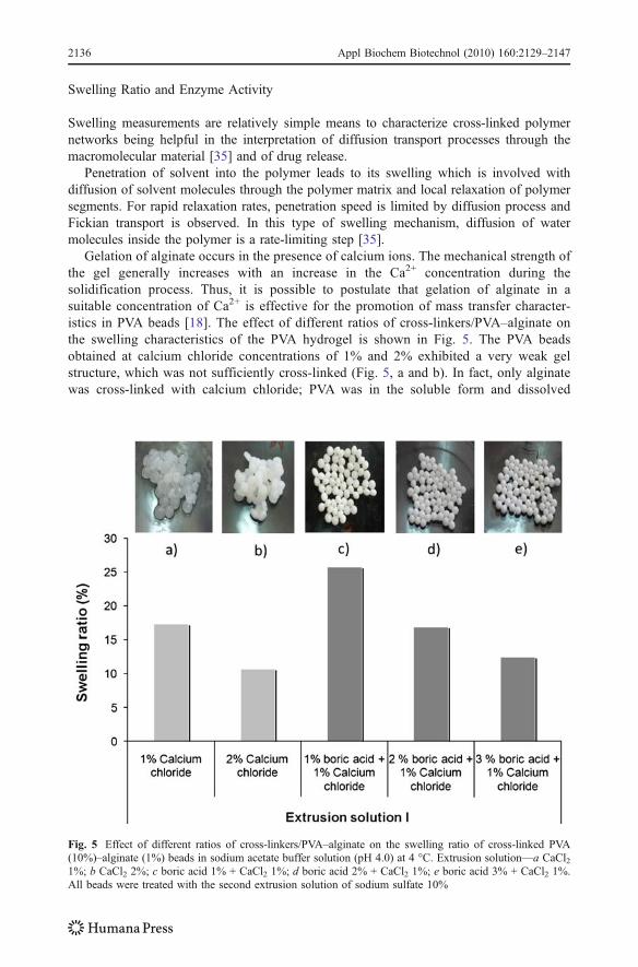
Gelation of alginate occurs in the presence of calcium ions. The mechanical strength ofthe gel generally increases with an increase in the Ca2+ concentration during thesolidification process. Thus, it is possible to postulate that gelation of alginate in asuitable concentration of Ca2+ is effective for the promotion of mass transfer character-istics in PVA beads [18]. The effect of different ratios of cross-linkers/PVA–alginate onthe swelling characteristics of the PVA hydrogel is shown in Fig. 5. The PVA beadsobtained at calcium chloride concentrations of 1% and 2% exhibited a very weak gelstructure, which was not sufficiently cross-linked (Fig. 5, a and b). In fact, only alginatewas cross-linked with calcium chloride; PVA was in the soluble form and dissolved
Fig. 5 Effect of different ratios of cross-linkers/PVA–alginate on the swelling ratio of cross-linked PVA(10%)–alginate (1%) beads in sodium acetate buffer solution (pH 4.0) at 4 °C. Extrusion solution—a CaCl21%; b CaCl2 2%; c boric acid 1% + CaCl2 1%; d boric acid 2% + CaCl2 1%; e boric acid 3% + CaCl2 1%.All beads were treated with the second extrusion solution of sodium sulfate 10%
2136 Appl Biochem Biotechnol (2010) 160:2129–2147
almost completely after transferring the beads into acetate buffer. This loss of mass(dissolution of PVA to buffer) corresponds to lower wet weights resulting in lowerswelling ratios.
This observation was confirmed after naringin hydrolysis by naringinase immobilized inPVA (10%)–alginate (1%) beads treated with calcium chloride 1% and 2%, respectively, andwith the second extrusion solution of sodium sulfate (10%). The sulfate ions would probablyform thiosulfate linkages among the cross-linked PVA. Treatment of beads with sodiumsulfate resulted in PVA beads, with a rubberlike elasticity and strength almost identical tobeads that were produced by previous researchers [15, 29, 30] but with improved properties.
Different ratios of boric acid/PVA led to significant difference in water uptake by thebeads. Higher ratios led to lower swelling ratio. Chain entanglement along with increase incross-linking agent concentration would result in a decreased network expansion [35](Fig. 5, c, d, and e). Lower boric acid/PVA ratios caused a significant increase in swellingproperties. In general, it may be concluded that the increase of acid boric/PVA ratioincreases the cross-linking density, which significantly reduces the swelling ratio of the gels[35] and that cross-linking of hydrogels contributes to the reduction of the molecular meshsize of the gel [36]. These results show the suitability of combined saturated boric acidsolution and calcium chloride solution together to cross-link PVA–alginate.
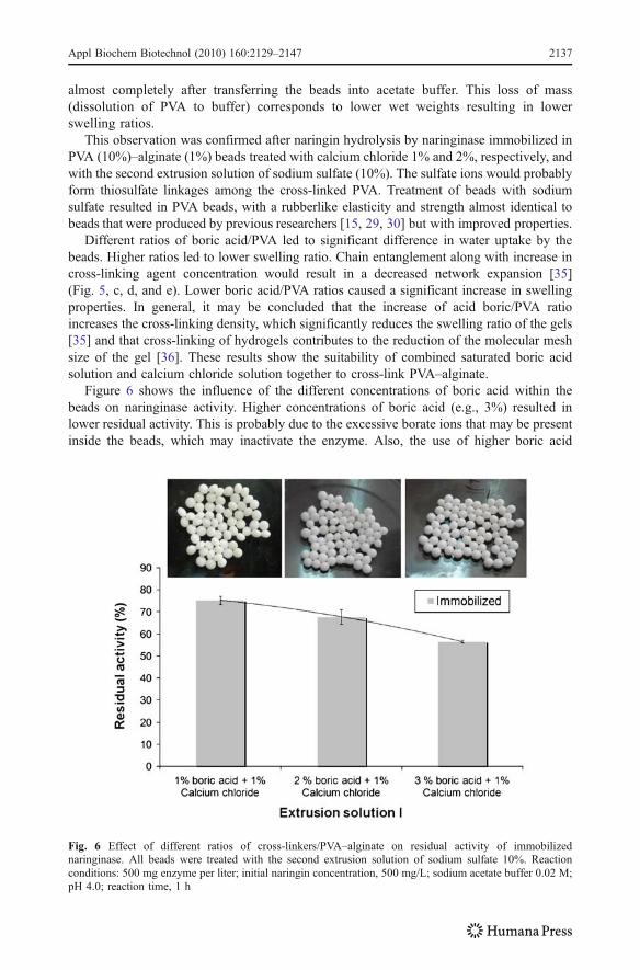
Figure 6 shows the influence of the different concentrations of boric acid within thebeads on naringinase activity. Higher concentrations of boric acid (e.g., 3%) resulted inlower residual activity. This is probably due to the excessive borate ions that may be presentinside the beads, which may inactivate the enzyme. Also, the use of higher boric acid
Fig. 6 Effect of different ratios of cross-linkers/PVA–alginate on residual activity of immobilizednaringinase. All beads were treated with the second extrusion solution of sodium sulfate 10%. Reactionconditions: 500 mg enzyme per liter; initial naringin concentration, 500 mg/L; sodium acetate buffer 0.02 M;pH 4.0; reaction time, 1 h
Appl Biochem Biotechnol (2010) 160:2129–2147 2137
concentrations resulted in denser beads with smaller pores [15]. This property seems tolimit the diffusivity of the substrate naringin and/or product naringenin. On this basis, botheffects may contribute to the decrease in the activity of the enzyme, naringinase. Beadstreated at lower boric acid concentration (1%) lead to higher residual activities but wereshown to be more fragile. In fact, equilibrium between mechanical stability of the matrixand residual activity has to be considered.
PVA–alginate-immobilized beads have advantages on the simple operation of immobi-lization and on the high strength [37]. Calcium alginate facilitates the formation ofhydrogen bond between PVA molecules, contributing to the formation of interpenetratinggel network [37].
The kinetic studies of swelling in water showed that pure PVA and PVA–alginatehydrogels reaches equilibrium after 4 h (Fig. 4).
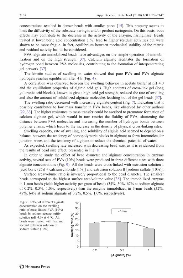
A correlation was observed between the swelling behavior in acetate buffer at pH 4.0and the equilibrium properties of alginic acid gels. High contents of cross-link gel (longguluronic acid blocks), known to give a high acid gel strength, reduced the rate of swellingand also the amount of solubilized alginate molecules leaching out of the gel beads [37].
The swelling ratio decreased with increasing alginate content (Fig. 7), indicating that itpossibly contributes to low mass transfer in PVA beads, like observed by other authors[22, 33]. The higher resistance to mass transfer could be ascribed to premature formation ofcalcium alginate gel, which would in turn restrict the fluidity of PVA, shortening thedistance between PVA molecules and increasing the number of hydrogen bonds betweenpolymer chains, which leads to the increase in the density of physical cross-linking sites.
Swelling capacity, rate of swelling, and solubility of alginic acid seemed to depend on abalance between the tendency of homopolymeric blocks in alginate to form intermolecularjunction zones and the tendency of alginate to reduce the chemical potential of water.
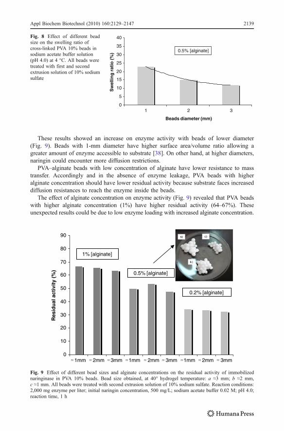
As expected, swelling rate increased with decreasing bead size, as it is evidenced fromthe results of bead size effect, presented in Fig. 8.
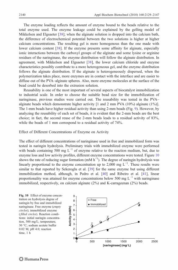
In order to study the effect of bead diameter and alginate concentration in enzymeactivity, several sets of PVA (10%) beads were produced in three different sizes with threealginate concentrations (Fig. 9). All the beads were cross-linked with extrusion solution I[acid boric (2%) + calcium chloride (1%)] and extrusion solution II [sodium sulfate (10%)].
Surface area/volume ratio is inversely proportional to the bead diameter. The smallestbeads correspond to the highest surface area/volume value [38]. The immobilized enzymein 1-mm beads yields higher activity per gram of beads (34%, 50%, 67% at sodium alginateof 0.2%, 0.5%, 1.0%, respectively) than the enzyme immobilized in 3-mm beads (32%,48%, 64% at sodium alginate of 0.2%, 0.5%, 1.0%, respectively).
0
5
10
15
20
25
30
35
40
0.2 0.5 1
[Alginate] (%)
Sw
ellin
g ra
tio
(%) 1mm
Fig. 7 Effect of different alginateconcentration on the swellingratio of cross-linked PVA (10%)beads in sodium acetate buffersolution (pH 4.0) at 4 °C. Allbeads were treated with first andsecond extrusion solution ofsodium sulfate (10%)
2138 Appl Biochem Biotechnol (2010) 160:2129–2147
These results showed an increase on enzyme activity with beads of lower diameter(Fig. 9). Beads with 1-mm diameter have higher surface area/volume ratio allowing agreater amount of enzyme accessible to substrate [38]. On other hand, at higher diameters,naringin could encounter more diffusion restrictions.
PVA–alginate beads with low concentration of alginate have lower resistance to masstransfer. Accordingly and in the absence of enzyme leakage, PVA beads with higheralginate concentration should have lower residual activity because substrate faces increaseddiffusion resistances to reach the enzyme inside the beads.
The effect of alginate concentration on enzyme activity (Fig. 9) revealed that PVA beadswith higher alginate concentration (1%) have higher residual activity (64–67%). Theseunexpected results could be due to low enzyme loading with increased alginate concentration.
0
5
10
15
20
25
30
35
40
1 2 3
Beads diameter (mm)
Sw
ellin
g r
atio
(%
) 0.5% [alginate]
Fig. 8 Effect of different beadsize on the swelling ratio ofcross-linked PVA 10% beads insodium acetate buffer solution(pH 4.0) at 4 °C. All beads weretreated with first and secondextrusion solution of 10% sodiumsulfate
0
10
20
30
40
50
60
70
80
90
1mm 2mm 3mm 1mm 2mm 3mm 1mm 2mm 3mm
Res
idu
al a
ctiv
ity
(%)
1% [alginate]
0.5% [alginate]
0.2% [alginate]
Fig. 9 Effect of different bead sizes and alginate concentrations on the residual activity of immobilizednaringinase in PVA 10% beads. Bead size obtained, at 40° hydrogel temperature: a ≈3 mm; b ≈2 mm,c ≈1 mm. All beads were treated with second extrusion solution of 10% sodium sulfate. Reaction conditions:2,000 mg enzyme per liter; initial naringin concentration, 500 mg/L; sodium acetate buffer 0.02 M; pH 4.0;reaction time, 1 h
Appl Biochem Biotechnol (2010) 160:2129–2147 2139
The enzyme loading reflects the amount of enzyme bound to the beads relative to thetotal enzyme used. The enzyme leakage could be explained by the gelling model ofMikkelsen and Elgsaeter [38]; when the alginate solution is dropped into the calcium bath,the difference of electrochemical potential between the two solutions is higher at highercalcium concentrations. The resulting gel is more homogenous than the one made withlower calcium content [38]. If the enzyme presents some affinity for alginate, especiallyionic interactions between the carboxyl groups of the alginate and some lysine or arginineresidues of the naringinase, the enzyme distribution will follow the alginate distribution. Inagreement, with Mikkelsen and Elgsaeter [38], the lower calcium chloride and enzymecharacteristics possibly contribute to a more heterogeneous gel, and the enzyme distributionfollows the alginate distribution. If the alginate is heterogeneously dispersed, when thepolymerization takes place, more enzymes are in contact with the interface and are easier todiffuse out of the PVA–alginate spheres. Also, more enzyme molecules at the surface of thebead could be desorbed into the extrusion solution.
Reusability is one of the most important of several aspects of biocatalyst immobilizationto industrial scale. In order to choose the suitable bead size for the immobilization ofnaringinase, previous studies were carried out. The reusability was tested in the PVA–alginate beads which demonstrate higher activity [1 and 2 mm PVA (10%) alginate (1%)].The 1-mm beads have higher residual activity than using 2-mm beads (Fig. 9). However, byanalyzing the reusability of each set of beads, it is evident that the 2-mm beads are the bestchoice; in fact, the second reuse of the 2-mm beads leads to a residual activity of 83%,while the beads of 1 mm correspond to a residual activity of 74%.
Effect of Different Concentrations of Enzyme on Activity
The effect of different concentrations of naringinase used in free and immobilized form wastested in naringin hydrolysis. Preliminary trials with immobilized enzyme were performedwith beads containing 500 mg L−1 of enzyme relative to the reaction medium, but, due toenzyme loss and low activity profiles, different enzyme concentrations were tested. Figure 10shows the rate of reducing sugar formation (mM h−1). The degree of naringin hydrolysis waslinearly proportional to the enzyme concentration up to 2,000 mg L−1. These results weresimilar to that reported by Sekeroglu et al. [39] for the same enzyme but using differentimmobilization method, although, in Pedro et al. [40] and Ribeiro et al. [41], linearproportionality was attained for enzyme concentrations below 500 mg L−1 with naringinaseimmobilized, respectively, on calcium alginate (2%) and K-carrageenan (2%) beads.
0
1
2
3
4
5
6
7
8
9
0 500 1000 1500 2000 2500[naringinase] (mg/ L)
Act
ivit
y (m
M/h
)
Free
Immobilized
Fig. 10 Effect of enzyme concen-tration on hydrolysis degree ofnaringin by free and immobilizednaringinase. Free enzyme (emptycircles); immobilized enzyme( filled circles). Reaction condi-tions: initial naringin concentra-tion, 500 mg/L; temperature,30 °C; sodium acetate buffer0.02 M, pH 4.0; reactiontime, 1 h
2140 Appl Biochem Biotechnol (2010) 160:2129–2147

The naringinase concentration used in further experiments was 2,000 mg L−1 relative tothe reaction media for both free and immobilized enzyme.
Effect of pH on the Activity of Free and Immobilized Naringinase
The effect of pH on the activity of free and immobilized naringinase for the naringinhydrolysis was examined from pH 3.0 to 6.0. The activities obtained are presented inFig. 11. The maximum activity was observed at optimum pH around 4 for both free andimmobilized naringinase. The pH profile of the immobilized enzyme reveals a broaderprofile of enzyme activity, which is suggestive of a protective role played by the beads,more noticeable at extreme pH values tested. The lower activity of the immobilizednaringinase may be attributed to alteration of enzyme structure during entrapment into theacid boric solution or to diffusion limitations.
Similar observations has been frequently reported in the literature [5], namely in thework of Sekeroglu et al. [39], with naringinase from P. decumbens, and of Busto et al. [42],with naringinase from Aspergillus niger for free and immobilized forms, in PVA cryogel.Also, optimum pH values of 4.5 for naringinase from A. niger [43] and from P. decumbens[44] were referred. Lower pH values were reported for immobilized naringinase from P.decumbens, like 3.5 on Celite by the adsorption/cross-link with glutaraldehyde [45] and onactivated seeds of Ocimum basilicum with covalent binding method [46]. Tsen et al. [47]referred an optimum pH value of 3.7 from Penicillium sp. with naringinase immobilized incellulose triacetate fiber by the entrapment method.
Effect of Temperature on the Activity of Free and Immobilized Naringinase
The use of enzymes in bioprocesses often encounters the problem of its thermalinactivation. Under high temperature, enzymes may undergo partial unfolding by heat-induced destruction of noncovalent interactions [21].
The temperature dependence of the free and immobilized naringinase activity wasstudied in the temperature range 30–90 °C. The results obtained are depicted in Fig. 12.Optimum catalytic activity was observed at 70 °C, for both free and immobilizednaringinase; however, the immobilized enzyme had a lower activity than the free one. Thismay be due to decreased affinity of the enzyme for the substrate caused by internaldiffusion restriction of the immobilized naringinase. The immobilized naringinase exhibiteda broader profile at the optimum temperature, showing a better thermal stability, whereasthe free enzyme was less stable towards heat [48]. The polymer network is supposed topreserve the tertiary structure of the enzyme. It has been suggested that enzyme
0
1
2
3
4
5
6
7
2 3 4 5 6 7pH
Act
ivit
y (m
M/h
)
FreeImmobilized
Fig. 11 Effect of pH on degreeof hydrolysis of naringin by freeand immobilized naringinase.Free enzyme (empty circles);immobilized enzyme ( filledcircles). Reaction conditions:2,000 mg enzyme per liter;initial naringin concentration,500 mg/L; temperature, 30 °C;sodium acetate buffer; reactiontime, 1 h
Appl Biochem Biotechnol (2010) 160:2129–2147 2141
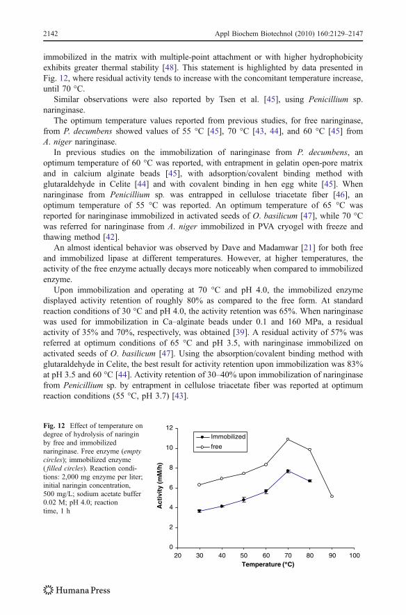
immobilized in the matrix with multiple-point attachment or with higher hydrophobicityexhibits greater thermal stability [48]. This statement is highlighted by data presented inFig. 12, where residual activity tends to increase with the concomitant temperature increase,until 70 °C.
Similar observations were also reported by Tsen et al. [45], using Penicillium sp.naringinase.
The optimum temperature values reported from previous studies, for free naringinase,from P. decumbens showed values of 55 °C [45], 70 °C [43, 44], and 60 °C [45] fromA. niger naringinase.
In previous studies on the immobilization of naringinase from P. decumbens, anoptimum temperature of 60 °C was reported, with entrapment in gelatin open-pore matrixand in calcium alginate beads [45], with adsorption/covalent binding method withglutaraldehyde in Celite [44] and with covalent binding in hen egg white [45]. Whennaringinase from Penicillium sp. was entrapped in cellulose triacetate fiber [46], anoptimum temperature of 55 °C was reported. An optimum temperature of 65 °C wasreported for naringinase immobilized in activated seeds of O. basilicum [47], while 70 °Cwas referred for naringinase from A. niger immobilized in PVA cryogel with freeze andthawing method [42].
An almost identical behavior was observed by Dave and Madamwar [21] for both freeand immobilized lipase at different temperatures. However, at higher temperatures, theactivity of the free enzyme actually decays more noticeably when compared to immobilizedenzyme.
Upon immobilization and operating at 70 °C and pH 4.0, the immobilized enzymedisplayed activity retention of roughly 80% as compared to the free form. At standardreaction conditions of 30 °C and pH 4.0, the activity retention was 65%. When naringinasewas used for immobilization in Ca–alginate beads under 0.1 and 160 MPa, a residualactivity of 35% and 70%, respectively, was obtained [39]. A residual activity of 57% wasreferred at optimum conditions of 65 °C and pH 3.5, with naringinase immobilized onactivated seeds of O. basilicum [47]. Using the absorption/covalent binding method withglutaraldehyde in Celite, the best result for activity retention upon immobilization was 83%at pH 3.5 and 60 °C [44]. Activity retention of 30–40% upon immobilization of naringinasefrom Penicillium sp. by entrapment in cellulose triacetate fiber was reported at optimumreaction conditions (55 °C, pH 3.7) [43].
0
2
4
6
8
10
12
20 30 40 50 60 70 80 90 100Temperature ( C)
Act
ivit
y (m
M/h
)
Immobilized
free
Fig. 12 Effect of temperature ondegree of hydrolysis of naringinby free and immobilizednaringinase. Free enzyme (emptycircles); immobilized enzyme( filled circles). Reaction condi-tions: 2,000 mg enzyme per liter;initial naringin concentration,500 mg/L; sodium acetate buffer0.02 M; pH 4.0; reactiontime, 1 h
2142 Appl Biochem Biotechnol (2010) 160:2129–2147
Kinetic Parameters
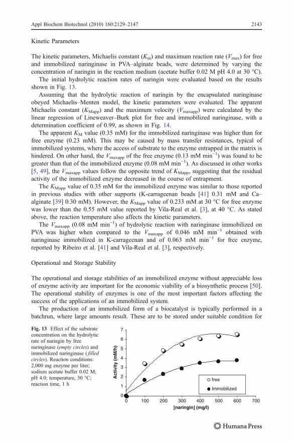
The kinetic parameters, Michaelis constant (Km) and maximum reaction rate (Vmax) for freeand immobilized naringinase in PVA–alginate beads, were determined by varying theconcentration of naringin in the reaction medium (acetate buffer 0.02 M pH 4.0 at 30 °C).
The initial hydrolytic reaction rates of naringin were evaluated based on the resultsshown in Fig. 13.
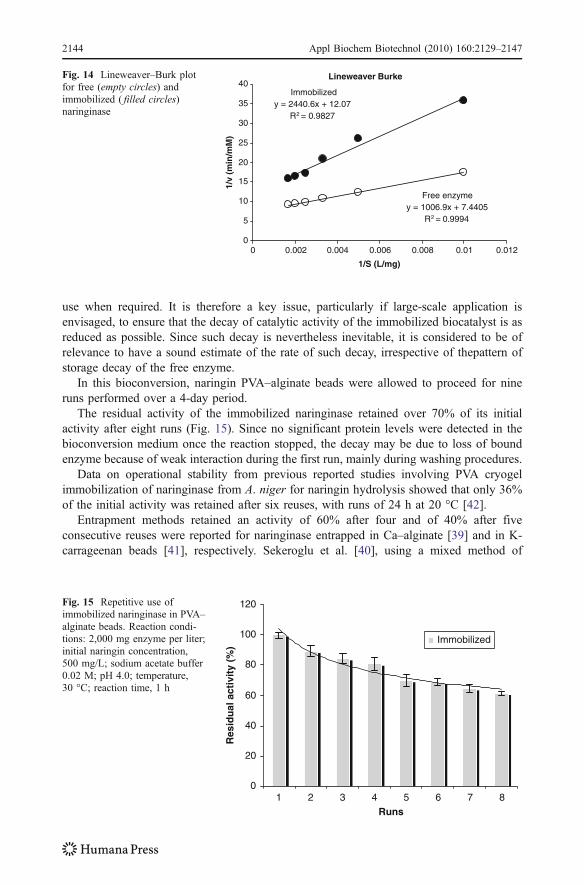
Assuming that the hydrolytic reaction of naringin by the encapsulated naringinaseobeyed Michaelis–Menten model, the kinetic parameters were evaluated. The apparentMichaelis constant (KMapp) and the maximum velocity (Vmaxapp) were calculated by thelinear regression of Lineweaver–Burk plot for free and immobilized naringinase, with adetermination coefficient of 0.99, as shown in Fig. 14.
The apparent KM value (0.35 mM) for the immobilized naringinase was higher than forfree enzyme (0.23 mM). This may be caused by mass transfer resistances, typical ofimmobilized systems, where the access of substrate to the enzyme entrapped in the matrix ishindered. On other hand, the Vmaxapp of the free enzyme (0.13 mM min−1) was found to begreater than that of the immobilized enzyme (0.08 mM min−1). As discussed in other works[5, 49], the Vmaxapp values follow the opposite trend of KMapp, suggesting that the residualactivity of the immobilized enzyme decreased in the course of entrapment.
The KMapp value of 0.35 mM for the immobilized enzyme was similar to those reportedin previous studies with other supports (K-carrageenan beads [41] 0.31 mM and Ca–alginate [39] 0.30 mM). However, the KMapp value of 0.233 mM at 30 °C for free enzymewas lower than the 0.55 mM value reported by Vila-Real et al. [3], at 40 °C. As statedabove, the reaction temperature also affects the kinetic parameters.
The Vmaxapp (0.08 mM min−1) of hydrolytic reaction with naringinase immobilized onPVA was higher when compared to the Vmaxapp of 0.046 mM min−1 obtained withnaringinase immobilized in K-carrageenan and of 0.063 mM min−1 for free enzyme,reported by Ribeiro et al. [41] and Vila-Real et al. [3], respectively.
Operational and Storage Stability
The operational and storage stabilities of an immobilized enzyme without appreciable lossof enzyme activity are important for the economic viability of a biosynthetic process [50].The operational stability of enzymes is one of the most important factors affecting thesuccess of the applications of an immobilized system.
The production of an immobilized form of a biocatalyst is typically performed in abatchrun, where large amounts result. These are to be stored under suitable condition for
0
1
2
3
4
5
6
7
0 100 200 300 400 500 600 700[naringin] (mg/l)
Act
ivit
y (m
M/h
)
free
Immobilized
Fig. 13 Effect of the substrateconcentration on the hydrolyticrate of naringin by freenaringinase (empty circles) andimmobilized naringinase ( filledcircles). Reaction conditions:2,000 mg enzyme per liter;sodium acetate buffer 0.02 M;pH 4.0; temperature, 30 °C;reaction time, 1 h
Appl Biochem Biotechnol (2010) 160:2129–2147 2143
use when required. It is therefore a key issue, particularly if large-scale application isenvisaged, to ensure that the decay of catalytic activity of the immobilized biocatalyst is asreduced as possible. Since such decay is nevertheless inevitable, it is considered to be ofrelevance to have a sound estimate of the rate of such decay, irrespective of thepattern ofstorage decay of the free enzyme.
In this bioconversion, naringin PVA–alginate beads were allowed to proceed for nineruns performed over a 4-day period.
The residual activity of the immobilized naringinase retained over 70% of its initialactivity after eight runs (Fig. 15). Since no significant protein levels were detected in thebioconversion medium once the reaction stopped, the decay may be due to loss of boundenzyme because of weak interaction during the first run, mainly during washing procedures.
Data on operational stability from previous reported studies involving PVA cryogelimmobilization of naringinase from A. niger for naringin hydrolysis showed that only 36%of the initial activity was retained after six reuses, with runs of 24 h at 20 °C [42].
Entrapment methods retained an activity of 60% after four and of 40% after fiveconsecutive reuses were reported for naringinase entrapped in Ca–alginate [39] and in K-carrageenan beads [41], respectively. Sekeroglu et al. [40], using a mixed method of
Lineweaver Burke
Immobilizedy = 2440.6x + 12.07
R2 = 0.9827
Free enzymey = 1006.9x + 7.4405
R2 = 0.9994
0
5
10
15
20
25
30
35
40
0 0.002 0.004 0.006 0.008 0.01 0.012
1/S (L/mg)
1/v
(min
/mM
)
Fig. 14 Lineweaver–Burk plotfor free (empty circles) andimmobilized ( filled circles)naringinase
0
20
40
60
80
100
120
1 2 3 4 5 6 7 8Runs
Res
idu
al a
ctiv
ity
(%)
Immobilized
Fig. 15 Repetitive use ofimmobilized naringinase in PVA–alginate beads. Reaction condi-tions: 2,000 mg enzyme per liter;initial naringin concentration,500 mg/L; sodium acetate buffer0.02 M; pH 4.0; temperature,30 °C; reaction time, 1 h
2144 Appl Biochem Biotechnol (2010) 160:2129–2147
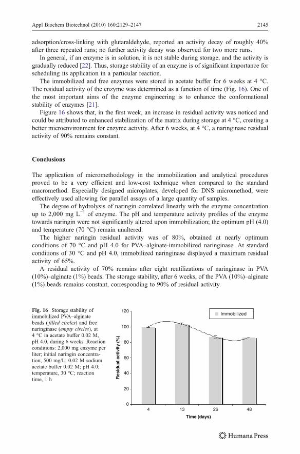
adsorption/cross-linking with glutaraldehyde, reported an activity decay of roughly 40%after three repeated runs; no further activity decay was observed for two more runs.
In general, if an enzyme is in solution, it is not stable during storage, and the activity isgradually reduced [22]. Thus, storage stability of an enzyme is of significant importance forscheduling its application in a particular reaction.
The immobilized and free enzymes were stored in acetate buffer for 6 weeks at 4 °C.The residual activity of the enzyme was determined as a function of time (Fig. 16). One ofthe most important aims of the enzyme engineering is to enhance the conformationalstability of enzymes [21].
Figure 16 shows that, in the first week, an increase in residual activity was noticed andcould be attributed to enhanced stabilization of the matrix during storage at 4 °C, creating abetter microenvironment for enzyme activity. After 6 weeks, at 4 °C, a naringinase residualactivity of 90% remains constant.
Conclusions
The application of micromethodology in the immobilization and analytical proceduresproved to be a very efficient and low-cost technique when compared to the standardmacromethod. Especially designed microplates, developed for DNS micromethod, wereeffectively used allowing for parallel assays of a large quantity of samples.
The degree of hydrolysis of naringin correlated linearly with the enzyme concentrationup to 2,000 mg L−1 of enzyme. The pH and temperature activity profiles of the enzymetowards naringin were not significantly altered upon immobilization; the optimum pH (4.0)and temperature (70 °C) remain unaltered.
The higher naringin residual activity was of 80%, obtained at nearly optimumconditions of 70 °C and pH 4.0 for PVA–alginate-immobilized naringinase. At standardconditions of 30 °C and pH 4.0, immobilized naringinase displayed a maximum residualactivity of 65%.
A residual activity of 70% remains after eight reutilizations of naringinase in PVA(10%)–alginate (1%) beads. The storage stability, after 6 weeks, of the PVA (10%)–alginate(1%) beads remains constant, corresponding to 90% of residual activity.
0
20
40
60
80
100
120
4 13 26 48
Time (days)
Res
idu
al a
ctiv
ity (
%)
ImmobilizedFig. 16 Storage stability ofimmobilized PVA–alginatebeads ( filled circles) and freenaringinase (empty circles), at4 °C in acetate buffer 0.02 M,pH 4.0, during 6 weeks. Reactionconditions: 2,000 mg enzyme perliter; initial naringin concentra-tion, 500 mg/L; 0.02 M sodiumacetate buffer 0.02 M; pH 4.0;temperature, 30 °C; reactiontime, 1 h
Appl Biochem Biotechnol (2010) 160:2129–2147 2145

In contrast to biopolymers, PVA hydrogels are hardly biodegradable and show anexcellent mechanical stability with a large elongation at break, and even in long-timereactions no significant abrasion could be observed [11]. In addition, the PVA–alginatesupport matrices developed evidenced physical stability at high temperatures (>50 and<80 °C). This increases the potential of the immobilized naringinase application as apractical catalyst on an industrial scale because higher temperatures reduce the viscosity ofthe reaction medium, minimize the mechanical energy in agitation, reduce the risk ofcontamination, and improve naringin solubility.
In conclusion, these very promising results achievedwith immobilized naringinase on PVA–alginate beads are illustrative of the potential of this strategy for biocatalyst immobilization.
References
1. Ferreira, L., Afonso, C., Vila-Real, H., Alfaia, A., & Ribeiro, M. H. L. (2008). Food Technology andBiotechnology, 46(2), 146–150.
2. Ladaniya, M. (2008). Citrus fruit: Biology, technology and evaluation. Amsterdam: Elsevier.3. Vila Real, H. J., Alfaia, A. J., Calado, A. R. T., & Ribeiro, M. H. L. (2007). Food Chemistry, 102(3),
565–570.4. Ribeiro, I. A., Rocha, J., Sepodes, B., Mota-Filipe, H., & Ribeiro, M. H. L. (2008). Journal of Molecular
Catalysis B Enzymatic, 52–53, 13–18.5. Bajpai, A. K., & Bhanu, S. (2003). Colloid & Polymer Science, 282, 76–83.6. Lozinsky, V. L., & Plieva, F. M. (1998). Enzyme and Microbial Technology, 23, 227–242.7. Durieux, A., Nicolay, X., & Simon, J. P. (2000). Biotechnology Letters, 22, 1679–1684.8. Czichocki, G., Dautzenberg, H., Capan, E., & Vorlop, K.-D. (2001). Biotechnology Letters, 23(16),
1303–1307.9. Gröger, H., Capan, E., Barthuber, A., & Vorlop, K. D. (2001). Organic Letters, 3, 1969–1972.10. Wilson, L., Illanes, A., Pessela, B. C. C., Abian, O., Fernández-Lafuente, R., & Guisán, J. M. (2004).
Biotechnology and Bioengineering, 86(5), 558–562.11. Parascandola, P., Branduardi, P., & Alteris, E. (2006). Enzyme and Microbial Technology, 38, 184–189.12. Schlieker, M., & Vorlop, K. D. (2006). A novel immobilization method for entrapment LentiKats®
methods in biotechnology. In J. M. Guisan (Ed.), Immobilization of enzymes and cells (2nd ed., pp. 333–343). Heidelberg: Springer.
13. Ariga, O., Takagi, H., Nishizawa, H., & Sano, Y. (1987). Journal of Fermentation Technology, 65, 651–658.14. Imai, K., Shiomi, T., Uchida, K., & Miya, M. (1986). Biotechnology and Bioengineering, 28, 1721–1726.15. Idris, A., Zain, N., & Suhaimi, M. (2008). Process Biochemistry, 43, 331–338.16. Long, Z., Huang, Y., Cai, Z., Cong, W., & Ouyang, F. (2004). Process Biochemistry, 39, 2129–2133.17. Chen, K., & Houng, J. (1994). Cell immobilization with phosphorylated polyvinyl alcohol (PVA) gel. In
G. F. Bickerstaff (Ed.), Immobilization of enzymes and cells: Methods biotechnology (Vol. 1).Heidelberg: Springer.
18. Chen, K., Chen, S., & Houng, J. (1996). Enzyme and Microbial Technology, 18, 502–506.19. Chang, C., & Tseng, S. (1998). Biotechnology Techniques, 12(12), 865–868.20. Li-sheng, Z., Wei-zhong, W., & Jian-long, W. (2007). Journal of Environmental Science, 19, 1293–1297.21. Dave, R., & Madamwar, D. (2006). Process Biochemistry, 41, 951–955.22. Queiroz, A., Passos, E., Alves, S., Silva, G., Higa, O., & Vítolo, M. (2005). Journal of Applied Polymer
Science, 102, 1553–1560.23. Hsia, T., Feng, Y., Ho, C., Chou, W., & Tseng, S. (2008). Journal of Industrial Microbiology &
Biotechnology, 35, 721–727.24. Wu, A. K. Y., & Wiesecarver, K. D. (1992). Biotechnology and Bioengineering, 39, 447–449.25. Grishin, S. I., & Tuovinen, O. H. (1989). Applied Microbiology and Biotechnology, 31, 505–511.26. Lin, H., Liu, W., Liu, Y., & Cheng, C. (2002). Journal Polymer Research, 9, 233–238.27. Kurokawa, H., Shibayama, M., Ishimaru, T., Nomura, S., & Wu, W. (1992). Polymer, 33(10), 2182–
2188.28. Leibler, L., Pezron, E., & Pincus, P. A. (1988). Polymer, 29, 1105–1109.29. Keita, G., Ricard, A., Audebert, R., Pezron, E., & Leibler, L. (1995). Polymer, 36, 49.30. Pattanapitpaisal, P., Brown, N. L., & Macaskie, L. E. (2001). Biotechnology Letters, 23, 61–65.
2146 Appl Biochem Biotechnol (2010) 160:2129–2147

31. Miller, G. L. (1959). Analytical Chemistry, 31, 426–428.32. Bradford, M. M. (1976). Analytical Biochemistry, 72, 248–254.33. Kim, J., Choi, H., et al. (2008). International Journal of Pharmaceutics, 359, 79–86.34. Li, M., Cheng, S., & Yan, H. (2007). Green Chemistry, 9, 894–898.35. Braze, C. S., & Peppas, N. A. (2000). European Journal of Pharmaceutics and Biopharmaceutics, 49,
47–58.36. Kim, C., & Lee, P. (1992). Pharmaceutical Research, 9, 10–16.37. Nizam Horia, M., Abd Alla Safaa, G., & El-Naggar Abdel Wahab, M. (2007). Journal of
Macromolecular Science A, 44(3), 291–297.38. Mikkelsen, A., & Elgsaeter, A. (1995). Biopolymers, 36(1), 17–41.39. Sekeroglu, G., Fadıloglu, S., & Gogus, F. (2006). European Food Research and Technology, 224, 55–60.40. Pedro, H. A., Alfaia, A. J., Marques, J., Vila-Real, H. J., Calado, A. T., & Ribeiro, M. H. L. (2007).
Enzyme and Microbial Technology, 40, 442–446.41. Ribeiro, I. A., & Ribeiro, M. H. L. (2008). Journal of Molecular Catalysis B Enzymatic, 51, 10–18.42. Busto, M. D., Meza, V., Ortega, N., & Perez-Mateos, M. (2007). Food Chemistry, 104, 1177–1182.43. Soares, N., & Hotchkiss, J. (1998). Journal of Food Science, 63, 61–65.44. Norouzian, F., Hosseinzadeh, A., Inanlou, D., & Moazami, N. (1999). World Journal of Microbiology
and Biotechnology, 15(4), 501–502.45. Puri, M., Kaur, H., & Kennedy, J. F. (2005). Journal of Chemical Technology & Biotechnology, 80(10),
1160–1165.46. Norouzian, D. (2003). Iranian Journal of Biotechnology, 1(4), 197–206.47. Tsen, H., Tsai, S., & Yu, G. (1989). Journal of Fermentation and Bioengineering, 67, 186–189.48. Buchholz, K., & Klein, J. (1987). In K. Mosbach (Ed.), Methods in enzymology: Immobilized enzymes
and cells (pp. 3–30). London: Academic.49. Manjón, A., Bastida, J., Romero, C., Jimeno, A., & Iborra, J. L. (1985). Biotechnology Letters, 7(7),
477–482.50. Luckarift, H. R., Spain, J. C., Naik, R. R., & Stone, M. O. (2004). Nature Biotechnology, 22, 211–213.
Appl Biochem Biotechnol (2010) 160:2129–2147 2147