Smart porous microparticles based on gelatin/sodium alginate
http://informahealthcare.com/mncISSN: 0265-2048 (print), 1464-5246 (electronic)
J Microencapsul, Early Online: 1–11! 2014 Informa UK Ltd. DOI: 10.3109/02652048.2014.898706
RESEARCH ARTICLE
Behaviour and ultrastructure of human bone marrow-derivedmesenchymal stem cells immobilised in alginate-poly-L-lysine-alginatemicrocapsules
Noha Attia1*, Edorta Santos2,3*, Hala Abdelmouty1, Samia Arafa1, Nahed Zohdy1, Rosa Marıa Hernandez2,3,Gorka Orive2,3, and Jose Luis Pedraz2,3
1Department of Histology and Cell Biology, Faculty of Medicine, Alexandria University, Alexandria, Egypt, 2Laboratory of Pharmaceutics, School of
Pharmacy, NanoBioCel Group, University of the Basque Country, Vitoria, Spain, and 3Biomedical Research Networking Center in Bioengineering,
Biomaterials and Nanomedicine (CIBER-BBN), Vitoria, Spain
Abstract
Context: Human bone marrow mesenchymal stem cells (hBM-MSCs) show a great promise forthe treatment of a variety of diseases. Despite the previous trials to encapsulate hBM-MSCs inalginate-poly-L-lysine-alginate (APA) systems, the various changes that follow immobilisationhave not been ascertained yet. Objective: Determine the various consequences derivedfrom entrapment on cell behaviour, putting special emphasis on the ultrastructure.Methods: hBM-MSCs were immobilised in APA microcapsules to further characterise theirviability, metabolic activity, proliferation, VEGF-secretability, and morphology. Results: The VEGFproduced by monolayer hBM-MSCs increased significantly 1 d post-encapsulation, and wasmaintained for at least 4 weeks. TEM imaging of cells revealed well preserved ultrastructureindicating protein synthesis and high metabolic activity. Conclusion: Although APAmicroencapsulation did not support 100% of fully viable hBM-MSCs for long-term cultures, itwas conceived to enhance both VEGF secretion and metabolic activity while not losing theirstemness characteristics.
Keywords
Alginate, bone marrow-derived mesenchymalstem cells, cell microencapsulation,ultrastructure, VEGF secretion
History
Received 2 July 2013Revised 27 December 2013Accepted 10 February 2014Published online 24 April 2014
Introduction
Human bone marrow mesenchymal stem cells (hBM-MSCs) areconsidered promising adult multipotent stem cell populations(Giordano et al., 2007). They are able to differentiate both intomultiple mesoderm- and non-mesoderm-type lineages (Sanchez-Ramos, 2002; Baksh et al., 2004). Unlike other bone marrow(BM) resident cells, they can be easily isolated by their selectiveadherence to culture vessels. MSCs represent an ideal candidatefor cell-based therapies as they are a robust cell type exhibiting aremarkable genetic stability (Karaoz et al., 2011). Moreover,they can interact with cells of both the innate and adaptiveimmune systems, leading to the modulation of several effectorfunctions (Griffin et al., 2010). MSCs are well known for theirhypoimmunogenic properties (Nauta and Fibbe, 2007). Theywere found to immunoregulate cytotoxic T lymphocytes,both directly and indirectly (Beyth et al., 2005). Furthermore,they can significantly affect the progression of autoimmunediseases, as they are involved in alloantigen recognition andelimination (Abumaree et al., 2012). Finally, as adult stemcells, clinical applications of MSC bring up a powerful alternative
to ethically controversial use of embryonic stem cells (Chanet al., 2007).
In addition to their intrinsic ‘‘stem’’ properties for cell-basedtherapy, numerous lines of evidence suggest that MSCs secreteseveral soluble factors, which can stimulate the regeneration ofsurrounding microenvironment after local transplantation(Phinney and Prockop, 2007; Horie et al., 2011). Amongthese factors, vascular endothelial growth factor (VEGF) is akey pro-angiogenic factor that increases vascularisation andperfusion, thus considered one of the key players behindclinical improvement of patients with myocardial infarction,limb ischaemia, or stroke that receive MSC therapy (Nagayaet al., 2004). MSCs are well known for their capacity to hometo injured tissues, differentiate into distinctive cell types, andpromote the formation of regenerative microenvironments(Jones and McGonagle, 2008). However, even if systemicdelivery of MSCs appears to be a safe, clinically relevantapproach for cell therapy, Pittenger and Martin have shownthat less than 3% of MSCs delivered by direct injection werestill present after 2 weeks (Pittenger and Martin, 2004). Such alow cell survival, together with the poor organ-specific cellretention and the difficulty to control the phenotype of trans-planted cells over sufficiently long times, have greatly hamperedthe success of cell-based therapies (Menasche, 2005; Correaet al., 2007).
For over a decade, researchers have explored the use ofalginate hydrogels as artificial extracellular matrices to
*These authors contributed equally to this work.
Address for correspondence: Jose Luis Pedraz, Laboratory of Pharmacyand Pharmaceutical Technology, Faculty of Pharmacy UPV/EHU, Paseode la Universidad nº 7, 01006 Vitoria-Gasteiz, Spain. Tel: 34-945013091.Fax: +34-945013040. E-mail: [email protected]
Jour
nal o
f M
icro
enca
psul
atio
n D
ownl
oade
d fr
om in
form
ahea
lthca
re.c
om b
y 19
7.12
1.12
1.15
on
04/2
5/14
For
pers
onal
use
onl
y.
encapsulate living cells and improve their transplantation (Oriveet al., 2005; Ponce et al., 2006). These scaffolds provide a morenatural 3D microenvironment for the cells, resembling in vivosituation where cells develop their function (Abbah et al., 2008).Alginate is considered one of the biomaterials of choice for manyresearchers (Garate et al., 2012), since their hydrogels are non-cytotoxic, semi-permeable, and have been shown to provideimmune protection for many cell types (Yu et al., 2010).Furthermore, alginate allows gel formation under gentle condi-tions, which is an attractive feature for cell encapsulation (Oriveet al., 2006). Entrapped cells can be thoroughly studied, as theyconsistently achieve a longer lifespan in culture compared withnon-encapsulated cells (Ghidoni et al., 2008). Transplantation ofencapsulated MSCs – as a measure to immunoisolate them – canopen the door for the use of non-autologous cells, allogenic oreven xenogenic (Ngoc et al., 2011).
In this work, we aimed to encapsulate hBM-MSCs into APAmicrocapsules in order to further characterise cell behaviour interms of viability, metabolic activity, proliferation and VEGFsecretion. In addition, we also investigated the impact ofmicroencapsulation on their morphology, ultrastructure, andpreservation of their differentiation potential following recoveryat different time intervals.
Methods
Monolayer culture of hBM-MSCs
P1 cryopreserved hBM-MSCs (24-years-old female donor) wereobtained from the Texas A&M Health Science Center College ofMedicine Institute for Regenerative Medicine at Scott & Whitethrough a grant from NCRR of the NIH, grant # P40RR017447.Primary hBM-MSCs were cultured at 500–1000 cells/cm2
in T-175 cm2 tissue culture flasks (Corning) maintained at 37 �Cin a humidified 5% CO2 environment. Cells were culturedin Complete Culture Medium (CCM) composed of AlphaMinimum Essential Medium (a-MEM; Life Technologies,Carlsbad, CA) supplemented with 16.5% foetal bovine serum(FBS, Life Technologies, Carlsbad, CA), 1% L-glutamine (LifeTechnologies, Carlsbad, CA), and 1% Antibiotic-Antimycotic(Life Technologies, Carlsbad, CA). All experiments were con-ducted on cells at passage 2–4 with medium changed twice a week.
Encapsulation
hBM-MSCs were immobilised into APA microcapsules using anelectrostatic droplet generator (8 V) with brief modificationsof the procedure designed by Lim and Sun (1980). Ultra purelow-viscosity and high-guluronic acid alginate (LVG) waspurchased from FMC Biopolymer, Norway, and poly-L-lysine(PLL; hydrobromide MW¼ 15–30 kDa) was obtained fromSigma-Aldrich (St. Louis, MO). Briefly, cells were harvestedfrom monolayer cultures using trypsin-EDTA 0.25% (LifeTechnologies, Carlsbad, CA), filtered through a 40 lM poremesh and resuspended in 1.5% (w/v) LVG-alginate sterilesolution, obtaining a cell density of 5� 106 cells/mL. Thissuspension was extruded through a 0.35 mm needle using a10 mL sterile syringe with a peristaltic pump (flow rate 5.9 mm/h). The resulting alginate beads were collected in a CaCl2 solution(55 mM) and maintained in agitation for 10 min for complete ionicgelation. Subsequently, the beads were coated with 0.05% (w/v)PLL for 5 min, followed by a second coating with 0.1% alginatefor another 5 min. Finally, the microcapsules were cultured inCCM. The whole process was carried out at room temperature(RT) and under aseptic conditions. The diameters and overallmorphology were characterised using inverted optical microscopy(Nikon TSM).
Characterisation of hBM-MSCs before and afterencapsulation
In vitro adipogenic and osteogenic differentiation
As for pre encapsulated hBM-MSCs were seeded onto 6-wellplates (BD Falcon) for 2 weeks with medium changed twice aweek till 70–80% confluence. After 2, 4, and 6 weeks ofencapsulation, cells were de-encapsulated with 0.5 mg/mL ofalginate lyase (Sigma-Aldrich, St. Louis, MO) and cultured inCCM till 70–80% confluence. The same differentiation protocolwas applied on all samples. For Adipogenic differentiation, hBM-MSCs were cultured in CCM supplemented with 0.5 lMDexamethasone (Sigma-Aldrich), 0.5 lM isobutylmethylxanthine(Sigma-Aldrich), and 50 lM Indomethacin (Sigma-Aldrich). Forosteogenic differentiation, cells were cultured in CCM supple-mented with 10 nM Dexamethasone, 20 mM b-glycerolphosphate(Sigma-Aldrich) and 50 lM L-ascorbic acid 2-phosphate (Sigma-Aldrich). After 2 weeks differentiation protocol, intracellular lipiddroplets indicating adipogenic differentiation were confirmed byOil Red O (ORO) (Sigma-Aldrich) staining. Osteogenic differen-tiation was assessed by staining with Alizarin Red S (ARS)(Sigma-Aldrich).
CFU assay
One hundred pre-encapsulated hBM-MSCs were cultured in 10 mLof sterile CCM in a 100 mm diameter dish (about 55 cm2 culturearea). Cells were incubated for 14 d, with medium change twice aweek. Then, the plate was stained with 3% Crystal Violet in 100%methanol to distinguish colonies. The same assay was repeated onde-encapsulated hBM-MSCs after 6 weeks of entrapment.
Encapsulated cell viability and proliferation
Live/dead assay
Encapsulated cells were stained with the LIVE/DEAD kit(Invitrogen) and after incubation for 30 min in dark at RT,fluorescence micrographs were taken with an epi-fluorescencemicroscope (Nikon TSM). Live cells fluoresced in green viaenzymatic conversion of acetoxymethyl ester of calcein (calcein-AM) to calcein, while the nuclei of dead cells fluoresced in redvia labeled Ethidium homodimer-1 (EthD-1) due to the impair-ment of the cell membrane integrity. This assay was performed ondays 1, 14, and 42 post-encapsulation.
Flow cytometry for quantitative assessment of cell viability
hBM-MSCs were de-encapsulated with 0.5 mg/mL of alginatelyase and filtered through a 40- mm mesh filter. Free cellsuspension was added to TrueCount� tubes (BD) and stainedwith 1 lM calcein-AM and 0.8 mM EthD-1 (LIVE/DEAD kit).After 20 min incubation in dark at RT, cell viability was assessedby means of flow cytometry (BD FACSCalibur). The populationcorresponding to de-encapsulated cells was gated in a FSC/SSCdot plot and further analysed in a bivariate FL1/FL3 dot plot. Thesignal of green fluorescing calcein was detected in the FL1channel, while FL3 channel was employed for red fluorescingEthD-1. Calcein positive and EthD-1 negative cells (upper leftquadrant) were considered viable, calcein negative and EthD-1positive cells (lower right quadrant) were considered dead.However, both calcein and EthD-1 positive (upper right quadrant)were considered as living damaged cells. TrueCount beads wereutilised for calculation of cell concentration per sample.
This assay was performed on days 1, 14, and 42. CellQuest Prosoftware was used both for acquisition and analysis of the data.For all samples, a total of 1� 104 events were examined. Datawere shown as mean of three independent samples ± SD.
2 N. Attia et al. J Microencapsul, Early Online: 1–11
Jour
nal o
f M
icro
enca
psul
atio
n D
ownl
oade
d fr
om in
form
ahea
lthca
re.c
om b
y 19
7.12
1.12
1.15
on
04/2
5/14
For
pers
onal
use
onl
y.
Cell counting kit-8 (CCK-8) assay
Metabolic activity was determined on the basis of tetrazoliumproduction using the Cell Counting Kit-8 (CCK-8) (Sigma-Aldrich). For all samples, this assay was performed on days 1, 7,14, 21, 28, 35, and 42. For 2D hBM-MSCs, cell suspensionswere seeded at 3000 cells per well in 12-well plates and incubatedin 5% CO2, 37 �C. At predetermined time points, wells werewashed twice with phosphate-buffer saline (PBS). 500 lL of CCMwere added to each well with 50 lL of CCK-8. After 4 hincubation, color development was read at 450 nm using aTecan M200 microplate reader. All values were corrected withthe reference wavelength at 690 nm and normalised against themean value of three blank wells (only CCM). Sampleswere examined in triplicates. Data were shown as mean ofthree independent samples ± S.D. As for the microcapsules,same procedure was carried out by inoculating 100 lL ofmicrocapsule suspension per well in a 96-well plate, and followedby addition of 10 lL of CCK-8. Samples were examined intriplicates. Data were shown as mean of seven independentsamples ± S.D.
Cell proliferation assay
Cell proliferation was monitored by 5-Bromo-20-deoxyuridine(BrdU) incorporation during DNA synthesis by using a BrdU-based cell proliferation ELISA. Briefly, The equivalent of 2� 104
cells/100 lL medium were placed into each well of 96-well plate,incubated with CCM supplemented with 10% FBS. The negativecontrol groups were incubated with starving medium (supple-mented with 0.1% FBS). After 24 h, the microcapsules wereincubated in the presence of 10 lM BrdU for an additional 24 h.Cells were de-encapsulated with 0.5 mg/mL of alginate lyase andassayed for BrdU uptake using Cell Proliferation Biotrak ELISASystem (Amersham, NJ) following manufacturer’s instructions.Absorbance was quantified at 450 nm using a Tecan M200microplate reader. Absorbances of the non-specific bindingcontrol groups (without BrdU) were subtracted from the rest ofthe groups, and results were normalised with the correspondingnegative control for each experiment. Data were shown as mean of5 independent samples ± S.D per study group.
VEGF secretion
50mL of microcapsules/mL were inoculated into each well in asix-well plate, and subsequently incubated for 48 h. The sameprocedure was repeated for 2D hBM-MSCs by placing15 000 cells/mL in each well. The VEGF concentration in theculture supernatants of both 2D and encapsulated cells wasdetermined using a Quantikine Human VEGF ImmunoassayELISA Kit (R&D Systems, Minneapolis, MN) according to themanufacturer’s instructions. All assays were performed in tripli-cate, and the results were expressed as mean ± SD.
Phalloidin staining for F actin
Phalloidin was used to detect F-actin in encapsulated cells 2 and4 weeks post-encapsulation. Microcapsules were washed withPBS and fixed for 10 min with 3.7% paraformaldehyde (Sigma-Aldrich). After washing twice with PBS, cell membranes werepermeabilised with 0.1% Triton X-100 (Sigma-Aldrich) for 5 minand then washed again with PBS. Samples were then incubated ina 6.6 mM Alexa Fluor�488 phalloidin (Life Technologies,Carlsbad, CA) and 1% BSA solution for 40 min at RT andprotected from light in order to label F-actin component of thecytoskeleton. Finally, samples were washed with 1% BSA in PBSsolution. Microcapsules were observed under confocal laserscanning microscope (CLSM; LEICA LCS SP2 AOBS).
Transmission electron microscopy (TEM)
Monolayer cell pellets and microcapsules were fixed with 2%gluteraldehyde in PBS (pH 7.4) for 24 h at 4 �C, washed and post-fixed with 1% OsO4 buffered with 0.1 mol/L PBS for 2 h. Allsamples were dehydrated in a graded series of ethanol andembedded in Epon 812. Ultrathin sections (90 nm) were double-stained with uranyl acetate and lead citrate and then examinedwith PHILIPS EM208S TEM.
Statistical analysis
In BrdU uptake test, Mann–Whitney test was used to comparesamples with control groups, and Wilcoxon Signed Ranks Testwas used to compare results of day 10 with those of day 30.Additionally, Mann–Whitney test was also used to compareVEGF secretion between (monolayer- day 1 microcapsules), and(monolayer- day 28 microcapsules), while Wilcoxon SignedRanks Test was used to compare between day1 microcapsules andday 28 microcapsules. A (*) indicated significant differencebetween groups for statistical significance of p� 0.05.
Results
Characterisation of pre-encapsulated hBM-MSCs
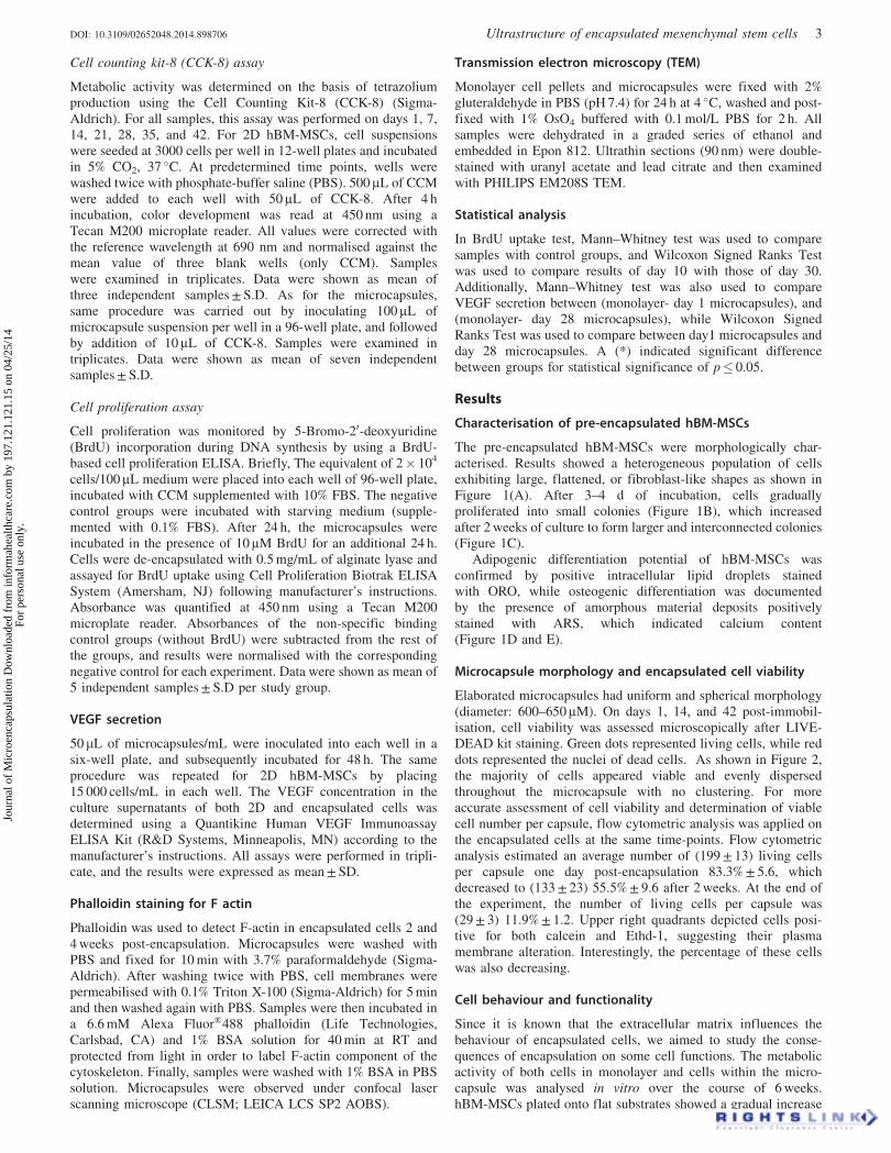
The pre-encapsulated hBM-MSCs were morphologically char-acterised. Results showed a heterogeneous population of cellsexhibiting large, flattened, or fibroblast-like shapes as shown inFigure 1(A). After 3–4 d of incubation, cells graduallyproliferated into small colonies (Figure 1B), which increasedafter 2 weeks of culture to form larger and interconnected colonies(Figure 1C).
Adipogenic differentiation potential of hBM-MSCs wasconfirmed by positive intracellular lipid droplets stainedwith ORO, while osteogenic differentiation was documentedby the presence of amorphous material deposits positivelystained with ARS, which indicated calcium content(Figure 1D and E).
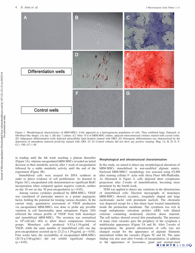
Microcapsule morphology and encapsulated cell viability
Elaborated microcapsules had uniform and spherical morphology(diameter: 600–650 lM). On days 1, 14, and 42 post-immobil-isation, cell viability was assessed microscopically after LIVE-DEAD kit staining. Green dots represented living cells, while reddots represented the nuclei of dead cells. As shown in Figure 2,the majority of cells appeared viable and evenly dispersedthroughout the microcapsule with no clustering. For moreaccurate assessment of cell viability and determination of viablecell number per capsule, flow cytometric analysis was applied onthe encapsulated cells at the same time-points. Flow cytometricanalysis estimated an average number of (199 ± 13) living cellsper capsule one day post-encapsulation 83.3% ± 5.6, whichdecreased to (133 ± 23) 55.5% ± 9.6 after 2 weeks. At the end ofthe experiment, the number of living cells per capsule was(29 ± 3) 11.9% ± 1.2. Upper right quadrants depicted cells posi-tive for both calcein and Ethd-1, suggesting their plasmamembrane alteration. Interestingly, the percentage of these cellswas also decreasing.
Cell behaviour and functionality
Since it is known that the extracellular matrix influences thebehaviour of encapsulated cells, we aimed to study the conse-quences of encapsulation on some cell functions. The metabolicactivity of both cells in monolayer and cells within the micro-capsule was analysed in vitro over the course of 6 weeks.hBM-MSCs plated onto flat substrates showed a gradual increase
DOI: 10.3109/02652048.2014.898706 Ultrastructure of encapsulated mesenchymal stem cells 3
Jour
nal o
f M
icro
enca
psul
atio
n D
ownl
oade
d fr
om in
form
ahea
lthca
re.c
om b
y 19
7.12
1.12
1.15
on
04/2
5/14
For
pers
onal
use
onl
y.
in reading until the 4th week reaching a plateau thereafter(Figure 3A), whereas encapsulated hBM-MSCs revealed an initialdecrease in their metabolic activity after 1 week of encapsulationfollowed by a stable metabolic activity until the end of theexperiment (Figure 3B).
Immobilised cells were assayed for DNA synthesis inorder to detect evidence of cell proliferation. As denoted inFigure 3(C), encapsulated cells demonstrated no significant BrdUincorporation when compared against negative controls, neitheron day 10 nor on day 30 post-encapsulation (p40.05).
Among various cytokines produced by hBM-MSCs, VEGFwas considered of particular interest as a potent angiogenicfactor, holding the potential for treating various disorders. In thecurrent study, quantitative assessment of VEGF productionvia encapsulated hBM-MSCs was done to probe any possiblevariation in cell functionality upon entrapment. Figure 3(D)reflected the release profile of VEGF from both monolayerand immobilised hBM-MSCs. The secretion was normalisedto 106 cell/ mL/ 48 h to allow direct comparisons betweengroups. Monolayer cells produced 9.53 ± 1.54 mg/mL ofVEGF, while the same number of immobilised cells one daypost-encapsulation secreted up to 23.23 ± 1.39 mg/mL (p¼ 0.05).Four weeks later, the secretability of encapsulated hBM-MSCs(20.74 ± 5.86 mg/mL) did not exhibit significant changes(p40.05).
Morphological and ultrastructural characterisation
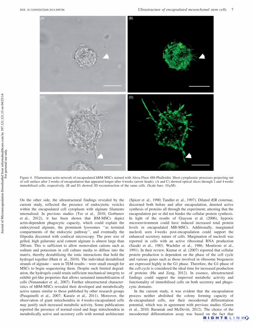
In this study, we aimed to detect any morphological alterations ofhBM-MSCs immobilised in non-modified alginate matrix.Enclosed hBM-MSCs’ morphology was assessed using CLSMafter staining cellular F- actin with Alexa Fluor 488-Phalloidin.As illustrated in Figure 4, cells depicted short cytoplasmicprojections after 2 weeks of immobilisation, becoming moreprominent by the fourth week.
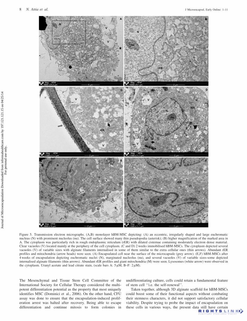
TEM was applied to detect any variations in the ultrastructureof immobilised cells. Electron micrographs of monolayerhBM-MSCs showed eccentric, irregularly shaped and largeeuchromatic nuclei with prominent nucleoli. The chromatinwas dispersed except for a thin dense layer located immediatelyinside the perinuclear membrane. The cytoplasm was rich inrough endoplasmic reticulum profiles (rER) with dilatedcisternae containing moderately electron dense material.The cell surface showed several thin pseudopodia. The presenceof many clear vacuoles gave the periphery of the cytoplasm amultilocular appearance (Figure 5A and B). After 2 weeks ofencapsulation, the general ultrastructure of cells was notchanged except for the appearance of alginate filamentsinternalised within the vacuoles (Figure 5C and D). The samefinding was also seen after 4 weeks of encapsulation, in additionto the appearance of lysosomes, giant and normal-sized
Figure 1. Morphological characteristics of hBM-MSCs. Cells appeared as a heterogeneous population of cells. They exhibited large, flattened, orfibroblast-like shapes. (A) day 1, (B) day 7 culture. (C) After 14 d of hBM-MSC culture, adjacent interconnected colonies stained with crystal violet.(D) Adipogenic differentiation wells depicted intracellular lipid droplets stained with ORO. (E) Osteogenic differentiation was characterised by thedeposition of amorphous material positively stained with ARS. (F, G) Control cultures did not show any positive staining. Mag. (A, B, D, E, F,G)� 100, (C)� 40.
4 N. Attia et al. J Microencapsul, Early Online: 1–11
Jour
nal o
f M
icro
enca
psul
atio
n D
ownl
oade
d fr
om in
form
ahea
lthca
re.c
om b
y 19
7.12
1.12
1.15
on
04/2
5/14
For
pers
onal
use
onl
y.
mitochondria (Figure 5E) together with marginated nucleolus(Figure 5F).
Differentiation potential of de-encapsulated hBM-MSCs
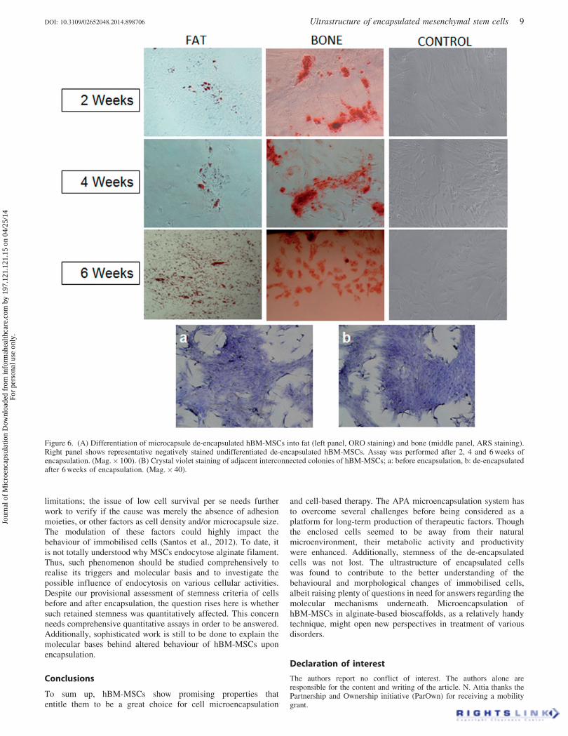
The ability of encapsulated cells to retain their stem cellsproperties upon de-encapsulation was evaluated. These cellsretained both adipogenic and osteogenic differentiation potentialas confirmed by ORO and ARS staining, respectively(Figure 6A). Additionally, hBM-MSCs de-encapsulated after6 weeks of encapsulation were observed to retain theirpre-encapsulation morphology and colony forming ability(Figure 6B).
Discussion
hBM-MSCs are truly attractive adult cells, which reside inspecialised but still largely unknown tissue compartments‘‘niches’’ (Shin and Peterson, 2013). They have been in focusof many researchers in the field of medicine for decades due totheir enormous therapeutic potential and stable properties. SinceMSCs cultured in vitro onto 2D substrates (e.g. ‘‘tissue cultureplastic’’) may lose cell-specific properties with time and so,poorly reflect their behaviour in vivo (Han et al., 2012), in thisstudy we aimed to elucidate various facets of hBM-MSCbehaviour within one of the most studied 3D scaffolds, ‘‘alginatemicrocapsules’’. Few studies have attempted to encapsulate hBM-MSCs, even though their homing capacity, hypoimmunogenic andimmunomodulatory properties make them likely the cells ofchoice in therapeutic cell encapsulation. Moreover, none havefocused on the ultrastructural alterations that may occur to thosecells upon encapsulation. In the current work, hBM-MSCs wereencapsulated into APA microcapsules, and then were thoroughlycharacterised as regards their viability, metabolic activity,
proliferation, VEGF secretability, and morphology, putting spe-cial emphasis on cellular ultrastructure.
In any microencapsulation-based cell therapy strategy, theaccurate assessment of the viability and proliferation ofencapsulated cells is essential for predicting the potential long-term secretion profiles of their therapeutic products. In the presentstudy, Live/Dead kit was used to assess cell viability both bymicroscopy and by flow cytometry. Even though the microscopicevaluation depicted no clear difference in green fluorescing dotsbetween day 1 and 14, flow cytometry – as a much accurate assay– demonstrated reduction of cell viability over time. Grossmann(2002) explained such decreasing viability of immobilised cellsby the lack of appropriate cell-matrix adhesion, thereby increasingdetachment-induced cell death (anoikis). Similar to epithelial andendothelial cells, MSCs are anchorage-dependent cells in whichcell-extracellular matrix interaction plays a paramount role intheir survival and function (Rowlands et al., 2008).
Metabolic activity of monolayer cells increased gradually,reaching a plateau after 4 weeks. Such pattern was expected inlight of the consensus that once the MSCs reach confluence, theirrate of proliferation decreases (Neuhuber et al., 2008). On theother side, immobilised cells showed an initial decrease in themetabolic activity/capsule after 1 week of encapsulation, followedby a plateau. Thus, given the gradual decline in cell viabilitytogether with the absent cell proliferation, present results denoteda gradual increase of cellular metabolic activity after 2 weeks ofentrapment till the end of the experiment. The fore mentionedcessation of mitotic activity in encapsulated cells was inaccordance with other researchers reporting that encapsulatedMSCs had minimal space for growth and, thus, became contact-inhibited and growth arrested (Markusen et al., 2006; Duggalet al., 2008). In turn, Bidarra et al. (2010) had attributed suchlack of cell proliferation to the spatially constraining polymeric
Figure 2. (A) Bright field (Upper panel) and fluorescence (Middle panel) micrographies of encapsulated hBM-MSCs stained with the LIVE/DEAD kit.Microcapsules depicted uniform rounded contour. Green dots represented cytoplasm of living cells, while red dots represented the nuclei of dead cells.Majority of cells appeared evenly dispersed and viable throughout the microcapsule. Scale bar: 200 mm. Lower panel show flow cytometry-basedviability for hBM-MSCs exposed to alginate lyase de-encapsulation procedure, performed on days 1, 14, and 42 post-immobilisation. The FL1/FL3dot-plots of calcein/ EthD-1 labeled cells. Upper left quadrants represented live cells. Lower right quadrants represented dead cells, while upper rightquadrants represented cells with impaired plasma membranes. The dimensions of all the quadrants were held constant when calculating cell viability inall experiments. (B) Line graph representing number of living cells/capsule.
DOI: 10.3109/02652048.2014.898706 Ultrastructure of encapsulated mesenchymal stem cells 5
Jour
nal o
f M
icro
enca
psul
atio
n D
ownl
oade
d fr
om in
form
ahea
lthca
re.c
om b
y 19
7.12
1.12
1.15
on
04/2
5/14
For
pers
onal
use
onl
y.

network. Moreover, Ma and co-researchers (Ma et al., 2003)documented that MSCs seized to proliferate upon immobilisation.Additionally, they reported that alginate encapsulation provides asuspension condition, unsuitable for division of the anchorage-dependent MSCs, thus became arrested in G0G1 phase.G1-arrested cells are metabolically more active than non-arrestedones (Hu and Zeng, 2012) and such increased cellular metabolicactivity could refer to cell activities other than proliferation(Bidarra et al., 2010). On the other hand, our data were not inagreement with those of Goren and co-workers (Goren et al.,2010). Those authors reported that the viability and proliferationof the encapsulated hBM-MSCs remained stable throughout their70 d-experiments. Discrepant findings could be explained by thedifferent experimental design (e.g. lower alginate concentrationand/or much lower cell density) compared to that adopted in thecurrent study.
Beneficial effects of hBM-MSCs are believed to be mainlydue to their paracrine activity, so that we next investigated ifAPA-encapsulated hBM-MSCs would retain their pre-encapsula-tion paracrine function. Several research groups documented theability of monolayer MSCs to secrete VEGF and other angiogeniccytokines (Tang et al., 2006; Burchfield and Dimmeler, 2008;Boomsma and Geenen, 2012). Moreover, Bidarra et al. (2010)proved that the VEGF secreted by encapsulated MSCs was able to
diffuse through the hydrogel network into the surroundingmedium. However, to the best of our knowledge, this is the firststudy performing direct quantitative comparison between VEGFsecretability of pre- and post-encapsulated hBM-MSCs. Wediscerned what it seems to be a significant upregulation ofVEGF secretion after encapsulation that was retained even after4 weeks of culture. Lee et al. (2013) have recently explained suchincreased VEGF secretion by the relative hypoxic environ-ment inside the microcapsules. Additionally, several micro-environmental cues such as shear stress and substrate compliancehave been reported to control MSC paracrine activity (Ranganathet al., 2012). Anyway, the molecular forces that increase thesecretory function of encapsulated hBM-MSCs are intriguing butstill unclear.
Contrary to expectations, our results showed that hBM-MSCsembedded in non-modified alginate matrix attained several mm-sized cortical protrusions into the surrounding matrix that becamemore evident by the fourth week of encapsulation. Thosefilopodia-like branches were further evidenced by fluorescentstaining of actin filament. Since the alginate hydrogel used forencapsulation was not modified with adhesion moieties, suchcellular extensions could not be due to cell-matrix adhesion, asreported by other several studies (Lawson et al., 2004; Markusenet al., 2006; Bidarra et al., 2010; Sayyar et al., 2012).
Figure 3. (A, B) Line graphs reported the metabolic activity of viable cells by CCK-8 assay on days 1, 7, 14, 21, 28, 35 and 42 of culture.(A) Monolayer cells (n¼ 3), (B) Encapsulated cells (n¼ 7). (C) Bar graph showing BrdU uptake of encapsulated hBM-MSCs, demonstrated nosignificant increase in BrdU incorporation, neither on day 10 nor on day 30 post-encapsulation (p40.05) (n¼ 5). (D) Bar graph showing VEGFsecretion of hBM-MSCs. As monolayer cultures, day1 and day 28 post-encapsulation (n¼ 3). Their secretion significantly increased after encapsulation(p¼ 0.05), with no significant change later (p40.05). Vertical error bars represented the standard deviation of the reported mean values. (*¼ statisticalsignificance) (NS¼ no significance).
6 N. Attia et al. J Microencapsul, Early Online: 1–11
Jour
nal o
f M
icro
enca
psul
atio
n D
ownl
oade
d fr
om in
form
ahea
lthca
re.c
om b
y 19
7.12
1.12
1.15
on
04/2
5/14
For
pers
onal
use
onl
y.
On the other side, the ultrastructural findings revealed by thecurrent study, reflected the presence of endocytotic vesicleswithin the encapsulated cell cytoplasm with alginate filamentsinternalised. In previous studies (Tso et al., 2010; Gorbunovet al., 2012), it has been shown that BM-MSCs depictactin-dependent phagocytic capacity, which could explain theendocytosed alginate, the prominent lysosomes ‘‘as terminalcompartments of the endocytic pathway’’, and eventually thefilipodia discerned with confocal microscopy. The pore size ofgelled, high guluronic acid content alginate is almost large than200 nm. This is sufficient to allow monovalent cations such assodium and potassium in cell culture media to diffuse into thematrix, thereby destabilising the ionic interactions that hold thehydrogel together (Hunt et al., 2010). The individual destabilisedstrands of alginate – seen in TEM results – were small enough forMSCs to begin sequestering them. Despite such limited degrad-ation, the hydrogels could retain sufficient mechanical integrity toexhibit gel-like properties that allows sustained immobilisation ofcells (Nunamaker et al., 2007). Further ultrastructural character-istics of hBM-MSCs revealed their developed and metabolicallyactive nature similar to those published by other research groups(Pasquinelli et al., 2007; Karaoz et al., 2011). Moreover, theobservation of giant mitochondria in 4 weeks-encapsulated cellsmay justify such increased metabolic activity. Some publicationsreported the presence of normal-sized and huge mitochondria inmetabolically active and secretory cells with normal architecture
(Spicer et al., 1990; Tandler et al., 1997). Dilated rER cisternae,discerned both before and after encapsulation, denoted activesynthesis of proteins all through the experiment; attesting that theencapsulation per se did not hinder the cellular protein synthesis.In light of the results of Grayson et al. (2006), hypoxicmicroenvironment could have induced increased total proteinlevels in encapsulated MB-MSCs. Additionally, marginatednucleoli seen 4 weeks post-encapsulation could support theenhanced secretory nature of cells. Margination of nucleoli wasreported in cells with an active ribosomal RNA production(Sasaki et al., 1983; Wachtler et al., 1986; Montironi et al.,1991). In their review, Kumar et al. (2007) reported that cellularprotein production is dependent on the phase of the cell cycleand various genes such as those involved in ribosome biogenesisare expressed highly in the G1 phase. Therefore, the G1-phase ofthe cell cycle is considered the ideal time for increased productionof proteins (Hu and Zeng, 2012). In essence, ultrastructuralfindings could support the improved metabolic activity andfunctionality of immobilised cells on both secretory and phago-cytic domains.
In the current study, it was evident that the encapsulationprocess neither abolished the colony forming capacity ofde-encapsulated cells, nor their mesodermal differentiationpotential, which was in agreement with previous studies (Gorenet al., 2010; Baraniak and McDevitt, 2012). The choice of themesodermal differentiation assay was based on the fact that
Figure 4. Filamentous actin network of encapsulated hBM-MSCs stained with Alexa Fluor 488-Phalloidin. Short cytoplasmic processes projecting outof cell surface after 2 weeks of encapsulation that appeared longer after 4 weeks (arrow heads). (A and C) showed optical slices through 2 and 4 weeksimmobilised cells, respectively. (B and D) showed 3D reconstruction of the same cells. (Scale bars: 10 lM).
DOI: 10.3109/02652048.2014.898706 Ultrastructure of encapsulated mesenchymal stem cells 7
Jour
nal o
f M
icro
enca
psul
atio
n D
ownl
oade
d fr
om in
form
ahea
lthca
re.c
om b
y 19
7.12
1.12
1.15
on
04/2
5/14
For
pers
onal
use
onl
y.
The Mesenchymal and Tissue Stem Cell Committee of theInternational Society for Cellular Therapy considered the multi-potent differentiation potential as the property that most uniquelyidentifies MSC (Dominici et al., 2006). On the other hand, CFUassay was done to ensure that the encapsulation-induced prolif-eration arrest was halted after recovery. Being able to escapedifferentiation and continue mitosis to form colonies in
undifferentiating culture, cells could retain a fundamental featureof stem cell ‘‘i.e. the self-renewal’’.
Taken together, although 3D alginate scaffold for hBM-MSCscould boost some of their functional aspects without combatingtheir stemness characters, it did not support satisfactory cellularviability. Despite trying to probe the impact of encapsulation onthese cells in various ways, the present data still have certain
Figure 5. Transmission electron micrographs. (A,B) monolayer hBM-MSC depicting: (A) an eccentric, irregularly shaped and large euchromaticnucleus (N) with prominent nucleolus (nu). The cell surface showed many thin pseudopodia (asterisk); (B) higher magnification of the marked area inA. The cytoplasm was particularly rich in rough endoplasmic reticulum (rER) with dilated cisternae containing moderately electron dense material.Clear vacuoles (V) located mainly at the periphery of the cell cytoplasm. (C and D) 2 weeks immobilised hBM-MSCs. The cytoplasm depicted severalvacuoles (V) of variable sizes with alginate filaments internalised in some of them similar to the extra cellular ones (thin arrows). Abundant rERprofiles and mitochondria (arrow heads) were seen. (A) Encapsulated cell near the surface of the microcapsule (grey arrow). (E,F) hBM-MSCs after4 weeks of encapsulation depicting euchromatic nuclei (N), marginated nucleolus (nu), and several vacuoles (V) of variable sizes-some depictedinternalised alginate filaments (thin arrows). Abundant rER profiles and giant mitochondria (M) were seen. Lysosomes (white arrow) were observed inthe cytoplasm. Uranyl acetate and lead citrate stain, (scale bars A: 5 lM, B–F: 2 lM).
8 N. Attia et al. J Microencapsul, Early Online: 1–11
Jour
nal o
f M
icro
enca
psul
atio
n D
ownl
oade
d fr
om in
form
ahea
lthca
re.c
om b
y 19
7.12
1.12
1.15
on
04/2
5/14
For
pers
onal
use
onl
y.
limitations; the issue of low cell survival per se needs furtherwork to verify if the cause was merely the absence of adhesionmoieties, or other factors as cell density and/or microcapsule size.The modulation of these factors could highly impact thebehaviour of immobilised cells (Santos et al., 2012). To date, itis not totally understood why MSCs endocytose alginate filament.Thus, such phenomenon should be studied comprehensively torealise its triggers and molecular basis and to investigate thepossible influence of endocytosis on various cellular activities.Despite our provisional assessment of stemness criteria of cellsbefore and after encapsulation, the question rises here is whethersuch retained stemness was quantitatively affected. This concernneeds comprehensive quantitative assays in order to be answered.Additionally, sophisticated work is still to be done to explain themolecular bases behind altered behaviour of hBM-MSCs uponencapsulation.
Conclusions
To sum up, hBM-MSCs show promising properties thatentitle them to be a great choice for cell microencapsulation
and cell-based therapy. The APA microencapsulation system hasto overcome several challenges before being considered as aplatform for long-term production of therapeutic factors. Thoughthe enclosed cells seemed to be away from their naturalmicroenvironment, their metabolic activity and productivitywere enhanced. Additionally, stemness of the de-encapsulatedcells was not lost. The ultrastructure of encapsulated cellswas found to contribute to the better understanding of thebehavioural and morphological changes of immobilised cells,albeit raising plenty of questions in need for answers regarding themolecular mechanisms underneath. Microencapsulation ofhBM-MSCs in alginate-based bioscaffolds, as a relatively handytechnique, might open new perspectives in treatment of variousdisorders.
Declaration of interest
The authors report no conflict of interest. The authors alone areresponsible for the content and writing of the article. N. Attia thanks thePartnership and Ownership initiative (ParOwn) for receiving a mobilitygrant.
Figure 6. (A) Differentiation of microcapsule de-encapsulated hBM-MSCs into fat (left panel, ORO staining) and bone (middle panel, ARS staining).Right panel shows representative negatively stained undifferentiated de-encapsulated hBM-MSCs. Assay was performed after 2, 4 and 6 weeks ofencapsulation. (Mag.� 100). (B) Crystal violet staining of adjacent interconnected colonies of hBM-MSCs; a: before encapsulation, b: de-encapsulatedafter 6 weeks of encapsulation. (Mag.� 40).
DOI: 10.3109/02652048.2014.898706 Ultrastructure of encapsulated mesenchymal stem cells 9
Jour
nal o
f M
icro
enca
psul
atio
n D
ownl
oade
d fr
om in
form
ahea
lthca
re.c
om b
y 19
7.12
1.12
1.15
on
04/2
5/14
For
pers
onal
use
onl
y.
References
Abbah S, Lu W, Chan D, Cheung K, Liu W, Zhao F, Li Z, Leong J,Luk K. Osteogenic behavior of alginate encapsulated bone marrowstromal cells: An in vitro study. J Mater Sci Mater Med, 2008;19:2113–19.
Abumaree M, Al Jumah M, Pace RA, Kalionis B. Immunosuppressiveproperties of mesenchymal stem cells. Stem Cell Rev, 2012;8:375–92.
Baksh D, Song L, Tuan R. Adult mesenchymal stem cells:Characterization, differentiation, and application in cell and genetherapy. J Cell Mol Med, 2004;8:301–16.
Baraniak PR, Mcdevitt TC. Scaffold-free culture of mesenchymal stemcell spheroids in suspension preserves multilineage potential. CellTissue Res, 2012;347:701–11.
Beyth S, Borovsky Z, Mevorach D, Liebergall M, Gazit Z, Aslan H,Galun E, Rachmilewitz J. Human mesenchymal stem cells alterantigen-presenting cell maturation and induce T-cell unresponsiveness.Blood, 2005;105:2214–19.
Bidarra SLJ, Barrias CC, Barbosa MRA, Soares R, Granja PL.Immobilization of human mesenchymal stem cells within RGD-graftedalginate microspheres and assessment of their angiogenic potential.Biomacromolecules, 2010;11:1956–64.
Boomsma RA, Geenen DL. Mesenchymal stem cells secrete multiplecytokines that promote angiogenesis and have contrasting effects onchemotaxis and apoptosis. PLoS One, 2012;7:e35685.
Burchfield JS, Dimmeler S. Role of paracrine factors in stem andprogenitor cell mediated cardiac repair and tissue fibrosis. FibrogenesisTissue Repair, 2008;1:4. doi: 10.1186/1755-1536-1-4.
Chan BP, Hui TY, Yeung CW, Li J, Mo I, Chan GCF. Self-assembledcollagen–human mesenchymal stem cell microspheres for regenerativemedicine. Biomaterials, 2007;28:4652–66.
Correa PL, Mesquita CT, Felix RM, Azevedo JC, Barbirato GB, FalcaoCH, Gonzalez C, Mendonca ML, Manfrim A, De Freitas G.Assessment of intra-arterial injected autologous bone marrow mono-nuclear cell distribution by radioactive labeling in acute ischemicstroke. Clin Nucl Med, 2007;32:839–41.
Dominici M, Le Blanc K, Mueller I, Slaper-Cortenbach I, Marini FC,Krause DS, Deans RJ, Keating A, Prockop DJ, Horwitz EM. Minimalcriteria for defining multipotent mesenchymal stromal cells. TheInternational Society for Cellular Therapy position statement.Cytotherapy, 2006;8(4):315–17.
Duggal S, Frønsdal KB, Szoke K, Shahdadfar A, Melvik JE,Brinchmann JE. Phenotype and gene expression of human mesenchy-mal stem cells in alginate scaffolds. Tissue Eng Part A, 2008;15:1763–73.
Garate A, Murua A, Orive G, Hernandez RM, Pedraz JL. Stem cells inalginate bioscaffolds. Ther Deliv, 2012;3:761–74.
Ghidoni I, Chlapanidas T, Bucco M, Crovato F, Marazzi M, Vigo D,Torre ML, Faustini M. Alginate cell encapsulation: New advances inreproduction and cartilage regenerative medicine. Cytotechnology,2008;58:49–56.
Giordano A, Galderisi U, Marino IR. From the laboratory bench to thepatient’s bedside: An update on clinical trials with mesenchymal stemcells. J Cell Physiol, 2007;211:27–35.
Gorbunov N, Garrison B, Zhai M, Mcdaniel D, Ledney G, Elliott T,Kiang J. Autophagy-mediated defense response of mouse mesenchy-mal stromal cells (MSCs) to challenge with Escherichia coli. DTICDocument 2012.
Goren A, Dahan N, Goren E, Baruch L, Machluf M. Encapsulated humanmesenchymal stem cells: A unique hypoimmunogenic platform forlong-term cellular therapy. FASEB J, 2010;24:22–31.
Grayson WL, Zhao F, Izadpanah R, Bunnell B, Ma T. Effects of hypoxiaon human mesenchymal stem cell expansion and plasticity in 3Dconstructs. J Cell Physiol, 2006;207:331–9.
Griffin MD, Ritter T, Mahon BP. Immunological aspects of allogen-eic mesenchymal stem cell therapies. Hum Gene Ther, 2010;21:1641–55.
Grossmann J. Molecular mechanisms of ‘‘detachment-induced apoptosis– anoikis’’. Apoptosis, 2002;7:247–60.
Han S, Zhao Y, Xiao Z, Han J, Chen B, Chen L, Dai J. The three-dimensional collagen scaffold improves the stemness of rat bonemarrow mesenchymal stem cells. J Genet Genomics, 2012;39(12):633–41.
Horie N, Pereira MP, Niizuma K, Sun G, Keren-Gill H, Encarnacion A,Shamloo M, Hamilton SA, Jiang K, Huhn S. Transplanted stemcell-secreted vascular endothelial growth factor effects poststroke
recovery, inflammation, and vascular repair. Stem cells, 2011;29:274–85.
Hu WS, Zeng AP. 2012. Genomics and systems biology of mammaliancell culture. Heidelberg, Germany: Springer.
Hunt NC, Smith AM, Gbureck U, Shelton RM, Grover LM.Encapsulation of fibroblasts causes accelerated alginate hydrogeldegradation. Acta Biomaterialia, 2010;6(9):3649–56.
Jones E, Mcgonagle D. Human bone marrow mesenchymal stem cellsin vivo. Rheumatology (Oxford), 2008;47:126–31.
Karaoz E, Okcu A, Gacar G, Saglam O, Yuruker S, Kenar H.A comprehensive characterization study of human bone marrowMSCs with an emphasis on molecular and ultrastructural properties.J Cell Physiol, 2011;226:1367–82.
Kumar N, Gammell P, Clynes M. Proliferation control strategies toimprove productivity and survival during CHO based productionculture. Cytotechnology, 2007;53:33–46.
Lawson M, Barralet J, Wang L, Shelton R, Triffitt JT. Adhesion andgrowth of bone marrow stromal cells on modified alginate hydrogels.Tissue Eng, 2004;10:1480–91.
Lee CS, Watkins EA, Burnsed OA, Schwartz Z, Boyan B. Tailoringadipose stem cell trophic factor production with differentiation mediumcomponents to regenerate chondral defects. Tissue Eng Part A, 2013;19:1451–64.
Lim F, Sun AM. Microencapsulated islets as bioartificial endocrinepancreas. Science, 1980;210:908–10.
Ma HL, Hung SC, Lin SY, Chen YL, Lo WH. Chondrogenesis of humanmesenchymal stem cells encapsulated in alginate beads. J BiomedMater Res A, 2003;64:273–81.
Markusen JF, Mason C, Hull DA, Town MA, Tabor AB, Clements M,Boshoff CH, Dunnill P. Behavior of adult human mesenchymal stemcells entrapped in alginate-GRGDY beads. Tissue Eng, 2006;12:821–30.
Menasche P. Stem cells for clinical use in cardiovascular medicine.Thromb Haemost, 2005;94:697–701.
Montironi R, Scarpelli M, Braccischi A, Galluzzi CM, Diamanti L,Alberti R. Quantitative analysis of nucleolar margination in diagnosticcytopathology. Virchows Arch A, 1991;419:505–12.
Nagaya N, Takafumi F, Takashi I, Hajime O, Takefumi I, Masaaki U,Masakazu Y, Hidezo M, Kenji K, Soichiro K. Intravenous adminis-tration of mesenchymal stem cells improves cardiac function in ratswith acute myocardial infarction through angiogenesis and myogen-esis. Am J Physiol Heart Circ Physiol, 2004;287:H2670–6.
Nauta AJ, Fibbe WE. Immunomodulatory properties of mesenchymalstromal cells. Blood, 2007;110:3499–506.
Neuhuber B, Swanger SA, Howard L, Mackay A, Fischer I. Effects ofplating density and culture time on bone marrow stromal cellcharacteristics. Exp Hematol, 2008;36:1176–85.
Ngoc PK, Van Phuc P, Nhung TH, Thuy DT, Nguyet NTM. Improving theefficacy of type 1 diabetes therapy by transplantation of immunoiso-lated insulin-producing cells. Hum Cell, 2011;24:86–95.
Nunamaker, EA, Erin KP, Daryl RK. In vivo stability and biocompati-bility of implanted calcium alginate disks. J Biomed Mater Res A,2007;83.4:1128–37.
Orive G, Carcaboso A, Hernandez R, Gascon A, Pedraz J.Biocompatibility evaluation of different alginates and alginate-basedmicrocapsules. Biomacromolecules, 2005;6:927–31.
Orive G, Hernandez RM, Gascon AR, Pedraz JL. 2006. Encapsulation ofcells in alginate gels. In: Guisan JM, ed. Immobilization of enzymesand cells. New Jersey: Humana Press, pp. 345–55.
Pasquinelli G, Tazzari P, Ricci F, Vaselli C, Buzzi M, Conte R, Orrico C,Foroni L, Stella A, Alviano F. Ultrastructural characteristics of humanmesenchymal stromal (stem) cells derived from bone marrow and termplacenta. Ultrastruct Pathol, 2007;31:23–31.
Phinney DG, Prockop DJ. Concise review: Mesenchymal stem/multi-potent stromal cells: The state of transdifferentiation and modes oftissue repair—current views. Stem cells, 2007;25:2896–902.
Pittenger MF, Martin BJ. Mesenchymal stem cells and their potential ascardiac therapeutics. Circ Res, 2004;95:9.
Ponce S, Orive G, Hernandez R, Gascon AR, Pedraz JL, De Haan BJ,Faas MM, Mathieu H, De Vos P. Chemistry and the biological responseagainst immunoisolating alginate–polycation capsules of differentcomposition. Biomaterials, 2006;27:4831–9.
Ranganath SH, Levy O, Inamdar MS, Karp JM. Harnessing themesenchymal stem cell secretome for the treatment of cardiovasculardisease. Cell Stem Cell, 2012;10:244–58.
10 N. Attia et al. J Microencapsul, Early Online: 1–11
Jour
nal o
f M
icro
enca
psul
atio
n D
ownl
oade
d fr
om in
form
ahea
lthca
re.c
om b
y 19
7.12
1.12
1.15
on
04/2
5/14
For
pers
onal
use
onl
y.
Rowlands AS, George PA, Cooper-White JJ. Directing osteogenicand myogenic differentiation of mscs: Interplay of stiffness andadhesive ligand presentation. Am J Physiol Cell Physiol, 2008;295:C1037–C44.
Sanchez-Ramos JR. Neural cells derived from adult bone marrow andumbilical cord blood. J Neurosci Res, 2002;69:880–93.
Santos E, Orive G, Calvo A, Catena R, Fernandez-Robredo P,Layana AG, Hernandez RM, Pedraz JL. Optimization of 100 lmalginate-poly-L-lysine-alginate capsules for intravitreous administra-tion. J Control Release. 2012;158(3):443–50.
Sasaki K, Matsumura G, Ito T. A quantitative morphology of nucleoli oferythroblasts in the mouse spleen: An electron microscopic study. ArchHistol Jpn, 1983;46:43–9.
Sayyar B, Dodd M, Wen J, Ma S, Marquez-Curtis L, Janowska-Wieczorek A, Hortelano G. Encapsulation of factor IX-engineeredmesenchymal stem cells in fibrinogen–alginate microcapsulesenhances their viability and transgene secretion. J Tissue Eng, 2012;3(1):2041731412462018.
Shin L, Peterson DA. Human mesenchymal stem cell grafts enhancenormal and impaired wound healing by recruiting existing endo-genous tissue stem/progenitor cells. Stem Cells Transl Med, 2013;2:33–42.
Spicer S, Parmley R, Boyd L, Schulte B. Giant mitochondria distinctfrom enlarged mitochondria in secretory and ciliated cells of gerbiltrachea and bronchioles. Am J Anat, 1990;188:269–81.
Tandler B, Nagato T, Phillips CJ. Megamitochondria in the serous acinarcells of the submandibular gland of the neotropical fruit bat, artibeusobscurus. Anat Rec, 1997;248:13–17.
Tang J, Xie Q, Pan G, Wang J, Wang M. Mesenchymal stem cellsparticipate in angiogenesis and improve heart function in rat model ofmyocardial ischemia with reperfusion. Eur J Cardiothorac Surg 2006;30:353–61.
Tso GHW, Law HKW, Tu W, Chan GCF, Lau YL. Phagocytosis ofapoptotic cells modulates mesenchymal stem cells osteogenic differ-entiation to enhance IL-17 and RANKL expression on CD4+ T cells.Stem cells, 2010;28:939–54.
Wachtler F, Hopman A, Wiegant J, Schwarzacher H. On the position ofnucleolus organizer regions (NORs) in interphase nuclei: Studies witha new, non-autoradiographic in situ hybridization method. Exp CellBiol, 1986;167:227–40.
Yu J, Du KT, Fang Q, Gu Y, Mihardja SS, Sievers RE, Wu JC, Lee RJ.The use of human mesenchymal stem cells encapsulated in RGDmodified alginate microspheres in the repair of myocardial infarctionin the rat. Biomaterials, 2010;31:7012–20.
DOI: 10.3109/02652048.2014.898706 Ultrastructure of encapsulated mesenchymal stem cells 11
Jour
nal o
f M
icro
enca
psul
atio
n D
ownl
oade
d fr
om in
form
ahea
lthca
re.c
om b
y 19
7.12
1.12
1.15
on
04/2
5/14
For
pers
onal
use
onl
y.
Copyright © 2022 FDOKUMEN