Development of an EGFR-targeting Alginate-based Injectable ...

�9 1995 by Humana Press Inc. All rights of any nature whatsoever reserved. 0273-2289/95/5502--0095509.40
High-Alkaline Protease from Bacillus PB92 Entrapped in Calcium Alginate Gel
Physicochemical and Microscopic Studies
M. G. ROIG, 1 D. H. RASHID, 2 AND J. F. KENNEDY ",2
l Departamento de Quimica Fisica, Facultad de Farmacia, Universidad de Salamanca, Spain; and 2Birmingham Carbohydrate
and Protein Technology Group, Research Laboratory for the Chemistry of Bioactive Carbohydrates and Proteins, School of Chemistry, Birmingham University, B15 2TT, UK
Received June 22, 1994; Accepted October 12, 1994
ABSTRACT
High-alkaline protease (HAP) has been entrapped in Manugel DMB (an alginate gel) and assayed with two sizes and types of sub- strates: neutral protein casein and synthetic chromogenic tripeptide substrate, Z-Gly-Pro-Cit-PNA. Increasing the concentration of calcium chloride used for capsule formation decreased the measured enzyme activity with both substrates. Capsules were found to be stable in water for long periods of time, but they dissolved in both phosphate and carbonate-bicarbonate buffers. The pH vs activity profiles of encap- sulated enzyme showed pH optima between 10 and 11 with both substrates. The calcium alginate matrix surrounding the enzyme was quite effective in stabilizing the enzyme at 20-25~ and even more so at 4~ Enzyme stability at 50~ was quite impressive, some enzyme activity being evident even after remaining for 1 wk at this temperature in water. Increasing concentrations of sodium dodecyl sulfate (SDS) were also found to inhibit the protease progressively, whereas a polyhexamethylene biguanidium chloride (PHMBH+CI-) and SDS:PHMBH+C1 - combination showed the opposite effect.
Optical microscopy, especially polarized light microscopy, pro- vided a sensitive physical means of ascertaining some of the structural properties (sphericity, disorganization or organization, distinct layer
*Author to whom all correspondence and reprint requests should be addressed.
Applied Biochemistry and Biotechnology 9 5 Vol. 55, 1995

96 Roig, Rashid, and Kennedy
enveloping the capsules, intensity of the maltese cross) of the capsules with and without enzyme before and after different chemical treat- ments and the presence or absense of the substrate.
Index Entries: Protease; high-alkaline protease; immobilized; algin- ate; pH; stability; microscopy; SDS; polyhexamethylene biguanidium.
INTRODUCTION
Alkalophilic Bacillus strains produce a number of different proteolytic enzymes, some of them having properties similar to those of the known subtilisins, but others being characterized by having maximum activity up to pH 12.0 (1). In 1934, Vedder (2) succeeded in isolating 16 strains of a bacterium belonging to the genus Bacillus, which he called Bacillus alcalo- philus because this species only grew at pH values above 7, preferably within the 8.6-10 range. Aunstrup et al. (3) described the isolation of nearly one hundred strains of bacteria from samples of soil, animal manure, and various other natural sources. With regard to the proteolytic activity of the enzymes produced by these species, strains have been divided into three groups or types in which the proteolytic activity is measured at pH 12. All the enzymes produced were also tested for their stability in solu- tion at 50~ in the presence of:
1. Sodium tripolyphosphate; 2. Sodium tetraborate; and 3. Surfactants, with varying results.
The temperature optima were in 40-60~ range. These enzymes have been shown to be serine active site proteases
and are stabilized by the presence of Ca 2+ (4). They are single peptide chains free of disulfide bridges. The N-terminal residue is alanine, as in subtilisins. The specificity for peptide bond hydrolysis is broad, and the enzymes have esterase activity (5). The alkaline protease from B. licheni- formis indicates a preference for leucine, cysteine, and glutamate peptide bonds (6). Morhihara (6) stresses that subtilisin BPN (NOVO) attacks the Leu--Tyr bond in oxidized insulin B-chain more rapidly than any other bond. Other important bonds are Ser--His and Leu--Val for scission by the enzyme, and it has been concluded that this general class of serine alkaline protease attacks Glu--His, Leu--Val, Leu--Tyr, Tyr--Leu, Phe-- Phe, Phe--Tyr, or Tyr--Thr. BPN and Carlsberg subtilisins exhibit higher activity toward esters of aromatic amino acids than those of aliphatic ones. From the cleavage pattern of small peptides by subtilisin BPN (7) it seems that proline is an invariable factor as the second amino acid residue. The molecular weights are in the region of 20.000-30.000 and the isoelectric point is between 10 and 11.
Applied Biochemistry and Biotechnology Vol. 55, 1995

Entrapped Alkaline Protease 97
High-alkaline protease (HAP) is produced by high yields by a novel Bacillus species designated PB92 in a nutrient medium containing carbon, nitrogen, and trace element sources under aerobic circumstances, and the proteolytic enzyme formed has been isolated from the fermentation broth (8). The microorganism producing the enzyme was isolated from an earth sample from Zambia by a specific procedure at a pH within the range of 8.0-9.5. The protease was obtained at high yields by enriching the growth medium with high-energy sources, such as glucose, sucrose, dextrins, and starch, and a nitrogen source of organic origin, such as casein, yeast, and soya flour. Certain amounts of calcium and magnesium salts and several trace elements were also added. A good aeration is also necessary during fermentation, and the pH of the medium is kept on the alkaline side. The enzyme is recovered by filtering the broth to remove the micro- organisms and insoluble material. For dry washing powder compositions, the enzyme is either precipitated by adding water-miscible organic sol- vents or inorganic salts, such as Na2SO4 or (NH4)2SO4, to the filtrate. After precipitation or centrifugation, the resulting cake is dried. Alkaline pro- tease is an enzyme of significant interest with respect to decontamination and cleansing of materials to which protein has inadvertently been added. They are regarded as potent proteases with a wide specificity range, and for these reasons, this particular enzyme, which is available, was chosen.
In principle, protein globules can be prevented from unfolding if placed in a certain cell that does not interact with the molecules either chemically or sorptionally, but that is sufficiently " t ight" to prevent for- mation of an unfolded conformation for steric reasons. In the entrapment in "t ight" support pores, the native conformation of a protein globule could be maintained in a purely mechanical fashion (9). This mechanism has been hypothesized more than once in the literature (10-13) and ex- perimental evidence for it was furnished (14-16) when the properties of c~-chymotrypsin and trypsin in polyacrylamide gel or in polysaccharide support were studied.
Calcium alginate gel has proven to be a reliable, versatile, and useful mode of immobilization of a variety of cell types, subcellular organelles, and enzyme and multicomponent systems (17-22). The easy way in which the gel can be generated from soluble alginate and calcium ion is also an advantage giving rise to entrapped enzyme immobilization rather than covalent derivatization immobilization, which may be damaging to the enzyme-active site and so destroy some of its activity. Differences in algi- nate gel properties result from variations in alginate grades and also the effect of this variation on the diffusion of low and high molecular com- pounds into and from gels. Furthermore, the effects on diffusion of gel concentrations have been reported (23). Most commonly, the formation of spherical calcium alginate beads has been reported. A method for the production of spherical alginate gels ranging from a few millimeter up to several millimeter diameter has been described (24). Alginate beads with
Applied Biochemistry and Biotechnology Vol. 55, 1995

98 Roig, Rashid, and Kennedy
diameters between 0.5 and 3 mm are used to immobilize microorganisms as biocatalysts. Micro-size alginate gels are used as column packing mate- rial for inverse steric exclusion chromatography to determine their pore sizes from dextran standards of known molecular weight. In spite of this extended use, there have been only a few contributions related to detailed aspects of the structure of these biocatalyst carriers as a basis for under- standing structure-property relationships. Some data have been presented on mechanical strength pressure stability (25,26), abrasion resistance (27), and diffusion (25).
This research examined the ability of calcium to form gels from alginate solution with a view to investigating the nature of the interaction occurring in an alginate gel present as a capsule and a protease enzyme entrapped within it. Interactions between the enzyme and polyhexamethylenebigu- anidinium chloride were also studied, as were mixtures of enzyme and detergents. The activity of the enzyme proved to be a sensitive measure of the interaction. Viscometry was used to follow the other interactions in solution, and polarized light microscopy was used to study the changes in the organized structure within the capsules.
EXPERIMENTAL
Materials
Sodium-dodecyl sulfate (SDS) (specially purified for biochemical work) was obtained from BDH Biochemicals Ltd., Poole, Dorset. Polyhexamethyl- enebiguanidinium chloride (PHMBH+C1 -) at an initial concentration of 20% (w/v) was obtained from ICI Ltd., Pollution Control Division, Hyde, Cheshire. Sodium alginate samples were donated by Alginate Industries Ltd. Merck. Casein (Hammarsten) was also obtained from BDH Chemicals Ltd. Tripeptide substrate (Z-glycyl-L-prolyl-L-citrulline) p-nitroanilide was obtained from Koch-Light Laboratories Ltd., Col- nbrook, Bucks., England. HAP PB92 (3281000 ADU/g) was provided by Gist-Brocades, Holland, as a liquid formulation containing glycerol.
Methods
Preparation of Polysaccharide Solutions Solutions of alginate were prepared by slowly adding the dry poly-
saccharide to vigorously stirred cold water, SDS, or SDS/PHMBH*C1 - solutions. The solutions obtained were left standing at room temperature for at least 2 h.
Preparation of Casein Substrate Casein (2.5 g) was dissolved in glycine-sodium hydroxide buffer
(0.1M), pH 11.0. This was heated in a boiling water bath for 10 rain. After cooling, the pH was checked and adjusted by either 2M glycine or 1.0M
Applied Biochemistry and Biotechnology Vol. 55, 1995

Entrapped Alkaline Protease 99
NaOH solution to the appropriate pH value. The denatured casein solu- tion (2 mL) was used in the assay.
Preparation of Tripeptide Substrate Ten milligrams of Z-Gly-Pro-Cit-PNA were initially dissolved in 1.0
cm 3 of dimethylsulfoxide, and then 4.0 cm 3 of acetonitrile were added and mixed thoroughly. In the assay, 400/~L of this concentrated sub- strate solution were utilized; thus, the assay cell contained 400/~L of the substrate and 1.6 mL of buffer.
Preparation of Calcium Alginate.HAP Capsules Sodium alginate (Manugel DMB batch no. 433101) at 1% (w/v) in water
was mixed with liquid HAP PB92 and water at a volume ratio of 9.5:0.5:5.0 mL. The resulting solution was dropped into a calcium chloride solution (2% w/v, 20 mL) using the tube end of a peristaltic pump to produce trans- lucent capsules. The capsules were left in the CaC12 solution for 5 min and later washed several times with water. They were then stored in water for I h. This ensured that any remaining enzyme that was not entrapped within the capsules would leak out into the surrounding liquid. (Thus, when the capsules were assayed later for their enzyme activity, it was assumed that the enzyme activity was owing totally to the enzyme en- trapped within the capsule and not to any residual enzyme remaining on the capsule surface or in the surrounding liquid.) The capsules were washed again and finally stored in water for a further 2 h at 20-25~ before being assayed.
HAP Activity Assay A new method of assay for HAP PB92 was developed for use in this
work, in conjunction with the well-established casein assay. The new assay involves the use of z-Glycyl-L-prolyl-L-citrulline-4-nitroanilide (tri- peptide substrate) in which 4-nitroaniline (as a chromogenic group) is attached by an amide bond to the carbonyl group of citrulline. The tripep- tide substrate was first synthesized by (28).
Ideally, synthetic substrates for proteolytic enzymes should comply with the following criteria:
1. They should have only one cleavage site, for example, the C-terminal peptide bond;
2. Chromogenic substrates should show no absorbance in the region where the chromophore absorbs. This is the case with this substrate (29). The higher the molar extinction coefficient of the chromophore, the higher the sensitivity in assaying very small amounts of substrate (e.g., for 4-nitroaniline, X = 9850M-1/cm with absorption maximum at 405 nm);
3. Enzymatic hydrolysis should follow Michaelis Menten kinetics; 4. Neither excess substrate nor the hydrolysis product(s) should
cause enzyme inhibition; and
Applied Biochemistry and Biotechnology Vol. 55, 1995

100 Roig, Rashid, and Kennedy
5. The water solubility of the substrate should be at least five times the KM value obtained for the hydrolysis of the substrate with the enzyme concerned (30).
Although the casein assay obeys some of the above criteria, it does, how- ever, have major disadvantages--the assay is not linear with respect to enzyme concentration and is time-consuming and tedious. The tripeptide assay overcomes both these problems. In general, the assay of the enzyme was done in quadruplicate with casein and in triplicate with tripeptide substrate at 37 and 25~ respectively. The pH of the denatured casein was 11.0, whereas the tripeptide substrate assay was performed at pH 10.0.
The advantages of the tripeptide substrate over the casein assay may be summarized as follows:
1. More sensitive; 2. Linear with respect to enzyme concentration over the range
studied; 3. Much simpler operationally and can be rapidly executed; 4. Useful when measurement of initial velocity is required (this
becomes a cumbersome affair when measured via the casein assay); and
5. More easily automated.
The tripeptide substrate assay, however, may not be used under certain circumstances--the substrate is precipitated with certain compounds that may be present in the assay system either in the buffer or in the enzyme mixture. From the many chromogenic and fluorogenic substrates synthe- sized (28,31), only four have been found to be successful for the HAP assay. Best results are achieved by the substrate z-Gly-Pro-Cit-PNA (tri- peptide substrate).
Free HAP Assays Enzyme solutions were prepared (e.g., 250 mg of liquid HAP in 5 mL
of distilled water) to give 50 mg/mL solution. To a series of 2-mL samples of denatured casein, pH 11, 50-/~L aliquots of enzyme (10, 20, 30, and 40 mg/mL) were added. The samples were incubated at 37~ for 10 min, and the reaction terminated by the addition of 2 mL of 6.5% w/v TCAA solu- tion. The samples were centrifuged for 5 min and 100-#L samples of the supernatant were added to 1.0 mL of sodium hydroxide solution (0.5M). To this mixture, 50 #L of Folin Ciocalteu's phenol reagent was added and mixed. The absorbance at 660 nm was measured after the samples had been standing for 10 min at room temperature. A nonlinear dependence was observed between mean absorbance at 660 nm and enzyme concen- tration. However, linearity of the tripeptide assay was tested with respect to enzyme concentration. Fifty microliters of enzyme, prepared as above, were used in the assays. Each assay was performed at 25~ for 6 min, using 0.1M, pH 10, glycine-NaOH buffer in sets of three and the mean absorbance at 400 nm recorded against enzyme concentration.
Applied Biochemistry and Biotechnology Vol. 55, 1995
Entrapped Alkaline Protease 101
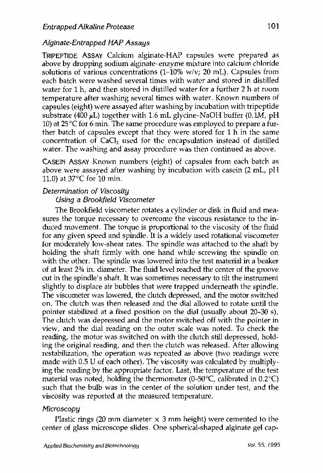
Alginate.Entrapped HAP Assays
TRIPEPTIDE ASSAY Calcium alginate-HAP capsules were prepared as above by dropping sodium alginate--enzyme mixture into calcium chloride solutions of various concentrations (1--10% w/v; 20 mL). Capsules from each batch were washed several times with water and stored in distilled water for I h, and then stored in distilled water for a further 2 h at room temperature after washing several times with water. Known numbers of capsules (eight) were assayed after washing by incubation with tripeptide substrate (400 #L) together with 1.6 mL glycine-NaOH buffer (0.1M, pH 10) at 25~ for 6 rain. The same procedure was employed to prepare a fur- ther batch of capsules except that they were stored for 1 h in the same concentration of CaC12 used for the encapsulation instead of distilled water. The washing and assay procedure was then continued as above.
CASEIN ASSAY Known numbers (eight) of capsules from each batch as above were assayed after washing by incubation with casein (2 mL, pH 11.0) at 37~ for 10 rain.
Determination of Viscosity Using a Brookfield Viscometer The Brookfield viscometer rotates a cylinder or disk in fluid and mea-
sures the torque necessary to overcome the viscous resistance to the in- duced movement. The torque is proportional to the viscosity of the fluid for any given speed and spindle. It is a widely used rotational viscometer for moderately low-shear rates. The spindle was attached to the shaft by holding the shaft firmly with one hand while screwing the spindle on with the other. The spindle was lowered into the test material in a beaker of at least 23/4 in. diameter. The fluid level reached the center of the groove cut in the spindle's shaft. It was sometimes necessary to tilt the instrument slightly to displace air bubbles that were trapped underneath the spindle. The viscometer was lowered, the clutch depressed, and the motor switched on. The clutch was then released and the dial allowed to rotate until the pointer stabilized at a fixed position on the dial (usually about 20-30 s). The clutch was depressed and the motor switched off with the pointer in view, and the dial reading on the outer scale was noted. To check the reading, the motor was switched on with the clutch still depressed, hold- ing the original reading, and then the clutch was released. After allowing restabilization, the operation was repeated as above (two readings were made with 0.5 U of each other). The viscosity was calculated by multiply- ing the reading by the appropriate factor. Last, the temperature of the test material was noted, holding the thermometer (0-50~ calibrated in 0.2~ such that the bulb was in the center of the solution under test, and the viscosity was reported at the measured temperature.
Microscopy Plastic rings (20 mm diameter x 3 mm height) were cemented to the
center of glass microscope slides. One spherical-shaped alginate gel cap-
Applied Biochemistry and Biotechnology Vol. 55, 1995

102 Roig, Rashid, and Kennedy
sule (2-3 mm diameter) was placed in the ring and completely submerged in distilled water, buffer, or substrate. The alginate capsule was then ex- amined with transmitted light under bright-field and polarized light micro- scopy using a low-power microscope objective (xl or x2 and x4 magnifica- tion). The capsules were then photographed with a Leitz automatic camera system on a Leitz Orthoplan microscope.
Protein ,Staining of HAP-Alginate Capsules STAINING Coomassie Brilliant Blue R-250 (0.1 g) was dissolved in 50 mL of ethanol and filtered (to remove insoluble material that can stick to the sur- face of the gels and is then difficult to remove). Then, 50 mL of 25% w/v acetic acid were added to the resulting solution. Both capsules with and without enzyme were stained overnight with shaking.
DESTAINING The capsules were drained and rinsed with water and drained before each destain step. The capsules were also shaken for 1--2 h in each destain:
1. First destain: 40 mL ethanol + 60 mL 5% w/v acetic acid; 2. Second and third destain: 30 mL ethanol + 70 mL 5% w/v
acetic acid; 3. Fourth destain: 20 mL ethanol + 80 mL 5% w/v acetic acid.
The microphotograph of capsules was taken after they were sufficiently destained and left in water for 1 h.
RESULTS AND DISCUSSION
Interaction of PHMBH+CI- and SDS with HAP PB92
Several studies on the binding of the detergents to protein have shown that the nature of the binding varies according to the amount of detergent bound. Putnam and Neurath (32) found that two electrophoretically stable complexes could be formed between horse serum albumin and SDS, con- taining 55 and 110 SDS mol/mol of protein. Hunter and McDuffie (33) found that SDS binds to human serum albumin in the molar ratios 63-67/1, and that reduction of S--S groups of the protein increased the binding to 92-94/1. Reynolds et al. (34) showed that native bovine serum albumin possesses ten primary binding sites for SDS and that the protein is only grossly disorganized when the SDS/protein molar ratio is increased above 10. SDS is an anionic surfactant and complexes with both hydrophobic and positive charge regions of the protein, e.g., tryptophan residues, a hydrophobic tail (possibly more than eight carbons in an alkyl chain), and the negative charge of the detergent is required for the formation of the complex. Imoto et al. (35) showed that SDS forms a stable complex with lysozyme without causing conformational changes in the enzyme mole- cule. The above reference indicated that sites for the interaction were the
Applied Biochemistry and Biotechnology Vol. 55, 1995

Entrapped Alkaline Protease
Table 1 Effect of SDS and PHMBH § C1- on HAP PB92 Activity
103
SDS, Activity, PHMBH § CI-, Activity, SDS/PHMBH + CI-, Activity, % w/v % % w/v % % w/v/% w/v %
0 100 0 97 0 97 1 37 0.4 100 10/4 100 2 25 0.8 100 5/2 96 3 18 1.2 97 2.5/1 84 4 13 1.6 96 1/0.5 80 7 12 2 96 10/4 a 80
10 8 4 91
aThe molar ratio in this case was 10.4 instead of 15.6.
hydrophobic regions, especially the appropriate pocket-like active site cleft, and positive charges of the protein side, and that the hydrophobic moiety and negative charge of the SDS side were required for the forma- tion of complex.
In this work, aliquots (100 #L) of HAP (50 #L concentrated enzyme diluted with 1 mL of 1--10% w/v SDS) were digested with casein (2 mL, 2--5% w/v, pH 11), and the mixtures incubated at 37~ for 10 min. Tri- chloroacetic acid (6.5% w/v, 2 mL) was then added to each mixture to stop the reaction. The mixture was stirred and centrifuged for 5 min. Ali- quots (400 #L) of the supernatant were then added to NaOH (0.5M, 1000 #L) together with Folin's reagent (50/~L). The samples were allowed to stand at 20-25~ for 10 rain, and absorbance was measured at 660 nm. Results were always the average of two completely separate determina- tions. HAP is progressively inhibited by increasing concentrations of SDS (Table 1).
Since SDS appears to react with HAP, possibly by denaturation, an investigation was made of protective agents against the effect of SDS on protein. Since PHMBH § C1- gives a complex with SDS (36), this property of PHMBH+ C1- was exploited as a potential protective agent. The pro- tective effects of PMBH § against SDS denaturation of HAP were in- vestigated using the casein assay at pH 11 and 37~ In this sense, a similar procedure was applied to 100 #L of enzyme (50 #L concentrated enzyme diluted with 1 mL of PHMBH § C1- [0.4--4% w/v] digested with casein [2 mL, 2.5% w/v, pH 11] at 37~ and 100 #L of enzyme [50 #L concentrated enzyme diluted with I mL of 1--10% w/v SDS to 0.4--4% w/v PHMBH§ C1- aqueous solution, molar ratio [15.6], digested with casein [2 mL, pH 11] at 37~ The results indicate that SDS inhibited the enzyme activity, but PHMBH § C1- slightly activated the enzyme, this being most prominent at 0.4% w/v concentration (Table 1).
Additional evidence for this was found in the literature. Adam and Barrat (37) used o~-D-glucose polymers containing the 1-4-linkages in a
Applied Biochemistry and Biotechnology Vol. 55, 1995

104 Roig, Rashid, and Kennedy
protective system for the retention of oL-amylase activity in detergent com- positions. The preferred structure was a branched chain starch dextrin. Alginate (0.1--7%) has been used (38) in the purification of amylase and catalase with organic solvents. Binders, such as alginate gum arabic and guar gum, at 0.01--4% have been used during the preparation of bacterial protease granules for incorporation in detergent powders and have been claimed to stabilize the enzyme (39). Treatment of a bovine hepatocatalase with either sodium chondroitin sulfate or tamarind seed polysaccharide improved the stability of the enzyme by approx 70% (40). Soluble starch and alginic acid were also reported to be effective. Dhariwal (29) reported the effect of amino acid alginate, gum acacia, and PHMBH§ on the activity of HAP PB92 and found that the above-mentioned compounds have a stabilizing effect on the enzyme. It was also found that polyhydric compounds, such as sugars and glycerol, are very effective stabilizing agents of protein conformation and enzyme activity (41-43). Moreover, diamine or polyamines have also been claimed to stabilize solutions of protease or amylases (44).
Effect of Temperature Aliquots (50 #L) of HAP (25 #L of concentrated enzyme diluted with
1 mL of various percentages of both [1--10% {w/v} SDS and 0.4--4.4% { w/v} PHMBH+CI -, molar ratio 15.6 at pH 9.8]) were digested with casein (2 mL, pH 11) at 37~ for 10 min by the same method as before. A similar procedure was applied to 50/~L enzyme--SDS--PHMBH+C1 - mixture after heating the mixture at 50~ for 0.5 and 2 h. A significant decrease in activity was shown by heating for 0--0.5 h and no effect between 0.5 and 2.0 h. One important effect was observed when 10% w/v SDS was mixed with 4% w/v PHMBH+ Cl- molar ratio 15.6; the mixture was viscous and the enzyme was active at 37~ when left at 20~ but not at 50~ (Table 2). Increasing the concentration of PHMBH § decreased the activity (Table 1). However, decreased amounts of PHMBH+C1 - at SDS molar ratio 15.6 mixture also decreased the activity of the enzyme (Table 2).
Obviously, PHMBH+C1 - provides the enzyme with protection by interacting with the SDS molecule. However, at high temperature, this interaction could be avoided. On the other hand, PHMBH § C1- on its own caused the activity of the enzyme to increase slightly at the 0.4% w/v level or perhaps less. This may be owing to some inhibitor being removed from the crude enzyme preparation. Furthermore, the above increase in enzyme activity by PHMBH+ C1- may also be attributed to the possibility of extra crosslinking via carboxylate (--COO--) groups. Existing crosslinks are usually the result of disulfide bridges, and these certainly protect the ter- tiary structure required for enzyme activity. In comparison, Dhariwal (29) also found that the activity of HAP increases sharply with both PHMBH+ C1- (0.25% w/v) and SDS:PHMBH+C1 - ratio 138:1.
Applied Biochemistry and Biotechnology Vol. 55, 1995

Entrapped Alkaline Protease
Table 2 Effect of Temperature on HAP PB92 Activity in the Presence
of SDS, SDS/Sodiurn Alginate, and SDS/PHMBH+C1 -
105
SDS, SDS/Na + alginate, SDS/PHMBH+C1 - , Before % w/v % w/v/% w/v % w/v/% w/v heating
Activity, %
Heating at 50~ for 0.5 h
0 0.6 1.5 3 6
0/1 0.6/1 1.5/1
3/1 6/1
0/0 10/4 a 5/2 a
2.511 a 110.5 a
100 90.5 17.3 15.0 14.6 14.4 13.1 12.2 12.2 11.2
100 77.0 72.4 13.6 39.0 13.5 28.0 13.0 24.0 12.8 97 90
100 18.5 95.7 14.7 84 13 69 12.3
aMolar ratio was 15.6 and pH 9.8.
Apparently both stabilizing and protective effects of alginate and PHMBH+ C1- are the result of ionic interactions. The enzyme at pH 11 has negatively charged side chains, e.g., aspartic acid and glutamic acid and also has positively charged side chains, e.g., lysine and possibly arginine. PHMBH+C1 - would interact with the negative charges of the enzyme. Undoubtedly, the PHMBH+C1 - ions interact with SDS molecules, and thus hinder their interaction with the enzyme. However, sodium alginates are negatively charged polymers that are also capable of interacting with the positively charged side chain of the enzyme.
In conclusion, the above results in this section showed that the anionic detergent (SDS) inhibited HAP activity, whereas a biocide (PHMBH § C1-) activated the enzyme at a concentration of 0.4--0.8% (w/v). PHMBH § C1- can also provide the enzyme with protection by interacting with SDS molecules. However, the most interesting effect was observed when 10% w/v SDS was mixed with 4% w/v PHMBH+C1 - in a molar ratio of 15.6:1. The mixture became viscous and the enzyme was not only totally pro- tected, but exhibited greater activity compared with the activity of the enzyme in buffer alone. Furthermore, it was found that alginate protected the protease against SDS denaturation by approx 55% at 20~ but failed to do so at 50~ Alginate alone also had a stabilizing effect on the enzyme.
Applied Biochemistry and Biotechnology Vol. 55, 1995

106 Roig, Rashid, and Kennedy
Interaction of Alginate and SD8 with HAP PB92
Sodium alginate was tested for its ability to protect and stabilize the alkaline protease against the denaturation effect of SDS. Aliquots (50 ~L) of HAP (25 #L of concentrated enzyme diluted with I mL of SDS of various concentrations [0.6, 1.5, 3, and 6% w/v in water at pH 11]) were digested with casein (2 mL, pH 11) at 37~ for 10 rain. As a control, 50 #L of HAP (25/~L of concentrated enzyme diluted with 1 mL of water, pH 11) was also digested with casein (2 mL, pH 11). A similar procedure was applied to 50/~L of HAP (25/~L of concentrated enzyme diluted with 1 mL of SDS at various concentrations [0.6, 1.5, 3, and 6% w/v in 1% w/v sodium alginate at pH 11]) and a control (50 #L) of HAP (25/~L of concentrated enzyme diluted with 1 mL of 1% w/v sodium alginate at pH 11). These samples were left at 50~ for 0.5 and 2 h and also assayed with casein (Table 2). It was found that sodium alginate containing SDS at pH 11 restored the HAP activity compared with enzyme containing SDS alone. In fact, sodium alginate containing 0.6% w/v SDS restored HAP activity by approx 55% of the original, but failed to do so at 50~ Alginate itself showed stabilizing effects on enzyme activity even after it had been stored for 2 h at 50~
The possible explanation of the above effect is that the alginate residue provides the enzyme with protection by interacting with the surfactant molecule at 20--25~ This may prevent the hydrophobic component of the detergent from entering into the internal hydrophobic structure of proteins. However at high temperature, this effect is very reduced. On the other hand, it has been proposed (45,46) that the stabilization of en- zyme by glycerol might be explained in terms of the formation of a water-- glycerol structure around the protein molecules. Furthermore, Gerlsma (47,48) and Gerlsma and Stuur (49, 50) have suggested that the stabilizing effect of polyhydric alcohols is the result of a decrease in the hydrogen- bond-rupturing capacity of the medium. Since alginate interacts favorably with water by entering into the water lattice and strengthening solvent structure, its presence in the aqueous medium could increase the hydro- phobicity of the protein; this would increase the thermodynamically unfavorable situation and require the use of more energy for unfolding than in water. As a result, the presence of alginate should tend to favor the more folded or native state.
Behavior of HAP Entrapped in Manugel DMB in the Form of Calcium Alginate Gel Capsules
Calcium alginate has been used extensively as a carrier for the immo- bilization of enzymes and cells (51). It has also been used as a new method for coimmobilization of microorganism with enzyme (52). Thus, the cur- rently popular use of calcium alginate gels for the immobilization of bio- logical material was applied to HAP in the present work. One important
Applied Biochemistry and Biotechnology Vol. 55, 1995
Entrapped Alkaline Protease 107
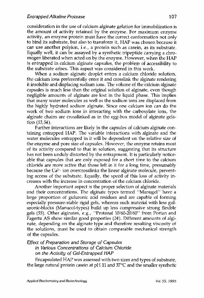
consideration in the use of calcium alginate gelation for immobilization is the amount of activity retained by the enzyme. For maximum enzyme activity, an enzyme protein must have the correct conformation not only to bind its substrate, but also to transform it. HAP was chosen because it can use another polyion, i.e., a protein such as casein, as its substrate. Equally well, it can be assayed by a synthetic tripeptide carrying a chro- mogen liberated when acted on by the enzyme. However, when the HAP is entrapped in calcium alginate capsules, the problem of accessibility to the substrate arises. This aspect was considered in this work.
When a sodium alginate droplet enters a calcium chloride solution, the calcium ions preferentially enter it and crosslink the alginate rendering it insoluble and displacing sodium ions. The volume of the calcium alginate capsules is much less than the original solution of alginate, even though negligible amounts of alginate are lost in the liquid phase. This implies that many water molecules as well as the sodium ions are displaced from the highly hydrated sodium alginate. Since one calcium ion can do the work of two sodium ions in interacting with the carboxylate ions, the alginate chains are crosslinked as in the egg-box model of alginate gela- tion (53,54).
Further interactions are likely in the capsules of calcium alginate con- taining entrapped HAP. The variable interactions with alginate and the water molecules entrapped in it will be dependent on the relative size of the enzyme and pore size of capsules. However, the enzyme retains most of its activity compared to that in solution, suggesting that its structure has not been unduly distorted by the entrapment. It is particularly notice- able that capsules that are only exposed for a short time to the calcium chloride are more active that those left in it for a long time, presumably because the Ca 2+ ion overcrosslinks the linear alginate molecule, prevent- ing access of the substrate. Equally, the speed of this loss of activity in- creases with the increase in concentration of the calcium chloride.
Another important aspect is the proper selection of alginate materials and their concentrations. The alginate types termed "Manugel" have a large proportion of guluronic acid residues and are capable of forming especially pressure-stable rigid gels, whereas such material with less gul- uronic-blocks (Manucol-types) build up less compressive strong flexible gels (55). Other alginates, e.g., "Protanal 10/60-20/60" from Portan and Fagertu AS show similar good properties (24). Different amounts of algi- nate, depending on the alginate type and therefore resulting viscosity of the solutions, must be used to obtain comparable mechanical strength of the capsules.
Effect of Preparation and Storage of Capsules in Various Concentrations of Calcium Chloride on the Activity of Gel-Entrapped HAP Encapsulated HAP was assessed with two sizes and types of substrate,
the large natural protein casein at pH 11 and 37~ and the smaller synthetic
Applied Biochemistry and Biotechnology Vol. 55, 1995

108 Roig, Rashid, and Kennedy
chromogenic tripeptide at pH 10 and 25 ~ which was found to be a sub- strate for the enzyme. Before assay, capsules were stored for 3 h in water to remove loosely bound enzyme. Capsules were prepared (50) in CaC12 (2% w/v, 10 mL) at various pHs (5, 7, 9, and 11). Capsules from each batch were washed several times and stored in water for 1 h at 20-25 ~ The same capsules from each batch were stored in water for a further 2 h after washing several times with distilled water at room temperature. Known numbers (eight) of capsules from each batch were assayed after washing by incubation with both tripeptides (400 #L) together with 1.6 mL glycine- NaOH buffer (0.1M, pH 10) at 25~ for 6 min, and the absorbance was measured at 400 nm. In assays using 2 mL casein, pH 11, at 37~ for 10 min, absorbance was measured at 660 nm. The same procedure was applied to capsules from each batch made in 2% w/v calcium chloride at various pHs (5, 7, 9, and 11) kept in the same pH of 2% CaC12 for i h, and then stored for a further 2 h in water after washing several times with distilled water. Known numbers (eight) of capsules from each batch were assayed after washing by incubation with both tripeptide substrate and casein.
As shown in Fig. 1, with a given amount of enzyme available for en- trapment, the use of calcium chloride at increasing concentrations (1-10% w/v) to form capsules with 0.63% w/v Manugel DMB caused correspond- ing progressive decreases in the amount of enzyme activity as assayed by both casein and tripeptide substrates. The effect may be the result of the rapidity of and increasing level of crosslinking by the Ca 2§ ions at increas- ing concentrations, preventing access to the substrate. However, after standing the capsules for I h in the same concentration of calcium chloride as the one in which they were prepared, capsules of fairly constant, but low enzyme activity, irrespective of the calcium chloride concentration (1-10% w/v), were produced (Fig. 1). Low calcium chloride concentrations are favored for highest useable enzyme activity. Probably this is because of more accessibility of the enzyme to both of the substrates. The above results suggest that the substrate can diffuse into calcium-alginate-HAP capsules. The diffusion of these substrates is altered by increasing the concentration of calcium chloride used in the gel preparation (Fig. 1). The decrease of enzyme activity may be the result of "crosslinkage" of calcium ions with alginate, which is caused by the reduction of pore sizes.
As mentioned above, when calcium alginate-entrapped enzymes cap- sules were stored for I h in calcium chloride solution, a constant but lower immobilized enzyme activity was observed than when the just-formed capsules were stored in water without any further contact with the calcium chloride solution. This is because in calcium chloride solutions, the CaC12 diffuses inside the matrix and a highly ionic crosslinked calcium alginate preparation is obtained. This increases the internal diffusional resistances to the substrate. With the recently formed calcium alginate capsule, the ionic crosslinking is only present on the surface of the capsules. Thus, in- side the capsules (i.e., in the microenvironment of the enzyme), there is
Applied Biochemistry and Biotechnology Vol. 55, 1995

Entrapped Alkaline Protease 109
",~ 2o
A
~ 100
20
100
I 1 l I l I I l l I
f 1 T 1 1 I F r I , , T 1 5 i0
[CaCI2]/% w/v
Fig. 1. Effect of making and storing capsule in various concentrations of calcium chloride on the activity of gel-entrapped HAP with (A) casein at pH 11.0 and 37.0~ and (B) tripeptide substrate at pH 10.0 and 25.0~ Activity at 1% w/v CaC12 is taken as 100%. (&, •) Storage in water for I h and for a further 2 h. (A, I ) Storage in CaC12 for 1 h and water for a further 2 h.
no diffusional resistance, and so enzyme activity is higher. However, in terms of stability, it was observed that the highly ionic crosslinked matrix led to more stable preparations. This arises from the same internal diffu- sional resistances, which do not allow the enzyme to be released into the substrate solution for each activity determination.
In comparison, the diffusion characteristics of several substrates of varying molecular sizes into and from calcium alginate gel beads in well- stirred solutions have been investigated (56). It was observed that the dif- fusion of high-mol-wt substrates showed similar characteristics to those observed in the present study. Furthermore, it has been reported (23) that polymannuronic alginate gels possess good porosity characteristics, whereas polyguluronic acid alginates form gels with lower porosity. Manugel DJ and Manugel DMB, both form satisfactory gels, but with slower diffusion characteristics for higher-mol-wt compounds than Man- ucol LH (23). The preparation batch used in this work for the solidifica- tion of the Manugel DMB-HAP solution droplets was usually 2% w/v
Applied Biochemistry and Biotechnology VoL .55, 1995

1 10 Roig, Rashid, and Kennedy
100
6O
"~ 2C im
O
(~) 100
J ~
Pc' 6o
2(:
1 1 1 I
5.0 7.0 9.0 11.0
pH of CaCI 2 solut ion
Fig. 2. The effect of making and storing encapsulated HAP at various pHs of calcium chloride. Assay (A) casein substrate at pH 11.0 and 37.0~ and (B) tri- peptide substrate at pH 10.0 and 25.0~ The activity from 2% CaC12 at pH 5.0 is taken as 100%. (I-7, &) Storage in water for I h and for a further 2 h. (B, &) Stor- age in CaC12 for 1 h and water for a further 2 h.
calcium chloride solution. It was noticeable that the resulting encap- sulated enzyme alginate was freely accessible to both casein and tripep- tide substrates. However, a concentration of the calcium chloride solution as low as 0.05% w/v can be used (24). Also, other cations, such as AP § Fe B+, and so on (57) can be used for alginate gelation, as has been shown at pilot plant scale (58).
Clearly, the pH of calcium chloride at which the alginate capsules containing HAP are made is important. A pH of 5 is preferred for capsules stored in water for 3 h, and pHs 5 and 9 are preferred for capsules stored in calcium chloride for i h and water for a further 2 h, using either casein or tripeptide substrate (Fig. 2). The increase in enzyme activity may be the result of more accessibility of the substrate to the capsules from the surrounding solution under these conditions.
Applied Biochemistry and Biotechnology Vol. :55, 1995

Entrapped Alkaline Protease 1 11
Effect of pH on the Activity of HAP.Alginate Capsules The pH profile was constructed by measuring the activity of both the
soluble and encapsulated HAP at various pH values ranging from 7.5 to 11.5. The buffering system used was 0.1M glycine-NaOH with and with- out phosphate buffer (0.1M), and the assay was conducted with either the tripeptide substrate or denatured casein. The activities at various pH values with both substrates were measured.
It was found that the pH profile of the soluble enzyme-alginate control mixture with tripeptide substrate in glycine-NaOH buffer alone showed an optimum activity at pH ranges from 9 to 10. In comparison, tripeptide substrate in glycine-NaOH-phosphate mixed buffer showed an increased level of activity at pH ranges from 7.5 to 8.5 with greatest increase of activ- ity at pH 10.5 (see Fig. 3). However, the pH profile of the encapsulated enzyme with tripeptide substrate in glycine-NaOH buffer alone showed a clear optimum at pH ranges from 10 to 11. In the presence of a combina- tion of phosphate buffer and glycine-NaOH buffer, maximum activity was observed at pH 10.5. Further, increased activity was observed at almost all ranges of pH compared with that of the glycine-NaOH buffer alone (see Fig. 3). This was in marked contrast to the pH profile of the soluble high HAP-alginate control mixture with casein in glycine-NaOH buffer with and without phosphate buffer (see Fig. 3). Here, the small shoulder at pH 9 was accompanied by a very strong peak at pH 11 using casein in glycine-NaOH buffer alone. Casein in glycine-NaOH-phosphate mixed buffer showed an increase of activity at ranges of pH from 8 to 11.5 with a strong optimum at pH 11 (see Fig. 3). However, encapsulated enzymes produced a somewhat different pH activity profile, as can be seen from Fig. 3. Casein in glycine-NaOH buffer showed a clear peak at pH 9 and was accompanied by another strong peak at pH 11. Again, the addition of phosphate buffer increased the activity at all ranges of pH from 7.5 to 11.5 with high activity at pH 11.
The overall effect was in some cases that two pH optima were en- countered (e.g., both capsules and control liquid mixture with glycine- NaOH-phosphate mixed buffer and casein, at pHs 9 and 11). However, with tripeptide substrate in glycine-NaOH-phosphate mixed buffer, the capsules showed only one pH optima at pH 10.5, higher than the equiva- lent liquid enzyme alginate mixture pH (9-10) without phosphate, but the same with it. The important point is that with both substrates the enzyme shows good activity from pH 8.5 to 11. However, the addition of phos- phate buffer to glycine-NaOH buffer showed an increased level of activity at almost all pH ranges from 7.5 to 11, increasing at higher pH. Phosphate was found to cause breakdown of the capsules using either substrate (see below). The effect of phosphate buffer may be due to chelation of phos- phate with calcium. Here, the pH at which calcium chelation occurs is
Applied Biochemistry and Biotechnology Vol. 55, 1995

1 12 Roig, Rashid, and Kennedy
160
100
>, 40
C
i m
> . m
0 0
G) . i
,4 , , . . I
(~ 12(~ 0)
r r
Fig. 3.
60
A
2C
d / 60
d
B 12(
6(]
8.0 9.0 10.0 11.0
D
pH The effect of pH on the activity of HAP entrapped in capsules of
calcium alginate gel (A, C) and in free solution of sodium alginate (B, D), using tripeptide substrate (A, B) and casein (C, D) for assay. The activity in glycine- NaOH buffer at pH 10.0 (A, B) and 11.0 (C, D) is takejn as 100%. (r~, �9 glycine- NaOH buffer. (A, e)(glycine-NaOH) + phosphate buffer.
most important, and the effectiveness of the phosphate ions will vary greatly over the pH ranges.
The effect of phosphate on enzyme activity was further studied using capsules made from 1% w/v sodium alginate in phosphate buffer, pH 6.6, of various concentrations (0.05-0.2M) ejected into 2% w/v calcium chloride (2% w/v, pH 9). The results from Table 3 show that when capsules were assayed immediately after washing, the maximum activity was obtained with the control alginate in water, and no difference was observed if the
Applied Biochemistry and Biotechnology Vol. 55, 1995

Entrapped Alkaline Protease
Table 3 The Effect of Phosphate Buffer (pH 6.6)
on HAP Activity During Encapsulation in Alginate and on Storage
113
Medium
Relative activity, %
During encapsulation Capsule storage
Oh 2h Oh 2h
Water 100 48 100 55 0.05M phosphate 92 77 0.1M phosphate 95 67 100 41 0.2M phosphate 76 73 Enzyme/alginate solution a 100 90
a In 0.1M phosphate buffer, pH 6.6. The encapsulation was made by dropping the enzyme solution into CaC12 2% (w/v) pH 9.0.
alginate was in 0.05 or 0.1M phosphate buffer. However, in 0.2M phos- phate buffer, the resulting activity was lower. Despite this, the preferred level of activity was obtained with 0.05M phosphate when these capsules were stored in water for 2 h to remove unbound enzyme and the three concentrations of phosphate used were above of alginate in water (see Table 3). Probably, the presence of phosphate resulted in fewer cross- linkages between Ca 2§ ion and alginate molecules and, consequently, in producing higher capsule porosity, which increases the accessibility of the substrate to entrapped enzyme molecules.
Moreover, with a further batch of capsules using alginate in 0.1M phosphate buffer, pH 6.6, the capsules were found to redissolve in phos- phate buffer. The rate of solubility was dependent on the pH of the phos- phate buffer. At basic pH (11), the effect was immediate, and at neutral pH (7) it took 5 rain. However, capsules lasted for 2 h in phosphate buffer at slightly acidic pH 5. The control in water was still intact after 24 h, and the enzyme activity was greater compared to that of the capsules stored in phosphate buffer at pH 5. The solubilization of capsules in phosphate buffer is probably the result of the formation of mono-calcium phosphate, hence removing the crosslinking agent. Higher enzyme activity with phosphate buffer in some cases occurs possibly because of the fact that calcium ions undoubtedly interact with phosphate ions and, thus, hinder its interaction with the enzyme.
Storage Stability of Immobilized HAP Surprisingly, preservation is quite good in water alone after the less
effectively entrapped HAP elutes out (half-life tl/2 at 4, 25, and 50 ~ were 11.3, 6.8, and 4.0 d, respectively). Conversely, in calcium chloride solu- tion, low activity is monitored, presumably because the Ca 2+ ions "over- crosslink" the linear alginate molecule, preventing access of the large
Applied Biochemistry and Biotechnology Vol. 55, 1995

1 14 Roig, Rashid, and Kennedy
substrate. Subsequent immersion in sodium chloride solution does not affect enzyme recovery. Two and 10% w/v calcium alginate solutions surrounding the HAP are quite effective in stabilizing the enzyme at 25 ~ (t,/2 = 48.0 h; 45.0 h) and even more so at 4~ (G = 51.7 h; 54.1 h). In addi- tion, the stability at 50~ is quite promising in water (see above), whereas in 2 and 10% w/v calcium alginate is decreased (G = 8.8 h; 7.8 h). Appar- ently, calcium can maintain the conformational integrity of the neutral protease as a group and the B. subtilis enzyme in particular (59). This could be regarded as another reason for calcium alginate gel to be used as a suit- able method of enzyme stabilization.
Microscopic Studies of HAP-Alginate Capsules Microscopic examinations of calcium alginate capsules with and with-
out enzyme were carried out under both bright-field and polarized micro- scopy. The effects of various buffers at various pHs and substrates were also investigated.
Microscopic Tests Except for the presence of a single small protrusion, HAP-Manugel
DMB (alginate) capsules were virtually spherical-shaped structures that measured 2-3 mm in diameter. Under bright-field transmitted light optical microscopy, capsules without enzyme were translucent and amorphous in appearance, whereas a small brown area was evident in the center of the capsules containing the enzyme (HAP). This brown central region was not evident in the enzyme-containing capsules made from freshly prepared Manugel DMB, whereas it was present in those capsules with enzyme made from 24-h-old ("aged") Manugel DMB, and was visible to the unaided eye. A definitive blue coloration was evident mainly in this central region of the enzyme-containing capsules, which had been stained with the protein stain Coomassie Brilliant Blue R-250, and was also visible to the unaided eye. When examined between crossed polars (polarization microscopy), both the capsules with and without enzyme manifested the polarization (maltese) cross, which is characteristic of the structures pos- sessing spherulytic symmetry, e.g., starch granules (60), and bordered pits in plant cell walls. Figures 4-7 summarize the effects of various chem- ical treatments on the optical properties of the control (enzyme-less) and enzyme-containing capsules.
Diagrams (i)-(iii) in Fig. 4 represent three different structural config- urations that will manifest the maltese cross when viewed between crossed polars. The two enlarged photomicrographs (Fig. 5C and 6C) reveal the presence of radial striations similar to that represented diagrammatically in Fig. 4 (iii), thus pointing to this structural configuration as being respon- sible for the maltese cross manifested by these capsules with and wihout enzyme when examined between crossed polars. A spherical-shaped iso- tropic object, such as that represented by diagram Fig. 4 (iv), can at times
Applied Biochemistry and Biotechnology Vol. 55, 1995

Entrapped Alkaline Protease 115
Spiral Concentr ic R o s e t t e Isotropic sphere sphere sphere sphere
i ii i i i iv Fig. 4. Various structural configurations of calcium alginate capsules, with
and without enzyme, which are responsible for the maltese cross when examined under polarized light.
Fig. 5. Photomicrograph of calcium alginate capsules when viewed with plain light and green filter after different treatments. (A) Stored in water for 24 h; (B) stained with Coomassie Blue; (C) viewed with high numerical aperture objective, see the presence of radial striation; (D) in glycine--NaOH buffer, pH 7.5, 10.0, and 11.0; (E) with tripeptide substrate in glycine-NaOH buffer, pH 10.0; (F) with casein in glycine-NaOH, pH 11.0; notice a formation of outer layer.
Applied Biochemistry and Biotechnology Vol. 55, 1995

1 16 Roig, Rashid, and Kennedy
Fig. 6. Photomicrograph of HAP-Manugel capsules when viewed with plain light and green filter after different treatments. (A) Stored in water for 24 h; (B) stained with Coomassie Blue; (C) capsule A viewed with high numerical aperture objective; notice the presence of radial striation; (D) capsule A stored in water for 1 mo; notice the enzyme appeared to be scattered all over the capsule; (E) capsule A with tripeptide substrate in glycine--NaOH buffer, pH 10.0; (F) capsule A with casein in glycine-NaOH, pH 11.0; notice the formation of thin outer layer.
also manifest a maltese cross between crossed polars by virtue of the rota- tion of polarized light by the object, especially when viewed with high numerical aperture objectives. However, a low numerical aperture objec- tive and condenser were used for examining the capsules with and with- out enzyme, thus avoiding the undesirable optical phenomemon described above. In addition, the maltese cross was no longer visible between crossed polars in capsules that had been placed in carbonate--bicarbonate or phosphate buffer for brief periods, whereas the spherical outline of the
Applied Biochemistry and Biotechnology VoL 55, 1995

Entrapped Alkaline Protease 117
Fig. 7. Photomicrograph of HAP-Manugel capsules when viewed with polarized light, after different treatments. (A) Stored in water for 24 h; (B) stained with Coomassie Blue; (D) capsule A stored in water for I mo; (E) capsule A with tripeptide substrate in glycine-NaOH buffer, pH 10.0; notice how much weaker the polarization cross is; (F) capsule A with casein in glycine--NaOH, pH 11.0; notice the polarization cross for enzyme-alginate capsules bounded by polariza- tion cross of opposite sign for casein outer layer.
capsules was still clearly evident. This indicates that the alginate capsules with and without enzyme are highly ordered anisotropic entities, and that the maltese cross manifested by these spherical-shaped entities is not a result of depolarization phenomenon associated with an isotropic, spherical-shaped body.
Chemical Interpretation Microscopic observation shows HAP entrapped within the Manugel
capsule under bright field (Fig. 6). There appears to be a concentrated
Applied Biochemistry and Biotechnology Vol. 55, 1995

118 Roig, Rashid, and Kennedy
brown spot in the middle of the capsule as well as numerous tiny spots that are not present in the enzyme-less capsules. The absence of a central brown region in the enzyme-containing capsules made from freshly pre- pared alginate solution is possibly the result of the fact that freshly prepared alginate solution is not as fully hydrated or oriented as similar "aged" alginate solutions. A definite blue coloration was evident in the central region of the enzyme-containing capsules when stained with Coomassie Blue. However, the amount of enzyme around the surface of the capsules is insufficient to give a dark blue coloration in the capsule containing en- zyme (Fig. 6B). Compared with the light purple color observed in enzyme- less capsules (Fig. 5B), it is therefore assumed that the dark spot is the result of the presence of enzyme (protein) molecules, with the majority being concentrated in the middle of the capsules.
As shown in Fig. 6E, capsules containing enzyme treated with tripep- tide substrate underwent a change in appearance under bright and espec- ially under polarized light microscopy (Fig. 7E). However, the addition of the same substrate to the control did not have much effect on the appear- ance of the capsule.
The disorientation of the structure of the capsule containing enzyme with tripeptide substrate may be attributed to the fact that the enzyme molecules would interact with the tripeptide substrate that diffused from the surrounding solution into the enzyme-alginate capsule via an inter- mediate enzyme-substrate complex. The products tripeptide and 4-nitro- aniline then diffuse out from the center so that the yellow color of the 4-nitroaniline is seen diffusing out.
The addition of capsules with and without enzyme to casein brought about slight changes in the appearance of the capsule under bright field and especially under polarized light microscopy (Fig. 7F). The high-mol-wt substrate casein appears to be adsorbed onto the surface of the capsules with and without enzyme, forming an enveloping layer around the cal- cium alginate capsule. The casein layer is thick around enzyme-less cap- sules and thinner around the enzyme-containing capsules. It is possible that a thinner casein envelope might result in the enzyme-containing cap- sule from partial digestion by the enzyme, besides being adsorbed by calcium alginate. In the enzyme-less capsule, this is not possible. Calcium must be thought of as interacting not only with alginate, but also with a certain group in the (protein) enzyme.
Glycine-NaOH buffer, pH 10 or 11, or 10 min brought about changes in appearance of the enzyme-containing capsules under bright-field and especially under polarized light microscopy without affecting the control enzyme-less capsules stained with Coomassie Blue. The original structure is irrecoverable by supporting them in water for I h further. The ordered structure could be restored by holding them in 2% calcium chloride for 1 h further. Thus, calcium chloride is in its own right an agent that brings
Applied Biochemistry and Biotechnology Vol. 55, 1995

Entrapped Alkaline Protease 119
order to the capsule molecule, even if previous disorder has been induced by certain interactions.
If the capsules with and without enzyme are stored for 5 min either in carbonate-bicarbonate or phosphate buffer, pH 7.5, the birefringence of the capsule is completely lost. However, the spherical outline of the cap- sule is still clearly evident under bright-field microscopy. This may be owing to chelation of calcium with carbonate-bicarbonate or phosphate ions, which results in disorganization of the molecular structure initially responsible for optical anisotropy.
The previous disorder has also been observed in completely dried capsules. This shows the significance of water molecules in maintaining the ordered structure.
The storage of enzyme alginate capsules in water for I mo at 20-25 ~ caused a significant change in the position of enzyme, within the alginate capsules. Under bright-field microscopy, instead of being concentrated mainly in the center, the enzyme appeared to be scattered all over the capsule (Fig. 6D). The polarization cross was not appreciably affected (Fig. 7D).
It is well known that the structure and molecular weight of alginates are different according to the different kinds, ages, and parts of seaweeds used and to the different extraction processes of alginate. These results were obtained from only one type of alginate. Other types of alginate may give different results. It has been reported that alginate containing predominantly mannuronic units forms a gel with high apparent porosity, whereas polyguluronic alginate forms gels with lower porosity, especially with respect to high-mol-wt compounds (23). However, the method of investigation covered in this work should be useful for judging the capacity of calcium alginate gel as an immobilization matrix and for selecting the most effective system for immobilizing enzyme and whole cells.
CONCLUSIONS
Calcium alginate gels (Manugel DMB) were found to be a very good immobilizing system for high-alkaline protease and could be extended to other enzymes. Details of the effectiveness of immobilization as a function of alginate type should also be made available. Since the microscopic studies, especially those with polarized light, showed that organized structures occur in encapsulated alginate-HAP, a thorough investigation in this area is to be recommended, particularly in view of the observation that a similar organization has been reported with starch granules before gelation. There is opportunity for the development of an enzyme system that behaves as a pseudo-cell, with the added advantage that the enzyme could be released from the cell by the action of SDS:PHMBH+C1 - .
Applied Biochemistry and Biotechnology Vol. 55, 1995

120 Roig, Rashid, and Kennedy
REFERENCES
1. Nakanishi, J., Kita, Y., and Isono, M. (1974), Agr. Biol. Chem. 38(1), 37. 2. Vedder, A. (1934), Antonie Van Leeuwenhoek, vol. 1, 141. 3. Aunstrup, K., Anderson, O., and Oyttrup, H. (1968), British Patent No.
1,243, 784. 4. Horikoshi, K. and Ikeda, Y. (1977), US Patent No. 4,052,262. 5. Aunstrup, K. (1980), in Microbial Enzymes and Bioconversions, Rose, A. H.,
ed., Academic, New York, p. SO. 6. Morhihara, K. (1974), Adv. Enzym. 41. 7. Markland, F. S. and Smith, E. L. (1971), The Enzymes, vol 3, 3rd ed., Boyer,
P., ed., Academic, New York, London, p. 561. 8. Nijenhuis, B. (1977), US Patent No. 4,002,572. 9. Martinek, K., Mozhaev, V. V., and Berezin, I. V. (1980), in Enzyme Engineer-
ing, Lemuel, B., Wingard, Jr., and Berezin, I. V., ed., Plenum, New York and London.
10. Chapelle, E. W., Rich, E., and MacLeod, N. H. (1967), Science 155, 1287. 11. Horigome, T., Kasal, H., and Okuyama, T. (1974), J. Biochem. 75, 299. 12. Schreider, Z., Stroinski, A., and Pawelkiewicz, H. (1968), Bull. Acad. Polan.
Sci. Ser. Sci. Biol. 16, 203. 13. Norris, R. D. and Pawelkiewicz, H. (1975), Phytochemistry 14, 1701. 14. Goldmacher, V. S. (1977), Ph.D. Thesis, Lomonosov. State University,
Moscow. 15. Goldmacher, V. S., Klibanov, A. M., and Martinek, K., Vestnik, M. G. U.
(1978), Bull. Moscow University, 19(3). 16. Martinek, K., Goldmacher, V. S., Mishin, A., Torchinin, V. P., Smirnov,
V. N., and Brezin, I. V. (1978), Dokl. Akad. Nauk-SSSR 239, 227. 17. Klerstan, M. and Bucke, C. (1977), Biotechnol. Bioeng. 19, 387. 18. Paul, F. and Vignais, P. M. (1980), Enzyme Microb. Technol. 2, 281. 19. Gisby, P. E. and Hall, D. (1980), Nature 287, 251. 20. Brodelius, P. and Mosbach, K. (1982), Adv. Appl. Microbiol. 28, 1. 21. Voflop, K. D. and Klein, J. (1983), in New Developments in the Field of Cell
Immobilization in Formation of Biocatalysts by Ionotropic Gelation, 3rd Rotenburg Symposium-Enzyme Technology, Springer, New York, Wien.
22. Brodelius, P. (1985), in Immobilized Cells and Enzymes: A Practical Approach, Woodwards, J., ed., IRL Press, Oxford.
23. Kierstan, M., Darcy, G., and Reilly, J. (1982), Biotechnol. Bioeng. 24, 1507. 24. Klein, J., Stock, J., and Vorlop, K. D. (1983), J. Appl. Microbiol. Biotechnol. 18,
86. 25. Klein, J. and Washausen, P. (1979), Dechema. Monogr. 84, 277. 26. Cheetham, P. S. J. (1979), Enzyme Microb. Technol. 1, 183. 2Z Klein, J. and Eng. H. (1979), Dechema. Monogr. 84, 292. 28. Boukovalas, J. (1980), Ph.D. Thesis, University of Birmingham. 29. Dhariwal, M. S. (1983), Ph.D. Thesis, University of Birmingham. 30. Beck, E. A. (1977), in New Method for the Analysis of Chromogenic Substrates,
Witt, I., ed., De Gruyter, W., Berlin, New York. 31. Blake, N. (1983), Ph.D. Thesis, University of Birmingham. 32. Putnam, F. W. and Neurath, H. (1945), J. Biol. Chem. 159, 195.
Applied Biochemistry and Biotechnology Vol. 55, 1995

Entrapped Alkaline Protease 121
33. Hunter, M. J. and McDuffie, F. C. (1959), J. Am. Chem. Soc. 81, 1400. 34. Reynolds, J. A., Herbert, S., Polet, H., and Steinhardt, J. (1967), Biochemistry
6, 737. 35. Imoto, T., Sumi, Si., Tsuru, M., and Yagishita, K. (1979), Agric. Biol. Chem.
43(9), 1809. 36. Roig, M. G., Rashid, D. H., and Kennedy, J. F. (in press), Carb. Potym. 37. Adam, W. E. and Barrat, C. (1973), US Patent No. 3,773,674. 38. Miyaka, H. (1977), Japan Patent No. 7,048,49. 39. Knapsach, A. G. (1971), Ger. Often. 1958, 104. 40. Eisai, Co. Ltd. (1969), Japan Patent No. 8072. 41. Stauff, J. and Metrotra, K. N. (1961), Kolloid Z. 176, 1. 42. Timasheff, S. N., Lee, J. C., Plttz, E. P., and Tweedy, N. (1976), J. Colloid
Interfac. Sci. 55, 658. 43. Bull, H. B. and Breese, K. (1978), Biopolymers 17, 2121. 44. Schreider, W. (1972), German Patent No. 2,058,826. 45. Jarabak, J., Seeds, A. E., and Talalay, P. (1966), Biochemistry 5, 1269. 46. Ruwart, M. J. and Suelter, C. H. (1971), J. Biol. Chem. 246, 5990. 47. Gerlsma, S. Y. (1968), J. Biol. Chem. 243, 957. 48. Gerlsma, S. Y. (1970), Eur. J. Biochem. 14, 150. 49. Gerlsma, S. Y. and Stuur, E. R. (1972), Int. J. Peptide Protein Res. 4, 377. 50. Gerlsma, S. Y. and Sturr, E. R. (1974), Int. J. Peptide Protein Res. 6, 65. 51. Avrameas, S., Brown, G., Selegny, E., and Thomas, D. (1969), Ger. Often. 1,
915, 970. 52. Hartmeier, W. (1984), Process. Biochem. 40. 53. Grant, G. T., Morris, E. R., Rees, D. A., Smith, P. J. C., and Thom, D.
(1973), FEBS Lett. 32, 195. 54. Kohn, R. (1975), Pure Appl. Chem. 42, 371. 55. McDoweU, R. H. (1977), in Properties of Alginates, Alginate Industries Limited,
London. 56. Hideo, T., Matsumura, M., and Veliky, I. A. (1984), Biotech. Bioeng. 26, 53. 57. Klein, J. and Manecke, G. (1982), Enzyme Eng. 6, 181. 58. Fukushima, S. and Hanai, S. (1982), in Enzyme Engineering, Fukui, S. and
Wingard, L. B., Jr., eds., Plenum, New York, p. 347. 59. Matsubara, H. and Feder, J. (1971), The Enzymes, vol. 3, 3rd ed., Boyer, P.,
ed., Academic, New York, London. p. 721. 60. Coughlin, L. J. (1970), in Handbook Pulp and Paper Technology, Britt, X. W.,
ed., Van Nostrand Reinhold, New York, p. 631.
Applied Biochemistry and Biotechnology Vol. 55, 1995
Copyright © 2022 FDOKUMEN