Preparation of Vesicles Entrapped Lycopene Extract - J-Stage
8
1 Journal of Oleo Science Copyright ©2014 by Japan Oil Chemists’ Society J-STAGE Advance Publication date : May 15, 2014 doi : 10.5650/jos.ess13216 J. Oleo Sci. Preparation of Vesicles Entrapped Lycopene Extract Dhitaree Luxsuwong, Ratana Indranupakorn * and Paveena Wongtrakul Faculty of Pharmaceutical Sciences, Huachiew Chalermprakiet University, 18/18, K. M. 18, Bangplee, Samuthprakarn 10540, THAILAND 1 INTRODUCTION In the living system, various metabolic processes and en- vironmental stresses generate various reactive species in many different types 1-3) . For instance, ultraviolet radiation (UV) exposure obviously activates free radical formation on the skin 4-7) . The skin of the living system generally pro- tects itself against these harmful radical species by numer- ous natural antioxidants such as antioxidant enzymes and low molecular weight antioxidants; however, their scaveng- ing capacities are limited by the excessive free radicals 4, 5, 6, 8) . Antioxidants are widely distributed to the skin by consum- ing various foods, fruit and dietary supplements but physi- ological processes related to absorption, solubility, and transport limit the amount of antioxidants that can be de- livered to the skin 6, 7) . Recently, many researchers have focused on natural antioxidants and devices that were used to deliver them to the targeted skin area. One of the most important carotenoid antioxidants found in the skin is lycopene 4, 5) . Lycopene is the most effective quencher of singlet oxygen and peroxy radicals 4, 9-11) . To- matoes are abundant natural sources of lycopene 12-14) . Ly- copene has been used in food, beverages, animal fodder, dietary supplements, pharmaceutics and also cosmetics 4, 11, 15, 16) . Similar to other antioxidants, lycopene is a powerful natural antioxidant, so its stability in products is a crucial * Correspondence to: Ratana Indranupakorn, Faculty of Pharmaceutical Sciences, Huachiew Chalermprakiet University 18/18, K. M. 18, Bangplee, Samuthprakarn 10540, THAILAND E-mail: [email protected] Accepted February 18, 2014 (received for review December 11, 2013) Journal of Oleo Science ISSN 1345-8957 print / ISSN 1347-3352 online http://www.jstage.jst.go.jp/browse/jos/ http://mc.manusriptcentral.com/jjocs concern 17) . In order to solve this problem, various tech- niques have been used to stabilize the active antioxidants, e.g., complexation, encapsulation, chemical structure mod- ification, etc. Vesicular delivery systems have also been re- ported to be able to stabilize and enhance skin penetration of the entrapped compounds 18-20) . They are generally ob- tained from a self-assembly of hydrated amphiphlic mole- cules that, in water, formed a structure similar to the inter- cellular lipid lamellae of skin stratum corneum. The aim of this study was to isolate lycopene from toma- toes cultivated in Thailand. The isolated lycopene was then encapsulated in vesicles. The vesicle membrane was pre- pared from cholesterol and ascorbic acid-6-palmitate, which was also recognized as a potent antioxidant. Both of the blank vesicles and the lycopene loaded vesicles were kept for the evaluation of stability and antioxidant activity of lycopene. Fourier transform infrared spectroscopy (FT-IR) was employed to observe lycopene-vesicle interac- tion 21) . The scavenging activity of free and entrapped lyco- pene against free radical, DPPH, was measured in order to investigate the stability of lycopene. Abstract: Lycopene, a lipophilic carotenoid, has been known as an effective antioxidant in supporting the cutaneous defensive system. However, it is unstable when exposed to light and water. In this study, lycopene was isolated from tomatoes and a vesicular delivery system was developed to entrap and stabilize the lycopene in the aqueous system. A simple process, maceration in ethyl acetate, was used to extract lycopene from the tomatoes. The extract was then chromatographed on the Sephadex LH20 column using acetone as a solvent system to yield 995 μg of lycopene per gram of dried tomato weight. The vesicular delivery system was prepared from a combination of ascorbic acid-6-palmitate (AP), cholesterol and dicetyl phosphate using a thin film hydration method. The formulation was composed of AP, cholesterol and dicetyl phosphate at a 44:44:12 molar ratio and with 2.12 μmol/ml of the isolated lycopene. Both blank vesicles and lycopene loaded vesicles were kept for a period of 3 months at 4±2℃ and at the room temperature (28±2℃) to evaluate the effect of the encapsulation on the characteristic of the vesicles and on the antioxidant activity of the encapsulated lycopene. The result implied that lycopene could be stabilized in the vesicles and its scavenging activity against DPPH free radicals was superior to that of the free lycopene solution. Key words: lycopene, vesicles, high performance thin layer chromatography, antioxidant
-
Upload
khangminh22 -
Category
Documents
-
view
0 -
download
0
Transcript of Preparation of Vesicles Entrapped Lycopene Extract - J-Stage
1
Journal of Oleo ScienceCopyright ©2014 by Japan Oil Chemists’ SocietyJ-STAGE Advance Publication date : May 15, 2014doi : 10.5650/jos.ess13216J. Oleo Sci.
Preparation of Vesicles Entrapped Lycopene ExtractDhitaree Luxsuwong, Ratana Indranupakorn* and Paveena WongtrakulFaculty of Pharmaceutical Sciences, Huachiew Chalermprakiet University, 18/18, K. M. 18, Bangplee, Samuthprakarn 10540, THAILAND
1 INTRODUCTIONIn the living system, various metabolic processes and en-
vironmental stresses generate various reactive species in many different types1-3). For instance, ultraviolet radiation(UV)exposure obviously activates free radical formation on the skin4-7). The skin of the living system generally pro-tects itself against these harmful radical species by numer-ous natural antioxidants such as antioxidant enzymes and low molecular weight antioxidants; however, their scaveng-ing capacities are limited by the excessive free radicals4, 5, 6, 8). Antioxidants are widely distributed to the skin by consum-ing various foods, fruit and dietary supplements but physi-ological processes related to absorption, solubility, and transport limit the amount of antioxidants that can be de-livered to the skin6, 7). Recently, many researchers have focused on natural antioxidants and devices that were used to deliver them to the targeted skin area.
One of the most important carotenoid antioxidants found in the skin is lycopene4, 5). Lycopene is the most effective quencher of singlet oxygen and peroxy radicals4, 9-11). To-matoes are abundant natural sources of lycopene12-14). Ly-copene has been used in food, beverages, animal fodder, dietary supplements, pharmaceutics and also cosmetics4, 11, 15, 16). Similar to other antioxidants, lycopene is a powerful natural antioxidant, so its stability in products is a crucial
*Correspondence to: Ratana Indranupakorn, Faculty of Pharmaceutical Sciences, Huachiew Chalermprakiet University 18/18, K. M. 18, Bangplee, Samuthprakarn 10540, THAILANDE-mail: [email protected] February 18, 2014 (received for review December 11, 2013)Journal of Oleo Science ISSN 1345-8957 print / ISSN 1347-3352 onlinehttp://www.jstage.jst.go.jp/browse/jos/ http://mc.manusriptcentral.com/jjocs
concern17). In order to solve this problem, various tech-niques have been used to stabilize the active antioxidants, e.g., complexation, encapsulation, chemical structure mod-ification, etc. Vesicular delivery systems have also been re-ported to be able to stabilize and enhance skin penetration of the entrapped compounds18-20). They are generally ob-tained from a self-assembly of hydrated amphiphlic mole-cules that, in water, formed a structure similar to the inter-cellular lipid lamellae of skin stratum corneum.
The aim of this study was to isolate lycopene from toma-toes cultivated in Thailand. The isolated lycopene was then encapsulated in vesicles. The vesicle membrane was pre-pared from cholesterol and ascorbic acid-6-palmitate, which was also recognized as a potent antioxidant. Both of the blank vesicles and the lycopene loaded vesicles were kept for the evaluation of stability and antioxidant activity of lycopene. Fourier transform infrared spectroscopy(FT-IR)was employed to observe lycopene-vesicle interac-tion21). The scavenging activity of free and entrapped lyco-pene against free radical, DPPH, was measured in order to investigate the stability of lycopene.
Abstract: Lycopene, a lipophilic carotenoid, has been known as an effective antioxidant in supporting the cutaneous defensive system. However, it is unstable when exposed to light and water. In this study, lycopene was isolated from tomatoes and a vesicular delivery system was developed to entrap and stabilize the lycopene in the aqueous system. A simple process, maceration in ethyl acetate, was used to extract lycopene from the tomatoes. The extract was then chromatographed on the Sephadex LH20 column using acetone as a solvent system to yield 995 μg of lycopene per gram of dried tomato weight. The vesicular delivery system was prepared from a combination of ascorbic acid-6-palmitate (AP), cholesterol and dicetyl phosphate using a thin film hydration method. The formulation was composed of AP, cholesterol and dicetyl phosphate at a 44:44:12 molar ratio and with 2.12 μmol/ml of the isolated lycopene. Both blank vesicles and lycopene loaded vesicles were kept for a period of 3 months at 4±2℃ and at the room temperature (28±2℃) to evaluate the effect of the encapsulation on the characteristic of the vesicles and on the antioxidant activity of the encapsulated lycopene. The result implied that lycopene could be stabilized in the vesicles and its scavenging activity against DPPH free radicals was superior to that of the free lycopene solution.
Key words: lycopene, vesicles, high performance thin layer chromatography, antioxidant

D. Luxsuwong, R. Indranupakorn and P. Wongtrakul
J. Oleo Sci.
2
2 EXPERIMENTAL PROCEDURE2.1 Materials
Tomatoes were acquired from the fresh market in Bangkok, Thailand. The chemicals including lycopene stan-dard, Sephadex LH20®, ascorbic acid-6-palmitate(AP), cholesterol, dicetyl phosphate(DCP)and 2,2-diphenyl-1-picrylhydrazyl(DPPH)were purchased from Sigma-Al-drich. Ethyl acetate, acetone, dichloromethane and hexane were obtained from Vwr International ltd., England and Qrec Asia Sdn Bhd, Malaysia. Ethanol, methanol and alu-minium HPTLC silica gel 60 F254 were supplied from Merck, Germany.
2.2 ApparatusThe structure analysis of lycopene extract was per-
formed by electrospray Ionization-Mass spectrometry: ESI–MS(MicrOTOF, Bruker Daltonics, USA)and Nuclear Mag-netic Resonance Spectrometry: 1H NMR(Fourier 300, Bruker, USA). Analysis and characterization of lycopene loaded vesicles were carried out by using High Perfor-mance Thin Layer Chromatography(HPTLC: CAMAG®, Switzerland), particle analyzer(DelsaTM Nano C, Japan), polarized light microscope(DS-Fi1, Nikon, Japan), Fourier Transform Infrared Spectrophotometer(Spectrum 100, PerkinElmer®, USA), UV-Vis spectrophotometer V-630(Jasco, Japan), Transmission electron microscopy: TEM(TECNAI, The Netherlands).
2.3 Measurements2.3.1 Extraction and isolation of lycopene
Fresh tomatoes were chopped and dried at 65-70℃. The dried tomatoes, 10 g each, were then macerated with 100 mL of ethyl acetate in a dark place. The ethyl acetate was filtered and dried under reduced pressure at 45℃ by using a rotary evaporator(Heizbad HB digit, Heidolph ®, Germany). The dried extract was redissolved in acetone and subjected to Sephadex LH20® column chromatography in order to isolate the red pigment, lycopene, from interfer-ing substances using acetone as eluent. Analysis of all frac-tions that contained lycopene by developed HPTLC was then performed to verify that isolation was successful by comparison with lycopene standard. The pure lycopene fractions were pooled, concentrated and quantitatively an-alyzed by developed HPTLC. The amount of lycopene was calculated using the linear regression equation derived from the calibration curves. The chemical identification of the isolated lycopene was done using NMR and ESI-MS.2.3.2 1H-NMR and Mass spectrometry
Lycopene in the fractions obtained from chromatography was identified by both 1H-NMR and ESI-Mass spectrometry. The 1H NMR spectrometer was carried out at 300 MHz. The lycopene extract was dissolved in deuterated solvents(CDCl3)then the solution was poured in a NMR tube and observed on the applied magnetic field. The 1H-Chemical
shifts were reported in parts per million. For ESI-MS mea-surement, a lycopene sample was dissolved in acetone and directly injected to ESI-MS. ESI source was operated at 150℃ and spray voltage at 5.0 kV in the positive ion mode within the m/z range 100–800. Sodium formate was used for calibration. The flow rate of nitrogen gas was set at 4.0 L/min.2.3.3 Chromatographic conditions and mobile phase selec-
tion22, 23)
The chromatographic separations were performed on HPTLC aluminium precoated plates with silica gel G60 F254. 1 μl of the isolated lycopene and its standard were applied to the layer as bands by using a Camag Linomat 5 applica-tor equipped with a 100 μL syringe. The two different mobile phase compositions, hexane: ethyl acetate(85:15)and methanol: hexane: ethyl acetate(45:40:15), were em-ployed to achieve the separation of the lycopene band from the others in a chamber previously saturated for 30 min. After development, densitometric scanning at wavelength 472 nm was then performed with a TLC Scanner-3 equipped with WinCATS software. A suitable solvent system, which gave good resolution, was then selected for quantitative determination of lycopene.2.3.4 Validation of analytical method
The ICH guidelines were followed for the validation of the proposed HPTLC procedure23). The method was vali-dated in terms of linearity, precision and accuracy. For lin-earity study, standard solutions of lycopene(0.30 mg/ml)were applied on HPTLC plates in the range of 0.6 to 3 μg in triplicate at five levels of concentration and the following regression equation was found by plotting the peak area(y)versus the lycopene concentration(x)expressed in μg. The accuracy of the proposed method was evaluated by per-forming recovery studies. Amounts of lycopene standard 0.46, 0.57 and 0.71 μg at three different levels(80, 100 and 120%)were added to the pre-analysed, isolated lycopene solutions. The experiment was conducted in triplicate. The percent recovery as well as average percent recovery was calculated. To study intra-day and inter-day precisions, three different concentrations of the isolated lycopene(0.3, 0.4 and 0.5 μL)were analyzed in triplicate on the same day and on five different days afterwards. The results were re-ported in terms of relative standard deviation(%RSD).2.3.5 Vesicle preparation
AP, cholesterol and dicetyl phosphate were used as main compounds to prepare vesicles by a modified film hydra-tion method. Ingredients of each formula showed in Table 1 were completely dissolved in 10 ml of dichloromethane in a round bottle. Organic solvent was then evaporated by rotary evaporator at 55℃ under normal pressure until a smooth and dry thin film was formed at the bottom of the bottle. In order to completely eliminate solvent residue, ni-trogen gas was purged above the dried film, which was then detached from the glass vessel wall under mechanical

VESICLES ENTRAPPED LYCOPENE EXTRACT
J. Oleo Sci.
3
stirring with 10 ml of deionized water at 65℃. The obtained vesicles were subjected to particle size reduction by soni-cation in an ultrasonic bath for 30 minutes.
Lycopene loaded vesicles were prepared by dissolving a certain amount of lycopene in dichloromethane solution containing AP, cholesterol and DCP. After solvent evapora-tion, the film was hydrated with 10 mL. of deionized water as mentioned above. Blank and lycopene loaded vesicles were kept at room temperature(28±2℃)and 4±2℃ for a period of 3 months.2.3.6 Vesicle characterization
Vesicular dispersions were characterized by transmission electron microscope and optical light polarized microscope for vesicle formation and morphology. A drop of vesicle dispersion was applied to a carbon film-covered copper grid and was stained with a 2% uranyl acetate. The excess solution was removed by using the tip of a filter paper. Then samples were examined and photographed under TEM.
Size distribution of the vesicles and polydispersivity index(PI)were determined by dynamic laser light scatter-ing at 25℃. Each sample was measured three times in trip-licate during a period of 3 months.2.3.7 IR measurement
Lycopene intercalation in vesicles was evaluated by fourier transform infrared spectroscopy(FT-IR). Attenuat-ed total reflectance(ATR)technique(PerkinElmer, Inc. USA)was used for this analysis. After cleaning a top-plate, an infrared air background was collected. Each sample of pure lycopene, blank and lycopene loaded vesicle as well as physical mixture of lycopene and vesicle dispersion was placed on top-plate. The infrared spectra were recorded over the range of 4000 and 650 cm-1.2.3.8 2,2-diphenyl-1-picrylhydrazyl(DPPH)radical scav-
enging activity assayThe ability of the vesicles to stabilize the entrapped sub-
stance was assessed by keeping the dispersion at two dif-ferent conditions, i.e., a dark place at room temperature(28±2℃), refrigerator(4±2℃)for a period of 3 months and a lighted place at room temperature(28±2℃)for a period of 5 days. Then radical scavenging activities against DPPH of free lycopene and lycopene loaded vesicles were examined according to the method reported by Brand-Wil-liams et al.(1995)4) with some modification. In order to
avoid absorption crossover in DPPH scavenging activity measurement, the absorbance was measured at 540 nm since lycopene and DPPH-lycopene complex have almost no absorption at 540 nm15). Lycopene solutions were pre-pared at 2.12, 1.70, 1.48 and 1.27 μmol/ml, which were of the same lycopene concentration as those in vesicles. Briefly, 400 μl lycopene loaded vesicles or lycopene solu-tion was mixed with 3600 μl of 3×10⁻³ mol/l DPPH solu-tion. The mixtures were shaken quickly, placed into the cell holder and the absorbance was measured at 540 nm. Absorbance was measured every 10 min over a 60 min time period. To make sure that they were not exposed to light, the cuvettes were covered with an opaque aluminium foil.
Antioxidant activity was expressed as percentage scav-enging capacity and calculated by using the following equation10):
% Radical Scavenging Capacity=100-[(Abssample-Absblank)×100] / Abscontrol
where Abssample was absorbance value of the sample plus DPPH solution, Absblank was absorbance value of the sample plus ethanol and Abscontrol was absorbance value of DPPH solution plus ethanol.
3 RESULTS AND DISCUSSION3.1 Extraction and isolation of lycopene
The dried tomatoes(100 g)were macerated in ethyl acetate. After filtration, the solution was evaporated under reduced pressure to give the ethyl acetate extract(3.23 g), which was further chromatographed on Sephadex LH20® column using acetone as the solvent system. The combina-tion of fractions was based on similar HPTLC profiles to yield the pure lycopene fraction(995 μg/g of dried weight, calculated from the linear regression equation).
3.2 1H NMR and Mass SpectrometryThe 1H NMR and ESI-MS experiments were performed
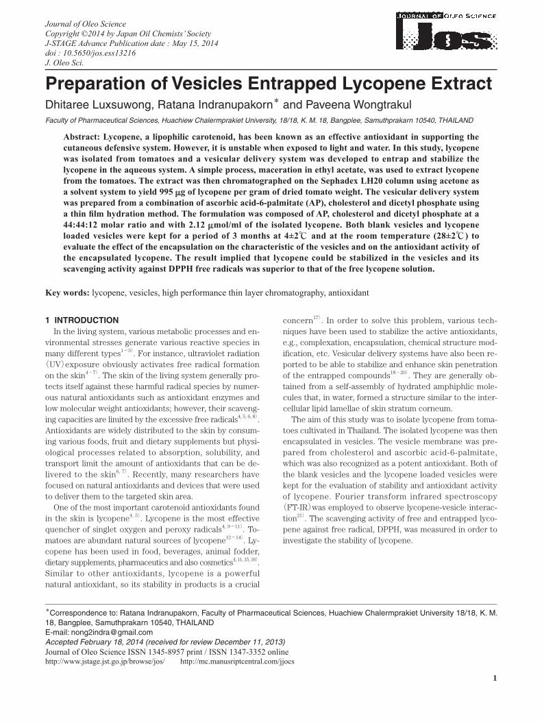
on the lycopene fraction obtained from chromatography in order to assure that its main compound was lycopene. The results of 1H NMR(Fig. 1)were as follow: 1H NMR(CDCl3, ppm), 5.33(2,2’-H), ca 2.33(3,3’-H), ca 2.33(4,4’-H), 6.52
Table 1 Formulations of vesicles.
Ingredients Blank vesicles (% μmole)
Lycopene loaded vesicles(% μmole)
Ascorbic acid-6-palmitate 44 44Cholesterol 44 44Dicetyl phosphate 12 12Lycopene - 2.12
D. Luxsuwong, R. Indranupakorn and P. Wongtrakul
J. Oleo Sci.
4
(7,7’-H), 6.23(8,8’-H), 6.22(9,9’-H), ca 6.89(11,11’-H), ca 6.51(14,14’-H), ca 6.87(15,15’-H), 1.60(16,16’-H), 1.59(17,17’-H), 2.03(18,18’-H), 2.04(19,19’-H), 2.05(20,20’-H). The NMR spectrum revealed a strong solvent(CDCl3)peak at 7.260 ppm. and the high-field part between 0-3 ppm. and low-field part of key area between 5-6.5 ppm. due to the presence of acyclic aliphatic group and olefinic group, respectively. The NMR signals corresponded well to the structure of lycopene, which is aliphatic hydrocarbons
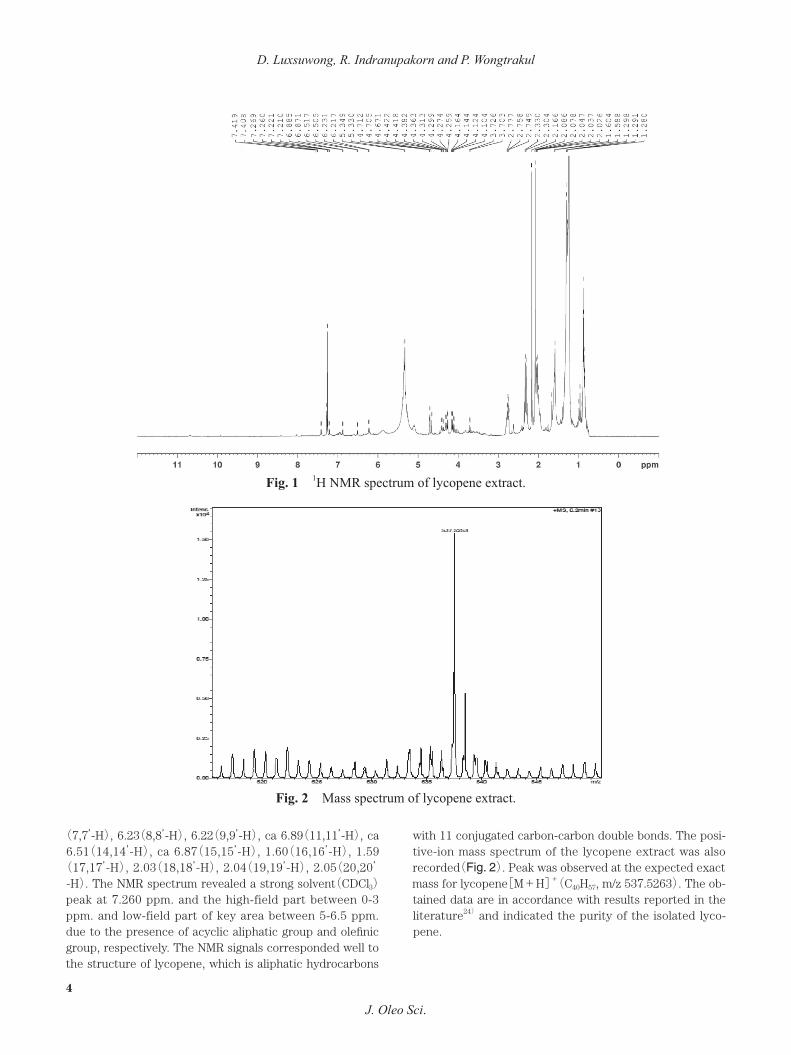
with 11 conjugated carbon-carbon double bonds. The posi-tive-ion mass spectrum of the lycopene extract was also recorded(Fig. 2). Peak was observed at the expected exact mass for lycopene[M+H]+(C40H57, m/z 537.5263). The ob-tained data are in accordance with results reported in the literature24) and indicated the purity of the isolated lyco-pene.
Fig. 1 1H NMR spectrum of lycopene extract.
Fig. 2 Mass spectrum of lycopene extract.

VESICLES ENTRAPPED LYCOPENE EXTRACT
J. Oleo Sci.
5
3.3 Mobile phase selection and HPTLC analysisOptimization of the mobile phase was performed by
using two different solvent systems. A combination of hexane:ethyl acetate(85:15)and methanol:hexane:ethyl acetate(45:40:15)was evaluated for abilities to cause movements(Rf values)of isolated lycopene(b)and its standard(a). It was observed that the former produced too high Rf values of lycopene(0.90). The latter was found to be of a more suitable level of lycopene with Rf values of 0.76 as shown in Fig. 3. Moreover, it gave speedy develop-ment and sharp peaks.3.3.1 Linearity, precision and accuracy
The developed HPTLC method was validated according to ICH guidelines23) with respect to linearity, precision and accuracy. The linear regression data for the calibration plot showed good linear relationship over the concentration range of 0.6-3.0 μg/band. The regression equation was found to be y=1,653.5 x+77.1 with a correlation coeffi-cient(r)of 0.999. The percent relative standard deviations were found to be 1.33 and 0.93 for intra-day and inter-day precision studies, respectively, which indicated that the method was highly precise. To study the accuracy of the method, the percent average recoveries obtained were found to be 100.91%, 101.04% and 101.25% at three dif-ferent concentration levels(80%, 100% and 120% addi-tion)with an average relative standard deviation of 1.70. It has been shown to be linear, precise and accurate in ac-ceptance criteria25) and can be used successfully for the si-multaneous quantitation of the small amount of lycopene.
3.4 Preparation and characterization of vesicles Ascorbyl palmitate is known as a free radical fighting an-
tioxidant. It has an amphiphilic character, which can as-semble to form bilayer vesicles in water26). Addition of cho-lesterol and negative charged lipid, DCP, resulted in an appropriated structure for the formation of stable vesicles. From the preliminary study, it was found that mole ratio of AP:cholesterol:DCP at 44:44:12 was an appropriated for-mulation for the entrapment of lycopene. Vesicles prepared by the film hydration method were translucent dispersions after passing a 30-minute sonication. The birefringent characteristics of lamellar structure of vesicular membrane could be observed under an optical light polarized micro-scope. The lycopene loaded vesicles detected under TEM were spherical in shape as shown in Fig. 4 suggesting that vesicles could be successfully prepared from a mixture of ascorbyl palmitate and cholesterol. Lycopene is a lipophilic substance; therefore, it is possible for lycopene to be solu-bilized in a hydrophobic region of the vesicles.
The results of vesicle size, polydispersity index and zeta potential measured within a period of 3 months were shown in Table 2. All vesicles were small about 200 nm. in size.
3.5 IR measurementFT-IR spectra of blank vesicles(a), free lycopene(b), ly-
copene loaded in vesicles(c)and lycopene-vesicles mixture(d)were shown in Fig. 5. Spectra of blank vesicle in aqueous system obviously showed broad peaks of O-H stretching at 3338 cm-1 and O-H scissors bending at 1638 cm-1. While, FT-IR peaks of free lycopene showed C-H stretching at 3010, 2922, 2853 cm-1, C=C stretching at 1742, 1711, 1662 cm-1 and C-H bending at 1457, 1377 cm-1.
Fig. 3 HPTLC chromatogram of isolated lycopene (b) and its standard (a) with the Rf values of 0.76, stationary phase: HPTLC silica gel 60 F254, mo-bile phase : methanol:hexane:ethyl acetate (45:40:15).
Fig. 4 �Transmission electron microscope images of ly-copene loaded vesicles, Scale bar 250 nm.

D. Luxsuwong, R. Indranupakorn and P. Wongtrakul
J. Oleo Sci.
6
FT-IR data of lycopene are in accordance with results re-ported in the literature21). The spectra of physical mixture of lycopene and vesicles still showed the characteristic signals of lycopene, located at around 1600 and 1000 cm-1, which had clearly disappeared in the spectra of lycopene
loaded vesicle. This may be attributed to the intercalation of lycopene into the lipophilic part of the vesicles resulting in weak FT-IR peak intensity of lycopene. Meanwhile, this phenomenon was not detected when lycopene was uncap-tured in physical mixture.
3.6 2,2-diphenyl-1-picrylhydrazyl(DPPH)radical scaveng-ing activity assay
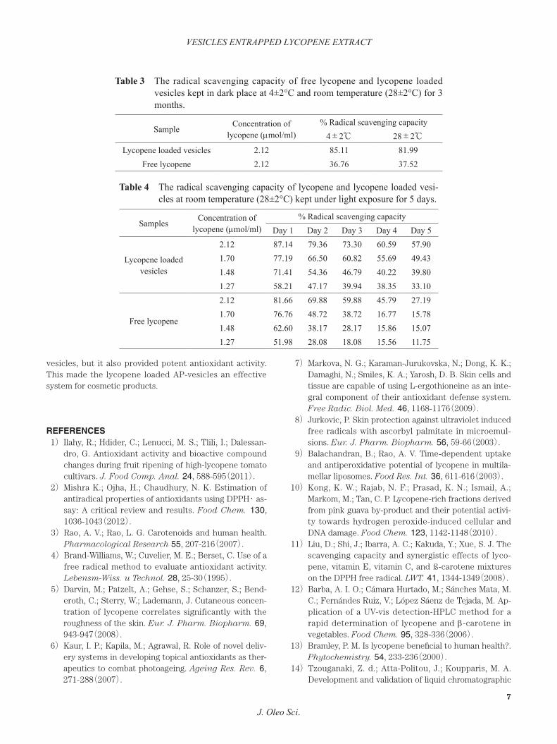
Scavenging activities against the DPPH radical of free ly-copene and lycopene loaded in vesicles, which were kept in a dark place for 3 months and under light exposure for 5 days, were shown in Tables 3 and 4 respectively. After storage, the scavenging activity of free lycopene was much lower than that of the loaded lycopene. The dramatic de-crease of activity was observed when the lycopene solution was exposed to sunlight. However, the activity could be prolonged by the incorporation of the lycopene in vesicles. Therefore, the encapsulation could significantly sustain free radical scavenging activity of lycopene in an aqueous system.
4 CONCLUSIONThe isolated lycopene from the tomato was successfully
entrapped in vesicles prepared from a mixture of ascorbic acid-6-palmitate, cholesterol and dicetyl phosphate. The lycopene loaded vesicles were stable in size and dispers-ibility in the period of 3 months storage in a dark place. The encapsulation of lycopene in a vesicle membrane was able to stabilize antioxidant activity of lycopene from light induced degradation. Not only did AP form a membrane of
Table 2 Particle size, polydispersity index and zeta potential of free and lycopene loaded vesicles kept at 4±2°C and room temperature (28±2°C) for 3 months.
Formula Blank vesicles Lycopene loaded vesiclesTemp. (℃) 4±2 28±2 4±2 28±2
Mean particle size (nm)
Starting 208.4 ±6.5 208.4 ±6.5 195.0 ±3.0 195.0 ±3.01st month 218.8 ±2.1 229.4 ±7.6 219.8 ±0.9 235.6 ±4.52nd month 288.6 ±1.4 259.0 ±12.8 284.9 ±0.5 276.2 ±6.03rd month 294.3 ±4.2 293.5 ±5.1 288.7 ±2.1 291.6 ±3.5
Polydispersity index
Starting 0.256±0.023 0.256±0.023 0.34 ±0.017 0.34 ±0.0171st month 0.252±0.012 0.266±0.012 0.342±0.003 0.251±0.0142nd month 0.265±0.009 0.212±0.009 0.260±0.017 0.264±0.0023rd month 0.227±0.022 0.320±0.013 0.251±0.050 0.261±0.004
Zeta potential (mV)
Starting -53.96 ±0.95 -53.96 ±0.95 -55.66 ±1.71 -55.66 ±1.711st month -48.39 ±0.63 -52.40 ±1.33 -55.58 ±1.07 -51.30 ±0.542nd month -48.2 ±3.22 -49.50 ±1.73 -54.80 ±1.93 -52.10 ±0.173rd month -46.2 ±2.08 -48.60 ±1.16 -52.09 ±0.99 -52.10 ±1.21
Remark : Each data point represented the average value±standard deviation from triplicate measurements
Fig. 5 FT-IR spectra of vesicles (a), lycopene (b), lyco-pene loaded vesicles (c) and lycopene-vesicle mixture (d).
VESICLES ENTRAPPED LYCOPENE EXTRACT
J. Oleo Sci.
7
vesicles, but it also provided potent antioxidant activity. This made the lycopene loaded AP-vesicles an effective system for cosmetic products.
REFERENCES1) Ilahy, R.; Hdider, C.; Lenucci, M. S.; Tlili, I.; Dalessan-
dro, G. Antioxidant activity and bioactive compound changes during fruit ripening of high-lycopene tomato cultivars. J. Food Comp. Anal. 24, 588-595(2011).
2) Mishra K.; Ojha, H.; Chaudhury, N. K. Estimation of antiradical properties of antioxidants using DPPH・ as-say: A critical review and results. Food Chem. 130, 1036-1043(2012).
3) Rao, A. V.; Rao, L. G. Carotenoids and human health. Pharmacological Research 55, 207-216(2007).
4) Brand-Williams, W.; Cuvelier, M. E.; Berset, C. Use of a free radical method to evaluate antioxidant activity. Lebensm-Wiss. u Technol. 28, 25-30(1995).
5) Darvin, M.; Patzelt, A.; Gehse, S.; Schanzer, S.; Bend-eroth, C.; Sterry, W.; Lademann, J. Cutaneous concen-tration of lycopene correlates significantly with the roughness of the skin. Eur. J. Pharm. Biopharm. 69, 943-947(2008).
6) Kaur, I. P.; Kapila, M.; Agrawal, R. Role of novel deliv-ery systems in developing topical antioxidants as ther-apeutics to combat photoageing. Ageing Res. Rev. 6, 271-288(2007).
7) Markova, N. G.; Karaman-Jurukovska, N.; Dong, K. K.; Damaghi, N.; Smiles, K. A.; Yarosh, D. B. Skin cells and tissue are capable of using L-ergothioneine as an inte-gral component of their antioxidant defense system. Free Radic. Biol. Med. 46, 1168-1176(2009).
8) Jurkovic, P. Skin protection against ultraviolet induced free radicals with ascorbyl palmitate in microemul-sions. Eur. J. Pharm. Biopharm. 56, 59-66(2003).
9) Balachandran, B.; Rao, A. V. Time-dependent uptake and antiperoxidative potential of lycopene in multila-mellar liposomes. Food Res. Int. 36, 611-616(2003).
10) Kong, K. W.; Rajab, N. F.; Prasad, K. N.; Ismail, A.; Markom, M.; Tan, C. P. Lycopene-rich fractions derived from pink guava by-product and their potential activi-ty towards hydrogen peroxide-induced cellular and DNA damage. Food Chem. 123, 1142-1148(2010).
11) Liu, D.; Shi, J.; Ibarra, A. C.; Kakuda, Y.; Xue, S. J. The scavenging capacity and synergistic effects of lyco-pene, vitamin E, vitamin C, and ß-carotene mixtures on the DPPH free radical. LWT. 41, 1344-1349(2008).
12) Barba, A. I. O.; Cámara Hurtado, M.; Sánches Mata, M. C.; Fernándes Ruiz, V.; López Sáenz de Tejada, M. Ap-plication of a UV-vis detection-HPLC method for a rapid determination of lycopene and β-carotene in vegetables. Food Chem. 95, 328-336(2006).
13) Bramley, P. M. Is lycopene beneficial to human health?. Phytochemistry. 54, 233-236(2000).
14) Tzouganaki, Z. d.; Atta-Politou, J.; Koupparis, M. A. Development and validation of liquid chromatographic
Table 3 The radical scavenging capacity of free lycopene and lycopene loaded vesicles kept in dark place at 4±2°C and room temperature (28±2°C) for 3 months.
Sample Concentration of lycopene (μmol/ml)
% Radical scavenging capacity4±2℃ 28±2℃
Lycopene loaded vesicles 2.12 85.11 81.99Free lycopene 2.12 36.76 37.52
Table 4 The radical scavenging capacity of lycopene and lycopene loaded vesi-cles at room temperature (28±2°C) kept under light exposure for 5 days.
Samples Concentration of lycopene (μmol/ml)
% Radical scavenging capacityDay 1 Day 2 Day 3 Day 4 Day 5
Lycopene loaded vesicles
2.12 87.14 79.36 73.30 60.59 57.901.70 77.19 66.50 60.82 55.69 49.431.48 71.41 54.36 46.79 40.22 39.801.27 58.21 47.17 39.94 38.35 33.10
Free lycopene
2.12 81.66 69.88 59.88 45.79 27.191.70 76.76 48.72 38.72 16.77 15.781.48 62.60 38.17 28.17 15.86 15.071.27 51.98 28.08 18.08 15.56 11.75
D. Luxsuwong, R. Indranupakorn and P. Wongtrakul
J. Oleo Sci.
8
method for the determination of lycopene in plasma. Analytica Chimica Acta. 467, 115-123(2002).
15) Liu, Y.; Liu, J.; Chen, X.; Liu, Y.; Di, D. Preparative sep-aration and purification of lycopene from tomato skin extracts by macroporous adsorption resins. Food Chem. 123, 1027-1034.(2010).
16) Shi, J.; Yi, C.; Xue, S. J.; Jiang, Y.; Ma, Y.; Li, D. Effect of modifiers on the profile of lycopene extracted from tomato skins by supercritical CO2. J. Food Eng. 93, 431-436(2009).
17) Shi, J.; Dai, Y.; Kakuda, Y.; Mittal, G.; Xue, S. J. Effect of heating and exposure to light on the stability of ly-copene in tomato purée. Food Control. 19, 514-520(2008).
18) Kumar, G. P.; Rajeshwarrao, P. Nonionic surfactant ve-sicular systems for effective drug delivery-an overview. Acta Pharm. Sin. B. 1(4), 208-219(2011).
19) Manosroi, A.; Wongtrakul, P.; Manosroi, J.; Sakai, H.; Sugawara, F.; Yuasa, M.; Abe, M. Characterization of vesicles prepared with various non-ionic surfactants mixed with cholesterol. Colloids and Surfaces B: Biointerfaces 30, 129-138(2003).
20) Uchegbu, I. F.; Vyas, S. P. Non-ionic surfactant based vesicles(niosomes)in drug delivery. Int. J. Pharm. 172, 33-70(1998).
21) Kamil, M.; M., Mohamed, G. F.; Shaheen, M. S. Fourier Transformer Infrared Spectroscopy for Quality Assur-ance of Tomato Products. Journal of American Sci-ence 7(6), 559-572(2011).
22) Shewiyo, D. H.; Kaale, E.; Risha, P. G.; Dejaegher, B.; Smeyers-Verbeke, J.; Heyden, Y. V. HPTLC methods to assay active ingredients in pharmaceutical formula-tions: A review of the method development and valida-tion steps. J. of Pharmaceut. Biomed. Anal. 66, 11-23(2012).
23) The International Conference on Harmonization, Q2(R1), Validation of Analytical Procedure: Text and Methodology,(2005).
24) El-Raey, M. A.; Ibrahim, G. E.; Eldahshan, O. A. Lyco-pene and lutein ; A review for their chemistry and medical uses. Journal of Pharmacognosy and Phy-tochemistry 2, 245-254(2013).
25) Ferenczi-Fodor, K.; Végh, Z.; Nagy-Turák, A; Renger, B; Zeller, M. Validation and quality assurance of planar chromatographic procedures in pharmaceutical analy-sis. J. AOAC Int. 84, 1265-1276(2001).
26) Gopinath, D.; Ravi, D.; Rao, B. R.; Apte, S.S.; Renuka, D.; Rambhau, D. Ascorbyl palmitate vesicles(Aspa-somes): formation characterization and applications. Int. J. Pharm. 271, 95-113(2004).