mRNA Inventory of Extracellular Vesicles from Ustilago maydis
21
Fungi Journal of Article mRNA Inventory of Extracellular Vesicles from Ustilago maydis Seomun Kwon 1 , Oliver Rupp 2 , Andreas Brachmann 3 , Christopher Frederik Blum 4 , Anton Kraege 1 , Alexander Goesmann 2 and Michael Feldbrügge 1, * Citation: Kwon, S.; Rupp, O.; Brachmann, A.; Blum, C.F.; Kraege, A.; Goesmann, A.; Feldbrügge, M. mRNA Inventory of Extracellular Vesicles from Ustilago maydis. J. Fungi 2021, 7, 562. https://doi.org/ 10.3390/jof7070562 Academic Editor: Kuang R. Chung Received: 8 June 2021 Accepted: 7 July 2021 Published: 14 July 2021 Publisher’s Note: MDPI stays neutral with regard to jurisdictional claims in published maps and institutional affil- iations. Copyright: © 2021 by the authors. Licensee MDPI, Basel, Switzerland. This article is an open access article distributed under the terms and conditions of the Creative Commons Attribution (CC BY) license (https:// creativecommons.org/licenses/by/ 4.0/). 1 Institute for Microbiology, Cluster of Excellence on Plant Sciences, Heinrich Heine University Düsseldorf, 40225 Düsseldorf, Germany; [email protected] (S.K.); [email protected] (A.K.) 2 Bioinformatics and Systems Biology, Justus-Liebig-Universität, 35392 Giessen, Germany; [email protected] (O.R.); [email protected] (A.G.) 3 Biocenter of the LMU Munich, Genetics Section, Grosshaderner Str. 2-4, 82152 Planegg-Martinsried, Germany; [email protected] 4 Institute for Mathematical Modelling of Biological Systems, Heinrich Heine University Düsseldorf, 40225 Düsseldorf, Germany; [email protected] * Correspondence: [email protected]; Tel.: +49-211-81-14720 Abstract: Extracellular vesicles (EVs) can transfer diverse RNA cargo for intercellular communi- cation. EV-associated RNAs have been found in diverse fungi and were proposed to be relevant for pathogenesis in animal hosts. In plant-pathogen interactions, small RNAs are exchanged in a cross-kingdom RNAi warfare and EVs were considered to be a delivery mechanism. To extend the search for EV-associated molecules involved in plant-pathogen communication, we have charac- terised the repertoire of EV-associated mRNAs secreted by the maize smut pathogen, Ustilago maydis. For this initial survey, we examined EV-enriched fractions from axenic filamentous cultures that mimic infectious hyphae. EV-associated RNAs were resistant to degradation by RNases and the presence of intact mRNAs was evident. The set of mRNAs enriched inside EVs relative to the fungal cells are functionally distinct from those that are depleted from EVs. mRNAs encoding metabolic enzymes are particularly enriched. Intriguingly, mRNAs of some known effectors and other pro- teins linked to virulence were also found in EVs. Furthermore, several mRNAs enriched in EVs are also upregulated during infection, suggesting that EV-associated mRNAs may participate in plant-pathogen interactions. Keywords: extracellular vesicles (EVs); mRNA; fungal pathogen; plant pathogen; Ustilago maydis 1. Introduction Extracellular vesicles (EVs) are ubiquitously secreted from cells, carrying a diverse array of molecular cargos. The role of EVs in intercellular signalling and communication is particularly interesting, as they can facilitate mass delivery of otherwise intracellular molecules across the extracellular space. EV-associated molecules can induce physiological changes in the recipient cells [1]. In pathogenic microbes, EVs can facilitate both intraspecies coordination of pathogen cells during infection [2], and broader cross-kingdom interaction with host cells [3,4]. Investigations on fungal EVs have identified associated proteins [5], RNAs [6], lipids [7], polysaccharides [8], and metabolites [9]. At the level of individual cells, EVs have been implicated in structural functions such as cell wall remodelling [10] and glucuronoxylo- mannan capsule formation [8]. At the population level, secretion of EVs in Candida albicans is important for biofilm formation and antifungal resistance [11]. Furthermore, EVs of Cryptococcus gattii effectuate long-distance coordination of virulence between fungal cells engulfed in different macrophages; EVs from a hypervirulent strain trigger rapid prolifera- tion of less virulent strains in the phagosome [3]. While EVs of some clinically important J. Fungi 2021, 7, 562. https://doi.org/10.3390/jof7070562 https://www.mdpi.com/journal/jof
-
Upload
khangminh22 -
Category
Documents
-
view
0 -
download
0
Transcript of mRNA Inventory of Extracellular Vesicles from Ustilago maydis

FungiJournal of
Article
mRNA Inventory of Extracellular Vesicles from Ustilago maydis
Seomun Kwon 1 , Oliver Rupp 2, Andreas Brachmann 3 , Christopher Frederik Blum 4, Anton Kraege 1,Alexander Goesmann 2 and Michael Feldbrügge 1,*
�����������������
Citation: Kwon, S.; Rupp, O.;
Brachmann, A.; Blum, C.F.; Kraege,
A.; Goesmann, A.; Feldbrügge, M.
mRNA Inventory of Extracellular
Vesicles from Ustilago maydis. J. Fungi
2021, 7, 562. https://doi.org/
10.3390/jof7070562
Academic Editor: Kuang R. Chung
Received: 8 June 2021
Accepted: 7 July 2021
Published: 14 July 2021
Publisher’s Note: MDPI stays neutral
with regard to jurisdictional claims in
published maps and institutional affil-
iations.
Copyright: © 2021 by the authors.
Licensee MDPI, Basel, Switzerland.
This article is an open access article
distributed under the terms and
conditions of the Creative Commons
Attribution (CC BY) license (https://
creativecommons.org/licenses/by/
4.0/).
1 Institute for Microbiology, Cluster of Excellence on Plant Sciences, Heinrich Heine University Düsseldorf,40225 Düsseldorf, Germany; [email protected] (S.K.); [email protected] (A.K.)
2 Bioinformatics and Systems Biology, Justus-Liebig-Universität, 35392 Giessen, Germany;[email protected] (O.R.);[email protected] (A.G.)
3 Biocenter of the LMU Munich, Genetics Section, Grosshaderner Str. 2-4, 82152 Planegg-Martinsried, Germany;[email protected]
4 Institute for Mathematical Modelling of Biological Systems, Heinrich Heine University Düsseldorf,40225 Düsseldorf, Germany; [email protected]
* Correspondence: [email protected]; Tel.: +49-211-81-14720
Abstract: Extracellular vesicles (EVs) can transfer diverse RNA cargo for intercellular communi-cation. EV-associated RNAs have been found in diverse fungi and were proposed to be relevantfor pathogenesis in animal hosts. In plant-pathogen interactions, small RNAs are exchanged ina cross-kingdom RNAi warfare and EVs were considered to be a delivery mechanism. To extendthe search for EV-associated molecules involved in plant-pathogen communication, we have charac-terised the repertoire of EV-associated mRNAs secreted by the maize smut pathogen, Ustilago maydis.For this initial survey, we examined EV-enriched fractions from axenic filamentous cultures thatmimic infectious hyphae. EV-associated RNAs were resistant to degradation by RNases and thepresence of intact mRNAs was evident. The set of mRNAs enriched inside EVs relative to the fungalcells are functionally distinct from those that are depleted from EVs. mRNAs encoding metabolicenzymes are particularly enriched. Intriguingly, mRNAs of some known effectors and other pro-teins linked to virulence were also found in EVs. Furthermore, several mRNAs enriched in EVsare also upregulated during infection, suggesting that EV-associated mRNAs may participate inplant-pathogen interactions.
Keywords: extracellular vesicles (EVs); mRNA; fungal pathogen; plant pathogen; Ustilago maydis
1. Introduction
Extracellular vesicles (EVs) are ubiquitously secreted from cells, carrying a diversearray of molecular cargos. The role of EVs in intercellular signalling and communicationis particularly interesting, as they can facilitate mass delivery of otherwise intracellularmolecules across the extracellular space. EV-associated molecules can induce physiologicalchanges in the recipient cells [1]. In pathogenic microbes, EVs can facilitate both intraspeciescoordination of pathogen cells during infection [2], and broader cross-kingdom interactionwith host cells [3,4].
Investigations on fungal EVs have identified associated proteins [5], RNAs [6], lipids [7],polysaccharides [8], and metabolites [9]. At the level of individual cells, EVs have beenimplicated in structural functions such as cell wall remodelling [10] and glucuronoxylo-mannan capsule formation [8]. At the population level, secretion of EVs in Candida albicansis important for biofilm formation and antifungal resistance [11]. Furthermore, EVs ofCryptococcus gattii effectuate long-distance coordination of virulence between fungal cellsengulfed in different macrophages; EVs from a hypervirulent strain trigger rapid prolifera-tion of less virulent strains in the phagosome [3]. While EVs of some clinically important
J. Fungi 2021, 7, 562. https://doi.org/10.3390/jof7070562 https://www.mdpi.com/journal/jof

J. Fungi 2021, 7, 562 2 of 21
fungi carry virulence-associated molecules [12] and promote infection [3,13,14], many stud-ies also indicate that fungal EVs stimulate host immune responses to the detriment of thepathogen [15].
The role of EVs in plant-pathogen interaction is not yet well understood, although theyhave been frequently observed at various plant-fungal interfaces [16–18] Biological signifi-cance of plant EVs and their cargos have been elucidated in only a few cases. For instance,EVs of the model plant Arabidopsis thaliana carry small RNAs (sRNAs) that silence virulencegenes in the grey mould fungus Botrytis cinerea [19] and the oomycete pathogen Phytoph-thora capsici [20] during infection. A. thaliana EVs additionally contain “tiny RNAs” [21]and various defence-related proteins [22]. In another example, sunflower EVs inhibit sporegermination and growth of the white mould pathogen, Sclerotinia sclerotiorum [23].
EVs of plant pathogenic fungi are only recently being characterised. So far, EV-associated proteomes of the wheat pathogen, Zymoseptoria tritici [24], and the cottonpathogen, Fusarium oxysporum f. sp. vasinfectum (Fov; [9]) have been examined. Fov secretesEVs with polyketide synthases and a purple pigment. The fractions containing these EVstrigger hypersensitive cell-death in plants, reflecting the necrotrophic lifestyle of this highlyprolific mycotoxin producer [9]. While studies on plant pathogen effectors to date haveprimarily focused on conventionally secreted proteins, such efforts to examine EV cargoscould broaden the spectrum of effector candidates, not only to include unconventionallysecreted proteins, but also RNAs and metabolites.
Fungal EVs have been found to contain all types of RNA, the majority of the cargobeing shorter sRNAs and tRNAs, but also mRNAs and rRNAs [6]. sRNA effectors havebeen discovered in at least five different filamentous phytopathogens to date [25–29].These participate in the bidirectional, cross-kingdom RNAi warfare between plants andpathogens [26]. The diversity of RNAs associated with fungal EVs suggest that RNAspecies other than sRNAs could also be transferred from a pathogenic fungus to functionas effectors in host cells. Particularly interesting would be the concept of effector deliveryin the form of full-length mRNAs in pathogen EVs. Such mRNAs could theoretically betranslated in the recipient host cells to yield multiple proteins and transfer the cost ofeffector protein production to the host.
Ustilago maydis is a biotrophic fungal pathogen of maize [30], which can cause upto 20% yield losses [31]. It is an established model organism for endosome-associatedmRNA transport [32] and has secondarily lost the RNAi machinery, so it does not producecanonical sRNAs [33]. This makes it an interesting organism to examine the mRNA cargoof EVs. EV-like structures have long been observed at the interface between U. maydisand maize cells during biotrophic infection [16], suggesting their relevance in the inter-action. Furthermore, engineered strains are available, where filamentous growth and theconcomitant infection program can be induced in axenic culture [34].
In nature, the infectious form of U. maydis is the dikaryotic filament, formed by matingof compatible sporidia [30]. Filamentation is brought about by heterodimerisation ofcomplementary bE and bW homeodomain transcription factors from each sporidium,which initiates a transcriptional cascade for infectious development [35,36]. Here wehave taken advantage of a laboratory strain, AB33, where complementary bE and bWare both present in the same strain and are inducible by switching the nitrogen source,allowing facile and reproducible filamentation in culture [34]. AB33 induced filamentstranscriptionally and developmentally mimic infectious dikaryotic filaments and havebeen used as a surrogate to study the initial stage of infection. Evidently, many effectorsand genes relevant for infection are expressed in AB33 filaments in culture [35,36]. Hence,we have utilised U. maydis as an ideal system for an initial survey of EV cargo mRNAs inplant pathogens.
J. Fungi 2021, 7, 562 3 of 21
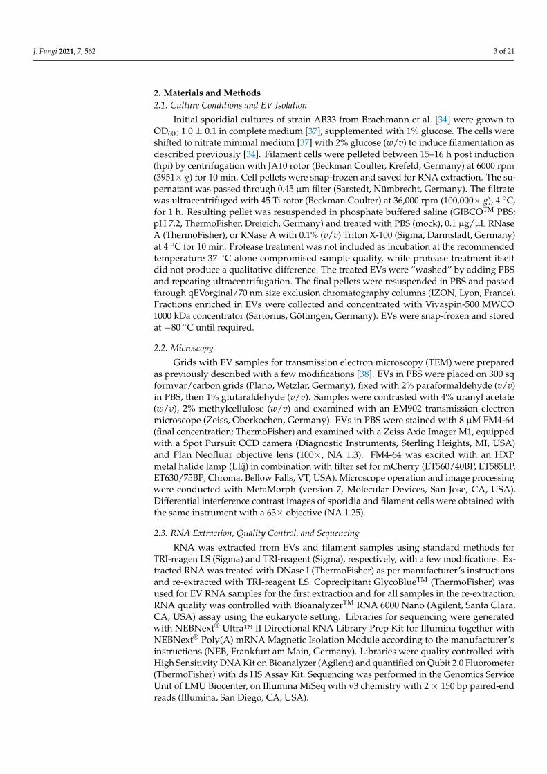
2. Materials and Methods2.1. Culture Conditions and EV Isolation
Initial sporidial cultures of strain AB33 from Brachmann et al. [34] were grown toOD600 1.0 ± 0.1 in complete medium [37], supplemented with 1% glucose. The cells wereshifted to nitrate minimal medium [37] with 2% glucose (w/v) to induce filamentation asdescribed previously [34]. Filament cells were pelleted between 15–16 h post induction(hpi) by centrifugation with JA10 rotor (Beckman Coulter, Krefeld, Germany) at 6000 rpm(3951× g) for 10 min. Cell pellets were snap-frozen and saved for RNA extraction. The su-pernatant was passed through 0.45 µm filter (Sarstedt, Nümbrecht, Germany). The filtratewas ultracentrifuged with 45 Ti rotor (Beckman Coulter) at 36,000 rpm (100,000× g), 4 ◦C,for 1 h. Resulting pellet was resuspended in phosphate buffered saline (GIBCOTM PBS;pH 7.2, ThermoFisher, Dreieich, Germany) and treated with PBS (mock), 0.1 µg/µL RNaseA (ThermoFisher), or RNase A with 0.1% (v/v) Triton X-100 (Sigma, Darmstadt, Germany)at 4 ◦C for 10 min. Protease treatment was not included as incubation at the recommendedtemperature 37 ◦C alone compromised sample quality, while protease treatment itselfdid not produce a qualitative difference. The treated EVs were “washed” by adding PBSand repeating ultracentrifugation. The final pellets were resuspended in PBS and passedthrough qEVorginal/70 nm size exclusion chromatography columns (IZON, Lyon, France).Fractions enriched in EVs were collected and concentrated with Vivaspin-500 MWCO1000 kDa concentrator (Sartorius, Göttingen, Germany). EVs were snap-frozen and storedat −80 ◦C until required.
2.2. Microscopy
Grids with EV samples for transmission electron microscopy (TEM) were preparedas previously described with a few modifications [38]. EVs in PBS were placed on 300 sqformvar/carbon grids (Plano, Wetzlar, Germany), fixed with 2% paraformaldehyde (v/v)in PBS, then 1% glutaraldehyde (v/v). Samples were contrasted with 4% uranyl acetate(w/v), 2% methylcellulose (w/v) and examined with an EM902 transmission electronmicroscope (Zeiss, Oberkochen, Germany). EVs in PBS were stained with 8 µM FM4-64(final concentration; ThermoFisher) and examined with a Zeiss Axio Imager M1, equippedwith a Spot Pursuit CCD camera (Diagnostic Instruments, Sterling Heights, MI, USA)and Plan Neofluar objective lens (100×, NA 1.3). FM4-64 was excited with an HXPmetal halide lamp (LEj) in combination with filter set for mCherry (ET560/40BP, ET585LP,ET630/75BP; Chroma, Bellow Falls, VT, USA). Microscope operation and image processingwere conducted with MetaMorph (version 7, Molecular Devices, San Jose, CA, USA).Differential interference contrast images of sporidia and filament cells were obtained withthe same instrument with a 63× objective (NA 1.25).
2.3. RNA Extraction, Quality Control, and Sequencing
RNA was extracted from EVs and filament samples using standard methods forTRI-reagen LS (Sigma) and TRI-reagent (Sigma), respectively, with a few modifications. Ex-tracted RNA was treated with DNase I (ThermoFisher) as per manufacturer’s instructionsand re-extracted with TRI-reagent LS. Coprecipitant GlycoBlueTM (ThermoFisher) wasused for EV RNA samples for the first extraction and for all samples in the re-extraction.RNA quality was controlled with BioanalyzerTM RNA 6000 Nano (Agilent, Santa Clara,CA, USA) assay using the eukaryote setting. Libraries for sequencing were generatedwith NEBNext® Ultra™ II Directional RNA Library Prep Kit for Illumina together withNEBNext® Poly(A) mRNA Magnetic Isolation Module according to the manufacturer’sinstructions (NEB, Frankfurt am Main, Germany). Libraries were quality controlled withHigh Sensitivity DNA Kit on Bioanalyzer (Agilent) and quantified on Qubit 2.0 Fluorometer(ThermoFisher) with ds HS Assay Kit. Sequencing was performed in the Genomics ServiceUnit of LMU Biocenter, on Illumina MiSeq with v3 chemistry with 2 × 150 bp paired-endreads (Illumina, San Diego, CA, USA).
J. Fungi 2021, 7, 562 4 of 21
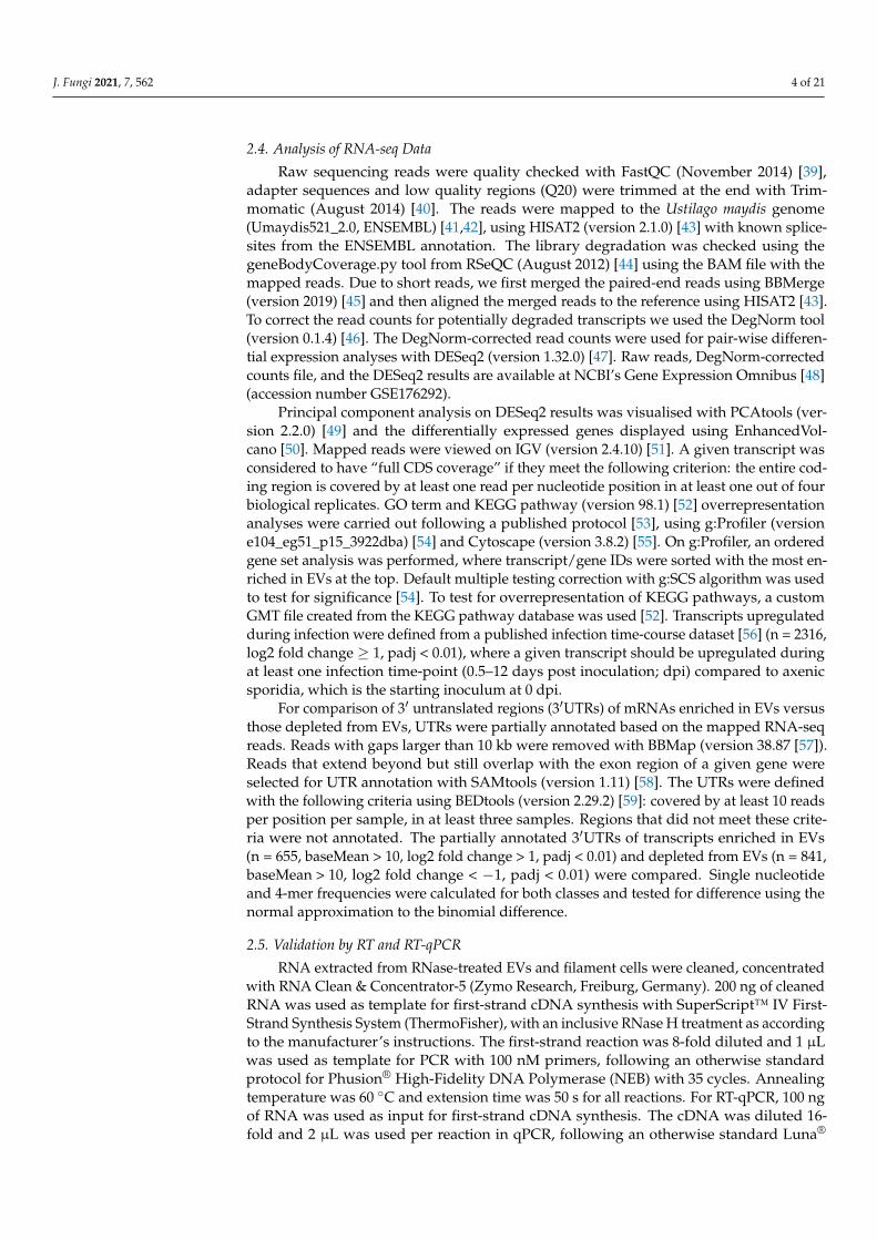
2.4. Analysis of RNA-seq Data
Raw sequencing reads were quality checked with FastQC (November 2014) [39],adapter sequences and low quality regions (Q20) were trimmed at the end with Trim-momatic (August 2014) [40]. The reads were mapped to the Ustilago maydis genome(Umaydis521_2.0, ENSEMBL) [41,42], using HISAT2 (version 2.1.0) [43] with known splice-sites from the ENSEMBL annotation. The library degradation was checked using thegeneBodyCoverage.py tool from RSeQC (August 2012) [44] using the BAM file with themapped reads. Due to short reads, we first merged the paired-end reads using BBMerge(version 2019) [45] and then aligned the merged reads to the reference using HISAT2 [43].To correct the read counts for potentially degraded transcripts we used the DegNorm tool(version 0.1.4) [46]. The DegNorm-corrected read counts were used for pair-wise differen-tial expression analyses with DESeq2 (version 1.32.0) [47]. Raw reads, DegNorm-correctedcounts file, and the DESeq2 results are available at NCBI’s Gene Expression Omnibus [48](accession number GSE176292).
Principal component analysis on DESeq2 results was visualised with PCAtools (ver-sion 2.2.0) [49] and the differentially expressed genes displayed using EnhancedVol-cano [50]. Mapped reads were viewed on IGV (version 2.4.10) [51]. A given transcript wasconsidered to have “full CDS coverage” if they meet the following criterion: the entire cod-ing region is covered by at least one read per nucleotide position in at least one out of fourbiological replicates. GO term and KEGG pathway (version 98.1) [52] overrepresentationanalyses were carried out following a published protocol [53], using g:Profiler (versione104_eg51_p15_3922dba) [54] and Cytoscape (version 3.8.2) [55]. On g:Profiler, an orderedgene set analysis was performed, where transcript/gene IDs were sorted with the most en-riched in EVs at the top. Default multiple testing correction with g:SCS algorithm was usedto test for significance [54]. To test for overrepresentation of KEGG pathways, a customGMT file created from the KEGG pathway database was used [52]. Transcripts upregulatedduring infection were defined from a published infection time-course dataset [56] (n = 2316,log2 fold change ≥ 1, padj < 0.01), where a given transcript should be upregulated duringat least one infection time-point (0.5–12 days post inoculation; dpi) compared to axenicsporidia, which is the starting inoculum at 0 dpi.
For comparison of 3′ untranslated regions (3′UTRs) of mRNAs enriched in EVs versusthose depleted from EVs, UTRs were partially annotated based on the mapped RNA-seqreads. Reads with gaps larger than 10 kb were removed with BBMap (version 38.87 [57]).Reads that extend beyond but still overlap with the exon region of a given gene wereselected for UTR annotation with SAMtools (version 1.11) [58]. The UTRs were definedwith the following criteria using BEDtools (version 2.29.2) [59]: covered by at least 10 readsper position per sample, in at least three samples. Regions that did not meet these crite-ria were not annotated. The partially annotated 3′UTRs of transcripts enriched in EVs(n = 655, baseMean > 10, log2 fold change > 1, padj < 0.01) and depleted from EVs (n = 841,baseMean > 10, log2 fold change < −1, padj < 0.01) were compared. Single nucleotideand 4-mer frequencies were calculated for both classes and tested for difference using thenormal approximation to the binomial difference.
2.5. Validation by RT and RT-qPCR
RNA extracted from RNase-treated EVs and filament cells were cleaned, concentratedwith RNA Clean & Concentrator-5 (Zymo Research, Freiburg, Germany). 200 ng of cleanedRNA was used as template for first-strand cDNA synthesis with SuperScript™ IV First-Strand Synthesis System (ThermoFisher), with an inclusive RNase H treatment as accordingto the manufacturer’s instructions. The first-strand reaction was 8-fold diluted and 1 µLwas used as template for PCR with 100 nM primers, following an otherwise standardprotocol for Phusion® High-Fidelity DNA Polymerase (NEB) with 35 cycles. Annealingtemperature was 60 ◦C and extension time was 50 s for all reactions. For RT-qPCR, 100 ngof RNA was used as input for first-strand cDNA synthesis. The cDNA was diluted 16-fold and 2 µL was used per reaction in qPCR, following an otherwise standard Luna®

J. Fungi 2021, 7, 562 5 of 21
Universal qPCR Master Mix (NEB) protocol in Stratagene Mx3000P (Agilent). Relative geneexpression analysis was carried out using the 2ˆ-ddCT method [60], with UMAG_02361 asa reference gene between EVs and filament samples (Log2 fold change = −0.09 in RNA-seq;Table S1). Primers used for full-length RT-PCR and RT-qPCR are shown in Table S2.
3. Results3.1. EVs from Axenic Filamentous Cultures of U. maydis Contain RNA
First, to check whether U. maydis hyphae secrete EVs with appreciable RNA cargo,we have developed a robust protocol for EV isolation (or enrichment) from U. maydiscultures. EVs were isolated from filaments of strain AB33 [34], induced from yeast-like,budding sporidia (Figure 1a) in axenic culture. Isolated particles were examined by TEM,which confirmed typical cup-shaped form of fixed EVs (Figure 1b). The samples weresubjected to staining with the lipophilic dye FM4-64, which further verified the presence oflipid-containing particles (Figure 1c).
In order to determine the presence of extracellular RNA protected within EVs, BioanalyzerTM
profiles of EV-associated RNAs were examined following RNase treatment of EVs prior toRNA extraction. While RNA extracted from EVs treated with RNase alone (Figure 2b) stillproduced a profile comparable to mock-treated EVs (Figure 2a), RNase treatment in thepresence of a detergent (Figure 2c) at a concentration that should disrupt EV membraneintegrity [61], led to extensive degradation of EV-associated RNA. This supports that theextracellular RNA isolated is likely to be encased in EVs, protected from the RNase-richculture environment.
J. Fungi 2021, 7, x FOR PEER REVIEW 5 of 21
16-fold and 2 µL was used per reaction in qPCR, following an otherwise standard Luna® Universal qPCR Master Mix (NEB) protocol in Stratagene Mx3000P (Agilent). Relative gene expression analysis was carried out using the 2^-ddCT method [60], with UMAG_02361 as a reference gene between EVs and filament samples (Log2 fold change = −0.09 in RNA-seq; Table S1). Primers used for full-length RT-PCR and RT-qPCR are shown in Table S2.
3. Results 3.1. EVs from Axenic Filamentous Cultures of U. maydis Contain RNA
First, to check whether U. maydis hyphae secrete EVs with appreciable RNA cargo, we have developed a robust protocol for EV isolation (or enrichment) from U. maydis cul-tures. EVs were isolated from filaments of strain AB33 [34], induced from yeast-like, bud-ding sporidia (Figure 1a) in axenic culture. Isolated particles were examined by TEM, which confirmed typical cup-shaped form of fixed EVs (Figure 1b). The samples were subjected to staining with the lipophilic dye FM4-64, which further verified the presence of lipid-containing particles (Figure 1c).
In order to determine the presence of extracellular RNA protected within EVs, Bio-analyzerTM profiles of EV-associated RNAs were examined following RNase treatment of EVs prior to RNA extraction. While RNA extracted from EVs treated with RNase alone (Figure 2b) still produced a profile comparable to mock-treated EVs (Figure 2a), RNase treatment in the presence of a detergent (Figure 2c) at a concentration that should disrupt EV membrane integrity [61], led to extensive degradation of EV-associated RNA. This supports that the extracellular RNA isolated is likely to be encased in EVs, protected from the RNase-rich culture environment.
Figure 1. Extracellular vesicles (EVs) from axenic filamentous culture of Ustilago maydis. (a) Infec-tious filamentous development was induced in axenic culture using the laboratory strain AB33 [34]. In this strain, the transcriptional cascade of genes necessary for infection and dimorphic switch from yeast-like budding sporidia (left) to hyphal filament (right), can be induced by switching the nitro-gen source (both scale bars = 10 µm). EVs were prepared from cultures of filaments between 15–16 h post induction as the one shown on the right. (b) Transmission electron micrograph of EVs from filamentous culture of AB33 (scale bar = 250 nm). Typical cup-shaped morphology of EVs is due to fixation. Examples of a smaller and a larger EV are indicated with black arrowheads and an EV lysed during sample preparation is marked with an empty arrowhead. (c) Staining of AB33 filament EVs with the lipophilic dye FM4-64 (scale bar = 10 µm). Larger brighter spots are most likely aggre-gates of EVs formed due to ultracentrifugation.
Figure 1. Extracellular vesicles (EVs) from axenic filamentous culture of Ustilago maydis. (a) Infectiousfilamentous development was induced in axenic culture using the laboratory strain AB33 [34]. In thisstrain, the transcriptional cascade of genes necessary for infection and dimorphic switch from yeast-like budding sporidia (left) to hyphal filament (right), can be induced by switching the nitrogensource (both scale bars = 10 µm). EVs were prepared from cultures of filaments between 15–16 hpost induction as the one shown on the right. (b) Transmission electron micrograph of EVs fromfilamentous culture of AB33 (scale bar = 250 nm). Typical cup-shaped morphology of EVs is due tofixation. Examples of a smaller and a larger EV are indicated with black arrowheads and an EV lysedduring sample preparation is marked with an empty arrowhead. (c) Staining of AB33 filament EVswith the lipophilic dye FM4-64 (scale bar = 10 µm). Larger brighter spots are most likely aggregatesof EVs formed due to ultracentrifugation.

J. Fungi 2021, 7, 562 6 of 21J. Fungi 2021, 7, x FOR PEER REVIEW 6 of 21
Figure 2. Extracellular RNA associated with U. maydis EVs. (a) Bioanalyzer profile of RNA extracted from EVs suspended in PBS, incubated with additional PBS as a mock treatment. (b) RNA from EVs treated with 0.1 µg/µL RNase A in PBS. (c) RNA from EVs treated with 0.1 µg/µL RNase A and 0.1% (v/v) triton X-100 in PBS. (d) Total RNA of induced hyphal filament cells, from which above EV samples were derived.
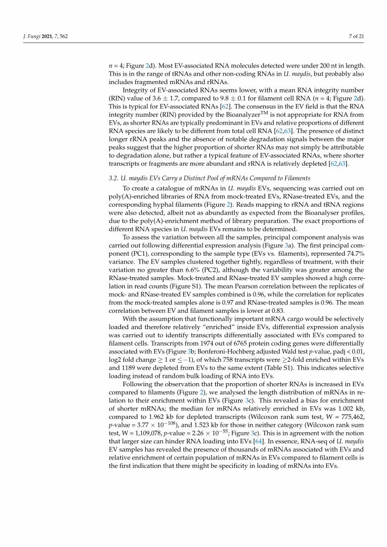
BioanalyzerTM profiles of EV-associated RNAs showed the presence of distinct 18S and 28S ribosomal RNA (rRNA) peaks and a larger, broader peak of less than 200 nt (Fig-ure 2a,b). 18S and 28S rRNAs occupy a lesser proportion (7.3% ± 1.7; n = 4) in EV samples (Figure 2a) compared to total RNA samples of filamentous cells (45.2% ± 3.0; n = 4; Figure
Figure 2. Extracellular RNA associated with U. maydis EVs. (a) Bioanalyzer profile of RNA extractedfrom EVs suspended in PBS, incubated with additional PBS as a mock treatment. (b) RNA from EVstreated with 0.1 µg/µL RNase A in PBS. (c) RNA from EVs treated with 0.1 µg/µL RNase A and0.1% (v/v) triton X-100 in PBS. (d) Total RNA of induced hyphal filament cells, from which above EVsamples were derived.
BioanalyzerTM profiles of EV-associated RNAs showed the presence of distinct 18Sand 28S ribosomal RNA (rRNA) peaks and a larger, broader peak of less than 200 nt(Figure 2a,b). 18S and 28S rRNAs occupy a lesser proportion (7.3% ± 1.7; n = 4) in EVsamples (Figure 2a) compared to total RNA samples of filamentous cells (45.2% ± 3.0;
J. Fungi 2021, 7, 562 7 of 21
n = 4; Figure 2d). Most EV-associated RNA molecules detected were under 200 nt in length.This is in the range of tRNAs and other non-coding RNAs in U. maydis, but probably alsoincludes fragmented mRNAs and rRNAs.
Integrity of EV-associated RNAs seems lower, with a mean RNA integrity number(RIN) value of 3.6 ± 1.7, compared to 9.8 ± 0.1 for filament cell RNA (n = 4; Figure 2d).This is typical for EV-associated RNAs [62]. The consensus in the EV field is that the RNAintegrity number (RIN) provided by the BioanalyzerTM is not appropriate for RNA fromEVs, as shorter RNAs are typically predominant in EVs and relative proportions of differentRNA species are likely to be different from total cell RNA [62,63]. The presence of distinctlonger rRNA peaks and the absence of notable degradation signals between the majorpeaks suggest that the higher proportion of shorter RNAs may not simply be attributableto degradation alone, but rather a typical feature of EV-associated RNAs, where shortertranscripts or fragments are more abundant and rRNA is relatively depleted [62,63].
3.2. U. maydis EVs Carry a Distinct Pool of mRNAs Compared to Filaments
To create a catalogue of mRNAs in U. maydis EVs, sequencing was carried out onpoly(A)-enriched libraries of RNA from mock-treated EVs, RNase-treated EVs, and thecorresponding hyphal filaments (Figure 2). Reads mapping to rRNA and tRNA regionswere also detected, albeit not as abundantly as expected from the Bioanalyser profiles,due to the poly(A)-enrichment method of library preparation. The exact proportions ofdifferent RNA species in U. maydis EVs remains to be determined.
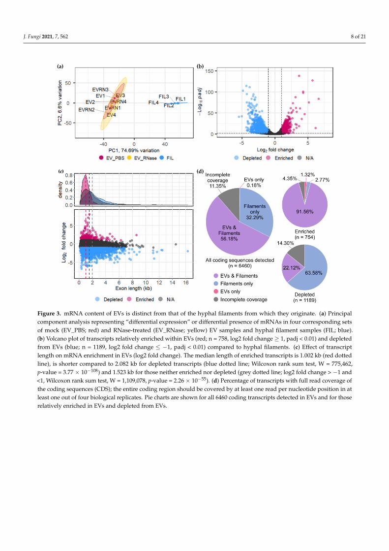
To assess the variation between all the samples, principal component analysis wascarried out following differential expression analysis (Figure 3a). The first principal com-ponent (PC1), corresponding to the sample type (EVs vs. filaments), represented 74.7%variance. The EV samples clustered together tightly, regardless of treatment, with theirvariation no greater than 6.6% (PC2), although the variability was greater among theRNase-treated samples. Mock-treated and RNase-treated EV samples showed a high corre-lation in read counts (Figure S1). The mean Pearson correlation between the replicates ofmock- and RNase-treated EV samples combined is 0.96, while the correlation for replicatesfrom the mock-treated samples alone is 0.97 and RNase-treated samples is 0.96. The meancorrelation between EV and filament samples is lower at 0.83.
With the assumption that functionally important mRNA cargo would be selectivelyloaded and therefore relatively “enriched” inside EVs, differential expression analysiswas carried out to identify transcripts differentially associated with EVs compared tofilament cells. Transcripts from 1974 out of 6765 protein coding genes were differentiallyassociated with EVs (Figure 3b; Bonferoni-Hochberg adjusted Wald test p-value, padj < 0.01,log2 fold change ≥ 1 or ≤−1), of which 758 transcripts were ≥2-fold enriched within EVsand 1189 were depleted from EVs to the same extent (Table S1). This indicates selectiveloading instead of random bulk loading of RNA into EVs.
Following the observation that the proportion of shorter RNAs is increased in EVscompared to filaments (Figure 2), we analysed the length distribution of mRNAs in re-lation to their enrichment within EVs (Figure 3c). This revealed a bias for enrichmentof shorter mRNAs; the median for mRNAs relatively enriched in EVs was 1.002 kb,compared to 1.962 kb for depleted transcripts (Wilcoxon rank sum test, W = 775,462,p-value = 3.77 × 10−108), and 1.523 kb for those in neither category (Wilcoxon rank sumtest, W = 1,109,078, p-value = 2.26 × 10−55; Figure 3c). This is in agreement with the notionthat larger size can hinder RNA loading into EVs [64]. In essence, RNA-seq of U. maydisEV samples has revealed the presence of thousands of mRNAs associated with EVs andrelative enrichment of certain population of mRNAs in EVs compared to filament cells isthe first indication that there might be specificity in loading of mRNAs into EVs.
J. Fungi 2021, 7, 562 8 of 21J. Fungi 2021, 7, x FOR PEER REVIEW 8 of 21
Figure 3. mRNA content of EVs is distinct from that of the hyphal filaments from which they originate. (a) Principal component analysis representing “differential expression” or differential presence of mRNAs in four corresponding sets of mock (EV_PBS; red) and RNase-treated (EV_RNase; yellow) EV samples and hyphal filament samples (FIL; blue). (b) Volcano plot of transcripts relatively enriched within EVs (red; n = 758, log2 fold change ≥ 1, padj < 0.01) and depleted from EVs (blue; n = 1189, log2 fold change ≤ −1, padj < 0.01) compared to hyphal filaments. (c) Effect of transcript length on mRNA enrichment in EVs (log2 fold change). The median length of enriched transcripts is 1.002 kb (red dotted line), is shorter compared to 2.082 kb for depleted transcripts (blue dotted line; Wilcoxon rank sum test, W = 775462, p-value = 3.77 × 10−108) and 1.523 kb for those neither enriched nor depleted (grey dotted line; log2 fold change > −1 and < 1, Wilcoxon rank sum test, W = 1109078, p-value = 2.26 × 10−55). (d) Percentage of transcripts with full read coverage of the coding sequences (CDS); the entire coding region should be covered by at least one read per nucleotide position in at least one out of four biological replicates. Pie charts are shown for all 6460 coding transcripts detected in EVs and for those relatively enriched in EVs and depleted from EVs.
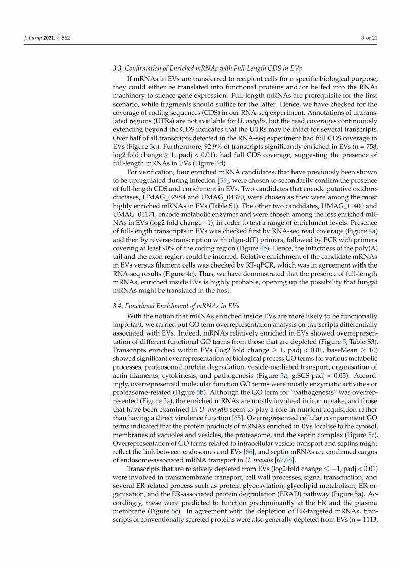
3.3. Confirmation of Enriched mRNAs with Full-Length CDS in EVs If mRNAs in EVs are transferred to recipient cells for a specific biological purpose,
they could either be translated into functional proteins and/or be fed into the RNAi ma-chinery to silence gene expression. Full-length mRNAs are prerequisite for the first sce-nario, while fragments should suffice for the latter. Hence, we have checked for the cov-erage of coding sequences (CDS) in our RNA-seq experiment. Annotations of untranslated regions (UTRs) are not available for U. maydis, but the read coverages continuously ex-tending beyond the CDS indicates that the UTRs may be intact for several transcripts. Over half of all transcripts detected in the RNA-seq experiment had full CDS coverage in EVs (Figure 3d). Furthermore, 92.9% of transcripts significantly enriched in EVs (n = 758, log2 fold change ≥ 1, padj < 0.01), had full CDS coverage, suggesting the presence of full-length mRNAs in EVs (Figure 3d).
Figure 3. mRNA content of EVs is distinct from that of the hyphal filaments from which they originate. (a) Principalcomponent analysis representing “differential expression” or differential presence of mRNAs in four corresponding setsof mock (EV_PBS; red) and RNase-treated (EV_RNase; yellow) EV samples and hyphal filament samples (FIL; blue).(b) Volcano plot of transcripts relatively enriched within EVs (red; n = 758, log2 fold change ≥ 1, padj < 0.01) and depletedfrom EVs (blue; n = 1189, log2 fold change ≤ −1, padj < 0.01) compared to hyphal filaments. (c) Effect of transcriptlength on mRNA enrichment in EVs (log2 fold change). The median length of enriched transcripts is 1.002 kb (red dottedline), is shorter compared to 2.082 kb for depleted transcripts (blue dotted line; Wilcoxon rank sum test, W = 775,462,p-value = 3.77 × 10−108) and 1.523 kb for those neither enriched nor depleted (grey dotted line; log2 fold change > −1 and<1, Wilcoxon rank sum test, W = 1,109,078, p-value = 2.26 × 10−55). (d) Percentage of transcripts with full read coverage ofthe coding sequences (CDS); the entire coding region should be covered by at least one read per nucleotide position in atleast one out of four biological replicates. Pie charts are shown for all 6460 coding transcripts detected in EVs and for thoserelatively enriched in EVs and depleted from EVs.
J. Fungi 2021, 7, 562 9 of 21
3.3. Confirmation of Enriched mRNAs with Full-Length CDS in EVs
If mRNAs in EVs are transferred to recipient cells for a specific biological purpose,they could either be translated into functional proteins and/or be fed into the RNAimachinery to silence gene expression. Full-length mRNAs are prerequisite for the firstscenario, while fragments should suffice for the latter. Hence, we have checked for thecoverage of coding sequences (CDS) in our RNA-seq experiment. Annotations of untrans-lated regions (UTRs) are not available for U. maydis, but the read coverages continuouslyextending beyond the CDS indicates that the UTRs may be intact for several transcripts.Over half of all transcripts detected in the RNA-seq experiment had full CDS coverage inEVs (Figure 3d). Furthermore, 92.9% of transcripts significantly enriched in EVs (n = 758,log2 fold change ≥ 1, padj < 0.01), had full CDS coverage, suggesting the presence offull-length mRNAs in EVs (Figure 3d).
For verification, four enriched mRNA candidates, that have previously been shownto be upregulated during infection [56], were chosen to secondarily confirm the presenceof full-length CDS and enrichment in EVs. Two candidates that encode putative oxidore-ductases, UMAG_02984 and UMAG_04370, were chosen as they were among the mosthighly enriched mRNAs in EVs (Table S1). The other two candidates, UMAG_11400 andUMAG_01171, encode metabolic enzymes and were chosen among the less enriched mR-NAs in EVs (log2 fold change ~1), in order to test a range of enrichment levels. Presenceof full-length transcripts in EVs was checked first by RNA-seq read coverage (Figure 4a)and then by reverse-transcription with oligo-d(T) primers, followed by PCR with primerscovering at least 90% of the coding region (Figure 4b). Hence, the intactness of the poly(A)tail and the exon region could be inferred. Relative enrichment of the candidate mRNAsin EVs versus filament cells was checked by RT-qPCR, which was in agreement with theRNA-seq results (Figure 4c). Thus, we have demonstrated that the presence of full-lengthmRNAs, enriched inside EVs is highly probable, opening up the possibility that fungalmRNAs might be translated in the host.
3.4. Functional Enrichment of mRNAs in EVs
With the notion that mRNAs enriched inside EVs are more likely to be functionallyimportant, we carried out GO term overrepresentation analysis on transcripts differentiallyassociated with EVs. Indeed, mRNAs relatively enriched in EVs showed overrepresen-tation of different functional GO terms from those that are depleted (Figure 5; Table S3).Transcripts enriched within EVs (log2 fold change ≥ 1, padj < 0.01, baseMean ≥ 10)showed significant overrepresentation of biological process GO terms for various metabolicprocesses, proteosomal protein degradation, vesicle-mediated transport, organisation ofactin filaments, cytokinesis, and pathogenesis (Figure 5a; g:SCS padj < 0.05). Accord-ingly, overrepresented molecular function GO terms were mostly enzymatic activities orproteasome-related (Figure 5b). Although the GO term for “pathogenesis” was overrep-resented (Figure 5a), the enriched mRNAs are mostly involved in iron uptake, and thosethat have been examined in U. maydis seem to play a role in nutrient acquisition ratherthan having a direct virulence function [65]. Overrepresented cellular compartment GOterms indicated that the protein products of mRNAs enriched in EVs localise to the cytosol,membranes of vacuoles and vesicles, the proteasome, and the septin complex (Figure 5c).Overrepresentation of GO terms related to intracellular vesicle transport and septins mightreflect the link between endosomes and EVs [66], and septin mRNAs are confirmed cargosof endosome-associated mRNA transport in U. maydis [67,68].
Transcripts that are relatively depleted from EVs (log2 fold change ≤ −1, padj < 0.01)were involved in transmembrane transport, cell wall processes, signal transduction, andseveral ER-related process such as protein glycosylation, glycolipid metabolism, ER or-ganisation, and the ER-associated protein degradation (ERAD) pathway (Figure 5a). Ac-cordingly, these were predicted to function predominantly at the ER and the plasmamembrane (Figure 5c). In agreement with the depletion of ER-targeted mRNAs, tran-scripts of conventionally secreted proteins were also generally depleted from EVs (n = 1113,
J. Fungi 2021, 7, 562 10 of 21
log2 fold change ≤ −1, padj < 0.01, baseMean ≥ 10, g:SCS padj = 2.08 × 10⁻¹³). These re-sults suggest that subcellular localisation of mRNAs may affect their loading into EVs: mR-NAs associated with intracellular vesicles are more likely to be loaded into EVs, while thosethat require translation at the rough ER are relatively depleted from EVs.
Since the 3′UTR region is particularly important for subcellular localisation of mR-NAs [69], we have partially annotated the UTRs based on the mapped sequencing readsand carried out a preliminary analysis to test if there is a difference between the 3′UTRsof mRNAs enriched in EVs and those depleted from EVs. 3′UTRs of enriched mRNAsshowed a higher frequency of adenine nucleotides (p = 1.3 × 10⁻¹⁹) and less cytosine(p = 1.1 × 10⁻⁷) and uridine (p = 9.6 × 10⁻³) compared to the depleted sequences. Ac-cordingly, A-rich 4-mers were significantly increased in frequency among the enriched3′UTR sequences compared to the depleted (p < 0.05; Figure S2b). This prompts deeperinvestigation into EV-targeting motifs in the future.
J. Fungi 2021, 7, x FOR PEER REVIEW 10 of 21
Figure 4. Presence of full-length mRNAs enriched within EVs. (a) RNA-seq read coverage of se-lected infection-relevant, EV-associated mRNA candidates in four biological replicates each of EV and filament samples. Y-axis shows normalised coverage in bases per million (bpm) and the range is indicated in brackets. X-axis is length in kb (scale bar = 1 kb). (b) Confirmation of full-length mRNA candidates by RT-PCR. Primers to yield amplicons covering ≥ 90% of transcript coding re-gion length were used. RT indicates that the reverse-transcribed first-strand cDNA was used as a template for PCR and “-“ sign indicates a -RT negative control. (c) Confirmation of relative transcript enrichment in EVs compared to filaments by RT-qPCR. Fold relative enrichment within EVs calcu-lated as 2^-ddCt. Inset shows fold enrichment of UMAG_11400 and UMAG_01171 with an adjusted y-axis.
Figure 4. Presence of full-length mRNAs enriched within EVs. (a) RNA-seq read coverage of selectedinfection-relevant, EV-associated mRNA candidates in four biological replicates each of EV andfilament samples. Y-axis shows normalised coverage in bases per million (bpm) and the range isindicated in brackets. X-axis is length in kb (scale bar = 1 kb). (b) Confirmation of full-length mRNAcandidates by RT-PCR. Primers to yield amplicons covering ≥ 90% of transcript coding region lengthwere used. RT indicates that the reverse-transcribed first-strand cDNA was used as a template forPCR and “-“ sign indicates a -RT negative control. (c) Confirmation of relative transcript enrichmentin EVs compared to filaments by RT-qPCR. Fold relative enrichment within EVs calculated as 2ˆ-ddCt.Inset shows fold enrichment of UMAG_11400 and UMAG_01171 with an adjusted y-axis.

J. Fungi 2021, 7, 562 11 of 21J. Fungi 2021, 7, x FOR PEER REVIEW 12 of 21
Figure 5. Gene ontology (GO) term analysis of mRNAs differentially loaded into EVs. Biological process (a), cellular com-partment (b), and molecular function (c) GO terms significantly overrepresented (g:SCS padj < 0.05) in sets of transcripts enriched (red clusters; n= 748, baseMean ≥ 10, log2 fold change ≥ 1, padj < 0.01) and depleted from EVs (blue clusters; n= 1113, baseMean ≥ 10, log2 fold change ≤ −1, padj < 0.01).
Since the 3′UTR region is particularly important for subcellular localisation of mRNAs [69], we have partially annotated the UTRs based on the mapped sequencing reads and carried out a preliminary analysis to test if there is a difference between the 3′UTRs of mRNAs enriched in EVs and those depleted from EVs. 3′UTRs of enriched mRNAs showed a higher frequency of adenine nucleotides (p = 1.3 × 10⁻¹⁹) and less cyto-sine (p = 1.1 × 10⁻⁷) and uridine (p = 9.6 × 10⁻³) compared to the depleted sequences. Ac-cordingly, A-rich 4-mers were significantly increased in frequency among the enriched
Figure 5. Gene ontology (GO) term analysis of mRNAs differentially loaded into EVs. Biological process (a), molecularfunction (b), cellular compartment (c). GO terms significantly overrepresented (g:SCS padj < 0.05) in sets of transcriptsenriched (red clusters; n = 748, baseMean ≥ 10, log2 fold change ≥ 1, padj < 0.01) and depleted from EVs (blue clusters;n = 1113, baseMean ≥ 10, log2 fold change ≤ −1, padj < 0.01).

J. Fungi 2021, 7, 562 12 of 21
3.5. mRNAs Upregulated during Infection Are Present in EVs from Axenic Filaments
Since U. maydis is a plant pathogen, we asked whether a portion of EV-associatedmRNAs are relevant for infection. Many genes pertinent for infection are expressed inAB33 filaments in culture due to the transcriptional cascade instigated by bE/bW [35].Indeed, mRNAs of at least nine previously characterised bona fide effectors and secretedproteins linked to virulence were reliably detected in EVs: Stp2 [70], ApB73 [71], Scp2 [72],UMAG_01690 [73], Sta1 [74], Stp1 [75], Nuc1 [76], Cmu1 [77], and UmFly1 [78] (in theorder of enrichment in EVs; Table S4).
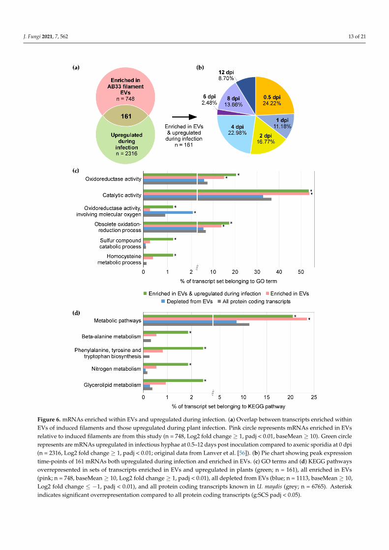
Next, we searched for mRNAs enriched in EVs, that are also upregulated during plantinfection. For this, we referred to the published time-course transcriptomic analysis ofU. maydis infection [56]. 161 mRNAs were found to be both enriched in EVs and upreg-ulated during infection compared to the axenic sporidia at 0 dpi (Figure 6a & Table S5).Over three-quarters of these were induced early on, during the first four days of infection(Figure 6b). GO term analysis of these 161 mRNAs found an overrepresentation of oxi-doreductase and other catalytic enzyme activities, as well as functions linked to sulphurcompound catabolism and homocysteine metabolism (g:SCS padj < 0.05; Figure 6c). We fur-ther examined KEGG pathways [52] and found significant overrepresentation of functionsin metabolic pathways, including beta-alanine metabolism, aromatic amino acid biosyn-thesis, nitrogen metabolism, and glycerolipid metabolism (g:SCS padj < 0.05; Figure 6d).If pathogen EV-associated mRNAs can act as effectors, such metabolic enzymes may berelevant, as U. maydis is known to reprogram plant host metabolism [79].
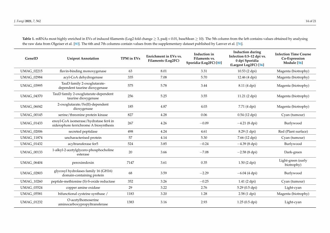
We have examined the most highly enriched mRNAs (n = 17, Log2 fold change ≥ 3,padj < 0.01, baseMean ≥ 10), with the assumption that these are more likely to have beenloaded in EVs to serve a biological function (Table 1). Many of the most enriched mRNAsencode oxidoreductases with similar annotations, suggesting related activities. This reflectsthe general overrepresentation of GO terms for oxidoreductases and metabolic enzymes(Figures 5 and 6). Secondly, 10 out of the 17 most enriched mRNAs are induced concomitantwith filamentous growth (Log2 fold change ≥ 1, padj < 0.01; [80]), and are up-regulatedduring infection (Log2 fold change ≥ 1, padj < 0.01; [56]). Furthermore, with reference tothe previously defined co-expression modules from an extensive infectious time-coursestudy [56], we found an overrepresentation of the “magenta” infection-related expres-sion module representative of biotrophic proliferation in planta (g:SCS padj = 1.43 × 10⁻⁴).In essence, we observe an enrichment of mRNAs in EVs that can be linked to filamentinduction and infection.
J. Fungi 2021, 7, 562 13 of 21J. Fungi 2021, 7, x FOR PEER REVIEW 14 of 21
Figure 6. mRNAs enriched within EVs and upregulated during infection. (a) Overlap between transcripts enriched within EVs of induced filaments and those upregulated during plant infection. Pink circle represents mRNAs enriched in EVs relative to induced filaments are from this study (n = 748, Log2 fold change ≥ 1, padj < 0.01, baseMean ≥ 10). Green circle represents are mRNAs upregulated in infectious hyphae at 0.5–12 days post inoculation compared to axenic sporidia at 0 dpi (n = 2316, Log2 fold change ≥ 1, padj < 0.01; original data from Lanver et al. [56]). (b) Pie chart showing peak expression time-points of 161 mRNAs both upregulated during infection and enriched in EVs. (c) GO terms and (d) KEGG pathways overrepresented in sets of transcripts enriched in EVs and upregulated in plants (green; n = 161), all enriched in EVs (pink; n = 748, baseMean ≥ 10, Log2 fold change ≥ 1, padj < 0.01), all depleted from EVs (blue; n = 1113, baseMean ≥ 10, Log2 fold change ≤ −1, padj < 0.01), and all protein coding transcripts known in U. maydis (grey; n = 6765). Asterisk indicates signifi-cant overrepresentation compared to all protein coding transcripts (g:SCS padj < 0.05).
Figure 6. mRNAs enriched within EVs and upregulated during infection. (a) Overlap between transcripts enriched withinEVs of induced filaments and those upregulated during plant infection. Pink circle represents mRNAs enriched in EVsrelative to induced filaments are from this study (n = 748, Log2 fold change ≥ 1, padj < 0.01, baseMean ≥ 10). Green circlerepresents are mRNAs upregulated in infectious hyphae at 0.5–12 days post inoculation compared to axenic sporidia at 0 dpi(n = 2316, Log2 fold change ≥ 1, padj < 0.01; original data from Lanver et al. [56]). (b) Pie chart showing peak expressiontime-points of 161 mRNAs both upregulated during infection and enriched in EVs. (c) GO terms and (d) KEGG pathwaysoverrepresented in sets of transcripts enriched in EVs and upregulated in plants (green; n = 161), all enriched in EVs(pink; n = 748, baseMean ≥ 10, Log2 fold change ≥ 1, padj < 0.01), all depleted from EVs (blue; n = 1113, baseMean ≥ 10,Log2 fold change ≤ −1, padj < 0.01), and all protein coding transcripts known in U. maydis (grey; n = 6765). Asteriskindicates significant overrepresentation compared to all protein coding transcripts (g:SCS padj < 0.05).
J. Fungi 2021, 7, 562 14 of 21
Table 1. mRNAs most highly enriched in EVs of induced filaments (Log2 fold change ≥ 3, padj < 0.01, baseMean ≥ 10). The 5th column from the left contains values obtained by analysingthe raw data from Olgeiser et al. [80]. The 6th and 7th columns contain values from the supplementary dataset published by Lanver et al. [56].
GeneID Uniprot Annotation TPM in EVs Enrichment in EVs vs.Filaments (Log2FC)
Induction inFilaments vs.
Sporidia (Log2FC) [80]
Induction duringInfection 0.5–12 dpi vs.
0 dpi Sporidia(Largest Log2FC) [56]
Infection Time CourseCo-Expression
Module [56]
UMAG_02215 flavin-binding monoxygenase 63 8.01 3.31 10.53 (2 dpi) Magenta (biotrophy)
UMAG_02984 acyl-CoA dehydrogenase 335 7.08 5.70 12.46 (4 dpi) Magenta (biotrophy)
UMAG_03995 TauD family 2-oxoglutarate-dependent taurine dioxygenase 575 5.78 3.44 8.11 (4 dpi) Magenta (biotrophy)
UMAG_04370 TauD family 2-oxoglutarate-dependenttaurine dioxygenase 256 5.25 3.55 11.21 (2 dpi) Magenta (biotrophy)
UMAG_06042 2-oxoglutarate/Fe(II)-dependentdioxygenase 185 4.87 4.03 7.71 (4 dpi) Magenta (biotrophy)
UMAG_00145 serine/threonine protein kinase 827 4.28 0.06 0.54 (12 dpi) Cyan (tumour)
UMAG_01433 enoyl-CoA isomerase/hydratase fer4 insiderophore ferrichrome A biosynthesis 267 4.26 −0.09 −4.21 (8 dpi) Burlywood
UMAG_02006 secreted peptidase 498 4.24 4.61 8.29 (1 dpi) Red (Plant surface)
UMAG_11874 uncharacterised protein 57 4.14 5.30 7.66 (12 dpi) Cyan (tumour)
UMAG_01432 acyltransferase fer5 524 3.85 −0.24 −4.39 (8 dpi) Burlywood
UMAG_00133 1-alkyl-2-acetylglycero-phosphocholineesterase 20 3.66 −7.08 −2.58 (8 dpi) Dark-green
UMAG_06404 peroxiredoxin 7147 3.61 0.35 1.50 (2 dpi) Light-green (earlybiotrophy)
UMAG_02803 glycosyl hydrolases family 16 (GH16)domain-containing protein 68 3.59 −2.29 −6.04 (4 dpi) Burlywood
UMAG_10260 peptide-methionine (S)-S-oxide reductase 352 3.26 −0.25 1.41 (2 dpi) Cyan (tumour)
UMAG_03524 copper amine oxidase 29 3.22 2.76 5.29 (0.5 dpi) Light-cyan
UMAG_05581 bifunctional cysteine synthase / 1183 3.20 1.28 2.58 (1 dpi) Magenta (biotrophy)
UMAG_01232 O-acetylhomoserineaminocarboxypropyltransferase 1383 3.16 2.93 1.25 (0.5 dpi) Light-cyan
J. Fungi 2021, 7, 562 15 of 21
4. Discussion
EVs are emerging as mediators of plant-pathogen communication, particularly asvehicles for transfer of RNA (reviewed in [81]). On the plant side, studies to date havemostly focused on the role of sRNAs [19,20] and proteins [22,23,82] in EVs, while onlyprotein cargos have been examined in EVs of phytopathogenic fungi [9,24]. To extend thesearch for EV cargo molecules in plant-pathogen communication, we have characterisedthe repertoire of mRNAs associated with EVs of the fungus U. maydis.
For this purpose, we developed a robust EV isolation (or enrichment) protocol andexamined EVs produced in axenic cultures of U. maydis filaments, used as a surrogate forinfectious hyphae in planta (Figure 1). Omics studies on EVs of phytopathogenic fungi haveso far examined EVs from axenic cultures [9,24]. While there are limitations to using axeniccultures of pathogenic fungi to identify EV cargos linked to virulence, isolation of fungalEVs from apoplastic washing fluid of maize plants is inherently destructive [83,84] andwould first require development of markers for U. maydis EVs. Induced filaments of thelab strain AB33 in axenic culture mimic the morphology and, partially, the gene expressionof infectious filaments [34–36] Therefore, we have used these cultures for an initial surveyof EV-associated mRNAs in U. maydis.
We have reliably detected transcripts of bona fide effectors and several secreted proteinslinked to virulence in AB33 filaments and their EVs (Table S4), which supports that oursystem has the potential to lead to discovery of novel EV-associated effectors. Thousandsof mRNAs were detected in association with U. maydis EVs, the majority of which havefull-length coverage (Figures 3d and 4) and are protected from external RNases (Figure 2).Protease activity in U. maydis cultures is high, requiring deletion of multiple proteases toobtain intact secreted proteins from U. maydis cultures [85]. Likewise, U. maydis secretesRNases in culture [76]. Therefore, it is unlikely that so many mRNAs can preserve integrityin the culture medium, unless they are protected inside EVs. Over 90% of the mRNAs en-riched inside EVs are likely to be full-length (Figure 3d), suggesting that there is a biologicalreason for loading these mRNAs into EVs.
mRNA loading into EVs may be determined by the intracellular location of the mRNAsinside the fungal filament. The two most relevant EV subtypes are exosomes and microvesi-cles. Exosomes are originally intraluminal vesicles (ILVs) in multivesicular endosomes(MVEs) that are released upon fusion with the plasma membrane, while microvesiclesare formed by direct budding from the plasma membrane [66]. Hence, localisation onthe surface of maturing endosomes or at the cell periphery would increase the likelihoodof being loaded into exosomes and microvesicles, respectively. This might explain whymRNAs encoding proteins linked to intracellular vesicles and vacuoles are enriched in EVsof U. maydis (Figure 5). Discovery of EV-associated RNA-binding proteins should helpelucidate the mechanism of mRNA loading.
Since mRNAs enriched within EVs encode proteins with functions distinct fromthose that are depleted, we suspect a biological reason for preferentially exporting thesemRNAs. Since several transcripts upregulated during infection are enriched in U. maydisEVs (Table 1; Figure 6a), such mRNAs could be studied further as effector candidates.There are two non-mutually exclusive hypotheses for the role of EV cargo mRNAs inU. maydis-maize interaction: (1) fungal mRNA fragments lead to silencing of maize genesor (2) full-length fungal mRNAs are translated into multiple effector proteins in maize cells.
Bidirectional, cross-kingdom RNA interference (RNAi) is a widespread mechanismof plant-pathogen interaction [86]. Diverse fungal and oomycete pathogens send sRNAeffectors that hijack the plant RNAi machinery to silence host defence genes [20,25–29]As is the case for plant sRNAs that target pathogen genes [19], EVs are thought to be thevehicles of pathogen sRNA effector delivery to host plant cells. U. maydis has lost theconventional RNAi machinery [33]. Therefore, it might employ other RNA species, such asmRNA or tRNA fragments for the same purpose. For example, tRNA-fragments of the
J. Fungi 2021, 7, 562 16 of 21
bacterial symbiont Bradyrhizobium japonicum participate in silencing of plant genes involvedin root hair development to promote nodulation [87].
Effector delivery in the form of mRNA could be highly cost-effective for the pathogen,if they can be translated in the correct location at required amplitude in the host cell.In support of this hypothesis, proof of principle studies using elegant reporter systemshave demonstrated that EV-associated mRNAs are transferred and translated de novo in therecipient cells [88,89]. A recent in vivo study has shown that mRNAs in glioblastoma EVsare most likely translated in recipient astrocytes and lead to metabolic reprogramming [90].Also, in the medically important Paracoccidioides spp., mRNA cargos of EVs were foundto be translation-competent in a heterologous, in vitro system [91]. Given these examplesand the evidence for full-length EV cargo mRNAs from this study (Figures 3d and 4b),translation of EV-associated fungal mRNAs into functional proteins in recipient cellsseems possible.
It is interesting that the set of mRNAs enriched in EVs and upregulated during infec-tion are overrepresented in metabolic enzymes and oxidoreductases (Figure 6). Biotrophiccolonisation by U. maydis is accompanied by extensive reprogramming of metabolism,redox status, and hormone signalling in the infected plant tissues [79]. The fungus deployseffectors to divert metabolites away from biosynthesis of lignin [92] and salicylic acid(SA; [77]), and induces the jasmonate/ethylene signalling pathway to counter SA-mediateddefence [93]. U. maydis also harbours metabolic enzymes lacking signal peptides that cansynthesise [94,95], degrade [96], or potentially alter metabolic flux into biosynthesis ofplant hormones [41]. Intriguingly, isochorismatases, which divert isochorismate away fromSA biosynthesis in the host cell, are unconventionally secreted effectors of the filamentousphytopathogens, Verticillium dahliae and Phytophthora sojae [97]. Similarly, fungal metabolicand redox enzymes, that are upregulated during infection and loaded into EVs in the formof mRNA or protein, have the potential to “moonlight” as effectors if delivered to thehost cell. Thus, the presence of intact, enriched mRNAs in EVs of U. maydis present anopportunity to discover novel RNA effectors in plant pathogenic fungi.
5. Conclusions
We have isolated EVs from the phytopathogenic fungus U. maydis and identifiedmRNAs that are enriched within EVs compared to the cells. Many of the highly enrichedmRNAs are also upregulated during infection and are likely to be full-length. The inventoryof these mRNAs now forms the foundation for future research addressing the mechanismof mRNA loading into EVs and their function in a recipient cell.
Supplementary Materials: The following are available online at https://www.mdpi.com/article/10.3390/jof7070562/s1. Figure S1: Heatmap showing Pearson correlation between the biologicalreplicates of mock- and RNase- treated EV samples and the corresponding filament samples se-quenced. Figure S2: Overrepresented nucleotides and k-mers in the 3′UTRs of mRNAs enriched inEVs. Table S1: Differential enrichment of transcripts in EVs and filaments. Table S2: Primers used inthis study. Table S3: Significantly overrepresented GO terms among transcripts enriched in EVs anddepleted from EVs. Table S4: Transcripts of known secreted proteins linked to virulence that weredetected in this study. Table S5: 161 Transcripts enriched in EVs and upregulated during infection.
Author Contributions: Conceptualisation by S.K. and M.F.; methodology, investigation, originaldraft preparation, visualisation by S.K.; library preparation and RNA sequencing by A.B.; RNA-seqanalysis by O.R. and S.K.; UTR annotation by A.K.; nucleotide and k-mer frequency analysis byC.F.B.; review and editing by M.F. and O.R.; supervision and project administration by M.F.; fundingacquisition by M.F., A.G. and S.K. All authors have read and agreed to the published version ofthe manuscript.
Funding: This research was funded by grants from the Deutsche Forschungsgemeinschaft (DFG, Ger-man Research Foundation) under Germany’s Excellence Strategy EXC-2048/1—Project ID 39068111to MF; DFG-FOR5116 subproject B3 (FE448/15-1) to M.F. as well as DFG-FOR5116 subproject C1to A.G., S.K. was funded by the Deutscher Akademischer Austausch Dienst (DAAD, German Aca-demic Exchange Service; Graduate School Scholarship Programme 57243780) in the framework of

J. Fungi 2021, 7, 562 17 of 21
iGRAD-Plant Graduate School (DFG/GRK152) and DFG-FOR5116. C.F.B. was funded by the JürgenManchot Stiftung Düsseldorf (Jürgen Manchot Foundation Düsseldorf).
Institutional Review Board Statement: Not applicable.
Informed Consent Statement: Not applicable.
Data Availability Statement: Raw and processed RNA-seq data are openly available at the GEOdatabase under accession number GSE176292: https://www.ncbi.nlm.nih.gov/geo/query/acc.cgi?acc=GSE176292 (2 July 2021).
Acknowledgments: We thank Vera Göhre for critical reading of the manuscript and Libera Lo Prestifor invaluable discussions in the initial phase of the project. We are grateful for excellent technicalassistance provided by Marion Nissen at Center for Advanced Imaging (CAi, HHU Düsseldorf),who has operated TEM, and Gisela Brinkmann, who has assisted in RNA-seq library preparation.We thank the Bioinformatics Core Facility at the chair of Systems Biology at JLU Giessen for assistancein RNA-seq analysis, as well as provision of computational resources and general support by theBiGi service center (BMBF grant 031A533) within the de.NBI network.
Conflicts of Interest: The authors declare no conflict of interest. The funders had no role in the designof the study; in the collection, analyses, or interpretation of data; in the writing of the manuscript,or in the decision to publish the results.
References1. Tkach, M.; Thery, C. Communication by extracellular vesicles: Where we are and where we need to go. Cell 2016, 164, 1226–1232.
[CrossRef]2. Regev-Rudzki, N.; Wilson, D.W.; Carvalho, T.G.; Sisquella, X.; Coleman, B.M.; Rug, M.; Bursac, D.; Angrisano, F.; Gee, M.;
Hill, A.F.; et al. Cell-cell communication between malaria-infected red blood cells via exosome-like vesicles. Cell 2013, 153,1120–1133. [CrossRef] [PubMed]
3. Bielska, E.; Sisquella, M.A.; Aldeieg, M.; Birch, C.; O’Donoghue, E.J.; May, R.C. Pathogen-derived extracellular vesicles mediatevirulence in the fatal human pathogen Cryptococcus gattii. Nat. Commun. 2018, 9, 1556. [CrossRef] [PubMed]
4. Kuipers, M.E.; Hokke, C.H.; Smits, H.H. Pathogen-derived extracellular vesicle-associated molecules that affect the host immunesystem: An overview. Front. Microbiol. 2018, 9, 2182. [CrossRef] [PubMed]
5. Bleackley, M.R.; Dawson, C.S.; Anderson, M.A. Fungal extracellular vesicles with a focus on proteomic analysis. Proteomics 2019,19, 1800232. [CrossRef] [PubMed]
6. Peres da Silva, R.; Puccia, R.; Rodrigues, M.L.; Oliveira, D.L.; Joffe, L.S.; César, G.V.; Nimrichter, L.; Goldenberg, S.; Alves, L.R.Extracellular vesicle-mediated export of fungal RNA. Sci. Rep. 2015, 5, 7763. [CrossRef] [PubMed]
7. Vallejo, M.C.; Nakayasu, E.S.; Longo, L.V.G.; Ganiko, L.; Lopes, F.G.; Matsuo, A.L.; Almeida, I.C.; Puccia, R. Lipidomic analysis ofextracellular vesicles from the pathogenic phase of Paracoccidioides brasiliensis. PLoS ONE 2012, 7, e39463. [CrossRef]
8. Rodrigues, M.L.; Nimrichter, L.; Oliveira, D.L.; Frases, S.; Miranda, K.; Zaragoza, O.; Alvarez, M.; Nakouzi, A.; Feldmesser, M.;Casadevall, A. Vesicular polysaccharide export in Cryptococcus neoformans is a eukaryotic solution to the problem of fungaltrans-cell wall transport. Eukaryot. Cell 2007, 6, 48–59. [CrossRef]
9. Bleackley, M.; Samuel, M.; Garcia-Ceron, D.; Mckenna, J.A.; Lowe, R.G.T.; Pathan, M.; Zhao, K.N.; Ang, C.S.; Mathivanan, S.;Anderson, M.A. Extracellular vesicles from the cotton pathogen Fusarium oxysporum f. sp. vasinfectum induce a phytotoxicresponse in plants. Front. Plant Sci. 2020, 10, 1610. [CrossRef]
10. Zhao, K.N.; Bleackley, M.; Chisanga, D.; Gangoda, L.; Fonseka, P.; Liem, M.; Kalra, H.; Al Saffar, H.; Keerthikumar, S.; Ang, C.S.;et al. Extracellular vesicles secreted by Saccharomyces cerevisiae are involved in cell wall remodelling. Commun. Biol. 2019, 2, 305.[CrossRef]
11. Zarnowski, R.; Sanchez, H.; Covelli, A.S.; Dominguez, E.; Jaromin, A.; Bernhardt, J.; Mitchell, K.F.; Heiss, C.; Azadi, P.; Mitchell, A.;et al. Candida albicans biofilm-induced vesicles confer drug resistance through matrix biogenesis. PLoS Biol. 2018, 16, e2006872.[CrossRef]
12. Rodrigues, M.L.; Nakayasu, E.S.; Oliveira, D.L.; Nimrichter, L.; Nosanchuk, J.D.; Almeida, I.C.; Casadevall, A. Extracellularvesicles produced by Cryptococcus neoformans contain protein components associated with virulence. Eukaryot. Cell 2008, 7, 58–67.[CrossRef]
13. Hai, T.P.; Tuan, T.L.; Van Anh, D.; Mai, T.N.; Phu Huong, L.N.; Thwaites, G.; Johnson, E.; Van Vinh Chau, N.; Baker, S.; Ashton, P.;et al. The virulence of the Cryptococcus neoformans vnia-5 lineage is highly plastic and associated with isolate background. bioRxiv2020. [CrossRef]
14. Ikeda, M.A.K.; de Almeida, J.R.F.; Jannuzzi, G.P.; Cronemberger-Andrade, A.; Torrecilhas, A.C.T.; Moretti, N.S.; da Cunha, J.P.C.;de Almeida, S.R.; Ferreira, K.S. Extracellular vesicles from Sporothrix brasiliensis are an important virulence factor that induce anincrease in fungal burden in experimental sporotrichosis. Front. Microbiol. 2018, 9, 2286. [CrossRef] [PubMed]

J. Fungi 2021, 7, 562 18 of 21
15. Freitas, M.S.; Bonato, V.L.D.; Pessoni, A.M.; Rodrigues, M.L.; Casadevall, A.; Almeidaa, F. Fungal extracellular vesicles aspotential targets for immune interventions. Msphere 2019, 4, e00747-19. [CrossRef] [PubMed]
16. Snetselaar, K.M.; Mims, C.W. Light and electron-microscopy of Ustilago maydis hyphae in maize. Mycol. Res. 1994, 98, 347–355.[CrossRef]
17. An, Q.; Ehlers, K.; Kogel, K.H.; van Bel, A.J.; Hückelhoven, R. Multivesicular compartments proliferate in susceptible andresistant MLA12-barley leaves in response to infection by the biotrophic powdery mildew fungus. New Phytol. 2006, 172, 563–576.[CrossRef] [PubMed]
18. Roth, R.; Hillmer, S.; Funaya, C.; Chiapello, M.; Schumacher, K.; Lo Presti, L.; Kahmann, R.; Paszkowski, U. Arbuscular cellinvasion coincides with extracellular vesicles and membrane tubules. Nat. Plants 2019, 5, 204–211. [CrossRef]
19. Cai, Q.; Qiao, L.; Wang, M.; He, B.; Lin, F.M.; Palmquist, J.; Huang, S.D.; Jin, H. Plants send small RNAs in extracellular vesicles tofungal pathogen to silence virulence genes. Science 2018, 360, 1126–1129. [CrossRef]
20. Hou, Y.; Zhai, Y.; Feng, L.; Karimi, H.Z.; Rutter, B.D.; Zeng, L.; Choi, D.S.; Zhang, B.; Gu, W.; Chen, X.; et al. A Phytophthoraeffector suppresses trans-kingdom rnai to promote disease susceptibility. Cell Host Microbe 2019, 25, 153–165.e155. [CrossRef]
21. Baldrich, P.; Rutter, B.D.; Karimi, H.Z.; Podicheti, R.; Meyers, B.C.; Innes, R.W. Plant extracellular vesicles contain diverse smallRNA species and are enriched in 10-to 17-nucleotide “tiny” rnas. Plant Cell 2019, 31, 315–324. [CrossRef]
22. Rutter, B.D.; Innes, R.W. Extracellular vesicles isolated from the leaf apoplast carry stress-response proteins. Plant Physiol. 2017,173, 728–741. [CrossRef]
23. Regente, M.; Pinedo, M.; San Clemente, H.; Balliau, T.; Jamet, E.; de la Canal, L. Plant extracellular vesicles are incorporated bya fungal pathogen and inhibit its growth. J. Exp. Bot. 2017, 68, 5485–5495. [CrossRef] [PubMed]
24. Hill, E.H.; Solomon, P.S. Extracellular vesicles from the apoplastic fungal wheat pathogen Zymoseptoria tritici. Fungal Biol.Biotechnol. 2020, 7, 13. [CrossRef] [PubMed]
25. Weiberg, A.; Wang, M.; Lin, F.M.; Zhao, H.W.; Zhang, Z.H.; Kaloshian, I.; Huang, H.D.; Jin, H.L. Fungal small RNAs suppressplant immunity by hijacking host RNA interference pathways. Science 2013, 342, 118–123. [CrossRef] [PubMed]
26. Wang, M.; Weiberg, A.; Lin, F.M.; Thomma, B.P.H.J.; Huang, H.D.; Jin, H.L. Bidirectional cross-kingdom RNAi and fungal uptakeof external rnas confer plant protection. Nat. Plants 2016, 2, 16151. [CrossRef]
27. Wang, B.; Sun, Y.; Song, N.; Zhao, M.; Liu, R.; Feng, H.; Wang, X.; Kang, Z. Puccinia striiformis f. sp. tritici microRNA-like rna 1 (Pst-milR1), an important pathogenicity factor of Pst, impairs wheat resistance to Pst by suppressing the wheat pathogenesis-related 2gene. New Phytol. 2017, 215, 338–350. [CrossRef]
28. Jian, J.; Liang, X. One small RNA of Fusarium graminearum targets and silences cebip gene in common wheat. Microorganisms 2019,7, 425. [CrossRef] [PubMed]
29. Dunker, F.; Trutzenberg, A.; Rothenpieler, J.S.; Kuhn, S.; Prols, R.; Schreiber, T.; Tissier, A.; Kemen, A.; Kemen, E.; Huckelhoven, R.;et al. Oomycete small RNAs bind to the plant RNA-induced silencing complex for virulence. eLife 2020, 9, e56096. [CrossRef]
30. Brefort, T.; Doehlemann, G.; Mendoza-Mendoza, A.; Reissmann, S.; Djamei, A.; Kahmann, R. Ustilago maydis as a pathogen. Annu.Rev. Phytopathol. 2009, 47, 423–445. [CrossRef]
31. Fisher, M.C.; Henk, D.A.; Briggs, C.J.; Brownstein, J.S.; Madoff, L.C.; McCraw, S.L.; Gurr, S.J. Emerging fungal threats to animal,plant and ecosystem health. Nature 2012, 484, 186–194. [CrossRef]
32. Haag, C.; Steuten, B.; Feldbrügge, M. Membrane-coupled mRNA trafficking in fungi. Annu. Rev. Microbiol. 2015, 69, 265–281.[CrossRef]
33. Laurie, J.D.; Linning, R.; Bakkeren, G. Hallmarks of RNA silencing are found in the smut fungus Ustilago hordei but not in its closerelative Ustilago maydis. Curr. Genet. 2008, 53, 49–58. [CrossRef] [PubMed]
34. Brachmann, A.; Weinzierl, G.; Kamper, J.; Kahmann, R. Identification of genes in the bW/bE regulatory cascade in Ustilago maydis.Mol. Microbiol. 2001, 42, 1047–1063. [CrossRef] [PubMed]
35. Wahl, R.; Zahiri, A.; Kamper, J. The Ustilago maydis b mating type locus controls hyphal proliferation and expression of secretedvirulence factors in planta. Mol. Microbiol. 2010, 75, 208–220. [CrossRef] [PubMed]
36. Heimel, K.; Scherer, M.; Vranes, M.; Wahl, R.; Pothiratana, C.; Schuler, D.; Vincon, V.; Finkernagel, F.; Flor-Parra, I.; Kamper, J. Thetranscription factor Rbf1 is the master regulator for b-mating type controlled pathogenic development in Ustilago maydis. PLoSPathog. 2010, 6, e1001035. [CrossRef] [PubMed]
37. Holliday, R. Ustilago maydis. In Bacteria, Bacteriophages, and Fungi: Volume 1; King, R.C., Ed.; Springer US: Boston, MA, USA,1974; pp. 575–595.
38. Cicero, A.L.; Delevoye, C.; Gilles-Marsens, F.; Loew, D.; Dingli, F.; Guéré, C.; André, N.; Vié, K.; van Niel, G.; Raposo, G. Exosomesreleased by keratinocytes modulate melanocyte pigmentation. Nat. Commun. 2015, 6, 7506. [CrossRef] [PubMed]
39. Andrews, S. FastQC: A Quality Control Tool for High Throughput Sequence Data. Available online: http://www.bioinformatics.babraham.ac.uk/projects/fastqc/ (accessed on 10 November 2014).
40. Bolger, A.M.; Lohse, M.; Usadel, B. Trimmomatic: A flexible trimmer for Illumina sequence data. Bioinformatics 2014, 30, 2114–2120.[CrossRef]
41. Kämper, J.; Kahmann, R.; Bölker, M.; Ma, L.-J.; Brefort, T.; Saville, B.J.; Banuett, F.; Kronstad, J.W.; Gold, S.E.; Müller, O.; et al.Insights from the genome of the biotrophic fungal plant pathogen Ustilago maydis. Nature 2006, 444, 97–101. [CrossRef]

J. Fungi 2021, 7, 562 19 of 21
42. Howe, K.L.; Contreras-Moreira, B.; De Silva, N.; Maslen, G.; Akanni, W.; Allen, J.; Alvarez-Jarreta, J.; Barba, M.; Bolser, D.M.;Cambell, L.; et al. Ensembl genomes 2020-enabling non-vertebrate genomic research. Nucleic Acids Res. 2020, 48, D689–D695.[CrossRef]
43. Kim, D.; Paggi, J.M.; Park, C.; Bennett, C.; Salzberg, S.L. Graph-based genome alignment and genotyping with HISAT2 andHISAT-genotype. Nat. Biotechnol. 2019, 37, 907–915. [CrossRef]
44. Wang, L.; Wang, S.; Li, W. Rseqc: Quality control of RNA-seq experiments. Bioinformatics 2012, 28, 2184–2185. [CrossRef]45. Bushnell, B.; Rood, J.; Singer, E. BBMerge—Accurate paired shotgun read merging via overlap. PLoS ONE 2017, 12, e0185056.
[CrossRef]46. Xiong, B.; Yang, Y.; Fineis, F.R.; Wang, J.-P. DegNorm: Normalization of generalized transcript degradation improves accuracy in
RNA-seq analysis. Genome Biol. 2019, 20, 75. [CrossRef]47. Love, M.I.; Huber, W.; Anders, S. Moderated estimation of fold change and dispersion for RNA-seq data with DESeq2. Genome
Biol. 2014, 15, 550. [CrossRef] [PubMed]48. Edgar, R.; Domrachev, M.; Lash, A.E. Gene expression omnibus: NCBI gene expression and hybridization array data repository.
Nucleic Acids Res. 2002, 30, 207–210. [CrossRef] [PubMed]49. Blighe, K.L.A. PCAtools: Everything Principal Component Analysis. R Package Version 2.2.0. Available online: https://
bioconductor.org/packages/release/bioc/vignettes/PCAtools/inst/doc/PCAtools.html (accessed on 18 March 2021).50. Blighe, K.R.S.; Lewis, M. EnhancedVolcano: Publication-Ready Volcano Plots with Enhanced Colouring and Labeling. R Package
Version 1.8.0. Available online: https://github.com/kevinblighe/EnhancedVolcano (accessed on 18 March 2021).51. Robinson, J.T.; Thorvaldsdóttir, H.; Winckler, W.; Guttman, M.; Lander, E.S.; Getz, G.; Mesirov, J.P. Integrative genomics viewer.
Nat. Biotechnol. 2011, 29, 24–26. [CrossRef]52. Kanehisa, M.; Goto, S. Kegg: Kyoto encyclopedia of genes and genomes. Nucleic Acids Res. 2000, 28, 27–30. [CrossRef] [PubMed]53. Reimand, J.; Isserlin, R.; Voisin, V.; Kucera, M.; Tannus-Lopes, C.; Rostamianfar, A.; Wadi, L.; Meyer, M.; Wong, J.; Xu, C.; et al.
Pathway enrichment analysis and visualization of omics data using g:Profiler, GSEA, Cytoscape and EnrichmentMap. Nat. Protoc.2019, 14, 482–517. [CrossRef]
54. Raudvere, U.; Kolberg, L.; Kuzmin, I.; Arak, T.; Adler, P.; Peterson, H.; Vilo, J. G:Profiler: A web server for functional enrichmentanalysis and conversions of gene lists (2019 update). Nucleic Acids Res. 2019, 47, W191–W198. [CrossRef]
55. Shannon, P.; Markiel, A.; Ozier, O.; Baliga, N.S.; Wang, J.T.; Ramage, D.; Amin, N.; Schwikowski, B.; Ideker, T. Cytoscape:A software environment for integrated models of biomolecular interaction networks. Genome Res. 2003, 13, 2498–2504. [CrossRef]
56. Lanver, D.; Muller, A.N.; Happel, P.; Schweizer, G.; Haas, F.B.; Franitza, M.; Pellegrin, C.; Reissmann, S.; Altmuller, J.; Rensing,S.A.; et al. The biotrophic development of Ustilago maydis studied by RNA-seq analysis. Plant Cell 2018, 30, 300–323. [CrossRef]
57. Bushnell, B. BBMap Short Read Aligner, and Other Bioinformatic Tools. Available online: https://sourceforge.net/projects/bbmap/ (accessed on 5 November 2020).
58. Li, H.; Handsaker, B.; Wysoker, A.; Fennell, T.; Ruan, J.; Homer, N.; Marth, G.; Abecasis, G.; Durbin, R.; Genome Project DataProcessing, S. The sequence alignment/map format and Samtools. Bioinformatics 2009, 25, 2078–2079. [CrossRef]
59. Quinlan, A.R.; Hall, I.M. BEDtools: A flexible suite of utilities for comparing genomic features. Bioinformatics 2010, 26, 841–842.[CrossRef]
60. Livak, K.J.; Schmittgen, T.D. Analysis of relative gene expression data using real-time quantitative PCR and the 2(-Delta DeltaC(T)) method. Methods 2001, 25, 402–408. [CrossRef]
61. Osteikoetxea, X.; Sodar, B.; Nemeth, A.; Szabo-Taylor, K.; Paloczi, K.; Vukman, K.V.; Tamasi, V.; Balogh, A.; Kittel, A.; Pallinger, E.;et al. Differential detergent sensitivity of extracellular vesicle subpopulations. Org. Biomol. Chem. 2015, 13, 9775–9782. [CrossRef][PubMed]
62. Mateescu, B.; Kowal, E.J.K.; van Balkom, B.W.M.; Bartel, S.; Bhattacharyya, S.N.; Buzas, E.I.; Buck, A.H.; de Candia, P.;Chow, F.W.N.; Das, S.; et al. Obstacles and opportunities in the functional analysis of extracellular vesicle RNA—An ISEV positionpaper. J. Extracell. Vesicles 2017, 6, 1286095. [CrossRef]
63. O’Brien, K.; Breyne, K.; Ughetto, S.; Laurent, L.C.; Breakefield, X.O. RNA delivery by extracellular vesicles in mammalian cellsand its applications. Nat. Rev. Mol. Cell Biol. 2020, 21, 585–606. [CrossRef] [PubMed]
64. Wei, Z.; Batagov, A.O.; Schinelli, S.; Wang, J.; Wang, Y.; El Fatimy, R.; Rabinovsky, R.; Balaj, L.; Chen, C.C.; Hochberg, F.; et al.Coding and noncoding landscape of extracellular RNA released by human glioma stem cells. Nat. Commun. 2017, 8, 1145.[CrossRef] [PubMed]
65. Eichhorn, H.; Lessing, F.; Winterberg, B.; Schirawski, J.; Kämper, J.; Müller, P.; Kahmann, R. A ferroxidation/permeation ironuptake system is required for virulence in Ustilago maydis. Plant Cell 2006, 18, 3332–3345. [CrossRef]
66. van Niel, G.; D’Angelo, G.; Raposo, G. Shedding light on the cell biology of extracellular vesicles. Nat. Rev. Mol. Cell Biol. 2018,19, 213–228. [CrossRef] [PubMed]
67. Baumann, S.; Konig, J.; Koepke, J.; Feldbrügge, M. Endosomal transport of septin mRNA and protein indicates local translationon endosomes and is required for correct septin filamentation. EMBO Rep. 2014, 15, 94–102. [CrossRef]
68. Zander, S.; Baumann, S.; Weidtkamp-Peters, S.; Feldbrügge, M. Endosomal assembly and transport of heteromeric septincomplexes promote septin cytoskeleton formation. J. Cell Sci. 2016, 129, 2778–2792. [CrossRef] [PubMed]
69. Andreassi, C.; Riccio, A. To localize or not to localize: mRNA fate is in 3′UTR ends. Trends Cell Biol. 2009, 19, 465–474. [CrossRef]
J. Fungi 2021, 7, 562 20 of 21
70. Ludwig, N.; Reissmann, S.; Schipper, K.; Gonzalez, C.; Assmann, D.; Glatter, T.; Moretti, M.; Ma, L.S.; Rexer, K.H.; Snetselaar, K.;et al. A cell surface-exposed protein complex with an essential virulence function in Ustilago maydis. Nat. Microbiol. 2021, 6,722–730. [CrossRef] [PubMed]
71. Stirnberg, A.; Djamei, A. Characterization of ApB73, a virulence factor important for colonization of Zea mays by the smut Ustilagomaydis. Mol. Plant Pathol. 2016, 17, 1467–1479. [CrossRef]
72. Krombach, S.; Reissmann, S.; Kreibich, S.; Bochen, F.; Kahmann, R. Virulence function of the Ustilago maydis sterol carrier protein2. New Phytol. 2018, 220, 553–566. [CrossRef]
73. Schilling, L.; Matei, A.; Redkar, A.; Walbot, V.; Doehlemann, G. Virulence of the maize smut Ustilago maydis is shaped byorgan-specific effectors. Mol. Plant Pathol. 2014, 15, 780–789. [CrossRef] [PubMed]
74. Tanaka, S.; Gollin, I.; Rossel, N.; Kahmann, R. The functionally conserved effector Sta1 is a fungal cell wall protein required forvirulence in Ustilago maydis. New Phytol. 2020, 227, 185–199. [CrossRef]
75. Schipper, K. Charakterisierung Eines Ustilago Maydis Genclusters, das für Drei Neuartige Sekretierte Effektoren Kodiert; Philipps-Universität Marburg: Marburg, Germany, 2009.
76. Mukherjee, D.; Gupta, S.; Ghosh, A.; Ghosh, A. Ustilago maydis secreted T2 ribonucleases, Nuc1 and Nuc2 scavenge extracellularRNA. Cell Microbiol. 2020, 22, e13256. [CrossRef]
77. Djamei, A.; Schipper, K.; Rabe, F.; Ghosh, A.; Vincon, V.; Kahnt, J.; Osorio, S.; Tohge, T.; Fernie, A.R.; Feussner, I.; et al. Metabolicpriming by a secreted fungal effector. Nature 2011, 478, 395–398. [CrossRef] [PubMed]
78. Ökmen, B.; Kemmerich, B.; Hilbig, D.; Wemhoner, R.; Aschenbroich, J.; Perrar, A.; Huesgen, P.F.; Schipper, K.; Doehlemann, G.Dual function of a secreted fungalysin metalloprotease in Ustilago maydis. New Phytol. 2018, 220, 249–261. [CrossRef]
79. Doehlemann, G.; Wahl, R.; Horst, R.J.; Voll, L.M.; Usadel, B.; Poree, F.; Stitt, M.; Pons-Kuhnemann, J.; Sonnewald, U.; Kahmann, R.;et al. Reprogramming a maize plant: Transcriptional and metabolic changes induced by the fungal biotroph Ustilago maydis. PlantJ. 2008, 56, 181–195. [CrossRef]
80. Olgeiser, L.; Haag, C.; Boerner, S.; Ule, J.; Busch, A.; Koepke, J.; König, J.; Feldbrügge, M.; Zarnack, K. The key protein ofendosomal mRNP transport Rrm4 binds translational landmark sites of cargo mRNAs. EMBO Rep. 2019, 20, e46588. [CrossRef][PubMed]
81. Kwon, S.; Tisserant, C.; Tulinski, M.; Weiberg, A.; Feldbrügge, M. Inside-out: From endosomes to extracellular vesicles in fungalRNA transport. Fungal Biol. Rev. 2020, 34, 89–99. [CrossRef]
82. De Palma, M.; Ambrosone, A.; Leone, A.; Del Gaudio, P.; Ruocco, M.; Turiák, L.; Bokka, R.; Fiume, I.; Tucci, M.; Pocsfalvi, G. Plantroots release small extracellular vesicles with antifungal activity. Plants 2020, 9, 1777. [CrossRef] [PubMed]
83. Witzel, K.; Shahzad, M.; Matros, A.; Mock, H.P.; Muhling, K.H. Comparative evaluation of extraction methods for apoplasticproteins from maize leaves. Plant Methods 2011, 7, 48. [CrossRef]
84. Gentzel, I.; Giese, L.; Zhao, W.Y.; Alonso, A.P.; Mackey, D. A simple method for measuring apoplast hydration and collectingapoplast contents. Plant Physiol. 2019, 179, 1265–1272. [CrossRef]
85. Terfrüchte, M.; Wewetzer, S.; Sarkari, P.; Stollewerk, D.; Franz-Wachtel, M.; Macek, B.; Schleputz, T.; Feldbrügge, M.; Buchs, J.;Schipper, K. Tackling destructive proteolysis of unconventionally secreted heterologous proteins in Ustilago maydis. J. Biotechnol.2018, 284, 37–51. [CrossRef]
86. Huang, C.-Y.; Wang, H.; Hu, P.; Hamby, R.; Jin, H. Small RNAs—Big players in plant-microbe interactions. Cell Host Microbe 2019,26, 173–182. [CrossRef]
87. Ren, B.; Wang, X.; Duan, J.; Ma, J. Rhizobial tRNA-derived small RNAs are signal molecules regulating plant nodulation. Science2019, 365, 919–922. [CrossRef] [PubMed]
88. Ridder, K.; Sevko, A.; Heide, J.; Dams, M.; Rupp, A.K.; Macas, J.; Starmann, J.; Tjwa, M.; Plate, K.H.; Sultmann, H.; et al.Extracellular vesicle-mediated transfer of functional RNA in the tumor microenvironment. Oncoimmunology 2015, 4, e1008371.[CrossRef]
89. Lai, C.P.; Kim, E.Y.; Badr, C.E.; Weissleder, R.; Mempel, T.R.; Tannous, B.A.; Breakefield, X.O. Visualization and tracking of tumourextracellular vesicle delivery and RNA translation using multiplexed reporters. Nat. Commun. 2015, 6, 7029. [CrossRef]
90. Zeng, A.; Wei, Z.; Rabinovsky, R.; Jun, H.J.; El Fatimy, R.; Deforzh, E.; Arora, R.; Yao, Y.; Yao, S.; Yan, W.; et al. Glioblastoma-derived extracellular vesicles facilitate transformation of astrocytes via reprogramming oncogenic metabolism. iScience 2020, 23,101420. [CrossRef] [PubMed]
91. Peres da Silva, R.; Longo, L.G.V.; Cunha, J.; Sobreira, T.J.P.; Rodrigues, M.L.; Faoro, H.; Goldenberg, S.; Alves, L.R.; Puccia, R.Comparison of the RNA content of extracellular vesicles derived from Paracoccidioides brasiliensis and Paracoccidioides lutzii.Cells-Basel 2019, 8, 765. [CrossRef]
92. Tanaka, S.; Brefort, T.; Neidig, N.; Djamei, A.; Kahnt, J.; Vermerris, W.; Koenig, S.; Feussner, K.; Feussner, I.; Kahmann, R.A secreted Ustilago maydis effector promotes virulence by targeting anthocyanin biosynthesis in maize. eLife 2014, 3, e01355.[CrossRef]
93. Darino, M.; Chia, K.-S.; Marques, J.; Aleksza, D.; Soto-Jiménez, L.M.; Saado, I.; Uhse, S.; Borg, M.; Betz, R.; Bindics, J.; et al.Ustilago maydis effector Jsi1 interacts with topless corepressor, hijacking plant jasmonate/ethylene signaling. New Phytol. 2021,229, 3393–3407. [CrossRef]

J. Fungi 2021, 7, 562 21 of 21
94. Reineke, G.; Heinze, B.; Schirawski, J.; Buettner, H.; Kahmann, R.; Basse, C.W. Indole-3-acetic acid (IAA) biosynthesis in the smutfungus Ustilago maydis and its relevance for increased IAA levels in infected tissue and host tumour formation. Mol. Plant Pathol.2008, 9, 339–355. [CrossRef] [PubMed]
95. Bruce, S.A.; Saville, B.J.; Neil Emery, R.J. Ustilago maydis produces cytokinins and abscisic acid for potential regulation of tumorformation in maize. J. Plant Growth Regul. 2011, 30, 51–63. [CrossRef]
96. Rabe, F.; Ajami-Rashidi, Z.; Doehlemann, G.; Kahmann, R.; Djamei, A. Degradation of the plant defence hormone salicylic acid bythe biotrophic fungus Ustilago maydis. Mol. Microbiol. 2013, 89, 179–188. [CrossRef] [PubMed]
97. Liu, T.; Song, T.; Zhang, X.; Yuan, H.; Su, L.; Li, W.; Xu, J.; Liu, S.; Chen, L.; Chen, T.; et al. Unconventionally secreted effectors oftwo filamentous pathogens target plant salicylate biosynthesis. Nat. Commun. 2014, 5, 4686. [CrossRef] [PubMed]