Photodynamic diagnosis of bladder cancer in ex vivo urine cytology
Upload
johnshopkinsCategory
view
3download
0
Candidate biomarkers in exosome-like vesicles purified from ratand mouse urine samples
Javier Conde-Vancells1, Eva Rodriguez-Suarez2, Esperanza Gonzalez1, Agustin Berisa3,David Gil4, Nieves Embade1, Mikel Valle4, Zigmund Luka5, Felix Elortza2, ConradWagner5,6, Shelly C. Lu7, Jose M. Mato1, and Juan M. Falcon-Perez11Metabolomics Unit, CICbioGUNE, CIBERehd, Bizkaia Technology Park, Derio, Bizkaia, Spain2Proteomics Unit, CICbioGUNE, ProteoRed, CIBERehd, Bizkaia Technology Park, Derio, Bizkaia,Spain3R&D and Innovation Department, FAES FARMA S.A., Leioa, Bizkaia, Spain4Structural Biology Unit, CICbioGUNE, Bizkaia Technology Park, Derio, Bizkaia, Spain5Department of Biochemistry, Vanderbilt University, Nashville, TN, USA6Tenessse Valley Department of Medical Affairs, Medical Center, Nashville, TN, USA7Division of Gastrointestinal and Liver Diseases, Keck School of Medicine, University SouthernCalifornia, Los Angeles, CA, USA
AbstractPurpose—There is a compelling clinical imperative to identify discerning molecular biomarkersof hepatic disease in order to inform the diagnosis, prognosis and treatment.
Experimental design—We have investigated the proteome of urinary vesicles present in urinesamples obtained from experimental models for the study of liver injury, as an approach foridentifying potential biomarkers for hepatic disease.
Results—The biochemical and proteomic characterization of highly purified exosome-like urinaryvesicles has identified 28 proteins previously unreported in these vesicles, and many that have beenpreviously associated with diseases, such as the prion-related protein. Furthermore, in urine samplesfrom D-galactosamine-treated rats, a well-characterized experimental model for acute liver injury,we have detected a severe reduction in some proteins that normally are clearly detected in urinaryvesicles. Finally, differential protein content on urinary vesicles from a mouse model for chronicliver injury has been also identified.
Conclusions and clinical relevance—Our results argue positively that urinary vesicles couldbe a source for identifying non-invasive biomarkers of liver injury. We proposed some proteins suchas Cd26, Cd81, Slc3A1 and Cd10 that have been found to be differentially expressed in urinaryvesicles from some of the analyzed models as potential biomarkers for liver injury.
© 2010 WILEY-VCH Verlag GmbH & Co. KGaA, WeinheimCorrespondence: Dr. Juan Manuel Falcon-Perez, Metabolomics Unit, CIC bioGUNE, CIBERehd, Bizkaia Technology Park, Bldg.801-A, Derio, 48160, Bizkaia, Spain, [email protected], Fax: +34-944-061-301.All row data have been uploaded to the public data repository PRIDE (Accession nos. 9764–9767).The authors have declared no conflict of interest.
NIH Public AccessAuthor ManuscriptProteomics Clin Appl. Author manuscript; available in PMC 2010 June 8.
Published in final edited form as:Proteomics Clin Appl. 2010 February 3; 4(4): 416–425. doi:10.1002/prca.200900103.
NIH
-PA Author Manuscript
NIH
-PA Author Manuscript
NIH
-PA Author Manuscript

KeywordsBiomarker; Exosomes; Subproteome; Urine; Vesicles
1 IntroductionUrine is an ideal non-invasive source of biomarkers because of the convenience of its collectionin large amounts and the ability to collect it repeatedly over lengthy time periods. Because ofthis, its proteome has been extensively studied for the discovery of biological markers for thediagnosis and classification of diseases mainly – but not exclusively – from the urological tract,and for monitoring the efficacy of treatments. However, the presence of highly abundantproteins (e.g. albumin) may mask the identification of under-represented protein that haspotential pathophysiologic significance, so the analysis of urinary sub-proteomes would helpto overcome this problem. Regarding that, urinary vesicles including exosomes are nanometersized particles that have been described as good sources for biomarker discovery, unveilingpromising results since they allow the detection of relatively low-abundant proteins anddecrease the complexity of the urinary proteome [1,2]. In the past few years, several vesicle-associated candidates of potential diagnostic value have been identified. Aquoporin-2, presentin these vesicles, appears to correlate with circulating vasopressin levels, and measurementsof its excretion have begun to be exploited for the study of water balance abnormalities inhumans [3,4]. Exosomal Fetuin-A protein is elevated in patients with acute kidney injury, butnot in prerenal azotemia [5]. Proteins such as resistin, GTPase NRas or galectin-3-bindingprotein has been reported to be differentially expressed in urinary vesicles of patient withbladder cancer versus healthy controls [6]. Recently, the analysis of these vesicles in prostatecancer patients has shown that they contain well-established prostate markers (PSA and PSMA)and the tumour-associated marker 5T4 [7]. Furthermore, urinary vesicles from patients withBartter syndrome type I, associated with mutations in the SLC12A1 gene, which encodes forthe NKCC2 sodium–potassium–chloride co-transporter protein, were shown to contain verylow concentrations of this transporter, compared with normal individuals [1]. In addition,Gonzales et al. have recently detected up to 1132 unique proteins in human urinary vesicles,which includes 177 disease-related supporting the usefulness of these vesicles to biomarkerdiscovery [1]. Remarkably, results obtained from many of these laboratories have revealed theprofound variability observed among different specimens, even those from healthy individuals,which may delay or prevent the biomarker definition of a determined protein [6,7].
Animal models provide the necessary tools to overcome typical confounding variables, suchas genetic heterogeneity, gender differences and environmental factors, including diet andlifestyle. Different rat and mouse models have been extensively used to address an array ofphysiological questions including: metabolism, toxicology and multiple disease processes. Ourresearch group has similarly investigated the urinary vesicles present in rat and mouse urinesamples in an effort to identify potential biomarkers for diseases prior to attempting humantrials. We performed for the first time cryo-electron microscopy on these urinary vesicles,revealing the presence of repetitive “mushroom”-shape structures on their surfaces. We alsoperformed the first proteomic analysis of highly purified exosome-like vesicles from urinesamples. In total, we have detected 134 proteins, including metabolic enzymes, solutetransporters, peptidases and proteins involved in cell signaling and in cytoskeletonorganization. Many of these proteins have previously been associated with diseases. Thepresence of different vesicle populations with a size smaller than 220 nm was also demonstratedbased on their protein composition. Finally, using two animal models, one for acute and otherfor chronic hepatic damage, we are able to detect significant changes in these vesicles that canconstitute indicators of cellular damage. This further supports the hypothesis that the study of
Conde-Vancells et al. Page 2
Proteomics Clin Appl. Author manuscript; available in PMC 2010 June 8.
NIH
-PA Author Manuscript
NIH
-PA Author Manuscript
NIH
-PA Author Manuscript

these vesicles in animal models may provide us with potential biomarkers with respect to betterdisease diagnosis and prognosis.
2 Materials and methods2.1 Reagents
All reagents were of analytical grade and primarily acquired from Sigma-Aldrich (St. Louis,MO, USA). Mouse monoclonal antibodies were purchased from the indicated vendors: anti-CD63 (clone AD1), anti-flotillin (clone 18) from BD Biosciences (Mountain View, CA, USA),anti-CD10 (neprilysin) (clone F-4) from Santa Cruz Biotech. (Santa Cruz, CA, USA), anti-Hsp70 (clone BRM-22) from Sigma-Aldrich, and anti-TSG101 (clone 4A10) from Abcam(Cambridge, UK). Rabbit polyclonal antibodies were acquired from the indicated vendors:anti-SLC3A1 (rBAT, H-300) from Santa Cruz Biotech, anti-Caveolin from BD Biosciences,anti-CD26 (DPP4) and anti-LIMPII from Abcam, and anti-AQP1 from Sigma-Aldrich. Goatpolyclonal antibody anti-Probasin (R-15) was purchased from Santa Cruz Biotech. Sheeppolyclonal anti-albumin was purchased from Abcam. Hamster Armenian anti-mouse Cd81(clone Eat2) was purchased from Serotec (Oxford, UK). Rabbit polyclonal anti-PrP (EP1802Y)was purchased from Abcam. HRP-conjugated secondary antibody was from GE Healthcare(Buckinghamshire, UK).
2.2 Animal studiesMale Wistar rats, 14 wk of age (body weight 300–400 g), were maintained in anenvironmentally controlled room at 221C on 12-h light/dark cycle and provided with standarddiet (Rodent Maintenance Diet, Harlan Teklad Global Diet 2014) and water ad libitum.Different groups were made for the study (Supporting Information Fig. S1): A group (I) of 24animals was used to collect urine samples to perform the biochemical and proteomiccharacterization of rat urinary vesicles. Another group (II) including 12 rats received anintraperitoneal injection of 1 g/kg/5 mL of D(+)-galactosamine hydrochloride (GalN, Sigma-Aldrich). A final control group (III) of 12 animals received the same volume of saline solution.Six hours after injection, GalN- and saline-treated animals were housed in metabolic cages tocollect urine samples. After urine sample collection, GalN-treated and saline-treated animalswere sacrificed and livers were formalin-fixed, embedded in paraffin and 5-µm thick sectionswere cut and stained with hematoxylin and eosin according to the standard procedure.Furthermore, wild-type (WT) and glycine N-methyltransferase (GNMT) knockout (GNMT-KO) mice were also maintained in an environmentally controlled room at 22°C on 12-h light/dark cycle and provided with standard diet and water ad libitum. In this case, ten animals fromeach of the two groups (WT and GNMT-KO) were housed in metabolic cages to collect urineat the indicated ages. In all studies, the rats and mice were fasted for 6 h prior to starting a 16-h urine collection in the absence of food and the presence of water ad libitum. The urine samplescollected for same group were pooled together due to the small amount (23 and 5 mL as meanvalues in rats and mice, respectively) of individual urine samples. The pooled samples werekept at −80°C until the purification of urinary vesicles. All animal experimentation wasconducted in accordance with the Spanish Guide for the Care and Use of Laboratory Animals,and protocols were approved by the CIC bioGUNE Ethical Review Committee that has beenaccredited by AAALAC and OLAW organizations.
2.3 Urinary vesicle isolationUrinary vesicles from 200 mL (rat) or 50 mL (mouse) of urine were isolated as described byThery et al. [8]. Briefly, collected urine samples were centrifuged for 30 min at 1500 × g. Theresultant supernatants were subjected to filtration on 0.22 µm pore filters, followed by ultra-centrifugation at 10 000 × g and 100 000 × g for 30 and 60 min, respectively. The resultingpellets were suspended in PBS, pooled and again ultracentrifuged at 100 000 × g for 60 min.
Conde-Vancells et al. Page 3
Proteomics Clin Appl. Author manuscript; available in PMC 2010 June 8.
NIH
-PA Author Manuscript
NIH
-PA Author Manuscript
NIH
-PA Author Manuscript

The final pellet of urinary vesicles was suspended in 150 µL of PBS, aliquoted and stored at−80°C.
For proteomic analysis, rat urinary exosomes isolated from group I were further purified on30% sucrose cushion as described previously [8]. Briefly, the PBS-suspended urinary vesiclepreparation was diluted in 20 mL of PBS and under-layered on top of a density cushioncomposed of 20 mM Tris/30% sucrose/deuterium oxide (D2O), pH 7.4 (4 mL), forming avisible interphase. The samples were ultracentrifuged at 100 000 × g at 4°C for 75 min in aSW-32 Ti swinging bucket rotor. The ultracentrifuged tubes were pierced on the side with an18-gauge needle and 3.5 mL were withdrawn from the bottom. Vesicles contained in the 30%sucrose/D2O cushion were collected, diluted a minimum of ten times with PBS and centrifugedat 100 000 × g and 4°C for 60 min. The final pellet, highly enriched in exosome-like vesicles,was resuspended in PBS to half of the initial volume, aliquoted and stored at −80°C.
2.4 Fractionation of urinary vesicles on continuous sucrose gradientHundred microliter of rat urinary vesicles isolated from group I were diluted in 2 mL of HEPES/sucrose stock solution (20 mM HEPES, 2.5 M sucrose) and poured at the bottom of a SW32centrifuge tube. A continuous 0.25–2 M sucrose gradient was created on the top by means ofa gradient maker device coupled to an auto densi-flow density gradient fractionator (Labconco,Kansas City, MO, USA). In the gradient maker, 6 mL of 2 M sucrose solution (20 mM HEPES,2 M sucrose) were poured in the proximal compartment of the gradient maker, and 6 mL of0.25 M sucrose solution (20 mM HEPES, 0.25 M sucrose) were poured in the distalcompartment. The continuous sucrose gradient was ultracentrifuged overnight (≥ 14 h) at 210000 × g, 4°C, in an SW32Ti swinging-bucket rotor. Onemilliliter fractions were collected fromtop to bottom and 10 µL of each fraction were used for measurement of the refractive index todensity determination. Each fraction was diluted with 2 mL of 20 mM HEPES (pH 7.4) andultra-centrifuged during 1 h at 110 000 × g, 4°C, in a TLA-110 rotor. Supernatants wereaspirated and the pellets were resuspended in 25 µL PBS and frozen at −80°C.
2.5 NanoLC-MS/MS analysis and database searchProteomic profiles were performed with rat urinary vesicles isolated from group I. Theexosome-enriched preparation derived from the 30% sucrose cushion or selected urinaryvesicular fractions obtained from the continuous sucrose gradient were separated on a 4–12%SDS-PAGE gel, fixed and stained with Coomassie blue. The corresponding gel lane was sliced,and each slice was subjected to trypsin digestion. Nanoflow LC-MS/MS analysis wasperformed using a nanoflow UPLC system coupled to a QTOF Premier mass spectrometer(Waters). Samples were loaded in 1% aqueous formic acid (FA) and analyzed by reverse phaseLCMS/MS in a UPLC reverse phase chromatography system. Tryptic peptides were desaltedon a Symmetry C18 trapping cartridge and further separated on an analytical column with anintegrated electrospray ionization emitter tip. Peptides were eluted at a flow rate of 250 nL/min from the analytical column directly to electrospray ionization emitter tip by using a 30min gradient from 0 to 30% solvent B (solvent A: 1% aqueous FA and solvent B: 100% ACN,1% FA). Data was acquired in the data-dependent acquisition mode, in which a full scan massspectrum (m/z: 300–1500) was followed by MS/MS (m/z: 50–1995) in the three most abundantmulti-charged ions (+2 and +3) every 4 s. Argon was used as the collision gas. Collisionenergies were interpolated linearly as a function of a charge state and m/z of each peptide.Dynamic exclusion was incorporated for 30 s. A scan of the reference compound(Glufibrinopeptide B) was acquired every ten scans of the analyte through the entire run. Rawdata were processed using ProteinLynx Global Server v2.2.5. The resulting pkl file wassearched against v54 of Swiss-Prot sequence database (Rattus: 6719 sequences) with rat astaxonomy using an in-house MASCOT server (Version 2.2.03, Matrix Sciences, London, UK).One miss cleavage was allowed; carbamidomethyl was chosen as fixed modification and
Conde-Vancells et al. Page 4
Proteomics Clin Appl. Author manuscript; available in PMC 2010 June 8.
NIH
-PA Author Manuscript
NIH
-PA Author Manuscript
NIH
-PA Author Manuscript
methionine oxidation as variable modification. A peptide mass tolerance of 10 ppm and 0.1Da of fragment mass tolerance were allowed. To make these data available for future studies,all the raw data (exosome-enriched preparation as 9764, fraction 6 as 9765, fraction 9 as 9766and fraction 12 as 9767) have been uploaded to the public data repository PRIDE(http://www.ebi.ac.uk/pride/). Only proteins with at least two specific peptides and with a falsediscovery rate (FDR) lower than 1% were included in the study. FDR was performed accordingthe automated tool that is included in MASCOT search engine following the instructions ofMatrix Science, and the Supporting Information Table S6 shows the data obtained from thisanalysis.
The lists with the proteins with all the parameters (protein accession number, description, massand score) are provided in Supporting Information as Table S1 for exosome-enrichedpreparation, Table S2 for fraction 6, Table S3 for fraction 9 and Table S4 for fraction 12. Anadditional Supporting Information Table S5 is also provided with information (number,sequence and charge state) regarding the peptides considered to identify the proteins.
2.6 Western blot analysisThe protein concentration of the preparations was determined by means of a Bradford proteinassay using BSA as the standard. Samples were incubated for 5 min at 37, 65 and 951C, andseparated on 4–12% pre-casted gels from Invitrogen (Carlsbad, CA, USA). After beingtransferred to nitrocellulose membranes and blocked overnight (5% milk and 0.05% Tween-20in PBS), primary antibody was added for 1 h, followed by PBS washing and the applicationof secondary HRP-conjugated antibody. All proteins, except for prion-related protein (PrPc),were detected under non-reducing conditions. Chemioluminiscence detection of bands wasperformed with ECL Plus reagent.
2.7 Electron microscopyFor cryo-electron microscopy, vesicle preparations were directly adsorbed onto glow-discharged holey carbon grids (QUANTIFOIL, Germany). Grids were blotted at 95% humidityand rapidly plunged into liquid ethane with the aid of VITROBOT (Maastricht InstrumentsBV, The Netherlands). For negative staining, 2.5 µL drops of purified vesicles were adsorbedonto glow-discharged carbon-coated copper grids, washed with distilled water and stained withfreshly prepared 2.0% uranyl acetate in aqueous suspension. Vitrified samples were imaged atliquid nitrogen temperature using a JEM-2200FS/CR transmission cryo-electron microscope(JEOL, Japan) equipped with a field emission gun and operated at an acceleration voltage of200 kV. In cryo-EM sessions, digital images were taken using low-dose technique by meansof an ULTRASCAN 4000SP (4096 × 4096 pixels) cooled slow-scan CCD camera (GATAN,UK). An in-column energy filter (Omega Filter) was used to improve the signal-to-noise ratio(Frank, 1995) of these images, by zero-loss filtering. Negative stained samples were imagedat room temperature using a JEM-1230 transmission electron microscope (JEOL, Japan)equipped with a thermionic tungsten filament and operated at an acceleration voltage of 120kV. Images were taken using the ORIUS SC1000 (4008 × 2672 pixels) cooled slow-scan CCDcamera (GATAN, UK).
3 Results3.1 Structural, biochemical and proteomic characterization of rat urinary vesicles
We first characterized the nanometer-sized vesicles present in rat urine samples. Membranevesicles less than 220 nm in size were purified from pooled samples of rat urine by filtrationand differential centrifugation, as described above. The analysis by cryo-electron microscopydemonstrates that the isolated material contains rounded vesicles that have their surfacescovered by mushroom-shaped structures (Fig. 1, inset). Quantitative analysis of cryo-electron
Conde-Vancells et al. Page 5
Proteomics Clin Appl. Author manuscript; available in PMC 2010 June 8.
NIH
-PA Author Manuscript
NIH
-PA Author Manuscript
NIH
-PA Author Manuscript

micro-graphs (Fig. 1B) illustrates that the mean diameter of these vesicles in two independentpreparations were 95.8 ± 50.7 nm (n = 95) and 92.6 ± 43.9 nm (n = 94). Equal amounts ofprotein derived from urine samples, and the urinary vesicles purified from these same urinesamples, were subjected to Western blot analysis using antibodies specific for differentproteins. The enrichment in exosomal markers such as the proteins Cd63, Cd81, Flotillin orTsg101, as shown in Fig. 1C, argues positively for the elevated presence of exosome-likevesicles in this material, as has been previously reported in human urine samples [1,2].Remarkably, we have also detected the PrPc in urinary vesicles (Fig. 1C). This protein isbelieved to be present in exosomes from a number of different sources [9–11]. This Westernblot analysis also shows the clear reduction of highly abundant proteins such as albumin (Fig.1c), supporting the hypothesis that a better characterization of these vesicles could be a goodapproach to identify biomarkers that are under-represented in urine samples. At present, theproteome of the whole urinary vesicles has been reported [1]; therefore, our interest was tomore specifically characterize the protein content of exosome-like vesicles. To do that, wehave performed a further purification step involving a flotation on 30% sucrose cushion thathave been widely described to purify these exosomes in other systems [8]. The exosome highlyenriched vesicles were resolved by loading in SDS-PAGE, the gel lanes were sliced into pieces,subjected to in-gel trypsinization and analyzed by nano-spray LC-MS/MS. Overall, 134 uniqueproteins were identified by at least two peptides with a FDR lower than 1% (SupportingInformation Table S1). These included several solute transporters, peptidases, proteinsinvolved in redox homeostasis, cytoskeleton reorganization, cell signaling and cytosolicenzymes that were most likely engulfed during the exosome formation. Proteins identified inthis study included 28 proteins reported for the first time present in urinary vesicles (SupportingInformation Table S1), and many proteins that have been associated with different diseases(Table 1 and Supporting Information Table S1) that may be considered as candidate urinaryexosomal biomarkers.
A more exhaustive characterization of the different vesicle populations existing in urinaryvesicles was obtained using a continuous sucrose gradient. As shown in Fig. 2, at least threedifferent sub-populations based on their size, density and protein content could be clearlyidentified. These populations were obtained in fractions corresponding to densities of 1.180,1.246 and 1.252 g/mL, respectively. Their median diameter sizes determined by cryo-electronmicroscopy were 99.4, 74.4 and 72.4 nm, respectively. At the protein level, by Western blotanalysis the population with the lowest density has enriched its content in the protein flotillincompared to the content in Cd63. On the contrary, the densest population has less amount offlotillin compared with Cd63, and the intermediate population present more or less similaramount of these two proteins. Other proteins such as Cd10 appear to be equally distributed inthese three populations. To refine the proteomic profile of these three populations, a LC-MS/MS experimental approach was undertaken (Fig. 3 and Supporting Information Tables S2–S4), and from the 105 different proteins identified, 20 were widely distributed in the differentvesicle populations as indicated by their identification in the three analyzed fractions. Amongstthis group that could be essential constituents of these urinary vesicles were found many solutetransporters such as Slc3a1 or S23a1, peptidases as Cd26, Cd10, AmpE or AmpN, and somecytoskeletal-related and anchoring proteins including Nherf, Pdzd1, ezrin or actin.
3.2 Indicators of hepatic damage detected in urinary vesiclesTo obtain specific indicators of damage in urinary vesicles, we have studied them inexperimental models resembling two pathological conditions. D-galactosamine (GalN) is awell-established hepatotoxin frequently used in animal experiments. Administration of GalNcauses severe liver damage in rats closely resembling those seen in human viral hepatitis[12]. A group of 12 rats were intraperitoneally injected with GalN (1 g/kg/5 mL), whereasanother group of 12 rats received a similar volume of saline solution. Six hours later the animals
Conde-Vancells et al. Page 6
Proteomics Clin Appl. Author manuscript; available in PMC 2010 June 8.
NIH
-PA Author Manuscript
NIH
-PA Author Manuscript
NIH
-PA Author Manuscript

were individually housed and urine samples were collected during a 16-h period. No significantdifferences were found in the amount of urine samples collected between GalN- or saline-treated rats obtaining mean values (n = 12) of 26.4 ± 14.8 and 20.0 ± 12.2 mL, respectively.After collecting urine samples, the animals were sacrificed and the liver of each animal wasprocessed for histological hematoxylin–eosin staining. In all GalN-treated rats were observedliver-damaged regions similar to that one shown in the representative image of Fig. 4A, andno liver alterations were appreciated in none of the saline-treated rats, confirming the efficiencyand the consistency of the GalN treatment. Urinary vesicles from GalN-treated rats werepurified and compared by Western blotting to those vesicles obtained from urine samples ofthe control saline-treated group (Figs. 4B and C). As shown in Fig. 4C, no significantdifferences were found between the two urine samples regarding to the levels of albumin. Asexpected, after the purification of urinary vesicles the presence of albumin was undetectable(Fig. 4C) indicating the good efficacy of the vesicle isolation procedure. In contrast, thepresence of several proteins normally present in urinary vesicles including Cd26 (DPP4),Slc3a1 (rBAT) or Cd81 was dramatically reduced in sample obtained from GalN-treated rats(Fig. 4B). Other proteins (e.g. LimpII and Cd10) were also reduced in this sample although atless extent. All these data indicate a severe effect on the urinary vesicles content in urinesamples caused by this hepatotoxic compound.
We have also analyzed the urinary vesicles in mice carrying a deletion for the gene coding forGNMT, a liver-specific enzyme involved in the homeostasis of S-adenosylmethionine, whichis a universal donor of methyl groups in biological methylation processes [13]. These knockoutmice have been shown to develop steatosis, fibrosis and hepato-cellular carcinoma (HCC),resembling many of the features observed in human chronic liver fatty acid disease [14–16].We have obtained urinary vesicles from these mice at different ages and compared them withthose obtained from WT animals by analyzing the presence of some common vesicularproteins. As shown in Fig. 5, while no significant differences were found for the presence ofproteins Cd81 and flotillin, a clear increment in the level of Cd10 protein was observed inurinary vesicles derived from the knockout mice. Remarkably, this increased concentrationwas detected in very early disease stages and was consistently observed over the time courseof disease progression (Fig. 5), supporting the fact that these vesicles are suitable biologicalsources to identify biomarkers in early stages of diseases.
4 DiscussionWe have performed an extensive proteomic and biochemical characterization of urinaryvesicles smaller than 220 nm in diameter, which include exosome-like vesicles. In ourknowledge, all of the existing electron micrographs of these urinary vesicles have been obtainedusing negative staining procedures involving techniques that alter the shape of these vesicles.We have used a cryo-electron microscope to study their real size and morphology, andconsistently observed the presence of repetitive mushroom-shape structures on the surface ofthese vesicles. Interestingly, these superficial structures were not found in cryo-electronmicrograph of exosome-like vesicles secreted by hepatocytes [17], suggesting that this featurecould be unique to urinary vesicles; further studies of additional exosome populations by cryo-electron microscopy will help to establish this fact.
The whole proteome of urinary vesicles has already been reported [1,2]; we herein report onthe sub-proteome of vesicle populations with a diameter of less than 220 nm that can be foundin urine samples. First, we have obtained a preparation highly enriched in exosome-like vesicleswhen applying the methodology that has been well established to characterize these vesiclesin cell culture supernatants [8]. In these preparations, we accurately identified 134 proteins,20% of which are being reported for first time to be present in urinary vesicles. This stronglysuggests that the analysis of sub-proteomes is a suitable methodology to detect the presence
Conde-Vancells et al. Page 7
Proteomics Clin Appl. Author manuscript; available in PMC 2010 June 8.
NIH
-PA Author Manuscript
NIH
-PA Author Manuscript
NIH
-PA Author Manuscript
of additional proteins. Among these newly detected exosomal proteins are themetalloproteinases Mephrin A and B, which have been associated with inflammatory boweldisease [18]. At least 40% of the identified proteins have been reported to be associated withseveral diseases and further characterizations of these exosome-detected proteins under normaland pathological conditions will help to define them as truly biomarkers of diseases. In anotherapproach, by fractioning these urinary vesicles (<220 nm) in a continuous sucrose gradient,we have demonstrated the presence of at least three vesicle populations defined according totheir content of proteins such as flotillin, Cd63 or Cd10. A further proteomic characterizationof these populations revealed that more than half of the identified proteins were detected in thefraction with a similar density to the exosomes, supporting the view that urinary vesicles below220 nm in diameter contain primarily, but not exclusively, exosome-like vesicles. Furthermore,these findings raise an important issue concerning those proteomes that have been obtained sofar with urinary vesicles because previous studies related to these vesicles have been performedwith a heterogeneous population of vesicles which probably have different cellular origins.Our work is the first study to characterize different vesicle populations that exist in urinesamples, in order to obtain additional insight that may help to define the origin of the differentvesicles that could be useful to obtain the localization of the damage. However, our studyindicates that the proteomic approach alone is not enough to determinate the cellular origin ofthe different urinary vesicles because the identified proteins so far have a wide cell typedistribution providing low information in terms of their origins. Further studies in this directionusing well-characterized animal models with pathologies affecting specific cell types wouldbe needed to obtain the right origin of the different vesicles.
Several laboratories have previously reported the presence of the prion-causing disease proteinin exosomes derived from different, primarily neuronal cell types [9–11]. Remarkably, we haveshown here that the native protein PrPc was also detected in two independent preparations ofurinary vesicles. The band that we have detected corresponds to the highest glycosylated formof the protein in clear agreement with previous exosomal detections of this protein in whichalso just one band was observed. While in cellular extracts additional bands are detectable, inexosomal preparations just one band with the highest glycosylation state is observed [9,19].This finding could open new avenues to investigate the metabolism and clearance mechanismof PrPc during prion infection and disease progression. It may also provide opportunities toimpact diagnosis and prognosis applications.
Regarding the liver pathology, we have analyzed urinary vesicles in two animal models andeven though a low number of biological replicas have been used in this study, our results supportthe usefulness of these vesicles to serve as biological sources to look for biomarkers of hepaticdiseases. The development of acute liver injury induced by the administration of GalN is welldocumented [12,20], and due to the similar morphological and pathophysiological lesions, ithas been widely used as model for the study of human viral hepatitis [21,22]. Our analysis ofurinary vesicles in GalN-treated rats has shown severe reduction on their content in Cd26,Slc3a1 and Cd81 proteins, supporting the potential use of these three proteins as candidateurinary indicators of acute liver damage. Further studies with other hepatotoxins will berequired to confirm the potential of urinary vesicles as biological sources for liver biomarkerdiscovery.
Previously, it has been shown that the activity of Cd26 in the serum of GalN-treated rats andpatients with hepatitis was elevated [23], and more recently, it has been reported that patientswith chronic hepatitis C also have elevated serum activity of this enzyme [24,25] and a negativecorrelation with the response to interferon therapy was observed [25]. According to our results,these elevated levels of Cd26 activity in the serum could be generated by a deficiency in theurine elimination of this protein. We have also focused on the study of another animal modelrecently reported for the study of liver disease, the knockout mouse for the hepatic specific
Conde-Vancells et al. Page 8
Proteomics Clin Appl. Author manuscript; available in PMC 2010 June 8.
NIH
-PA Author Manuscript
NIH
-PA Author Manuscript
NIH
-PA Author Manuscript

GNMT gene. This genetic model develops chronic liver damage resembling many of the clinicalmanifestations observed in patients of non-alcoholic fatty acid disease, such as steatosis,fibrosis and HCC [14–16]. The analysis of urinary vesicles from WT and GNMT-KO micehave revealed that Cd10 protein is elevated in vesicles isolated from knockout samples evenin 2-month-old animals that are in the first stages of the disease, supporting the notion that anincrease in this protein within urinary vesicles could be considered an early indicator of chronicliver disease. Cd10 is a 100-kDa transmembrane glycoprotein, also known as neprilysin,neutral endopeptidase, membrane metallo-endopeptidase, or enkephalinase, which is involvedin the cleavage and inactivation of multiple physiologically active peptides. Loss or decreasein Cd10 expression has been reported in many types of malignancies, including renal cancer[26], invasive bladder cancer [27], poorly differentiated stomach cancer [28], small cell andnon-small cells lung cancer [29], endometrial cancer [30] and prostate cancer [31].Furthermore, Cd10 has been found to be over-expressed in liver samples from HCC [32,33],and recently, its canalicular immunostaining has been shown to be useful in discriminatingHCC and metastatic carcinoma of the liver [34]. These studies, combined with our results,suggest that Cd10 could be a good disease biomarker that can be analyzed in urinary samples.
In summary, this study investigates the proteome of different vesicle populations that exist inurine samples and indicates several candidate biomarkers including PrPc, Cd26, Slc3a1, Cd81and Cd10 that are detected in urinary vesicles and may be useful for diagnostic purposes. Inaddition, our results emphasize the effectiveness using urinary vesicle analysis to identifypotential diagnostic markers.
Clinical Relevance
Liver injury ranging from mild infection to life-threatening liver failure is a seriousworldwide health issue that our society is currently facing. A major goal in liver pathologyis the identification of biomarkers for early detection of the different liver alterations.Currently, differentiation of these alterations depends mainly on histological examinationof liver biopsies. However, the numbers of patients with liver injury means that use of liverbiopsy in their investigation to reach a reliable diagnosis is both practically and financiallyimpractical; consequently, there is a compelling clinical imperative to identify non-invasivediscerning molecular biomarkers of hepatic injury.
Recently, several reports have shown that urine samples contain vesicles that could be usedin the biomarker discovery area. In this study, we have characterized urinary vesicles fromnormal and from two well-characterized animal models for the study of liver disease, andwe have demonstrated that several proteins localized in these vesicles including Cd26 andCd10 are sensitive to liver injury. Our findings suggest these urinary vesicles could be abiological source to identify non-invasive biomarkers of liver disease, and support that abetter characterization of these vesicles in urine samples from patients with liver pathologieswill provide new hepatic biomarkers.
Supplementary MaterialRefer to Web version on PubMed Central for supplementary material.
Abbreviations
FA formic acid
FDR false discovery rate
GNMT glycine N-methyltransferase
Conde-Vancells et al. Page 9
Proteomics Clin Appl. Author manuscript; available in PMC 2010 June 8.
NIH
-PA Author Manuscript
NIH
-PA Author Manuscript
NIH
-PA Author Manuscript

HCC hepatocellular carcinoma
PrPc prion-related protein
WT wild type
AcknowledgmentsThe authors gratefully thank Richard Finnell for his critical reading of the manuscript, and J. Rodriguez, A. Peña, V.Perez, N. Teso, B. Martínez de la Pera, C. Oceja, I. Iturriza and L. María for their technical assistance and FAESFARMA S.A. for its support with rat experimentation procedures and sample collection. This work was supported bygrants from the Fondo de Investigaciones Sanitarias (Instituto de Salud Carlos III, 06/ 0621 to J. M. F. P.); Program“Ramon y Cajal” of Spanish Ministry (to J. M. F. P); PN I1D SAF 2005-00855 (to J. M. M.); NIH grant (AT-1576 toS. C. L. and J. M. M.); DK15289 (to C. W.); HEPADIP consortium (HEPADIP-EULSHM-CT-205); BBVAfoundation. CIBERehd is funded by the Instituto de Salud Carlos III. Mass spectrometry analysis was performed atCIC bioGUNE Proteomics Core Facility, member of ProteoRed.
References1. Gonzales PA, Pisitkun T, Hoffert JD, Tchapyjnikov D, et al. Large-scale proteomics and
phosphoproteomics of urinary exosomes. J. Am. Soc. Nephrol 2009;20:363–379. [PubMed:19056867]
2. Pisitkun T, Shen R-F, Knepper MA. Identification and proteomic profiling of exosomes in humanurine. Proc. Natl. Acad. Sci. USA 2004;101:13368–13373. [PubMed: 15326289]
3. Kanno K, Sasaki S, Hirata Y, Ishikawa S, et al. Urinary excretion of aquaporin-2 in patients withdiabetes insipidus. N. Engl. J. Med 1995;332:1540–1545. [PubMed: 7537863]
4. Wen H, Frokiaer J, Kwon TH, Nielsen S. Urinary excretion of aquaporin-2 in rat is mediated by avasopressin-dependent apical pathway. J. Am. Soc. Nephrol 1999;10:1416–1429. [PubMed:10405197]
5. Zhou H, Pisitkun T, Aponte A, Yuen PS, et al. Exosomal Fetuin-A identified by proteomics: a novelurinary biomarker for detecting acute kidney injury. Kidney Int 2006;70:1847–1857. [PubMed:17021608]
6. Smalley DM, Sheman NE, Nelson K, Theodorescu D. Isolation and identification of potential urinarymicro-particle biomarkers of bladder cancer. J. Proteome Res 2008;7:2088–2096. [PubMed:18373357]
7. Mitchell PJ, Welton J, Staffurth J, Court J, et al. Can urinary exosomes act as treatment responsemarkers in prostate cancer? J. Transl. Med 2009;7:4. [PubMed: 19138409]
8. Thery C, Amigorena S, Raposo G, Clayton A. Isolation and characterization of exosomes from cellculture super-natants and biological fluids. Chapter 3. Curr. Protoc. Cell Biol. 2006 Unit 3.22.
9. Fevrier B, Vilette D, Archer F, Loew D, et al. Cells release prions in association with exosomes. Proc.Natl. Acad. Sci. USA 2004;101:9683–9688. [PubMed: 15210972]
10. Vella LJ, Greenwood DL, Cappai R, Scheerlinck JP, Hill AF. Enrichment of prion protein in exosomesderived from ovine cerebral spinal fluid. Vet. Immunol. Immunopathol 2008;124:385–393.[PubMed: 18501435]
11. Vella LJ, Sharples RA, Lawson VA, Masters CL, et al. Packaging of prions into exosomes is associatedwith a novel pathway of PrP processing. J. Pathol 2007;211:582–590. [PubMed: 17334982]
12. Keppler D, Lesch R, Reutter W, Decker K. Experimental hepatitis induced by d-galactosamine. Exp.Mol. Pathol 1968;9:279–290. [PubMed: 4952077]
13. Lu SC, Mato JM. S-adenosylmethionine in cell growth, apoptosis and liver cancer. J. Gastroenterol.Hepatol 2008;23:S73–S77. [PubMed: 18336669]
14. Luka Z, Capdevila A, Mato JM, Wagner C. A glycine N-methyltransferase knockout mouse modelfor humans with deficiency of this enzyme. Transgenic Res 2006;15:393–397. [PubMed: 16779654]
15. Martinez-Chantar ML, Vazquez-Chantada M, Ariz U, Martinez N, et al. Loss of the glycine N-methyltransferase gene leads to steatosis and hepatocellular carcinoma in mice. Hepatology2008;47:1191–1199. [PubMed: 18318442]
Conde-Vancells et al. Page 10
Proteomics Clin Appl. Author manuscript; available in PMC 2010 June 8.
NIH
-PA Author Manuscript
NIH
-PA Author Manuscript
NIH
-PA Author Manuscript

16. Wagner C, Luka Z, Mato JM. Hepatocellular carcinoma in GNMT−/− mice. Toxicol. Appl. Pharmacol2009;237:246. [PubMed: 19345238]
17. Conde-Vancells J, Rodriguez-Suarez E, Embade N, Gil D, et al. Characterization and comprehensiveproteome profiling of exosomes secreted by hepatocytes. J. Proteome Res 2008;7:5157–5166.[PubMed: 19367702]
18. Sun Q, Jin HJ, Bond JS. Disruption of the meprin alpha and beta genes in mice alters homeostasis ofmonocytes and natural killer cells. Exp. Hematol 2009;37:346–356. [PubMed: 19110362]
19. Robertson C, Booth SA, Beniac DR, Coulthart MB, et al. Cellular prion protein is released onexosomes from activated platelets. Blood 2006;107:3907–3911. [PubMed: 16434486]
20. Decker K, Keppler D. Galactosamine induced liver injury. Prog. Liver Dis 1972;4:183–199. [PubMed:4631501]
21. El-Mofty SK, Scrutton MC, Serroni A, Nicolini C, Farber JL. Early, reversible plasma membraneinjury in galactosamine-induced liver cell death. Am. J. Pathol 1975;79:579–596. [PubMed:1137005]
22. Komano T, Egashira Y, Sanada H. L-Gln and L-Ser suppress the d-galactosamine-induced IL-18expression and hepatitis. Biochem. Biophys. Res. Commun 2008;372:688–690. [PubMed:18514057]
23. Eggstein S, Kreisel W, Gerok W, Eggstein M. Dipeptidyl aminopeptidase IV in hospitalized patientsand in galactosamine hepatitis of the rat: activity and lectin affinity chromatography in serum andhepatic plasma membranes. J. Clin. Chem. Clin. Biochem 1989;27:547–554. [PubMed: 2575117]
24. Andrieu T, Thibault V, Malet I, Laporte J, et al. Similar increased serum dipeptidyl peptidase IVactivity in chronic hepatitis C and other viral infections. J. Clin. Virol 2003;27:59–68. [PubMed:12727530]
25. Firneisz G, Lakatos PL, Szalay F. Serum dipeptidyl peptidase IV (DPP IV, CD26) activity in chronichepatitis C. Scand. J. Gastroenterol 2001;36:877–880. [PubMed: 11495085]
26. Gohring B, Holzhausen HJ, Meye A, Heynemann H, et al. Endopeptidase 24.11/CD10 is down-regulated in renal cell cancer. Int. J. Mol. Med 1998;2:409–414. [PubMed: 9857225]
27. Koiso K, Akaza H, Ohtani M, Miyanaga N, Aoyagi K. A new tumor marker for bladder cancer. Int.J. Urol 1994;1:33–36. [PubMed: 7627835]
28. Sato Y, Itoh F, Hinoda Y, Ohe Y, et al. Expression of CD10/neutral endopeptidase in normal andmalignant tissues of the human stomach and colon. J. Gastroenterol 1996;31:12–17. [PubMed:8808423]
29. Cohen AJ, Bunn PA, Franklin W, Magill-Solc C, et al. Neutral endopeptidase: variable expressionin human lung, inactivation in lung cancer, and modulation of peptide-induced calcium flux. CancerRes 1996;56:831–839. [PubMed: 8631021]
30. Pekonen F, Nyman T, Ammala M, Rutanen EM. Decreased expression of messenger RNAs encodingendothelin receptors and neutral endopeptidase 24.11 in endometrial cancer. Br. J. Cancer1995;71:59–63. [PubMed: 7819049]
31. Papandreou CN, Usmani B, Geng Y, Bogenrieder T, et al. Neutral endopeptidase 24.11 loss inmetastatic human prostate cancer contributes to androgen-independent progression. Nat. Med1998;4:50–57. [PubMed: 9427606]
32. Borscheri N, Roessner A, Rocken C. Canalicular immunostaining of neprilysin (CD10) as a diagnosticmarker for hepatocellular carcinomas. Am. J. Surg. Pathol 2001;25:1297–1303. [PubMed:11688465]
33. Xiao SY, Wang HL, Hart J, Fleming D, Beard MR. cDNA arrays and immunohistochemistryidentification of CD10/CALLA expression in hepatocellular carcinoma. Am. J. Pathol2001;159:1415–1421. [PubMed: 11583969]
34. Ahuja A, Gupta N, Kalra N, Srinivasan R, et al. Role of CD10 immunochemistry in differentiatinghepatocellular carcinoma from metastatic carcinoma of the liver. Cytopathology 2008;19:229–235.[PubMed: 18070122]
Conde-Vancells et al. Page 11
Proteomics Clin Appl. Author manuscript; available in PMC 2010 June 8.
NIH
-PA Author Manuscript
NIH
-PA Author Manuscript
NIH
-PA Author Manuscript

Figure 1.Characterization of rat urinary vesicles. (A) Cryo-electron micrograph of material isolated byfiltration and differential centrifugation from rat urine samples. Note the presence of roundedvesicles and repetitive structures decorating the surface of these vesicles. Inset, a surface areawas 4 × magnified to underline that this structures have a mushroom-shaped appearance.Bar, 100 nm. (B) Quantitative analysis of vesicle diameter was performed in cryo-electronmicrographs acquired and processed by using Digital Micrograph Software. Distributions ofpercentage of vesicles, number of vesicles analyzed (n) and mean and SD of two independentpreparations are indicated. (C) Western blot analysis of two independent urine preparations.
Conde-Vancells et al. Page 12
Proteomics Clin Appl. Author manuscript; available in PMC 2010 June 8.
NIH
-PA Author Manuscript
NIH
-PA Author Manuscript
NIH
-PA Author Manuscript
Similar amount (5 µg) of protein from urine and vesicles isolated from same sample wereanalyzed by Western blotting using antibodies against the indicated proteins.
Conde-Vancells et al. Page 13
Proteomics Clin Appl. Author manuscript; available in PMC 2010 June 8.
NIH
-PA Author Manuscript
NIH
-PA Author Manuscript
NIH
-PA Author Manuscript
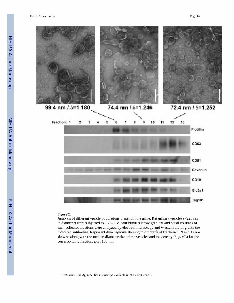
Figure 2.Analysis of different vesicle populations present in the urine. Rat urinary vesicles (<220 nmin diameter) were subjected to 0.25–2 M continuous sucrose gradient and equal volumes ofeach collected fractions were analyzed by electron microscopy and Western blotting with theindicated antibodies. Representative negative staining micrograph of fractions 6, 9 and 12 areshowed along with the median diameter size of the vesicles and the density (δ, g/mL) for thecorresponding fraction. Bar, 100 nm.
Conde-Vancells et al. Page 14
Proteomics Clin Appl. Author manuscript; available in PMC 2010 June 8.
NIH
-PA Author Manuscript
NIH
-PA Author Manuscript
NIH
-PA Author Manuscript

Figure 3.Proteomic overlap of three vesicle populations existing in rat urine samples. Urinary vesicles(<220 nm in diameter) were subjected to continuous sucrose gradient and collected fractionsF6, F9 and F12 corresponding to the indicated densities (δ, g/mL) and diameter size vesicleswere subjected to proteomic analysis by LC-MS/MS. Only proteins identified by at least twopeptides were considered in the analysis. Protein accession numbers were used to perform thecomparative analysis and number of proteins found in common among the different fractionsis indicated. Based on their activities, proteins (indicated by accession name) were categorizedin metabolic enzymes (brown), protein processing and modification (red), transporters (blue),cytoskeleton reorganization, protein localization, membrane trafficking or cell signaling(pink), redox homeostasis (green) and miscellaneous (black).
Conde-Vancells et al. Page 15
Proteomics Clin Appl. Author manuscript; available in PMC 2010 June 8.
NIH
-PA Author Manuscript
NIH
-PA Author Manuscript
NIH
-PA Author Manuscript
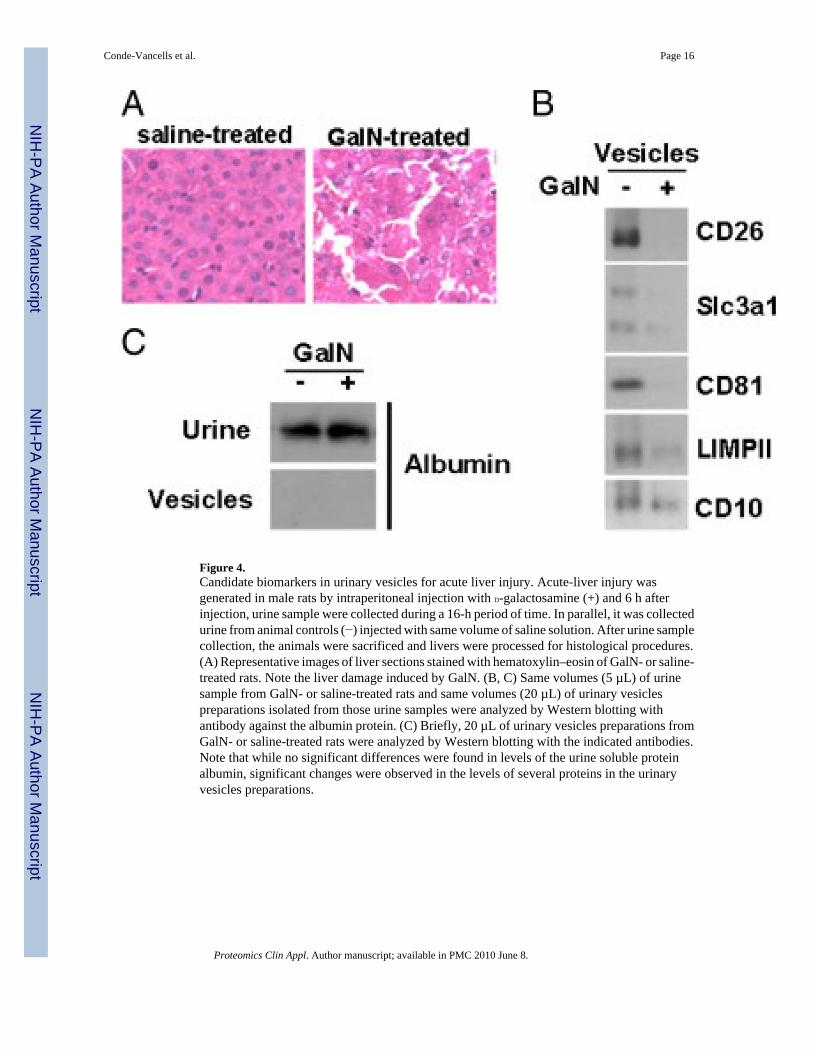
Figure 4.Candidate biomarkers in urinary vesicles for acute liver injury. Acute-liver injury wasgenerated in male rats by intraperitoneal injection with D-galactosamine (+) and 6 h afterinjection, urine sample were collected during a 16-h period of time. In parallel, it was collectedurine from animal controls (−) injected with same volume of saline solution. After urine samplecollection, the animals were sacrificed and livers were processed for histological procedures.(A) Representative images of liver sections stained with hematoxylin–eosin of GalN- or saline-treated rats. Note the liver damage induced by GalN. (B, C) Same volumes (5 µL) of urinesample from GalN- or saline-treated rats and same volumes (20 µL) of urinary vesiclespreparations isolated from those urine samples were analyzed by Western blotting withantibody against the albumin protein. (C) Briefly, 20 µL of urinary vesicles preparations fromGalN- or saline-treated rats were analyzed by Western blotting with the indicated antibodies.Note that while no significant differences were found in levels of the urine soluble proteinalbumin, significant changes were observed in the levels of several proteins in the urinaryvesicles preparations.
Conde-Vancells et al. Page 16
Proteomics Clin Appl. Author manuscript; available in PMC 2010 June 8.
NIH
-PA Author Manuscript
NIH
-PA Author Manuscript
NIH
-PA Author Manuscript
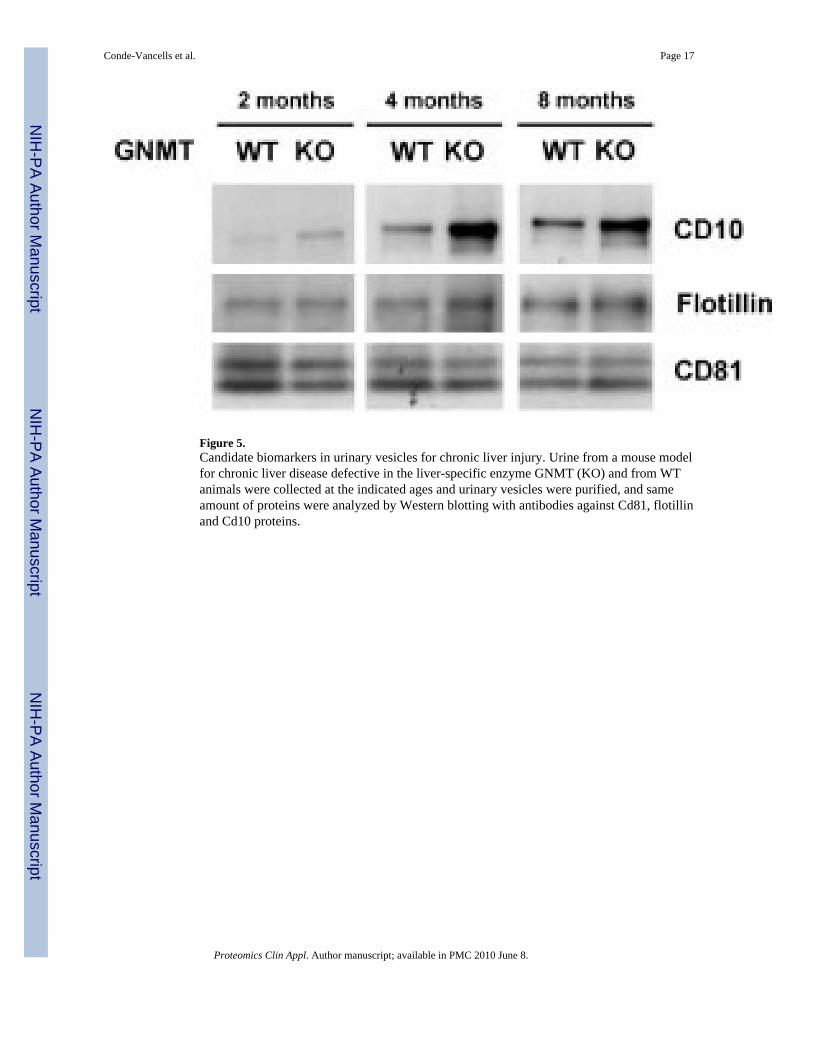
Figure 5.Candidate biomarkers in urinary vesicles for chronic liver injury. Urine from a mouse modelfor chronic liver disease defective in the liver-specific enzyme GNMT (KO) and from WTanimals were collected at the indicated ages and urinary vesicles were purified, and sameamount of proteins were analyzed by Western blotting with antibodies against Cd81, flotillinand Cd10 proteins.
Conde-Vancells et al. Page 17
Proteomics Clin Appl. Author manuscript; available in PMC 2010 June 8.
NIH
-PA Author Manuscript
NIH
-PA Author Manuscript
NIH
-PA Author Manuscript

NIH
-PA Author Manuscript
NIH
-PA Author Manuscript
NIH
-PA Author Manuscript
Conde-Vancells et al. Page 18
Table 1
Disease-associated proteins detected in exosome-like urinary vesiclesa)
Disease Proteinb)
Polycystic kidney disease
AQP1_RAT, TMM27_RAT
Inflammatory bowel disease
MEP1A_RAT, MEP1B_RAT, MDR1_RAT
Triosephosphate isomerase deficiency
TPIS_RAT
Aminoacylase 1 deficiency
ACY1A_RAT
Fructosuria, essential KHK_RAT
Fructose-1,6- bisphosphatase deficiency
F16P1_RAT
Fructose intolerance, hereditary
ALDOB_RAT
Phosphoglycerate kinase 1 deficiency
PGK1_RAT
Cystinuria SLC3A1_RAT
Cystathioninuria CGL_RAT
Glutathionuria GGT1_RAT
Dystonia, juvenile- onset
ACTB_RAT
Obesity PTER_RAT
Hypertension HPCL1_RAT, GSH1_RAT
Myocardial infarction, susceptibility to
GSH0_RAT
Osteoarthritis CALM_RAT
Neurological diseases SODC_RAT, G3P_RAT, AADAT_RAT, 3HAO_RAT, DHPR_RAT, GLNA_RAT, PARK7_RAT
Obsessive-compulsive disorder
EAA3_RAT
Chronic liver disease DPP4_RAT
Acute liver injury RGN_RAT
Cancer, angiogenesis, metastasis
NEP_RAT, DPP4_RAT, CLIC4_RAT, UBIQ_RAT, GSTP1_RAT, COF1_RAT, PDZ1I_RAT, ANXA2_RAT, 1433E_RAT, 1433Z_RAT, 1433T_RAT, RAP1A_RAT, PEBP1_RAT, G3P_RAT, CD82_RAT, UK114_RAT, PARK7_RAT
Pharmacogenetics S22A5_RAT, S47A1_RAT, MDR1_RAT, GSH1_RAT, GSTA1_RAT, GSTP1_RAT
a)Data mining analysis in PubMed database was performed with identified proteins. References supporting these associations are provided in
Supporting Information Table S1.
b)Protein accession number is provided. Bold indicater the proteins described in exosome-like urinary vesicles for first time.
Proteomics Clin Appl. Author manuscript; available in PMC 2010 June 8.
Copyright © 2022 FDOKUMEN