Chapter 4 Manipulation of Purified DNA
33
Chapter 4 Manipulation of Purified DNA enzyrnes, joining DNA molecules 55 Enzymes for cutting DNA - restriction endOllucleases,61 Once pure samples of DNA have been prepared, the next step in a gene cloning experiment is construction of the recombinant DNA molecule (Figure 1.1). To produce this recombinant molecule, the vector, as well as the DNA to be cloned, must be cut at specific points and then joined together in a con- trolled manner. Cutting and joining are two examples of DNA manipulative techniques, a wide variety of which have been developed over the past few years. As well as being cut and joined, DNA molecules can be shortened, lengthened, copied into RNA or into new DNA molecules, and modified by the addition or removal of specific chemical groups. These manipulations, all of which can be carried out in the test tube, provide the foundation not only for gene cloning, but also for studies of DNA biochemistry, gene structure and the control of gene expression. Almost all DNA manipulative techniques make use of purified enzymes. Within the cell these enzymes participate in essential processes such as DNA replication and transcription, breakdown of unwanted or foreign DNA (e.g. invading virus DNA), repair of mutated DNA and recombination between dif- ferent DNA molecules. After purification from cell extracts, many of these enzymes can be persuaded to carry out their natural reactions, or something ; closely related to them, under artificial conditions. Although these enzymatic reactions are often straightforward, most are absolutely impossible to perform by standard chemical methods. Purified enzymes are therefore crucial to genetic engineering and an important industry has sprung up around their preparation, characterization and marketing. Commercial suppliers of high purity enzymes provide an essential service to the molecular biologist. The cutting and joining manipulations that underlie gene cloning are carried out by enzymes called restriction endonucleases (for cutting) and ligases (for joining). Most of this chapter will be concerned with the ways in which these two types of enzyme are used. First, however, we must consider the whole range of DNA manipulative enzymes, to see exactly what types of 54
-
Upload
khangminh22 -
Category
Documents
-
view
3 -
download
0
Transcript of Chapter 4 Manipulation of Purified DNA

Chapter 4 Manipulation of Purified DNA
enzyrnes, joining DNA molecules 55
Enzymes for cutting DNA - restriction endOllucleases,61
Once pure samples of DNA have been prepared, the next step in a gene cloning experiment is construction of the recombinant DNA molecule (Figure 1.1). To produce this recombinant molecule, the vector, as well as the DNA to be cloned, must be cut at specific points and then joined together in a controlled manner. Cutting and joining are two examples of DNA manipulative techniques, a wide variety of which have been developed over the past few years. As well as being cut and joined, DNA molecules can be shortened, lengthened, copied into RNA or into new DNA molecules, and modified by the addition or removal of specific chemical groups. These manipulations, all of which can be carried out in the test tube, provide the foundation not only for gene cloning, but also for studies of DNA biochemistry, gene structure and the control of gene expression.
Almost all DNA manipulative techniques make use of purified enzymes. Within the cell these enzymes participate in essential processes such as DNA replication and transcription, breakdown of unwanted or foreign DNA (e.g. invading virus DNA), repair of mutated DNA and recombination between different DNA molecules. After purification from cell extracts, many of these enzymes can be persuaded to carry out their natural reactions, or something
; closely related to them, under artificial conditions. Although these enzymatic reactions are often straightforward, most are absolutely impossible to perform by standard chemical methods. Purified enzymes are therefore crucial to genetic engineering and an important industry has sprung up around their preparation, characterization and marketing. Commercial suppliers of high purity enzymes provide an essential service to the molecular biologist.
The cutting and joining manipulations that underlie gene cloning are carried out by enzymes called restriction endonucleases (for cutting) and ligases (for joining). Most of this chapter will be concerned with the ways in which these two types of enzyme are used. First, however, we must consider the whole range of DNA manipulative enzymes, to see exactly what types of
54

,r
v j
,. \.
~.
e
3 c _1
')
,r
n
The
reaction can be performed. Many of these enzymes will be mentioned in later chapters when procedures that make use of them are described.
4.1 The range of DNA manipulative enzymes
DNA manipulative enzymes can be grouped into five broad classes depending on the type of reaction that they catalyse:"""
(1) Nucleases are enzymes that cut, shorten or degrade nucleic acid molecules. (2) Ligases join nucleic acid molecules together. (3) Polymerases make copies of molecules. (4) Modifying enzymes remove or add chemical groups. (5) Topob,omerases introduce or remove supercoils from covalently closed
cireular DNA.
Before considering in detail each of these classes of enzyme, two points should be made. The first is that, although most enzymes can be assigned to a particular class, a few display multiple activities that span two or more classes. Most importantly, many polymerases combine their ability to make new DNA molecules with an associated DNA degradative (i.e. nuclease) activity.
Second, it should be appreciated that, as well as the DNA manipulative enzymes, many similar enzymes able to act on RNA are known. The ribonuclease used to remove eontaminating RNA from DNA preparations (p. 35) is an example of such an enzyme. Although some RNA manipulative enzymes have applications in gene cloning and will be mentioned in later chapters, we will in general restrict our thoughts to those enzymes that act on DNA.
4.1.1 Nueleases Nucleases degrade DNA molecules by breaking the phosphodiester bonds that link one nucleotide to the next in a DNA strand. There are two different kinds of nuclease (Figure 4.1):
(1) Exonucleases remove nucleotides one at a time from the end of a DNA molecule.
(2) Endonucleases are able to break internal phosphodiester bonds within a DNA molecule.
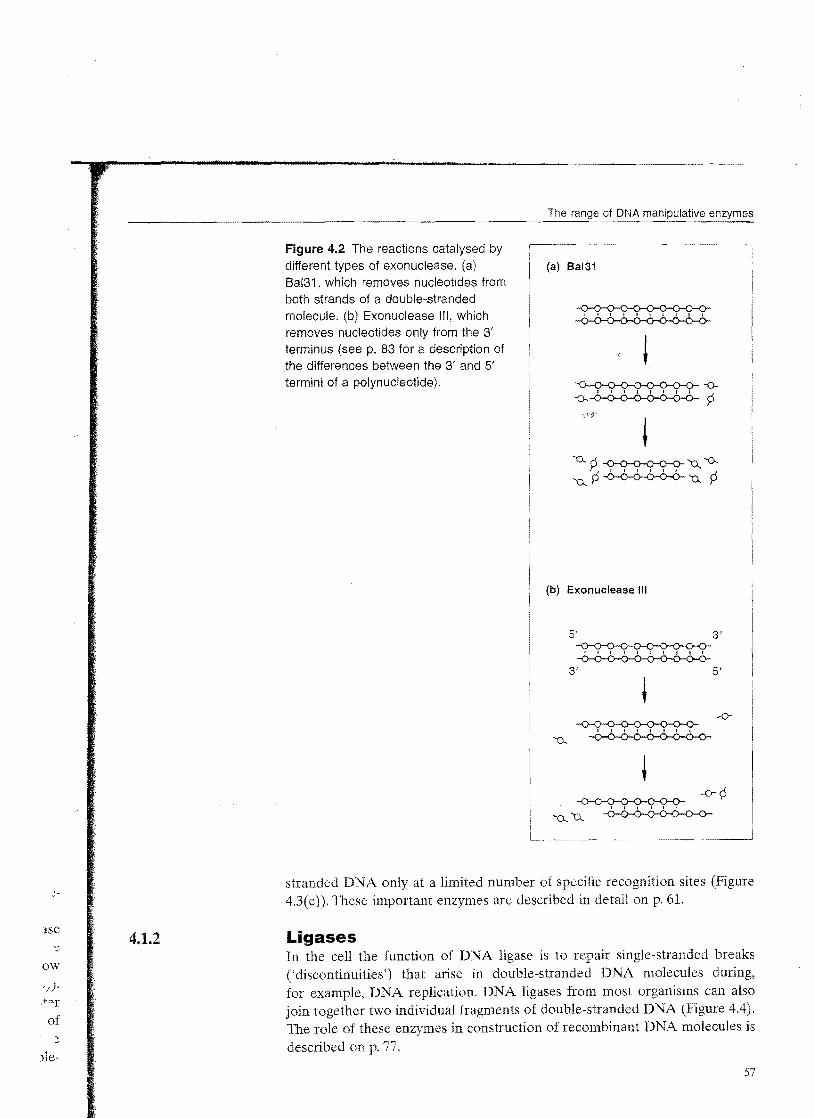
The main distinction between different exonucleases lies in the number of strands that are degraded when a double-stranded molecule is attacked. The enzyme called Bal31 (purified from the bacterium Alteromonas espejiana) is an example of an exonuclease that removes nucleotides from both strands of a double-stranded molecule (Figure 4.2(a»). The greater the length of time that Ba131 is allowed to act on a group of DNA molecules, the shorter the resulting DNA fragments will be. In contrast, enzymes such as E. coli exonuclease
55

Manipulation of Purified DNA
(a) An exonuclease
Cleavage
, , I I I I I I I I I I I I , I ~ Hydrogen bond
'0...
"'0-. , , , I ,
~ I , I I , ,
~ Nucleotide Phosphodiester bond
Cleavage i, ~ , I , -':>
Figure 4.1 The reactions catalysed by the two different kinds of nuclease. (a) An exonuclease, which removes nucleotides from the end of a DNA molecule. (b) An endonuclease, which breaks internal phosphodiester bonds.
t '0... -0 , , , , , , , '0-.. -0
(b) An endonuclease
t f I J I I I
I I , JY""(), , A::r -0
-0-0--0--0 --0-0-0 --0-0-0-0--0 -0-0--6-0-0-6 -6-6-6 -0-6-6-6-0 -6--6
In degrade just one strand of a double-stranded molecule, leaving singlestranded DNA as the product (Figure 4.2(b»).
The same criterion can be used to classify endonucleases. Sl endonuclease (from the fungus Aspergillus oryzae) only cleaves single strands (Figure 4.3(a)), whereas deoxyribonuclease I (DNase I), which is prepared from cow pancreas, cuts both single and double-stranded molecules (Figure 4.3(b»). DNase I is non-specific in that it attacks DNA at any internal phosphodiester bond; the end result of prolonged DNase I action is therefore a mixture of mononucleotides and very short oligonucleotides. On the other hand, the special group of enzymes called restriction endonucleases cleave double
;
l::;e
ow '/). ,tar
. of ~
)le-
Figure 4.2 The reactions catalysed by different types of exonuclease. (a) 8al31 , which removes nucleotides from both strands of a double-stranded molecule. (b) Exonuclease Ill, which removes nucleotides only from the 3' terminus (see p. 83 for a description of the differences between the 3' and 5' termini of a polynucleotide).
The
(a) Bal31
(b) Exonuclease III
5' 3' -0--0-0-0--0--o
I t r I I I I I I I
-0--0-0-0--0-o3' 5'
stranded DNA only at a limited number of specific recognition sites (Figure 4.3(c). These important enzymes are described in detail on p. 61.
4.1.2 Ligases In the cell the function of DNA ligase is to repair single-stranded breaks ('discontinuities') that arise in double-stranded DNA molecules during, for example. DNA replication. DNA ligases from most organisms can also join together two individual fragments of double-stranded DNA (Figure 4.4) . The role of these enzymes in construction of recombinant DNA molecules is described on p. 77.
0
57

of Purified DNA
(a) S1 nuclease
(i) (ii)
-0-0 -0 -0-0-0 -0-0
(b) DNase I
(i) (ii)
-0-0- -0- -0-0-0-- -0-0
(c) A restriction endonuclease
~ I I r I I I I 1 , I
--0-0-0-0--0-o
/
-0-0-0- -0-0-0-01 I 1 I I I l
-0-0-0- -0-0-0-0
q q
-0-0-0-0-0-o-o-I I I I I I I I
-0-0-0-0-0-0-0-
-0--0- -0- -0-0-0, , ,-6-6- -6- -0-0-0
Figure 4.3 The reactions catalysed by different types of endonuclease. (a) S1 nuclease, which cleaves only singlestranded DNA including singlestranded nicks in mainly double-stranded molecules. (b) DNase I, which cleaves both single- and double-stranded DNA. (c) A restriction endonuclease, which cleaves double-stranded DNA, but only at a limited number of sites.
4.1.3 Polymerases DNA polymerases are enzymes that synthesize a new strand of DNA complementary to an existing DNA or RNA template (Figure 4.5(a)). Most polymerases can function only if the template possesses a double-stranded region that acts as a primer for initiation of polymerization.
Four types of DNA polymerase are used routinely in genetic engineering. The first is DNA polymerase I, which is usually prepared from E. coli. This
58

Figure 4.5 The reactions catalysed by DNA polymerases. (a) The basic reaction: a new DNA strand is synthesized in the 5' to 3' direction. (b) DNA polymerase I, which initially fills in nicks but then continues to synthesize a new strand, degrading the existing one as it proceeds. (c) The Klenow fragment, which only fills in nicks. (d) Reverse transcriptase, which uses a template of RNA.
Figure 4.4 The two reactions catalysed by DNA ligase. (a) Repair of a discontinuity a missing phosphodiester bond in one strand of a double-stranded molecule. (b) Joining two molecules together.
(a) Discontinuity repair
/' A discontinuity
-0-0-0-0 (0-0-0-0-6-6-6-6-6-6-6
~ DNA ligase
-0-0-0-0-0-o-o~
(b),,~pining two molecules
!DNA ligase
-<)-;.,~I I I I I , I l: ,
-0-0-0-0-0-0-0-0-0
(a) The basic reaction Newly syntnesized
Primer strand
5' .f 3' 5' ,------··--···--·---,3' -A-T-G- -A-T-G-C-A-T-T-G-C-A-T
, . . . .. . ... -T-A-C-G-T-A-A-C-G-T-A
5' 3' 5'3' Template
(b) DNA polymerase I Existing nucleotides
are replaced A nick
-A-T-G-~
G-C-A-T- -A-T-G-C-A-T-T-G-C-A-T-T-A-C-G-T-A-A-C-G-T-A- -T-A-C-G-T-A-A-C-G-T-A
(c) The Klenow fragment
Existing nucleotides in are not replaced
_ ...A~~ f -A-T-G- G-C-A-T- -A-T -G-C-A-T--T G-C-A-T... , ..... '
-T-A-C-G-T-A-A-C-G-T-A- -T-A-C-G-T-A-A-C-G-T-A
(d) Reverse transcriptase Nevv strand Of DN,i\
-A-T-G- -A-T -G-C-A-T-T -G-C-A-T -u-a-c-g-u-a-a-c-g-u-a -u-a-c-g-u-a-a-c-g-u-a
~ RNA template
59

of Purified DNA
enzyme attaches to a short single-stranded region (or nick) in a mainly doublestranded DNA molecule, and then synthesizes a completely new strand, degrading the existing strand as it proceeds (Figure 4.5(b»). DNA polymerase I is therefore an example of an enzyme with a dual activity DNA polymerization and DNA degradation.
The polymerase and nuclease activities of DNA polymerase I are controlled by different parts of the enzyme molecule. The nuclease activity is contained in the first 323 amino acids of the polypeptide, so removal of this segment leaves a modified enzyme that retains the polymerase function but is unable to degrade DNA. This modified enzyme, called the Klenow fragment, can still synthesize a complementary DNA strand on a single-stranded template. but as it has no nuclease activity it cannot continue the synthesis once the nick is filled in (Figure 4.5( c». Several other enzymes natural polymerases and modified versions have similar properties to the Klenow fragment. The major application of the Klenow fragment and these related polymerases is in manual DNA sequencing (p. 207).
The Taq DNA polymerase used in the polymerase chain reaction (PCR) (Figure 1.2) is the DNA polymerase I enzyme of the bacterium Thermus aquaticus. This organism lives in hot springs, and many of its enzymes, including the Taq DNA polymerase, are thermostable, meaning that they are resistant to denaturation by heat treatment. This is the special feature of Taq DNA polymerase that makes it suitable for PCR, because if it was not thermostable it would be inactivated when the temperature of the reaction is raised to 94°C to denature the DNA.
The final type of DNA polymerase that is important in genetic engineering is reverse transcriptase, an enzyme involved in the replication of several kinds of virus. Reverse transcriptase is unique in that it uses as a template not DNA but RNA (Figure4.5(d».The ability of this enzyme to synthesize a DNA strand complementary to an RNA template is central to the technique called complementary DNA (cDNA) cloning (p. 166).
4.1.4 DNA modifying enzymes There are numerous enzymes that modify DNA molecules by addition or removal of specific chemical groups. The most important are as follows:
.il) Alkaline phosphatase (from E. coli, calf intestinal tissue or arctic shrimp), which removes the phosphate group present at the 5' terminus of a DNA molecule (Figure 4.6(a»).
(2) Polynucleotide kinase (from E. coli infected with T4 phage), which has the reverse effect to alkaline phosphatase, adding phosphate groups onto free 5' termini (Figure 4.6(b».
(3) Terminal deoxynucleotidyl transferase (from calf thymus tissue), which adds one or more deoxyribonucleotides onto the 3' terminus of a DNA molecule (Figure 4.6(c»).
60
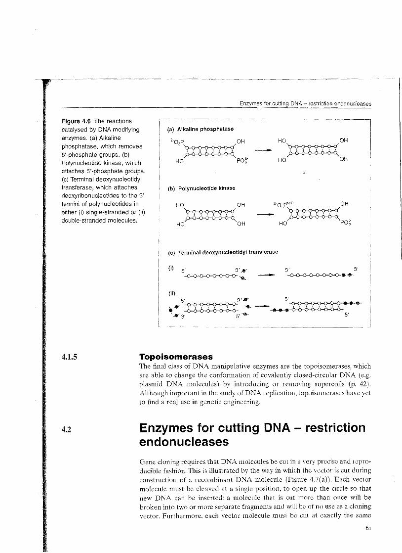
Figure 4.6 The reactions catalysed by DNA modifying enzymes. (a) Alkaline phosphatase, which removes 5'-phosphate groups. (b) Polynucleotide kinase, which attaches 5'-phosphate groups. (c) Terminal deoxynucleotidyl transferase, which attaches deoxyribonucleotides to the 3' termini of polynucleotides in either (i) single-stranded or (ii) double-stranded molecules.
DNA - restriction endonucleases
(a) Alkaline phosphatase
2·03PD-o-o-o-o-o-o-dOH HO OH D-o-o-o-o-o-o-d-
HO OHHO PO~'
(b) Polynucleotide kinase
HO OH D-o-o-o-o-o-o-d
HO OH
(c) Terminal deoxynucleotidyl transferase
(i) 5' 3' K 5' 3' ~-0-0-0--0--0-o- ...... -
4.1.5 Topoisomerases The final class of DNA manipulative enzymes are the topoisomerases, which are able to change the conformation of covalently closed-circular DNA (e.g. plasmid DNA molecules) by introducing or removing supercoils (p. 42). Although important in the study of DNA replication, topoisomerases have yet to find a real use in genetic engineering.
4.2 Enzymes for cutting DNA - restriction endonucleases
Gene cloning requires that DNA molecules be cut in a very precise and reproducible fashion. This is illustrated by the way in which the vector is cut during construction of a recombinant DNA molecule (Figure 4.7(a». Each vector molecule must be cleaved at a single position, to open up the circle so that new DNA can be inserted: a molecule that is cut more than once will be broken into two or more separate fragments and will be of no use as a cloning vector. Furthermore, each vector molecule must be cut at exactly the same
61

of Purified DNA
Figure 4.7 The need for very (a) Vector molecules precise cutting manipulations in
a gene cloning experiment.
Each vector molecule must be cut once, each at the same position
(b) The DNA molecule containing the gene to be cloned
..
Fragments small Large DNA molecule enough to be cloned
position on the circle as will become apparent in later chapters, random cleavage is not satisfactory. It should be clear that a very special type of nuclease is needed to carry out this manipulation.
Often it is also necessary to cleave the DNA that is to be cloned (Figure 4.7(b».There are two reasons for this. First, if the aim is to clone a single gene, which may consist of only 2 or 3 kb of DNA, then that gene will have to be cut out of the large (often greater than 80lcb) DNA molecules produced by skilful use of the preparative techniques described in Chapter 3. Second, large
'l')NA molecules may have to be broken down simply to produce fragments small enough to be carried by the vector. Most cloning vectors exhibit a preference for DNA fragments that fall into a particular size range; M13-based vectors, for example, are very inefficient at cloning DNA molecules more than 3kb in length.
Purified restriction endonucleases allow the molecular biologist to cut DNA molecules in the precise, reproducible manner required for gene cloning. The discovery of these enzymes, which led to Nobel Prizes for W. Arber, H. Smith and D. Nathans in 1978, was one of the key breakthroughs in the development of genetic engineering.
62

4.2.1
DNA - restriction endonucleases
The discovery and function of restriction endonucleases The initial observation that led to the eventual discovery of restriction endonucleases was made in the early 1950s, when it was shown that some strains of bacteria are immune to bacteriophage infection, a phenomenon referred to as host-controlled restriction.
The mechanism of restriction is not very complicated, even though it took over 20 years to be fully understood. Restriction occurs because the bacterium produces an enzyme that degrades the phage DNA before it has time to replicate and direct synthesis of ne,v phage partjcle~--(Figure 4.8(a». The bacterium's own DNA, the destruction of which would of course be lethal, is protected from attack because it carries additional methyl groups that block the degradative enzyme action (Figure 4.8(b».
Figure 4.8 The function of a restriction endonuclease in a (a) Restriction of phage DNA bacterial cell: (a) phage DNA is Phage injects cleaved, but (b) bacterial DNA -DNA into a
bacteriumis not.
: Restriction I
Phage DNAendonucleases is cleavedbind to the and inactivated phage DNA
(b) Bacterial DNA is not cleaved /'
\ ,/
)) ~r
\ ~Restriction endonuclease
cannot bind to the recognition sequence
Recognition sequences are methylated

of Purified DNA
These degradative enzymes are called restriction endonucleases and are synthesized by many, perhaps all, species of bacteria; over 1200 different ones have now been characterized. Three different classes of restriction endonuclease are recognized, each distinguished by a slightly different mode of action. Types I and III are rather complex and have only a limited role in genetic engineering. Type II restriction endonucleases, on the other hand, are the cutting enzymes that are so important in gene cloning.
4.2.2 Type II restriction endonucleases cut DNA at specific nucleotide sequences The central feature oftype II restriction endonucleases (which will be referred to simply as 'restriction endonucleases' from now on) is that each enzyme has a specific recognition sequence at which it cuts a DNA molecule. A particular enzyme cleaves DNA at the recognition sequence and nowhere else. For example, the restriction endonuclease called Pvul (isolated from Proteus vulgaris) cuts DNA only at the hexanucleotide CGATCG. In contrast, a second enzyme from the same bacterium, called PvuII, cuts at a different hexanucleotide, in this case CAGCTG.
Many restriction endonucleases recognize hexanucleotide target sites, but others cut at four, five, eight or even longer nucleotide sequences. Sau3A (from Staphylococcus aureus strain 3A) recognizes GATC, and Alul (Arthrobacter [uteus) cuts at AGCT. There are also examples of restriction endonucleases with degenerate recognition sequences, meaning that they cut DNA at anyone of a family of related sites. HinfI (Haemophilus influenzae strain R t), for instance, recognizes GANTC, so cuts at GAATC, GATTC, GAGTC and GACTC.
The recognition sequences for some of the most frequently used restriction endonucleases are listed in Table 4.1.
4.2.3 Blunt ends and sticky ends The exact nature of the cut produced by a restriction endonuclease is of considerable importance in the design of a gene cloning experiment. Many restriction endonucleases make a simple double-stranded cut in the middle of the recognition sequence (Figure 4.9(a», resulting in a blunt end or flush end.
"'hull and Alul are examples of blunt end cutters. Other restriction endonucleases cut DNA in a slightly different way. With
these enzymes the two DNA strands are not cut at exactly the same position. Instead the cleavage is staggered, usually by two or four nuc1eotides, so that the resulting DNA fragments have short single-stranded overhangs at each end (Figure 4.9(b». These are called sticky or cohesive ends, as base pairing between them can stick the DNA molecule back together again (recall that sticky ends were encountered on p. 23 during the description of A phage replication). One important feature of sticky end enzymes is that restriction endonucleases with different recognition sequences may produce the same

DNA restriction endonucleases
Table 4.1 The recognition sequences for some of the most frequently used restriction endonucleases.
Enzyme Organism Recognition Bhmt or sequence" sticky end
EcoRI BamHI BgllI PvuI PvuII Hind III HinfI Sau3A AZul TaqI HaeIII NotI SfiI
Escherichia coli Bacillus amyloliquefaciens Bacillus globigii Proteus vulgaris Proteus vulgaris Haemophilus infiuenzae Rd Haemophilus infiuenzae Rf Staphylococcus aureus Arthrobacter iuteus Thermus aquaticus Haemophilus aegyptius Nocardia otitidis-caviarum Streptomyces fimbriatus
GAATTC GGATCC AGATCT CGATCG
;-;y:<J"
CAGCTG AAGCTT GANTC GATC AGCT TCGA GGCC GCGGCCGC GGCCNNNNNGGCC
Sticky Sticky Sticky Sticky Blunt Sticky Sticky Sticky Blunt Sticky Blunt Sticky Sticky
• The sequence shown is that of one strand, given in the 5' to 3' direction. 'N' indicates any nucleotide. Note that almost all recognition sequences are palindromes: when both strands are considered they read the same in each direction, for example:
5'-GAATTC-3'
EcoRI i I I I I I
3'-C TTAAG-5'
sticky ends. BamHI (recognition sequence GGATCC) and BglII (AGATCT) are examples - both produce GATC sticky ends (Figure 4.9(c». The same sticky end is also produced by Sau3A, which recognizes only the tetranucleotide GATe. Fragments of DNA produced by cleavage with either of these enzymes can be joined to each other, as each fragment carries a complementary sticky end.
4.2.4 The frequency of recognition sequences in a DNA molecule The number of recognition sequences for a particular restriction endonuclease in a DNA molecule of known length can be calculated mathematically. A tetranucleotide sequence (e.g. Gf\TC) should occur once every 44 "= 256 nucleotides, and a hexanuc1eotide (e.g. GGATCC) once every 46 = 4096 nucleotides. These calculations assume that the nucleotides are ordered in a random fashion and that the four different nucleotides are present in equal proportions (i.e. the GC content =50%). In practice, neither of these assumptions is entirely valid. For example, the A DNA molecule, at 49kb, should
65

of Purified DNA
Figure 4.9 The ends produced (a) Production of blunt ends
-N-N-A-G-C-T-N-N- ~~
-N-N-T-C-G-A-N-N
'N' A, G, C, orT
(b) Production of sticky ends
-N-N-G-A-A-T"-T-C-N-N- EcoRI . . . . . . . . ~ ~
-N-N-C-T-T-A-A -G-N-N
-N-N-,i,,-G C T-N-N
-N-N-T-C G-A-N-N
Blunt ends
-N-N-G A-A-T - T··C-N-N
-N-N-C"T-T-A-A \ 3-N-N \
Sticky ends
by cleavage of DNA with different restriction endonucieases. (a) A blunt end produced by Alul. (b) A sticky end produced by EcoRI. (c) The same sticky ends produced by BamHI, Bgnl and Sau3A.
(c) The same sticky ends produced by different restriction endonucleases
-N-N-G G-A- T--C-C-N-NBamHI
-N-N-C-C-T-A-G G-N-N-
Bgill --N-N-A
-N-N-T-C-T-A-G
G-A-T-C-T-N-N
A-N-N
Sau3A -N-N-N
-N-N-N-C-T-A-G
G-A-T·-C-N-N-N
N-N-N
contain about sites for a restriction endonuclease with a hexanucleotide recognition sequence. In fact, many of these recognition sites occur less fre'quently six for BglII, five for BamHI and only two for SaLI), a reflection of the fact that the GC content for -A is rather less than 50% (Figure 4.10(a)).
Furthermore, restriction sites are generally not evenly spaced out along a DNA molecule. If they were, then digestion with a particular restriction endonuclease would give fragments of roughly equal Figure 4.1O(b) shows the fragments produced by cutting t.. DNA with BglII, BamHI and Sall. In each case therc is a considerable spread of fragment indicating that in , .. DNA the nucleotides are not randomly ordered.
The lesson to be learned from Figure 4.10 is that although mathematics may give an idea of how many restriction sites to in a given DNA
66

DNA restriction endonucleases
(a) Cleavage sites on A DNA
o 10 20 30 40 49 kb
1.,--1--'---1--L.-!---'------r'11"--1,------L-'-,-,-,----;I~l~r-,---l---"
• 8g/ll- 6 sites
l 8amHI- 5 sites
~ S8/1- 2 sites
(b) Fragment sizes
8gll1 r------------------------------------,122010 ,-------------------~113286
.--------------.19688 ,..---, 2392 11651 n 415 D60
8amHI .---------------------------'116841
'7233 ,---------" 6770 ,---------., 6527
'5626 .--------., 5505
Sa/I r-----------------------------------------------------~132745
.------------------------.,15258 n499
Figure 4.10 Restriction of the /" DNA molecule. (a) The positions of the recognition sequences for Bgnl, BamHI and San. (b) The fragments produced by cleavage with each of these restriction endonucleases. The numbers are the fragment sizes in base pairs.

4.2.5
of Purified DNA
(a)
Add 2pi Bg/li buffer
\~
'~ 2 J.l9 /, DNA (16 J.l1)
(e)
IT ..
I HV
molecule, only experimental analysis can provide the true picture. We must therefore move on to consider how restriction endonucleases are used in the laboratory.
Performing a restriction digest in the laboratory As an example, we will consider how to digest a sample of A DNA (concentration 1251-lg/ml) with BglIL
First of all the required amount of DNA must be pipetted into a test tube. The amount of DNA that will be restricted depends on the nature ofthe experiment. In this case we shall digest 21-lg of A DNA, which is contained in 161-l1 of the sample (Figure 4.11(a»). Very accurate micropipettes will therefore be needed.
The other main component in the reaction will be the restriction endonuobtained from a commercial supplier as a pure solution of known con
centration. But before adding the enzyme, the solution containing the DNA must be adjusted to provide the correct conditions to ensure maximal activity of the enzyme..\-lost restriction endonucleases function adequately at pH 7.4, but different enzymes vary in their requirements for ionic strength (usually
Figure 4.11 Performing a restriction digest in the laboratory (b) (c)
(see text for details), Add 0.5)11 BglIi + 1.5 fll H20
I
lnCUbate at 3TC for 111
) (d)
/ ":), '
.\~~ Cleaved A DNA
~d,d p~enol or EDTA or Ileat at 70c C for 15 min
--- .._ ......._._.........._--- -
68

DNA - restriction endonucleases
Table 4.2 A lOx buffer suitable for restriction of DNA with BglII.
Component Concentration (mM)
Tris-HCl, pH 7.4 500 MgClz 1{)0
NaCl 500 Dithiothreitol 10
provided by sodium chloride (NaCl) and magnesium (Mg2+) concentration (all type II restriction endonucleases require Mg2
->. in order to function). It is also advisable to add a reducing agent, such as dithiothreitol (DIT), which stabilizes the enzyme and prevents its inactivation. Providing the right conditions for the enzyme is very important - incorrect NaCl or Mg2+ concentrations may not only decrease the activity of the restriction endonuclease, but may also cause changes in the specificity of the enzyme, so that DNA cleavage occurs at additional, non-standard recognition sequences.
The composition of a suitable buffer for BglII is shown in Table 4.2. "This buffer is ten times the working concentration, and is diluted by being added to the reaction mixture. In our example. a suitable final volume for the reaction lllixture would be 201.11, so we add 2}l1 of lOx BglII buffer to the 16111 of DKA already present (Figure 4.11(b»).
The restriction endonuclease can now be added. By convention, 1 unit of enzyme is defined as the quantity needed to cut 1 j.lg of DKA in 1 hour, so we need 2 units of BglII to cut 21lg of tv DNA. BglII is frequently obtained at a concentration of 4 unitS/ill, so 0.5 III is sufflcient to cleave the DNA. The final ingredients in the reaction mixture are therefore 0.5111 BglII + 1.5 III water, giving a final volume of 20111 (Figure 4.1l(c).
'The last factor to consider is incubation temperature. Most restriction endonuc1eases, including BglII, work best at 3TC, but a few have different requirements. TaqI, for example, is a restriction enzyme from Thermus aquaticus and, like Taq DNA polymerase, has a high working temperature. Restriction digests with TaqI must be incubated at 65°C to obtain maximum enzyme activity.
After 1 hour the restriction should be complete (Figure 4.1l(d». If the DNA fragments produced by restriction are to be used in cloning experiments, the enzyme must somehow be destroyed so that it does not accidentally digest other DNA molecules that may be added at a later stage. There are several ways of 'killing' the enzyme. For many a short incubation at 70°C is sufficient, for others phenol extraction or the addition of ethylenediamine tetra acetate (EDTA), which binds Mg2+ ions preventing restriction endonuclease action, is used (Figure 4.11(e)).

of Purified DNA
4.2.6 Analysing the .cesult of restriction endonuclease cleavage A restriction digest results in a number of DNA fragments, the sizes of which depend on the exact positions of the recognition sequences for the endonuclease in the original molecule (Figure 4.1O).A way of determining the number and sizes of the fragments is needed if restriction endonucleases are to be of use in gene cloning. Whether or not a D~A molecule is cut at all can be determined fairly easily by testing the viscosity of the solution. Larger DNA molecules result in a more viscous solution than smaller ones, so cleavage is associated with a decrease in viscosity. However, working out the number and sizes of the individual cleavage products is more difficult. In fact, for several years this \vas one of the most tedious aspects of experiments involving DNA. Eventually the problems were solved in the early 1970s when the technique of gel electrophoresis was developed.
Separation of molecules by gel electrophoresis Electrophoresis, like ion-exchange chromatography (see p. 35), is a technique that uses differences in electrical charge to separate the molecules in a mixture. DNA molecules have negative charges, and so when placed in an electric field they migrate towards the positive pole (Figure 4.12 (a). The rate of migration of a molecule depends on two factors, its shape and its charge-tomass ratio. Unfortunately, most DNA molecules are the same shape and all have very similar charge-to-mass ratios. Fragments of different sizes cannot therefore be separated by standard electrophoresis.
The size of the DNA molecule does, however, become a factor if the electrophoresis is performed in a gel. A gel, which is usually made of agarose, polyacrylamide, or a mixture of the two, comprises a complex network of pores. through which the DNA molecules must travel to reach the positive electrode. The smaller the DNA molecule, the faster it can migrate through the gel. Gel electrophoresis therefore separates DNA molecules according to their size (Figure 4.12(b).
In practice the composition of the gel determines the sizes of the DNA molecules that can be separated. A 0.5 cm thick slab of 0.5% agarose, which has relatively large pores, would be used for molecules in the size range
.$1"'-30kb, allowing, for example, molecules of 10 and 12kb to be clearly distinguished. At the other end of the scale, a very thin (0.3mm) 40% polyacrylamide gel, with extremely small pores, would be used to separate much smaller DNA molecules, in the range 1-300bp, and could distinguish molecules differing in length by just a single nucleotide.
Visualizing DNA molecules by staining a gel The easiest way to see the results of a gel electrophoresis experiment is to stain the gel with a compound that makes the DNA visible. Ethidium bromide (EtBr), already described on p. 45 as a means of visualizing DNA in caesium
70

- --
DNA - restriction endonucleases
Figure 4.12 (a) Standard electrophoresis does not separate DNA fragments of different sizes, (a) Standard electrophoresis
whereas (b) gel electrophoresis does.
h; / \
DNA Buffer
DNA migrates towards the anode, but little separation into size classes occurs
(b) Gel electrophoresis
~~r_~~~~~,;~ d_!-Buffer
'-DNA, loaded \ Gelinto a well cut
out of the gel
IElectrophorese
t
\\;DNA separates into bands of different-sized fragments
----Smailest
chloride gradients, is also routinely used to stain DNA in agarose and polyacrylamide gels (Figure 4.13). Bands showing the positions of the different size classes of DNA fragment are clearly visible under ultraviolet irradiation after EtBr staining, so long as sufficient DNA is present. Unfortunately, the procedure is very hazardous because ethidium bromide is a powerful mutagen and the ultraviolet radiation used to visualize the DNA can cause severe burns. For this reason, non-mutagenic dyes that stain DNA green or blue, and which do not require ultraviolet irradiation in order for the results to be seen, are now used in many laboratories.
71

of Purified DNA
- ...-~
Figure 4.13 Visualizing DNA Wells for samples bands in an agarose gel by
EtBr staining and ultraviolet (UV) irradiation.
- Agarose gel
UV transparent plastic support
Soak in 0.5 J.l.9/ml solution
-...........,
of EtBr, 15 min
=
-TAO<---. ·Sands of DNA ............- - fluoresce
; \
) UV
Visualizing DNA molecules by autoradiography The one drawback with staining is that there is a limit to its sensitivity. If less than about lOng of DNA is present per band, it is unlikely that the bands will show up after staining. For small amounts of DNA a more sensitive detection method is needed.
Autoradiography provides an answer. If the DNA is 1abelled before electrophoresis, by incorporation of a radioactive marker into the individual 1].glecules, then the DNA can be visualized by placing an X-ray-sensitive photographic film over the gel. The radioactive DNA exposes the film, revealing the banding pattern (Figure 4.14).
A DNA molecule is usually labelled by incorporating nucleotides that carry a radioactive isotope of phosphorus, 32p (Figure 4.15(a). Several methods are available, the most popular being nick trans1ation and end filling.
Nick translation refers to the activity of DNA polymerase I (p. 58). Most purified samples of DNA contain some nicked molecules, however carefully the preparation has been carried out, which means that DNA polymerase I is able to attach to the DNA and catalyse a strand replacement reaction (Figure 4.5(b». This reaction requires a supply of nucleotides: if one of these is
72
r

1
s
, " t
Y s e s
Figure 4.14 The use of autoradiography to visualize radioactively labelled DNA in an agarose gel.
---,--~--
= = Glass plate
Agarose gel, dried in an oven
\ X-ray-sensitlve film laid over the gel
\ ~xpose tor 12-100 h, \deVeiOP the film
DNA have exposed the film
Autoradiograph
radioactively 13belled, the DNA molecule will itself become labelled (Figure 4.1S(b)).
Nick translation can be used to label any DNA molecule but may under some circumstances also cause DNA cleavage. End filling is a gentler method that rarely causes breakage of the DNA, but unfortunately can only be used to label DNA molecules that have sticky ends. The enzyme used is the Klenow fragment (p. 60), which 'fills in' a sticky end by synthesizing the complementary strand (Figure 4.15(c)). As with nick translation, if the end fiIling reaction is carried out in the presence of labelled nucleotides, the DNA itself becomes labelled.
Both nick translation and end filling can label DNA to such an extent that very small quantities can be detected in gels by autoradiography. As little as 2ng of DNA per band can be visualized under ideal conditions.
73

Figure 4.15 Radioactive (a) [0: - 32PJ dATP labelling: (a) the structure of a-
NHz 32P-deoxyadenosine triphosphate 1
([a.-32P]dATP); (b) labelling DNA /y----C/~N by nick translation; (c) labelling O· 0- 0- HC/' II I 1 1 I \ C h CH DNA by end filling.
-0-P-O- P-O- P-0-CH2 N--- "N;'/'
6 6 /6 1/.0~1/ C H H C
/ I'\.I 1/i H c-c
( I 1 Radioactive OH H 32 0
(b) Labelling by nick translation
DNA Poll
+ 32p-dATP
(c) Labelling by end-filling
Klenow fragment
+ 32p_dATP\ -~ EcoRI sticky end Labelled end
4.2.7 jistimation of the sizes of DNA molecules Gel electrophoresis separates different sized DNA molecules, with the smallest molecules travelling the greatest distance towards the positive electrode. If several DNA fragments of varying sizes are present (the result of a successful restriction digest, for example), then a series of bands appears in the gel. How can the sizes of these fragments be determined?
The most accurate method is to make use of the mathematical relationship that links migration rate to molecular mass. TIle relevant formula is:
D a-b(logM)
74

ll.e. cle
ip
DNA restriction endonucleases
where D is the distance moved, M is the molecular mass, and a and b are constants that depend on the electrophoresis conditions.
Because extreme accuracy in estimating DNA fragment sizes is not always necessary, a much simpler though less precise method is more generally used. A standard restriction digest, comprising fragments of known size, is usually included in each electrophoresis gel that is run. Restriction digests of /. DNA are often used in this way as size markers. For example, HindIII cleaves ADNA into eight fragments, ranging in size from 125 bp for the smallest to over 23 kb for the largest. As the sizes of the fragments in this digest are known, the fragment sizes in the experimental digest can be estimated by comparing the positions of the bands in the two tracks (Figure 4.16). Although not precise, this method can be performed with as little as a 5% error, which is quite satisfactory for most purposes.
Figure 4.16 Estimation of the sizes of DNA fragments in an agarose gel. (a) A rough (a) Rough estimation by eye
estimate of fragment size can be obtained by eye. (b) A more accurate measurement of
Hind II I Unknownfragment size is gained by using the mobilities 1 of the Hindill-t, fragments to construct a calibration curve; the sizes of the unknown
23130bpfragments can then be determined from the 9416 distances they have migrated. 6557
c.5000bp4361
2 c.3200bp 2322 ---,. 2027 ----- ----~ 3 c.2000bp
564 ----
(b) Accurate graphical estimation
10
:2 ::s 7.5 « z 0 5 a (J) N 2.5
Cl5 0
0 2 3 4 5
Distance migrated (em)
75

4.2.8
of Purified DNA
Mapping the positions of different restriction sites in a DNA molecule So far we have considered how to determine the number and sizes of the DNA fragments produced by restriction endonuclease cleavage. Ine next step in restriction analysis is to construct a map showing the relative positions in the DNA molecule of the recognition sequences for a number of different enzymes. Only when a restriction map is available can the correct restriction endonucleases be selected for the particular cutting manipulation that is required (Figure 4.17).
To construct a restriction map, a series of restriction digests must be performed. First, the number and sizes of the fragments produced by each restriction endonuclease must be determined by gel electrophoresis, followed by comparison with size markers (Figure 4.18). "This information must then be supplemented by a series of double digestions, in which the DNA is cut by two restriction endonucleases at once. It may be possible to perform a double digestion in one step if both enzymes have similar requirements for pH. Mg2+ concentration, etc. Alternatively, the two digestions may have to be carried out one after the other, adjusting the reaction mixture after the first digestion to provide a different set of conditions for the second enzyme.
gene A gene 8 gene C
l8gl11 18amHI ~ Sail
To obtain gene B, digest with 8gl11
To ol)tain gene D, digest with 8amHI + Sail
A \3t:---? --D"\\!/
~io=j
Figure 4.17 Using a restriction map to work out which restriction endonucleases should be used to obtain DNA fragments containing individual genes.

~----------------------------------~------------------------------------~---------------
DNA - restriction endonucleases
Single and double digestions
Enzyme Number of fragments Sizes (kb)
Xbal 2 24.0,24.5 Xhol 2 15.0,33.5 Kpnl 3 1.5, 17.0,30.0 Xbal + Xhol 3 9.0;,-15.0, 24.5 Xbal+ 4 1.5, 6.0, 24.0
Conclusions:
(1) As), DNA is linear, the number of restriction sites for each enzyme is Xbal 1, Xhol 1, Kpnl 2.
(2) The Xbal and Xhol sites can be mapped:
Xba I fragments
Xba I - Xho I fragments
Xho I fragments
Xhoi Xbai The only possibility is:
(3) All the Kpnl sites fall in the 24.5 kb Xbal fragment, as the 24.0 kb fragment is intact after Xbal-Kpnl double digestion. The order of the Kpnl fragments can be determined only by partial digestion.
Partial digestion
Enzyme Fragment sizes (kb)
Kpnl - limiting conditions 1.5, 17.0, 18.5, 30.0, 31.5, 48.5
Conclusions:
(1) 48.5kb fragment uncun.
(2) 1.5, 17.0 and 30.0 kb fragments are products of complete digestion.
(3) 18.5 and 31.5 kb fragments are products of partial digestion.
Kpnls The Kpnl map must be:
30.0 1.5 17,0
XhOI )(,iJal Kpn!s ____l-___"'--_Ll_____
15,0 9.0 6,0 15 '17,0 Therefore the complete map is:
Figure 4.18 Restriction mapping. This example shows how the positions of the Xbal, Xhol and Kpnl sites on the 'f- DNA molecule can be determined.
77

of Purified DNA
Comparing the results of single and double digests will allow many, if not all, of the restriction sites to be mapped (Figure 4.18). Ambiguities can usually be resolved by partial digestion, carried out under conditions that result in cleavage of only a limited number of the restriction sites on any DNA molecule. Partial digestion is usually achieved by reducing the incubation period, so the enzyme does not have time to cut all the restriction sites, or by incubating at a low temperature (e.g. 4°C rather than 37°C), which limits the activity of the enzyme.
The result of a partial digestion is a complex pattern of bands in an electrophoresis gel. As well as the standard fragments, produced by total digestion, additional sizes are seen. These are molecules that comprise two adjacent restriction fragments, separated by a site that has not been cleaved. Their size8 indicate which restriction fragments in the complete digest are next to one another in the uncut molecule (Figure 4.18).
4.3 Ligation - joining DNA molecules together
The final step in construction of a recombinant DNA molecule is the joining together of the vector molecule and the DNA to be cloned (Figure 4.19). This process is referred to as ligation, and the enzyme that catalyses the reaction is called DNA ligase.
4.3.1 The mode of action of DNA ligase All living cells produce DNA ligases, but the enzyme used in genetic engineering is usually purified from E. coli bacteria that have been infected with T4 phage. Within the cell the enzyme carries out the very important function of repairing any discontinuities that may arise in one of the strands of a double-stranded molecule (Figure 4.4(a)). A discontinuity is quite simply a position where a phosphodiester bond between adjacent nucleotides is missing (contrast this with a nick, where one or more nucleotides are absent), Although discontinuities may arise by chance breakage of the cell's DNA molecules, they are also a natural resu1t of processes such as DNA replication and 'recombination. Ligases therefore play several vital roles in the celL
In the test tube, purified DNA ligases, as well as repairing single-strand discontinuities, can also join together individual DNA molecules or the two ends of the same molecule, 'The chemical reaction involved in ligating two molecules is exactly the same as discontinuity repair, except that two phosphodiester bonds must be made, one for each strand (Figure 4.20(a)),
4.3.2 Sticky ends increase the efficiency of ligation The ligation reaction in Figure 4.20(a) shows two blunt~ended fragments being joined together. Although this reaction can be carried out in the test tube, it

•••••••
s
g .t
-------- .... _._--_...._--
DNA molecules t","""th,,.
Figure 4.19 Ligation: the final step in construction of \ __ Gene
a recombinant DNA molecule.
Figure 4.20 The different joining reactions catalysed by DNA ligase: (a) ligation of blunt-ended molecules; (b) ligation of stickyended molecules.
1...- DNA to be cloned
Vector G lDNA
Recombinant DNA molecule
(a) Ligating blunt ends
(b) Ligating sticky ends
-0-0-0I , ,
-0-0-0-0-0-o-o
Discontinuities / .,\ / '
; \ -0-0-0 It. It fIJI •• 1\ Transient base
1 , I I I I I 1 1 1 j
-0-0--0-0-0-0-6 ........ paired structure
\ -0-0-0:-. It • ., ....
~H+ \ /
DNA ligase seals the discontinuities
1
79

of Purified DNA
4.3.3
80
-"_.'-----" ----,---"'-'"' "'"--,--"""
is not very efficient. 111is is because the ligase is unable to 'catch hold' of the molecule to be ligated, and has to wait for chance associations to bring the ends together. If possible, blunt end ligation should be performed at high DNA concentrations, to increase the chances of the ends of the molecules coming together in the correct way.
In contrast, ligation of complementary sticky ends is much more efficient. This is because compatible sticky ends can base pair with one another by hydrogen bonding (Figure 4.20(b)), forming a relatively stable structure for the enzyme to work on, If the phosphodiester bonds are not synthesized fairly quickly then the sticky ends fall apart again. These transient, base-paired structures do, however, increase the efficiency of ligation by increasing the length of time the ends are in contact with one another,
Putting sticky ends onto a blunt-ended molecule For the reasons detailed in the preceding section, compatible sticky ends are desirable on the DNA molecules to be ligated together in a gene cloning experiment. Often these sticky ends can be provided by digesting both the vector and the DNA to be cloned with the same restriction endonuclease, or with different enzymes that produce the same sticky end, but it is not always possible to do this. A common situation is where the vector molecule has sticky ends, but the DNA fragments to be cloned are blunt-ended. Under these circumstances one of three methods can be used to put the correct sticky ends onto the DNA fragments.
Linkers The first of these methods involves the use of linkers. 111ese are short pieces of double-stranded DNA, of known nucleotide sequence, that are synthesized in the test tube. A typical linker is shown in Figure 4.21(a). It is blunt-ended, but contains a restriction site, BamHI in the example shown. DNA ligase can attach linkers to the ends of larger blunt-ended DNA molecules. Although a blunt end ligation, this particular reaction can be performed very efficiently because synthetic oligonucleotides, such as linkers, can be made in very large amounts and added into the ligation mixture at a high concentration.
More than one linker will attach to each end of the DNA molecule, pro·dtlcing the chain structure shown in Figure 4.21 (b). However, digestion with BamHI cleaves the chains at the recognition sequences, producing a large number of cleaved linkers and the original DNA fragment, now carrying BamHI sticky ends. This modified fragment is ready for ligation into a cloning vector restricted with BamHI.
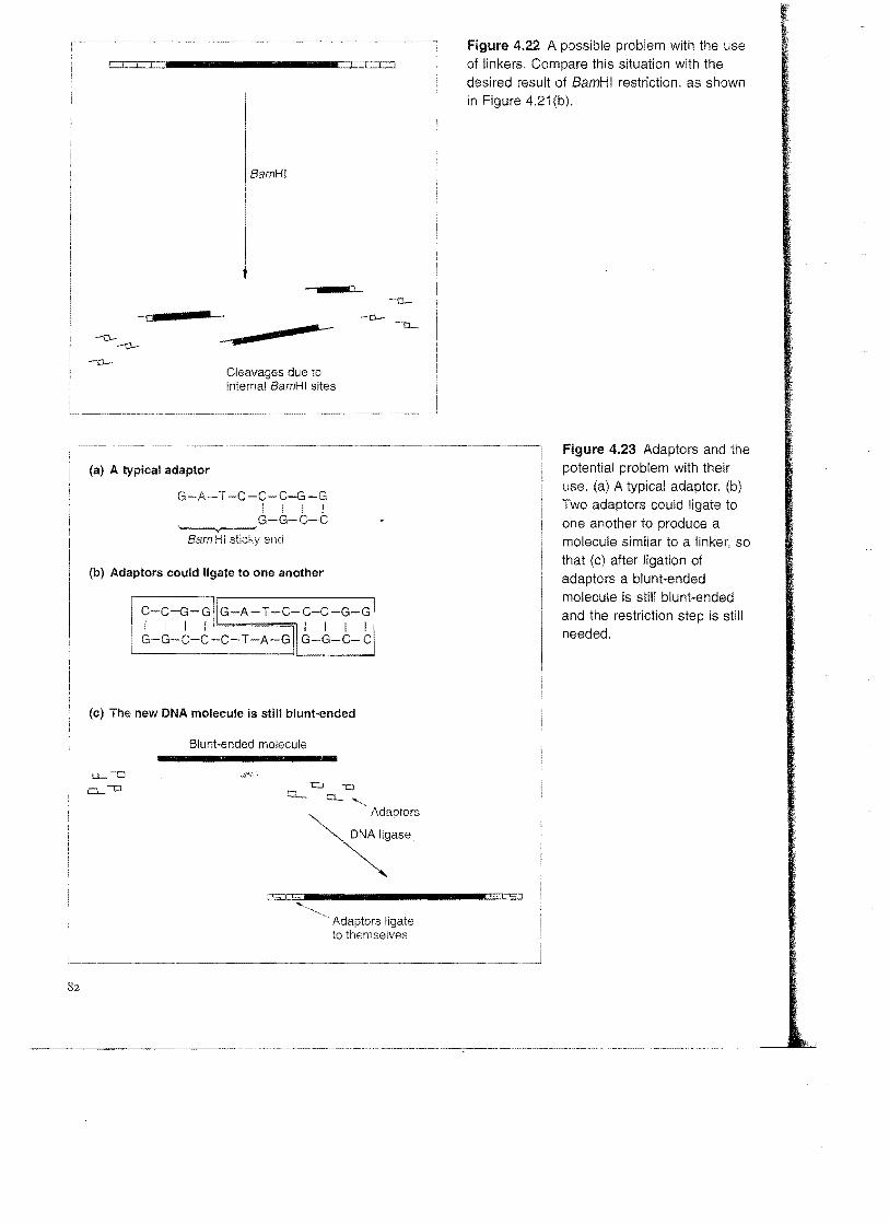
Adaptors There is one potential drawback with the use of linkers. Consider what would happen if the blunt-ended molecule shown in Figure 4.21(b) contained one or more BamHI recognition sequences. If this was the case the restriction step

Z!'r""----~------------~- ~-.-- -~--------..... ......
DNA molecules tnt'1oth,o>y
Figure 4.21 Linkers and their use: (a) A typical linker(a) the structure of a typical linker;
(b) the attachment of linkers to a C-G-A-T-G-G-A-T-C-C-A-T-C-Cblunt-ended molecule. I I I I 1 I I I I I I I I I G-C-T-A-C-C-T-A-G-G-T-A-G-C
'----,y ,
BamHI site
(b) The use of linkers
Blunt-ended molecule linkers
,,~i=
Linkers attached
BamHI sticky end
/-c=....................~i ~ ~
I
! )
needed to cleave the linkers and produce the sticky ends would also cleave the blunt-ended molecule (Figure 4.22). The resulting fragments will have the correct sticky ends, but that is no consolation if the gene contained in the blunt-ended fragment has now been broken into pieces.
The second method of attaching sticky ends to a blunt-ended molecule is designed to avoid this problem. Adaptors, like linkers, are short synthetic oligonucleotides. But unlike linkers, an adaptor is synthesized so that it already has one sticky end (Figure 4.23( a». The idea is of course to ligate the blunt end of the adaptor to the blunt ends of the DNA fragment, to produce a new molecule with sticky ends. This may appear to be a simple method but in practice a new problem arises. The sticky ends of individual adaptor molecules could base pair with each other to form dimers (Figure 4.23(b», so that the new DNA molecule is still blunt-ended (Figure 4.23(c». The sticky ends could be recreated by digestion with a restriction endonuclease, but that would defeat the purpose of using adaptors in the first place.
The answer to the problem lies in the precise chemical structure of the ends of the adaptor molecule. Normally the two ends of a polynucleotide strand are chemically distinct, a fact that is clear from a careful examination of the polymeric structure of DNA (Figure 4.24(a». One end, referred to as
81
~~=C~."""""""""~~I=~~~
BamHI
mo - - -
Cleavages due to internal BamHI sites
(a) A typical adaptor
G-A-T -C-C-C-G-G I I I I G-G-C-C
~ BamHI sticky end
(b) Adaptors could ligate to one another
(c) The' new DNA molecule is still blunt-ended
Blunt-ended molecule
I:J Cl....,
~D~:·~~aPtors ~a"
: c., I ,~,
~.. Adaptors ligate to themselves
Figure 4.22 A possible problem with the use of linkers. Compare this situation with the desired result of 8amHI restriction, as shown in Figure 4.21 (b).
Figure 4.23 Adaptors and the potential problem with their use. (a) A typical adaptor. (b) Two adaptors could ligate to one another to produce a molecule similar to a linker, so that (c) atter ligation of adaptors a blunt-ended molecule is still blunt-ended and the restriction step is still needed.
1.,1 to,!
--_..._--- ......._..- ..•.. - .....- ... - .....---.~--..-.--.--.-..-.-.--.-..--.----~---~

DNA molecules tnnAtn,Ar
Figure 4.24 The distinction between the 5' and 3' termini of a (a) The structure of a
polynucleotide strand polynucleotide. showing the chemical distinction between the S'·P and 3'·OH terminiOne ~
",o,otid, I
I L
(b) In the double helix the polynucleotide strands are anti parallel
5' 3'»»»»)««««(
3' 5'
(c) Ligation takes place between 5'-P and 3'·OH termini
5' -0--0--0-0 3' 5'~3' I I 1 I
-0-0-0-0-0-o--o ~ 3' 5' 3' 5'
~ 5' 3'
-O-O-O-O-It ...... .
3/~~51
the 5' terminus, carries a phosphate group (5' -P); the other, the 3' terminus, has a hydroxyl group (3'-OH). In the double helix the two strands are antiparaIlel (Figure 4.24(b)), so each end of a double-stranded molecule consists of one 5'-P terminus and one 3'-OH terminus. Ligation takes place between the 5'·P and 3'-OH ends (Figure 4.24(e».
Adaptor molecules are synthesized so that the blunt end is the same as 'natural' DNA, but the sticky end is different. The 3'-OH terminus of the sticky
83

of Purified DNA
end is the same as usual, but the 5'-P terminus is modified: it lacks the phosphate group, and is in fact a S'-OH terminus (Figure 4.2S(a». DNA ligase is unable to form a phosphodiester bridge between S'-OH and 3'-OH ends. TIle result is that, although base pairing is always occurring between the sticky ends of adaptor molecules. the association is never stabilized by ligation (Figure 4.2S(b) ).
Adaptors can therefore be ligated to a blunt-ended DNA molecule but not to themselves. After the adaptors have been attached, the abnormal5'-OH terminus is converted to the natural 5'-P form by treatment with the enzyme polynucleotide kinase (p. 60), producing a sticky-ended fragment that can be inserted into an appropriate vector.
Producing sticky ends by homopolymer tailing The technique of homopolymer tailing offers a radically different approach to the production of sticky ends on a blunt-ended DNA molecule. A homopolymer is simply a polymer in which all the subunits are the same. A DNA strand
Figure 4.25 The use of (a) The precise structure of an adaptor adaptors: (a) the actual
structure of an adaptor, HO, .,./OH
showing the modified 5'-OH G-A-T-C-C-C-G-G I I I ! terminus; (b) conversion of G-G-C-C
blunt ends to sticky ends .,./ 'P02.The modified HO 3 5' ·OH terminus through the attachment of
adaptors.
(b) Ligation using adaptors
HO t::l _ Adaptors HO-o Blunt-ended molecule O--OH
~g"" O-O~O-WI__________________D-~~~
POlynUCleotide kinase
)
2·03P-.:::___________\PO~·
5'·P Terminus

DNA molecules tnn"th,,,r
made up entirely of, say, deoxyguanosine is an example of a homopolymer, and is referred to as polydeoxyguanosine or poly(dG).
Tailing involves using the enzyme terminal deoxynucleotidyl transferase (p. 60) to add a series of nucleotides onto the 3/-OH termini of a double-
Figure 4.26 Homopolymer tailing: (a) synthesis of a (a) Synthesis of a homopolymer tail
homopolymer tail; (b) construction of a recombinant DNA molecule from a tailed vector plus tailed insert DNA; (c) repair of the recombinant DNA molecule. dCTP ::: 2' -deoxycytidine 5' -triphosphate.
3' 5'
(b) Ligation of homopolymer tails
Vector - poly(dC) tails
Insert DNApoly(dG) tails
(c) The repair steps
repairs the discontinuities
85

Manipulation of Purified DNA
stranded DNA molecule. If this reaction is carried out in the presence of one deoxyribonucleotide a homopolymer tail is produced (Figure 4.26(
Of course, to be able to ligate together two tailed molecules, tIle liUllHJ'LJVI
mers must be complementary, Frequently polydeoxycytosine (poly(dC» are attached to the vector and poly(dG) to the DNA to be cloned. Base between the two occurs when the DNA moLecules are mixed (Figure
In practice the poly(dG) and poly( dC) tails are not usually exactly same length, and the base-paired recombinant molecules that result have as well as discontinuities (Figure 4.26( c». Repair is therefore a two process, using Klenow polymerase to fIll in the nicks followed by DNA ligase to synthesize the final phosphodiester bonds. This repair reaction does not always have to be performed in the test tube. If the complementary homopolymer tails are longer than about 20 nucleotides, then quite stable base-paired associations are formed. A recombinant DNA molecuLe, held together by base pairing although not completely ligated, is often stable enough to be introduced into the host cell in the next stage of the cloning experiment (Figure 1.1). Once inside the host, the cell's own DNA polymerase and DNA ligase repair the recombinant DNA molecule, completing the construction begun in the test tube.
Further reading
Brown, T.A. (1998) Molecular Biology Labfax. Volume I: Recombinant DNA, 2nd edn. Academic Press, London. [Contains details of all types of enzymes used to manipulate DNA and RNA]
Jacobsen, H., Klenow, H. & Overgaard-Hansen, K. (1974) The N-terminal amino acid sequences of DNA polymerase I from Escherichia coli and of the large and small fragments obtained by a limited proteolysis. European Journal of Biochemistry, 45, 623-7. [Production of the Klenow fragment of DNA polymerase 1.]
Powell, R & Gannon, F. (2000) Construction of recombinant DNA molecules. In: Essential Molecular Biology: A Practical Approach, Vol. 1 (ed. T.A Brown), 2nd edn, pp. 129-50. Oxford University Press, Oxford. [Details of restriction and ligation.]
REBASE: http://rebase.neb.com!rebase/ [A comprehensive list of all the known restriction endonucleases and their recognition sequences.]
Robinson, D.H. & Lafleche. G.J. (2000) Nucleic acid electrophoresis in agarose gels. In: Essential Molecular Biology: A Practical Approach, Vol. 1 (ed. TA Brown), 2nd edn, pp. 89-119. Oxford University Press, Oxford.
Rothstein, RJ., Lau, L.F., BahL c.P., Narang, N.A & Wu, R (1979) Synthetic adaptors for cloning DNA .Methods in Enzymology, 68, 98-109.
Smith, H.o. & Wilcox, K.W. (1970) A restriction enzyme from Haemophilus influenzae. Journal of Molecular Biology, 51, 379-91. [One of the first full descriptions of a restriction endonuclease.]
86