Autoacetylation of Purified Calreticulin Transacetylase Utilizing Acetoxycoumarin as the Acetyl...
14
Autoacetylation of Purified Calreticulin Transacetylase Utilizing Acetoxycoumarin as the Acetyl Group Donor Seema Bansal & Prija Ponnan & Hanumantharao G. Raj & Susan T. Weintraub & Madhu Chopra & Ranju Kumari & Daman Saluja & Ajit Kumar & Tapesh K. Tyagi & Prabhjot Singh & Ashok K. Prasad & Luciano Saso & Ramesh C. Rastogi & Virinder S. Parmar Received: 17 February 2008 / Accepted: 26 August 2008 / Published online: 16 September 2008 # Humana Press 2008 Abstract Our earlier reports documented that calreticulin, a multifunctional Ca 2+ -binding protein in endoplasmic reticulum lumen, possessed protein acetyltransferase function termed Calreticulin Transacetylase (CRTAase). The autoacetylation of purified human placental CRTAase concomitant with the acetylation of receptor proteins by a model acetoxycoumarin, 7,8-Diacetoxy-4-methylcoumarin, was observed. Here, we have examined the autoacetylation property of CRTAase by immunoblotting and mass spectrometry. Ca 2+ was found to inhibit CRTAase activity. The inhibition of both autoacetylation of CRTAase as well as acetylation of the receptor protein was apparent when Ca 2+ was included in the reaction mixture as visualized by interaction with anti-acetyl lysine antibody. The acetylation of lysines residues: −48, −62, −64, −153, and −159 in N-domain and −206, −207, −209, and −238 in P-domain of CRTAase were located by high- performance liquid chromatography-electronspray ionization tandem mass spectrometry. Further, computer assisted protein structure modeling studies were undertaken to probe the effect of autoacetylation of CRTAase. Accordingly, the predicted CRTAase 3D model showed that all the loop regions of both N- and P-domain bear the acetylated lysines. Appl Biochem Biotechnol (2009) 157:285–298 DOI 10.1007/s12010-008-8357-2 S. Bansal : P. Ponnan : H. G. Raj (*) : R. Kumari : A. Kumar : T. K. Tyagi : P. Singh Department of Biochemistry, V. P. Chest Institute, University of Delhi, Delhi 1100 07, India e-mail: [email protected] P. Ponnan : A. K. Prasad : R. C. Rastogi : V. S. Parmar Department of Chemistry, University of Delhi, Delhi 1100 07, India M. Chopra : D. Saluja Dr.B.R.Ambedkar Center for Biomedical Research, University of Delhi, Delhi 1100 07 Delhi, India S. T. Weintraub Department of Biochemistry, The University of Texas Health Science Center, 7703 Floyd Curl Drive, San Antonio, TX 78229-3900, USA L. Saso Department of Human Physiology and Pharmacology, “Vittorio Erspamer” Sapienza University of Rome, P.le Aldo Moro 5, 00185 Rome, Italy
-
Upload
independent -
Category
Documents
-
view
4 -
download
0
Transcript of Autoacetylation of Purified Calreticulin Transacetylase Utilizing Acetoxycoumarin as the Acetyl...
Autoacetylation of Purified Calreticulin TransacetylaseUtilizing Acetoxycoumarin as the Acetyl Group Donor
Seema Bansal & Prija Ponnan & Hanumantharao G. Raj &Susan T. Weintraub & Madhu Chopra & Ranju Kumari &Daman Saluja & Ajit Kumar & Tapesh K. Tyagi &Prabhjot Singh & Ashok K. Prasad & Luciano Saso &
Ramesh C. Rastogi & Virinder S. Parmar
Received: 17 February 2008 /Accepted: 26 August 2008 /Published online: 16 September 2008# Humana Press 2008
Abstract Our earlier reports documented that calreticulin, a multifunctional Ca2+-bindingprotein in endoplasmic reticulum lumen, possessed protein acetyltransferase functiontermed Calreticulin Transacetylase (CRTAase). The autoacetylation of purified humanplacental CRTAase concomitant with the acetylation of receptor proteins by a modelacetoxycoumarin, 7,8-Diacetoxy-4-methylcoumarin, was observed. Here, we haveexamined the autoacetylation property of CRTAase by immunoblotting and massspectrometry. Ca2+ was found to inhibit CRTAase activity. The inhibition of bothautoacetylation of CRTAase as well as acetylation of the receptor protein was apparentwhen Ca2+ was included in the reaction mixture as visualized by interaction with anti-acetyllysine antibody. The acetylation of lysines residues: −48, −62, −64, −153, and −159 inN-domain and −206, −207, −209, and −238 in P-domain of CRTAase were located by high-performance liquid chromatography-electronspray ionization tandem mass spectrometry.Further, computer assisted protein structure modeling studies were undertaken to probe theeffect of autoacetylation of CRTAase. Accordingly, the predicted CRTAase 3D modelshowed that all the loop regions of both N- and P-domain bear the acetylated lysines.
Appl Biochem Biotechnol (2009) 157:285–298DOI 10.1007/s12010-008-8357-2
S. Bansal : P. Ponnan :H. G. Raj (*) : R. Kumari : A. Kumar : T. K. Tyagi : P. SinghDepartment of Biochemistry, V. P. Chest Institute, University of Delhi, Delhi 1100 07, Indiae-mail: [email protected]
P. Ponnan : A. K. Prasad : R. C. Rastogi : V. S. ParmarDepartment of Chemistry, University of Delhi, Delhi 1100 07, India
M. Chopra :D. SalujaDr.B.R.Ambedkar Center for Biomedical Research, University of Delhi, Delhi 1100 07 Delhi, India
S. T. WeintraubDepartment of Biochemistry, The University of Texas Health Science Center, 7703 Floyd Curl Drive,San Antonio, TX 78229-3900, USA
L. SasoDepartment of Human Physiology and Pharmacology,“Vittorio Erspamer” Sapienza University of Rome, P.le Aldo Moro 5, 00185 Rome, Italy

Energy minimization of the acetylated residues revealed charge neutralization of lysines dueto the N-ε-acetylation which may facilitate the interaction of CRTAase with the proteinsubstrate and the subsequent transacetylase action.
Keywords Protein acetyltransferase . Calreticulin . Acetoxycoumarin . Protein acetylation .
Mass spectrometry
Introduction
Calreticulin (CR) is one of the most important Ca2+-binding proteins in the endoplasmicreticulum (ER) of eukaryotic cells. CR is divided into three structural and functionaldomains [1]. The amino terminal (N-domain) is highly conserved and binds Zn2+ with highcapacity [2, 3]. It is followed by proline rich P-domain that binds Ca2+ with high affinity(Kd=0.05–10 μM and 1 mol of Ca2+/mol of protein) [4]. The carboxy terminal (C-domain)is highly acidic bearing clusters of aspartate and glutamate residues, binds Ca2+ with highcapacity (Kd=∼1–2 mM and ∼25–50 mol of Ca2+/mol of protein) [4, 5]. CR in coordinationwith its paralog calnexin functions as molecular chaperones in the correct folding of nascentN-linked glycoproteins in the ER lumen [6]. Even though CR is a soluble ER protein andcalnexin is type 1 ER membrane protein [1, 6], they are known to possess remarkableamino acid structural and sequence similarity. Peptide segments of these two homologousproteins have varying sequence identity ranging from 42–78% [7].
Recently the X-ray crystal structure of calnexin luminal domain from dog [8] and NMRsolution structure of rat CR P-domain has been determined [9].
We have established the transfer of acetyl group from 7,8-Diacetoxy-4-methylcoumarin(DAMC), a model acetoxy drug to certain receptor proteins such as glutathione S-transferase (GST), nicotinamide adenine dinucleotide phosphate (NADPH) cytochromeP-450 reductase and nitric oxide synthase (NOS) [10–12]. In our recent report, we haveprovided mass spectrometric evidence for the placental CRTAase-mediated acetylation ofnNOS by DAMC [13]. The analysis of protein acetylation by mass spectrometry has gainedpopularity because of its sensitivity and rapid characterization of post-translationalmodifications [14, 15].We observed the concomitant acetylation of CR during the CRTAasecatalyzed reaction. Such autoacetylation of CR was investigated recently [13, 16]. In thisstudy, electrospray ionization (ESI)-tandem mass spectrometry (MS/MS) was used tolocalize the acetylated residues within the sequence of human placental CRTAase. In orderto visualize the acetylated sites, a theoretical three dimensional structure of CRTAase waspredicted and the modified amino acids were analyzed with respect to the neighboringamino acid groups of the secondary structure of CRTAase.
Materials and Methods
Materials
Anti-acetyl lysine polyclonal antibody was purchased from Cell Signaling Technology,Beverly, MA, USA. Goat anti-rabbit horse radish peroxidase (HRP) conjugated secondaryantibody and diaminobenzidine (DAB) were purchased from Bangalore Genei, Bangalore,India. Acryl amide, ammonium per sulphate, TEMED, bis-acrylamide, pre-stainedmolecular weight markers were purchased from BioRad, Hertfordshire, United Kingdom.
286 Appl Biochem Biotechnol (2009) 157:285–298

Glycine, Trizma base, sodium dodecyl sulphate (SDS), reduced glutathione (GSH), 1-chloro-2,4 dinitrobenzene (CDNB), EDTA, were procured from Sigma Chemicals Co.,St. Louis, MO, USA. Standard molecular weight markers for SDS-PAGE were procuredfrom MBI Fermentas. Modified trypsin was supplied by Promega, Madison, WI, USA. Allother chemicals used were of high purity and were obtained from local suppliers.
Purification of CRTAase from Human Placenta
Placenta was obtained from Gauri Nursing Home, Malka Ganj, Delhi, India from healthy,25–40-year-old patients who had no symptoms of any infectious disease. Placenta wasstored and brought to the laboratory in an ice box within 30 min of delivery. Placentalmicrosomes and cytosol were prepared as described earlier [16]. Protein content wasestimated by the method of Lowry et al. [17]. Placental CRTAase was purified as describedearlier [16]. For this purpose, microsomal pellet was solubilized by brief homogeniza-tion in 0.1M phosphate buffer, pH 7.4 and 5% sodium cholate using the method of Deyet al. with modifications [18]. The clear supernatant obtained after solubilization wasdialyzed overnight against 10 mM potassium phosphate buffer (pH 7.4) containing 1 mMEDTA, 1 mM DTT, and 1 mM PMSF. The dialyzed fraction was subjected to CM-Sepharose, DEAE-Sepharose, and Q-Sepharose chromatographies to purify the protein tohomogeneity.
Assay of CRTAase
CRTAase in placental microsomes was assayed routinely using DAMC as the first substrate(unless otherwise mentioned) and cytosolic GST as the second substrate, using the methodof Raj et al. [12]. The percentage inhibition of cytosolic GST under the conditions of theassay was considered proportional to CRTAase activity.
SDS PAGE
Autoacetylation experiments were carried out using DAMC and the purified CRTAase [16],the production of acetylated CRTAase was quantified after separating the components ofthe reaction on sodium dodecyl sulfate polyacrylamide gel electrophoresis (SDS-PAGE).Appropriate controls were taken to elucidate the role of CRTAase in catalyzing theautoacetylation. Purified CRTAase was incubated with DAMC (100 μM), 10 mMphosphate buffer (pH 7.2) without any receptor protein and incubated for 30 min at 37 °Cin a water bath. After the completion of the reaction, sample buffer (loading dye) was added tothe reaction mixture to stop the reaction. This reaction mixture was loaded onto SDS-PAGE.The gel electrophoresis was carried out with discontinuous buffer system originally devisedby Orstein [19] and Davis [20] followed by the procedure of Laemmli [21]. The samplesand the stacking gel were prepared in Tris–HCl (pH 6.8). All components of the systemcontained 0.1% SDS.
Western Blot Analysis
For the detection of the acetylated lysine residues, western blot analysis was carried outusing anti-acetyl lysine polyclonal antibody. The electrophoretically separated protein weretransblotted to polyvinylidene fluoride (PVDF) membrane at 300 mA for 3 h at 4 °C. Non-specific sites on the PVDF were blocked with blocking reagent (5% Blotto). Primary
Appl Biochem Biotechnol (2009) 157:285–298 287
antibody dilution (1:1,000) was prepared in TBST (Tris-buffered saline (TBS) with 0.05%Tween) containing 1% BSA and incubation was carried out in 4 °C overnight with slightagitation.
The PVDF membrane was extensively washed with TBST. Goat anti-rabbit HRPconjugated secondary antibody approximately diluted in TBST was then added andincubation for 1 h at room temperature was carried out. The sheets were then washedextensively with TBST/TBS.
Acetylated protein was immunodetected with rabbit polyclonal anti-acetylated lysine(1:1,000) and developed with goat anti-rabbit IgG secondary antibody coupled to HRPusing DAB system.
Effect of Calcium on CRTAase Catalyzed Acetylation of NADPH Cytochrome P-450Reductase and Autoacetylation
In order to demonstrate the effect of calcium on CRTAase catalyzed acetylation of receptorprotein, DAMC (100 μM) was separately preincubated with or without calcium (5 μM)along with the purified CRTAase (M. wt. 60 KDa) and receptor protein (NADPHcytochrome P-450 reductase) followed by western blot using anti-acetyl lysine antibodyas described above. Appropriate controls having DMSO in place of DAMC and otherone having no enzyme were taken. In the same manner, DAMC (100 μM) waspreincubated with calcium (5 μM) followed by addition of CRTAase [16] to elucidate therole of calcium on autoacetylation.
In-Gel Protein Reduction
The acetylated bands were cut and placed in a 0.6-ml tube filled with ultra pure water. Thebands were washed with 40 mM ammonium bicarbonate, 50% acetonitrile. Wash reagent,500 μl, was added to each tube followed by slow inversion mixing for 10 min. The washingstep was repeated and the tube was filled with acetonitrile. The band was allowed toincubate end over end for another 10 min. At this point, the bands were dehydrated piece ofplastic. The acetonitrile was removed by spinning at 1,000 rpm for 5 min and the bandswere ready for trypsin digestion.
HPLC-ESI-MS/MS
The excised bands were digested in situ with trypsin in 40mMNH4HCO3 at 37 °C for 4 h. Thedigests were analyzed without further purification by capillary high-performance liquidchromatography (HPLC)-electrospray ionization tandem mass spectra (HPLC-ESI-MS/MS)acquired on a Thermo Fisher LTQ linear ion trap mass spectrometer (San Jose, CA, USA)fitted with a New Objective Pico View 550 nanospray interface. The source was coupledonline to an Eksigent Nano LC micro HPLC (Dublin, CA, USA). The digest were dissolvedin mobile phase A (0.5% acetic acid (HAc)/0.005% trifluroacetic acid (TFA)) and loaded ontoa micro HPLC column PicoFrit™ (Woburn, MA, USA; 10 cm length×75 μ i.d.) packed withC18 adsorbant (Vydac, Hesperia, CA; 218MS 5 μM particle size, 300 Å pore diameter). Thepeptides were eluted from the column with a gradient of 2% to 42% mobile phase B (90%acetonitrile/0.5% HAc/0.005% TFA) for 30 min with a flow rate of 0.4 μl/min. The elutedpeptides were electrosprayed into the linear ion trap mass spectrometer (2.9 KV ESI voltage,three isolation window for MS/MS and 35% relative collision energy).
288 Appl Biochem Biotechnol (2009) 157:285–298
Mass Spectrometric Data Analysis
The spectrum were surveyed by scan strategy followed by acquisition of data-dependentcollision-induced dissociation (CID) mode that determined seven most intense ions in thesurvey scan above a set threshold. The uninterpreted CID spectra were searched against theNCBI-nr database with MASCOT search engine (Matrix Science, London, UK) [22].Methionine oxidation and lysine acetylation were considered as variable modification forall searches. Cross correlation of the Mascot results with X! Tandem [23] was done anddetermination of protein identity probabilities was accomplished by Scaffold™ (ProteomeSoftware Inc.).
Molecular Modeling
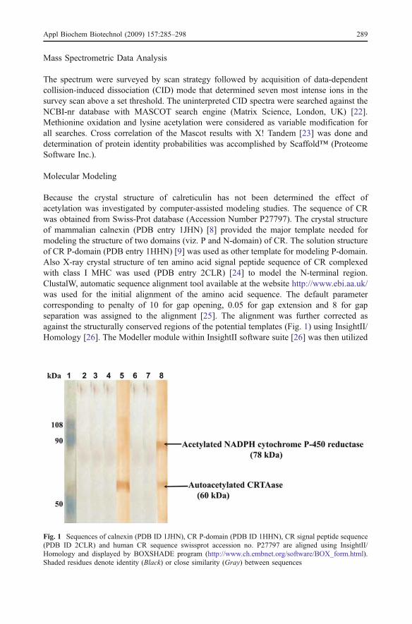
Because the crystal structure of calreticulin has not been determined the effect ofacetylation was investigated by computer-assisted modeling studies. The sequence of CRwas obtained from Swiss-Prot database (Accession Number P27797). The crystal structureof mammalian calnexin (PDB entry 1JHN) [8] provided the major template needed formodeling the structure of two domains (viz. P and N-domain) of CR. The solution structureof CR P-domain (PDB entry 1HHN) [9] was used as other template for modeling P-domain.Also X-ray crystal structure of ten amino acid signal peptide sequence of CR complexedwith class I MHC was used (PDB entry 2CLR) [24] to model the N-terminal region.ClustalW, automatic sequence alignment tool available at the website http://www.ebi.aa.uk/was used for the initial alignment of the amino acid sequence. The default parametercorresponding to penalty of 10 for gap opening, 0.05 for gap extension and 8 for gapseparation was assigned to the alignment [25]. The alignment was further corrected asagainst the structurally conserved regions of the potential templates (Fig. 1) using InsightII/Homology [26]. The Modeller module within InsightII software suite [26] was then utilized
Fig. 1 Sequences of calnexin (PDB ID 1JHN), CR P-domain (PDB ID 1HHN), CR signal peptide sequence(PDB ID 2CLR) and human CR sequence swissprot accession no. P27797 are aligned using InsightII/Homology and displayed by BOXSHADE program (http://www.ch.embnet.org/software/BOX_form.html).Shaded residues denote identity (Black) or close similarity (Gray) between sequences
Appl Biochem Biotechnol (2009) 157:285–298 289
to assign coordinates to regions structurally aligned to template, build intervening loops,optimize the rotamers of amino acid side chains and perform an initial energy optimizationof the structure. The disulfide bond in CR N-domain (Cys105-Cys137) which is conservedin calnexin (Cys161-Cys195) was assigned to the initial model as predicted by homologywith calnexin.
Biopolymer module implemented in InsightII [26] was used to modify lysines −48,−62, −64, −153, −159, −206, −207, −209, and −238 by the addition of an acetyl group,from the InsightII fragment library, to the side chain N-ε-atom. Using the same module,hydrogen atoms were added to the acetylated and non-acetylated protein structures at pH7.0. The default consistent valence force field (cvff) force field [27] was applied to boththe structures, the net charge of resulting protonated state of the acetylated protein was−68 and non-acetylated protein was −59. Further, a series of energy minimization stepswere performed on both the protein structures by InsightII/Discover [26] using followingprotocol: (a) In the first step of minimization, all the heavy (all non-hydrogen) atoms wereconstrained, the hydrogen atoms were allowed to minimize using 100 cycles of steepestdecent until the maximum derivative (|dE/dr|) of the system was <1 kcal/(mole Ǻ). (b)This step was followed by another steepest descent minimization with the sameparameter as in step (a), but constraining the protein backbone atoms and relaxing allother atoms of the molecule. (c) In the final step, the protein molecule was minimizedusing 50 cycles of conjugate gradient method with the backbone atom fixed andallowing all other atoms relax until the maximum derivative was <0.01 kcal/(mole Ǻ).The final RMS derivative for both the minimized structure was found to be <0.001.The difference in the interaction after N-ε atom modifications of lysine residues wereanalyzed by InsightII [26].
Results
We have earlier documented isolation, purification, and characterization of CRTAase fromhuman placental microsomes [16]. The placental CRTAase was found to have molecularweight of 60 kDa and exhibited differential specificities to various classes of polyphenolicacetates. Placental CRTAase was incubated with a receptor protein NADPH cytochromeP-450 reductase and DAMC followed by western blot analysis using anti-acetyl lysineantibody. CRTAase when preincubated with CaCl2 (3 μM) was found to be inhibited [16].These observations bring home the influence of Ca2+-binding site on CRTAase activity.
Effect of Calcium on CRTAase Catalyzed Acetylation of NADPHCytochrome P-450 Reductase
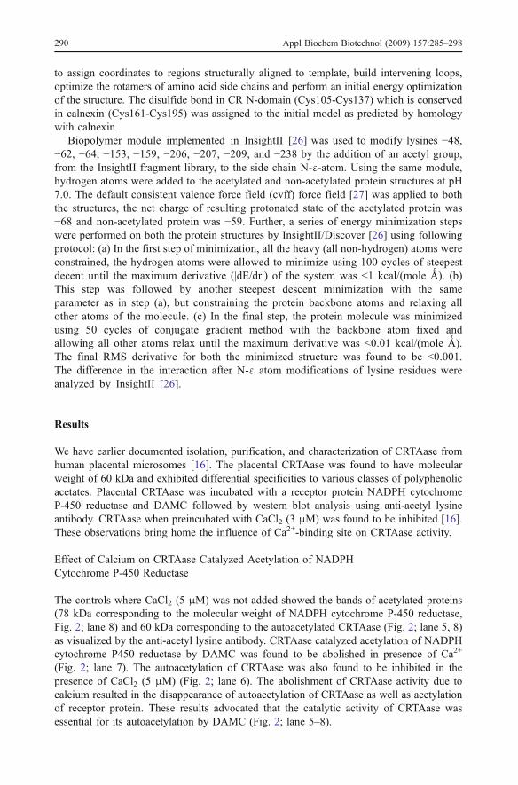
The controls where CaCl2 (5 μM) was not added showed the bands of acetylated proteins(78 kDa corresponding to the molecular weight of NADPH cytochrome P-450 reductase,Fig. 2; lane 8) and 60 kDa corresponding to the autoacetylated CRTAase (Fig. 2; lane 5, 8)as visualized by the anti-acetyl lysine antibody. CRTAase catalyzed acetylation of NADPHcytochrome P450 reductase by DAMC was found to be abolished in presence of Ca2+
(Fig. 2; lane 7). The autoacetylation of CRTAase was also found to be inhibited in thepresence of CaCl2 (5 μM) (Fig. 2; lane 6). The abolishment of CRTAase activity due tocalcium resulted in the disappearance of autoacetylation of CRTAase as well as acetylationof receptor protein. These results advocated that the catalytic activity of CRTAase wasessential for its autoacetylation by DAMC (Fig. 2; lane 5–8).
290 Appl Biochem Biotechnol (2009) 157:285–298
ESI-MS/MS
The results from ESI-MS/MS revealed the localization of acetylated lysines ofautoacetylated CRTAase. For this purpose, the CRTAase was incubated with DAMC for30 min as described under “Materials and Methods” section. The reaction mixture wassubjected to SDS-PAGE for the separation of autoacetylated CRTAase. The autoacetylated
Fig. 2 Effect of Ca2+ onCRTAase catalyzed acetylationof NADPH cytochrome P-450reductase by DAMC. Lane 1Prestained molecular weightmarkers; lane 2 CRTAase+DMSO; lane 3 NADPH cyto-chrome P-450 reductase+DAMC;lane 4 CRTAase+DMSO+CaCl2+(5 μM)+NADPHcytochrome P-450 reductase;lane 5 CRTAase+DAMC; lane 6CRTAase+CaCl2+(5 μM)+DAMC; lane 7 CRTAase+CaCl2+(5 μM)+DAMC+NADPHcytochrome P-450 reductase;lane 8 CRTAase+DAMC+NADPH cytochrome P-450reductase
Appl Biochem Biotechnol (2009) 157:285–298 291
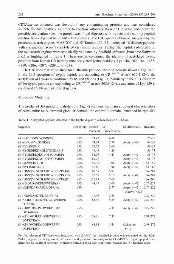
CRTAase so obtained was devoid of any contaminating proteins and was consideredsuitable for MS analysis. In order to confirm autoacetylation of CRTAase and locate thepossible acetylation sites, the protein was in-gel digested with trypsin and resulting peptidemixture was subjected to ESI-MS/MS analysis. The CID spectra obtained analyzed by theproteome search engines MASCOT and X! Tandem [22, 23], indicated 18 distinct peptideswith a significant score as acetylated on lysine residues. Further the peptides identified bythe two search engines were statistically validated by Scaffold software (Proteome SoftwareInc.) as highlighted in Table 1. These results confirmed the identity of acetylated trypticpeptides from human CR bearing nine acetylated lysine residues, Lys −48, −62, −64, −153,−159, −206, −207, −209, and −238.
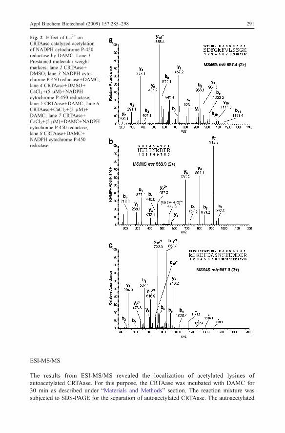
The CID spectra were obtained for all the nine peptides, three of them are shown in Fig. 3a–c.In the CID spectrum of tryptic peptide corresponding to CR 44–55 at m/z 657.4 (2+), theacetylation of Lys-48 is confirmed by b5 and y8 ions (Fig. 3a). Similarly in the CID spectrumof the tryptic peptide corresponding to CR154–162 at m/z 563.9 (2+), acetylation of Lys-159 isconfirmed by b6 and y4 ions (Fig. 3b).
Molecular Modeling
The predicted 3D model of calreticulin (Fig. 4) contains the main structural characteristicsof calreticulin: an N-terminal globular domain, the central P-domain “extended hairpin-like
Table 1 Acetylated peptides detected in the tryptic digest of autoacetylated CRTAase.
Sequence Probability Mascotion score
X!Tandem score
Modifications Residues
(K)EQFLDGDGWTSR(W) 95% 73.42 6.80 25–36(K)SDFGkFVLSSGK(F) 95% 74.14 2.70 Acetyl (+42) 44–55(K)FVLSSGK(F) 95% 37.72 2.00 49–55(K)FYGDEEKDKGLQTSQDAR(F) 95% 46.48 4.20 56–73(K)FYGDEEkDKGLQTSQDAR(F) 95% 54.08 4.47 Acetyl (+42) 56–73(K)FYGDEEKDkGLQTSQDAR(F) 95% 61.27 Acetyl (+42) 56–73(K)GkNVLINK(D) 95% 49.39 3.00 Acetyl (+42) 152–159(K)NVLINkDIR(C) 95% 45.86 1.96 Acetyl (+42) 154–162(K)IDNSQVESGSLEDDWDFLPPKK(I) 95% 67.59 5.82 186–207(K)IDNSQVESGSLEDDWDFLPPkK(I) 95% 55.59 5.15 Acetyl (+42) 186–207(K)IDNSQVESGSLEDDWDFLPPK(K) 95% 135.14 7.68 186–206(K)kIKDPDASKPEDWDER(A) 95% 44.45 3.09 Acetyl (+42) 207–222(K)kIkDPDASKPEDWDER(A) 95% 2.77 Acetyl (+42),
Acetyl (+42)207–222
(K)IKDPDASKPEDWDER(A) 95% 39.95 2.55 208–222(R)AKIDDPTDSKPEDWDkPEHIPD
PDAK(K)95% 63.97 5.85 Acetyl (+42) 223–248
(K)IDDPTDSKPEDWDkPEHIPDPDAK(K)
95% 4.31 Acetyl (+42) 225–248
(K)KPEDWDEEMDGEWEPPVIQNPEYK(G)
95% 58.11 7.35 249–272
(K)KPEDWDEEmDGEWEPPVIQNPEYK(G)
95% 68.83 5.44 Oxidation(+16)
249–272
Purified placental CRTAase was incubated with DAMC, the modified protein was separated on the SDS-PAGE, digested with trypsin at 37° for 4 h and processed for analysis by LC-MS/MS. Tryptic peptides areidentified by Scaffold software (Proteome Software Inc.) with significant Mascot and X! Tandem score
292 Appl Biochem Biotechnol (2009) 157:285–298

arm” comprising the two types of repeated motifs PXXIXDPDAXKPEDWDE (type 1) andGXWXPPXIXNPXYX (type 2) arranged in 111222 fashion and the C-terminal domainbearing clusters of aspartate and glutamate residues. The modeling study provided us toanalyze the localization of acetylated lysine residues in the 3D structure of CR. All theacetylated lysine residues located by LC-MS/MS were visualized in the N- and P-domain ofCR (Fig. 4). The predicted 3D structure of CR revealed that the five acetylated lysines in
1JHN 1 SKSKPDTSAPTSPKVTYKAPVPSGEVYFADSFDRGTLSG--WILSKAKKDDTDDEIAKYD1HHN 1 ------------------------------------------------------------2CLR 1 ----MLLSVPLLLG----------------------------------------------P27797 1 ----MLLSVPLLLGLLGLAVAEP-AVYFKEQFLDGDGWTSRWIESKHKSD--FGKFVLSSconsensus 1 mllsvplllgv a vyf d f g wi sk k d
1JHN 59 GKWEVDEMKETKLPGDKGLVLMSRAKHHAISAKLNKPFLFDTKPLIVQYEVNFQNGIECG1HHN 1 ------------------------------------------------------------2CLR ------------------------------------------------------------P27797 54 GKFYGDEEKD------KGLQTSQDARFYALSASFEPFSNKGQTLVVQFTVKHEQNIDCGGconsensus 61 gkw de ke kgl ak aisa li qn g
1JHN 119 GAYVKLLSKTPELNLDQFHDKTPYTIMFGPDKCGEDYKLHFIFRHKNPKTGVYEEKHAKR1HHN 1 ------------------------------------------------------------2CLR ------------------------------------------------------------P27797 108 GYVKLFPNS---LDQTDMHGDSEYNIMFGPDICGPGTKKVHVIFNYKGKNVLINKDIRCKconsensus 121 g l h t y imfgpd cg k i k v r
1JHN 179 PDADLKTYFTDKKTHLYTLILNPDNSFEILVDQSIVNSGNLLNDMTPPVNPSREIEDPED1HHN 1 ---------------------------------------------------SKKIKDPDA2CLR ------------------------------------------------------------P27797 165 -----DDEFTHLYTLIVRPDNTYEVKIDNSQVESGSLEDDWDFL------PPKKIKDPDAconsensus 181 ft t l d e s pskkikdpda
1JHN 239 QKPEDWDERPKIPDPDAVKPDDWNEDAPAKIPDEEATKPDGWLDDEPEYVPDPDAEKPED1HHN 10 AKPEDWDERAKIDDPTDSKPEDW---------------------DKPEHIPDPDAKKPED2CLR ------------------------------------------------------------P27797 214 SKPEDWDERAKIDDPTDSKPEDW---------------------DKPEHIPDPDAKKPEDconsensus 241 kpedwderakiddptdskpedw dkpehipdpdakkped
1JHN 299 WDEDMDGEWEAPQIANPKCESAPGCGVWQRPMIDNPNYKGKWKPPMIDNPNYQGIWKPRK1HHN 49 WDEEMDGEWEP------------------PVIQNPEYKGEWKPRQIDNPDYKGTWIHPEI2CLR ------------------------------------------------------------P27797 253 WDEEMDGEWEP------------------PVIQNPEYKGEWKPRQIDNPDYKGTWIHPEIconsensus 301 wdeemdgewep pviqnpeykgewkprqidnpdykgtwihpei
1JHN 359 IPNPDFFEDLEPFKMTPFSAIGLELWSMTSDIFFDNFIVCGDRRVVDDWANDGWGLKKAA1HHN 91 DNPEYSPDANI-------------------------------------------------2CLR ------------------------------------------------------------P27797 295 DNPEYSPDPSIYAYDNFGVLGLDLWQVKSGTIFDNFLITNDEAYAEEFGNETWGVTKAAEconsensus 361 dnpeyspd i t if i d d k a
1JHN 419 DGAAEP------------------------------------------------------1HHN ------------------------------------------------------------2CLR ------------------------------------------------------------P27797 355 KQMKDKQDEEQRLKEEEEDKKRKEEEEAEDKEDDEDKDEDEEDEEDKEEDEEEDVPGQAKconsensus 421 e
1JHN ---1HHN ---2CLR ---P27797 415 DELconsensus 481
Fig. 3 Tandem mass spectra of selected CR tryptic peptides containing acetyl-lysine. Residue numbersshown in boldface type above and below the peptide sequences indicate detected collision-induceddissociation fragments for y-series (C-terminal) and b-series (N-terminal) ions, respectively. a SequenceSDFGkFVLSSGK (Corresponding to residues 45–55 of CR) observed in the tryptic digest of CR, the ion atm/z 648.4 was generated by loss of water from doubly charged precursor; b sequence NVLINkDIR(Corresponding to residues 154–162 of CR) peak at m/z 554.9 represents the ion due to loss of water in thedoubly charged precursor; c sequence kIKDPDASKPEDWDER (Corresponding to residues 207–222 of CR)observed in the tryptic digest of CR. MS/MS m/z 657.9 (3+). Asterisk corresponds to internal fragments
Appl Biochem Biotechnol (2009) 157:285–298 293

the N-domain (Lys −48, −62, −64, −153, and −159) were found to be in the exposedsurface of the protein. Similar trend was observed in the case of the four acetylated lysinesof P-domain (Lys −206, −207, −209, and −238). To examine effect of acetylated residueson enzyme stability, energy minimization calculations were performed on the modeledstructure CR bearing acetyl group. The interactions of lysine residues with neighboringamino acid side chains and backbone atoms were explored. Examination of all the energeticstate of protein by different rounds of energy minimization have evidenced that in mostcases of the acetylated CR, a loss of H-bonds of the ε-amino group lysine residues andacidic counterparts such as aspartate and glutamate residues as observed in comparison withnon-acetylated form (Figs. 5 and 6). This is due to the loss of positive charges on thespecific lysine residues owing to acetylation and thus failing their ability to formintermolecular H-bonds with the aspartate and glutamate residues.
Discussion
Our earlier studies proposed the concept of enzymatic acetylation of protein independent ofacetyl CoA. The ER was shown to contain a unique enzyme TAase, as described in ourearlier reports [11, 12], catalyzing the transfer of acetyl group to certain functional proteins.TAase was purified to homogeneity from human placental microsomes and exhibited M.wt. of 60 kDa. The N-terminal sequence of purified TAase was found to exhibit 100%identity with N-terminal sequence of mature CR [28]. The identity of TAase with CRwas further confirmed from the properties of CR such as immunoreactivity with anti-CR,
Fig. 4 The predicted structure of CR is represented in Kabsch–Sander secondary structure with α-helices inred and β-sheets in yellow. The modified lysine residues are in stick representation with standard atomcolors. The figure was prepared using Pymol software [43]
294 Appl Biochem Biotechnol (2009) 157:285–298

Ca2+-binding and being a substrate for protein kinase [16]. We have in this communicationmade the efforts to establish the transacetylase-dependent acetylation of CR utilizingacetoxycoumarins as the acetyl group donors. CR is one of the major Ca2+-binding proteinsin the ER. Studies show that binding of Ca2+ to calreticulin appears to induce
Fig. 5 Comparison of intermolecular H-bonds (dotted lines) of lysines with glutamate and aspartate residuesin non-acetylated (a) and acetylated (b) CR, after energy minimization. The amino acid residues are renderedin sticks with standard atom colors. Loss of H-bonds between Lys-62 and Glu-25, Lys-64 and Asp-63 also inLys-48 and Glu-39 was observed due to neutralization of the positively charged lysine residue by acetylation.The acetylated lysine residues of N-domain are highlighted here. The figure was generated using InsightIIprogram [26]
Appl Biochem Biotechnol (2009) 157:285–298 295
conformational change in the protein and shown to modulate the activities of CR byforming a core which is resistant to proteolysis. Also this ion-induced conformationalchange is accountable for Ca2+-dependent interaction of CR with PDI and ERP57 [29]. Theinhibition of autoacetylation of CR by Ca2+ as recorded here may be viewed in this context.
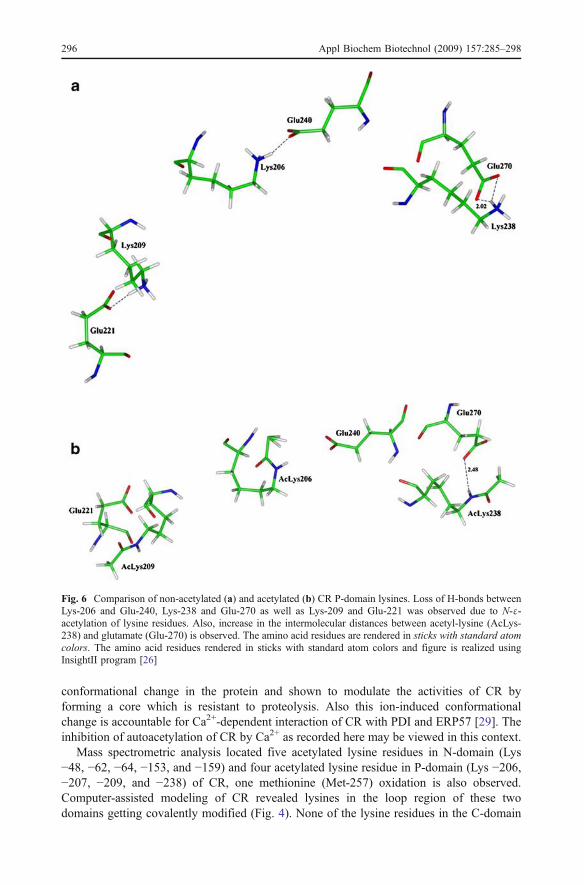
Mass spectrometric analysis located five acetylated lysine residues in N-domain (Lys−48, −62, −64, −153, and −159) and four acetylated lysine residue in P-domain (Lys −206,−207, −209, and −238) of CR, one methionine (Met-257) oxidation is also observed.Computer-assisted modeling of CR revealed lysines in the loop region of these twodomains getting covalently modified (Fig. 4). None of the lysine residues in the C-domain
Fig. 6 Comparison of non-acetylated (a) and acetylated (b) CR P-domain lysines. Loss of H-bonds betweenLys-206 and Glu-240, Lys-238 and Glu-270 as well as Lys-209 and Glu-221 was observed due to N-ε-acetylation of lysine residues. Also, increase in the intermolecular distances between acetyl-lysine (AcLys-238) and glutamate (Glu-270) is observed. The amino acid residues are rendered in sticks with standard atomcolors. The amino acid residues rendered in sticks with standard atom colors and figure is realized usingInsightII program [26]
296 Appl Biochem Biotechnol (2009) 157:285–298
was observed to be covalently modified by acetylation. The effect of covalent modificationN-ε-acetyl-lysine was analyzed by the energy minimization studies. Several rounds of energyminimization revealed neutralization of lysine residue and breaking of intermolecular H-bondwith neighboring acidic residues (Figs. 5 and 6). These effects of modification by way ofacetylation of lysine residues could facilitate interaction of CR with other proteins.
Cellular protein acetylation is mediated by the prototype of histone acetyltransferases(HAT). Several classes of HAT, nuclear (Type A) as well as cytosolic (Type B), are knownto exist. Gcn5/PCAF, MYST and p300/CBP are the major examples of nuclear HAT andHAT1 is termed cytoplasmic HAT [30, 31]. Although, these enzymes catalyze proteinacetylation by acetyl CoA, a number of distinct features among them are recognized in thecase of nuclear HAT [32]. A few of such distinguishing features include sequencedivergence and some HAT is found to acetylate non-histone proteins such as transcriptionfactors p53 and MyoD [33, 34]. Lately, the existence of protein acetyltransferase in the ERlumen is reported [35]. Our recent studies conclusively established protein acetylationmediated by purified CR [13, 16, 28]. CR lacked any structural or sequence resemblancesto prototypes of HAT. In this context, transcription factor IIB (TFIIB) could be cited as anexample of protein acetyltransferase distinct from the HAT family. CR could be similar toTFIIB which is also known to undergo autoacetylation [36]. The results described in thispaper have shown that the intact transacetylase function is necessary for the autoacetylationof CR (Fig. 2). There are reports of self-acetylated intermediate involving cysteineacetylation in the action of HAT (such as ESA1) activity [37]. Some experimental evidencehighlights autoacetylation of HAT as in the case of the action of p300 [38] and TFIIB [36]where acetylated lysine is an intermediate. CRTAase was also found to be an acetyl CoA-linked protein acetyltransferase (unpublished data). The acetylated CR residue (LYS-206) isreported in certain cases of acute myeloid leukemia due to cytogenetic abnormalities, asanalyzed by mass spectrometric studies. Also, the prognostic importance of the acetylationof lysine residue of CR has been highlighted [39]. It is pertinent to point out that the CRautoacetylation by DAMC in vitro has also resulted in the acetylation of Lys-206. Certainacetyltransferase mediated lysine acetylation is found to be abundant among mitochondrialproteins [40, 41]. Many energy metabolic pathway proteins and dehydrogenases were thekey proteins being acetylated in mitochondria but the exact functions of acetylation are stillunclear. Also, the proteomics survey carried out by Zhao and co-workers has highlightedthe possible importance of lysine acetylation of many regulatory proteins includingchaperones such as heat shock proteins, cyclophilin A, as well as other chaperones,subunits of TCP1 ring complex, and FK506-binding protein 4 [42]. Even the calcium-binding proteins are no exception with reports of Annexin V (a calcium-dependentphospholipids-binding protein) getting acetylated [42]. Accordingly, this investigation bythe integrated approach of bioinformatics method and proteomic experiments can beperceived to interpret the significant biological consequence by autoacetylation of CR.
Acknowledgement This work was supported by Department of Biotechnology, Government of India andItalian Ministry of University and Research, General Management of Strategies and Development ofInternationalization of Scientific and Technological Research.
References
1. Michalak, M., Milner, R. E., Burns, K., & Opas, M. (1992). Biochemical Journal, 285, 681–692.2. Khanna, N. C., Tokuda, M., &Waisman, D. M. (1986). Journal of Biological Chemistry, 261, 8883–8887.
Appl Biochem Biotechnol (2009) 157:285–298 297
3. Baksh, S., Spamer, C., Heilmann, C., & Michalak, M. (1995). FEBS Letters, 375, 53–57.4. Baksh, S., & Michalak, M. (1991). Journal of Biological Chemistry, 266, 458–465.5. Treves, S., DeMattei, M., Lanfredi, M., Villa, A., Green, N. M., MacLennan, D. H., et al. (1990).
Biochemical Journal, 271, 473–480.6. Williams, D. B. (1995). Biochemistry and Cell Biology, 73, 123–132.7. Wada, I., Rindress, D., Cameron, P. H., Ou, W. J., Doherty, J. J., Louvard, D., et al. (1991). Journal of
Biological Chemistry, 266, 19599–19610.8. Schrag, J. D., Bergeron, J. J., Li, Y., Borisova, S., Hahn, M., Thomas, D. Y., et al. (2001). Molecular
Cell, 8, 633–644.9. Ellgaard, L., Riek, R., Herrmann, T., Güntert, P., Braun, D., Helenius, A., et al. (2001). Procedings of
National Acadamy of Sciences of United States of America, 98, 3133–3138.10. Khurana, P., Kumari, R., Vohra, P., Kumar, A., Seema, Gupta, G., et al. (2006). Bioorganic and
Medicinal Chemistry, 14, 575–583.11. Raj, H. G., Parmar, V. S., Jain, S. C., Goel, S., Singh, A., Gupta, K., et al. (1999). Bioorganic and
Medicinal Chemistry, 7, 369–373.12. Raj, H. G., Parmar, V. S., Jain, S. C., Kohli, E., Ahmad, N., et al. (2000). Bioorganic and Medicinal
Chemistry, 8, 1707–1712.13. Bansal, S., Gaspari, M., Raj, H. G., Cuda, G., Verheij, E., Tyagi, Y. K., et al. (2008). Applied
Biochemistry and Biotechnology, 144, 37–45.14. Dormeyer, W., Ott, M., & Scnolzer, M. (2005). Molecular and Cellular Proteomics, 4, 1226–1239.15. Kim, J. Y., Kim, K. W., Kwon, H. J., Lee, D. W., & Yoo, J. S. (2002). Analytical Chemistry, 74, 5443–5449.16. Seema, Kumari, R., Gupta, G., Saluja, D., Kumar, A., Goel, S., et al. (2007). Cellular Biochemistry and
Biophysics, 47, 53–64.17. Lowry, O. H., Rosebrough, N. J., Farr, A. L., & Randall, R. J. (1951). Journal of Biological Chemistry,
193, 265–275.18. Dey, A. C., Rahal, S., Rimsay, R. L., & Senciall, I. R. (1981). Analytical Biochemitry, 110, 373–379.19. Ornstein, L. (1964). Annals of New York Academy of Sciences, 121, 321–349.20. Davis, B. J. (1964). Annals of New York Academy of Sciences, 121, 404–427.21. Laemmli, U. K. (1970). Nature (London), 227, 680–685.22. Perkins, D., Pappin, D., Creasy, D., & Cottrell, J. (1999). Electrophoresis, 20, 3551–3567.23. Craig, R., & Beavis, R. (2004). Bioinformatics, 20, 1466–1467.24. Collins, E. J., Garboczi, D. N., & Wiley, D. C. (1994). Nature, 371, 626–629.25. Higgins, D., Thompson, J., Gibson, T., Thompson, J. D., Higgins, D. G., & Gibson, T. J. (1994). Nucleic
Acids Research, 22, 4673–4680.26. InsightII, Version 2000 ed; Accelrys Inc.,: San Diego, CA.27. Dauber-Osguthorpe, P., Roberts, V. A., Osguthorpe, D. J., Wolff, J., Genest, M., & Hagler, A. T. (1988).
Proteins: Structure, Function and Genetics, 4, 31–47.28. Raj, H. G., Kumari, R., Seema, Gupta, G., Kumar, R., Saluja, D., et al. (2006). Pure Applied Chemistry,
78, 985–992.29. Elaine, F. C., Karolina, M. M., Kim, O., Steve, J., Iain, D. C., Paul, E., et al. (2000). Journal of
Biological Chemistry, 275, 27177–27185.30. Ruiz-Carillo, A. B., Sendra, R., Galiana, M., Pamblanco, M., Perez-Ortin, J. E., & Tordera, V. (1998).
Journal of Biological Chemistry, 273, 12599–12605.31. Marmorstein, R. (2001). Cellular and Molecular Life Sciences, 58, 693–703.32. Sterner, D. E., & Berger, S. L. (2000). Microbiology and Molecular Biology Reviews, 64, 435–459.33. Glozak, M. A., Sengupta, N., Zhang, X., & Seto, E. (2005). Gene, 363, 15–23.34. Zhang, K., & Dent, S. Y. (2005). Journal of Cellular Biochemistry, 96, 1137–1148.35. Costantini, C., Ko, M. H., Jonas, M. C., & Puglielli, L. (2007). Biochemical Journal, 407, 383–395.36. Chu, H. C., Makoto, H., & Anny, U. (2003). Nature, 424, 965–969.37. Yan, Y., Harper, S., Speicher, D. W., & Marmorstein, R. (2002). Nature (Structural Biology), 9, 862–869.38. Thompson, P. R., Wang, D., Wang, L., Fulco, M., Pediconi, N., Zhang, D., et al. (2004). Nature
(Structural and Molecular Biology), 11, 308–315.39. Balkhi, M. Y., Trivedi, A. K., Geletu, M., Christopeit, M., Bohlander, S. K., Behre, H. M., et al. (2006).
Nature (Oncogene), 25, 7041–7058.40. Mootha, V. K., Bunkenborg, J., Olsen, J. V., Hjerrild, M., Wisniewski, J. R., Stahl, E., et al. (2003). Cell,
115, 629–640.41. Taylor, S. W., Fahy, E., Zhang, B., Glenn, G. M., Warnock, D. E., Wiley, S., et al. (2003). Nature
(Biotechnology), 21, 281–286.42. Sung, C. K., Robert, S., Yue, C., Yingda, X., Haydn, B., Jimin, P., et al. (2006).Molecular Cell, 23, 607–618.43. The PyMOL Molecular Graphics System, version 0.99; DeLano Scientific: San Carlos, CA, USA, 2002
298 Appl Biochem Biotechnol (2009) 157:285–298