Human extracellular vesicles and correlation with two clinical ...
19
RESEARCH ARTICLE Human extracellular vesicles and correlation with two clinical forms of toxoplasmosis Allecineia Bispo da Cruz ID 1 , Marta Marques Maia 1 , Ingrid de Siqueira Pereira 1 , Noemi Nosomi Taniwaki 2 , Gislene Mitsue Namiyama 2 , João Paulo Marochi Telles 3 , Jose Ernesto Vidal 3,4,5 , Lı ´gia Cosentino Junqueira Franco Spegiorin 6,7 , Cinara Ca ´ ssia Brandão de Mattos ID 6 , Luiz Carlos de Mattos ID 6 , Cristina da Silva Meira-StrejevitchID 1 , Vera Lucia Pereira-ChioccolaID 1 * 1 Centro de Parasitologia e Micologia, Instituto Adolfo Lutz, Sao Paulo, Brazil, 2 Nucleo de Microscopia Eletro ˆ nica, Instituto Adolfo Lutz, Sao Paulo, Brazil, 3 Instituto de Infectologia Emilio Ribas, São Paulo, Brazil, 4 Faculdade de Medicina, Hospital das Clı ´nicas, da Universidade de São Paulo, São Paulo, Brazil, 5 Laborato ´ rio de Investigac ¸ ão Me ´ dica (LIM) 49, Instituto de Medicina Tropical da Universidade de São Paulo, São Paulo, Brazil, 6 Hospital de Base, São Jose ´ do Rio Preto, Brazil, 7 Faculdade de Medicina de São Jose ´ do Rio Preto, São Jose ´ do Rio Preto, Brazil * [email protected] Abstract Aim This study analyzed microvesicles and exosomes, called as extracellular vesicles (EVs) excreted in serum and cerebrospinal fluid (CSF) from patients with cerebral or gestational toxoplasmosis. Methods Clinical samples from 83 individuals were divided into four groups. Group I, 20 sera from healthy individuals and pregnant women (seronegative for toxoplasmosis); group II, 21 sera from seropositive patients for toxoplasmosis (cerebral or gestational forms); group III, 26 CSF samples from patients with cerebral toxoplasmosis/HIV co-infection (CT/HIV) (sero- positive for toxoplasmosis); and group IV, 16 CSF samples from seronegative patients for toxoplasmosis, but with HIV infection and other opportunistic infections (OI/HIV). Serum and CSF samples were ultracentrifuged to recover EVs. Next, vesicle size and concentra- tion were characterized by Nanoparticle Tracking Analysis (NTA). Results Concentrations of serum-derived EVs from toxoplasmosis patients (mean: 2.4 x 10 10 EVs/ mL) were statically higher than of non-infected individuals (mean: 5.9 x 10 9 EVs/mL). Con- centrations of CSF-derived EVs were almost similar in both groups. CT/HIV (mean: 2.9 x 10 9 EVs/mL) and OI/HIV (mean: 4.8 x 10 9 EVs/mL). Analyses by NTA confirmed that CSF- derived EVs and serum-derived EVs had size and shape similar to microvesicles and exo- somes. The mean size of EVs was similar in serum and CSF. Thus, the concentration, and not size was able distinguish patients with toxoplasmosis than healthy individuals. Presence of exosomes was also confirmed by transmission electron microscopy and evidence of PLOS ONE PLOS ONE | https://doi.org/10.1371/journal.pone.0229602 March 3, 2020 1 / 19 a1111111111 a1111111111 a1111111111 a1111111111 a1111111111 OPEN ACCESS Citation: da Cruz AB, Maia MM, Pereira IdS, Taniwaki NN, Namiyama GM, Telles JPM, et al. (2020) Human extracellular vesicles and correlation with two clinical forms of toxoplasmosis. PLoS ONE 15(3): e0229602. https://doi.org/10.1371/journal.pone.0229602 Editor: Gordon Langsley, Institut national de la sante ´ et de la recherche me ´dicale - Institut Cochin, FRANCE Received: September 3, 2019 Accepted: February 10, 2020 Published: March 3, 2020 Copyright: © 2020 da Cruz et al. This is an open access article distributed under the terms of the Creative Commons Attribution License, which permits unrestricted use, distribution, and reproduction in any medium, provided the original author and source are credited. Data Availability Statement: All relevant data are within the paper and its Supporting Information files. Funding: This study was supported by grants from the FAPESP - 2018/04709-8, ABC, MMM and ISP were supported by scholarship from CAPES; VLP- C, from CNPq (Conselho Nacional de Desenvolvimento Cientı ´fico e Tecnolo ´gico) 302327/2018-5.
-
Upload
khangminh22 -
Category
Documents
-
view
1 -
download
0
Transcript of Human extracellular vesicles and correlation with two clinical ...
RESEARCH ARTICLE
Human extracellular vesicles and correlation
with two clinical forms of toxoplasmosis
Allecineia Bispo da CruzID1, Marta Marques Maia1, Ingrid de Siqueira Pereira1, Noemi
Nosomi Taniwaki2, Gislene Mitsue Namiyama2, João Paulo Marochi Telles3, Jose
Ernesto Vidal3,4,5, Lıgia Cosentino Junqueira Franco Spegiorin6,7, Cinara Cassia Brandão
de MattosID6, Luiz Carlos de MattosID
6, Cristina da Silva Meira-StrejevitchID1, Vera
Lucia Pereira-ChioccolaID1*
1 Centro de Parasitologia e Micologia, Instituto Adolfo Lutz, Sao Paulo, Brazil, 2 Nucleo de Microscopia
Eletronica, Instituto Adolfo Lutz, Sao Paulo, Brazil, 3 Instituto de Infectologia Emilio Ribas, São Paulo, Brazil,
4 Faculdade de Medicina, Hospital das Clınicas, da Universidade de São Paulo, São Paulo, Brazil,
5 Laboratorio de Investigacão Medica (LIM) 49, Instituto de Medicina Tropical da Universidade de São Paulo,
São Paulo, Brazil, 6 Hospital de Base, São Jose do Rio Preto, Brazil, 7 Faculdade de Medicina de São Jose
do Rio Preto, São Jose do Rio Preto, Brazil
Abstract
Aim
This study analyzed microvesicles and exosomes, called as extracellular vesicles (EVs)
excreted in serum and cerebrospinal fluid (CSF) from patients with cerebral or gestational
toxoplasmosis.
Methods
Clinical samples from 83 individuals were divided into four groups. Group I, 20 sera from
healthy individuals and pregnant women (seronegative for toxoplasmosis); group II, 21 sera
from seropositive patients for toxoplasmosis (cerebral or gestational forms); group III, 26
CSF samples from patients with cerebral toxoplasmosis/HIV co-infection (CT/HIV) (sero-
positive for toxoplasmosis); and group IV, 16 CSF samples from seronegative patients for
toxoplasmosis, but with HIV infection and other opportunistic infections (OI/HIV). Serum
and CSF samples were ultracentrifuged to recover EVs. Next, vesicle size and concentra-
tion were characterized by Nanoparticle Tracking Analysis (NTA).
Results
Concentrations of serum-derived EVs from toxoplasmosis patients (mean: 2.4 x 10¹⁰ EVs/
mL) were statically higher than of non-infected individuals (mean: 5.9 x 10⁹ EVs/mL). Con-
centrations of CSF-derived EVs were almost similar in both groups. CT/HIV (mean: 2.9 x
109 EVs/mL) and OI/HIV (mean: 4.8 x 10⁹ EVs/mL). Analyses by NTA confirmed that CSF-
derived EVs and serum-derived EVs had size and shape similar to microvesicles and exo-
somes. The mean size of EVs was similar in serum and CSF. Thus, the concentration, and
not size was able distinguish patients with toxoplasmosis than healthy individuals. Presence
of exosomes was also confirmed by transmission electron microscopy and evidence of
PLOS ONE
PLOS ONE | https://doi.org/10.1371/journal.pone.0229602 March 3, 2020 1 / 19
a1111111111
a1111111111
a1111111111
a1111111111
a1111111111
OPEN ACCESS
Citation: da Cruz AB, Maia MM, Pereira IdS,
Taniwaki NN, Namiyama GM, Telles JPM, et al.
(2020) Human extracellular vesicles and
correlation with two clinical forms of
toxoplasmosis. PLoS ONE 15(3): e0229602.
https://doi.org/10.1371/journal.pone.0229602
Editor: Gordon Langsley, Institut national de la
sante et de la recherche medicale - Institut Cochin,
FRANCE
Received: September 3, 2019
Accepted: February 10, 2020
Published: March 3, 2020
Copyright: © 2020 da Cruz et al. This is an open
access article distributed under the terms of the
Creative Commons Attribution License, which
permits unrestricted use, distribution, and
reproduction in any medium, provided the original
author and source are credited.
Data Availability Statement: All relevant data are
within the paper and its Supporting Information
files.
Funding: This study was supported by grants from
the FAPESP - 2018/04709-8, ABC, MMM and ISP
were supported by scholarship from CAPES; VLP-
C, from CNPq (Conselho Nacional de
Desenvolvimento Cientıfico e Tecnologico)
302327/2018-5.
tetraspanins CD63 and CD9 in immunoblotting. Relative expressions of miR-146a-5p, miR-
155-5p, miR-21-5p, miR-29c-3p and miR-125b-5p were estimated in exosomal miRNA
extracted of EVs.
Serum-derived EVs from group II (cerebral and gestational toxoplasmosis) up-expressed
miR-125b-5p and miR-146a-5p. CSF-derived EVs from CT/HIV patients) up-expressed
miR-155-5p and miR-21-5p and were unable to express miR-29c-3p.
Conclusion
These data suggest the participation of EVs and exosomal miRNAs in unbalance of immune
response as elevation of TNF-α, IL-6; and downregulation of IFN-γ in cerebral and gesta-
tional forms of toxoplasmosis.
Introduction
Toxoplasmosis, caused by Toxoplasma gondii is an infection asymptomatic in immunocompe-
tent hosts. Chronic phase is characterized by persistence of encysted parasites in brain and
muscle. In asymptomatic or subclinical form, mild symptoms occur during first few weeks [1–
3]. Nevertheless, symptomatic forms may occur in 10–20% and complications such as, acute
respiratory failure or shock. Thus, toxoplasmosis could be a serious public health problem, as
it may lead to more severe symptoms [4,5]. Infections occurring during pregnancy, may led to
neonatal malformations and ocular difficulties in fetus [6–8]. Ocular forms are characterized
by necrotic lesions and are result from congenital or after birth-acquired infections. These
lesions may destroy neural retina architecture and sometimes choroid (retinochoroiditis) is
affected [9,10]. Reactivation of latent infection in immunodeficiency, such as people living
with HIV and without combination antiretroviral therapy, results, in cerebral toxoplasmosis.
In rare cases, due to failure of Th1 immune response disseminated toxoplasmosis might occur,
which involves at least two organs [5,11–14].
T. gondii induces changes in immune response of infected hosts in both, innate and adap-
tive ones. Thus, infected hosts develop life-long protective immunity against re-infection
against. Consequently, antigen secretion is essential in boosting the immune system since they
stimulate the T and B-cell responses [4,15]. These studies have been shown that development
of the clinical forms of toxoplasmosis is correlated with the different aspects of the relationship
between parasite and infected host [5,16–18].
Extracellular vesicles (EVs) are a group of small structures that are released by cells [19].
EVs are produced by most cell types and play an important role in cell-cell communication.
Among different functions, these nano-particles can delivery various cargos as proteins, lipids,
DNA, RNA, including micro-RNA (miRNA); and have different secretory components under
physiological or pathological, conditions [20,21]. EVs transfer biomarker and macromolecules
in specific diseases, contributing to disease pathogenesis [21–23].
EVs are purified by ultracentrifugation or chromatography. Among methods for examina-
tion include the electron microscopy and Nanoparticle Tracking Analysis (NTA) [19]. EVs
comprise a variety of membrane-limited vesicles released from cells, and are classified into
three subclasses, based on their origin or size. The subclass exosomes are the smallest and best
characterized vesicles (30–100 nm). They do not originate from plasma membrane, but they
are derived from internal budding of vesicles in the lumen of early endosome. Exosomes
PLOS ONE extracellular vesicles and toxoplasmosis
PLOS ONE | https://doi.org/10.1371/journal.pone.0229602 March 3, 2020 2 / 19
Competing interests: The authors have declared
that no competing interests exist.
contain different set of proteins such as the Alix, TSG101, HSP70 and the tetraspanins CD63,
CD81 and CD9 [24]. The microvesicles vary in size from 100 to 1,000 nm and are produced by
external budding from the plasma membrane. Finally, the apoptotic bodies represent the larg-
est EVs with size ranging from 1,000 to 5,000 nm; the name is due to their origin, since they
are released as blebs from cells undergoing programmed death cell [22,24–28].
The participation of EVs in interaction between T. gondii and hosts is not yet established.
Regarding on T. gondii, proteomic profile of exosomes containing a wide range of proteins
[29]. T. gondii derived-EVs (exosomes and microvesicles) contain miRNA and they were
immunologically recognized by host immune response inducing humoral and cellular
responses as IL-10, TNF-α, and iNOS [30,31]. Exosomes excreted by infected host cells consti-
tuted an effective non-cellular vaccine with antigen and adjuvant properties for the immune
response as well as, immunization [32,33]. T. gondii infection alters cell proliferation mecha-
nisms of infected cells and exosomes secreted by T. gondii-infected cells can mediate such
changes to neighboring cells [34–36]. However, the interaction between T. gondii infection
and EVs derived of humans is unknown. Thus, this study was aimed to characterize these
nanoparticles, including microvesicles and exosomes excreted in serum and cerebrospinal
fluid (CSF) from patients with cerebral or gestational toxoplasmosis.
Materials and methods
Ethical statements
The Ethic Committees of Instituto Adolfo Lutz (CONEP-IAL/SES number: 2922263), Instituto
Emilio Ribas (CONEP-IIER number: 1133380) and Faculdade de Medicina de São Jose do Rio
Preto (CEP-FAMERP number: 712142) by Plataforma Brasil (from Brazilian Ministry)
approved this study, with waive of the patient consent, which was performed according recom-
mendations of Plataforma Brasil.
Clinical samples
This study analyzed human clinical samples from 83 individuals (41 serum samples and 42
CSF) divided into four groups.
Group I was formed of 20 serum samples from healthy individuals and pregnant women.
All of them were seronegative for toxoplasmosis in ELISA. In period of blood collection, none
of them was in treatment for any infection or disease.
Group II was formed of 21 serum samples from seropositive patients for toxoplasmosis
(gestational or cerebral forms). Clinical diagnosis was established by clinical manifestations,
and laboratorial, by ELISA, and qPCR. HIV-infected patients were admitted and treated at
Instituto de Infectologia Emilio Ribas, in São Paulo, Brazil. Clinical diagnosis of cerebral toxo-
plasmosis in HIV-infected patients was based on: 1) progressive neurological deficits; 2) con-
trast-enhancing mass lesion(s) on computed tomography and/or magnetic resonance imaging;
and 3) successful clinical and radiological response to antiparasitic treatment within 10–14
days [13,14]. Pregnant women with suspicion of acute toxoplasmosis infection were admitted
and treated at the High-Risk Antenatal Care and Fetal Medicine Service (Hospital de Base, Sao
Jose do Rio Preto, Brazil) After enrolling in a high-risk pregnancy clinic, pregnant women
were routinely screened for TORSCH (Toxoplasmosis, Rubella, Syphilis, Cytomegalovirus,
Hepatitis and HIV) [37,38] Those clinically, epidemiologically suspected of toxoplasmosis and
having positive IgM or low IgG avidity for anti-T. gondii antibodies were underwent to amnio-
centesis and peripheral blood for PCR. Before collection pregnant women were informed of
the procedure and signing informed-consent forms.
PLOS ONE extracellular vesicles and toxoplasmosis
PLOS ONE | https://doi.org/10.1371/journal.pone.0229602 March 3, 2020 3 / 19
Group III was composed of 26 CSF samples from patients with cerebral toxoplasmosis/HIV
co-infection (CT/HIV). The diagnosis was determined by clinical manifestations, ELISA, and
qPCR, as described in Group II item.
Group IV was formed of 16 CSF samples from seronegative patients for toxoplasmosis, but
with HIV infection and other opportunistic infections (OI/HIV). These HIV-infected patients,
also, were admitted and treated at Instituto de Infectologia Emilio Ribas, in São Paulo, Brazil.
Negative diagnosis for toxoplasmosis was evaluated by ELISA, while the other opportunistic
infections, by clinical, laboratorial and radiological features.
All clinical samples (CSF, 3 mL and blood, 5 mL) were sent to laboratory within 48 hours
after collection and immediately processed.
Laboratorial diagnosis for toxoplasmosis
The laboratorial diagnosis for toxoplasmosis in the 83 clinical samples was evaluated by ELISA
using a tachyzoite lysate antigen (TLA) as described before [39,40]. In samples from patients
with cerebral or gestational, qPCR was performed using the REP-529 molecular marker,
which amplifies a highly repeatable 112 bp sequence in T. gondii genome [41].
Human EV purification by ultracentrifugation
CSF (600 μL) and serum (300 μL) samples were centrifuged at 13,500 x g for 15 minutes, to
remove pellets, containing dead cells and debris. The supernatants containing 250 μL and
500 μL of serum and CSF, respectively, were transferred into Ultra-Clear Centrifuge tubes (6
mL tube for SW-55 rotor), (Beckman Coulter, Brea, CA, USA) and the volume completed
until 6 mL with filtered phosphate-buffered saline (PBS), pH 7.2. The samples were ultracentri-
fuged at 100,000 x g for 60 minutes at 25˚ C in a Beckman1 Coulter L8-80M ultracentrifuge.
The pellets, containing EVs were resuspended in 100 μL of filtered PBS and stored at—20 ˚C
until analysis.
Nanoparticle Tracking Analysis (NTA)
After ultracentrifugation, concentration (particles/mL) and particle size (nm) CSF- derived
EVs and serum-derived EVs were evaluated by nanoparticle tracking analysis (NTA) using the
NanoSight NS300 instrument (Malvern—NanoSight™, NTA 3.0). NanoSITE-NTA computes
size and number of particles based on their measured Brownian motion. Capturing and ana-
lyzing settings were done according to the equipment protocol. The analyses of concentration
and size were performed with coefficient and hydrodynamic radius that were determined
using the Stokes–Einstein equation, produced in NanoSight NS300 instrument. Results dis-
played as a particle size distribution. Data were presented as the average and standard devia-
tion of three video recordings of 30–60 seconds per sample. Since NTA is accurate between
particle concentrations in the range of 2 x 10⁷ to 2 x 10⁹/mL, EV samples were diluted before
analysis in filtered PBS and the relative concentration calculated according to the dilution
factor.
Transmission electron microscopy (TEM)
Human EVs were previously fixed in 2% paraformaldehyde/PBS (v:v) for 1 hour. One drop of
suspension was put on EM grid and performed by negative staining technique with 2% potas-
sium phosphotungstate at pH 6,8, as previously described [31]. Grids were observed under a
JEOL Transmission Electron Microscope (model JEM1011) (JEOL/Massachusetts/USA) oper-
ating at 80 kV. Images were recorded with a Gatan 785 ES1000W Erlangshen camera.
PLOS ONE extracellular vesicles and toxoplasmosis
PLOS ONE | https://doi.org/10.1371/journal.pone.0229602 March 3, 2020 4 / 19
EVs and immunoblotting
Serum-derived EVs and CSF-derived EVs were solubilized in lysis buffer (2% SDS, 10% glyc-
erol, 5% 2-mercaptoethanol, 60 mM Tris-HCl, pH 6.8 and 0.002% bromophenol blue), boiled
and run in 10% polyacrylamide-SDS gels. The investigation of EV proteins was done after sil-
ver staining. With the intention to determine exosomes in purified EVs by immunoblotting,
EV proteins were transferred to nitrocellulose membranes. After blocked (for 60 minutes with
5% skim milk-PBS), membranes were incubated with anti-CD63 and anti-CD9 antibodies
(Invitrogen1) diluted 1: 500 at 4˚ C for 18 hours. These antibodies recognize specifically the
endosome-specific tetraspanins CD9 and CD63 (exosomal membranes). After washes, the
membranes were incubated for 1 hour at room temperature with a goat horseradish peroxi-
dase-conjugated anti-mouse IgG diluted (1:500) (Sigma1) in 5% skim milk-PBS. Bound anti-
bodies were visualized using the quimioluminescence western blotting substrate (Pierce ECL
Western Solution, Thermo Scientific1) and registered in Blot Scanner (C-DiGit1) according
to the manufacturer’s instructions.
RNA/miRNA isolation and cDNA synthesis
Total RNA including miRNA were extracted from serum-derived EVs (100μL). and CSF-
derived EVs (100μL). Previously, to minimize RNA degradation by RNases during the experi-
mental process, all materials and working surfaces were cleaned using RNaseZap1 RNase
Decontamination Solution (AmbionTM) prior to handling the samples. Purifications were
performed using the miRNeasy Mini Kit (Qiagen), according to manufacturing instructions.
EVs were mixed with a denaturing buffer in volumes described in the manufacture’s protocols.
The homogenate was incubated at room temperature for 5 minutes. As internal control for
extractions, 25 fmol of synthetic Caenorhabditis elegans miRNA (Cel-miR-39, Ambion) were
spiked to each sample (Mitchel et al., 2008). RNA pellets were dissolved in DEPEC (RNase-
free) water (30 μL). Next, 2 μL RNA including miRNA were reverse-transcripted (RT) using
the Taqman1Advanced miRNA cDNA Synthesis kit (Life Technologies) according to
manufacturing instructions. The reactions were performed in a Veriti1 96-Well Thermal
Cycler (Applied Biosystems1) following four steps under the following thermal conditions:
45 minutes at 37˚ C, 10 minutes at 65º for poly (A) tailing reaction; 60 minutes at 16˚ C for
ligation reaction; 15 minutes at 42˚ C, 5 minutes at 85º for reverse transcription reaction; and
5 minutes at 95˚ C, followed by 14 cycles of 95˚ C for 3 seconds, 60º C for 30 seconds; and a
stop reaction at 99˚ C for 10 minutes for miR-Amp reaction. cDNA samples were stored at
-70˚ C until use in qPCR assays.
Gene expression in qPCR
Assays were performed using 5 target genes (miR-146a-5p, miR-155-5p, miR-21-5p, miR-29c-
3p and miR-125b-5p). Gene expression of each sample was performed in duplicate by qPCR in
a custom assay, produced by Applied Biosystems1. qPCR amplification mixtures contained
5 μL of 2X TaqMan1 Fast Advanced Master Mix and 0.5 μL TaqMan1 Advanced miRNA
Assays (both Applied Biosystems1) for expression of each target and reference gene (Cel-
miR-39). The characteristics of genes used as molecular markers, including Assay IDs are
shown in Table 1. Template cDNA (2.5 μL) and RNase-free water (2 μL) were added to the
mixture in a total volume of 10 μL. Reactions were performed using a StepOne™ Real-Time
PCR Systems (Applied Biosystems1) using the following thermal profile in Fast mode: 95˚ C
for 20 seconds, followed by 40 cycles at 95˚ C for 1 second and 60˚ C for 20 seconds. In each
reaction a negative control was added (mix only). Negative results (without PCR amplifica-
tion) were repeated until three times.
PLOS ONE extracellular vesicles and toxoplasmosis
PLOS ONE | https://doi.org/10.1371/journal.pone.0229602 March 3, 2020 5 / 19
Data analysis
miRNA levels (five genes) were expressed as “Relative Quantification” (RQ). qPCR amplifica-
tion plots reflect the fluorescent signal in each cycle and were determined as threshold cycle
(CT) values for each sample. The mean of CT value was calculated after qPCR results (in dupli-
cate) for each sample. CT values were transformed into RQ by comparative CT method (2-
ΔΔCT) as described by Livak and Schimittgen [42]. miRNAs extracted from serum-derived
EVs of negative individuals and CSF-derived EVs of OI/HIV patients were considerate as cali-
brators for calculations of miRNA expression. According to the comparative CT method, the
expression values of calibrators are considerate as 1.0.
Comparisons of EV concentration and EV size between groups were determined by
Mann–Whitney test, one-tailed p value. In all cases, mean differences were considered statisti-
cally significant when p� 0.05 (� p<0.05, �� p<0.005, ��� p<0.0005, ���� p<0.00005). Statis-
tical analyses were performed using Graph Pad Prism software version 6.0 (San Diego, CA,
USA).
Results
Description of clinical samples
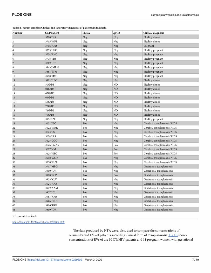
The clinical and laboratorial data of the 83 clinical samples used in this study are described in
Table 2 and Table 3 that included: patient code, laboratorial diagnosis for toxoplasmosis
(ELISA and qPCR), and clinical diagnosis. Table 2 describes group I, 20 negative sera for toxo-
plasmosis collected from healthy donors and healthy pregnant women; and group II, 21 sera
from cerebral toxoplasmosis/HIV-coinfected patients and pregnant women with gestational
toxoplasmosis. Table 3 describe group III, 26 CSF collected from CT/HIV patients; and group
IV, 16 CSF collected from OI/HIV patients.
Characterization by NTA of serum-derived EVs and CSF-derived EVs of
patients with toxoplasmosis
After purification by ultracentrifugation, CSF derived EVs and serum-derived EVs were ana-
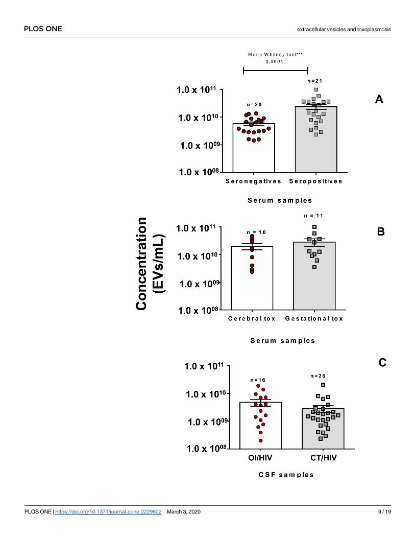
lyzed in NanoSight (size and concentration). Fig 1A shows the concentration of serum-derived
EVs (250 μL) of 21 seropositive patients and 20 seronegative individuals for toxoplasmosis.
Concentrations of serum-derived EVs of seropositive patients (mean: 2.4 x 10¹⁰ EVs/mL) were
statistically higher than seronegative individuals (mean: 5.9 x 10⁹ EVs/mL).
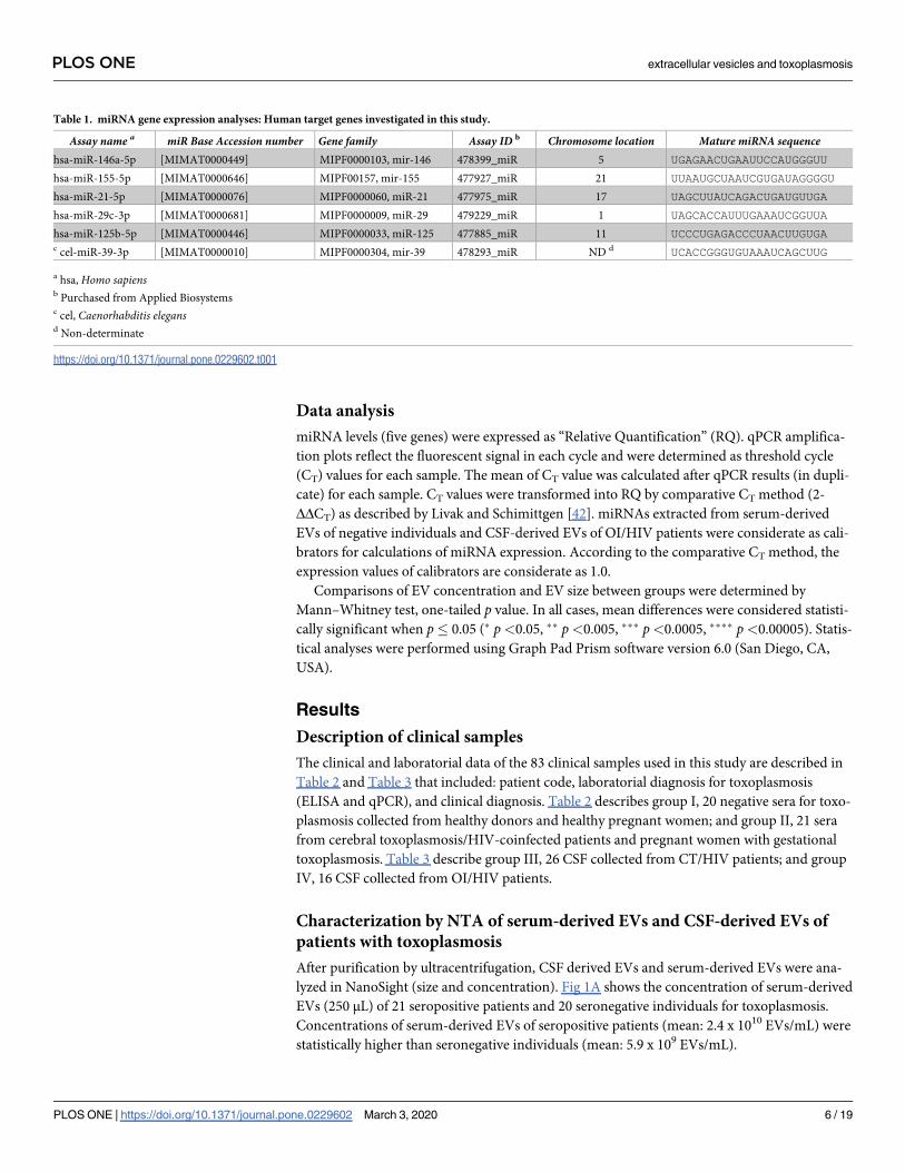
Table 1. miRNA gene expression analyses: Human target genes investigated in this study.
Assay name a miR Base Accession number Gene family Assay ID b Chromosome location Mature miRNA sequencehsa-miR-146a-5p [MIMAT0000449] MIPF0000103, mir-146 478399_miR 5 UGAGAACUGAAUUCCAUGGGUU
hsa-miR-155-5p [MIMAT0000646] MIPF00157, mir-155 477927_miR 21 UUAAUGCUAAUCGUGAUAGGGGU
hsa-miR-21-5p [MIMAT0000076] MIPF0000060, miR-21 477975_miR 17 UAGCUUAUCAGACUGAUGUUGA
hsa-miR-29c-3p [MIMAT0000681] MIPF0000009, miR-29 479229_miR 1 UAGCACCAUUUGAAAUCGGUUA
hsa-miR-125b-5p [MIMAT0000446] MIPF0000033, miR-125 477885_miR 11 UCCCUGAGACCCUAACUUGUGAc cel-miR-39-3p [MIMAT0000010] MIPF0000304, mir-39 478293_miR ND d UCACCGGGUGUAAAUCAGCUUG
a hsa, Homo sapiensb Purchased from Applied Biosystemsc cel, Caenorhabditis elegansd Non-determinate
https://doi.org/10.1371/journal.pone.0229602.t001
PLOS ONE extracellular vesicles and toxoplasmosis
PLOS ONE | https://doi.org/10.1371/journal.pone.0229602 March 3, 2020 6 / 19
The data produced by NTA were, also, used to compare the concentrations of
serum-derived EVs of patients according clinical form of toxoplasmosis. Fig 1B shows
concentrations of EVs of the 10 CT/HIV patients and 11 pregnant women with gestational
Table 2. Serum samples: Clinical and laboratory diagnoses of patients/individuals.
Number Cod/Patient ELISA qPCR Clinical diagnosis
1 3710/GJS Neg Neg Healthy donor
2 3715/WFS Neg Neg Healthy donor
3 3716/ARR Neg Neg Pregnant
4 3753/HSC Neg Neg Healthy pregnant
5 3754/AVO Neg Neg Healthy pregnant
6 3778/PRS Neg Neg Healthy pregnant
7 3809/IPT Neg Neg Healthy pregnant
8 3843/JMRM Neg Neg Healthy pregnant
9 3881/STM Neg Neg Healthy pregnant
10 3958/MSO Neg Neg Healthy pregnant
11 3991/JHVL Neg Neg Healthy donor
12 50G/DS Neg ND Healthy donor
13 01G/DS Neg ND Healthy donor
14 63G/DS Neg ND Healthy donor
15 65G/DS Neg ND Healthy donor
16 68G/DS Neg ND Healthy donor
17 70G/DS Neg ND Healthy donor
18 74G/DS Neg ND Healthy donor
19 75G/DS Neg ND Healthy donor
20 399/DPL Neg Neg Healthy pregnant
21 3621/JEC Pos Neg Cerebral toxoplasmosis/AIDS
22 3622/WRR Pos Neg Cerebral toxoplasmosis/AIDS
23 3623/RSL Pos Neg Cerebral toxoplasmosis/AIDS
24 3624/JAS Pos Neg Cerebral toxoplasmosis/AIDS
25 3625/CGC Pos Neg Cerebral toxoplasmosis/AIDS
26 3626/EMAS Pos Pos Cerebral toxoplasmosis/AIDS
27 3627/VM Pos Pos Cerebral toxoplasmosis/AIDS
28 3628/HSC Pos Pos Cerebral toxoplasmosis/AIDS
29 3918/WSO Pos Neg Cerebral toxoplasmosis/AIDS
30 3830/RLN Pos Neg Cerebral toxoplasmosis/AIDS
31 3717/MPG Pos Neg Gestational toxoplasmosis
32 3850/JDR Pos Neg Gestational toxoplasmosis
33 3910/RCP Pos Pos Gestational toxoplasmosis
34 3923/KLV Pos Neg Gestational toxoplasmosis
35 3924/AAZ Pos Neg Gestational toxoplasmosis
36 3929/AAM Pos Neg Gestational toxoplasmosis
37 3937/JCL Pos Neg Gestational toxoplasmosis
38 3967/KRB Pos Neg Gestational toxoplasmosis
39 3906/HRN Pos Neg Gestational toxoplasmosis
40 3916/SGO Pos Neg Gestational toxoplasmosis
41 3850/JDR Pos Neg Gestational toxoplasmosis
ND, non-determined.
https://doi.org/10.1371/journal.pone.0229602.t002
PLOS ONE extracellular vesicles and toxoplasmosis
PLOS ONE | https://doi.org/10.1371/journal.pone.0229602 March 3, 2020 7 / 19
toxoplasmosis. Among them, pregnant women had an increase in particle concentration
(mean: 2.8 x 10¹⁰ EVs/mL) than CT/HIV patients (mean: 2.0 x 10¹⁰ EVs/mL), but without sta-
tistical difference.
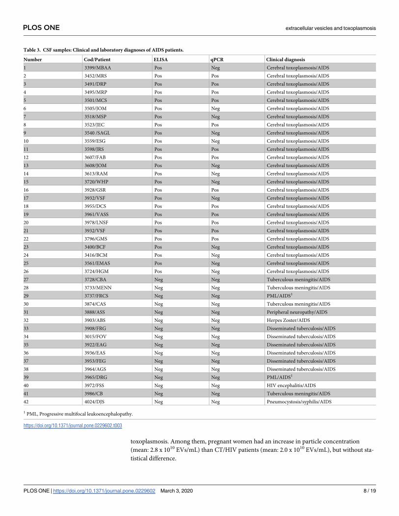
Table 3. CSF samples: Clinical and laboratory diagnoses of AIDS patients.
Number Cod/Patient ELISA qPCR Clinical diagnosis
1 3399/MBAA Pos Neg Cerebral toxoplasmosis/AIDS
2 3452/MRS Pos Pos Cerebral toxoplasmosis/AIDS
3 3491/DRP Pos Pos Cerebral toxoplasmosis/AIDS
4 3495/MRP Pos Pos Cerebral toxoplasmosis/AIDS
5 3501/MCS Pos Pos Cerebral toxoplasmosis/AIDS
6 3505/JOM Pos Neg Cerebral toxoplasmosis/AIDS
7 3518/MSP Pos Neg Cerebral toxoplasmosis/AIDS
8 3523/JEC Pos Pos Cerebral toxoplasmosis/AIDS
9 3540 /SAGL Pos Neg Cerebral toxoplasmosis/AIDS
10 3559/ESG Pos Neg Cerebral toxoplasmosis/AIDS
11 3598/JRS Pos Pos Cerebral toxoplasmosis/AIDS
12 3607/FAB Pos Pos Cerebral toxoplasmosis/AIDS
13 3608/JOM Pos Neg Cerebral toxoplasmosis/AIDS
14 3613/RAM Pos Neg Cerebral toxoplasmosis/AIDS
15 3720/WHP Pos Neg Cerebral toxoplasmosis/AIDS
16 3928/GSR Pos Pos Cerebral toxoplasmosis/AIDS
17 3932/VSF Pos Neg Cerebral toxoplasmosis/AIDS
18 3955/DCS Pos Pos Cerebral toxoplasmosis/AIDS
19 3961/VASS Pos Pos Cerebral toxoplasmosis/AIDS
20 3978/LNSF Pos Pos Cerebral toxoplasmosis/AIDS
21 3932/VSF Pos Pos Cerebral toxoplasmosis/AIDS
22 3796/GMS Pos Pos Cerebral toxoplasmosis/AIDS
23 3400/BCF Pos Neg Cerebral toxoplasmosis/AIDS
24 3416/BCM Pos Neg Cerebral toxoplasmosis/AIDS
25 3561/EMAS Pos Neg Cerebral toxoplasmosis/AIDS
26 3724/HGM Pos Neg Cerebral toxoplasmosis/AIDS
27 3728/CBA Neg Neg Tuberculous meningitis/AIDS
28 3733/MENN Neg Neg Tuberculous meningitis/AIDS
29 3737/FRCS Neg Neg PML/AIDS1
30 3874/CAS Neg Neg Tuberculous meningitis/AIDS
31 3888/ASS Neg Neg Peripheral neuropathy/AIDS
32 3903/ABS Neg Neg Herpes Zoster/AIDS
33 3908/FRG Neg Neg Disseminated tuberculosis/AIDS
34 3015/FOV Neg Neg Disseminated tuberculosis/AIDS
35 3922/EAG Neg Neg Disseminated tuberculosis/AIDS
36 3936/EAS Neg Neg Disseminated tuberculosis/AIDS
37 3953/FEG Neg Neg Disseminated tuberculosis/AIDS
38 3964/AGS Neg Neg Disseminated tuberculosis/AIDS
39 3965/DRG Neg Neg PML/AIDS1
40 3972/FSS Neg Neg HIV encephalitis/AIDS
41 3986/CB Neg Neg Tuberculous meningitis/AIDS
42 4024/DJS Neg Neg Pneumocystosis/syphilis/AIDS
1 PML, Progressive multifocal leukoencephalopathy.
https://doi.org/10.1371/journal.pone.0229602.t003
PLOS ONE extracellular vesicles and toxoplasmosis
PLOS ONE | https://doi.org/10.1371/journal.pone.0229602 March 3, 2020 8 / 19
PLOS ONE extracellular vesicles and toxoplasmosis
PLOS ONE | https://doi.org/10.1371/journal.pone.0229602 March 3, 2020 9 / 19

Analyzes in CSF were distinct from sera ones, since is unusual to obtain CSF samples from
health individuals. Thus, as criterion, CSF samples collected from patients with other opportu-
nistic diseases and AIDS were used as control to compare with those from CT/HIV group. Fig
1C shows the concentration CSF-derived EVs (500 μL). Among them, the 26 CT/HIV patients
had a decrease in particle concentration (mean: 2.9 x 10⁹ EVs/mL) than the 16 OI/HIV
patients (mean: 4.8 x 10⁹ EVs/mL), but without statistical difference.
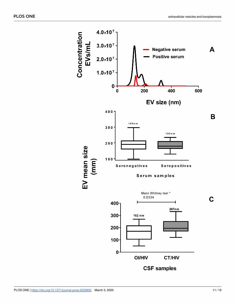
In the next step was the particle size was investigated by NTA. Values represent the mean of
three reads in NanoSight (concentration and size). Fig 2A shows the comparison between con-
centrations of serum-derived EVs from a seropositive patient and a seronegative individual. In
addition, shows the similar mean size of EVs in both samples.
Regarding to size, Fig 2B shows the mean± SEM (standard error of the mean) size of
serum-derived EVs, which was 189.0 ± 10.10 nm for negative and 185.3 ± 5.9 nm for positive
sera. Fig 2C shows the mean± SEM size of CSF-derived EVs that was 206.9 ± 11.49 nm for OI/
HIV patients. This value was statistically higher than OI/HIV patients, that was 162.8 ± 15.52
nm. These data show that a great part of both, serum-derived EVs and CSF-derived EVs were
microvesicles.
Electron microscopy and immunoblotting characterizing human EVs
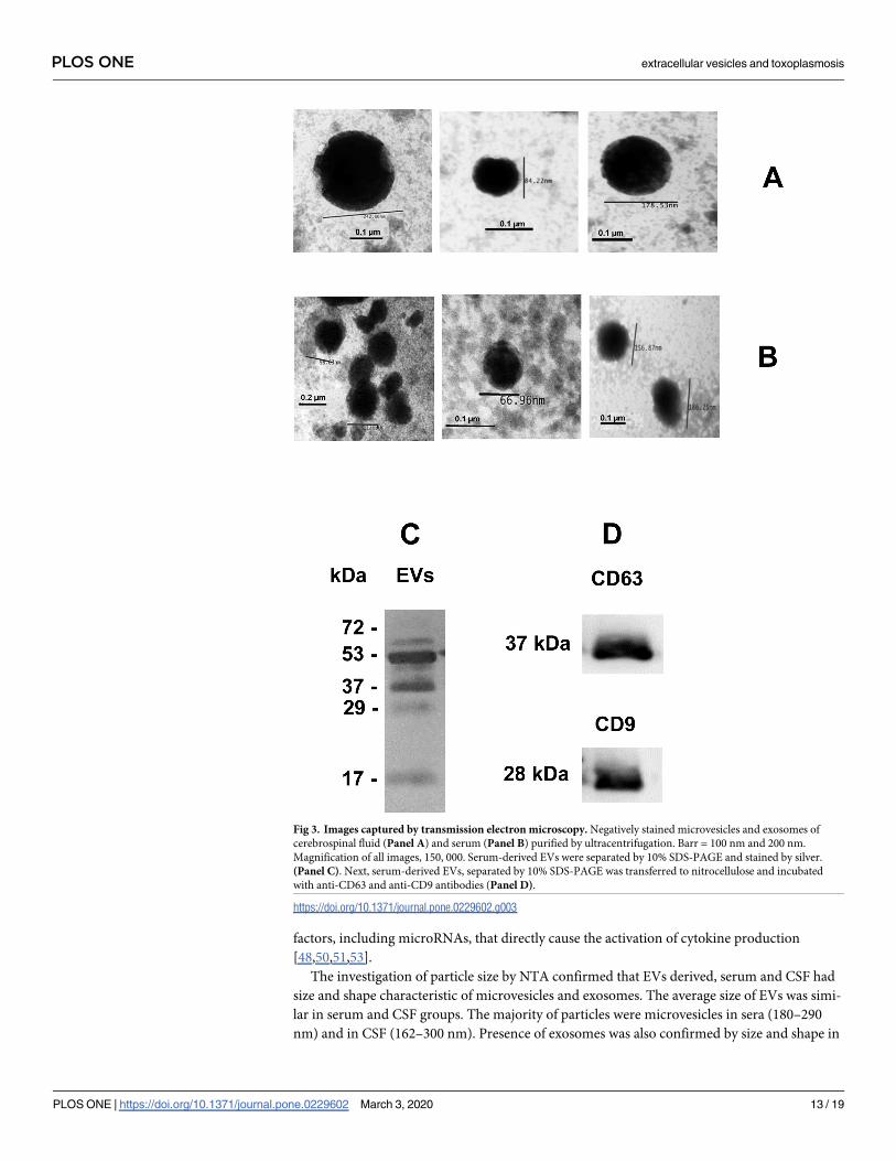
NTA results were confirmed after analysis by TEM and immunoblotting. Fig 3A and Fig 3B
show CSF-derived EVs and serum-derived EVs, respectively, after ultracentrifugation. The
images reveal vesicles with typical size and shape of microvesicles and exosomes.
Microvesicles and exosomes have different transmembrane proteins. Exosomes have tetra-
spanins, including CD9 and CD63, which were detected by immunoblotting. Serum-derived
EVs were separated by 10% SDS-PAGE and stained by silver (Fig 3C). After transference to
nitrocellulose and incubation with anti-CD9 and anti-CD63 antibodies, the tetraspanins
CD63, at 37 kDa, and CD9, at 28 kDa were reactive (Fig 3D). These results proved the presence
of exosomes in preparations.
Gene expression of miRNAs exosomal
Next, the relative expressions of miR-146a-5p, miR-155-5p, miR-21-5p, miR-29c-3p and miR-
125b-5p were estimated in miRNA purified from serum-derived EVs and CSF-derived EVs by
qPCR. Fig 4 shows the relative expression of miRNA (expressed as RQ) extracted of serum-
derived EVs (4A) and CSF-derived EVs (4B). RQ results of negative clinical samples (sera and
CSF) for toxoplasmosis were considered as calibrators. Serum-derived EVs of group II,
patients with gestational or cerebral toxoplasmosis up-expressed miR-125b-5p and miR-146a-
5p. Values were 4.9 and 3.7 times higher than serum-derived EVs of healthy individuals (cali-
brator), respectively. However, for miR-155-5p and miR-21-5p, patients expressed 2.2 and 1.3
times higher than serum-derived EVs of healthy individuals, respectively. miR-29c-3p was
similarly expressed in patients with toxoplasmosis and healthy individuals (Fig 4A).
Fig 1. EVS evaluated by representative NTA. (Panel A), distribution of concentration/mL of EVs purified by
ultracentrifugation from 41 serum samples (250 μL) from seronegative individuals (red circles) and seropositive
patients (gray squares). Differences in particle concentrations (EVs) between the two groups were statistically different
at ��� p = 0.0004 (Mann–Whitney test, one-tailed p value). (Panel B), comparison between distribution of
concentration (EVs/mL) from serum samples of cerebral toxoplasmosis patients (red circles) and pregnant women
with gestational toxoplasmosis (gray squares). (Panel C), distribution of concentration/mL of EVs purified by
ultracentrifugation of 42 cerebrospinal fluid samples (500 μL) from AIDS patients with other opportunistic infections
(OI/HIV) (red circles) and cerebral toxoplasmosis/HIV co-infection (CT/HIV) (gray squares).
https://doi.org/10.1371/journal.pone.0229602.g001
PLOS ONE extracellular vesicles and toxoplasmosis
PLOS ONE | https://doi.org/10.1371/journal.pone.0229602 March 3, 2020 10 / 19
PLOS ONE extracellular vesicles and toxoplasmosis
PLOS ONE | https://doi.org/10.1371/journal.pone.0229602 March 3, 2020 11 / 19
CSF-derived EVs from CT/HIV patients up-expressed miR-155-5p and miR-21-5p, 6.4 and
6.2 times more than calibrators (EVs of OI/HIVP, respectively. miR-125b-5p (1.6 times) and
miR-146a-5p (1.3 times) were similarly expressed in both groups of EVs. CSF-derived EVs
were unable to express miR-29c-3p (Fig 4B).
Discussion
Among EV functions produced in humans include the transference of different macromole-
cules and biomarkers interacting with specific molecules of immune system [20,21–23]. In T.
gondii infection, parasite EVs stimulate the host immune system to produce host EVs (includ-
ing microvesicles and exosomes). Infected host cells secret EVs with pathogen molecules able
to induce modifications in uninfected cells or may serve as antigen presenters for the immune
system [34]. At the same time, T. gondii EVs contain specific proteins as surface antigens,
which are release by tachyzoites and compose the excreted-secreted antigens. These proteins
participate of tachyzoite invasion, replication within host cells and are recognized by host
immune system [29,31,43,44].
The evidence of exosomes/microvesicles in clinical samples includes: purification of EVs by
ultracentrifugation; demonstration of exosomal proteins as tetraspanins; and presence of char-
acteristic structures (in size and shape) by NTA and electron microscopy. For analyses of
microvesicles and exosomes, purifications were performed in CSF and serum samples by ultra-
centrifugation. In this protocol, both types of EVs were recovered. Serum was chosen to mini-
mize residual platelets that occur with standard plasma protocols [45,46].
As this article was the first that evaluated EVs purified from infected human with parasitic
infection, this discussion was based in studies that described functions of human EVs pro-
duced in other diseases.
Concentrations of serum-derived EVs of seropositive patients were higher than those of
non-infected individuals. Among those infected, pregnant women developing acute infection
produced more EVs than CT/HIV patients, but without differences statistically significant. As
the mean size of EVs was similar in both groups, these data suggested that only the concentra-
tion is able to distinguish patients with toxoplasmosis than healthy individuals. Similar results
were shown in plasmas of patient with papillary thyroid carcinoma [47].
Analyzes of CSF-derived EVs were distinct from those of serum. As is unusual to obtain
CSF from health individuals, CSF collected of patients with other opportunistic diseases and
AIDS were used as negative control. Concentrations of CSF-derived EVs were almost similar
in both groups, but lower than those of sera. Similar results were shown before [48,49].
EVs are produced also by neurons, microglia, and dendritic cells in the CNS [48]. Neverthe-
less, function of EVs in nervous system is not totally established. Studies have shown that EVs
participate of the intercellular communication, maintenance of myelination, synaptic plastic-
ity, antigen presentation, and trophic support of neurons [48–52]. Here, CSF samples of both
patient groups, which were developing severe cerebral infection diseases, produced consider-
able concentration of EVs. According previous studies systemic inflammations caused by
severe infections increase the release of EVs and these vesicles contain pro-inflammatory
Fig 2. (Panel A), sera derived-EVs (seronegative—red line) and (seropositive—black line) were evaluated by
representative size distribution (nm) and particles/mL in NanoSight equipment. Data represent three reads per sample.
(Panel B), mean of EV size (in nm) purified by ultracentrifugation from 41 human sera. White column represents
mean size of EVs purified from seronegative individuals and gray column, from seropositive patients. (Panel C), mean
of EV size (in nm) purified by ultracentrifugation from 42 human CSF samples. White column represents mean size of
EVs purified from OI/HIV patients and gray columns, from CT/HIV patients. In Panels C and D, values are
represented by box and whiskers considering mean ±SEM.
https://doi.org/10.1371/journal.pone.0229602.g002
PLOS ONE extracellular vesicles and toxoplasmosis
PLOS ONE | https://doi.org/10.1371/journal.pone.0229602 March 3, 2020 12 / 19
factors, including microRNAs, that directly cause the activation of cytokine production
[48,50,51,53].
The investigation of particle size by NTA confirmed that EVs derived, serum and CSF had
size and shape characteristic of microvesicles and exosomes. The average size of EVs was simi-
lar in serum and CSF groups. The majority of particles were microvesicles in sera (180–290
nm) and in CSF (162–300 nm). Presence of exosomes was also confirmed by size and shape in
Fig 3. Images captured by transmission electron microscopy. Negatively stained microvesicles and exosomes of
cerebrospinal fluid (Panel A) and serum (Panel B) purified by ultracentrifugation. Barr = 100 nm and 200 nm.
Magnification of all images, 150, 000. Serum-derived EVs were separated by 10% SDS-PAGE and stained by silver.
(Panel C). Next, serum-derived EVs, separated by 10% SDS-PAGE was transferred to nitrocellulose and incubated
with anti-CD63 and anti-CD9 antibodies (Panel D).
https://doi.org/10.1371/journal.pone.0229602.g003
PLOS ONE extracellular vesicles and toxoplasmosis
PLOS ONE | https://doi.org/10.1371/journal.pone.0229602 March 3, 2020 13 / 19
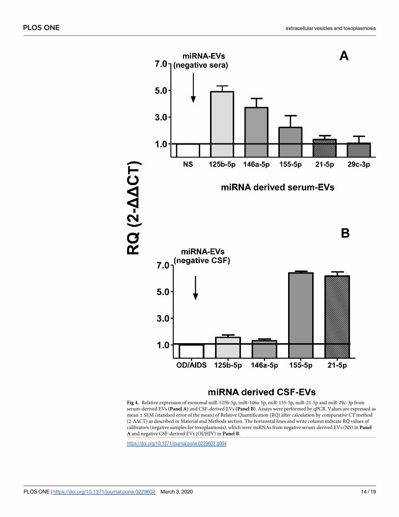
Fig 4. Relative expression of exosomal miR-125b-5p, miR-146a-5p, miR-155-5p, miR-21-5p and miR-29c-3p from
serum-derived EVs (Panel A) and CSF-derived EVs (Panel B). Assays were performed by qPCR. Values are expressed as
mean ± SEM (standard error of the mean) of Relative Quantification (RQ) after calculation by comparative CT method
(2-ΔΔCT) as described in Material and Methods section. The horizontal lines and write column indicate RQ values of
calibrators (negative samples for toxoplasmosis), which were miRNAs from negative serum-derived EVs (NS) in Panel
A and negative CSF-derived EVs (OI/HIV) in Panel B.
https://doi.org/10.1371/journal.pone.0229602.g004
PLOS ONE extracellular vesicles and toxoplasmosis
PLOS ONE | https://doi.org/10.1371/journal.pone.0229602 March 3, 2020 14 / 19
TEM and the evidence of the tetraspanins CD63 and CD9 confirmed in immunoblotting after
SDS-PAGE.
Small RNAs participate in transcriptional gene regulation in cell development and other
biological processes such as cell differentiation, proliferation and apoptosis [54]. Among small
RNAs, miRNAs, which can be transported by EVs are regulatory elements that can modulate
gene expression at the post-transcriptional level [24]. miRNAs, yet may be important in con-
trolling Th1 differentiation, and to produce increased levels of IFN-γ, implicating miRNAs in
the regulation of Th1 response [24,54].
HIV/AIDS patients with low number of CD4 normally develop opportunistic infections. At
the same time, EVs can cause inflammations in HIV/AIDS patients via host miRNAs and viral
miRNAs [54,55]. However, until now is unknown the participation of the miRNAs in different
co-infection caused by HIV and other pathogens. Patients developing all toxoplasmosis forms
had circulating tachyzoites, IFN-γ inhibition and high TNF-α production. This inflammatory
response contributes for damage of the choroid and retina and encephalitis [17,56].
Serum-derived EVs from CT/HIV patients and pregnant women, with acute toxoplasmosis
up-expressed miR-125b-5p and miR-146a-5p, when compared with serum-derived EVs of
healthy individuals. Elevation of TNF-α, IL-6, and downregulation of IFN-γ, as already
reported in AIDS patients, marked upregulation of miRNA 125b-5p [53,57]. In the same way,
miR-146a-containing exosomes reduce cytokine expression, as IFN-γ, IL-17 and IL-2 [57–59].
Nevertheless, CSF-derived EVs of both groups of patients, CT/HIV and OI/HIV expressed
equality miR-125b-5p, and miR-146a-5p. CSF-derived EVs of CT/HIV patients compared
with those of OI/HIV patients up-expressed miR-155-5p and miR-21-5p. These data can sug-
gest the induction of pro-inflammatory cytokine expression caused by miR-155 in CT/HIV
co-infection that was more intense than those from other co-infections analyzed here. At the
same time, the up-expression of miR-21-5p suggested the impact on the balance between Th1
and Th2 [58,59].
miR-21-5p expression by serum-derived EVs was almost similar in both patient groups
(with or without toxoplasmosis). Similar results were observed by Akers et al. [26] when stud-
ied EVs from glioblastoma patients. The authors demonstrated that miR-21-5p was more
expressed in CSF-derived EVs than those of serum. On the other hand, Lakhter et al. [60] have
shown that exosomal miR-21-5p may be up-expressed in the presence of IFN-γ and TNF-α.
Serum-derived EVs in patients with toxoplasmosis down-expressed miR-29c-3p. CSF-derived
EVs were unable to express miR-29c-3p. IFN-γ has a critical role in the immune response in intra-
cellular infections and miR29c-3p participates as a regulator of Th1 and IFN-γ in innate and adap-
tive immunity. According other authors [57,61,62], down or non-expression of miR29c-3p can
cause an increase in IFNγ. However, the data here presented could suggest that EVs produced in
AIDS patients, other miRNAs may play more significant role in suppressing IFN-γ.
Finally, all these data together demonstrate the presence of microvesicles and exosomes
expressing exosomal miRNAs in patients with cerebral or gestational toxoplasmosis Exosomes
containing miRNA can participate in modulation of cellular response. Results demonstrated
the presence of a heterogeneous population of EVs in serum and CSF samples.
Supporting information
S1 Data. Comp soro neg e pos humano2.
(ZIP)
S2 Data. Comparison concentr EVs in human serum1.
(ZIP)
PLOS ONE extracellular vesicles and toxoplasmosis
PLOS ONE | https://doi.org/10.1371/journal.pone.0229602 March 3, 2020 15 / 19
S3 Data. Concentr EVs in human CSF1.
(ZIP)
S4 Data. Concentr EVs in human serum1.
(ZIP)
S5 Data. CSF—All miRNAs4.
(ZIP)
S6 Data. Sera—All miRNAs4.
(ZIP)
S7 Data. Size human EVs in CSF—Mean2.
(ZIP)
S8 Data. Size human EVs in serum—Mean2.
(ZIP)
S1 File.
(PDF)
S1 Fig.
(PPTM)
S2 Fig.
(PPTM)
S1 Results.
(XLSX)
Author Contributions
Data curation: João Paulo Marochi Telles, Jose Ernesto Vidal, Lıgia Cosentino Junqueira
Franco Spegiorin, Cinara Cassia Brandão de Mattos, Vera Lucia Pereira-Chioccola.
Formal analysis: Jose Ernesto Vidal, Vera Lucia Pereira-Chioccola.
Funding acquisition: Vera Lucia Pereira-Chioccola.
Investigation: Allecineia Bispo da Cruz, Marta Marques Maia, Ingrid de Siqueira Pereira,
Noemi Nosomi Taniwaki, Gislene Mitsue Namiyama, Cristina da Silva Meira-Strejevitch.
Methodology: Allecineia Bispo da Cruz, Marta Marques Maia, Ingrid de Siqueira Pereira,
Noemi Nosomi Taniwaki, Gislene Mitsue Namiyama, Cristina da Silva Meira-Strejevitch.
Project administration: Vera Lucia Pereira-Chioccola.
Supervision: Vera Lucia Pereira-Chioccola.
Writing – original draft: Vera Lucia Pereira-Chioccola.
Writing – review & editing: Jose Ernesto Vidal, Cinara Cassia Brandão de Mattos, Luiz Carlos
de Mattos, Cristina da Silva Meira-Strejevitch, Vera Lucia Pereira-Chioccola.
References1. Dubey JP. Advances in the life cycle of Toxoplasma gondii. Int J Parasitol. 1998, 7: 1019–24.
2. Montoya JG, Liesenfeld O. Toxoplasmosis. Lancet. 2004, 363: 1965–76. https://doi.org/10.1016/
S0140-6736(04)16412-X PMID: 15194258
PLOS ONE extracellular vesicles and toxoplasmosis
PLOS ONE | https://doi.org/10.1371/journal.pone.0229602 March 3, 2020 16 / 19
3. Hill DE, Chirukandoth S, Dubey JP. Biology and epidemiology of Toxoplasma gondii in man and ani-
mals. Ann Health Res Rev. 2005, 6: 41–61.
4. Carruthers VB. Host cell invasion by the opportunistic pathogen Toxoplasma gondii. Acta Tropica.
2002, 81: 111–22. https://doi.org/10.1016/s0001-706x(01)00201-7 PMID: 11801218
5. Pereira-Chioccola VL, Vidal JE, Su C. Toxoplasma gondii infection and cerebral toxoplasmosis in HIV-
infected patients. Future Microbiol. 2009, 4: 1363–79. https://doi.org/10.2217/fmb.09.89 PMID:
19995194
6. Elbez-Rubinstein A, Ajzenberg D, Darde ML, Cohen R, Dumètre A, Year H, et al. Congenital toxoplas-
mosis and reinfection during pregnancy: case report, strain characterization, experimental model of
reinfection, and review. J Infect Dis. 2009, 199: 280–5. https://doi.org/10.1086/595793 PMID:
19032062
7. Olariu TR, Remington JS, McLeod R, Alam A, Montoya JG. Severe congenital toxoplasmosis in the
United States: clinical and serologic findings in untreated infants. Pediatr Infect Dis. 2011, 30: 1056–61.
8. Ferreira AI, De Mattos CC, Frederico FB, Meira CS, Almeida GC Jr, Nakashima F, et al. Risk factors for
ocular toxoplasmosis in Brazil. Epidemiol. Infect. 2014, 142: 142–8. https://doi.org/10.1017/
S0950268813000526 PMID: 23507508
9. Glasner PD, Silveira C, Kruszon-Moran D, Martins MC, Burnier Junior M, Silveira S, et al. An unusually
high prevalence of ocular toxoplasmosis in southern Brazil. Am J Ophthalmol. 1992, 114; 136–44.
https://doi.org/10.1016/s0002-9394(14)73976-5 PMID: 1642287
10. Previato M, Frederico FB, Murata FH, Siqueira RC, Barbosa AP, Silveira-Carvalho AP, et al. A Brazilian
report using serological and molecular diagnosis to monitoring acute ocular toxoplasmosis. BMC Res
Notes. 2015, 8: 746. https://doi.org/10.1186/s13104-015-1650-6 PMID: 26643197
11. Bossi P, Bricaire F. Severe acute disseminated toxoplasmosis. Lancet. 2004, 364: 579.
12. Hernandez AV, Thota P, Pellegrino D, Pasupuleti V, Benites-Zapata VA, Deshpande A., et al. A sys-
tematic review and meta-analysis of the relative efficacy and safety of treatment regimens for HIV-asso-
ciated cerebral toxoplasmosis: is trimethoprim-sulfamethoxazole a real option? HIV Med. 2017, 18:
115–24. https://doi.org/10.1111/hiv.12402 PMID: 27353303
13. Vidal JE, Hernandez AV, de Oliveira AC, Dauar RF, Barbosa SP Jr, Focaccia R. Cerebral toxoplasmo-
sis in HIV-positive patients in Brazil: clinical features and predictors of treatment response in the
HAART era. AIDS Patient Care STDS. 2005, 19: 840–8. https://doi.org/10.1089/apc.2005.19.840
14. Vidal JE, Oliveira AC. AIDS-related cerebral toxoplasmosis in São Paulo State, Brazil: marked improve-
ments in the highly active antiretroviral therapy-era but the challenges continue. Braz. J Infect Dis.
2013, 17: 379–80.
15. Prigione I, Facchetti P, Lecordier L, Deslee D, Chiesa S, Cesbron-Delauw MF, et al. T cell clones raised
from chronically infected healthy humans by stimulation with Toxoplasma gondii excretory–secretory
antigens cross-react with live tachyzoites: characterization of the fine antigenic specificity of the clones
and implications for vaccine development. J. Immunol. 2000, 7: 3741–8.
16. Dubey JP, Lago EG, Gennari SM, Su C, Jones JL. Toxoplasmosis in humans and animals in Brazil:
high prevalence, high burden of disease, and epidemiology. Parasitology. 2012, 139: 1375–424.
https://doi.org/10.1017/S0031182012000765 PMID: 22776427
17. Meira CS, Pereira-Chioccola VL, Vidal JE, de Mattos CC, Motoie G, Costa-Silva TA, et al. Cerebral and
ocular toxoplasmosis related with IFN-γ, TNF-α, and IL-10 levels. Front Microbiol. 2014, 13: 492.
18. Maia MM, Meira-Strejevitch CS, Pereira-Chioccola VL, de Hippolito DDC, Silva VO, Brandão de Mattos
CC, et al. Evaluation of gene expression levels for cytokines in ocular toxoplasmosis. Parasite Immunol.
2017, 39: pim.12462.
19. Thery C, Ostrowski M, Segura E. Membrane vesicles as conveyors of immune responses. Nat Rev
Immunol. 2009, 9: 581–93. https://doi.org/10.1038/nri2567 PMID: 19498381
20. Yang XX, Sun C, Wang L, Guo XL. New insight into isolation, identification techniques and medical
applications of exosomes. J Control Release. 2019, 17ii: S0168–3659.
21. Yu X, Harris SL, Levine AJ. The regulation of exosome secretion: a novel function of the p53 protein.
Cancer Res. 2006, 66: 4795–4801. https://doi.org/10.1158/0008-5472.CAN-05-4579 PMID: 16651434
22. Delabranch X, Berger A, Boisrame-Helms J, Meziani F. Microparticles and infectious diseases. Med
Mal Infect. 2012, 42: 335–43. https://doi.org/10.1016/j.medmal.2012.05.011 PMID: 22766273
23. Schwab A, Meyering SS, Lepene B, Iordanskiy S, van Hoek ML, Hakami RM, et al. Extracellular vesi-
cles from infected cells: potential for direct pathogenesis. Front Microbiol. 2015, 20: 1132 eCollection.
24. Mathivanan S, Ji H, Simpson RJ. Exosomes: extracellular organelles important in intercellular commu-
nication. J Proteomics. 2010, 73: 1907–20. https://doi.org/10.1016/j.jprot.2010.06.006 PMID:
20601276
PLOS ONE extracellular vesicles and toxoplasmosis
PLOS ONE | https://doi.org/10.1371/journal.pone.0229602 March 3, 2020 17 / 19
25. Cocucci E, Racchetti G, Meldolesi J. Shedding microvesicles: artefacts no more. Trends Cell Biol.
2009, 19: 43–51. https://doi.org/10.1016/j.tcb.2008.11.003 PMID: 19144520
26. Akers JC, Gonda D, Kim R, Carter BS. Biogenesis of extracellular vesicles (EV): exosomes, microvesi-
cles, retrovirus-like vesicles, and apoptotic bodies. J Neurooncol. 2013, 113: 1–11. https://doi.org/10.
1007/s11060-013-1084-8 PMID: 23456661
27. ExoCarta. Exosome, protein, RNA and lipid database. Available from http://www.exocarta.org. [Cited
2019 may].
28. Fleming A, Sampey G, Chung M, Bailey C, van Hoek ML, Kashanchi F, et al. The carrying pigeons of
the cell: exosomes and their role in infectious diseases caused by human pathogens. Pathog Dis 2014,
71: 109–20. https://doi.org/10.1111/2049-632X.12135 PMID: 24449527
29. Wowk PF, Zardo ML, Miot HT, Goldenberg S, Carvalho PC, Morking PA. Proteomic profiling of extracel-
lular vesicles secreted from Toxoplasma gondii. Proteomics. 2017, 17: 15–6.
30. Li Y, Liu Y, Xiu F, Wang J, Cong H, He S, et al. Characterization of exosomes derived from Toxoplasma
gondii and their functions in modulating immune responses. Int J Nanomed. 2018, 19: 467–77.
31. Silva VO, Maia MM, Torrecilhas AC, Taniwaki NN, Namiyama GM, Oliveira KC, et al. Extracellular vesi-
cles isolated from Toxoplasma gondii induce host immune response. Parasite Immunol. 2018, 40:
e12571. https://doi.org/10.1111/pim.12571 PMID: 29974519
32. Aline F, Bout D, Amigorena S, Roingeard P, Dimier-Poisson I. Toxoplasma gondii antigen-pulsed-den-
dritic cell-derived exosomes induce a protective immune response against T. gondii infection. Infect
Immun. 2004, 72: 4127–37. https://doi.org/10.1128/IAI.72.7.4127-4137.2004 PMID: 15213158
33. Beauvillain C, Ruiz S, Guiton R, Bout D, Dimier-Poisson I. A vaccine based on exosomes secreted by a
dendritic cell line confers protection against T. gondii infection in syngeneic and allogeneic mice.
Microbes Infect. 2007, 9: 1614–22. https://doi.org/10.1016/j.micinf.2007.07.002 PMID: 17905628
34. Bhatnagar S, Shinagawa K, Castellino FJ, Schorey JS. Exosomes released from macrophages infected
with intracellular pathogens stimulate a proinflammatory response in vitro and in vivo. Blood. 2007,
110: 3234–44. https://doi.org/10.1182/blood-2007-03-079152 PMID: 17666571
35. Kim MJ, Jung BK, Cho J, Song H, Pyo KH, Lee JM, et al. Exosomes secreted by Toxoplasma gondii-
Infected L6 cells: their effects on host cell proliferation and cell cycle changes. Korean J Parasitol. 2016,
54: 147–54. https://doi.org/10.3347/kjp.2016.54.2.147 PMID: 27180572
36. Pope SM, Lasser C. Toxoplasma gondii infection of fibroblasts causes the production of exosome-like
vesicles containing a unique array of mRNA and miRNA transcripts compared to serum starvation. J
Extracell Vesicles. 2013, 11: 2 eCollection.
37. Goncalves MA, Mattos CC, Spegiorin LC, Oliani DC, Oliani AH, Mattos LC. Seropositivity rates for toxo-
plasmosis, rubella, syphilis, cytomegalovirus, hepatitis and HIV among pregnant women receiving care
at a public health service, São Paulo state, Brazil. Braz J Infect Dis. 2010; 14: 601–5. PMID: 21340301
38. Murata FHA, Ferreira MN, Camargo NS, Santos GS, Spegiorin LCJF, Silveira-Carvalho AP, et al. Fre-
quency of the anti-Toxoplasma gondii antibodies class IgA, IgM and IgG in high-risk pregnancy women,
Brazil. Rev Soc Bras Med Trop. 2016; 49: 512–14. https://doi.org/10.1590/0037-8682-0046-2016
PMID: 27598642
39. Meira CS, Costa-Silva TA, Vidal JE, Ferreira IM, Hiramoto RM, Pereira-Chioccola VL. Use of the serum
reactivity against Toxoplasma gondii excreted-secreted antigens in cerebral toxoplasmosis diagnosis in
human immunodeficiency virus-infected patients. J Med Microbiol. 2008, 57: 845–50. https://doi.org/
10.1099/jmm.0.47687-0 PMID: 18566142
40. Costa-Silva TA, Borges MM, Galhardo CS, Pereira-Chioccola VL. Immunization with excreted/secreted
proteins in AS/n mice activating cellular and humoral response against Toxoplasma gondii infection.
Acta Trop. 2012, 124: 203–9. https://doi.org/10.1016/j.actatropica.2012.08.013 PMID: 22940015
41. Camilo LM, Pereira-Chioccola VL, Gava R, Meira-Strejevitch CS, Vidal JE, Brandão de Mattos CC.
Molecular diagnosis of symptomatic toxoplasmosis: a 9-year retrospective and prospective study in a
referral laboratory in São Paulo, Brazil. Braz J Infect Dis. 2017, 21: 638–47. https://doi.org/10.1016/j.
bjid.2017.07.003 PMID: 28968510
42. Livak KJ, Schmittgen TD. Analysis of relative gene expression data using real-time quantitative PCR
and the 2(-Delta Delta C(T)) Method Methods. 2001, 25: 402–8.
43. Daryani A, Hosseine AZ, Dalimi A. Immune response against excreted/secreted antigens of Toxo-
plasma gondii tachyzoites in the murine model. Vet Parasitol. 2003, 113: 123–34. https://doi.org/10.
1016/s0304-4017(03)00044-x PMID: 12695037
44. Długońska H, Gatkowska J. Exosomes in the context of Toxoplasma gondii–host communication. Ann
Parasitol. 2016, 1: 169–74.
PLOS ONE extracellular vesicles and toxoplasmosis
PLOS ONE | https://doi.org/10.1371/journal.pone.0229602 March 3, 2020 18 / 19
45. Arroyo JD, Cheville JR, Kroh EM, Ruf IK, Pritchard CC, Gibson DF, et al. Argonaute2 complexes carry
a population of circulating microRNAs independent of vesicles in human plasma. Proc Natl Acad Sci
USA. 2011, 22: 5003–8.
46. Kalra H, Adda CG, Liem M, Ang CS, Mechler A, Simpson RJ, et al. Comparative proteomics evaluation
of plasma exosome isolation techniques and assessment of the stability of exosomes in normal human
blood plasma. Proteomics. 2013, 13: 3354–64. https://doi.org/10.1002/pmic.201300282 PMID:
24115447
47. Rappa G, Puglisi C, Santos MF, Forte S, Memeo L, Lorico A. Extracellular vesicles from thyroid carci-
noma: The new frontier of liquid biopsy. Int J Mol Sci. 2019, 20, pii: E1114. https://doi.org/10.3390/
ijms20051114 PMID: 30841521
48. Spaull R, McPherson B, Gialeli A, Clayton A, Uney J, Heep A, et al. Exosomes populate the cerebrospi-
nal fluid of preterm infants with post-haemorrhagic hydrocephalus. Int J Dev Neurosci. 2019, 73: 59–
65. https://doi.org/10.1016/j.ijdevneu.2019.01.004 PMID: 30639393
49. Street JM, Barran PE, Mackay CL, Weidt S, Balmforth C, Walsh TS, et al. Identification and proteomic
profiling of exosomes in human cerebrospinal fluid. J Transl Med. 2012, 5: 5.
50. Alvarez-Erviti L, Seow Y, Yin H, Betts C, Lakhal S, Wood MJ. Delivery of siRNA to the mouse brain by
systemic injection of targeted exosomes. Nat Biotechnol. 2011, 29: 341–345. https://doi.org/10.1038/
nbt.1807 PMID: 21423189
51. Matsumoto J, Stewart T, Sheng L, Li N, Bullock K, Song N, et al. Transmission of alpha-synuclein-con-
taining erythrocyte-derived extracellular vesicles across the blood-brain barrier via adsorptive mediated
transcytosis: another mechanism for initiation and progression of Parkinson’s disease? Acta Neuro-
pathol Commun. 2017, 13: 71.
52. Shi M, Sheng L, Stewart T, Zabetian CP, Zhang J. New windows into the brain: Central nervous sys-
tem-derived extracellular vesicles in blood. Prog Neurobiol. 2019, 175: 96–106. https://doi.org/10.
1016/j.pneurobio.2019.01.005 PMID: 30685501
53. Nematian SE, Mamillapalli R, Kadakia TS, Majidi Zolbin M, Moustafa S, Taylor HS. Systemic inflamma-
tion induced by microRNAs: Endometriosis-derived alterations in circulating microRNA 125b-5p and
Let-7b-5p regulate macrophage cytokine production. J Clin Endocrinol Metab. 2018, 103: 64–74.
https://doi.org/10.1210/jc.2017-01199 PMID: 29040578
54. Bartel DP. MicroRNAs: genomics, biogenesis, mechanism, and function. Cell. 2004, 116: 281–97.
https://doi.org/10.1016/s0092-8674(04)00045-5 PMID: 14744438
55. Verma P, Pandey RK, Prajapati P, Prajapati VK. Circulating MicroRNAs: Potential and emerging bio-
markers for diagnosis of human infectious diseases. Front Microbiol. 2016, 7: 1274 eCollection. https://
doi.org/10.3389/fmicb.2016.01274 PMID: 27574520
56. Meira C.S., Vidal J.E., Costa-Silva T.A., Frazatti-Gallina N., Pereira-Chioccola V.L., 2011. Immunodiag-
nosis in cerebrospinal fluid of cerebral toxoplasmosis and HIV-infected patients using Toxoplasma gon-
dii excreted/secreted antigens. Diagn. Microbiol. Infect. Dis. 71, 279–285. https://doi.org/10.1016/j.
diagmicrobio.2011.07.008 PMID: 21907524
57. Amado T, Schmolka N, Metwally H, Silva-Santos B, Gomes AQ. Cross-regulation between cytokine
and microRNA pathways in T cells. Eur J Immunol. 2015, 45: 1584–95. https://doi.org/10.1002/eji.
201545487 PMID: 25865116
58. Alexander M, Hu R, Runtsch MC, Kagele DA, Mosbruger TL, Tolmachova T, et al. Exosome-delivered
microRNAs modulate the inflammatory response to endotoxin. Nat Commun. 2015, 6: 7321 https://doi.
org/10.1038/ncomms8321 PMID: 26084661
59. Yang L, Boldin MP, Yu Y, Liu CS, Ea CK, Ramakrishnan P, et al. miR-146a controls the resolution of T
cell responses in mice. J Exp Med. 2012, 209:1655–70. https://doi.org/10.1084/jem.20112218 PMID:
22891274
60. Lakhter A.J., Pratt R.E., Moore R.E., Doucette K.K., Maier B.F., DiMeglio L.A., et al. 2018. Beta cell
extracellular vesicle miR-21-5p cargo is increased in response to inflammatory cytokines and serves as
a biomarker of type 1 diabetes. Diabetologia. 61, 1124–1134. https://doi.org/10.1007/s00125-018-
4559-5 PMID: 29445851
61. Ma F., Xu S., Liu X., Zhang Q., Xu X., Liu M., et al. 2011. The microRNA miR-29 controls innate and
adaptive immune responses to intracellular bacterial infection by targeting interferon-γ. Nat. Immunol.
12, 861–869. https://doi.org/10.1038/ni.2073 PMID: 21785411
62. Steiner D. F., Thomas M. F., Hu J. K., Yang Z., Babiarz J. E., Allen C. D., et al. 2011. MicroRNA-29 reg-
ulates T-box transcription factors and interferon-γ production in helper T cells. Immunity. 35, 169–181.
https://doi.org/10.1016/j.immuni.2011.07.009 PMID: 21820330
PLOS ONE extracellular vesicles and toxoplasmosis
PLOS ONE | https://doi.org/10.1371/journal.pone.0229602 March 3, 2020 19 / 19