Scaled preparation of extracellular vesicles from ... - AutoCRAT
Upload
khangminh22Category
view
0download
0
Article
Extracellular Polysaccharide from Porphyridium cruentum (purpureum) stimulates the non-specific immune activities on white shrimp (Litopenaeus vannamei) Yenny Risjani1, Nurul Mutmainnah1, Siti Narsito Wulan2, Yunianta2
1 Faculty of Fisheries and Marine Science, Universitas Brawijaya, Malang, Indonesia; [email protected] 2 Department of Agricultural Technology, Faculty of Agricultural Technology, Universitas Brawijaya,
Malang, Indonesia Correspondence: [email protected]
Abstract: White shrimps are susceptible to outbreaks of vibriosis because they do not have any adaptive immune system, they only have a non-specific innate immune system. The administration of EPS from microalgae Porphyridium cruentum (synonym: P. purpureum) on shrimps Litopenaeus vannamei was investigated to determine the effect of this immunostimulant on their non specific immune response and to test if EPS can be used as a protective agent for shrimp related to Vibrio infection. EPS was given to shrimps by immersion method on day 1 and booster on day 8. Shrimp hemocytes were taken on day 1 (EPS administration), day 7 (no treatment), day 8 (EPS booster) and day 9 (Vibrio infection) and tested for their immune response on each treatment. Result shows an increase in values of all immune parameters in line with the increasing EPS concentration, except the Differential Haemocyte Count (DHC). In detail, an increase was noted in total hemocytes (THC) value, Phagocytotic Activity (PA), Respiratory Burst (RB) in line as the EPS concentration increase. Although there is a decrease after the infection, the value obtained is not lower than the control value. These results indicate that EPS from Porphyrydium enhances immune parameters in shrimp rapidly and has the ability as an immunostimulant or an immunomodulator. It is a good modulator for the non specific immune cells of Pacific white shrimps, and it can be used as a preventive agent against Vibrio.
Keywords: Hemocytes, innate immune cells, Phagocytic Activity, Respiratory Burst, microalgae, immunomodulator, shrimps L. vannamei, Vibrio harveyi, Extracellular Polysaccharide (EPS).
1. Introduction
Vaname, the white shrimp (Litopenaeus vannamei) is one type of the popular and easily cultivated shrimps. This shrimp is highly and quickly productive. Among other superiorities, it is able to live in a wide range of salinity (euryhaline) between 5 to 30 ppt, as well as being adaptable to high stocking densities, and growing well at low protein feeding levels. It entered Indonesia according to the Decree of the Minister of Marine and Fisheries of Republic of Indonesia Number 41 of 2001, as a substitute for the tiger shrimp commodity whose production declined due to disease attacks [1]. Nevertheless, along with the expansion of cultivation activities, the disease on vaname became increasingly familiar, one of which is vibriosis caused by a pathogenic bacteria Vibrio harveyi [2; 3].
Preprints (www.preprints.org) | NOT PEER-REVIEWED | Posted: 8 December 2020 doi:10.20944/preprints202012.0193.v1
© 2020 by the author(s). Distributed under a Creative Commons CC BY license.

Vibriosis by Vibrio harveyi is still an important discussion in the development of vaname shrimp cultivation. The pathogenic bacteria often attacks shrimp during nauplii, zoea, and mysis stages and it sometimes occurs in the post larval stage, even during maintenance in ponds at the age of 1 to 1.5 months. This disease can spread directly through the waters, as well as direct contact between shrimps, killing the population shortly after 1 to 3 days from the initial infection [4]. Pathogen occurrences on shrimp occur faster because their immunity is different from fish. They do not have any adaptive immune system, and they only have a non-specific or innate/natural immune system, which on a cellular basis includes phagocytic activity, encapsulation, and nodule formation. They do not have certain antibodies induced by certain antigens, so they are more susceptible to outbreaks [2; 5]. The emergence of disease in shrimp is often associated with poor aquatic environmental conditions, especially in Asia [6].
Various preventive efforts are done; one of which is the provision of immunostimulant using Porphyridium cruentum, a red microalga. In this study we suggest the provision of immunostimulant using Porphyridium cruentum, a red microalga. The use of immunostimulant is one of the alternatives to defend from pathogenic infections [7; 8; 9]. Immunostimulant compounds can be obtained from various sources known to contain lipopolysaccharide (LPS), vitamin C, glucan, levamisole, and some microalgae. One type of microalgae containing Extracellular Polysaccharide (EPS) for anti-bacteria and potentially as a source of immunostimulant through antioxidant activity is Porphyridium cruentum [10; 11; 12; 13].
The term EPS is used based on the previous research [27]. Exopolysacharides can also be
called Extracellular Polysaccharides, based on the source of the polysaccharides presence issued
from the cells as metabolite product into water media [16; 27]. Beside genus Phorphyridium, in
general, some strains of Cyanobacteria, i.e. Cyanothecesp., Oscillatoria sp., Nostoc sp., and
Nostoccarneum also secrete EPS [33].
In our previous study [16], we reported about Porphyridium growth and Extracellular
Polysaccharides (EPS) production from this species. Microalgae growth was observed every day for
14 days of culture with the provision of 1ml/L of Fe, silicate, and vitamins and 24-hours continuous
lighting. After 14 days of microalgae culture, 15% culture density resulted in EPS of + 10,000 mg/L,
20% culture density resulted in EPS of + 12,000 mg/L, and 25% culture density resulted in EPS of +
14,000 mg/L.
The FTIR test reported in our previous study showed that in EPS there were dominant bonds,
namely phenol bonds and polysaccharide bonds [16]. Polysaccharides, one of the ingredients that
can be used to produce immunostimulant, are classified into carbohydrates. They are the
combination of more than six monosaccharide molecules, which can be hydrolyzed back into many
monosaccharide molecules. They are able to stimulate immune system in shrimps, characterized by
THC increase [20]. The important component in an immune mechanism is the guarding process by organisms
that can detect the presence of foreign objects or the emergence of foreign molecules originating from the outside of the organism. A good system must be able to stimulate defense responses from foreign molecules, including those mediated by cells. Invertebrates innate immunity resemble to vertebrates immune response [30]. They do not have specific epitope immune activities and
Preprints (www.preprints.org) | NOT PEER-REVIEWED | Posted: 8 December 2020 doi:10.20944/preprints202012.0193.v1
immunoglobulins, but they are able to recognize and destroy invading or parasitic microorganisms [21]. Cellular response as biomarker for immune systems of aquatic animals has been studied as an effect not only associated to bacterial infection or disease, but also related to environmental changes [18; 39; 40].
Invertebrates cell wall have proteins such as lipopolysaccharide (LPS), β-1,3-glucans (BG)
and a specific protein called beta glucan binding protein (BGBP). BGBP in shrimp appears to be the
main plasma protein right after binding to β-glucans, where β-glucan is a compound composed of
polysaccharides, which react with the surface of the hemocytes and stimulate the formation of
granule cells. Granule cells are one of the hemocyte cells that are responsible for immune system in
shrimps [21].
For immune system in shrimp, this study used EPS from Porphyridium cruentum. Porphyridium
cruentum is a single-celled red microalgae belonging to the class of Rhodophyceae, currently has
other synonym: Porphyridium purpureum (Bory) . It is capable of living freely or colonizing in marine
waters, and can be cultivated in a fast life cycle. The round shaped Porphyridium cruentum cells is 4 to
9 µm, and almost 60% is composed of carbohydrates. The cells are bound in mucilage, a compound
constantly excreted by the cell forming a capsule, surrounding the cell, containing polysaccharide
sulfate, known as EPS. EPS is one of the important components in the function of Porphyridium
cruentum as an antioxidant, antibacterial, antiviral, and anti-hyperglycemic substance [14; 15].
Various studies on the benefits of Porphyridium cruentum as an anti-bacterium have been widely
carried out, but the use of EPS from this species as immunostimulant in Vibrio harveyi-infected
shrimp has not been done. Therefore, in addition to becoming the initial foundation of research
related to future problems, this study is needed to determine the effectiveness of the Extracellular
Polysaccharide Sulfate (EPS) of Porphyridium cruentum as a source of immunostimulant.
2. Results
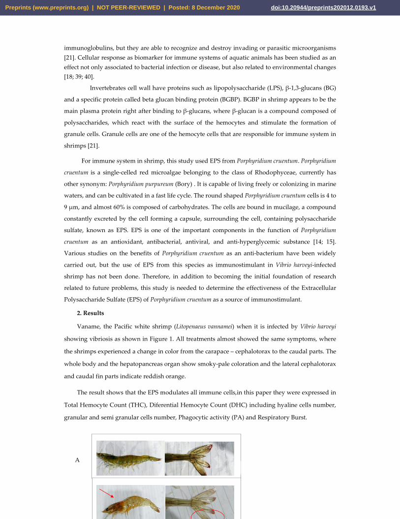
Vaname, the Pacific white shrimp (Litopenaeus vannamei) when it is infected by Vibrio harveyi
showing vibriosis as shown in Figure 1. All treatments almost showed the same symptoms, where
the shrimps experienced a change in color from the carapace – cephalotorax to the caudal parts. The
whole body and the hepatopancreas organ show smoky-pale coloration and the lateral cephalotorax
and caudal fin parts indicate reddish orange.
The result shows that the EPS modulates all immune cells,in this paper they were expressed in
Total Hemocyte Count (THC), Diferential Hemocyte Count (DHC) including hyaline cells number,
granular and semi granular cells number, Phagocytic activity (PA) and Respiratory Burst.
A
a b
a b
Preprints (www.preprints.org) | NOT PEER-REVIEWED | Posted: 8 December 2020 doi:10.20944/preprints202012.0193.v1

B C
Figure 1. Morphology of health Vaname (Litopenaeus vannamei) (A) and shrimp infected by Vibrio harveyi showing vibriosis as indicated by smoky body and organ coloration and reddish color change at cephalotorax and caudal fin parts (arrows). (B). Left : whole body, center: uropod. C. Hemocyte prelevement with a noodle.
2.1. Total Hemocyte Count (THC)
Total Hemocyte Count (THC) was observed on the 1st day of EPS administration, day 7 (no treatment), and on the 8th day, EPS booster administration was conducted with immersion method using 14 ppt, 12 ppt, and 10 ppt. On the 9th day, THC values were obtained after infection with Vibrio harveyi 107 cells/ml. The THC value is shown in Figure 2. After immunostimulant addition on day 1 and day 7 (without booster), we observed changes in THC value, where the highest value was obtained form 14 ppt EPS resulting in 39.9 x 10⁵ cells/ml, followed by 12 ppt EPS resulting the THC value of 39.4 x 10⁵ cells/ml, and the lowest THC value was obtained from the control (0 ppt) resulting in 36.4 x 10⁵ cells/ml. On the 8th day,after the booster was given, or through re-soaking with EPS, the highest THC value was obtained at the EPS treatment of 14 ppt resulting in 49.2 x 10⁵ cells/ml, followed by EPS treatment of 12 ppt resulting in 41.9 x 10⁵ cells/ml, and 10 ppt EPS treatment resulting in 39.0 x 10⁵ cells/ml.The lowest THC value was obtained from the control (without EPS) resulting in 36.1 x 10⁵ cells/ml. After 9 days of infection, there was a decrease in THC values in all treatments.The EPS treatment of 14 ppt resulted a decrease of THC valueto 43.2 x 10⁵ cells/ml, while treatment of 12 ppt resulted in a decrease to 39.5 x 10⁵ cells/ml, and the treatment of 10 ppt decreased the value to 32.8 x 10⁵ cells/ml. For the control, without EPS,THC value decreased very rapidly to 23.7 x 10⁵ cells/ml.
2.2. Differential Hemocyte Count (DHC)
Differential Hemocyte Count (DHC) consists of hyaline, semi-granular and granular cells. 2.2.1. Hyaline cells
On day 1 the highest hyaline cell was obtained from the 0 ppt control (37.2%), followed by 10
ppt (31.5%), 12 ppt (28.2%), and 14 ppt (24.3%). On day 7, without immunostimulant administration,
there was an increase in the number of hyaline cells in all treatments, where the highest value was
obtained from 12 ppt (37.9%), followed by 14 ppt gives (37.7%), and the 0 ppt control (37.3%).
On the 8th day, immunostimulant booster was given, resulting in a significant decrease in the number of hyaline cells in three treatments. The decrease of 35%, 33.3%, and 27.4% occurred for 10, 12, and 14 ppt EPS treatment respectively. The hyaline of the control slightly increased for 37.5%.On the 9th day (after infection), the hyaline cell significantly increased. The highest hyaline cell value
Preprints (www.preprints.org) | NOT PEER-REVIEWED | Posted: 8 December 2020 doi:10.20944/preprints202012.0193.v1

was obtained from 14 ppt EPS treatment (55.1%), followed 12 ppt (47.8%), and 10 ppt (43.4%).The hyaline value of the control (no treatment, 0 ppt EPS) is 41.1%.(Figure 3).
2.2.2. Semi-granular and granular cells
The lowest number of semi-granular cells for all days was obtained from the treatment of 14 ppt EPS; 11.50% on day 1, 16.80%on day 7, 14.30% on day 8,and 8.10%on day 9. (Figure 4).
The highest granular cell value was obtained from all 14 ppt EPS treatment: 64.20% on day 1
immunostimulant administration, 46.60% on day 7 without immunostimulant administration,
54.60% on day 8 after the booster, and 47.20 % on day 9 after infection. On the 7th day without EPS
addition, the value of granular cells appeared to decrease; this indicates a decrease in shrimp’s
immunity (Figure 5).
Figure 2.Total Haemocyte Count (THC) of Litopenaeus vannamei after EPS administration on. Day 1: EPSadministration, Day 8: EPS booster. Day 9: post-infection with Vibrio harveyi, 107 cells/ml.
2.3. Phagocytic Activity (PA)
Figure 6 and 7 showed Phagocytic Activity (PA) of homocyte cells of L. vannamae related to
Vibrio harveyi infection. On day 1 after immunostimulant administration, the highest phagocytosis
level was obtained from 14 ppt EPS treatment (22.9%), followed by 12 ppt EPS treatment (18.7%),
and 10 ppt EPS treatment (14.6%). The lowest value was on the control, without EPS, which is 12.3%.
On the 7th day without immunostimulant, values of 13.6 % and 13.3% were obtained from the
control and 10 ppt EPS treatment. The treatment of 12 and 14 produced the valueof 15.1% and 16.4%.
In all treatments, the phagocytosis level in day 7 is lower than that in day 1, except the control,
probably due to no immunostimulant treatment. On day 8 after booster administration, the
phagocytosis level is higher than the level in day 7, except for control (EPS= 0 ppt).
aa a
a
aa ab
b
aa
c c
b
a
dc
0.0
10.0
20.0
30.0
40.0
50.0
60.0
Day 1 Day 7 Day 8 Day 9
Tota
l Hae
moc
yte
Coun
t (10
5 ce
ll/m
l)
0 ppt 10 ppt 12 ppt 14 ppt
Preprints (www.preprints.org) | NOT PEER-REVIEWED | Posted: 8 December 2020 doi:10.20944/preprints202012.0193.v1

Figure 3. Percentage of Hyalin cells of Litopenaeus vannamei after EPS administration with different immersion concentration. Day 1: EPS administration; Day 7: no treatment; Day 8: EPS booster; Day 9: post-infection with Vibrio harveyi, 107 cells/ml.
Figure 4. Semi Granular cells of Litopenaeus vannamei after EPS administration with different immersion concentration. Day 1: EPS administration; Day 7: no treatment; Day 8: EPS booster; Day 9: post-infection with Vibrio harveyi, 107 cells/ml.
The use of 10 ppt of EPS concentration increased the level to 15.5%, 12 ppt of EPS concentration
increased the level to 18.8%, and 14 ppt treatment increased the level to 22.8%. A significant increase
in phagocytic activity was on day 9 after infection, but the control and 10 ppt EPS treatment did not
make any significant increase (26.3% and 27.8%). The highest phagocytic activity was from 12 and 14
ppt EPS treatment that produced 33.8% and 36.1% respectively.
2.4. Respiratory Burst (RB)
d b ca
ca d
b
b
bb
c
a
b
a
d
0.0%
10.0%
20.0%
30.0%
40.0%
50.0%
60.0%
70.0%
Day 1 Day 7 Day 8 Day 9
Hya
lin c
ells
(%)
0 ppt 10 ppt 12 ppt 14 ppt
d
dd
dc c c
cb b b
ba
aa
a
0.00%
5.00%
10.00%
15.00%
20.00%
25.00%
30.00%
Day 1 Day 7 Day 8 Day 9
Sem
i Gra
nula
r cel
ls (%
)
0 ppt 10 ppt 12 ppt 14 ppt
Preprints (www.preprints.org) | NOT PEER-REVIEWED | Posted: 8 December 2020 doi:10.20944/preprints202012.0193.v1

It was found that respiratory burst activity increases along EPS concentration. An interesting
phenomenon was on the 9th day post-infectious treatment with Vibrio harveyi, where the respiratory
burst (RB) value is lower than of the 1st, 7th, and 8th (0.511, 0.395, 0.537, and 0.383 respectively)
(Figure 8).
Figure 5. Percentage of granular cells of Litopenaeus vannamei after EPS administration with different immersion concentration. Day 1: EPS administration, Day 7: no treatment; Day 8: EPS booster. Day 9: post-infection with Vibrio harveyi, 107 cells/ml.
Figure 6. Phagocytic Activity (PA) of hemocyte cells of vaname post 24 hrs Vibrio harveyi bacterial infection.
Arrows showed the cells phagocyte yeasts actively. Photo was done under a light microscope (zoom 750).
aa a
a
bb b
b
c
c c c
d
dd
d
0.00%
10.00%
20.00%
30.00%
40.00%
50.00%
60.00%
70.00%
80.00%
Day 1 Day 7 Day 8 Day 9
Gra
nula
r cel
ls (%
)
0 ppt 10 ppt 12 ppt 14 ppt
Preprints (www.preprints.org) | NOT PEER-REVIEWED | Posted: 8 December 2020 doi:10.20944/preprints202012.0193.v1

Figure 7. Phagocytosis activity on Litopenaeus vannamei after EPS administration
with different immersion concentration. Day 1: EPS administration; Day 7: no
treatment; Day 8: EPS booster. Day 9: post-infection with Vibrio harveyi, 107 cells/ml.
Figure 8. Respiratory Burst Activity on Litopenaeus vannamei after EPS
administration with different immersion concentration. Day 1: EPS administration;
Day 7: no treatment; Day 8: EPS booster. Day 9: post-infection with Vibrio harveyi,
107 cells/ml.
3. Discussion This paper showed the first study of EPS application as a “stimulator” or “modulator” of
immune system in Litopenaeus vannamei from Vibrio harveyi infection. We revealed that the EPS from
Phorphyridium cruentum (P. purpureum) modulates all immune cells rapidly, as shown by the number
of Total Hemocyte Count (THC), Diferential Hemocyte Count (DHC), Phagocytic Activity (PA) and
Respiratory Burst (RB), where increasing EPS concentration will influence in a stronger effect of
stimulation and this indicates that EPS is a good modulator for the non specific immunity of Pacific
white shrimps.
Based on our preliminary experiment of infection, we used a certain number of Vibrio
harveyi (107 cells/ml), so that the shrimps become infected. Other previous studies reported the same
a a a
a
b ab
b
cb
c
c
d
c
d
d
0.0%
5.0%
10.0%
15.0%
20.0%
25.0%
30.0%
35.0%
40.0%
Day 1 Day 7 Day 8 Day 9
Phag
ocyt
otic
ind
ex (%
)
0 ppt 10 ppt 12 ppt 14 ppt
a a a
a
a ab
ab
b
b
c
bc
c
b
d
c
0.000
0.100
0.200
0.300
0.400
0.500
0.600
Day 1 Day 7 Day 8 Day 9
Resp
irato
ry B
urst
0 ppt 10 ppt 12 ppt 14 ppt
Preprints (www.preprints.org) | NOT PEER-REVIEWED | Posted: 8 December 2020 doi:10.20944/preprints202012.0193.v1
bacterial cells concentration [3; 9]. After infection with Vibrio harveyi, all individuals showed the
same indicators of color change, where the shrimps infected experienced a change in color from the
carapace to the caudal part, i.e. from a clear grey to a grey- reddish color on certain parts, and to the
smoky-pale coloration on the whole body. However, no softened carapace was found until a day
after infection. Softened shrimp carapace conditions occurred after three days post bacterial
infection.In some cases, the sign of disease can be consisted one or more indicators i.e. lesions,
reddish color change, melanization or discoloration and loss of function of affected parts. In general,
vibriosis shows several clinical symptoms in the infection, among others, the hepatopancreas
changes the color to brownish red from its original black color, the body's color changes to brownish
red either on the uropod, carapace to the feet, and carapace becomes soft. If vibriosis is very severe,
the body of the infected shrimp will look interrupted at night [34; 35; 36].
Generally in all treatment, the higher concentration of EPS influenced the modulation of
immune cells. This effect can be interpreted as "immunostimulatory" effect caused by the EPS.
Although after infection can be assessed as unfavorable or, in other words, the infection attacks and
reduces the number of immune cells, but the addition of EPS concentration give stimulatory effect
on shrimp’s health. Stimulation or modulation of immune cells occurred very rapidly, and this is the
reason we conducted the experiments for nine days. This study is in line with other studies which
had revealed that, due to bacterial infection, hemocyte in invertebrate changes in hours [23; 37].
Cellular immune response triggered within minute of bacterial introduction, and bacterial immune
challenges to see hemocyte and phagocytosis assay is 24 hours [38]. Decreasing and increasing THC values after bacterial infection can occur rapidly due to the
body's defense efforts. The pattern of immune system parameter increase and decrease in Vibrio alginolyticus-infected vaname was recorded in the previous study, where THC values began to decline from 0 to 24 hours and increased again after 36 hours as a form of shrimp natural immune system recovery [23]. By increasing the number of hemocytes, shrimp protect itself from infection, but when the infection occurs and the process of resistance to foreign matter is successful, the number of hemocytes decrease since a large number of them died after destroying foreign macromolecules.
Hyaline cells in shrimp are important for defense system. These cells are the smallest cell type with a ratio of high cytoplasmic nuclei and relatively a few cytoplasmic granules. The number of the semi-granular cells is related to the increase and decrease in the number of hyaline cells, where semi-granular cells are formed from the advanced stages of hyaline cell development. As a result, these cells cannot develop into semi-granular cells, so this makes the number of semi-granular cells decrease.
The increase of hyaline cells is associated with phagocytic activity; where in hyaline cells are infected, it will get a significant increase, as well as when immunostimulant, which can stimulate the body's defense activities, is given so that hyaline cells increase as the first defense response [22]. Hyaline cells decrease after being given immunostimulant. This can be caused by granular cell formation through the process of hyaline cell maturation. Semi granular cells are the cellular type between hyaline cells and granular cells that play an active role in the encapsulation of larger-size foreign bodies that cannot be phagocyted by hyaline [24]. The effect of EPS after infection can be assessed as unfavorable or, in the other words, the infection attacks and reduces the number of
Preprints (www.preprints.org) | NOT PEER-REVIEWED | Posted: 8 December 2020 doi:10.20944/preprints202012.0193.v1

semi-granular cells and granular cells increased at day 9, although in all treatment, the higher concentration of EPS influenced the modulation of immune cells.
Granular cells are the largest hemocyte cell type whose the nucleus is active in the process of storing and releasing prophenoloxydase and cytotoxicity systems [24]. These cells are characterized by the presence of granules in their cytoplasm. They are able to respond to polysaccharides from bacterial cell walls or β-glucan derived from fungi [25]. They play a role in phenoloxidase enzymes production for non-specific body defense activities, which are driven by the influence of immunostimulatory components such as β-glucan, composed of polysaccharides [26]. They are involved in a fast defense response to the virus attack two hours post infection [31].
A significant increase of phagocytic activity was noted in shrimps post infected with Vibrio harveyi as seen in this study. The results above reveal that shrimp has phagocytic activity against Vibrio harveyi infection, where the phagocytic activity itself is a reaction of cellular defense and is an important process to maintain and eliminate microorganisms or other foreign particles that enter the body [23; 18]. In general, phagocytic activity and respiratory burst increase in line with the increasing EPS concentration.
The measurement of phagocytic activity aims to determine the level of phagocytosis that occurs through several treatments. The entry of microbial components in the body can activate the body's defense response cellularly [18]; this can be observed through phagocytic activity, which is the main activity in the process of defending against foreign infections. Respiratory bursts were related, and they are monitored to identify the body's defense level associated with the activity of superoxide anion (O2-) which was characterized by the ability of blood cells to reduce NBT (nitrobluetetrazolium). In addition, the value of RB is related to the level of phagocytosis. The higher the respiratory burst value, the better the shrimp’s defense system [24].
Respiratory burst is an advanced activity of the phagocytosis process, where particles planted in phagolysosomes will be destroyed by digestive respiratory burst enzymes until free radical release occurs in phagolysosomes. In line with this study, RB activity of turbot phagocytes increases in high concentration water soluble seaweed extracts [28]. While, RB activity decreased when the treatment is added with serum, it might cause suppressive effects on cellular parameter. In contrast, RB increased when aquatic animal is in under osmotic stress [29].
A question can be arising regarding the toxicity of the compound for the testing animals. It exist various methods, approaches and animal models to analyze toxicity, from biochemical, cellular or mortality assays [39]. Indeed, the toxicity test using rat model shows that Phorphyridium biomass was not toxic [41]. Moreover, EPS has been evaluated to be not toxic, our study using EPS didn’t show any toxicity as has been revealed in our work (unpublished reports). Various preventive efforts have been done; among others using macroalgae extracts, to overcome vibriosis in shrimps particularly in black tiger shrimps [32] and in white shrimps [42]. Algae has polysaccharides contents which is potential for various purposes [43], among other, EPS. EPS has been tested to have the antimicrobial activities, particularly for HSV virus, types 1 and 2, Vaccinia virus and Vesicular stomatitis virus and two Gram-negative (Escherichia coli and Salmonella enteritidis) and one Gram-positive (Staphylococcus aureus) bacteria. All EPS extracts revealed a strong activity against V. stomatitis virus, higher than the activity of all chemical compounds tested [27]. In this study, we revealed that the EPS stimulates or modulates all immune biomarkers rapidly, and this indicates that EPS from microalgae is a good modulator for the non specific immunity of Pacific white shrimps. In spite of the fact that the EPS can modulate immune response of white shrimps rapidly, there is still a need for further research on the function of EPS not only as an
Preprints (www.preprints.org) | NOT PEER-REVIEWED | Posted: 8 December 2020 doi:10.20944/preprints202012.0193.v1

immunostimulant for preventive objective but also as curative material with the addition of higher doses of EPS or with a longer time treatment post Vibrio infection. 4. Materials and Methods
4.1. General This study uses experimental methodwith a simple Complete Randomized Design (CRD) and threedifferent dosage treatments and 1 control, each with three replications. Red microalgae (Porphyridium cruentum) was obtained from Situbondo Brackish Aquaculture Center (BBAP) in East Java.The test animals are vaname (Litopenaeus vannamei) obtained from UPT Brackish Water Aquaculture Center of Bangil, East Java. The research was conducted at Fish Reproduction Laboratory of Parasites and Fish Diseases Laboratory and Marine Sciences Laboratory of Fisheries and Marine Sciences Faculty of Universitas Brawijaya, Malang, Indonesia. 4.2. Culture condition Vaname (Litopenaeus vannamei) of the young stage, aged 45 days or more, with the length of 7 cm and weight of 5 grams are maintained in aquariums. Acclimatization was done one day before treatment, using pellet feeding and cultured in laboratory condition. Temperature, pH, dissolved oxygen (DO) and salinity were controlled during the study. Daily temperature was between 25 and 26oC, pH was 7.5-7.8, DO value was between 5.64 and 6.5 mg/L, and salinity was 35 ppt.
4.3. Extracellular Polysaccharides Extraction Extracellular Polysaccharides (EPS) can be obtained from microalgae as a supernatant. The EPS supernatant was obtained from the centrifugation process of Porphyridium cruentum together with the medium of its life. Centrifugation was carried out at a speed of 10,000 rpm for 15-20 minutes. The acquired supernatant was separated using microalgae pellets; only the supernatant was used. The combination of all Porphyridium cruentum supernatants was carried out by maceration using Ethanol 96% solvent with a solvent and media ratio of 1: 0.75 v/v. The samples were stored and allowed to stand at room temperature for 72 hours until white EPS deposits were formed. After 72 hours, the sample was put in a water bath for 1 hour at 80oC. The samples were filtered using simple filter paper, and precipitation was carried out using cold ethanol. EPS suspensions obtained in the freeze dryer and dialysis were carried out by resuspending the dry EPS into distilled water, and this process was carried out several times. We have reported the Chemical functional group of EPS in our previous study which used the Fourier Transformed Infrared (FTIR) method to determine the chemical groups of the compounds contained in EPS as initial information about the chemical composition of EPS [16].
4.4. Shrimp Treatment The test animals were white shrimps (Litopenaeus vannamei), 7 cm in length and 5 gram in weight. The shrimps were acclimatized before treatment at laboratory in a controlled condition. The test treatment was carried out by challenging the vaname shrimp (L. vannamei) using Vibrio harveyi with the density of 107 cells/ml using immersion method. The test shrimp samples were maintained by administering EPS as immunostimulant with variations in dosages of 10 ppt, 12 ppt, and 14 ppt. Immunostimulant addition was carried out on the 1st day. The shrimps were then left alive without
Preprints (www.preprints.org) | NOT PEER-REVIEWED | Posted: 8 December 2020 doi:10.20944/preprints202012.0193.v1

immunostimulant until the 7th day and were given a booster immunostimulant on the 8th day. On the 8th day (3-4 hours after immunostimulant was given), the shrimps were infected with 107 cells/ml of Vibrio harveyifor 24 hours. Hemolymph was taken from the shrimps at each treatment as the parameter test material for the study of immune cells. 4.5. Immune parameters The parameters which included Total Hemocyte Count (THC), Differential Hemocyte Count (DHC), Phagocytotic Activity (PA) and Respiratory Burst (RB) were calculated using a haemocytometer with the help of a light microscope with 400x magnification. Total Hemocyte Count (THC), Differential Hemocyte Count (DHC), and Phagocytotic Activity (PA) were based on the procedure as described on the previous studies [17, 18]. The respiratory burst activity was measured using the reduction of nitro-blue tetrazolium (NBT) assay [19]. 4.6 Toxicity Test To test the toxicity of EPS, other experimaental study has been performed [44]. Mortality test shows that the the EPS is safe up to concentration of 15% (v/v), thus, the concentration used in this study is appropriate. 4.7. Data Analyses
The data was analyzed using one-way Analysis of Variance (ANOVA) with a confidence interval of 95%. This analysis was used to analyze differences in the average value between groups of treatments or variations obtained between test groups. Thus, if F count > F table 5% and F table 1%, it can be concluded that the results of this research are significantly different. Then, the test was continued with the Smallest Significant Difference test (LSD) and Tukey test.
4.8. Ethical Consideration
The approval according to the regulation on the use of animals in our study is not necessary because our research used a limited number of common shrimps frequently consumed by the wider community. 5. Conclusions
The provision of immunostimulant EPS from Porphyridium cruentum can stimulate the non-specific immune activity of Litopenaeus vannamei, with the best dose being 14 ppt. Immune activity in this paper is characterized by an increase in THC value, DHC (hyaline and granular cells), Phagocytotic Activity (PA), and Respiratory Burst (RB) before being infected with Vibrio harveyi. Post-infection, decreases were identified in all parameters, except PA, but the treatment of EPS was higher than the control. All these results indicate that the EPS from Porphyrydium is a good modulator for the non specific immune cells of Pacific white shrimps, and it can be used as a preventive agent against Vibrio. Author Contributions: Conceptualization, YR and YY; Funding acquisition, YR; Investigation, NM; Resources,
SNW; Supervision, YR and YY; Visualization, YY and AW; Writing – review & editing, YR and SNW.
Funding:This research was funded by The Dikti of the Ministry of Research and Technology Republic Indonesia, with contract number 055/SP2H/LT/DRPM/2019.
Preprints (www.preprints.org) | NOT PEER-REVIEWED | Posted: 8 December 2020 doi:10.20944/preprints202012.0193.v1

Conflicts of Interest: The authors declare no conflict of interest.The funders had no role in the design of the study; in the collection, analyses, or interpretation of data; in the writing of the manuscript, or in the decision to publish the results. References [1] WWF, 2014. Better Management Practices Budidaya Udang Vannamei, Tambak Semi Intensif dengan Instalasi Pengolahan Air Limbah (IPAL).Versi 1.WWF-Indonesia. [2] Martinez F. S., 2007. The Immune System of Shrimp. Boletines Nicovita. Nicovita-ALICORP SAA Technical Service. [3] Widanarni, D. Meha, S. Nuryati, Sukendadan A. Suwanto, 2004. Uji Patogenisitas Vibrio harveyi pada Larva Udang Windu Menggunakan Resistensi Rifampisin Sebagai Penanda Molekuler. Jurnal Akuakultur Indonesia.3 (3): 23-27. [4] Supriyadi H., and A. Rukyani, 1992. The Use of Chemotherapuetic Agents for the Treatment of Bacterial Disease of Fish and Shrimp in Indonesia. Asian Fisheries Society, Manila, Philippines.515-517. [5] Widowati, I., Zainuri, M., Kusumaningrum, H.P., Maesaroh, Y., Hardivillier, Y., Leignel, V., Bourgougnon, N. and Mouget, J.L., 2018. Identification of agents causing vibriosis in Litopenaeus vannamei shrimp culture in Kendal, Central Java, Indonesia and application of microalgae Dunaliellasalina and Tetraselmischui as bio-control agents against vibriosis. Aquaculture, Aquarium, Conservation & Legislation, 11(1), pp.101-107. [6] Leung, K.M., Yeung, K.W., You, J., Choi, K., Zhang, X., Smith, R., Zhou, G.-J., Yung, M.M., Arias-Barreiro, C., An, Y.-J., Burket, S.R., Dwyer, R., Goodkin, N., Hii, Y.S., Hoang, T., Humphrey, C., Iwai, C.B., Jeong, S.-W., Juhel, G., Karami, A., Kyriazi-Huber, K., Lee, K.-C., Lin, B.-L., Lu, B., Martin, P., Nillos, M.G., Oginawati, K., Rathnayake, I., Risjani, Y., Shoeb, M., Tan, C.H., Tsuchiya, M.C., Ankley, G.T., Boxall, A.B., Rudd, M.A. and Brooks, B.W. (2020), Toward Sustainable Environmental Quality: Priority Research Questions for Asia. Environ Toxicol Chem, 39: 1485-1505. doi:10.1002/etc.4788 [7] Tseng, C.C., Chu, T.W., Danata, R.H., Risjani, Y., Shih, H.T. and Hu, S.Y., 2020. Hepcidin-Expressing Fish Eggs as A Novel Food Supplement to Modulate Immunity against Pathogenic Infection in Zebrafish (Daniorerio). Sustainability, 12(10), p.4057. [8] Maftuch, Toban, M. H., &Risjani, Y. (2012). Administration of marine algae (Gracilaria verrucosa) immunostimulant enhances some innate immune parameters in black tiger shrimp (Penaeus monodon fabricus) against vibrio harveyi infection. Journal of Applied Sciences Research, 8(2), 1052-1058. [9] Dangeubun, J.L., Hardoko, A.S. and Risjani, Y., 2013. The Effect of Treatment of Alstonia acuminata Bark-Based Active Compound on the Hematology and Histology of Tiger Grouper Fish (Epinephelus fuscoguttatus). Journal of Applied Biotechnology, 1, 11-24. [10] Pelczar, M. J., E. C. S. Chan, 2005. Elements of Microbiology. (Dasar-dasar Mikrobiologi). Translated by Hadioetomo).UI-Press. Jakarta. [11] Jawetz, E., Melnick and Adelberg, 1996. Review of Medical Microbiology. Translated : Gerard Bonang K. C. V., EGC. Penerbit Buku Kedokteran, Jakarta. [12] Sun L., C. Wang, Q. Shi, C. Ma, 2009. Preparation of different molecular weight polysaccharides from Porphyridium cruentum and their antioxidant activities. International Journal of Biological Macromolecules. 45 (1): 42-47.
Preprints (www.preprints.org) | NOT PEER-REVIEWED | Posted: 8 December 2020 doi:10.20944/preprints202012.0193.v1

[13] Trianto A., E. Wibowo, Suryono, R. S. Sapta, 2004. Ekstrak Daun Mangrove Aegiceras corniculatum sebagai Antibakteri Vibrio harveyi dan Vibrio parahaemolyticus. Ilmu Kelautan. 9 (4): 186-189. ISSN 0853-7291. [14] Borowitzka, M. A. and L. J. Borowitzka, 1988. Micro-algal Biotechnology. Cambridge: Cambridge University Press. [15] Garcia I. R., J. L. G. Guerrero, 2008. Evaluation of the antioxidant activity of three microalgal species for use as dietary supplements and in the preservation of foods. Food Chemistry 108 (3) : 1023-1026. [16] Mutmainnah, N., Risjani, Y. and Hertika, A.M.S., 2018. Growth Rate and Chemical Composition of Secondary Metabolite Extracellular Polysaccharide (EPS) Microalga Porphyridium cruentum. The Journal of Experimental Life Science, 8(2): 97-102. [17] Wootton, E.C., Dyrynda, E.A., Pipe, R.K. and Ratcliffe, N.A., 2003. Comparisons of PAH-induced immunomodulation in three bivalve molluscs. Aquatic toxicology, 65(1), pp.13-25. [18] Risjani, Y., Yunianta, Couteau, J. and Minier, C., 2014. Cellular immune responses and phagocytic activity of fishes exposed to pollution of volcano mud. Marine Environmental research, 96: 3-80. [19] Anderson, D.P.; Siwicki, A.K. Basic haematology and serology for fish health programs. In: SHARIFF, M.; ARTHUR, J.R.; SUBASINGHE, R.P. (Eds.) Diseases in Asian Aquaculture II. Manila: Fish Health Section, Asian Fisheries Society, 1995. P.185-202. [20] Manilal A., S. Sujith, G. S. Kiran, J. Selvin, C. Shakir, R. Gandhimathi, A. P. Lipton, 2009. Antimicrobial potential and seasonality of red algae collected from the southwest coast of India tested against shrimp, human and phytopathogens. Annals of Microbiology. 59 (2): 207-219. [21] Vargas-Albores, F., and G. Yepiz-Plascencia, 2000. Beta Glucan Binding protein and Its Role In Shrimp Immune Response. Aquaculture. 191(1-3):13-21. [22] Sari, A.H.W., Risjani, Y. and Mahendra, A.P.W., 2012. Efek Konsentrasi Sublethal Fenol Terhadap Total Haemocyte Count (THC) dan Histologi Insang Kepiting Bakau (Scylla serata). The Journal of Experimental Life Science, 2(2), pp.82-88. [23] Hsieh Shu-Ling, R. Yuan-Hwa, L. Yi-Chen, H. Pei-Shan, H. Chin-Hwa, K. Ching-Ming, 2008. Immune and Physiological Responses in Pacific White Shrimp (Panaeus vannamei) to Vibrio alginolyticus. Aquaculture.275 (1-4) : 335-341. [24] Rodriguez, J., and G. Le Moullac, 2000. State of the Art of Immunological Tools and Health Control of Penaeid Shrimp. Aquaculture. Page 109-119. [25] Johansen M. W., and K. Sonderhall, 2000. Cellular Immunity in Crustacean and The Pro PO System. Parasit (5). [26] Supamattaya, K., J. Pongmaneerat, and T. Klowklieng. 2000. The Effect of β-glucan (Macro Gard) on Growth Performance, Immune Response and Disease Resistance in Black Tiger Shrimp, Penaeus monodon Fabricus. Songklanakarin Journal Science Technology. 22:677-688.
[27] deJesus Raposo, M.F., de Morais, A.M.M.B. and de Morais, R.M.S.C., 2014. Influence of sulphate
on the composition and antibacterial and antiviral properties of the exopolysaccharide from
Porphyridium cruentum. Life sciences, 101(1-2), pp.56-63.
[28] Castro, R., Zarra, I. and Lamas, J., 2004. Water-soluble seaweed extracts modulate the
respiratory burst activity of turbot phagocytes. Aquaculture, 229(1-4), pp.67-78.
Preprints (www.preprints.org) | NOT PEER-REVIEWED | Posted: 8 December 2020 doi:10.20944/preprints202012.0193.v1
[29] Velmurugan BK, IF Jiang, HY Shih, DN Lee, CF Weng. 2012. Respiratory Burst Activity in Head Kidney and Spleen Leukocytes of Tilapia (Oreochromis mossambicus) under Acute Osmotic Stress. Zoological Studies 51(8): 1290-1297. [30] Salzet M. 2001. Vertebrate innate immunity resembles a mosaic of invertebrate immune responses. Trends in immunology22 (6) pp: 285-8 [31] Andrade, Fábio Goulart de, Negreiro, Maria Cláudia Cordeiro de, Levy, Sheila Michele, Fonseca, Inês Cristina de Batista, Moscardi, Flávio, & Falleiros, Ângela Maria Ferreira. 2010. Hemocyte quantitative changes in Anticarsiagemmatalis (Lepidoptera: Noctuidae) larvae infected by AgMNPV. Brazilian Archives of Biology and Technology, 53(2), 279-284. https://dx.doi.org/10.1590/S1516-89132010000200005 [32] Kanjana K, Radtanatip T, Asuvapongpatana S, Withyachumnarnkul B, Wongprasert K. 2011. Solvent extracts of the red seaweed Gracilariafisheri prevent Vibrio harveyi infections in the black tiger shrimp Penaeusmonodon. Fish Shellfish Immunol.30(1):389-396. doi:10.1016/j.fsi.2010.11.016 [33] Parikh, A. and Madamwar, D., 2006. Partial characterization of extracellular polysaccharides from cyanobacteria.Bioresource Technology, 97(15), pp.1822-1827. [34] L. Jayasree, P. Janakiram, R. Madhavi, 2006. Characterization of Vibrio spp. Associated with Diseased Shrimp from Culture Ponds of Andhra Pradesh (India). Journal of The World Aquaculture Society. 37 (4), pp : 523-532. https://doi.org/10.1111/j.1749-7345.2006.00066.x [35] Nasi L., Prayitno S. B., Sarjito. 2011. Kajian Bakteri Penyebab vibriosis pada Udang Secara Biomolekuler. Manajemen Sumberdaya Pantai, Universitas Diponegoro. [36] Rozik, M. 2014. Pengaruh Imunostimulan OMP terhadap Histopatologi Hepatopankreas Udang Windu (Peneaus monodon fabricus) pasca Uji Tantang dengan Vibrio Harveyi. Journal of Tropical Fisheries, 10 (1), pp: 750-755. [37] Li, T., Yan, D., Wang, X., Zhang, L. and Chen, P., 2019. Hemocyte Changes During Immune Melanization in Bombyx Mori Infected with Escherichia coli. Insects, 10(9), p.301. [38]. Stoepler, T.M., Castillo, J.C., Lill, J.T. and Eleftherianos, I., 2013. Hemocyte density increases with developmental stage in an immune-challenged forest caterpillar. PLoS One, 8(8), p.e70978. [39] Risjani, Y., Loppion, G., Couteau, J., Yunianta, Y., Widowati, I., Hermawati, A. and Minier, C., 2020a. Genotoxicity in the rivers from the Brantas catchment (East Java, Indonesia): occurrence in sediments and effects in Oreochromis niloticus (Linnæus 1758). Environmental Science and Pollution Research, pp.1-9. [40] Risjani, Y., Santoso, D.R., Couteau, J., Hermawati, A., Widowati, I. and Minier, C., 2020, May. Impact of anthropogenic activity and lusi-mud volcano on fish biodiversity at the Brantas Delta, Indonesia. In IOP Conference Series: Earth and Environmental Science (Vol. 493, No. 1, p. 012007). IOP Publishing. [41] Kavitha MD, Shree MS, Vidyashankar S, Sarada R. Acute and subchronic safety assessment of Porphyridium purpureum biomass in the rat model. Journal of applied phycology. 2016 Apr 1;28(2):1071-83. [42] Esquer-Miranda E, Nieves-Soto M, Rivas-Vega ME, Miranda-Baeza A, Piña-Valdez P. 2016. Effects of methanolicmacroalgae extracts from Caulerpasertularioides and Ulvalactuca on Litopenaeusvannamei survival in the presence of Vibrio bacteria. Fish Shellfish Immunol. 2016;51:346-350. doi:10.1016/j.fsi.2016.02.028 [43] Risjani, Y. and Abidin, G., 2020. Genetic diversity and similarity between green and brown morphotypes of Kappaphycus alvarezii using RAPD. Journal of Applied Phycology, 32(4), pp.2253-2260.
Preprints (www.preprints.org) | NOT PEER-REVIEWED | Posted: 8 December 2020 doi:10.20944/preprints202012.0193.v1

Preprints (www.preprints.org) | NOT PEER-REVIEWED | Posted: 8 December 2020 doi:10.20944/preprints202012.0193.v1
Copyright © 2022 FDOKUMEN