A Sulfated-Polysaccharide Fraction from Seaweed
16
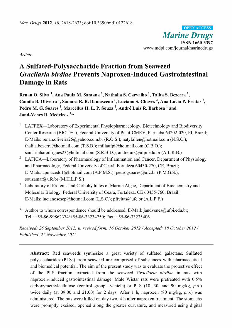
Mar. Drugs 2012, 10, 2618-2633; doi:10.3390/md10122618 Marine Drugs ISSN 1660-3397 www.mdpi.com/journal/marinedrugs Article A Sulfated-Polysaccharide Fraction from Seaweed Gracilaria birdiae Prevents Naproxen-Induced Gastrointestinal Damage in Rats Renan O. Silva 1 , Ana Paula M. Santana 2 , Nathalia S. Carvalho 1 , Talita S. Bezerra 1 , Camila B. Oliveira 1 , Samara R. B. Damasceno 1 , Luciano S. Chaves 3 , Ana Lúcia P. Freitas 3 , Pedro M. G. Soares 2 , Marcellus H. L. P. Souza 2 , André Luiz R. Barbosa 1 and Jand-Venes R. Medeiros 1, * 1 LAFFEX—Laboratory of Experimental Physiopharmacology, Biotechnology and Biodiversity Center Research (BIOTEC), Federal University of Piauí -CMRV, Parnaíba 64202-020, PI, Brazil; E-Mails: [email protected] (R.O.S.); [email protected] (N.S.C.); [email protected] (T.S.B.); [email protected] (C.B.O.); [email protected] (S.R.B.D.); [email protected] (A.L.R.B.) 2 LAFICA—Laboratory of Pharmacology of Inflammation and Cancer, Department of Physiology and Pharmacology, Federal University of Ceará , Fortaleza 60430-270, CE, Brazil; E-Mails: [email protected] (A.P.M.S.); [email protected] (P.M.G.S.); [email protected] (M.H.L.P.S.) 3 Laboratory of Proteins and Carbohydrates of Marine Algae, Department of Biochemistry and Molecular Biology, Federal University of Ceará , Fortaleza, CE 60455-760, Brazil; E-Mails: [email protected] (L.S.C.); [email protected] (A.L.P.F.) * Author to whom correspondence should be addressed; E-Mail: [email protected]; Tel.: +55-86-99862374/+55-86-33234750; Fax: +55-86-33235406. Received: 26 September 2012; in revised form: 16 October 2012 / Accepted: 18 October 2012 / Published: 22 November 2012 Abstract: Red seaweeds synthesize a great variety of sulfated galactans. Sulfated polysaccharides (PLSs) from seaweed are comprised of substances with pharmaceutical and biomedical potential. The aim of the present study was to evaluate the protective effect of the PLS fraction extracted from the seaweed Gracilaria birdiae in rats with naproxen-induced gastrointestinal damage. Male Wistar rats were pretreated with 0.5% carboxymethylcellulose (control group—vehicle) or PLS (10, 30, and 90 mg/kg, p.o.) twice daily (at 09:00 and 21:00) for 2 days. After 1 h, naproxen (80 mg/kg, p.o.) was administered. The rats were killed on day two, 4 h after naproxen treatment. The stomachs were promptly excised, opened along the greater curvature, and measured using digital OPEN ACCESS
Transcript of A Sulfated-Polysaccharide Fraction from Seaweed
Mar. Drugs 2012, 10, 2618-2633; doi:10.3390/md10122618
Marine Drugs
ISSN 1660-3397
www.mdpi.com/journal/marinedrugs
Article
A Sulfated-Polysaccharide Fraction from Seaweed
Gracilaria birdiae Prevents Naproxen-Induced Gastrointestinal
Damage in Rats
Renan O. Silva 1, Ana Paula M. Santana
2, Nathalia S. Carvalho
1, Talita S. Bezerra
1,
Camila B. Oliveira 1, Samara R. B. Damasceno
1, Luciano S. Chaves
3, Ana Lúcia P. Freitas
3,
Pedro M. G. Soares 2, Marcellus H. L. P. Souza
2, André Luiz R. Barbosa
1 and
Jand-Venes R. Medeiros 1,
*
1 LAFFEX—Laboratory of Experimental Physiopharmacology, Biotechnology and Biodiversity
Center Research (BIOTEC), Federal University of Piauí-CMRV, Parnaíba 64202-020, PI, Brazil;
E-Mails: [email protected] (R.O.S.); [email protected] (N.S.C.);
[email protected] (T.S.B.); [email protected] (C.B.O.);
[email protected] (S.R.B.D.); [email protected] (A.L.R.B.) 2 LAFICA—Laboratory of Pharmacology of Inflammation and Cancer, Department of Physiology
and Pharmacology, Federal University of Ceará, Fortaleza 60430-270, CE, Brazil;
E-Mails: [email protected] (A.P.M.S.); [email protected] (P.M.G.S.);
[email protected] (M.H.L.P.S.) 3 Laboratory of Proteins and Carbohydrates of Marine Algae, Department of Biochemistry and
Molecular Biology, Federal University of Ceará, Fortaleza, CE 60455-760, Brazil;
E-Mails: [email protected] (L.S.C.); [email protected] (A.L.P.F.)
* Author to whom correspondence should be addressed; E-Mail: [email protected];
Tel.: +55-86-99862374/+55-86-33234750; Fax: +55-86-33235406.
Received: 26 September 2012; in revised form: 16 October 2012 / Accepted: 18 October 2012 /
Published: 22 November 2012
Abstract: Red seaweeds synthesize a great variety of sulfated galactans. Sulfated
polysaccharides (PLSs) from seaweed are comprised of substances with pharmaceutical
and biomedical potential. The aim of the present study was to evaluate the protective effect
of the PLS fraction extracted from the seaweed Gracilaria birdiae in rats with
naproxen-induced gastrointestinal damage. Male Wistar rats were pretreated with 0.5%
carboxymethylcellulose (control group—vehicle) or PLS (10, 30, and 90 mg/kg, p.o.)
twice daily (at 09:00 and 21:00) for 2 days. After 1 h, naproxen (80 mg/kg, p.o.) was
administered. The rats were killed on day two, 4 h after naproxen treatment. The stomachs
were promptly excised, opened along the greater curvature, and measured using digital
OPEN ACCESS

Mar. Drugs 2012, 10 2619
calipers. Furthermore, the guts of the animals were removed, and a 5-cm portion of the
small intestine (jejunum and ileum) was used for the evaluation of macroscopic scores.
Samples of the stomach and the small intestine were used for histological evaluation,
morphometric analysis and in assays for glutathione (GSH) levels, malonyldialdehyde
(MDA) concentration, and myeloperoxidase (MPO) activity. PLS treatment reduced the
macroscopic and microscopic naproxen-induced gastrointestinal damage in a dose-dependent
manner. Our results suggest that the PLS fraction has a protective effect against
gastrointestinal damage through mechanisms that involve the inhibition of inflammatory
cell infiltration and lipid peroxidation.
Keywords: sulfated polysaccharide; gastrointestinal damage; naproxen; antioxidant activity
1. Introduction
In recent years, marine resources have attracted attention as a source of bioactive compounds for
the development of new drugs and healthy foods [1]. In particular, seaweeds are a very important and
commercially valuable resource for the food industry; they also serve as soil conditioners and are used
in traditional medicine because of their perceived health benefits [2,3]. In addition, sulfated
polysaccharides (PLSs) from marine algae are known to exhibit many biological and physiological
activities, including anticoagulant, antiviral, antitumor, anti-inflammatory, and antioxidant effects [4–7].
Red seaweeds synthesize a great variety of sulfated galactans, which are the major components of
the extracellular matrix. Red seaweed galactans are of great commercial importance and are used
widely in the food industry as gelling and thickening agents, due to their rheological properties [3].
These PLSs are primarily classified as agarans and carrageenans. In particular, galactans with 4-linked
α-galactose residues of the L-series are termed agarans and those of the D-series are termed
carrageenans [8]. Polysaccharides from the Gracilaria genus are composed mainly of the alternating
3-linked-β-D-galactopyranose unit (Gal) and the 4-linked-3,6-anhydro-α-L-galactopyranose unit
(AnGal). The Gal unit can be substituted by either methyl or sulfate ester groups [9].
PLSs comprise a complex group of macromolecules with a wide range of important biological
properties. The PLSs in seaweeds contain substances with great pharmaceutical and biomedical
potential [10–12]. Recent studies have shown that PLSs extracted from Gracilaria birdiae
demonstrated marked antioxidant and anti-inflammatory activities [11]. However, few studies have
correlated the potential of PLSs from seaweeds to the gastrointestinal tract damages associated with
the use of non-steroidal anti-inflammatory drugs (NSAIDs).
NSAIDs are often recommended clinically because of their anti-inflammatory, anti-pyretic, and
analgesic properties. However, the chronic use of these drugs is limited as a result of their capacity to
cause damage to the gastrointestinal tract, such as erosion, ulceration, perforation, and hemorrhage [13,14].
Naproxen is a non-selective NSAID that is widely prescribed for chronic treatments such as that for
arthritis; it is also one of the most likely drugs in this class to induce gastrointestinal damage [15–17].
The production of oxygen free radicals and lipid peroxidation play important roles in the
naproxen-induced gastric antral ulceration [18,19].

Mar. Drugs 2012, 10 2620
Thus, the aim of the present study was to investigate the protective effect of the PLS fraction
extracted from G. birdiae on naproxen-induced gastrointestinal damage in rats.
2. Results and Discussion
The polysaccharide fraction isolated from the red alga G. birdiae was previously identified [11,20].
This galactan is an agar-type polysaccharide composed mainly of β-D-galactopyranose linked to
3,6-anhydro-α-L-galactose with low methyl substituted groups. The structure is formed by
→4-3,6-anhydro-α-L-galp (1→3) β-D-galp 1→ segments, with the possibility of an α-L-galp unit
substituted at the 6-position by a sulfate ester [20]. In addition, the molar mass distribution was found
to be within 2.6 × 10⁶ and 3.75 × 10
5 g/mol, while the soluble carbohydrate, protein, and sulfate
contents were 85.5%, 2.5%, and 8.4%, respectively [11,20].
NSAIDs, such as naproxen, are the most widely used pharmacological agents for the treatment of
pain and inflammation [21]. However, 15%–30% of the patients who receive these drugs develop
gastrointestinal ulcers [22]. The gastrointestinal protective effect of PLS extracted from G. birdiae was
evaluated using a rat model of naproxen-induced gastrointestinal damage. In the present study, we
confirmed that treatment of the animals with naproxen for two days led to the formation of severe
macroscopic gastric and intestinal lesions (12.9 ± 4.0 mm and 16.4 ± 2.0 lesion scores, respectively).
Moreover, PLS treatment was found to reduce the macroscopic and microscopic naproxen-induced
gastrointestinal damage. Figure 1 shows that PLS prevented naproxen-induced gastropathy in a
dose-dependent manner, reaching its maximal effect at a dose of 90 mg/kg, with 93% and 78% lesion
inhibition in the stomach and small intestine (jejunum and ileum), respectively.
Figure 1. The sulfated-polysaccharide (PLS) fraction extracted from Gracilaria birdiae
reduces naproxen-induced gastric (A) and intestinal damage (B). Rats were treated by
gavage with either carboxymethylcellulose (C: control) or PLS (10, 30, and 90 mg/kg)
twice daily (at 09:00 hours and 21:00 hours) for two days. After 1 h, naproxen (80 mg/kg)
was administered by gavage. The results are expressed as the mean ± SEM of 5–7 animals
per group. * p < 0.05 vs. carboxymethylcellulose group; # p < 0.05 vs. naproxen group
(ANOVA and Newman-Keuls test).

Mar. Drugs 2012, 10 2621
Figure 1. Cont.
Gastric ulceration due to the ingestion of NSAIDs appears to be associated with a reduction in
gastric blood flow and an increase in leukocyte adherence within the gastric microcirculation,
secondary to a reduction in prostaglandin synthesis [23]. Several research groups have demonstrated
that serious NSAID-associated gastrointestinal complications develop not only in the upper, but also in
the lower gastrointestinal tract, including the small intestine; recently, this problem has become a topic
of great interest to gastroenterologists [24]. Therefore, determining the mechanisms of
NSAID-induced mucosal injury in the small intestine and identifying substances that effectively
protect the gastrointestinal mucosal against these aggressors is of great importance.
The gastroprotective effect of 90 mg/kg PLS was confirmed by histological analysis (Table 1).
Microscopic analysis revealed that naproxen increased edema, epithelial cell loss, and inflammatory
cell infiltration, but did not cause hemorrhagic damage. On the other hand, pretreatment with PLS
significantly decreased the infiltration of inflammatory cells, the formation of edema and the loss of
epithelial cells induced by naproxen (Table 1). Thus, the analysis of both the macro- and microscopic
findings revealed an excellent correlation, confirming the efficacy of the compound.
Table 1. Effect of the sulfated-polysaccharide fraction (PLS, 90 mg/kg) extracted from
Gracilaria birdiae on naproxen-induced microscopic gastric damage.
Experimental group
(n = 5)
Hemorrhagic
damage
(score, 0–4)
Edema
(score, 0–4)
Epithelial
cell loss
(score, 0–3)
Inflammatory
cells
(score, 0–3)
Total
(score, 0–14)
Control
0 0 (0–1) 0 0 (0–1) 0 (0–3)
Naproxen 0 3 (1–3) a 3 (3–3)
a 2 (2–3)
a 7 (5–9)
a
Naproxen + PLS (90 mg/kg) 0 0 (0–1) b 0 (0–2)
b 0 (0–1)
b 0 (0–3)
b
Data shown are the median values with the minimum and maximum scores given in parentheses. The
Kruskal-Wallis nonparametric test followed by Dunn’s test were used for multiple comparisons of
histological analyses. a p < 0.05 vs. control group (carboxymethylcellulose);
b p < 0.05 vs. naproxen group.

Mar. Drugs 2012, 10 2622
Figure 2 shows that in the small intestine of the animals treated with naproxen (90 mg/kg, p.o.)
twice daily for two days, significant villi shortening (Figure 2A), increased crypt depth (Figure 2B),
and a decreased villus/crypt ratio (Figure 2C) were seen. However, when the animals were pretreated
with PLS before naproxen administration, we observed a complete reversal of the intestinal
morphometry alterations in the small intestine (Figure 2A,B,C). Other studies have shown that the
development of NSAID-induced small intestinal ulcers is a multifactorial process, with a distinct
pathogenesis of gastric damage [25] that involves a combination of events, such as increased epithelial
permeability [26], intestinal hypermotility [27], and luminal bacterial invasion of the gut wall [28].
These effects lead to mucosal inflammation and eventually result in macroscopic damage. Taken
together, these results indicate that PLS has a significant protective effect against naproxen-induced
gastrointestinal damage.
Figure 2. Morphometric analyses of the small intestine tissues in rats (N = 6) treated with
naproxen alone or naproxen + the sulfated-polysaccharide fraction (PLS) extracted from
Gracilaria birdiae. Rats were treated by gavage with either carboxymethylcellulose
(C: control) or PLS (90 mg/kg) twice daily (at 09:00 and 21:00) for two days. After 1 h,
naproxen (80 mg/kg) was administered by gavage. After 4 h, the animals were killed and
segments of the small intestine were collected for the measurement of villus height (A),
crypt depth (B), and the villus/crypt (VH/CD) ratio (C). * p < 0.05 vs. control group;
# p < 0.05 vs. naproxen group (ANOVA and Newman-Keuls test).

Mar. Drugs 2012, 10 2623
Figure 2. Cont.
In recent years, studies have shown that PLSs interfere with cell migration at the site of
inflammation. Researchers have shown that these compounds, including those isolated from algae,
possess immunomodulatory activities that may stimulate the immune response, and thus control the
anti-inflammatory response [29]. In this study, the accumulation of leukocytes following the
appearance of naproxen-induced lesions was evaluated by measuring the activity of gastric
myeloperoxidase (MPO). MPO exists in polymorphonuclear leukocytes (PNL) that produce an
excessive amount of superoxide anions (O2−) and hydroxyl radicals (OH
•), which are free oxygen
radicals that are extremely toxic to the mucosa [30]. MPO levels have been shown to increase
concomitantly with NSAID-induced injury in rats [31]. Furthermore, this observation is consistent
with evidence indicating that mucosal damage induced by NSAIDs, such as naproxen, results in a
marked increase in mucosal MPO activity, which may be associated with an increase in neutrophil
infiltration and H2O2 in damaged tissues [32,33]. In this study, we observed significantly increased
levels of MPO in the stomach (5.9 ± 1.7 U/mg of tissue) and the small intestine (9.7 ± 1.6 U/mg of
tissue) of naproxen-treated rats compared to the control group (Figure 3). However, pretreatment with
90 mg/kg PLS significantly attenuated the naproxen-induced increase in MPO activity, in both the
stomach (0.9 ± 0.5 U/mg of tissue) and the intestine (1.4 ± 0.7 U/mg of tissue). Therefore, PLS may
protect the gastrointestinal tract by reducing the recruitment of leukocytes, thereby hindering
superoxide anion production.
Several studies have shown that NSAIDs also act as pro-oxidants, blocking the antioxidant systems
of mucosal cells and resulting in the formation of reactive oxygen species (ROS). As a consequence of
this process, oxidative damage occurs [34]. Recent works have demonstrated that ROS, such as
superoxide anion and hydroxyl radicals, play an important role in the pathogenesis of various diseases,
including the mucosal gastrointestinal damage induced by NSAIDs [35], ethanol [36], and others
agents [37]. However, organisms have enzymatic and non-enzymatic defense mechanisms against the
toxicity and tissue damage induced by these agents [38].

Mar. Drugs 2012, 10 2624
Figure 3. Effect of the sulfated-polysaccharide (PLS) fraction extracted from
Gracilaria birdiae on gastric myeloperoxidase (MPO) activity in a rat model of
naproxen-induced gastrointestinal damage. Rats were treated by gavage with either
carboxymethylcellulose (C: control) or PLS (90 mg/kg) twice daily (at 09:00 and 21:00)
for 2 days. After 1 h, naproxen (80 mg/kg) was administered by gavage. The results are
expressed as the mean ± SEM of 5–7 animals per group. * p < 0.05 vs. control group;
# p < 0.05 vs. naproxen group (ANOVA and Newman-Keuls test).
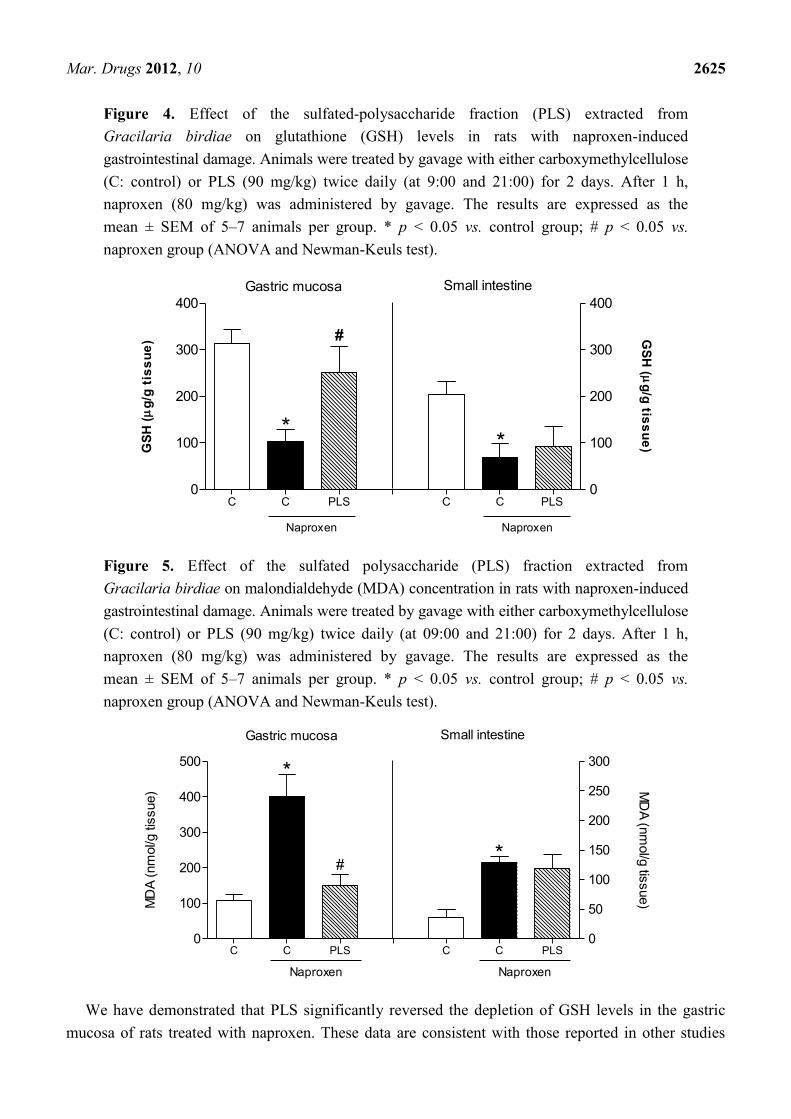
Therefore, the present study investigated two oxidative stress markers, glutathione (GSH) and
malondialdehyde (MDA). Naproxen significantly reduced the levels of GSH (103.4 ± 24.9 mg/g of
tissue) and increased the concentration of gastric mucosal MDA (401.7 ± 61.5 nmol/g of tissue) in the
stomachs of rats exposed to the drug compared to untreated normal controls (Figures 4 and 5,
respectively). Pretreatment with PLS (90 mg/kg) inhibited these effects in all of the naproxen-treated
rats, reducing the naproxen effects and resulting in values that approximated those of controls without
naproxen (251.1 ± 54.9 mg/g of tissue and 151.0 ± 29.8 nmol/g of tissue, respectively; Figure 4).
These results suggested that the gastroprotective effect of PLS involves the participation of oxidative
stress and its blockade. However, this alteration was not observed in the small intestine. Compared to
the naproxen group (69.8 ± 29.3 mg/g of tissue and 214.2 ± 19.5 nmol/g of tissue, respectively), the
group pretreated with PLS (90 mg/kg) did not show significant inhibition of GSH level depletion
(92.5 ± 42.2 mg/g of tissue) or reduction in the concentration of MDA (198.1 ± 38.6 nmol/g of tissue)
in the small intestine (Figures 4 and 5).
Antioxidant compounds play an important role in various pathological conditions, including
inflammation, neurodegenerative diseases, and cancer [39,40]. It has been systematically reported in
literature that PLSs that show antioxidant activity, such as those extracted from marine algae, protect
against cell death due to their ability to degrade excessive ROS [5]. However, few studies have
correlated antioxidant potential with NSAID-associated injury of the gastrointestinal tract.
Mar. Drugs 2012, 10 2625
Figure 4. Effect of the sulfated-polysaccharide fraction (PLS) extracted from
Gracilaria birdiae on glutathione (GSH) levels in rats with naproxen-induced
gastrointestinal damage. Animals were treated by gavage with either carboxymethylcellulose
(C: control) or PLS (90 mg/kg) twice daily (at 9:00 and 21:00) for 2 days. After 1 h,
naproxen (80 mg/kg) was administered by gavage. The results are expressed as the
mean ± SEM of 5–7 animals per group. * p < 0.05 vs. control group; # p < 0.05 vs.
naproxen group (ANOVA and Newman-Keuls test).
C C PLS C C PLS 0
100
200
300
400
0
100
200
300
400
Naproxen
*
#
Naproxen
*
Gastric mucosa Small intestine
GS
H (
g/g
tis
su
e) G
SH
( g
/g tis
su
e)
Figure 5. Effect of the sulfated polysaccharide (PLS) fraction extracted from
Gracilaria birdiae on malondialdehyde (MDA) concentration in rats with naproxen-induced
gastrointestinal damage. Animals were treated by gavage with either carboxymethylcellulose
(C: control) or PLS (90 mg/kg) twice daily (at 09:00 and 21:00) for 2 days. After 1 h,
naproxen (80 mg/kg) was administered by gavage. The results are expressed as the
mean ± SEM of 5–7 animals per group. * p < 0.05 vs. control group; # p < 0.05 vs.
naproxen group (ANOVA and Newman-Keuls test).
C C PLS C C PLS0
100
200
300
400
500
0
50
100
150
200
250
300
Naproxen
*
#
Naproxen
Gastric mucosa Small intestine
*
MD
A (
nm
ol/g
tis
sue) M
DA
(nm
ol/g
tissue)
We have demonstrated that PLS significantly reversed the depletion of GSH levels in the gastric
mucosa of rats treated with naproxen. These data are consistent with those reported in other studies

Mar. Drugs 2012, 10 2626
demonstrating that PLSs from algae inhibited ethanol-induced gastric damage by increasing tissue
GSH levels through the inactivation of ROS and products of lipid peroxidation [12]. Previous research
suggested that ROS-induced damage can be prevented in two ways: by suppressing free-radical
generation or by scavenging the free radicals generated [41]. Thus, we suggest that PLS prevents
naproxen-induced gastropathy by increasing GSH levels and that this effect may be secondary to a
decrease in free radical production. In addition, our results demonstrated that the administration of PLS
results in a significant decrease in the concentration of MDA in naproxen-induced gastropathy. Thus,
the mechanism through which PLS exerts its gastroprotective effect appears to involve a reduction in
the lipid peroxidation induced by naproxen administration.
Concomitantly, our results showed that naproxen also decreases GSH levels and increases lipid
peroxidation in the small intestine. Treatment with PLS did not inhibit the effects of naproxen on these
biochemical parameters in this organ. Thus, we suggest that the mechanisms underlying the
gastroprotective effect of PLS promote an increase in gastric mucosal resistance and a decrease in
aggressive factors. In contrast to the gastric tissue, the increase in endogenous GSH levels in the small
intestine does not appear to contribute to this effect. A possible mechanism may involve the
downregulation of the inflammatory response by inhibiting the synthesis and release of
pro-inflammatory mediators.
3. Experimental Section
3.1. Extraction of the Sulfated Polysaccharide Fraction
The marine alga G. birdiae was collected from the Atlantic coast of Brazil (Flecheiras Beach,
Trairí, Ceará). The samples were cleaned of epiphytes, washed with distilled water, and stored at
−20 °C. The polysaccharide extraction procedure was performed according to the method described by
Farias et al. [42]. The dried tissue (5 g) was milled and suspended in 250 mL of 0.1 M sodium acetate
buffer (pH 6.0) containing 510 mg of papain (E. Merck), 5 mM EDTA, and 5 mM cysteine, and
incubated at 60 °C for 12 h. The residue was removed by filtration and centrifugation (2700× g for
25 min at 4 °C), and the sulfated polysaccharides were precipitated by the addition of 48 mL of 10%
cetylpyridinium chloride (CPC, Sigma Chemical). The mixture was centrifuged (2700× g for 25 min at
4 °C) and the polysaccharides (kappa-carrageenan) in the pellet were washed with 200 mL of 0.05%
cetylpyridinium chloride solution, dissolved in 174 mL of a 2 M NaCl/ethanol (100:15, v/v) solution,
and precipitated with 200 mL of 70% ethanol (v/v) for 12 h at 4 °C. After further centrifugation (2700× g
for 25 min at 4 °C), the precipitate was washed twice with 200 mL of absolute ethanol and dried with
acetone under hot air flow (60 °C).
3.2. Chemical Characterization of the Sulfated Polysaccharide Fraction
The chemical characterization was previously determined [11,20]. Total sugar content of each
fraction was determined according to the method of Dubois [43]. Protein content was measured by
Bradford’s method [44]. Sulfate content in polysaccharides was determined by the barium
chlorideegelatin method [45] and the monosaccharide composition of red seaweed galactans was
obtained by reductive hydrolysis [46].

Mar. Drugs 2012, 10 2627
The peak molar masses (Mpk) were estimated by gel permeationchromatography (GPC) with a
Shimadzu equipment at room temperature using an Ultrahydrogel linear column (7.8 × 300 mm,
exclusion limits 106 g/mol), flow 0.5 mL/min, 0.5% polysaccharide concentration and 0.1 M NaNO3
as solvent.
3.3. Animals
Male Wistar albino rats, weighing 100–150 g, were obtained from the Department of Physiology
and Pharmacology, Federal University of Ceará. All animals were housed in temperature-controlled
rooms and received water and food ad libitum. The animals were deprived of food for 18–24 h before
the experimentation, but had free access to water. All surgical procedures and animal treatments were
conducted in accordance with the Guide for Care and Use of Laboratory Animals (National Institutes
of Health, Bethesda, MD) and were approved by the local ethics committee (Protocol No. 0066/10).
3.4. Effect of PLS on Naproxen-Induced Gastric Damage
Rats were pretreated with 0.5% carboxymethylcellulose (control group) or polysaccharide (PLS: 10,
30, and 90 mg/kg, p.o.) twice daily (at 09:00 and 21:00) for two days. After 1 h, naproxen (80 mg/kg,
p.o.) was administered twice daily (at 09:00 and 21:00) for two days as described by Kim et al., with
modifications [17]. The control group received only vehicle or vehicle + naproxen. The rats were
killed on the second day, 4 h after the naproxen treatment. The stomachs were promptly excised,
opened along the greater curvature, and washed with 0.9% saline. The gastric damage was measured
using digital calipers (Mitutoyo®). Samples of the stomachs were fixed in 10% formalin immediately
after removal for subsequent histological evaluation. Others samples were then weighed, frozen, and
stored at −70 °C until they were assayed for GSH levels [47], MDA concentration [48], and MPO
activity [49].
3.5. Effect of PLS on Naproxen-Induced Small Intestine Damage
Animals were treated with 0.5% carboxymethylcellulose (vehicle) or PLS (90 mg/kg, p.o.) twice
daily (at 09:00 and 21:00) for two days. One hour after the administration of PLS or vehicle, the
animals were treated with naproxen (80 mg/kg, p.o.), as described above. The control group received
vehicle or vehicle + naproxen. The rats were killed on the second day, 4 h after the naproxen
treatment. The abdomens were then opened, and after identification of the intestine, a 5-cm portion of
the small intestine (jejunum and ileum) was removed, washed with 0.9% saline, and pinned onto a wax
block for the evaluation of macroscopic scores by modifying the criteria described by Morris et al. [50]
(Table 2). All scoring of damage was performed in a randomized manner by an observer who was
unaware of the treatments that the rats had received. Samples of tissue were then removed for the
measurement of GSH levels [47], MDA concentration [48], and MPO activity [49]. Other samples
were fixed in 10% formalin for subsequent histopathological and morphometric analysis.

Mar. Drugs 2012, 10 2628
Table 2. Criteria for macroscopic scoring of intestinal damage.
Scores Criteria
0 No damage.
1 Focal hyperemia; no ulcers.
2 Ulceration without hyperemia or bowel wall thickening.
3
4
5
6–10
Ulceration with inflammation at 1 site.
≥2 Sites of ulceration and inflammation.
≥2 Major sites of ulceration and inflammation or 1 site
of ulceration extending >1 cm along length of intestinal.
If damage covered >2 cm along length of intestinal, score is increased by 1 for each
additional cm of involvement.
3.6. Histological Evaluation of Gastric Damage
For histological evaluation, the stomach samples were fixed in 10% formalin solution for 24 h.
After fixation, the samples were transferred to a solution of 70% alcohol. The material was then
embedded in paraffin and sectioned; 4-µm-thick sections were deparaffinized, stained with
hematoxylin and eosin (H & E), and then examined under a light microscope by an experienced
pathologist without knowledge of the treatments. The specimens were assessed according to the
criteria described by Laine and Weinstein, in which scores are assigned to the following parameters for
a maximum total score of 14: hemorrhagic damage (score of 0–4), edema in the upper mucosa (score
of 0–4), epithelial cell loss (score of 0–3), and presence of inflammatory cells (score of 0–3) [51].
3.7. Intestinal Morphometric Analysis
Morphometric analysis was performed using slides stained with H & E on a light microscope
equipped with a high-resolution Leica DFC 320 digital camera (Wetzlar, Germany) connected to a
computer with an image capture program. An average of 8 to 10 different linear measurements of crypt
depth and villus height were recorded. The height of the villus was measured from the top to the
bottom, corresponding to the junction of the crypt/villus. The depth of the crypts was defined as the
invagination between adjacent villi.
3.8. Glutathione Levels
The concentration of glutathione (GSH) in the samples of stomach and small intestine tissues was
estimated according to the method described by Sedlak and Lindsay [47]. A segment from each organ
was homogenized in 5 mL of cold 0.02 M EDTA solution (1 mL 100 mg/tissue). Aliquots (400 μL) of
the tissue homogenate were mixed with 320 μL of distilled water and 80 μL of 50% (w/v)
trichloroacetic acid in glass tubes and centrifuged at 3000 rpm for 15 min. Next, 400 μL of each
supernatant was mixed with 800 μL of Tris buffer (0.4 M, pH 8.9) and 20 μL of 0.01 M 5,5-dithio-bis
(2-nitrobenzoic acid). Subsequently, the samples were stirred for 3 min and read on a
spectrophotometer at 412 nm. GSH concentration was determined via a reduced GSH standard curve,
which was generated in parallel. The results are expressed as micrograms of GSH per gram of tissue.

Mar. Drugs 2012, 10 2629
3.9. Malondialdehyde Concentration
The MDA levels in the homogenate from each group were measured using the method described by
Mihara and Uchiyama [48], which is based on the reaction with thiobarbituric acid. Fragments of the
gastric mucosa and small intestine weighing between 100 and 150 mg were homogenized with cold
1.15% KCl to prepare 10% homogenates. Briefly, 250 μL of each homogenate was added to 1.5 mL of
1% phosphoric acid (H3PO4) and 0.5 mL of 0.6% tert-butyl alcohol (aqueous solution). Then, this
mixture was stirred and heated in a boiling water bath for 45 min. The mixture was then cooled
immediately in an ice water bath followed by the addition of 4 mL of n-butanol. This mixture was
shaken and the butanol layer was separated by centrifugation at 1200× g for 10 min. Optical density
was determined to be 535 and 520 nm, and the optical density difference between the two
determinations was calculated as the tert-butyl alcohol value. MDA concentrations are expressed as
millimoles per gram of tissue.
3.10. Myeloperoxidase Activity
Myeloperoxidase is an enzyme found primarily in neutrophil azurophilic granules which has been
used extensively as a biochemical marker for granulocyte infiltration into various tissues, including
the gastrointestinal tract. The extent of neutrophil accumulation in the gastric mucosa and small
intestinal was measured by MPO activity evaluation [49]. Briefly, 50–100 mg of tissue was
homogenized in 1 mL of potassium buffer with 0.5% of hexadecitrimetilamônio (HTAB) for each
50 mg of tissue. Then, homogenate was centrifuged at 40.000× g for 7 min at 4 °C. MPO activity in
the resuspended pellet was assayed by measuring the change in absorbance at 450 nm using
o-dianisidine dihydrochloride and 1% hydrogen peroxide. The results were reported as the MPO units
per mg of tissue. A unit of MPO activity was defined as that converting 1 µmol of hydrogen peroxide
to water in 1 min at 22 °C.
3.11. Statistical Analysis
Data were described as either means ± SEM or median, as appropriate. Analysis of Variance
(ANOVA) followed by Student-Newman-Keuls test was used to compare means and Kruskal-Wallis
nonparametric test, followed by Dunns tests to compare medians; P < 0.05 was defined as
statistically significant.
4. Conclusions
Our results suggest that PLS has a beneficial effect in this model of naproxen-induced
gastrointestinal damage, through mechanisms that involve the inhibition of inflammatory cell
infiltration and lipid peroxidation. We suggest that polysaccharides may have potential applications in
the development of novel therapeutics for preventing NSAID-induced adverse effects in the
gastrointestinal tract in humans.

Mar. Drugs 2012, 10 2630
Acknowledgments
The authors are grateful to the Brazilian Agency for Scientific and Technological Development—CNPq
(Brazil) and to the Research Foundation for Support of the State of Piauí—Brazil (FAPEPI) for
financially supporting this work.
References
1. Qi, H.; Zhao, T.; Zhang, Q.; Li, Z.; Zhao, Z. Antioxidant activity of different molecular weight
sulfated polysaccharides from Ulva pertusa Kjellm (Chlorophyta). J. Appl. Phycol. 2005, 17,
527–534.
2. Yang, Y.F.; Fei, X.G.; Song, J.M.; Hu, H.Y.; Wang, G.C.; Chung, I.K. Growth of Gracilaria
lemaneiformis under different cultivation conditions and its effects on nutrient removal in Chinese
coastal waters. Aquaculture 2006, 254, 248–255.
3. Jiao, G.; Yu, G.; Zhang, J.; Ewart, H.S. Chemical structures and bioactivities of sulfated
polysaccharides from marine algae. Mar. Drugs 2011, 9, 196–223.
4. Almeida-Lima, J.; Costa, L.S.; Silva, N.B.; Melo-Silveira, R.F.; Silva, F.V.; Felipe, M.B.M.C.;
Medeiros, S.R.B.M.; Leite, E.L.; Rocha, H.A.O. Evaluating the possible genotoxic, mutagenic
and tumor cell proliferation-inhibition effects of a non-anticoagulant, but antithrombotic algal
heterofucan. J. Appl. Toxicol. 2010, 30, 708–715.
5. Costa, L.S.; Fidelis, G.P.; Cordeiro, S.L.; Oliveira, R.M.; Sabry, D.A.; Câmara, R.B.G.;
Nobre, L.T.D.B.; Costa, M.S.S.P.; Almeida-Lima, J.; Farias, E.H.C. Biological activities of
sulfated polysaccharides from tropical seaweeds. Biomed. Pharmacother. 2010, 64, 21–28.
6. Cumashi, A.; Ushakova, N.A.; Preobrazhenskaya, M.E.; D’incecco, A.; Picooli, A.; Totani, L.;
Tinari, N.; Morozevich, G.E.; Berman, A.E.; Bilan, M.I. A comparative study of the
anti-inflammatory, anticoagulant, antiangiogenic, and antiadhesive activities of nine different
fucoidans from brown seaweeds. Glycobiology 2007, 17, 541–552.
7. Chaves, L.S.; Nicolau, L.A.D.; Silva, R.O.; Barros, F.C.N.; Freitas, A.L.P.; Aragao, K.S.;
Ribeiro, R.A.; Souza, M.H.L.P.; Barbosa, A.L.R.; Medeiros, J.V.R. Anti-inflammatory and
anti-nociceptive effects in mice of a sulfated polysaccharide fraction extracted from the marine
red algae Gracilaria caudata. Immunopharmacol. Immunotoxicol. 2012, in press.
8. Knutsen, S.H.; Myslabodski, D.E.; Larsen, B.; Usov, A.I. A modified system of nomenclature for
red algal galactans. Bot. Mar. 1994, 37, 163–170.
9. Rees, D.A. Biogenesis of 3,6-anhydro-L-galactose. Biochem. J. 1961, 81, 347–352.
10. Ye, H.; Wang, K.; Zhou, C.; Liu, J.; Zeng, X. Purification, antitumor and antioxidant activities
in vitro of polysaccharides from the brown seaweed Sargassum pallidum. Food Chem. 2008, 111,
428–432.
11. Souza, B.W.S.; Cerqueira, M.A.; Bourbon, A.I.; Pinheiro, A.C.; Martins, J.T.; Teixeira, J.A.;
Coimbra, M.A.; Vicente, A.A. Chemical characterization and antioxidant activity of sulfated
polysaccharide from the red seaweed Gracilaria birdiae. Food Hydrocoll. 2012, 27, 287–292.

Mar. Drugs 2012, 10 2631
12. Silva, R.O.; Santos, G.M.P.; Nicolau, L.A.D.; Lucetti, L.T.; Santana, A.P.M.; Chaves, L.S.C.;
Barros, C.N.; Freitas, A.L.P.; Souza, M.H.L.P.; Medeiros, J.V.R. Sulfated-polysaccharide fraction
from red algae Gracilaria caudata protects mice gut against ethanol-induced gastric damage.
Mar. Drugs 2011, 9, 2188–2200.
13. Fries, J.F. NSAID gastropathy: Epidemiology. J. Musculoskelet. Med. 1991, 8, 21–28.
14. Villegas, I.; La Casa, C.; De la Lastra, C.A.; Motilva, V.; Herrerías, J.M.; Martín, M.J. Mucosal
damage induced by preferential COX-1 and COX-2 inhibitors: Role of prostaglandins and
inflammatory response. Life Sci. 2004, 74, 873–884.
15. Beck, W.S.; Schneider, H.T.; Dietzel, K.; Nuernberg, B.; Brune, K. Gastrointestinal ulcerations
induced by anti-inflammatory drugs in rats. Arch. Toxicol. 1990, 64, 210–217.
16. Tenembaum, J. The epidemiology of nonsteroidal anti-inflammatory drugs. Can. J. Gastroenterol.
1999, 13, 119–122.
17. Kim, J.H.; Kim, Y.S.; Song, G.G.; Park, J.J.; Chang, H.I. Protective effect of astaxanthin on
naproxen-induced gastric antral ulceration in rats. Eur. J. Pharmacol. 2005, 514, 53–59.
18. Parks, D.A. Oxygen radicals: Mediators of gastrointestinal pathophysiology. Gut 1989, 30, 293–298.
19. Yoshikawa, T.; Naito, Y.; Ueda, S.; Oyamada, H.; Takemura, T.; Yoshida, N.; Sugino, S.; Kondo, M.
Role of oxygen-derived free radicals in the pathogenesis of gastric mucosal lesions in rats. J. Clin.
Gastroenterol. 1990, 12, 65–71.
20. Maciel, J.S.; Chaves, L.S.; Souza, B.W.S.; Teixeira, D.I.A.; Freitas, A.L.P.; Feitosa, J.P.A.;
De Paula, R.C.M. Structural characterization of cold extracted fraction of soluble sulfated
polysaccharide from red seaweed Gracilaria birdiae. Carbohydr. Polym. 2008, 71, 559–565.
21. Regula, J.; Butruk, E.; Dekkers, C.P.; de Boer, S.Y.; Raps, D.; Simon, L.; Terjung, A.; Thomas, K.B.;
Luhmann, R.; Fischer, R. Prevention of NSAID-associated gastrointestinal lesions: A comparison
study pantoprazole versus omeprazole. Am. J. Gastroenterol. 2006, 101, 1747–1755.
22. Nishida, T.; Tsujii, M.; Tsujii, S. Are COX-2 inhibitors truly able to prevent NSAIDs-associated
ulcers? Nippon Rinsho 2004, 62, 561–565.
23. Wallace, J.L. NSAID gastroenterophaty: Past, present and future. Can. J. Gastroenterol. 1996, 10,
451–459.
24. Kameda, N.; Higuchi, K.; Shiba, M. A prospective, single-blind trial comparing wireless capsule
endoscopy and double-balloon enteroscopy in patients with obscure gastrointestinal bleeding.
Gastroenterology 2008, 43, 434–440.
25. Pemberton, R.E.; Strand, L.J. A review of upper-gastrointestinal effects of the newer nonsteroidal
antiinflammatory agents. Dig. Dis. Sci. 1979, 24, 53–64.
26. Higuchi, K.; Umegaki, E.; Watanabe, T.; Yoda, Y.; Morita, E.; Murano, M.; Tokioka, S.;
Arakawa, T. Present status and strategy of NSAIDs-induced small bowel injury. J. Gastroenterol.
2009, 44, 879–888.
27. Takeuchi, K.; Miyazawa, T.; Tanaka, A.; Kato, S.; Kunikat, T. Pathogenic importance of
intestinal hypermotility in NSAID-induced small intestinal damage in rats. Digestion 2002, 66,
30–41.
28. Reuter, B.K.; Davies, N.M.; Wallace, J.L. Nonsteroidal anti-inflammatory drug enteropathy in
rats: Role of permeability, bacteria and enterohepatic circulation. Gastroenterology 1997, 112,
109–117.

Mar. Drugs 2012, 10 2632
29. Chen, D.; Wu, X.Z.; Wen, Z.Y. Sulfated polysaccharides and immune response: Promoter or
inhibitor? Panminerva Med. 2008, 50, 177–183.
30. Suzuki, M.; Mori, M.; Miura, S.; Suematsu, M.; Fukumura, D.; Kimura, H.; Ishii, H. Omeprazole
attenuates oxygen-derived free radical production from human neutrophils. Free Radic. Biol.
Med. 1996, 21, 727–731.
31. Zhang, X.; Tajima, K.; Kageyama, K.; Kyoi, T. Irsogladine maleate suppresses indomethacin-induced
elevation of proinflammatory cytokines and gastric injury in rats. World J. Gastroenterol. 2008,
14, 4784–4790.
32. Naito, Y.; Yoshikawa, T.; Matsuyama, K.; Yagi, N.; Arai, M.; Nakamura, Y.; Nishimura, S.;
Yoshida, N.; Kondo, M. Effects of oxygen radical scavengers on the quality of gastric ulcer
healing in rats. J. Clin. Gastroenterol. 1995, 21, 82–86.
33. Wallace, J.L. Pathogenesis of NSAID-induced gastroduedenal mucosal injury. Best Pract. Res.
Clin. Gastroenterol. 2001, 15, 691–703.
34. Odabasoglu, F.; Cakir, A.; Suleyman, H.; Aslan, A.; Bayir, Y.; Halici, M.; Kazaz, C.
Gastroprotective and antioxidant effects of usnic acid on indomethacine-induced gastric ulcer in
rats. J. Ethnopharmacol. 2006, 103, 59–65.
35. Basiviredy, J.; Jacob, M.; Ramamoorthy, P.; Pulimood, A.B.; Balasubramanian, K.A.
Indomethacin-induced free radical-mediated changes in the intestinal brush border membranes.
Biochem. Pharmacol. 2003, 65, 683–695.
36. Medeiros, J.V.; Gadelha, G.G.; Lima, S.J.; Garcia, J.A.; Soares, P.M.; Santos, A.A.; Brito, G.A.;
Ribeiro, R.A.; Souza, M.H. Role of the NO/cGMP/KATP pathway in the protective effects of
sildenafil against ethanol-induced gastric damage in rats. Br. J. Pharmacol. 2008, 153, 721–727.
37. Das, D.; Bandyopadhyay, D.; Bhattacharjee, M.; Banerjee, R.K. Hydroxyl radical is the major
cousative factor in stress-induced gastric ulceration. Free Radic. Biol. Med. 1997, 23, 8–18.
38. Bradley, P.P.; Christensen, R.D.; Rothstein, G. Cellular and extracellular myeloperoxidase in
pyogenic inflammation. Blood 1982, 60, 618–622.
39. Cuzzocrea, S.; Riley, D.P.; Caputi, A.P.; Salvemini, D. Antioxidant therapy: A new
pharmacological approach in shock, inflammation and ischemia/reperfusion injury. Pharmacol.
Rev. 2001, 53, 135–159.
40. Zhao, B. Natural antioxidants protect neurons in Alzheimer’s disease and Parkinson’s disease.
Neurochem. Res. 2009, 34, 630–638.
41. Xing, R.; Yu, H.; Liu, S.; Zhang, W.; Zhang, Q.; Li, Z.; Li, P. Antioxidant activity of differently
regioselective chitosan sulfates in vitro. Bioorg. Med. Chem. 2005, 13, 1387–1392.
42. Farias, W.R.L.; Valente, A.P.; Pereira, M.S.; Mourão, P.A.S. Structure and anticoagulant activity
of sulfated galactans. Isolation of a unique sulfated galactan from the red alga Botryocladia
occidentalis and comparison of its anticoagulant action with that of sulfated galactans from
invertebrates. J. Biol. Chem. 2000, 275, 29299–29307.
43. Dubois, M.; Gillis, K.A.; Hamilton, J.K.; Rebers, P.A.; Smith, F. Colorimetric method for
determination of sugars and related substances. Anal. Chem. 1956, 28, 350–356.
44. Bradford, M.M. A rapid and sensitive method for the quantitation of microgram quantities of
protein utilizing the principle of protein-dye binding. Anal. Biochem. 1976, 72, 248–254.

Mar. Drugs 2012, 10 2633
45. Lloyd, A.G.; Dodgson, K.S.; Price, R.G.; Rose, F.A.I. Infrared studies on sulphate esters. I.
Polysaccharide sulphates. Biochim. Biophys. Acta 1961, 46, 108–115.
46. Stevenson, T.T.; Furneaux, R.H. Chemical methods for the analysis of sulphated galactans from
red algae. Carbohydr. Res. 1991, 210, 277–298.
47. Sedlak, J.; Lindsay, R.H. Estimation of total, protein-bound, and nonprotein sulfhydryl groups in
tissue with Ellman’s reagent. Anal. Biochem. 1968, 24, 1992–2005.
48. Mihara, M.; Uchiyama, M. Determination of malonaldehyde precursor in tissues by thiobarbituric
acid test. Anal. Biochem. 1978, 86, 271–278.
49. Bradley, P.P.; Priebat, D.A.; Christensen, R.D.; Rothstein, G. Measurement of cutaneous
inflammation: Estimation of neutrophil content with an enzyme marker. J. Invest. Dermatol.
1982, 78, 206–209.
50. Morris, G.P.; Beck, P.L.; Herridge, M.S.; Depew, W.T.; Szewczuk, M.R.; Wallace, J.L.
Hapten-induced model of chronic inflammation and ulceration in the rat colon. Gastroenterology
1989, 96, 795–803.
51. Laine, L.; Weinstein, W.M. Histology of alcoholic hemorrhagic ―gastritis‖: A prospective
evaluation. Gastroenterology 1988, 94, 1254–1262.
Samples Availability: Available from the authors.
© 2012 by the authors; licensee MDPI, Basel, Switzerland. This article is an open access article
distributed under the terms and conditions of the Creative Commons Attribution license
(http://creativecommons.org/licenses/by/3.0/).