Current trends in capsular polysaccharide biosynthesis of Streptococcus pneumoniae
7
Current trends in capsular polysaccharide biosynthesis of Streptococcus pneumoniae Ernesto García a* , Daniel Llull a , Rosario Muñoz b , Marta Mollerach c , Rubens López a a Centro de Investigaciones Biológicas, CSIC, Velázquez 144, 28006 Madrid, Spain b Instituto de Fermentaciones Industriales, CSIC, Juan de la Cierva 3, 28006 Madrid, Spain c Facultad de Farmacia y Bioquímica, Universidad de Buenos Aires, Junín 956, 1113 Buenos Aires, Argentina Abstract – The capsular gene cluster (cap/cps) of 13 out of the 90 known pneumococcal types has been sequenced. The cap/cps operon, located between dexB and aliA in the Streptococcus pneumoniae chromosome, contains some of the genes responsible for the synthesis of the type-specific polysaccharide flanked by four conserved open reading frames. The biochemical function of only a few capsular genes has been established, whereas the role of the flanking regions is controversial. Remarkably, only one gene (tts) located outside the cap locus is required for the synthesis of type 37 capsule. Moreover, other genes not linked to the cap gene cluster are also needed for capsule synthesis in pneumococcus. © 2000 Éditions scientifiques et médicales Elsevier SAS [TRACE;eoi] 1. Introduction Streptococcus pneumoniae (pneumococcus) is a human pathogen of increasing clinical relevance causing important diseases such as meningitis, pneumonia, bacteremia and otitis. Nasopharyn- geal carriage of pneumococci is very common and newborns are colonized rapidly. Antibiotic resistance is now very frequent in pneumococ- cal isolates and several multiply antibiotic- resistant clones have spread worldwide. Increase in resistance to -lactam antibiotics is of particu- lar importance as decreased sensitivity to peni- cillin has been reported to occur in about 40% of the pneumococci isolated in different countries. Since it is unlikely that the upward trend in antibiotic resistance will be halted during the next few years much effort is currently being made in vaccine development. The pathogenic- ity of S. pneumoniae has been attributed to various factors such as the capsule, the autolytic LytA amidase and its cell wall target, the pneu- mococcal surface protein A (PspA), the pneu- molysin (Ply) and the IgA1 protease, among others. The capsule has been recognized as the major virulence factor of the pneumococcus since the pioneer work of Griffith [1] who reported that unencapsulated pneumococcal variants were virtually avirulent. The currently available 23-valent vaccine has several impor- tant limitations since it is poorly immunogenic in elderly people and immunocompromised patients and does not induce any immune response in infants below 2 years old. For these reasons, a new generation of polysaccha- ride–protein conjugate vaccines is currently being created [2]. Development of a conjugate vaccine against the pneumococcus also has sev- eral important limitations. First, the conjugate vaccine must contain as many of the polysac- charide types as possible but at least 90 different capsular polysaccharides have been described in S. pneumoniae. Second, the number of differ- * Correspondence and reprints [email protected] Res. Microbiol. 151 (2000) 429–435 © 2000 Éditions scientifiques et médicales Elsevier SAS. All rights reserved S092325080000173X/REV
-
Upload
independent -
Category
Documents
-
view
0 -
download
0
Transcript of Current trends in capsular polysaccharide biosynthesis of Streptococcus pneumoniae

Current trends in capsular polysaccharide biosynthesisof Streptococcus pneumoniae
Ernesto Garcíaa*, Daniel Llulla, Rosario Muñozb, Marta Mollerachc, Rubens Lópeza
a Centro de Investigaciones Biológicas, CSIC, Velázquez 144, 28006 Madrid, Spainb Instituto de Fermentaciones Industriales, CSIC, Juan de la Cierva 3, 28006 Madrid, Spain
c Facultad de Farmacia y Bioquímica, Universidad de Buenos Aires, Junín 956, 1113 Buenos Aires, Argentina
Abstract – The capsular gene cluster (cap/cps) of 13 out of the 90 known pneumococcal types has beensequenced. The cap/cps operon, located between dexB and aliA in the Streptococcus pneumoniae chromosome,contains some of the genes responsible for the synthesis of the type-specific polysaccharide flanked by fourconserved open reading frames. The biochemical function of only a few capsular genes has been established,whereas the role of the flanking regions is controversial. Remarkably, only one gene (tts) located outside thecap locus is required for the synthesis of type 37 capsule. Moreover, other genes not linked to the cap genecluster are also needed for capsule synthesis in pneumococcus. © 2000 Éditions scientifiques et médicalesElsevier SAS
[TRACE;eoi]
1. Introduction
Streptococcus pneumoniae (pneumococcus) is ahuman pathogen of increasing clinical relevancecausing important diseases such as meningitis,pneumonia, bacteremia and otitis. Nasopharyn-geal carriage of pneumococci is very commonand newborns are colonized rapidly. Antibioticresistance is now very frequent in pneumococ-cal isolates and several multiply antibiotic-resistant clones have spread worldwide. Increasein resistance to �-lactam antibiotics is of particu-lar importance as decreased sensitivity to peni-cillin has been reported to occur in about 40% ofthe pneumococci isolated in different countries.Since it is unlikely that the upward trend inantibiotic resistance will be halted during thenext few years much effort is currently beingmade in vaccine development. The pathogenic-
ity of S. pneumoniae has been attributed tovarious factors such as the capsule, the autolyticLytA amidase and its cell wall target, the pneu-mococcal surface protein A (PspA), the pneu-molysin (Ply) and the IgA1 protease, amongothers. The capsule has been recognized as themajor virulence factor of the pneumococcussince the pioneer work of Griffith [1] whoreported that unencapsulated pneumococcalvariants were virtually avirulent. The currentlyavailable 23-valent vaccine has several impor-tant limitations since it is poorly immunogenicin elderly people and immunocompromisedpatients and does not induce any immuneresponse in infants below 2 years old. For thesereasons, a new generation of polysaccha-ride–protein conjugate vaccines is currentlybeing created [2]. Development of a conjugatevaccine against the pneumococcus also has sev-eral important limitations. First, the conjugatevaccine must contain as many of the polysac-charide types as possible but at least 90 differentcapsular polysaccharides have been describedin S. pneumoniae. Second, the number of differ-* Correspondence and reprints [email protected]
Res. Microbiol. 151 (2000) 429–435© 2000 Éditions scientifiques et médicales Elsevier SAS. All rights reservedS092325080000173X/REV

ent polysaccharides that can be conjugated tothe protein is limited by the amount of theprotein required to elicit immunity. Third, sincethe pneumococcal types vary among differentparts of the world, a series of alternative formu-lations should be envisaged. An additional fac-tor that should be taken into account is that thepneumococcal types most frequently causingdisease in children (6A, 14, 19F and 23F) aredifferent from those associated with an increasedrelative risk in adulthood (types 3 and 8) [3].
An alternate approach to the fight againstpneumococcal diseases might be to developdrugs capable of inhibiting the synthesis of thecapsular polysaccharide. A rational approach tothis aim requires a deep knowledge of the genes(and proteins) involved in the biosynthetic pro-cess. Since most aspects of the classical [4] aswell as the current knowledge on this subjecthave been reviewed [5–8], we will only givehere an overview of the most salient aspects ofthis field.
2. Molecular organization of the capsulargene cluster in S. pneumoniae
Although Bernheimer and Wermundsen [9]first designated the capsular genes of S. pneu-moniae types 1, 2, 3, etc., as cap1, cap2, cap3, etc.,respectively, several authors employ the namecps (for capsular polysaccharide synthesis).Although priority rule for gene designationmust be preserved, for the sake of clarity, wehave maintained the nomenclature for pneumo-coccal capsular genes used in the original works.
Figure 1 shows an updated diagram of themolecular organization of the cap/cps gene clus-ters involved in the synthesis of 13 out of the 90different pneumococcal capsular polysaccha-rides. The cap37 locus is also included althoughit is not responsible for the synthesis of type 37capsule [10] (see below). It should be mentionedthat the classical studies suggesting that capsu-lar genes in S. pneumoniae are clustered together[11] have been confirmed. The cap/cps cluster,which is located between dexB and aliA (twogenes that do not participate in capsule biosyn-thesis) and close to the gene encoding PBP 1A,
has been mapped in the pneumococcal chromo-some [12] (figure 2). It can be observed that thewhole organization of the cap/cps operon (figure1) is similar to that found for the genes involvedin exopolysaccharide biosynthesis in severallactic acid bacteria [13]. The cap3 cluster ispeculiar as it is composed of only three genesand lacks the four open reading frames (ORFs)that are located at the 5’ end of all of the otherloci. The corresponding DNA sequences are,nevertheless, located upstream of the cap3 genecluster but they contain various mutations andare not transcribed, indicating that their geneproducts are not required for type 3 capsulesynthesis [14].
One of the most striking features of thepneumococcal cap/cps locus is its huge geneticdivergence since only a few genes are conservedamong different clusters (figure 1). It is worthmentioning that only the first gene of the cluster(cap/cpsA) is more than 90% identical in all thegene clusters sequenced so far, whereas at leasttwo classes of cap/cpsCDE genes have beenreported [15]. The case of genes coding foruridine diphosphoglucose dehydrogenases(UDP-GlcDH), namely, cap1K, cps2K and cap3A(figure 1) is particularly striking [16]. Neverthe-less, we have recently observed that the cap8Lgene is 92% identical to the cps2K gene encodingthe type 2 UDP-GlcDH although the overallarrangement of the genes varies from one genecluster to another [17]. In contrast, the group offour genes involved in the synthesis of deox-ythymidine diphosphorhamnose (dTDP-Rha)(cps2LMNO, cps19fLMNO and cps23fOPQR) arewell conserved in different serotypes even when,as in type 1 pneumococci, these ORFs (orf1through orf4) are not required for capsularpolysaccharide biosynthesis [18]. Even in thiscase, the location of the genes, i.e. immediately5’ of aliA, is also conserved (figure 1).
As noted before [6], only a few proteins havebeen biochemically characterized, whereas therole of the vast majority of the cap/cps geneproducts has been inferred only from sequenceand/or structural similarities. Moreover, a puta-tive function has not yet been envisaged forseveral gene products, namely, Cap1H, Cap8F,
430 E. García et al. / Res. Microbiol. 151 (2000) 429–435

Figure 1. Genetic organization of the S. pneumoniae DNA region containing the cap/cps gene cluster of different types. Thick and thinarrows represent complete or interrupted ORFs, respectively. Regions showing more than 90% identical nucleotides are representedby identical color and shading. The ‘inverted matchstick’ represents putative transcription terminators. The location of functional (bentgolden arrow) and putative (bent gray and red arrow) promoters is also shown. The cap/cps loci of types 2 [28], 4 (http://www.tigr.org),8 [17], 19C [29] and 37 [10] have been sequenced since the publication of a recent review [6].
E. García et al. / Res. Microbiol. 151 (2000) 429–435 431

Cap8J, Cps14F, Cps14K, Cps19f/a/bG, Cps19bP,Cps19b/cR, Cap33fI, Cap33fK and Cap33fM.
3. Possible role of recombinationalevents on the organizationof the cap/cps flanking regions
In addition to the variability characteristic ofthe cap/cps locus mentioned above, there arenoticeable differences in its flanking regions.This might be a consequence of the frequentcapsule changes that take place in pneumococ-cus [19]. Besides the presence of several inser-tion sequence-like elements that have been dis-cussed elsewhere [6], some additional interestingfeatures were observed when comparing theDNA regions flanking the cap/cps locus in iso-lates belonging to different types. With theexception of type 3 [14], the promoter of thecap/cps operon is located immediately upstreamof the ATG initiation codon of the cap/cpsA genepreceded by a copy of a repeated sequencecharacteristic of S. pneumoniae [18]. A putativetransposase-coding gene (indicated by a small,yellow arrow in figure 1) that is apparently
transcribed opposite the cap/cps operon wasfound in types 1, 2, 4, 8 and 37. Another genealso putatively coding for a transposase islocated further to the left (labeled as a small,light blue arrow in figure 1) in every typesequenced except in type 19A and in one of thetwo 23F isolates that have been examined [20,21]. Since the overall organization of these poten-tial transposase-coding genes is repeated in atleast seven different locations in the S. pneumo-niae chromosome (data not shown) it is tempt-ing to speculate that they might be part of aputative transposon.
At least three different sequence arrange-ments are apparent in the region located betweenthe capsular gene cluster and aliA, although the73 bp preceding this gene are common to all ofthe DNAs (not shown). In types 1, 2, 19F, 19Aand 23F that have the four genes involved in thesynthesis of dTDP-Rha, that region ranges from1 403 to 1 568 bp, and contains a putative tran-scription terminator followed 64 bp later by a25-bp perfect palindromic sequence (TAAAA-GAGTCCACTGGACTCTTTTA). This palin-drome appears to be characteristic of pneumo-coccus since up to four identical copies arepresent in the preliminary sequence data of theS. pneumoniae genome that were obtained fromThe Institute for Genomic Research through thewebsite at http://www.tigr.org. The shortestintergenic region (174 bp) has been found intype 33F DNA [22]. Recently, we observed thatin type 8 pneumococcal DNA the region betweencap8L and aliA (572 bp) is very similar to frag-ments of the cps33fN and cps33fO genes [17].
4. Molecular peculiarities underlyingcapsular polysaccharide formationin pneumococcus
With the exception only of the cap/cpsA gene,a single gene common to all the pneumococcalcapsular types does not appear to exist insidethe cap/cps locus. Since the regulatory role ofCap/CpsA on capsule gene expression has beenexclusively proposed on the basis of partialsequence similarities [23], the real function ofthis protein in capsular polysaccharide biosyn-
Figure 2. Partial physical and genetic maps of the S. pneumoniaestrain M24 DNA. The location of most of the restriction frag-ments and the genetic markers have been published [30]. Thepositions of the cap3 gene cluster and other relevant genes arealso shown. Reproduced from Llull et al. [10] by copyrightpermission of The Rockefeller University Press.
432 E. García et al. / Res. Microbiol. 151 (2000) 429–435
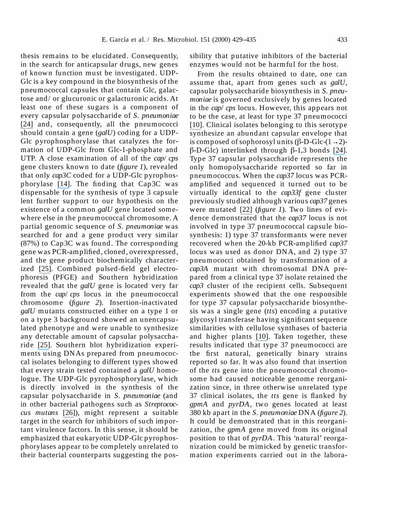
thesis remains to be elucidated. Consequently,in the search for anticapsular drugs, new genesof known function must be investigated. UDP-Glc is a key compound in the biosynthesis of thepneumococcal capsules that contain Glc, galac-tose and/or glucuronic or galacturonic acids. Atleast one of these sugars is a component ofevery capsular polysaccharide of S. pneumoniae[24] and, consequently, all the pneumococcishould contain a gene (galU) coding for a UDP-Glc pyrophosphorylase that catalyzes the for-mation of UDP-Glc from Glc-1-phosphate andUTP. A close examination of all of the cap/cpsgene clusters known to date (figure 1), revealedthat only cap3C coded for a UDP-Glc pyrophos-phorylase [14]. The finding that Cap3C wasdispensable for the synthesis of type 3 capsulelent further support to our hypothesis on theexistence of a common galU gene located some-where else in the pneumococcal chromosome. Apartial genomic sequence of S. pneumoniae wassearched for and a gene product very similar(87%) to Cap3C was found. The correspondinggene was PCR-amplified, cloned, overexpressed,and the gene product biochemically character-ized [25]. Combined pulsed-field gel electro-phoresis (PFGE) and Southern hybridizationrevealed that the galU gene is located very farfrom the cap/cps locus in the pneumococcalchromosome (figure 2). Insertion-inactivatedgalU mutants constructed either on a type 1 oron a type 3 background showed an unencapsu-lated phenotype and were unable to synthesizeany detectable amount of capsular polysaccha-ride [25]. Southern blot hybridization experi-ments using DNAs prepared from pneumococ-cal isolates belonging to different types showedthat every strain tested contained a galU homo-logue. The UDP-Glc pyrophosphorylase, whichis directly involved in the synthesis of thecapsular polysaccharide in S. pneumoniae (andin other bacterial pathogens such as Streptococ-cus mutans [26]), might represent a suitabletarget in the search for inhibitors of such impor-tant virulence factors. In this sense, it should beemphasized that eukaryotic UDP-Glc pyrophos-phorylases appear to be completely unrelated totheir bacterial counterparts suggesting the pos-
sibility that putative inhibitors of the bacterialenzymes would not be harmful for the host.
From the results obtained to date, one canassume that, apart from genes such as galU,capsular polysaccharide biosynthesis in S. pneu-moniae is governed exclusively by genes locatedin the cap/cps locus. However, this appears notto be the case, at least for type 37 pneumococci[10]. Clinical isolates belonging to this serotypesynthesize an abundant capsular envelope thatis composed of sophorosyl units (�-D-Glc-(1→2)-�-D-Glc) interlinked through �-1,3 bonds [24].Type 37 capsular polysaccharide represents theonly homopolysaccharide reported so far inpneumococcus. When the cap37 locus was PCR-amplified and sequenced it turned out to bevirtually identical to the cap33f gene clusterpreviously studied although various cap37 geneswere mutated [22] (figure 1). Two lines of evi-dence demonstrated that the cap37 locus is notinvolved in type 37 pneumococcal capsule bio-synthesis: 1) type 37 transformants were neverrecovered when the 20-kb PCR-amplified cap37locus was used as donor DNA, and 2) type 37pneumococci obtained by transformation of acap3A mutant with chromosomal DNA pre-pared from a clinical type 37 isolate retained thecap3 cluster of the recipient cells. Subsequentexperiments showed that the one responsiblefor type 37 capsular polysaccharide biosynthe-sis was a single gene (tts) encoding a putativeglycosyl transferase having significant sequencesimilarities with cellulose synthases of bacteriaand higher plants [10]. Taken together, theseresults indicated that type 37 pneumococci arethe first natural, genetically binary strainsreported so far. It was also found that insertionof the tts gene into the pneumococcal chromo-some had caused noticeable genome reorgani-zation since, in three otherwise unrelated type37 clinical isolates, the tts gene is flanked bygpmA and pyrDA, two genes located at least380 kb apart in the S. pneumoniae DNA (figure 2).It could be demonstrated that in this reorgani-zation, the gpmA gene moved from its originalposition to that of pyrDA. This ‘natural’ reorga-nization could be mimicked by genetic transfor-mation experiments carried out in the labora-
E. García et al. / Res. Microbiol. 151 (2000) 429–435 433

tory, although it was also observed that the ttsgene could also insert immediately downstreamof gpmA without a noticeable genome rear-rangement [10]. Interruption of the tts gene byinsertion-duplication mutagenesis resulted instrains unable to synthesize a type 37 capsule.Moreover, introduction of the tts gene clonedinto a suitable plasmid vector into differentstrains of S. pneumoniae or even in related spe-cies such as Streptococcus oralis rendered thesebacteria able to synthesize a type 37 capsularpolysaccharide, as demonstrated immunologi-cally (unpublished observations).
5. Concluding remarks
Much work has been carried out during thisdecade to decipher the molecular mechanismsunderlying the synthesis of the main virulencefactor of S. pneumoniae, the capsule. Neverthe-less, only a few gene products have been bio-chemically characterized and almost nothing isknown about aspects as important as regula-tion, transport and assembly of the polysaccha-ride chain subunits. It is generally thought thatthese polysaccharides are synthesized via lipid-linked repeat unit intermediates except in thecase of type 3 where capsule biosynthesis takesplace through the action of a processive syn-thase, the product of the cap3B gene [27]. Thisalso appears to be the case for type 37 capsulebut this is a peculiar case as the gene drivingcapsule formation is located far apart from thecap/cps locus. Whether type 37 pneumococcirepresent a unique case of capsular gene orga-nization or whether there are additional analo-gous examples in other serotypes remains to bedetermined. Furthermore, type 37 pneumococciare genetically binary and this property mightrepresent a selective advantage against theimmunological host defenses. Although cur-rently silent, the recipient cap37 locus mighteventually recover its capacity to synthesizetype 33F capsular polysaccharide, e.g. we canenvisage that transformation events involvingDNA fragments of the cap33f gene cluster wouldrestore to the wild-type genotype those genesmutated in cap37. De novo acquisition by S.
pneumoniae of a tts gene via genetic transforma-tion also appears to be a rather likely event. Theputative clinical relevance of genetically binarystrains as an alternative to the well-studiedphenomenon of capsule shifting [19] shouldalso be evaluated.
Acknowledgments
D. Llull was the recipient of a fellowship fromPrograma Sectorial de Formación de Profeso-rado Universitario y Personal Investigador (Min-isterio de Educación y Cultura). Research in ourlaboratory is supported by grant PB96-0809from the Dirección General de InvestigaciónCientífica y Técnica and the Programa de Coop-eración Científica con Iberoamérica from theSubdirección General de Cooperación Interna-cional.
References
[1] Griffith F., The significance of pneumococcal types, J. Hyg. 27 (1928)113–159.
[2] Briles D.E., Tart R.C., Swiatlo E., Dillard J.P., Smith P., Benton K.A.,Ralph B.A., Brooks-Walter A., Crain M.J., Hollingshead S.K.,McDaniel L.S., Pneumococcal diversity: considerations for newvaccine strategies with emphasis on pneumococcal surface proteinA (PspA), Clin. Microbiol. Rev. 11 (1998) 645–657.
[3] Scott J.A., Hall A.J., Dagan R., Dixon J.M., Eykyn S.J., Fenoll A.,Hortal M., Jetté L.P., Jorgensen J.H., Lamothe F., Latorre C., Macfar-lane J.T., Shlaes D.M., Smart L.E., Taunay A., Serogroup-specificepidemiology of Streptococcus pneumoniae: associations with age,sex, and geography in 7,000 episodes of invasive disease, Clin.Infect. Dis. 22 (1996) 973–981.
[4] Mäkelä P.H., Stocker B.A.D., Genetics of polysaccharide biosynthe-sis, Annu. Rev. Genet. 3 (1969) 291–322.
[5] García E., López R., Molecular biology of the capsular genes ofStreptococcus pneumoniae, FEMS Microbiol. Lett. 149 (1997) 1–10.
[6] García E., Llull D., López R., Functional organization of the genecluster involved in the synthesis of the pneumococcal capsule, Int.Microbiol. 2 (1999) 169–176.
[7] Yother J., in: Goldberg J.B. (Ed.), Genetics of Bacterial Polysaccha-rides, CRC Press, Boca Ratón, 1999, pp. 161–184.
[8] Paton J.C., Morona J.K., in: Fischetti V., Novick R., Ferreti J.,Portnoy D., Rood J. (Eds.), Gram-positive Pathogens, ASM Press,Washington, DC, 2000, pp. 201–213.
[9] Bernheimer H.P., Wermundsen I.E., Homology in capsular transfor-mation reactions in Pneumococcus, Mol. Gen. Genet. 116 (1972)68–83.
[10] Llull D., Muñoz R., López R., García E., A single gene (tts) locatedoutside the cap locus directs the formation of Streptococcus pneu-moniae type 37 capsular polysaccharide: type 37 pneumococci arenatural, genetically binary strains, J. Exp. Med. 190 (1999) 241–251.
[11] Ravin A.W., Linked mutations borne by deoxyribonucleic acidcontrolling the synthesis of capsular polysaccharide in pneumococ-cus, Genetics 45 (1960) 1387–1403.
434 E. García et al. / Res. Microbiol. 151 (2000) 429–435

[12] Arrecubieta C., López R., García E., Molecular characterization ofcap3A, a gene from the operon required for the synthesis of thecapsule of Streptococcus pneumoniae type 3: sequencing of muta-tions responsible for the unencapsulated phenotype and localiza-tion of the capsular cluster on the pneumococcal chromosome,J. Bacteriol. 176 (1994) 6375–6383.
[13] De Vuyst L., Degeest B., Heteropolysaccharides from lactic acidbacteria, FEMS Microbiol. Rev. 23 (1999) 153–177.
[14] Arrecubieta C., García E., López R., Sequence and transcriptionalanalysis of a DNA region involved in the production of capsularpolysaccharide in Streptococcus pneumoniae type 3, Gene 167 (1995)1–7.
[15] Morona J.K., Morona R., Paton J.C., Analysis of the 5′ portion of thetype 19A capsule locus identifies two classes of cpsC, cpsD, and cpsEgenes in Streptococcus pneumoniae, J. Bacteriol. 181 (1999)3599–3605.
[16] Muñoz R., García E., López R., Evidence for horizontal transfer fromStreptococcus to Escherichia coli of the kfiD gene encoding theK5-specific UDP-glucose dehydrogenase, J. Mol. Evol. 46 (1998)324–326.
[17] Muñoz R., Mollerach M., López R., García E., Characterization ofthe type 8 capsular gene cluster of Streptococcus pneumoniae,J. Bacteriol. 181 (1999) 6214–6219.
[18] Muñoz R., Mollerach M., López R., García E., Molecular organiza-tion of the genes required for the synthesis of type 1 capsularpolysaccharide of Streptococcus pneumoniae: formation of binaryencapsulated pneumococci and identification of cryptic dTDP-rhamnose biosynthesis genes, Mol. Microbiol. 25 (1997) 79–92.
[19] Coffey T.J., Enright M.C., Daniels M., Morona J.K., Morona R.,Hryniewicz W., Paton J.C., Spratt B.G., Recombinational exchangesat the capsular polysaccharide biosynthetic locus lead to frequentserotype changes among natural isolates of Streptococcus pneumo-niae, Mol. Microbiol. 27 (1998) 73–83.
[20] Ramirez M., Tomasz A., Molecular characterization of the complete23F capsular polysaccharide locus of Streptococcus pneumoniae,J. Bacteriol. 180 (1998) 5273–5278.
[21] Morona J.K., Miller D.C., Coffey T.J., Vindurampulle C.J.,Spratt B.G., Morona R., Paton J.C., Molecular and genetic charac-terization of the capsule biosynthesis locus of Streptococcus pneu-moniae type 23F, Microbiology 145 (1999) 781–789.
[22] Llull D., López R., García E., Muñoz R., Molecular structure of thegene cluster responsible for the synthesis of the polysaccharidecapsule of Streptococcus pneumoniae type 33F, Biochim. Biophys.Acta 1443 (1998) 217–224.
[23] Guidolin A., Morona J.K., Morona R., Hansman D., Paton J.C.,Nucleotide sequence analysis of genes essential for capsular polysac-charide biosynthesis in Streptococcus pneumoniae type 19F, Infect.Immun. 62 (1994) 5385–5396.
[24] van Dam J.E., Fleer A., Snippe H., Immunogenicity and immu-nochemistry of Streptococcus pneumoniae capsular polysaccharides,Antonie van Leeuwenhoek 58 (1990) 1–47.
[25] Mollerach M., López R., García E., Characterization of the galU geneof Streptococcus pneumoniae encoding a uridine diphosphoglucosepyrophosphorylase: a gene essential for capsular polysaccharidebiosynthesis, J. Exp. Med. 188 (1998) 2047–2056.
[26] Yamashita Y., Tsukioka Y., Nakano Y., Tomihisa K., Oho T., Koga T.,Biological functions of UDP-glucose synthesis in Streptococcusmutans, Microbiology 144 (1998) 1235–1245.
[27] Arrecubieta C., López R., García E., Type 3-specific synthase ofStreptococcus pneumoniae (Cap3B) directs type 3 polysaccharidebiosynthesis in Escherichia coli and in pneumococcal strains ofdifferent serotypes, J. Exp. Med. 184 (1996) 449–455.
[28] Iannelli F., Pearce B.J., Pozzi G., The type 2 capsule locus ofStreptococcus pneumoniae, J. Bacteriol. 181 (1999) 2652–2654.
[29] Morona J.K., Morona R., Paton J.C., Comparative genetics ofcapsular polysaccharide biosynthesis in Streptococcus pneumoniaetypes belonging to serogroup 19, J. Bacteriol. 181 (1999) 5355–5364.
[30] Gasc A.-M., Giammarinaro P., Ton-Hoang B., Geslin P., van derGiezen M., Sicard M., Structural organization of the Streptococcuspneumoniae chromosome and relatedness of penicillin-sensitive and-resistant strains in type 9V, Microb. Drug Resist. 3 (1997) 65–72.
E. García et al. / Res. Microbiol. 151 (2000) 429–435 435