Induction of Lytic Enzymes by the Interaction of Ustilago maydis with Zea mays Tissues

Book et al. Biotechnology for Biofuels 2014, 7:109http://www.biotechnologyforbiofuels.com/content/7/1/109
RESEARCH Open Access
Evolution of substrate specificity in bacterial AA10lytic polysaccharide monooxygenasesAdam J Book1,2†, Ragothaman M Yennamalli3,4†, Taichi E Takasuka1,3†, Cameron R Currie1,2,George N Phillips Jr1,3,4 and Brian G Fox1,3*
Abstract
Background: Understanding the diversity of lignocellulose-degrading enzymes in nature will provide insights forthe improvement of cellulolytic enzyme cocktails used in the biofuels industry. Two families of enzymes, fungal AA9and bacterial AA10, have recently been characterized as crystalline cellulose or chitin-cleaving lytic polysaccharidemonooxygenases (LPMOs). Here we analyze the sequences, structures, and evolution of LPMOs to understand thefactors that may influence substrate specificity both within and between these enzyme families.
Results: Comparative analysis of sequences, solved structures, and homology models from AA9 and AA10 LPMOfamilies demonstrated that, although these two LPMO families are highly conserved, structurally they have minimalsequence similarity outside the active site residues. Phylogenetic analysis of the AA10 family identified clades withputative chitinolytic and cellulolytic activities. Estimation of the rate of synonymous versus non-synonymous substitutions(dN/dS) within two major AA10 subclades showed distinct selective pressures between putative cellulolytic genes(subclade A) and CBP21-like chitinolytic genes (subclade D). Estimation of site-specific selection demonstrated thatchanges in the active sites were strongly negatively selected in all subclades. Furthermore, all codons in the subclade Dhad dN/dS values of less than 0.7, whereas codons in the cellulolytic subclade had dN/dS values of greater than 1.5.Positively selected codons were enriched at sites localized on the surface of the protein adjacent to the active site.
Conclusions: The structural similarity but absence of significant sequence similarity between AA9 and AA10 familiessuggests that these enzyme families share an ancient ancestral protein. Combined analysis of amino acid sites underDarwinian selection and structural homology modeling identified a subclade of AA10 with diversifying selection atdifferent surfaces, potentially used for cellulose-binding and protein-protein interactions. Together, these data indicatethat AA10 LPMOs are under selection to change their function, which may optimize cellulolytic activity. This workprovides a phylogenetic basis for identifying and classifying additional cellulolytic or chitinolytic LPMOs.
Keywords: Lytic polysaccharide monooxygenase, LPMO, Cellulase, Chitinase, Streptomyces, AA9, AA10, Enzymeevolution, Biofuels
BackgroundThe two most abundant polysaccharides in nature are cel-lulose and chitin [1]. Plants, insects, crustaceans, molluscs,and fungi all utilize these two highly stable polymers asprimary components of their cell walls. Deconstruction ofpolysaccharides is essential for ecosystem-level carbon
* Correspondence: [email protected]†Equal contributors1Department of Energy, Great Lakes Bioenergy Research Center, Madison,1552 University Avenue, Madison, WI 53726, USA3Department of Biochemistry, University of Wisconsin-Madison, BiochemistryAddition, 433 Babcock Dr., Madison, WI 53706, USAFull list of author information is available at the end of the article
© 2014 Book et al.; licensee BioMed Central LtCommons Attribution License (http://creativecreproduction in any medium, provided the orDedication waiver (http://creativecommons.orunless otherwise stated.
and nitrogen cycling. Moreover, polysaccharides are po-tential energy sources that could help supplement thecurrent massive demand for fossil fuels [2]. Intensive ef-forts worldwide focus on conversion of these energy-richbiomolecules into free sugars that can be fermented intobiofuels or other value-added bioproducts. However, hy-drolysis of these polymers is difficult due to their crystal-line structure, the stability of the β-glucosidic bond, andtheir close association with hemicellulose, lignin, andother modifying molecules [1,3]. Cellulolytic and chitino-lytic enzymes capable of this have been identified in amyriad of organisms, but most often in bacteria and fungi[4]. While the biochemical activities and mechanisms of
d. This is an Open Access article distributed under the terms of the Creativeommons.org/licenses/by/4.0), which permits unrestricted use, distribution, andiginal work is properly credited. The Creative Commons Public Domaing/publicdomain/zero/1.0/) applies to the data made available in this article,

Book et al. Biotechnology for Biofuels 2014, 7:109 Page 2 of 14http://www.biotechnologyforbiofuels.com/content/7/1/109
hydrolytic enzymes have been known for decades, oxyge-nolytic pathways for deconstruction of chitin and cellulosehave only recently been identified [5-8].CBH1, one of the first representatives of what are now
recognized to be lytic polysaccharides monooxygenases(LPMOs), was secreted by Streptomyces olivaceoviridisand interacted with α-chitin, but since it lacked classicalhydrolytic activity, it was thus considered to be a non-hydrolytic carbohydrate binding module (CBM) [9]. Anortholog of CBH1, chitin-binding protein 21 (CBP21) wasidentified in Serratia marcescens [10] and initially classi-fied as carbohydrate binding module 33 (CBM33, nowsystematically called Auxiliary Activity 10, AA10).a Thefunction of CBP21 was first demonstrated by Vaaje-Kolstad et al. [11], who showed cleavage of crystalline chi-tin in an O2-dependent reaction. Soon after this report,others showed that the eukaryotic counterpart, fungalglycoside hydrolase 61 (GH61, now systematically calledAuxiliary Activity 9, AA9) was a Cu2+-dependent enzyme[11-14]. An oxidative function has also been demonstratedfor CelS2, an AA10 from Streptomyces coelicolor [6],which reacts synergistically with hydrolytic cellobiohydro-lases and endoglucanases [15], and more recently forBlAA10A from Bacillus licheniformis and E8 from Ther-mobifida fusca [16] which react with chitin and cellulose,respectively, giving four AA10 enzymes whose functionhas been determined.AA9 and AA10 incorporate a single 18O from 18O2
into polysaccharide cleavage products, and so are nowclassified as LPMOs [11]. To date, structures of six AA9and five AA10 enzymes have been solved, including onenuclear magnetic resonance (NMR) structure [11,17-19].Overall, the two LPMO families share a conserved β-sandwich fold [11], and many residues on the substrate-binding surface are conserved. Moreover, Cu2+ has beenidentified in the active sites [8,17,18]. Although recentcomputational studies support the involvement of acopper-oxyl radical intermediate [20], the catalyticmechanism of this reaction is still largely unexplored.Oxidative polysaccharide cleavage results in the forma-
tion of an aldonic acid from C1 oxidation [21] or aketoaldose from C4 oxidation [21,22]. The monooxygen-ase reaction stoichiometry requires the addition of 2e−
from an oxidoreductase or other external electrondonor. The presence of oxidoreductases has been re-ported in various cellulolytic fungi [23], though an ac-tual, physiological electron partner for LPMOs has notbeen unambiguously determined.In this study, we compared amino acid sequences and
protein structures in order to explore the evolutionaryrelatedness of AA9 and AA10. Conserved sequence andstructural features were correlated with potential substrateinteractions and surfaces potentially used by electron do-nors. Phylogenetic analysis suggests that cellulose- and
chitin-specific enzymes are distributed into different sub-clades within bacterial AA10, as has been recently re-ported for the fungal AA9 [18,21]. Potential evolutionarilypressures within the AA10 family were examined in orderto understand how Darwinian selection might have influ-enced substrate specificity.
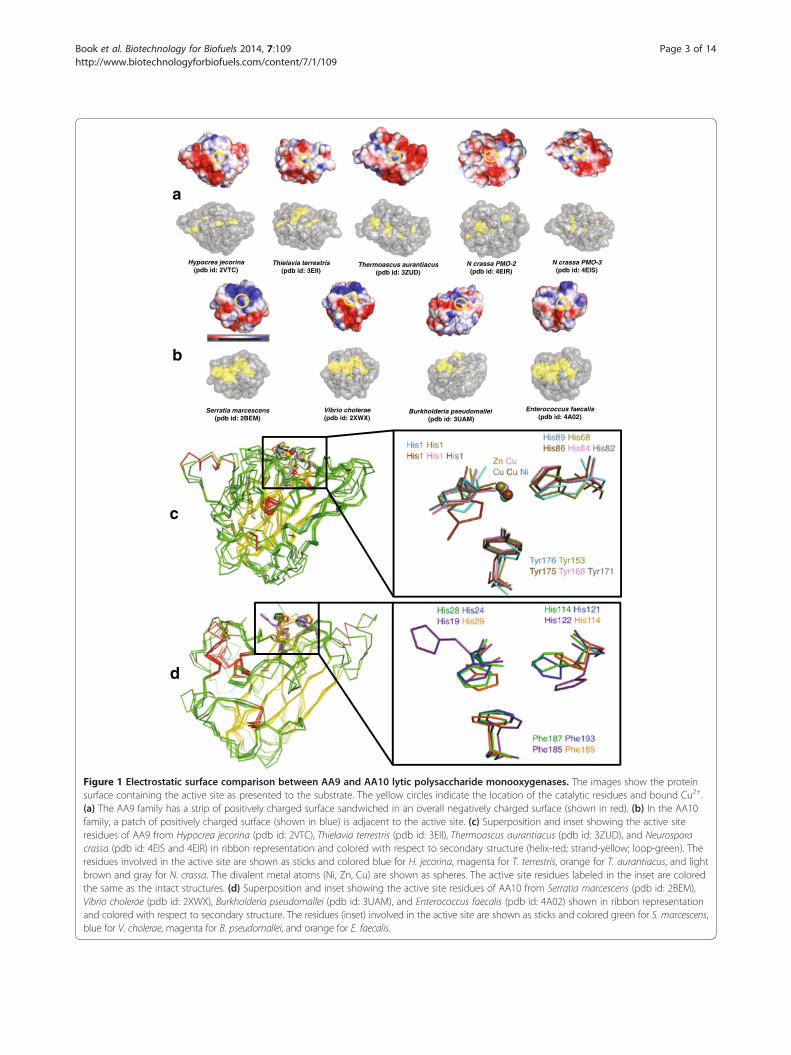
ResultsStructural comparison of LPMO families AA9 and AA10Figure 1a shows five crystal structures from the AA9family. These are from Hypocrea jecorina (Trichodermareesei, Protein Data Bank (pdb) id: 2VTC) [24], Thielaviaterrestris (pdb id: 3EII) [7], Thermoascus aurantiacus(pdb id: 2YET) [13], Neurospora crassa PMO-2 (pdb id:4EIR) [18], and N. crassa PMO-3 (pdb id: 4EIS) [18].Structures of four AA10 enzymes are also shown inFigure 1b. These are from S. marcescens (pdb id: 2BEM)[25], Vibrio cholerae O1 biovar EI Tor (pdb id: 2XWX)[26], Burkholderia pseudomallei (pdb id: 3UAM), andEnterococcus faecalis (pdb id: 4A02) [27]. Both AA9 andAA10 have a conserved β-sandwich fold with three tofour β-sheet strands (Figure 1c and 1d). The averageroot mean square (RMS) deviation of the aligned struc-tures is approximately 3 Å (Table 1) In addition to thefold-level similarity between AA9 and AA10, two keyhistidine (His) residues that coordinate a Cu2+ ion attheir active sites are also highly conserved in both families(Table 1 and Figure 1c). The structural superposition ofthe metal ligands suggests that this configuration is essen-tial for activity (inset in Figure 1c and 1d). A notable dif-ference between AA9 and AA10 is the third, non-coordinating active site residue; being primarily tyrosinein the former and primarily phenylalanine in the latter,with a relatively few exceptions presently also identified.Six AA10 structures from E. faecalis released in the
pdb show copper in the active site, and a recently pub-lished structure of AA9 from Phanerochaete chrysospor-ium (pdb id: 4B5Q) also shows copper bound in theactive site [28]. Copper binds with nanomolar affinity toAA10 [8,17]; its presence is consistent with O2 activationrequired for the LPMO reaction.
Surface electrostatic potential on the binding surfaces ofAA9 and AA10To explore factors that may contribute to substrate spe-cificity in the AA9 and AA10 families, we characterizedthe electrostatic potential present at the substrate-binding surface. In both families, the metal-binding his-tidine residues are part of a planar surface that consti-tutes the polysaccharide-binding surface [18]. Figure 1aand 1b show the surface electrostatic potential of repre-sentatives from both AA9 and AA10 families. For theAA9 proteins, which are biochemically characterized ascellulose monooxygenases, negatively charged residues
Serratia marcescens(pdb id: 2BEM)
Vibrio cholerae (pdb id: 2XWX)
Burkholderia pseudomallei (pdb id: 3UAM)
Enterococcus faecalis (pdb id: 4A02)
b
Hypocrea jecorina (pdb id: 2VTC)
Thielavia terrestris (pdb id: 3EII)
N crassa PMO-2 (pdb id: 4EIR)
N crassa PMO-3 (pdb id: 4EIS)
a
Thermoascus aurantiacus (pdb id: 3ZUD)
d
c
Figure 1 Electrostatic surface comparison between AA9 and AA10 lytic polysaccharide monooxygenases. The images show the proteinsurface containing the active site as presented to the substrate. The yellow circles indicate the location of the catalytic residues and bound Cu2+.(a) The AA9 family has a strip of positively charged surface sandwiched in an overall negatively charged surface (shown in red). (b) In the AA10family, a patch of positively charged surface (shown in blue) is adjacent to the active site. (c) Superposition and inset showing the active siteresidues of AA9 from Hypocrea jecorina (pdb id: 2VTC), Thielavia terrestris (pdb id: 3EII), Thermoascus aurantiacus (pdb id: 3ZUD), and Neurosporacrassa (pdb id: 4EIS and 4EIR) in ribbon representation and colored with respect to secondary structure (helix-red; strand-yellow; loop-green). Theresidues involved in the active site are shown as sticks and colored blue for H. jecorina, magenta for T. terrestris, orange for T. aurantiacus, and lightbrown and gray for N. crassa. The divalent metal atoms (Ni, Zn, Cu) are shown as spheres. The active site residues labeled in the inset are coloredthe same as the intact structures. (d) Superposition and inset showing the active site residues of AA10 from Serratia marcescens (pdb id: 2BEM),Vibrio cholerae (pdb id: 2XWX), Burkholderia pseudomallei (pdb id: 3UAM), and Enterococcus faecalis (pdb id: 4A02) shown in ribbon representationand colored with respect to secondary structure. The residues (inset) involved in the active site are shown as sticks and colored green for S. marcescens,blue for V. cholerae, magenta for B. pseudomallei, and orange for E. faecalis.
Book et al. Biotechnology for Biofuels 2014, 7:109 Page 3 of 14http://www.biotechnologyforbiofuels.com/content/7/1/109

Table 1 Structural homology of lytic polysaccharide monooxygenases
PDB ID % RMSD1 %id Source structure CAZy family Source organism Active site residues
2BEM 0 100 X-ray AA10 Serratia marcescens H28-H114-F187
2XWX 0.8 51 X-ray AA10 Vibrio cholerae H24-H121-F193
4A02 1 52 X-ray AA10 Enterococcus faecalis H29-H114-F185
2LHS 1.4 100 NMR AA10 Serratia marcescens H28-H114-F187
3UAM 1.4 39 X-ray AA10 Burkholderia pseudomallei H19-H122-F205
2VTC 3.2 9 X-ray AA9 Hypocrea jecorina H1-H89-Y176
4EIR 2.8 9 X-ray AA9 Neurospora crassa H1-H84-Y168
3ZUD 3.3 12 X-ray AA9 Thermoascus aurantiacus H1-H86-Y175
3EII 3.2 11 X-ray AA9 Thielavia terrestris H1-H68-Y153
4EIS 2.8 7 X-ray AA9 Neurospora crassa H1-H82-Y1711Root mean square deviation (RMSD) (%) for each structure from Protein Data Bank (pdb) compared to 2BEM determined by X-ray crystallography; %id indicatesthe percentage identity of each sequence to that of 2BEM. Three active site residues, His, His, and Phe/Tyr, are shown with residue numbers.
Book et al. Biotechnology for Biofuels 2014, 7:109 Page 4 of 14http://www.biotechnologyforbiofuels.com/content/7/1/109
(shown in red) prominently surround the active site(Figure 1a, yellow circle). In contrast, the AA10 chitinmonooxygenases contain both positively charged (shownin blue) and negatively charged residues (shown in red)surrounding the active site (Figure 1b, yellow circle).Aachmann et al. [17] used NMR to identify residuesfrom the chitinolytic AA10 enzyme from S. marcescens(pdb id: 2BEM) that are involved in chitin binding.These residues are Q53, Y54, E55, Q67, S58, L110, T111,A112, H114, and T116 [17]. The positions of the corre-sponding residues from the other AA10 enzymes thatalign with 2BEM are shown as yellow on a grey surfacein the lower parts of Figure 1a and 1b. In the othermembers of the AA10 family, most of these structurallyconserved residues are also surface-exposed (Figure 1b,bottom). However, in the AA9 family, only a few are ex-posed at the polysaccharide-binding surface (Figure 1a,bottom), indicating that different residues from thefolded structures will be involved in substrate binding inthe AA9 and AA10 families.
Diversity of domain structures in AA9 and AA10 proteinsAnother possible determinant of substrate specificitywithin the AA9 and AA10 families is the domain archi-tecture. LPMO enzymes have a diverse composition ofdomains: they can be single catalytic domains, associatedwith various CBMs, or even associated with other cata-lytic domains (such as glycoside hydrolase (GH) do-mains). Figure 2 shows a Cytoscape (The CytoscapeConsortium, San Diego, CA) protein sequence homologynetwork accounting for the variations in domains in theAA9 and AA10 families, where nodes represent enzymesor functional classes, and edges represent sequence simi-larity (bit score >200, evalue <1e−50). In order to preparethis network, sequences were collected from CAZy,compared via pairwise BLAST analysis, and then anno-tated with secondary CAZy domains. Nodes are colored
according to their phylum-level taxonomic identification.The network contains 184 AA9 sequences and 495AA10 sequences. All AA9 proteins were from eukary-otes, with a vast majority (99%) from the fungal phylaAscomycota (135 sequences) and Basidiomycota (34 se-quences). Of the protein sequence in the AA9 family,31% include a secondary carbohydrate binding module 1(CBM1), which has been reported to bind cellulose [29].Seven AA9 sequences are associated with CBM0, an un-classified CBM family [30,31].The AA10 family is exclusively from prokaryotes, with
226 sequences from Proteobacteria, 145 from Actinobac-teria, and 132 from Firmicutes (Figure 2). There were noedges linking members of the AA9 and AA10 families atthe similarity threshold of evalue <1e−50. Furthermore,when the similarity threshold was relaxed to 1e−5 therewere still no connections between the AA9 and AA10families. While Figure 2 shows that the AA9 networkcontains interspersed sequences from Ascomycota andBasidiomycota, the AA10 family shows clear taxonomicgroupings assembled from different bacterial phyla.These results also show that while the active site resi-dues of the AA9 and AA10 families are mostly con-served (Table 1), these two families do not share anyother significant sequence similarities or consistent link-ages to other domains.Figure 2 also shows that the AA10 family is combined
with a variety of secondary CBM domains, with 31% ofthe total sequences including cellulose-binding domainsCBM2 and CBM3 [32,33] or chitin-binding domainsCBM5 and CBM12 [34]. Further phylogenetic binning ofAA10 showed expansion within the genera of Streptomy-ces, Bacillus, and Vibrio (Additional file 1: Figure S1).Interestingly, 94% of the AA10 sequences that includeda cellulose-binding CBM were from the phylum Actino-bacteria, whereas 95% of sequences including a chitin-binding CBM were from the phyla Firmicutes and

0
_246
_267
_164
_420
_421
_446
_266_160
_135 _305_245
_357
_2
_308
_419
7_251
_45
CBM2
_331_416
_191
_287_443
CBM3_235
_194
_297 _482
_5_16_249
_242
_87
_93
_14
_110
_33_81
_91_278
_279
_299
_492
_99
_35_342 _456
_371
_472_321
_499_330
_44
_480
_444 _332
_49
_349
_295
_283
_385
_359
_155
_458
_174
_497
_1
_34_92_505
_241
_372_350_469
_101
_390
GH18
GH5GH44
_538 _625
_563
_532
_582
_593
_575
_558
_664
_546
_592
_633
_533
_515
_602
_657 _630
_589
_617
CBM0
_650
_550
_679
_545
_684
_540
_523
_628
_561
_641
_677
_124_88
_234
_106
0
_525
_539
_661_663
_616
_527_587
_674
_63
_666 _632
_596
_524
_691
_658
_667
_508
_635
_680
_612
_571
_689_685
_613
_554
_627
_605
_636_521
_652
_607
_513
_514_600
_567_624
_629
_551
_688
_656
_0_355
_185
_286
_288_285
_404
CBM12
CBM5
_496
_276
_90
_97_85
_394
_248
_193
_387
_71 _96
_74
_594
_683
_611_512
_614
_569
_568
_637
_522
_682
_601_659
_603
_553
_681
_669
_542
_552
_690
_623
_606
_595
_510
_675
_576
_686 _604
_676
_615
_608_655
_646
_649
_687
_588
_621
_562
_555
_543
_599
_642_597
_511
_678
_648_577
_626
_586
_509
_590
_565_526
CBM1
AscomycotaBasidiomycotaMetazoaUnknown
ActinobacteriaProteobacteriaFirmicutesUnknown
4
Figure 2 Domain and sequence similarity networks for the LPMO superfamily. Circles represent proteins from either the AA9 or AA10families, diamonds represent CAZy annotations. Edges represent either BLAST similarity with a bit score greater than 200 (evalue > e−50) orannotation to the indicated CAZy functional group. Colors represent taxonomic distribution of phyla of the source organisms. No sequencesimilarity above the indicated threshold was identified between the AA9 and AA10 superfamilies.
Book et al. Biotechnology for Biofuels 2014, 7:109 Page 5 of 14http://www.biotechnologyforbiofuels.com/content/7/1/109
Proteobacteria. Finally, two genes were identified thatalso encoded a glycoside hydrolase domain, suggesting arare but possibly synergistic pairing of glycoside hydro-lase and LPMO catalytic activities in a single enzyme.
Phylogenic analysis of LPMO familiesTo gain further insight into the evolutionary relationshipand possible functional roles of the distinct LPMO fam-ilies, we created phylogenetic trees representing the AA9and AA10 families (Figures 3 and 4, respectively). Briefly,sequences were collected, curated to remove redundantsequences with 100% identity, aligned, trimmed to theconserved catalytic domain, and then the tree was con-structed by MrBayes phylogenetic analysis [35]. Theresulting consensus tree was midpoint rooted and anno-tated with associated carbohydrate-binding modules inaddition to the AA9 or AA10 catalytic domains. The fivecrystal structures determined for AA9, 2YET, 2VTC,4EIS, 4EIR, and 3EII, were mapped onto the phylogen-etic tree. In Figure 3, the surfaces of these structures
have been colored to identify highly conserved residuesshared across the AA9 family. The tree was also anno-tated to indicate whenever a putative cellobiose dehydro-genase (AA3 family enzymes) was present in the hostgenome using a cutoff criterion of 35% identity to N.crassa CDH1. The ability of CDH to act as the proximalelectron donor for LPMO in cellulose oxidative cleavagehas been demonstrated in this organism [18,36-38].The AA9 LPMOs have been classified into four func-
tional types based on their reaction products [21]. Theseare shown in Figure 3 as red boxes. LPMO1 enzymes hy-droxylate the C1 position of pyranose rings and producean aldonolactone [18,21], while LPMO2 enzymes hy-droxylate the C4 position of pyranose rings and producea 4-ketoaldose [21,22]. LPMO3 enzymes are less specific[13,19,21,39], and produce both aldonolactone and non-reducing end oxidized products, while LPMO3* produceonly aldonic acids [21].Mapping of the four LPMO subgroups onto the global
AA9 phylogeny showed that the LPMO2, LPMO3, and

Figure 3 (See legend on next page.)
Book et al. Biotechnology for Biofuels 2014, 7:109 Page 6 of 14http://www.biotechnologyforbiofuels.com/content/7/1/109

(See figure on previous page.)Figure 3 Phylogenetic analysis of the AA9 LPMO superfamily. MrBayes phylogenetic tree for 254 AA9 protein sequences. The tree wasgenerated using the catalytic domain of the AA9 protein only. Additional carbohydrate-binding domains that are present in the full proteinsequence are indicated in the CBM column, but were not included in the calculation of the tree structure. Source organisms were searched forthe presence of a homolog to Neurospora crassa cellobiose dehydrogenase (CDH1). Protein identity scores are indicated in the CDH column, andcolors range from 30% identity (green) to 100% identity (red). Solved structures have been mapped onto the tree and colors represent conservation ofresidues across the whole AA9 family.
Book et al. Biotechnology for Biofuels 2014, 7:109 Page 7 of 14http://www.biotechnologyforbiofuels.com/content/7/1/109
LPMO3* subgroups are monophyletic, with each havinga single phylogenetic clade that corresponds to distinctfunctional classes (red boxes). In contrast, LPMO1 en-zymes span a major evolutionary division as two branchescross into this functional class, indicating more sequencediversity in the LPMO1 family. Examples where all fourLPMO functional types were fused to additional CBMdomains are identified in Figure 3. Moreover, Figure 3also shows that the majority of AA9 proteins come fromorganisms that also contain a cellobiose dehydrogenasehomolog.The AA10 phylogenetic tree was generated in a similar
manner using the catalytic domains of all non-redundantsequences present in the CAZy database. The AA10 treeshown in Figure 4 represents 374 non-redundant se-quences that are entirely bacterial in origin. The tree wasannotated with secondary CBM domains (central col-umn), and divided into two major clades (clade I and cladeII) that could be subdivided into four additional subclades(A through D). The biochemically characterized cellulose-oxidizing LPMOs from S. coelicolor (A3) and T. fusca arepresent in subclade A,b while all other LPMOs with ex-perimental confirmation of their reaction with chitin arepresent in subclades C and D [6].The tree was also anno-tated with microarray-based gene expression data for thesix variants of AA10 present in Streptomyces sp. SirexAA-E (SirexAA-E) [40]. Clade I contains a delineated mixtureof phyla, with subclade C containing sequences only fromActinobacteria and with subclade D containing sequencesfrom Firmicutes and Proteobacteria. Clade II is primarilycomposed of Actinobacteria and separates into subcladesA and B. Subclades A and B contain only cellulose-binding CBMs (CBM2 and CBM3) associated with thecatalytic AA10 domain, whereas subclades C and D con-tain only chitin-binding CBMs (CBM5 and 12). Further-more, expression data from SirexAA-E shows that genesfrom subclades A and B were selectively upregulated onlywhen cells were grown in medium containing cellulose asthe sole carbon source, while genes from subclade C wereupregulated only during growth on chitin [40].The cellulose-oxidizing LPMOs from AA10 are pri-
marily present in Actinobacteria, an aerobic filamentousbacterial phyla found in soil, but also associated with in-sects and other animals [40]. In Figure 4, the structuresof four AA10 enzymes are mapped to the phylogenetictree: 3UAM, 4A02, 2BEM, and 2XWX. Additionally,
predicted protein structures for expressed AA10 fromSirexAA-E are mapped onto the tree. There is highamino-acid sequence identity among the AA10 proteinswhose structures have been determined, with the highestsequence conservation observed at the active site (ma-genta color). Interestingly, homology models consistentlypredict an additional surface exposed loop region on thesame side of the protein as the active site in clade II pro-teins (chitin oxidation), but not in clade I (cellulose oxi-dation). The position of this loop can be recognized inpdb id: 4GBO, the E7 enzyme from T. fusca [16]. Re-cently, Vu et al. have identified a role for these extraloops in substrate recognition and control of specificityof reaction in the AA9 family [21].
Homology modeling of AA10 proteins and conservedsequence motif in LPMOsSeveral LPMOs from within the AA10 family have beenexperimentally verified to be either chitin or cellulosemonooxygenases (such as CBP21 and BlAA10A) whichreact with chitin, and CelS2 and E8, which react withcellulose [6,25].b To further explore structural determi-nants that control substrate specificity, we comparedhomology models for 43 proteins that spanned theAA10 family (Figure 5) across the clade I and clade II se-quences shown in Figure 4. Homology modeling usingI_TASSER [41], followed by superposition of the mod-eled structures showed that the most significant struc-tural differences were located in the substrate bindingregion (Figure 5, Additional file 2: Table S1). Specifically,the positions of loops (shown for illustration purposesonly) on the substrate-binding side of the protein hadmore variations than other parts of the modeled struc-tures. Correspondingly, the insertion observed in the se-quence alignments mapped to loops on the substrate-binding side of the AA10 family. Given the structuralvariability of clades I and II, and differences in measuredcatalytic functions, it is likely that these structural differ-ences help to modulate substrate selectivity between chi-tin and cellulose in AA10, as now predicted for AA9.To improve our mapping of potential functional deter-
minants onto the modeled structures, Multiple EM forMotif Elicitation (MEME) [42] was used. This approachidentified three sequence motifs among the 43 AA10proteins (Figure 5). These motifs were mapped backonto the structures and homology models. Simultaneously,

Figure 4 (See legend on next page.)
Book et al. Biotechnology for Biofuels 2014, 7:109 Page 8 of 14http://www.biotechnologyforbiofuels.com/content/7/1/109

(See figure on previous page.)Figure 4 Phylogenetic analysis of the AA10 LPMO superfamily. MrBayes phylogenetic tree for the 374 AA10 protein sequences. The tree wasgenerated using the catalytic domain of the AA10 protein only. Additional carbohydrate-binding domains that are present in the full proteinsequence are indicated in the CBM column, but were not included in the calculation of the tree structure. Solved structures have been mappedonto the tree and colors represent conservation of residues across the whole AA9 family, and in the three modeled structures for Streptomycessp. SirexAA-E and AA10 enzymes. Gene expression data for the six AA10 isoforms from SirexAA-E showing fold change in transcripts from glucosegrown cells to either cellulose or chitin grown cells.
Book et al. Biotechnology for Biofuels 2014, 7:109 Page 9 of 14http://www.biotechnologyforbiofuels.com/content/7/1/109
MEME was used to determine whether there were signifi-cant motifs observed in the published structures of AA9(Figure 5).In the homology-modeled proteins (shown in Figure 5
corresponding to the four AA10 clades shown inFigure 4), the three MEME motifs ranged from about 25to 41 residues in length. Motif1 was present in both
d
c
e
a
subclade Bsubclade AMotif location
2BEM3UAM4A022XWX3EII2YET2VTC
b AA10
AA9
Figure 5 Three Multiple Em for Motif Elicitation (MEME) motifs mappmotifs from four separate subclades A to D from the phylogenetic tree of Aof sequences in each clade is as follows: subclade A (68 sequences), cladeThese motifs were mapped to the structures that were predicted using iter(a). Motif1 is shown in cyan color, motif2 is shown in blue and motif3 is shavailable crystallographic structures of AA10 and AA9, showing the distribu(cyan), (d) motif2 (blue), and (e) motif3 (red) in the available structures.
AA9 and AA10, and contained the variable insertion re-gions that possibly yield substrate selectivity in theAA10 family. Motif2 and motif3 were observed only inAA10 (Figure 5b). It is interesting to note that the differ-ence in the number of motifs identified in AA9 as com-pared to AA10 provides an additional line of evidencesupporting the possibility of evolutionary selection in
motif 1E-value - 1.8e-26
subclade C subclade D
motif 1
motif 2
motif 3
motif 2E-value - 5.7e-26
motif 3E-value - 7.2e-6
ed to the predicted structures of AA10. MEME was used to identifyA10. The loops are shown for illustration purposes only. The numberB (29 sequences), clade C (77 sequences), and clade D (122 sequences).ative threading assembly refinement algorithm (I-TASSER), as shown inown in red. (b) MEME motifs mapped to the sequences from thetion of three motifs in the sequence. Sequence logo of (c) motif1
Book et al. Biotechnology for Biofuels 2014, 7:109 Page 10 of 14http://www.biotechnologyforbiofuels.com/content/7/1/109
these two families. Although experimental evidence as towhat these motifs (Figure 5c-e) contribute is currently lack-ing, it is clear from the superposition of the homology-modeled structures (Figure 5, cyan sequences) that motif1is well-positioned to play a role in substrate binding anddiscrimination between binding to chitin or cellulose.Interestingly, motif2 and motif3 span the breadth of theprotein and connect the substrate-binding surface to theopposite side of the protein where potential electrondonor proteins might interact.
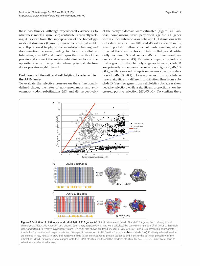
Evolution of chitinolytic and cellulolytic subclades withinthe AA10 familyTo evaluate the selective pressure on these functionallydefined clades, the rates of non-synonymous and syn-onymous codon substitutions (dN and dS, respectively)
AA10 subclade D
AA10 subclade A
0
0.1
0.2
0.3
0.4
0.5
0.6
0.7
0.8
0 0.2 0.4 0.6 0.8
Non
-syn
onym
ous
mut
atio
n ra
te (d
N)
Synonymous mutation r
b
a
c
Figure 6 Evolution of chitinolytic and cellulolytic AA10 genes. (a) Plotchitinolytic clades, clade A (circles) and clade D (diamonds), respectively. Vaclade and filtered to remove insignificant values (see text). Also shown arethresholds for positive and negative selection. Site-specific estimation of dNare colored in red, neutral in grey, and negative in blue (x-axis correspondsestimation). dN/dS ratios were also mapped onto the CBP21 structure 2BEMselection rates described above.
of the catalytic domain were estimated (Figure 6a). Pair-wise comparisons were performed against all geneswithin either subclade A or subclade D. Estimations withdN values greater than 0.01 and dS values less than 1.5were reported to allow sufficient mutational signal andto avoid the effect of back mutations that would artifi-cially increase dS and reduce dN with increased se-quence divergence [43]. Pairwise comparisons indicatethat a group of the chitinolytic genes from subclade Dare primarily under negative selection (Figure 6, dN/dS<0.2), while a second group is under more neutral selec-tion (1 > dN/dS >0.2). However, genes from subclade Ahave a significantly different distribution than from sub-clade D. Very few genes from cellulolytic subclade A shownegative selection, while a significant proportion show in-creased positive selection (dN/dS >1). To confirm these
SACTE_3159
1 1.2 1.4 1.6
ate (dS)
AA10 subclade D
AA10 subclade A
dN/dS = 1
dN/dS = 0.2
CBP21 - 2bem
of pairwise estimated dN and dS for genes from cellulolytic andlues were calculated by pairwise comparison of all genes within eachtrend lines for dN/dS ratios of 1 and 0.2, representing approximate/dS ratios for clade A (b) and clade D (c). Positively selected residuesto protein sequence and y-axis to the posterior probability of the, and the modeled structure for SACTE_3159. Colors correspond to
Book et al. Biotechnology for Biofuels 2014, 7:109 Page 11 of 14http://www.biotechnologyforbiofuels.com/content/7/1/109
results, site-specific dN/dS values were estimated for sub-clades A and D (Figure 6b). The results show that a sig-nificant number of residues in subclade A were indeedpositively selected, while residues in subclade D were allnegatively or neutrally selected. When plotted on theprotein structures, the negatively selected sites in bothsubclades A and D are primarily located around theactive-site residues. In contrast, most of the positively se-lected residues in subclade A are surface exposed, includ-ing regions on the putative substrate-binding surface andalong the interior of the protein traversing from thesubstrate-binding surface to the opposite surface of theprotein. Interestingly, this latter region may provide a sur-face for interaction with accessory redox proteins such ascellobiose dehydrogenase (AA3 enzymes).
DiscussionIn this study, we analyzed the AA9 and AA10 familiesusing available protein structures and sequence informa-tion to evaluate differences between and within the fam-ilies, to explore features that influence substrate specificity,and to characterize selective pressures that may have ledto functional diversification.LPMOs share a common structural fold and a spatial
conservation of active site residues, as seen by their lowroot mean square deviation (RMSD) values (ranging upto 3.3 Å, Table 1). While the core structural folds andthe active site geometry of these two LPMO families aresimilar, there is low homology at the amino-acid se-quence level, and the surface electrostatic potentials atthe substrate-binding surface show considerable differ-ences in charge distributions. Indeed, comparison of allAA9 and AA10 proteins available in the CAZy databasefailed to identify any sequences from across these twofamilies that have significant homology (evalue <1e−5).Our results indicate that although AA9 and AA10 familiesshare structural similarities, they have so significantly di-verged from a common ancestor that the only residue-level homology that remains is in the active site residues.Due to the low sequence similarity between AA9 and
AA10 families we analyzed their phylogenetic relation-ships separately. The AA9 phylogenetic tree is separatedinto three major evolutionarily related groups which par-tially correspond to the four types of enzyme activity ob-served for LPMOs [21]. LPMO2, LPMO3, and LPMO3*enzyme activities correspond to monophyletic clades,which suggests vertical inheritance and conserved en-zyme functions within each clade. In contrast, LPMO1enzymes are present in a polyphyletic clade, indicating amore diverse sequence space and potentially varied enzymefunction. Sequences from Ascomycetes and Basidiomy-cetes are scattered throughout the three major evolutionar-ily related groups in AA9, suggesting an ancestral sequencethat was shared before these two phyla separated.
The AA10 phylogenetic tree was separated into twomajor phylogenetic groups. When annotated with knownactivities, the two clades appear to separate enzymeswith different substrate specificities. Clade I contains allbiochemically defined chitin monooxygenases, whileclade II contains subclades that are either cellulose orchitin monooxygenases. Gene expression data fromSirexAA-E grown on either chitin or cellulose as the solecarbon source further corroborates this assessment [40].We also observed that CBM domain composition variesbetween clade I and II. Clade I is dominated by CBM5and 12 domains, which are primarily chitin binding, butpossibly can have a lignin-binding function as well [44].Clade II is enriched in CBM2 domains, which are pri-marily associated with cellulose binding. Most recently,Forsberg et al. showed the binding specificity of CelS2either with or without the associated CBM2 domain [16].Interestingly, although this CBM2 domain was tightlybound to either α or β-chitin, the corresponding AA10domain (CelS2) only reacted with cellulose. Further bio-chemical verification will be necessary to extend these ob-servations more broadly into phylogenetic space.To identify sequence and structural features that may
contribute to clade II activity against cellulose, we gener-ated homology-modeled structures for 43 sequencesthat span the phylogeny in AA10. Using MEME, thesehomology-modeled structures were identified to havethree highly significant motifs, where motif1 shows thelargest structural variability. Specifically, this variablemotif is contained in a loop of un-modeled sequence atthe substrate-binding surface and is only found in sub-clade A. Subclade A of AA10 contains biochemically char-acterized cellulose monooxygenases, and also contains themost highly upregulated AA10 enzyme when SirexAA-Eis grown on cellulose [40]. We hypothesize that this add-itional sequence at the binding surface is a defining featureof cellulose-active AA10 enzymes, paralleling the identifi-cation of a loop-modulating reaction specificity in theAA9 enzymes [21]. Motif2 and motif3, which span thebreadth of the protein, connect the substrate-binding sur-face to the opposite side of the protein. This suggests apossibility for modulation of electron donor interactions.Finally, we explored the selective pressures within two
clades of the AA10 family to understand how diversifica-tion may be distributed in this enzyme family. The resultsshow that chitinolytic enzymes in subclade D (chitinolyticenzymes) have mostly negative selection at both the wholegene and site-specific levels. In contrast, subclade A (bothchitinolytic and cellulolytic enzymes) contains more geneswith diversifying selection at both the whole gene andsite-specific levels. This result indicates that subclade Amay have undergone a change in substrate specificity andthat genes within this clade are potentially being selectedfor increased activity. Together, these data suggest that the

Book et al. Biotechnology for Biofuels 2014, 7:109 Page 12 of 14http://www.biotechnologyforbiofuels.com/content/7/1/109
ancestral form of AA10 may have been a chitin mono-oxygenase, and that clade II has apparently further spe-cialized for cellulose oxidation. Selection may be towardsmore favorable substrate binding, better interactions withaccessory redox proteins, such as cellobiose dehydrogen-ase enzymes, or perhaps both.
ConclusionsIn summary, this study provides a better understandingof the evolution of functional diversity within the re-cently discovered AA9 and AA10 LPMO families. To-gether, these data suggest that AA9 and AA10 familiesshare a distant common ancestor. Furthermore, cladeswithin the AA10 family are specialized for different sub-strates and subclade A has undergone diversifying selec-tion at surface-exposed regions of the protein.
Materials and methodsSequence similarity networkAA9 and AA10 protein-coding sequences were identi-fied on the Carbohydrate-Active Enzyme (CAZy) data-base [45], and harvested from the National Center forBiotechnology Information (NCBI) protein database. AllAA9 and AA10 sequences were compared against eachother using BLAST [46] to identify similar proteins. Allsequences were also re-annotated with CAZy families toidentify the domain structure of each protein. This datawas then used to build a similarity network using Cytos-cape 2.8.0 [47], and visualized as an organic layout.Nodes in the network represent unique protein se-quences and CAZy families. Edges represent a BLASTbit score of ≥200 (evalue ≥1 × e−50), or an annotation toa CAZy category. Nodes were annotated with taxonomicinformation at the phylum level.
Phylogenetic tree constructionAA9 and AA10 phylogenetic trees were constructed byfirst identifying proteins from the CAZy database, andthe harvesting sequence from NCBI. Sequences from ei-ther AA9 or AA10 families were aligned using MultipleSequence Comparison by Log-Expectation (MUSCLE)on the Cyberinfrastructure for Phylogenetic Research(CIPRES, https://www.phylo.org/portal2/login!input.action)Science Gateway [48]. Aligned sequences were thentrimmed to retain only the AA9 or AA10 domain; se-quences lacking the conserved active site His residueswere removed from the alignment. Phylogenetic treeswere generated using MrBayes code with a calculatedstandard deviation of ≤0.05. Non-default parameters wereset to mcmc, ngen = 10,000,000, temp = 0.200, burninfrac =0.25, stoprule = No, sump burnin = 4000, and sumt bur-nin = 4000. Resulting trees were annotated with pfam,phyla, solved structures, and cellobiose dehydrogenasehomolog information.
Evolutionary rate estimationCoding sequences for subclades A-D from the AA10family were collected and codon alignments were gener-ated with MUSCLE. Sequences were trimmed to retainonly the AA10 domain. Codon alignments were maskedwith Zorro (http://phylogenomics.wordpress.com/software/zorro/) to generate quality scores for codon positions [49],and then a phylogenetic tree was generated with RAxML(http://sco.h-its.org/exelixis/web/software/raxml/index.html) using the masking scores [50]. Pairwise codonsubstitution models (dN/dS values) were estimated usingthe CODEML program in the PAML package [51,52].Variables were set at CodonFreq = 0 and model = 0. Onlypairwise dN values with values ≥0.01 and dS values ≤1.5were reported so as to allow for sufficient mutational signaland to avoid the effects of back mutations. Site-specificcodon substitution models were generated using theCODEML program in PAML, with model = 0, NSsites = 3,ncatG = 3 fix_kappa = 0, fix_omega = 0, cleandata = 1, andfix_blength = 2.
Protein three-dimensional structure comparisonThe Dali protein structure alignment database (http://ekhidna.biocenter.helsinki.fi/dali_server/) was used to cal-culate %RMSD and %ID of LPMO enzymes whose struc-tures are known using 2BEM as a query [53]. Structureswith the ten best%RMSD are shown in Table 1.
Homology modelingThe 43 sequences highlighted in Figure 4 were consideredfor prediction of three-dimensional structures using itera-tive threading assembly refinement algorithm (I-TASSER)(http://zhanglab.ccmb.med.umich.edu/I-TASSER/) [41].For each sequence, the signal peptides and other domainsbesides the Cu2+-binding catalytic domain were includedin the homology modeling. Alignments used for modelingare tabulated in Additional file 2: Table S1. Models ob-tained with the highest C-score were retained for furtheranalysis. The homology models have been deposited atModel Archive (doi:10.5452/ma-asp8e) [54].
Structural analysisStructural comparisons were done using the Combina-torial Extension algorithm [55] implemented in PyMOL(Schrödinger, Portland, OR). Protein surface electrostat-ics calculations were carried using Adaptive Poisson-Boltzmann Solver (APBS) [56], where an externallygenerated pdb (P) file with per-atom charge (Q) and ra-dius (R) (PQR file) file was used to calculate the electro-statics. The parameters used were solvent and proteindielectrics of 78.0 and 2.0 respectively, solvent radius of1.4, and a monovalent ion concentration of 0.15 M. Thevisualization was depicted in PyMOL with positive andnegative molecular surface ranging from -2kT/e to 2kT/e.

Book et al. Biotechnology for Biofuels 2014, 7:109 Page 13 of 14http://www.biotechnologyforbiofuels.com/content/7/1/109
Motif identificationSequence-based motifs were identified using Multiple Emfor Motif Elicitation (MEME) (http://meme.nbcr.net/meme/) [42]. The occurrence of motifs in the sequencewas assumed to be distributed either zero or one per se-quence. Three motifs were identified for each set of se-quences given. The phylogenetic tree of AA10 was dividedinto four subclades (A to D) based on major phylogeneticclades. For each clade, the motifs were identified usingMEME.
EndnotesaCBM33 has recently been renamed as AA10; likewise
GH61 has been renamed as AA9 [11]. These names willbe used throughout.
bSACTE_3159 from the highly cellulolytic Streptomycessp. SirexAA-E, has also been confirmed to contain Cu2+
and have O2-dependent cellulose oxidation activity (M.Mbughuni and BG Fox, unpublished data).
Additional files
Additional file 1: Figure S1. Taxonomic diversity of AA10 sequences.AA10 sequences were collected from the CAZy database (608 sequences)and binned into taxonomic categories based on phylum and genus.Three phyla present were Proteobacteria, Firmicutes, and Actinobacteria(central pie chart). Smaller peripheral charts identify the number ofsequences within each genus.
Additional file 2: Table S1. Sequence alignments used for structuralmodeling.
AbbreviationsAA: Auxiliary activity; CBM: Carbohydrate-binding module; LPMO: Lyticpolysaccharide monooxygenases; GH: Glycoside hydrolase.
Competing interestsThe authors declare that they have no competing interests.
Authors’ contributionsAJB, RMY, and TET, designed and performed the experiments. All authors reviewed,discussed, and interpreted the results. AJB, RMY, TET, GNP, CRC, and BGF wrote andreviewed the manuscript. All authors read and approved the final manuscript.
AcknowledgementsThis work was funded by the Department of Energy Great Lakes BioenergyResearch Center (DOE BER Office of Science DE-FC-02-07ER64494), theDepartment of Energy Bringing Advanced Computational Techniques toEnergy Research program (DOE BACTER DE-FG02-04ER25627), the NationalInstitute of Health Protein Structure Initiative (U01 GM098248), and theNational Science Foundation GRAPE (CMMI-0941013).
Author details1Department of Energy, Great Lakes Bioenergy Research Center, Madison,1552 University Avenue, Madison, WI 53726, USA. 2Department ofBacteriology, University of Wisconsin-Madison, Microbial Sciences Building,1550 Linden Dr., Madison, WI 53706, USA. 3Department of Biochemistry,University of Wisconsin-Madison, Biochemistry Addition, 433 Babcock Dr.,Madison, WI 53706, USA. 4Current address: Biosciences at Rice, RiceUniversity, George R. Brown Hall, Houston, TX 77005, USA.
Received: 14 March 2014 Accepted: 7 July 2014Published: 6 August 2014
References1. Himmel ME, Ding SY, Johnson DK, Adney WS, Nimlos MR, Brady JW, Foust
TD: Biomass recalcitrance: engineering plants and enzymes for biofuelsproduction. Science 2007, 315:804–807.
2. Faaij APC: Bio-energy in Europe: changing technology choices. EnergPolicy 2006, 34:322–342.
3. Wilson DB: Microbial diversity of cellulose hydrolysis. Curr Opin Microbiol2011, 14:259–263.
4. Lynd LR, Weimer PJ, van Zyl WH, Pretorius IS: Microbial celluloseutilization: fundamentals and biotechnology. Microbiol Mol Biol Rev 2002,66:506–577. Table of contents.
5. Culpepper MA, Rosenzweig AC: Architecture and active site of particulatemethane monooxygenase. Crit Rev Biochem Mol Biol 2012, 47:483–492.
6. Forsberg Z, Vaaje-Kolstad G, Westereng B, Bunaes AC, Stenstrom Y, MacKenzieA, Sorlie M, Horn SJ, Eijsink VG: Cleavage of cellulose by a CBM33 protein.Protein Sci 2011, 20:1479–1483.
7. Harris PV, Welner D, McFarland KC, Re E, Navarro Poulsen JC, Brown K, SalboR, Ding H, Vlasenko E, Merino S, Xu F, Cherry J, Larsen S, Lo Leggio L:Stimulation of lignocellulosic biomass hydrolysis by proteins of glycosidehydrolase family 61: structure and function of a large, enigmatic family.Biochemistry 2010, 49:3305–3316.
8. Hemsworth GR, Taylor EJ, Kim RQ, Gregory RC, Lewis SJ, Turkenburg JP,Parkin A, Davies GJ, Walton PH: The copper active site of CBM33polysaccharide oxygenases. J Am Chem Soc 2013, 135:6069–6077.
9. Schnellmann J, Zeltins A, Blaak H, Schrempf H: The novel lectin-like proteinCHB1 is encoded by a chitin-inducible Streptomyces olivaceoviridis geneand binds specifically to crystalline alpha-chitin of fungi and otherorganisms. Mol Microbiol 1994, 13:807–819.
10. Suzuki K, Suzuki M, Taiyoji M, Nikaidou N, Watanabe T: Chitin bindingprotein (CBP21) in the culture supernatant of Serratia marcescens 2170.Biosci Biotechnol Biochem 1998, 62:128–135.
11. Vaaje-Kolstad G, Westereng B, Horn SJ, Liu Z, Zhai H, Sorlie M, Eijsink VG: Anoxidative enzyme boosting the enzymatic conversion of recalcitrantpolysaccharides. Science 2010, 330:219–222.
12. Levasseur A, Drula E, Lombard V, Coutinho PM, Henrissat B: Expansion ofthe enzymatic repertoire of the CAZy database to integrate auxiliaryredox enzymes. Biotechnol Biofuels 2013, 6:41.
13. Quinlan RJ, Sweeney MD, Lo Leggio L, Otten H, Poulsen JC, Johansen KS,Krogh KB, Jorgensen CI, Tovborg M, Anthonsen A, Tryfona T, Walter CP,Dupree P, Xu F, Davies GJ, Walton PH: Insights into the oxidativedegradation of cellulose by a copper metalloenzyme that exploitsbiomass components. Proc Natl Acad Sci U S A 2011, 108:15079–15084.
14. Westereng B, Ishida T, Vaaje-Kolstad G, Wu M, Eijsink VG, Igarashi K,Samejima M, Stahlberg J, Horn SJ, Sandgren M: The putative endoglucanasePcGH61D from Phanerochaete chrysosporium is a metal-dependentoxidative enzyme that cleaves cellulose. PLoS One 2011, 6:e27807.
15. Moser F, Irwin D, Chen S, Wilson DB: Regulation and characterization ofThermobifida fusca carbohydrate-binding module proteins E7 and E8.Biotechnol Bioeng 2008, 100:1066–1077.
16. Forsberg Z, Rohr AK, Mekasha S, Andersson KK, Eijsink VG, Vaaje-Kolstad G,Sorlie M: Comparative study of two chitin-active and two cellulose-activeAA10-type lytic polysaccharide monooxygenases. Biochemistry 2014,53:1647–1656.
17. Aachmann FL, Sorlie M, Skjak-Braek G, Eijsink VG, Vaaje-Kolstad G: NMRstructure of a lytic polysaccharide monooxygenase provides insight intocopper binding, protein dynamics, and substrate interactions. Proc NatlAcad Sci U S A 2012, 109:18779–18784.
18. Li X, Beeson WT, Phillips CM, Marletta MA, Cate JH: Structural basis forsubstrate targeting and catalysis by fungal polysaccharidemonooxygenases. Structure 2012, 20:1051–1061.
19. Phillips CM, Beeson WT, Cate JH, Marletta MA: Cellobiose dehydrogenaseand a copper-dependent polysaccharide monooxygenase potentiatecellulose degradation by Neurospora crassa. ACS Chem Biol 2011,6:1399–1406.
20. Kim S, Stahlberg J, Sandgren M, Paton RS, Beckham GT: Quantummechanical calculations suggest that lytic polysaccharidemonooxygenases use a copper-oxyl, oxygen-rebound mechanism. ProcNatl Acad Sci U S A 2014, 111:149–154.
21. Vu VV, Beeson WT, Phillips CM, Cate JHD, Marletta MA: Determinants ofregioselective hydroxylation in the fungal polysaccharidemonooxygenases. J Am Chem Soc 2014, 136:562–565.

Book et al. Biotechnology for Biofuels 2014, 7:109 Page 14 of 14http://www.biotechnologyforbiofuels.com/content/7/1/109
22. Isaksen T, Westereng B, Aachmann FL, Agger JW, Kracher D, Kittl R, LudwigR, Haltrich D, Eijsink VG, Horn SJ: A C4-oxidizing lytic polysaccharidemonooxygenase cleaving both cellulose and cello-oligosaccharides.J Biol Chem 2014, 289:2632–2642.
23. Hori C, Gaskell J, Igarashi K, Samejima M, Hibbett D, Henrissat B, Cullen D:Genomewide analysis of polysaccharides degrading enzymes in 11white- and brown-rot Polyporales provides insight into mechanisms ofwood decay. Mycologia 2013, 105:1412–1427.
24. Karkehabadi S, Hansson H, Kim S, Piens K, Mitchinson C, Sandgren M: Thefirst structure of a glycoside hydrolase family 61 member, Cel61B fromHypocrea jecorina, at 1.6 A resolution. J Mol Biol 2008, 383:144–154.
25. Vaaje-Kolstad G, Houston DR, Riemen AH, Eijsink VG, van Aalten DM: Crystalstructure and binding properties of the Serratia marcescens chitin-binding protein CBP21. J Biol Chem 2005, 280:11313–11319.
26. Wong E, Vaaje-Kolstad G, Ghosh A, Hurtado-Guerrero R, Konarev PV, IbrahimAF, Svergun DI, Eijsink VG, Chatterjee NS, van Aalten DM: The Vibriocholerae colonization factor GbpA possesses a modular structure thatgoverns binding to different host surfaces. PLoS Pathog 2012, 8:e1002373.
27. Vaaje-Kolstad G, Bohle LA, Gaseidnes S, Dalhus B, Bjoras M, Mathiesen G,Eijsink VG: Characterization of the chitinolytic machinery of Enterococcusfaecalis V583 and high-resolution structure of its oxidative CBM33enzyme. J Mol Biol 2012, 416:239–254.
28. Wu M, Beckham GT, Larsson AM, Ishida T, Kim S, Payne CM, Himmel ME,Crowley MF, Horn SJ, Westereng B, Igarashi K, Samejima M, Ståhlberg J,Eijsink VG, Sandgren M: Crystal structure and computationalcharacterization of the lytic polysaccharide monooxygenase GH61Dfrom the Basidiomycota fungus Phanerochaete chrysosporium. J BiolChem 2013, 288:12828–12839.
29. Blake AW, McCartney L, Flint JE, Bolam DN, Boraston AB, Gilbert HJ, Knox JP:Understanding the biological rationale for the diversity of cellulose-directed carbohydrate-binding modules in prokaryotic enzymes. J BiolChem 2006, 281:29321–29329.
30. Hogg D, Pell G, Dupree P, Goubet F, Martin-Orue SM, Armand S, Gilbert HJ:The modular architecture of Cellvibrio japonicus mannanases inglycoside hydrolase families 5 and 26 points to differences in their rolein mannan degradation. Biochem J 2003, 371:1027–1043.
31. Wang N, Zhang Y, Wang Q, Liu J, Wang H, Xue Y, Ma Y: Gene cloning andcharacterization of a novel alpha-amylase from alkaliphilic Alkalimonasamylolytica. Biotechnol J 2006, 1:1258–1265.
32. Lin ES, Wilson DB: Identification of a celE-binding protein and its potentialrole in induction of the celE gene in Thermomonospora fusca. J Bacteriol1988, 170:3843–3846.
33. Zhang S, Lao G, Wilson DB: Characterization of a Thermomonospora fuscaexocellulase. Biochemistry 1995, 34:3386–3395.
34. Huang L, Garbulewska E, Sato K, Kato Y, Nogawa M, Taguchi G, Shimosaka M:Isolation of genes coding for chitin-degrading enzymes in the novelchitinolytic bacterium, Chitiniphilus shinanonensis, and characterization ofa gene coding for a family 19 chitinase. J Biosci Bioeng 2012, 113:293–299.
35. Ronquist F, Teslenko M, van der Mark P, Ayres DL, Darling A, Hohna S,Larget B, Liu L, Suchard MA, Huelsenbeck JP: MrBayes 3.2: efficientBayesian phylogenetic inference and model choice across a large modelspace. Syst Biol 2012, 61:539–542.
36. Bey M, Zhou S, Poidevin L, Henrissat B, Coutinho PM, Berrin JG, Sigoillot JC:Cello-oligosaccharide oxidation reveals differences between two lyticpolysaccharide monooxygenases (family GH61) from Podosporaanserina. Appl Environ Microbiol 2013, 79:488–496.
37. Kittl R, Kracher D, Burgstaller D, Haltrich D, Ludwig R: Production of fourNeurospora crassa lytic polysaccharide monooxygenases in Pichiapastoris monitored by a fluorimetric assay. Biotechnol Biofuels 2012, 5:79.
38. Langston JA, Shaghasi T, Abbate E, Xu F, Vlasenko E, Sweeney MD:Oxidoreductive cellulose depolymerization by the enzymes cellobiosedehydrogenase and glycoside hydrolase 61. Appl Environ Microbiol 2011,77:7007–7015.
39. Beeson WT, Phillips CM, Cate JH, Marletta MA: Oxidative cleavage ofcellulose by fungal copper-dependent polysaccharide monooxygenases.J Am Chem Soc 2012, 134:890–892.
40. Takasuka TE, Book AJ, Lewin GR, Currie CR, Fox BG: Aerobic deconstructionof cellulosic biomass by an insect-associated Streptomyces. Sci Rep 2013,3:1030.
41. Roy A, Kucukural A, Zhang Y: I-TASSER: a unified platform for automatedprotein structure and function prediction. Nat Protoc 2010, 5:725–738.
42. Bailey TL, Williams N, Misleh C, Li WW: MEME: discovering and analyzingDNA and protein sequence motifs. Nucleic Acids Res 2006, 34:W369–W373.
43. Hurst LD: The Ka/Ks ratio: diagnosing the form of sequence evolution.Trends Genet 2002, 18:486.
44. Bianchetti CM, Harmann CH, Takasuka TE, Hura GL, Dyer K, Fox BG: Fusionof dioxygenase and lignin-binding domains in a novel secreted enzymefrom cellulolytic streptomyces sp SirexAA-E. J Biol Chem 2013,288:18574–18587.
45. Cantarel BL, Coutinho PM, Rancurel C, Bernard T, Lombard V, Henrissat B:The carbohydrate-active EnZymes database (CAZy): an expert resourcefor glycogenomics. Nucleic Acids Res 2009, 37:D233–D238.
46. Altschul SF, Madden TL, Schaffer AA, Zhang J, Zhang Z, Miller W, Lipman DJ:Gapped BLAST and PSI-BLAST: a new generation of protein databasesearch programs. Nucleic Acids Res 1997, 25:3389–3402.
47. Shannon P, Markiel A, Ozier O, Baliga NS, Wang JT, Ramage D, Amin N,Schwikowski B, Ideker T: Cytoscape: a software environment forintegrated models of biomolecular interaction networks. Genome Res2003, 13:2498–2504.
48. Edgar RC: MUSCLE: multiple sequence alignment with high accuracy andhigh throughput. Nucleic Acids Res 2004, 32:1792–1797.
49. Wu M, Chatterji S, Eisen JA: Accounting for alignment uncertainty inphylogenomics. PLoS One 2012, 7:e30288.
50. Stamatakis A: RAxML version 8: a tool for phylogenetic analysis andpost-analysis of large phylogenies. Bioinformatics 2014, 30:1312–1313.
51. Yang Z: PAML: a program package for phylogenetic analysis bymaximum likelihood. Comput Appl Biosci 1997, 13:555–556.
52. Yang Z, Rannala B: Bayesian phylogenetic inference using DNAsequences: a Markov Chain Monte Carlo Method. Mol Biol Evol 1997,14:717–724.
53. Holm L, Rosenstrom P: Dali server: conservation mapping in 3D. NucleicAcids Res 2010, 38:W545–W549.
54. The Model Archive. http://www.modelarchive.org/doi/10.5452/ma-asp8e.55. Shindyalov IN, Bourne PE: Protein structure alignment by incremental
combinatorial extension (CE) of the optimal path. Protein Eng 1998,11:739–747.
56. Baker NA, Sept D, Joseph S, Holst MJ, McCammon JA: Electrostatics ofnanosystems: application to microtubules and the ribosome. Proc NatlAcad Sci U S A 2001, 98:10037–10041.
doi:10.1186/1754-6834-7-109Cite this article as: Book et al.: Evolution of substrate specificity inbacterial AA10 lytic polysaccharide monooxygenases. Biotechnology forBiofuels 2014 7:109.
Submit your next manuscript to BioMed Centraland take full advantage of:
• Convenient online submission
• Thorough peer review
• No space constraints or color figure charges
• Immediate publication on acceptance
• Inclusion in PubMed, CAS, Scopus and Google Scholar
• Research which is freely available for redistribution
Submit your manuscript at www.biomedcentral.com/submit
Copyright © 2022 FDOKUMEN